
Juliana Eng and Yelena Y. Janjigian
Epidemiology
• In US, 21600 new cases gastric CA estimated for 2013 (CA Cancer J Clin 2013;63:11–30); Worldwide incidence 989600
• Western countries: Most common sites are prox lesser curvature, cardia, & EGJ
Risk Factors
• H. pylori (cagA-positive), tobacco, EtOH, obesity/high BMI, EBV, radiation, pernicious anemia, ↑ salt/nitrate diet (synergistic w/H. pylori)
• Precursor lesions: Adenomatous gastric polyps, dysplasia, chronic atrophic gastritis, & intestinal metaplasia
Etiology
• Intestinal type gastric CA could progress from precursor lesions
• Diffuse type CA: Carcinogenic event is loss of expression of E-cadherin
Genetics
• Familial cases account for up to 15% of all gastric CA
• Lynch: Germline Mts of MMR genes (MSH2, MLH1, MLH6, PMS1, PMS2)
• Diffuse Hereditary Gastric CA: Germline E-cadherin (CDH1) Mt, autosomal dominant, 60–80% penetrance, consider Ppx gastrectomy (NEJM 2001;344:1904–1909)
• Others: Li-Fraumeni Syndrome (LFS), FAP; Juvenile Polyposis, Peutz–Jeghers syn
Pathology
• Intestinal: Tumor cells arranged in tubular or glandular formations, spreads through gastric wall as part of a tumor mass
• Diffuse/Signet ring: Defective intercellular adhesion molecules allows tumor cells to invade w/o formation of tubules or glands; spreads as discohesive cells throughout stomach wall; highly met; signet ring histology is poor prognostic factor
• Others: Tubular, papillary, mucinous, adenosquamous, small cell, undifferentiated, squamous
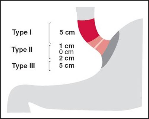
Figure 15-2
Siewert Classification for GE Junctions
• Type 1: Center of lesion 1–5 cm prox to GE jxn; Type 2: 1 cm prox & 2 cm distal to GE jxn
Type 3: 2–5 cm distal to GE jxn (Br J Surg 1998;85:1457–1459) **All are treated as Esophageal CA (AJCC 2010 7th ed.)
Clinical Manifestations
• Sx: Wt loss, abdominal pain, early satiety, GI bleed
• Avg of 6–9 mos from onset of sx to dx
• Mets most common to liver, peritoneal surfaces, & nonregional LN
Screening and Prevention
Screening programs implemented only in Japan due to higher incidence
Staging and Workup
• PET/CT may pick up occult M1 disease (bone, liver) in ∼10% pts, consider esp if locally adv disease
• EUS for T stage, suboptimal for LN staging, better if EUS-guided FNA
• Laparoscopy w/peritoneal washings: M1 disease in ∼20% pts
• T = depth of tumor invasion (T1 lamina propria, muscularis mucosae, or submucosa; T2 muscularis propria; T3 subserosal connective tissue w/o invasion of visceral peritoneum or adj structures; T4 serosa/visceral peritoneum (a) or adj structures (b))
• N = number pos LN in regional LN groups (N1 1–2 LN; N2 3–6 LN; N3a 7–15 LN; N3b ≥16 LN); Nodes in nonregional areas (hepatoduodenal, portal, paraaortic etc.) = distant mets (M1)
• Stage I: T1N1, T2N0, Stage II: T2–3N1, T1–2N2, T1N3, T3–4aN0, Stage III: T4bN0, T4a–bN1, T3–4bN2, T2–4bN3, Stage IV: M1 disease (AJCC TNM staging 7th edition 2010)
Locoregional Disease (Stage I–III)
• Gastrectomy (subtotal preferred) is potentially curative, >50% relapse
• Goal is R0 resection: Complete resection w/adequate margins (≥4 cm); R1: Microscopic residual disease (+margins); R2: Gross (macroscopic) residual disease; only ∼50% will end up w/R0 resection
• D0: Incomplete resection of N1 LN; D1: Removal of involved proximal or distal part of stomach or entire stomach (distal or total resection), including greater & lesser curvature LN; D2: Involves D1 & removal of celiac, gastrohepatic, & splenic LN (requires 15–30 LN for adequate staging)
• Standard of care: D2 LND—lower locoregional recurrence & CA-related death rates compared to D1 surgery (37% vs. 48%) (Lancet Oncol 2010;11:439–449)
Adjuvant Therapy for Locoregional Disease
• Postop 5-FU/LV w/RT (45 Gy): Compared to surgery-only, median OS 36 vs. 27 mos, <50% D1dissection (INT-0116 NEJM 2001;345:725–730)
• Periop ECF: 13% ↑ 5-y OS rate, 36% vs. 23%, postop Rx difficult to tolerate (MAGIC NEJM 2006;355:11–20)
• Postop oral fluoropyrimidine only: 3-y OS rate of 80.1% vs. 70.1% (NEJM 2007;357:1810–1820)
• Postop Cap +OX: 3-y DFS 74% vs. 60%, OS data pending (CLASSIC Lancet 2012;379:315–321)
• Postop Cap + CIS w/RT (45 Gy): Compared to Cap/CIS only, all D2 dissections, no change in DFS, subgroup w/pathologic LN mets may have ↑ DFS (ARTIST J Clin Oncol 2012;30:268–273)
Advanced Stage (IV) Treatment
• 5-FU & CIS or OX backbone, can add docetaxel (OS 9.2 vs. 8.6 mos (J Clin Oncol 2006;24:4991–4997)) or epirubicin
• ECF = ECX = EOF = EOX w/median OS of 9.9, 9.9, 9.3, & 11.2 mos respectively, noninferiority trial (NEJM 2008;358:36–46)
• 2nd-line chemo: Irinotecan vs. best supportive care (BSC) improves median survival, 4 vs. 2.4 mos (Eur J Cancer 2011;47:2306–2314); no median OS difference between Docetaxel & Irinotecan (5.2 vs. 6.5 mos), both better than BSC alone (3.8 mos) (J Clin Oncol 2012;30:1513–1518)
Targeted Therapy
• Trastuzumab (HER2 monoclonal Ab) for 1st-line tx of HER2+ gastric/GEJ adeno (∼20%): FDA approved w/CIS/fluoropyrimidine ↑ median OS 13.8 vs. 11.1 mos, ↑ PFS (ToGA Lancet 2010;376:687–697)
• Ramucirumab (VEGFR-2 monoclonal Ab) for 2nd-line tx adv gastric/GEJ adeno vs. BSC: ↑ Median OS 5.2 vs. 3.8 mos, ↑ PFS 2.1 vs. 1.3 mos (REGARD 2013 ASCO Abstract LBA5)
• Rilotumumab (hepatocyte growth factor/scatter factor [HGF/SF] monoclonal Ab, inhibits MET pathway) in phase II study adv gastric/GEJ adeno, 7.5 mg/kg or 15 mg/kg + ECX vs. placebo + ECX: ↑ PFS 5.6 vs. 4.2 mos; ↑ median OS 11.1 vs. 8.9 mos (p = 0.22) (J Clin Oncol 30 (suppl.) 2012;Abstract 4005)