
Brian D. Kavanagh, Jeffrey D. Bradley, and Robert D. Timmerman
Departing from the established traditions of conventionally fractionated external beam radiotherapy, in the late 1980s and early 1990s investigators in the United States, Sweden, and Japan began to explore the use of extremely brief hypofractionated radiation treatment regimens for spine, lung, liver, and selected other malignant extracranial tumors.1–3 In essence these clinical researchers were modifying techniques proven clinically valuable in the context of cranial and spine stereotactic radiosurgery in an effort to exploit the efficiency and biological potency of high–dose-per-fraction irradiation.4 This idea was soon appreciated for its clinical promise by other researchers in numerous countries across the world.
Pioneers in the field initially used customized ancillary equipment constructed in their own institutions to immobilize patients and to adapt ordinary linear accelerators for the task of precise internal tumor targeting. Now, however, the administration of high-dose, tightly focused external beam radiation therapy is greatly facilitated by a wide assortment of commercially available systems that immobilize patients, address the problem of respiratory motion during treatment, and ensure accurate treatment with the use of image guidance. The newest generation of linear accelerators from several manufacturers is either exclusively dedicated to cranial or extracranial stereotactic radiotherapy or is equipped with a built-in package of features that provide an easy means of administering this type of treatment.
Stereotactic body radiation therapy (SBRT) is the term applied in the United States by the American Society of Therapeutic Radiology and Oncology (ASTRO) for the management and delivery of image-guided high-dose radiation therapy with tumor-ablative intent within a course of treatment that does not exceed 5 fractions.5 Other descriptive terms have been occasionally applied to describe what is officially called SBRT, including the acronym SABR, an abbreviation for stereotactic ablative radiotherapy.6
 BIOLOGICAL AND ONCOLOGICAL RATIONALE FOR SBRT
The appeal of SBRT is based on the nonlinear relation between radiation dose and cytotoxic effect, whereby one or a few large individual doses of radiation therapy have substantially more cell-killing effect than the same dose of radiation given in smaller individual doses. Traditionally, the expected relation between radiation dose and tumor cell kill has been commonly estimated by the well-known linear-quadratic (LQ) model of radiation dose response, often relied on for the purpose of comparing the biological potency of different schedules of conventionally fractionated radiation therapy. In the range of dose per fraction used in SBRT, however, there has been an emerging appreciation that the LQ model overestimates the potency of fraction sizes on the order of 8 to 10 Gy or higher.
A variety of alternative mathematical models have been proposed to account for the observed inaccuracy of the LQ model for doses in this range. For example, Guerrero and Li7 have proposed a modification of Curtis’s8 lethal-potentially lethal model that accounts for ongoing repair processes occurring during the time of an individual radiation treatment, thus predicting lower cytotoxicity from high doses administered over time intervals resembling typical clinical treatment times. Experimental data modeling cranial radiosurgery offer support for this concept.9 Alternatively, Park et al.10 have offered the universal survival curve formalism, which combines the LQ model for doses in the range used in conventional fractionation with a multitarget model for doses in the range used for SBRT. This piecewise function effectively achieves the same key mathematical result as other departures from the LQ model applied to SBRT, namely a more linear slope in the relation between dose and log cell kill in the high dose region.
In the special case of prostate cancer, the rationale for evaluating SBRT for prostate cancer also includes a fundamentally different consideration. Here, applying the LQ model-based assumptions and interpretation, the α/β ratio for prostate cancer has been estimated to be very low, likely in the range of 1.5 to 3.0 Gy.11,12 If this estimate of α/β ratio for prostate cancer is correct, then higher doses per fraction should provide a more favorable therapeutic ratio than a conventionally fractionated regimen. Accumulating evidence from randomized clinical trials comparing conventional daily fractions of 2 Gy with hypofractionated regimens using fractions sized on the order of 3 Gy per day offer strong support for this hypothesis and the safety of using this approach.13–15 Clinical reports involving SBRT for prostate cancer have involved doses that are higher than 3 Gy and are discussed later in the chapter.
None of the aforementioned models of high–dose-per-fraction tumor cell–killing effects explicitly incorporate a mechanism of tumor cell kill that might be of equal or greater importance than tumor DNA damage-based injury, namely the antiangiogenic effect of endothelial cell apoptosis occurring above an apparent threshold dose on the order of 8 to 10 Gy. First observed preclinically and reported by Garcia-Barros et al.,16 clinical evidence indirectly supporting the importance of this mechanism includes measures of an increase in serum markers of apoptosis post-SBRT.17 Other similar threshold effects, which do not occur until the dose per fraction exceeds a minimum “unconventional” level, have been observed, suggesting new ways in which SBRT might be further exploited.18–20
Beyond its uses as primary therapy for selected early-stage cancers, SBRT has also been used as a noninvasive and efficient means of eradicating discrete metastatic tumors, and the argument for this application can be built on numerous overlapping lines of evidence or conceptual theories of cancer growth and dissemination or cytotoxic abscopal effects: (a) the empiric or phenomenological, (b) the patterns-of-failure concept, (c) the theory of oligometastases, (d) a lethal burden variation of the Norton-Simon hypothesis, or (e) immunological enhancement.21
The most straightforward argument for SBRT in the setting of isolated sites of metastatic disease is what might be termed an empiric or phenomenological rationale. There have been numerous reports of patients enjoying a high rate of 3- to 5-year survival following various forms of aggressive local treatment (e.g., surgical resection, radiofrequency ablation, cryotherapy) for limited metastases in the liver or lung from an assortment of solid tumor types. On one level, then, SBRT is a valid, noninvasive substitute for other local modalities if it provides similar efficacy and the same or less toxicity.
The indications for SBRT in the setting of metastatic disease might be alternatively couched in terms of a patterns-of-failure model. A traditional example of a patterns-of-failure approach would be combined modality therapy for lymphomas, where systemic treatment with chemotherapy is combined with involved field radiotherapy to the sites of disease that were grossly evident at the time of diagnosis, indicating that they contain the highest number of clonogenic cells and are thus least likely to be completely eliminated by the chemotherapy. A patterns-of-failure analysis by Rusthoven et al.,22 involving patients who received chemotherapy for metastatic non–small cell lung cancer, reveals a similar result: patients are most likely to manifest tumor recurrence in sites initially involved prior to chemotherapy, opening an opportunity for SBRT to sites of residual disease following some form of systemic therapy to be given with this goal in mind. An earlier analysis by Mehta et al.23 yielded very similar observations.
Yet another argument that has been advanced in support SBRT in this setting is the theory of oligometastases. As articulated by Hellman and Weichselbaum,24 this viewpoint considers that there seems to be a subgroup of patients with metastatic disease that is intermediate between completely absent and widely metastatic. For such patients the entire systemic disease burden is then entirely contained within the finite number of individual sites of gross disease recognized by the pertinent imaging studies. This condition would reflect an intermediate point in the natural history of that individual’s cancer; therefore, these patients might be cured if their limited numbers of metastatic sites are eradicated. Among the many studies lending support to this theory would be the report by Stinauer et al.25 involving patients treated with SBRT for metastases from melanoma or renal cell carcinoma, where there was significantly longer survival for patients with oligometastatic disease, there defined as three or fewer sites, than for patients with more extensive disease.
The theory of oligometastases may be extended beyond subdividing patients according to the integer number of detectable lesions present and, instead, toward characterizing prognosis according to a continuous rather than discrete metric of disease burden. In one sense this approach is a variation of the Norton-Simon hypothesis, as applied to the whole host rather than simply tumor cell kinetics. This conjecture arose from experimental observations in animal models where the number of cancer cells within the host increases from beneath the threshold of detectability, through a phase of rapid growth, and onward toward a plateau level that is lethal to the host.26 This observation was translated into clinical trials by postulating that traditional DNA-targeted chemotherapy is expected to render the greatest degree of cytotoxicity to cancer cells in the rapid phase of growth, when there is higher mitotic activity that renders DNA more vulnerable. Trials testing dose-dense chemotherapy were designed to exploit the enhanced chemosensitivity of rapidly growing cancer cells with the use of frequent dosing schedules that capture tumors repeatedly at relatively smaller volume, without allowing time for regrowth into relatively less sensitive, near-lethal tumor volumes.27
An example of evidence in support of the concept of a clinical lethal burden of disease, as measured by a continuous rather than discrete metric, includes the observations of Lee et al.,28 who used a positron emission tomography scan-based method of quantifying the metabolically active tumor burden in patients with lung cancer and observed that it independently predicted overall survival. SBRT given to a patient with metastatic disease could, in principle, have favorable effects that align with the tenets of the Norton-Simon hypothesis. First, the SBRT might reduce the patient’s total burden of disease in such a way that the remaining cancer within the patient’s body enters into a state of relatively higher growth fraction and is thus more susceptible to cytotoxic systemic agents. Second, the SBRT could prevent or delay the condition of lethal systemic tumor burden that is fatal to the patient.29
A fifth consideration that has emerged in recent years is the possibility that high–dose-per-fraction radiation therapy influences immune system responses in a manner that can be exploited for favorable therapeutic effect. Preclinical studies have demonstrated that high–dose-per-fraction ionizing radiation can induce antigen presentation within the tumor stroma.30 Furthermore, antibody-mediated induction of T cell activity can be combined with high–dose-per-fraction ionizing radiation to enhance not only the effect on the irradiated tumor but also to create an abscopal effect whereby tumor implants remote from the irradiated site regress.31 Findings such as these suggest ideas for new investigations into the combination of SBRT and immunomodulatory agents for patients with metastatic disease.
There is overlap among all of these perspectives, and it would be impossible to prove the exclusive validity of any one above another. Improved disease-free and overall survival following the use of SBRT to metastatic sites of disease would support any of these theories. Nevertheless, it is very important to consider the larger context of cancer treatment in which SBRT is applied for metastatic disease, and these theories can serve to frame the clinical or investigational objectives when SBRT is applied as a treatment for metastatic disease, either alone or in combination with a systemic agent.
STEREOTACTIC BODY RADIATION THERAPY GUIDELINES, PHYSICS OVERVIEW, AND SAFETY CONSIDERATIONS
ASTRO and the American College of Radiology (ACR) have published guidelines that characterize the personnel qualifications and responsibilities, documentation, quality control, and clinical operations recommended for the safe and proper administration of SBRT and follow-up care for patients treated.5 The ASTRO-ACR guidelines advise that the following components should be in place within and institution’s SBRT program:
1. Qualified personnel:
a. Board-certified radiation oncologist
b. Qualified medical physicist
c. Licensed radiation therapist
d. Other support staff as indicated (dosimetrists, oncology nurses, and so forth);
2. Ongoing machine quality assurance program;
3. Documentation in accordance with the ACR Practice Guideline for Communication: Radiation Oncology;
4. Quality control of treatment accessories;
5. Quality control of planning and treatment images;
6. Quality control of treatment planning system;
7. Simulation and treatment systems that account for systematic and random errors associated with setup and target motion in a manner that is based on actual measurement of organ motion and setup uncertainty.
The American Association of Physicist in Medicine Task Group 101 (TG101) moved forward from the ASTRO-ACR guidelines to generate a report that considers additional important nuances in the planning and treatment delivery of SBRT.32 Included within the TG101 report are discussions of potential imaging artifacts and their impact on treatment planning, the challenges of small field dosimetry, and the importance of using an acceptable dose calculation algorithm, among other issues.
Proper patient repositioning, target localization, and management of breathing-related motion are essential for SBRT. A variety of patient immobilization devices are available, including several types of body frames with external fiducial markers. So-called frameless systems incorporate ultrasound, kilovolt-range imaging, or near real-time computed tomography (CT) scanning to verify the location of internal targets relative to the beams to be used. Because SBRT treatment sessions are lengthier than conventional external beam treatments, patient comfort is an important issue.
Breathing-related motion control devices and systems fall into three general categories: (a) dampening, (b) gating, and (c) tracking or “chasing.” Respiratory dampening techniques include systems of abdominal compression intended to diminish one of the largest contributors to breathing-related motion, namely diaphragmatic excursion, by obliging the inspiratory–expiratory lung motion pattern to involve more intracostal expansion and shallower breathing overall. Also included in this category are the systems employing breath-holding maneuvers to stabilize the tumor in a reproducible stage of the respiratory cycle (e.g., deep inspiration). Gating systems for SBRT, as for any radiotherapy application, follow the respiratory cycle using a surrogate indicator for respiratory motion, for example, chest wall motion, and employ an electronic beam activation trigger allowing irradiation to occur only during a specified range of expected tumor locations. Tracking or “chasing” systems move the radiation beam or patient to follow the movement of the tumor.
Regardless of the system employed, the procedure of treatment planning must include the same consideration for respiratory motion management to be used during treatment. Despite available motion control equipment, some positional uncertainty will remain. The planning target volume (PTV) margins used to account for this residual motion of the gross tumor volume (GTV) will typically range from 5 to 10 mm.
The word stereotactic has heretofore usually implied that some sort of external reference markers indexed to internal structures facilitate internal target relocalization, but the definition has expanded to include systems of image-guided radiation therapy (IGRT) that relate the position of internal targets to a three-dimensional coordinate geometry registered to the treatment machine without the use of external markers on the patient. SBRT always involves some form of IGRT to guide for treatment delivery.
Most reports describing SBRT published to date have employed high-energy photons (x-rays) as the source of therapeutic radiation, although other particles can also be used. There is no absolute standard for the combination of beam or arc angles ideal for any given clinical situation, and each case can present unique challenges. In general to achieve a tightly focused high-dose distribution within the PTV and rapid dose falloff outside the PTV, a combination of multiple (often 10 or more) noncoplanar beams or multiple arcs are required. Intensity modulation across the individual beams or arc segments can be incorporated within SBRT.
As part of ASTRO’s Target Safely campaign, the Multidisciplinary Quality Assurance Subcommittee of the Clinical Affairs and Quality Committee of ASTRO commissioned a white paper on the topic of cranial radiosurgery and SBRT titled “Quality and Safety Considerations in Stereotactic Radiosurgery and Stereotactic Body Radiation Therapy.”33 Of particular importance within the white paper are sections emphasizing the importance of creating a proactive culture of safety with procedural checkpoints and error analysis mechanisms.
CLINICAL EXPERIENCE WITH STEREOTACTIC BODY RADIATION THERAPY IN SELECTED SITES
Liver
The two major reasons for considering SBRT for hepatocellular cancer (HCC) is that underlying severe liver disease often renders patients medically inoperable and that other nonsurgical therapies have generally achieved at best rather modest success in that setting. The natural history of untreated HCC has been reported to involve a median survival in the range of 3 to 8 months,34–36 and so a safe and effective therapy is needed in this setting.
The earliest observations following SBRT for HCC were reported by a group at Karolinska Hospital,2 and more recent formal prospective studies have followed. The Princess Margaret Hospital group utilized a 6-fraction regimen in a prospective phase I study in patients with HCC (n = 31) or intrahepatic cholangiocarcinoma (n = 10).37 Prescription doses were selected according to a normal tissue complication probability (NTCP) model based on conventionally fractionated radiotherapy to the liver. The median dose to the tumor was 36 Gy (range, 24 to 54 Gy). The NTCP model overestimated the chance of radiation-induced liver disease, and it is possible that the dose could have been escalated to a higher level in most cases. Nevertheless, in this group of heavily pretreated patients (over 60% had had at least one prior therapy for HCC), a remarkable median survival of 12 months was observed.
Méndez Romero et al.38 at Erasmus University Medical Center treated eight patients with 11 separate lesions of HCC in 3 to 5 fractions to a dose of 25.0 to 37.5 Gy. A 1-year survival of 75% was observed. Choi et al.39 at the Catholic University of Korea treated 23 patients with 32 individual lesions in a 3-fraction regimen to a median dose of 36 Gy (range, 30 to 39 Gy). No patient experienced severe toxicity, although follow-up was limited (median 11 months). Interestingly, in a subsequent analysis of predictors for decline in liver function after SBRT, on multivariate analysis the only predictor for a decrease in Child-Pugh classification (CPC) from baseline level was the volume of normal liver receiving 18 Gy or more (V18). The rate of negative impact on CPC rose sharply when the V18 exceeded 800 cc.40 This latter observation favors a model of SBRT effect on normal liver in line with a critical volume model, whereby it is important to preserve a certain minimum level of function by sparing an adequate volume of normal liver from receiving a dose above a certain threshold.
The group from the Korea Institute of Radiological and Medical Sciences treated a prospectively registered cohort of 38 patients with inoperable HCC with SBRT, all of whom had failed prior transarterial chemoembolization.41The median tumor volume was 40.5 cc (range, 11 to 464). The SBRT dose was 33 to 57 Gy in 3 or 4 fractions. Minimal grade 3 toxicity (<3%) was observed, and the 2-year overall survival was 61%.
Cardenes et al.42 from Indiana University and the University of Colorado reported a multi-institutional phase I dose escalation study of SBRT given in 3 fractions for HCC. Eligibility requirements were CPC-A or -B, medical or technical inoperability, and three or fewer lesions of cumulative tumor diameter 6 cm or less. The study enrolled 17 patients with 25 individual lesions. Dose was escalated from 36 to 48 Gy (16 Gy per fraction) in CPC-A patients without dose-limiting toxicity; however, two patients with CPC-B status at baseline developed grade 3 hepatic toxicity at the 42 Gy (14 Gy per fraction) dose level. Consequently, the dose for CPC-B patients was reduced to 40 Gy in 5 fractions. There were no local failures within the treated volume, and six patients proceeded to liver transplantation. The 2-year overall survival for the entire group was 60%.
The Indiana University group has separately reported their single institution experience involving a total of 60 patients: 34 CPC-A, 25 CPC-B, and 1 CPC-C.43 The median number of fractions, dose per fraction, and total dose, was 3, 14 Gy, and 44 Gy, respectively, for those with CPC-A cirrhosis and 5, 8 Gy, and 40 Gy, respectively, for those with CPC-B. With a median follow-up time of 27 months, the 2-year local control rate was 90%. Two-year overall survival was 67%. SBRT served as a bridge to liver transplant for 23 patients who underwent transplant at a median time of 7 months following SBRT. A progression in CPC was observed in 20% of patients within 3 months of treatment.
Outcomes following SBRT for liver metastases have also been reported by numerous groups44–51 and are summarized in Table 17.1. Regimens of 1 to 5 fractions have been employed, and total doses up to 60 Gy to the planning target volume have been administered. Taken together, the results show good survival outcomes achieved in heavily pretreated individuals and a trend toward improved local control with increasing dose.
Chang et al.52 reported a pooled analysis from three institutions with the use of liver SBRT for liver metastases from colorectal primary cancers. The combined experience from Stanford, Princess Margaret Hospital, and the University of Colorado included 65 patients with a total of 102 individual liver metastases from colorectal cancer treated. More than half of the patients had had at least one prior systemic therapy regimen, and over 40% of the patients had had two or more prior systemic regimens. The analysis indicated that to achieve durable local control of treated lesion, a 3-fraction total dose on the order of 48 Gy is needed. Sustained local control after SBRT was closely associated with improved survival on multivariate analysis (P = .06).
Technical issues and posttreatment imaging follow-up considerations unique to liver SBRT have been included in these reports and in review papers.53,54 Briefly, target delineation and image guidance can be difficult, because liver metastases are not well visualized on CT scans or in-room volumetric imaging used for IGRT. In many centers, radiopaque fiducial markers are place in or near the metastases to facilitate IGRT. In all centers, the planning target volume is an expansion of the gross tumor volume in consideration of the setup and intrafraction motion to be expected with the particular setup and delivery system employed. Typically the margin used to expand a GTV into a PTV is on the order of 5 mm axially and 5 to 10 mm craniocaudally.
The Colorado group first applied the critical volume approach to normal liver dose constraints in their initial phase I study55 and subsequent phase II study. Noted above in relation to the observations of the Catholic University of Korea in their treatment of primary liver tumors, the critical volume model liver SBRT is an adaptation of the early work of Yeas and Kalend.56 Applicable for organs of radiobiologically parallel structure, the crux of this application is to work backward, in a sense, from an estimate of how much volume of the organ is essential and must be protected from functional ablation. The estimate for liver that at least 700 cm3 should receive less than 15 Gy during a 3-fraction SBRT course was derived from a combination of prior reports of outcomes after partial hepatectomy documenting approximate minimum volumes required and estimates of the effects of that dose of radiation extrapolated from prior reports of conventionally fractionated treatment.
One feature of the normal tissue effect of liver SBRT consistently observed within the first few months after SBRT is a zone of hypodensity observed on follow-up CT scans corresponding to the volume that received approximately 30 Gy.55 This phenomenon, first described by Herfarth et al.57 following single-dose liver SBRT, is likely related to local veno-occlusive effects.58 There is no known clinical consequence associated with the finding, but it can cloud the assessment of tumor response within the first few months after liver SBRT.
TABLE 17.1 STEREOTACTIC BODY RADIATION THERAPY FOR LIVER METASTASES

CASE STUDY
Liver Stereotactic Body Radiation Therapy
A 45-year-old female had been diagnosed with stage IV breast cancer 2 years previously. Biopsy-proven liver metastases were present at the time of diagnosis. Numerous systemic agents had been given, most recently gemcitabine and trastuzumab. Although all other measurable or assessable sites of disease were stable or regressing, a mass in the liver had progressed from 2.5-by-2.9 cm to 6.0-by-4.2 cm within the past 3 months. Because the patient was tolerating the regimen well and apparently having a response in most sites, she was offered SBRT in an effort to eradicate tumor in the liver.
The lesion diameter (>6 cm) rendered the patient ineligible for an ongoing phase II trial of SBRT for liver metastases, and the dose given was lower than the protocol doses (Fig. 17.1). The 53 cm3 GTV was expanded by 5 mm radially and 10 mm in the superior-inferior direction to generate the PTV. The dose distribution shown was administered in 3 fractions within 1 week using multiple dynamic conformal arcs and a controlled breath-holding device. The nominal prescription dose was 45 Gy. The maximum point dose was 59 Gy, and the equivalent uniform dose was 54 Gy. The volume of normal liver receiving less than 15 Gy was 1,800 cm3. The portion of the right kidney receiving above 15 Gy was 13%. Follow-up scans at 6 months and 10 months show a Herfarth type 2 reaction with hyperdensity in the treated normal liver.57 There is also volume loss in the nearby normal liver parenchyma surrounding the lesion, a phenomenon that has also been described elsewhere.58 The lesion remained controlled for the duration of the patient’s life; she eventually died of complications related to central nervous system metastases.
Lung
Medically inoperable early-stage lung cancer has historically provided a substantial management challenge. Conventionally fractionated radiotherapy has yielded generally unsatisfactory outcomes with high rates of local failure and 3-year survivals in the range of approximately 30%. For this reason, medically inoperable early-stage lung cancer was the first clinical indication for which SBRT was studied in prospective clinical trials. Following the early exploratory studies of lung SBRT for stage I non–small cell lung cancer at the Karolinska Hospital in Stockholm59 and National Medical Defense Hospital in Saitama,60 numerous formal prospective studies of SBRT for medically inoperable non–small cell lung cancer have been now been reported. Table 17.2 lists the major prospective studies (N = 50 or more) with a minimum median follow-up of 24 months at the time of reporting, along with local control and overall survival at 3 years.61–64 The consistent observation is that 3-year survival on the order of 50% to 60% has been achieved, a noteworthy improvement relative to conventionally fractionated radiotherapy.
One important observation from the Indiana University studies was that although the treatment was generally well tolerated, tumor location near large airways in the vicinity of the pulmonary hilum (called the zone of the proximal bronchial tree) was associated with a markedly higher risk of toxicity. For this reason, in the Radiation Therapy Oncology Group’s (RTOG) study RTOG-0236 of SBRT for medically inoperable non–small cell lung cancer, patients with tumors located in the zone of the proximal bronchial tree were excluded.63 The RTOG has launched a separate dose-escalation study (ROTG-0813) in which tumors near the proximal bronchial tree are treated. Both RTOG and the Japanese Clinical Oncology Group have completed enrollment to studies that expand the use of SBRT to patients with early-stage lung cancer who are medically operable.
Although many retrospective studies of lung SBRT contain a mixture of both primary and metastatic lesions, a few prospective studies exclusively focused on SBRT for lung metastases have been reported. In the University of Colorado phase I SBRT trial for lung metastases,65 eligible patients had one to three pulmonary metastases from a solid tumor, cumulative tumor diameter less than 7 cm, and adequate pulmonary function (forced expiatory volume in the first second of expiration [FEV1] >1.0 L). The PTV was typically constructed from the GTV by adding a 5-mm radial and 10-mm craniocaudal margin. The first cohort received 48 Gy to the PTV in 3 fractions. The SBRT dose was escalated in subsequent cohorts up to a preselected maximum of 60 Gy in 3 fractions. The percentage of normal lung receiving more than 15 Gy (V15) was restricted to less than 35%. Dose-limiting toxicity (DLT) included acute grade 3 lung or esophageal toxicity or any acute grade 4 toxicity. No patient experienced a DLT, and the SBRT dose was escalated to 60 Gy in 3 fractions without reaching a maximum tolerated dose. No consistent significant effects on pulmonary functions tests were noted.
The phase II study of SBRT for lung metastases the Colorado study group included 38 patients with 63 lesions.66 Most had received at least one prior systemic regimen for metastatic disease, and approximately one-third had received two or more prior regimens. The incidence of any grade 3 toxicity was 8% (3/38), and no grade 4 toxicity was seen. Symptomatic pneumonitis occurred in one patient (2.6%). For 50 lesions assessable for local control, the median follow-up was 15.4 months. The median gross tumor volume was 4.2 cc. The actuarial 2-year local control was 96%. Median overall survival was 19 months.
FIGURE 17.1. Example of liver stereotactic body radiation therapy (SBRT). Top panel: Pre-SBRT planning computed tomography images with thin arrow pointing to the gross tumor volume (GTV) and wide arrow showing the planning target volume (PTV), which is outlined. Second panel: SBRT composite dose distribution. Third panel: Images obtained 6 months post-SBRT illustrating a Herfarth type 2 reaction in adjacent parenchyma and partial segmental atrophy. Bottom panel: Shows images 10 months post-SBRT, indicating continued tumor regression as the ablated liver volume continues to recede.
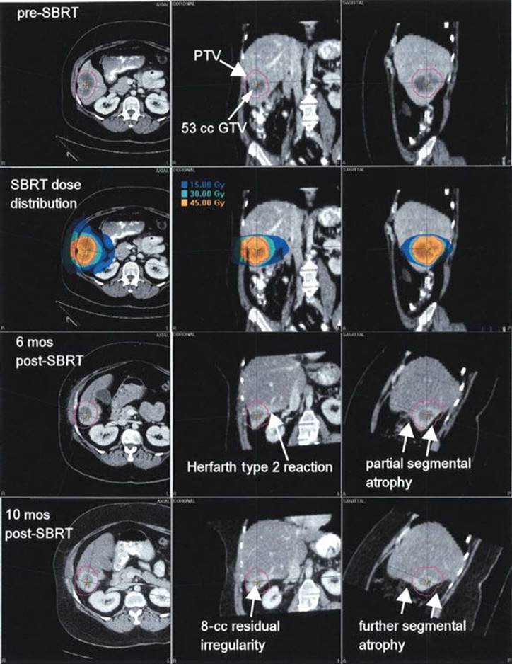
TABLE 17.2 MAJOR PROSPECTIVE STUDIES OF STEREOTACTIC BODY RADIATION THERAPY FOR MEDICALLY INOPERABLE NON–SMALL CELL LUNG CANCER

CASE STUDY
Lung Stereotactic Body Radiation Therapy
A 74-year-old female had undergone wedge resection for a pT1N0M0 non–small cell cancer of the right lung 7 years previously. She had a right pneumonectomy 3 years later as salvage treatment for a locoregional recurrence. She was later observed to have developed a left lung nodule on a surveillance chest x-ray, and needle biopsy proved it to be a non–small cell lung cancer, presumed to be a second primary. Staging studies revealed no other sites of disease. She was given systemic therapy and enjoyed a transient minor response and then regrowth of the lesion (Fig. 17.2).
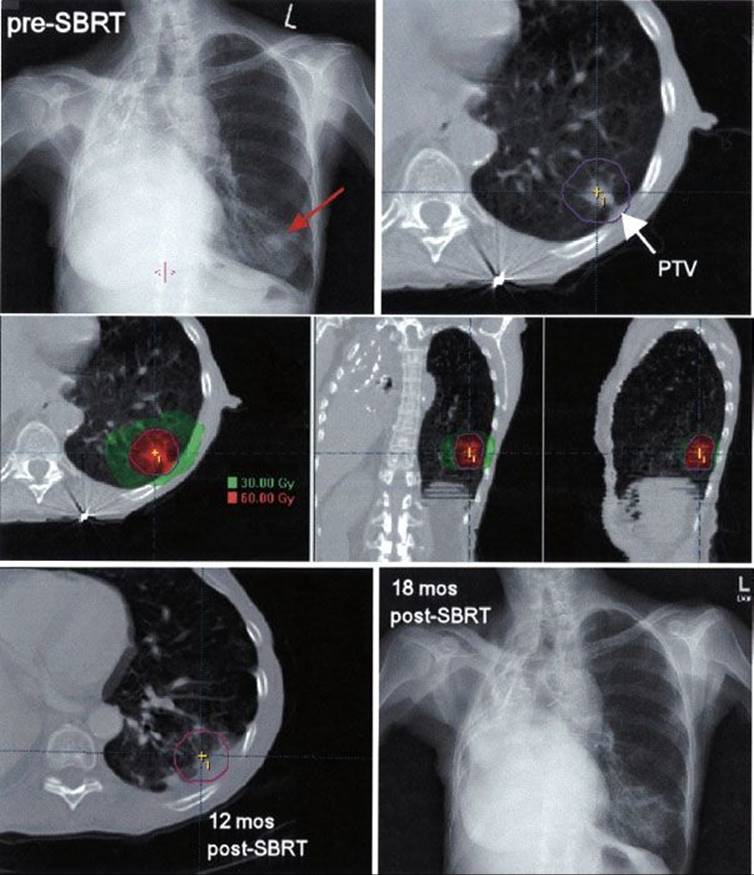
The patient used supplemental oxygen, 2 L/min at bedtime and occasionally during the day. She was offered SBRT as potentially curative therapy for a new T1N0M0 lung cancer. The 4 cm3 GTV was expanded by 5 mm radially and 10 mm in the superior-inferior direction to generate the 29 cm3 PTV. The dose distribution shown was administered in 3 fractions within 1 week using multiple dynamic conformal arcs. The patient did not comfortably tolerate a breath-holding technique because of her supplemental oxygen requirements; therefore, an abdominal compression technique was used during simulation and treatment. The nominal prescription dose was 60 Gy. The maximum point dose was 79 Gy, and the equivalent uniform dose was 72 Gy. The portion of normal lung receiving less than 15 Gy was 12.7%. The lesion remained controlled for the duration of the patient’s life; she died of unrelated causes more than 2 years after SBRT.
Spine
The earliest investigation into what would now be termed spine SBRT was that of Hamilton et al.,1,67 who used a rigid immobilization with a device surgically attached to the spinal column. Conservative doses in the range of 8 to 10 Gy were given in 1 fraction to nine patients with recurrent lesions in the spine following prior conventional radiotherapy. Spinal cord doses were very low using this technique (0.5 to 3.2 Gy). Limited follow-up suggested a favorable clinical effect in some patients, and no complications were observed. More recently, less invasive techniques have been investigated.
Ryu et al.68 at the Henry Ford Hospital initially studied the treatment of spine metastases with initial fractionated radiotherapy followed by a spinal radiosurgery boost (6 to 8 Gy), observing prompt relief of pain in nearly all 10 treated patients. In a subsequent study of single fraction spinal radiosurgery alone (10 to 16 Gy), this group observed complete or partial pain relief in 85% of the 49 patients treated.69 Perhaps even more importantly, pain relief was rapid after SBRT, sometimes within hours of treatment.
Chang et al.70,71 at the M.D. Anderson Cancer Center performed a prospective phase I dose escalation study in treating spinal metastases and later updated their institutional experience. The equipment used included a “CT on rails” that allowed for imaging immediately to guide patient repositioning. Sixty-three cancer patients underwent near-simultaneous CT-guided SBRT. Spinal magnetic resonance imaging was conducted at baseline and at each follow-up visit. The median tumor volume of 74 spinal metastatic lesions was 37.4 cc. Approximately half the patients received 30 Gy in 5 fractions, and the other half received 27 Gy in 3 fractions. A conservative constraint of 9 to 10 Gy maximum dose to the spinal cord was applied. No neuropathy or myelopathy was observed during a median follow-up period of nearly 2 years. The actuarial 1-year tumor progression-free rate was 84%. The investigators noted two characteristic patterns of failure: (a) recurrence in the bone adjacent to the site of previous treatment, and (b) recurrence in the epidural space adjacent to the spinal cord. A low rate of any grade 3 toxicity was observed.
FIGURE 17.2. Example of lung stereotactic body radiation therapy (SBRT). Top panel: Pre-SBRT chest x-ray showing the left lung nodule (red arrow) and axial planning computed tomography (CT) image with white arrow pointing to the planning target volume (PTV), which is outlined. Middle panel:SBRT composite dose distribution shown in axial, coronal, and sagittal perspectives. Bottom panel: Follow-up CT scan axial image obtained 12 months post-SBRT illustrating stable patchy fibrosis in the high dose region (left) and chest x-ray obtained 12 months post-SBRT, indicating minimal residual haziness in the region treated.
Similar observations of good tumor control and minimal toxicity have been reported from other centers.72–76 The largest is from Gerszten et al.74 of the University of Pittsburgh, who analyzed a cohort of 500 cases of spinal metastases. The maximum intratumoral dose ranged from 12.5 to 25 Gy (mean, 20). Tumor volume ranged from 0.20 to 264 mL (mean, 46). Durable pain reduction was achieved in 86% of patients, and durable tumor control was demonstrated for approximately 90% of the lesions treated. The vast majority of patients with a progressive neurologic deficit before treatment experienced at least some clinical improvement.
Regarding normal tissue toxicities, the Memorial Sloan-Kettering Cancer Center group reported that post-SBRT vertebral fracture is common when there is metastatic lytic disease involving more than 40% of the vertebral body and location at or below T10.77 The M.D. Anderson Cancer Center group also analyzed the risk of fracture after spine SBRT and noted that fractures were more common among patients of age over 55 years, those with a pre-existing fracture, and pain at the time of treatment,78 suggesting that patients at very high risk might appropriately be considered for prophylactic vertebral stabilization or augmentation procedures. Fortunately, spinal cord toxicity has only rarely been observed after SBRT. Case-control comparisons offer some suggestions of parameters that might elevate the risk, but the paucity of events evaluable make it difficult to draw firm conclusions.79,80 Constraints used to guide spine SBRT have included maximum point dose to the cord in the range of 10 to 14 Gy and limiting the volume of adjacent spinal cord receiving more than 10 Gy in a single fraction to 10% or less.
Prostate Cancer
The first publication on SBRT for prostate cancer was the work of Madsen et al.,81 who recently updated their observations.82 In a prospective trial, 40 patients with low-risk cancer (Gleason score ≤6 and prostate-specific antigen [PSA] ≤10 ng/mL) were treated to a dose of 33.5 Gy in 5 daily fractions The median age was 69 years (range, 50 to 82), and the median follow-up period was 5 years. The overall 5-year Phoenix definition (nadir plus 2 ng/mL) biochemical relapse-free survival rate was 93%. No patients died of prostate cancer. Late grade 3 genitourinary toxicity was rare, occurring in only one patient, and no late grade 3 or higher gastrointestinal was observed.
The groups at Winthrop University and the University of California–San Francisco have also reported trials of SBRT for early-stage prostate cancer.83,84 Using doses of 35 to 38 Gy in 4 or 5 fractions, both groups reported similarly low rates of grade 3 or higher toxicity of any kind (<1% in aggregate between the studies) after a median follow-up of 1 year. King et al.85 from Stanford University recently updated a prospective trial in which 67 patients with clinically localized low-risk prostate cancer were treated with SBRT to a dose of 36.25 Gy, administered in 5 fractions. The 4-year biochemical relapse-free survival was 94%, and no grade 3 or higher rectal toxicity was observed. There were only two cases (3%) of grade 3 or higher bladder toxicity, both of which were believed to have been caused or exacerbated by procedures performed for dysuria (cystoscopies or dilatation).
A separate recently published analysis combined a subset of the Stanford patients with a cohort of patients treated in an identical manner at a community center in Naples, Florida, all of whom had a minimum of 5 years of follow-up after SBRT.86 The 5-year biochemical relapse-free survival was 93%, and no severe treatment-related toxicity was observed. Another prospective trial conducted at several Harvard-affiliated centers involved a very similar regimen of 5 fractions of 7.25 to 7.5 Gy for low-risk prostate cancer.87 At a median follow-up of 44.5 months, the outcomes essentially replicated the Stanford-Naples experience, with a 3-year biochemical relapse-free survival of 98% and a 2% and 4% chance of grade 3 urinary or rectal toxicity, respectively.
Boike et al.88 from the University of Texas–Southwestern have completed a dose escalation for prostate SBRT, aiming for a more aggressive regimen potentially suitable for patients with intermediate or high-risk disease. In a prospective phase I trial, it was shown that with careful attention to the rectal and bladder doses, it was possible to administer safely a dose of 50 Gy in 5 fractions, with an observed risk of 2% and 4% for grade 3 or higher rectal and genitor-urinary toxicity, respectively. The maximum tolerated dose was not reached in this trial, and the 50 Gy in 5-fraction regimen was selected for continuation in an ongoing prospective phase II trial.
Pancreas Cancer
A major challenge in the application of concurrent chemotherapy and conventionally fractionated radiotherapy is the high rate of nonhematologic toxicity. Recent North American cooperative group studies involving various chemotherapy regimens and conventionally fractionated radiotherapy to a dose of 50.4 Gy given in 28 fractions have been associated with rates of grade 3 or higher nonhematologic toxicity in the range of 29% to 79%.89–92 SBRT would be an appealing alternative if equivalent survival could be achieved with lower rates of toxicity related to the reduction of the volume of normal tissue exposed to a high dose of radiation.
SBRT regimens for pancreas cancer have included treatments given in 1 to 5 fractions. The relation of risk of toxicity to the volume of normal tissue receiving a high dose is illustrated by the Danish cooperative group study. Here, a dose of 45 Gy in 3 fractions to a volume that included generous margins around the gross tumor volume, such that the median volume receiving more than 30 Gy, was 136 cc.93 The toxicity from this high-volume treatment was unacceptably high.
More recent studies have incorporated tighter planning margins around the primary tumor and achieved reduction in toxicity and improvement in survival. The Stanford experience included 55 patients treated with a single 25 Gy fraction to the gross tumor volume plus 3-mm margin, with gemcitabine (GEM) given for 1 cycle prior to and 4 to 6 cycles after SBRT.94 A median survival of 13 months was observed. The San Bartolo Hospital group gave 30 Gy in 3 fractions to a similar target volume, again with GEM given before and after SBRT, and observed an 11-month median survival.95 The Beth Israel Deaconess group used a dose of 24 to 36 Gy in 3 fractions, with the GEM given after SBRT for 6 cycles, and observed a 14-month median survival.96 The incidence of grade 3 or higher SBRT-related toxicity was very low for each study. In the San Bartolo Hospital and Beth Israel Deaconess studies, the rates of grade 3 or higher nonhematologic toxicity were 0% and 14%, respectively.
The Stanford group analyzed potential dosimetric factors that predicted for a risk of duodenal toxicity after single fraction SBRT for pancreatic cancer.97 Among 73 patients evaluable, 6 patients experienced grade 2 toxicity and 6 experienced grade 3 or 4 toxicity. Numerous interrelated metrics proved to be able to distinguish groups of lower versus higher risk of toxicity. For example, the volume of duodenum receiving a dose of 15 Gy or higher (V15) was significant: for V15 9.1 cc or more, the rate of toxicity was 52%, whereas for V15 less than 9.1 cc, the rate was 11% (P = .002).
CONCLUSIONS
SBRT has emerged as a versatile strategy with a wide range of applications for many different types and stages of cancer. As with any form of radiation therapy, careful attention to matters of patient selection and technical quality assurance is essential for the effective and safe implementation of SBRT. Future advances will refine our understanding of the biological mechanisms and optimal integration and sequencing of SBRT with other anticancer therapies.
REFERENCES
1. Hamilton AJ, Lulu BA, Fosmire H, et al. Preliminary clinical experience with linear accelerator-based spinal stereotactic radiosurgery. Neurosurgery 1995;36:311–319.
2. Blomgren H, Lax I, Naslund I, et al. Stereotactic high dose fraction radiation therapy of extracranial tumors using an accelerator. Clinical experience of the first thirty-one patients. Acta Oncol1995;34:861–870.
3. Uematsu M, Shioda A, Tahara K, et al. Focal, high dose, and fractionated modified stereotactic radiation therapy for lung carcinoma patients: a preliminary experience. Cancer 1998;82:1062–1070.
4. Kavanagh BD, Timmerman RD. Stereotactic radiosurgery and stereotactic body radiation therapy: an overview of technical considerations and clinical applications. Hematol Oncol Clin North Am2006;20:87–95.
5. Potters L, Kavanagh B, Galvin JM, et al. American Society for Therapeutic Radiology and Oncology (ASTRO) and American College of Radiology (ACR) practice guideline for the performance of stereotactic body radiation therapy. Int J Radiat Oncol Biol Phys 2010;76(2):326–332.
6. Loo BW, Chang JY, Dawson LA, et al. Stereotactic ablative radiotherapy: what’s in a name? Pract Rad Oncol 2011;1:38–39.
7. Guerrero M, Li X. Extending the linear–quadratic model for large fraction doses pertinent to stereotactic radiotherapy. Phys Med Biol 2004;49:4825–4835.
8. Curtis SB. Lethal and potentially lethal lesions induced by radiation—a unified repair model. Radiat Res 1986;106:252–270.
9. Benedict SH, Lin PS, Zwicker RD, et al. The biological effectiveness of intermittent irradiation as a function of overall treatment time: development of correction factors for LINAC-based stereotactic radiotherapy. Int J Radiat Oncol Biol Phys 1997;37:765–769.
10. Park C, Papiez L, Zhang S, et al. Universal survival curve and single fraction equivalent dose: useful tools in understanding potency of ablative radiotherapy. Int J Radiat Oncol Biol Phys 2008;70:847–852.
11. Brenner DJ, Hall EJ. Fractionation and protraction for radiotherapy of prostate carcinoma. Int J Radiat Oncol Biol Phys 1999;43:1095–1101.
12. Williams SG, Taylor JM, Liu N, et al. Use of individual fraction size data from 3756 patients to directly determine the alpha/beta ratio of prostate cancer. Int J Radiat Oncol Biol Phys 2007;68:24–33.
13. Arcangeli G, Saracino B, Gomellini S, et al. A prospective phase III randomized trial of hypofractionation versus conventional fractionation in patients with high-risk prostate cancer. Int J Radiat Oncol Biol Phys 2010;78:11–18.
14. Yeoh EE, Botten RJ, Butters J, et al. Hypofractionated versus conventionally fractionated radiotherapy for prostate carcinoma: final results of phase III randomized trial. Int J Radiat Oncol Biol Phys2011;81:1271–1278.
15. Dearnaley D, Syndikus I, Sumo G, et al. Conventional versus hypofractionated high-dose intensity-modulated radiotherapy for prostate cancer: preliminary safety results from the CHHiP randomised controlled trial. Lancet Oncol 2012;13:43–54.
16. Garcia-Barros M, Paris F, Cordon-Cardo C, et al. Tumor response to radiotherapy regulated by endothelial cell apoptosis. Science 2003;300(5622):1155–1159.
17. Zhang L, Kavanagh B, Thorburn A, et al. Preclinical and clinical estimates of a cancer’s basal apoptotic rate predict for the amount of apoptosis induced by subsequent pro-apoptotic stimuli. Clin Cancer Res 2010;16:4478–4489.
18. Lee Y, Auh SL, Wang Y, et al. Therapeutic effects of ablative radiation on local tumor require CD8+ T cells: changing strategies for cancer treatment. Blood 2009;114:589–595.
19. Kim DW, Huamani J, Niermann KJ, et al. Noninvasive assessment of tumor vasculature response to radiation-mediated, vasculature-targeted therapy using quantified power Doppler sonography: implications for improvement of therapy schedules. J Ultrasound Med 2006;25:1507–1517.
20. DeRose P, Thorpe PE, Gerber DE. Development of bavituximab, a vascular targeting agent with immune-modulating properties, for lung cancer treatment. Immunotherapy 2011;3:933–944.
21. Timmerman RD, Bizekis CS, Pass HI, et al. Local surgical, ablative, and radiation treatment of metastases. CA Cancer J Clin 2009;59:145–170.
22. Rusthoven K, Hammerman SF, Kavanagh BD, et al. Is there a role for consolidative stereotactic body radiation therapy following first-line systemic therapy for metastatic lung cancer? A patterns-of-failure analysis. Acta Oncol2009;48:578–583.
23. Mehta N, Mauer AM, Hellman S. Analysis of further disease progression in metastatic non-small cell lung cancer: implications for locoregional treatment. Int J Oncol 2004;25:1677–1683.
24. Hellman S, Weichselbaum RR. Oligometastases. J Clin Oncol 1995;13:8–10.
25. Stinauer MA, Kavanagh BD, Schefter TE, et al. Stereotactic body radiation therapy for melanoma and renal cell carcinoma: impact of single fraction equivalent dose on local control. Radiat Oncol2011;6(1):34.
26. Norton L, Simon R, Brereton HD, et al. Predicting the course of Gompertzian growth. Nature 1976;264(5586):542–545.
27. Fornier M, Norton L. Dose-dense adjuvant chemotherapy for primary breast cancer. Breast Cancer Res 2005;7(2):64–69.
28. Lee P, Weerasuriya DK, Lavori PW, et al. Metabolic tumor volume predicts for disease progression and death in lung cancer. Int J Radiat Oncol Biol Phys 2007;69:328–333.
29. Kavanagh BD, Kelly KA, Kane M. The promise of stereotactic body radiation therapy in a new era of oncology. Front Radiat Ther Oncol 2007;40:340–351.
30. Zhang B, Bowerman NA, Salama JK, et al.: Induced sensitization of tumor stroma leads to eradication of established cancer by T cells. J Exp Med 2007;204:49–55.
31. Dewan MZ, Galloway AE, Kawashima N, et al. Fractionated but not single-dose radiotherapy induces an immune-mediated abscopal effect when combined with anti–CTLA-4 antibody. Clin Cancer Res2009;15:5379–5388.
32. Benedict SH, Yenice KM, Followill D, et al. Stereotactic body radiation therapy: the report of AAPM Task Group 101. Med Phys 2010;37:4078–4101.
33. Solberg TD, Balter JM, Benedict SH, et al. Quality and safety considerations in stereotactic radiosurgery and stereotactic body radiation therapy: executive summary. Pract Radiat Oncol 2012;2:2–9.
34. Yeung YP, Lo CM, Liu CL, et al. Natural history of untreated nonsurgical hepatocellular carcinoma. J Gastroenterol 2005;100:1995–2004.
35. Ruzzenente A, Capra F, Pachera S, et al. Is liver resection justified in advanced hepatocellular carcinoma? Results of an observational study in 464 patients. J Gastrointest Surg 2009;13:1313–1320.
36. Meng M, Cui Y, She B, et al. Transcatheter arterial chemoembolization in combination with radiotherapy for unresectable hepatocellular carcinoma: a systematic review and meta-analysis. Radiother Oncol 2009;92:184–194.
37. Tse RV, Hawkins M, Lockwood G, et al. Phase I study of individualized stereotactic body radiotherapy for hepatocellular carcinoma and intrahepatic cholangiocarcinoma. J Clin Oncol 2008;26:657–664.
38. Méndez Romero A, Wunderink W, Hussain SM, et al. Stereotactic body radiation therapy for primary and metastatic liver tumors: a single institution phase i-ii study. Acta Oncol 2006;45(7):831–837.
39. Choi BO, Choi BI, Jang HS, et al. Stereotactic body radiation therapy with or without transarterial chemoembolization for patients with primary hepatocellular carcinoma: preliminary analysis. BMC Cancer 2008;8:351.
40. Son SH, Choi BO, Ryu MR, et al. Stereotactic body radiotherapy for patients with unresectable primary hepatocellular carcinoma: dose-volumetric parameters predicting the hepatic complication. Int J Radiat Oncol Biol Phys2010;78:1073–1080.
41. Seo YS, Kim M-S, Yoo S, et al. Preliminary result of stereotactic body radiotherapy as a local salvage treatment for inoperable hepatocellular carcinoma. J Surg Oncol 2010;102:209–214.
42. Cardenes HR, Price TR, Perkins SM, et al. Phase I feasibility trial of stereotactic body radiation therapy for primary hepatocellular carcinoma. Clin Transl Oncol 2010;12:218–225.
43. Andolino DL, Johnson CS, Maluccio M, et al. Stereotactic body radiotherapy for primary hepatocellular carcinoma. Int J Radiat Oncol Biol Phys 2011;81:e447–e453.
44. Herfarth KK, Debus J, Wannenmacher. Stereotactic radiation therapy of liver metastases: Update of the initial phase-I/II trial. Front Radiat Ther Oncol 2004;38:100–105.
45. Wulf J, Guckenberger M, Haedinger U, et al. Stereotactic radiotherapy of primary liver cancer and hepatic metastases. Acta Oncol 2006;45:838–847.
46. Hoyer M, Roed H, Traberg-Hansen A, et al. Phase II study on stereotactic body radiotherapy of colorectal metastases. Acta Oncol 2006;45:823–830.
47. van der Pool AE, Mendez-Romero A, Wunderink W, et al. Stereotactic body radiation therapy for colorectal liver metastases. Br J Surg 2010;97:377–382.
48. Rusthoven K, Kavanagh BD, Cardenes H, et al. Mature results of a multi-institutional phase I/II trial of stereotactic body radiation therapy for liver metastases. J Clin Oncol 2009;27:1572–1578.
49. Lee MT, Kim JJ, Dinniwell R, et al. Phase I study of individualized stereotactic body radiotherapy of liver metastases. J Clin Oncol 2009;27:1585–1591.
50. Goodman KA, Wiegner EA, Maturen KE, et al. Dose-escalation study of single-fraction stereotactic body radiotherapy for liver malignancies. Int J Radiat Oncol Biol Phys 2010;78:486–493.
51. Rule W, Timmerman R, Tong L, et al. Phase I dose-escalation study of stereotactic body radiotherapy in patients with hepatic metastases. Ann Surg Oncol 2011;18:1081–1087.
52. Chang DT, Swaminath A, Kozak M, et al. Stereotactic body radiotherapy for colorectal liver metastases: a pooled analysis. Cancer 2011;117:4060–4069.
53. Schefter TE, Kavanagh BD. Radiation therapy for liver metastases. Sem Rad Oncol 2011;21:264–270.
54. Høyer M, Swaminath A, Bydder S, et al. Radiotherapy for liver metastases: a review of evidence. Int J Radiat Oncol Biol Phy. 2012;82:1047–1057.
55. Schefter TE, Kavanagh BD, Timmerman RD, et al. A phase I trial of stereotactic body radiation therapy (SBRT) for liver metastases. Int J Radiat Oncol Biol Phys 2005;62:1371–1378.
56. Yeas RJ, Kalend A. Local stem cell depletion model for radiation myelitis. Int J Radiat Oncol Biol Phys 1988;14:1247–1259.
57. Herfarth KK, Hof H, Bahner ML, et al. Assessment of focal liver reaction by multiphasic CT after stereotactic single-dose radiotherapy of liver tumors. Int J Radiat Oncol Biol Phys 2003;57:444–451.
58. Olsen CC, Welsh J, Kavanagh BD, et al. Microscopic and macroscopic tumor and parenchymal effects of liver stereotactic body radiotherapy. Int J Radiat Oncol Biol Phys 2009;73(5):1414–1424.
59. Blomgren, H, Lax, I, Goranson, H, et al. Radiosurgery for tumors in the body: clinical experience using a new method. J Radiosurg 1998;1:63–74.
60. Uematsu M, Shioda A, Tahara K, et al. Focal, high dose, and fractionated modified stereotactic radiation therapy for lung carcinoma patients: a preliminary experience. Cancer 1998;82:1062–1070.
61. Fakiris AJ, McGarry RC, Yiannoutsos CT, et al. Stereotactic body radiation therapy for early-stage non-small-cell lung carcinoma: four-year results of a prospective phase II study. Int J Radiat Oncol Biol Phys 2009;75:677–682.
62. Baumann P, Nyman J, Hoyer M, et al. Outcome in a prospective phase II trial of medically inoperable stage I non–small-cell lung cancer patients treated with stereotactic body radiotherapy. J Clin Oncol2009;27:3290–3296.
63. Timmerman R, Paulus R, Galvin J, et al. Stereotactic body radiation therapy for inoperable early stage lung cancer. JAMA 2010;303:1070–1076.
64. Ricardi U, Filippi AR, Guarneri A, et al. Stereotactic body radiation therapy for early stage non-small cell lung cancer: results of a prospective trial. Lung Cancer 2010;68(1):72–77.
65. Schefter TE, Kavanagh BD, Raben D, et al. A phase I/II trial of stereotactic body radiation therapy (SBRT) for lung metastases: initial report of dose escalation and early toxicity. Int J Radiat Oncol Biol Phys 2006;66(4S): S120–S127.
66. Rusthoven K, Kavanagh BD, Burri SH, et al. Multi-institutional phase I/II trial of stereotactic body radiation therapy for lung metastases. J Clin Oncol 2009;27: 1579–1584.
67. Hamilton AJ, Lulu BA, Fosmire H, et al. LINAC-based spinal stereotactic radiosurgery. Stereotact Funct Neurosurg 1996;66:1–9.
68. Ryu S, Fang Yin F, Rock J, et al. Image-guided and intensity-modulated radiosurgery for patients with spinal metastasis. Cancer 2003;97(8):2013–2018.
69. Ryu S, Rock J, Rosenblum M, et al. Patterns of failure after single-dose radiosurgery for spinal metastasis. J Neurosurg 2004;101(Suppl 3):402–405.
70. Chang EL, Shiu AS, Lii MF, et al. Phase I clinical evaluation of near-simultaneous computed tomographic image-guided stereotactic body radiotherapy for spinal metastases. Int J Radiat Oncol Biol Phys2004;59:1288–1294.
71. Chang EL, Shiu AS, Mendel E, et al. Phase I/II study of stereotactic body radiotherapy for spinal metastasis and its pattern of failure. J Neurosurg Spine 2007;7:151–160.
72. Nelson JW, Yoo DS, Sampson JH, et al. Stereotactic body radiotherapy for lesions of the spine and paraspinal regions. Int J Radiat Oncol Biol Phys 2009;73(5):1369–1375.
73. De Salles AA, Pedroso AG, Medin P, et al. Spinal lesions treated with Novalis shaped beam intensity-modulated radiosurgery and stereotactic radiotherapy. J Neurosurg 2004;101(S3):435–440.
74. Gerszten PC, Burton SA, Ozhasoglu C, et al. Radiosurgery for spinal metastases: clinical experience in 500 cases from a single institution. Spine 2007;32:193–199.
75. Gibbs IC, Kamnerdsupaphon P, Ryu MR, et al. Image-guided robotic radiosurgery for spinal metastases. Radiother Oncol 2007;82:185–190.
76. Yamada Y, Bilsky MH, Lovelock DM, et al. High-dose, single fraction image-guided intensity-modulated radiotherapy for metastatic spinal lesions. Int J Radiat Oncol Biol Phys 2008;71:484–490.
77. Rose PS, Laufer I, Boland PJ, et al. Risk of fracture after single fraction image-guided intensity-modulated radiation therapy to spinal metastases. J Clin Oncol 2009;27(30):5075–5079.
78. Boehling NS, Grosshans DR, Allen PK. Vertebral compression fracture risk after stereotactic body radiotherapy for spinal metastases. J Neurosurg Spine 2012;16(4):379–386.
79. Sahgal A, Ma L, Gibbs I, et al. Spinal cord tolerance for stereotactic body radiotherapy. Int J Radiat Oncol Biol Phys 2010;77:548–553.
80. Sahgal A, Ma L, Weinberg V, et al. Reirradiation human spinal cord tolerance for stereotactic body radiotherapy. Int J Radiat Oncol Biol Phys 2012;82:107–116.
81. Madsen BL, His RA, Pham HT, et al. Stereotactic hypofractionated accurate radiotherapy of the prostate (SHARP), 33.4 Gy in five fractions for localized disease: first clinical trial results. Int J Radiat Oncol Biol Phys2007;67:1099–1105.
82. Pham HT, Song G, Badiozamani K, et al. Five-year outcome of stereotactic hypofractionated accurate radiotherapy of the prostate (SHARP) for patients with low-risk prostate cancer. Int J Radiat Oncol Biol Phys 2010;78:S58.
83. Katz A, Santoro M, Ashley R, et al. Stereotactic body radiotherapy for organ-confined prostate cancer. BMC Urol 2010;10:1.
84. Jabbari S, Weinberg VK, Kaprealian T, et al. Stereotactic body radiotherapy as monotherapy or post-external beam radiotherapy boost for prostate cancer: technique, early toxicity, and PSA response. Int J Radiat Oncol Biol Phys 2012;82:228–234.
85. King CR, Brooks JD, Harcharan G, et al. Long-term outcomes from a prospective trail of stereotactic body radiotherapy for low-risk prostate cancer. Int J Radiat Oncol Biol Phys 2012;82(2):877–882.
86. Freeman DE, King CR. Stereotactic body radiotherapy for low-risk prostate cancer: five-year outcomes. Rad Oncol 2011;6:3.
87. McBride SM, Wong DS, Dombrowski JJ, et al. Hypofractionated stereotactic body radiotherapy in low-risk prostate adenocarcinoma: preliminary results of a multi-institutional phase 1 feasibility trial. Cancer2012;118(15):3681–3690.
88. Boike TP, Lotan Y, Chinsoo Cho L, et al. Phase I dose-escalation study of stereotactic body radiation therapy for low-and intermediate-risk prostate cancer. J Clin Oncol 2011;29:2020–2026.
89. Rich TA, Harrision J, Abrams R, et al. Phase II study of external irradiation and weekly paclitaxel for nonmetastatic, unresectable pancreatic cancer: RTOG-98–12. Am J Clin Oncol 2004;27:51–56.
90. Rich TA, Myerson RJ, Harris J, et al. A randomized phase II trial of weekly gemcitabine (G), paclitaxel (P), and external irradiation followed by the farnesyl transferase inhibitor R115777 (NSC#702818) for locally advanced pancreatic cancer (RTOG 0020). Proceedings of the ASCO Gastrointestinal Cancers Symposium, San Francisco, CA, 2006. Abstract 121. Available at: www.asco.org/ascov2/Meetings/Abstracts?&vmview=abst_detail_view&confID=41&abstract ID=43.
91. Crane CH, Winter K, Regine WF, et al. Phase II study of bevacizumab with concurrent capecitabine and radiation followed by maintenance gemcitabine and bevacizumab for locally advanced pancreatic cancer: Radiation Therapy Oncology Group RTOG 0411. J Clin Oncol 2009;27:4096–4102.
92. Loerher PJ Sr, Feng Y, Cardenes H, et al. Gemcitabine alone versus gemcitabine plus radiotherapy in patients with locally advanced pancreatic cancer: an Eastern Cooperative Oncology Group trial. J Clin Oncol 2011;29:4105–4112.
93. Hoyer M, Roed H, Sengelov L, et al. Phase II study on stereotactic radiotherapy of locally advanced pancreatic carcinoma. Radiother Oncol 2005;76(1):48–53.
94. Schellenberg D, Quon A, Minn AY, et al. 18Fluorodeoxyglucose PET is prognostic of progression-free and overall survival in locally advanced pancreas cancer treated with stereotactic radiotherapy. Int J Radiat Oncol Biol Phys2010;77(5):1420–1425.
95. Polistina F, Constantin G, Cassamissima F, et al. Unresectable locally advanced pancreatic cancer: a multimodal treatment using neoadjuvant chemoradiotherapy (gemcitabine plus stereotactic radiosurgery) and subsequent surgical exploration. Ann Surg Oncol 2010;17:2092–2101.
96. Mahadevan A, Jain S, Goldstein M, et al. Stereotactic body radiotherapy and gemcitabine for locally advanced pancreatic cancer. Int J Radiat Oncol Biol Phys 2010;78(3):735–742.
97. Murphy JD, Christman-Skieller C, Kim J, et al. A dosimetric model of duodenal toxicity after stereotactic body radiotherapy for pancreatic cancer. Int J Radiat Oncol Biol Phys 2010;78(5):1420–1426.