
Benjamin H. Lok, Jeremy Setton, Felix Ho, Nadeem Riaz, Shyam S. Rao, and Nancy Y. Lee
 ANATOMY
The nasopharynx is a cuboidal chamber that is slightly broader in the transverse dimension than in the anterior–posterior dimension (Fig. 41.1). Anteriorly it is continuous with the nasal cavity via the posterior choanae, while inferiorly it communicates with the oropharynx. The roof of the nasopharynx is formed by the basilar portion of the sphenoid and occipital bones and the floor by the superior surface of the soft palate and nasopharyngeal isthmus. The lateral walls of the nasopharynx contain the pharyngotympanic tube (Eustachian tube) openings, which are bounded by a prominence known as the torus tubarius. The torus is formed by the cartilage of the pharyngotympanic tube elevating the mucous membrane of the lateral nasopharynx. Posterior to the torus is the pharyngeal recess otherwise known as the fossa of Rosenmüller. The lateral walls, including the pharyngeal recess (fossa of Rosenmüller), are the most common origin of nasopharyngeal malignancies. The posterior wall of the nasopharynx contains the superior pharyngeal constrictor muscle, pharyngobasilar fascia, and buccopharyngeal fascia.
The superior pharyngeal constrictor only extends superiorly to the skull base in the midline, and laterally the pharyngobasilar fascia serves to attach the constrictor muscle to the base of the skull at the basiocciput and petrous portion of the temporal bone. This lateral area of muscular deficiency is otherwise known as the sinus of Morgagni, through which the pharyngotympanic tube and levator veli palatini pass. The pharyngobasilar fascia is continuous with the foramen lacerum and is in close proximity to the foramen ovale, foramen spinosum, jugular foramen, hypoglossal canal, and carotid space. The proximity of these foramina to the sinus of Morgagni assumes importance in the consideration of intracranial extension (Fig. 41.2). A summary of the various foramina located in the base of skull is presented in Table 41.1.
The afferent innervation of the nasopharynx anterior to the pharyngotympanic tube orifice is provided by the maxillary division of the trigeminal nerve (V2), and posterior to the tubal orifice by the glossopharyngeal nerve. Motor supply is via the pharyngeal branches of the glossopharyngeal nerve, vagus nerve, and sympathetic fibers from the superior cervical ganglion. The arterial supply of the nasopharynx is provided by the ascending pharyngeal artery, sphenopalatine artery, and the artery of the pterygoid canal. Venous drainage is provided by the pharyngeal plexus, which drains into the internal jugular veins directly or via communication with the pterygoid plexus.
FIGURE 41.1. A: Midsagittal magnetic resonance image (MRI) of the head, showing the nasopharynx and related structures. B: Axial contrast-enhanced MRI showing a small tumor in the left fossa of Rosenmüller (arrow) and normal structures in the rest of the nasopharynx.

EPIDEMIOLOGY AND ETIOLOGY
Nasopharyngeal carcinoma is an uncommon cancer in most parts of the world. The age-adjusted incidence rate (per 100,000 people per year) among men ranges from 0.6 in the United States and Japan to 5.4 in Algeria, 5.8 in the Philippines, 11.0 in Singapore, 17.2 among Eskimos, Indians, and Aleuts in Alaska to 17.8 and 26.9 in Hong Kong and Guangdong Province in Southern China, respectively.1–2,3
A bimodal age distribution is observed in low-risk populations. The first peak incidence arises between 15 to 25 years of age, with the second peak at 50 to 59 years of age.4,5–6 In high-risk populations, the peak incidence occurs in the fourth and fifth decades of life.4 Both genders have a similar age distribution; however, the male-to-female incidence ratio is 2:1 to 3:1.7
This distinct racial and geographic distribution of nasopharyngeal carcinoma suggests a multifactorial cause. Current epidemiologic and experimental data identify at least three important etiologic factors: (i) genetic, (ii) environmental, and (iii) viral.
The high incidence of nasopharyngeal carcinoma among Southern Chinese and populations of Southern Chinese descent suggests a component of genetic susceptibility. A genome-wide association study of nasopharyngeal cancer found three susceptibility loci8 and confirmed a linkage study that found a gene closely linked to the HLA locus conferred a greatly increased risk of this disease.9 In addition, several HLA haplotypes, including A2, B46, and B17, are associated with an increased risk of developing nasopharyngeal carcinoma.10,11
The high consumption of salted fish in Southern China has been implicated as an important environmental factor.12–13,14–15 Dimethylnitrosamine, a carcinogen found in salted fish, has been shown to induce carcinoma in the upper respiratory tract in rats.13 Other potential environmental etiologic factors that have been associated with nasopharyngeal carcinoma include alcohol consumption and exposure to dust, fumes, formaldehyde, and cigarette smoke,16,17although definitive conclusion has been elusive. Descendents from Chinese who have migrated from endemic areas to Western countries show progressively lower risk, but their incidence remains higher than that of the indigenous populations.18–20 Buell18 observed that American-born second-generation Chinese had a lower risk than the Asian-born first generation, while whites born in Southeast Asia had an increased risk compared to American-born whites. Dickson and Flores20 reported that the incidence rate in Chinese who were natively born in China was 20.5, compared with 1.3 for Chinese and 0.2 for whites born in Canada. Taken together, environmental factors appear to play a role in the etiology of nasopharyngeal cancer.
Epstein-Barr virus (EBV) has been associated with nasopharyngeal carcinoma, especially the nonkeratinizing type, irrespectively of ethnic or geographic origin.21 Premalignant lesions of nasopharyngeal epithelium show increased levels of EBV, suggesting that EBV infection may influence the early stages of tumorigenesis in nasopharyngeal carcinoma (NPC).22 Detection of a single form of EBV DNA in tumors suggests that clonal expansion from an initial EBV infected and ultimately transformed cell is likely. EBV’s tumorigenic potential is due to a set of latent genes: latent membrane proteins (LMP1, LMP2A, and LMP2B) and EBV-determined nuclear antigens (EBNA1 and EBNA2), which are the proteins predominantly expressed in NPC.23 LMP1 is the principal oncogene, with evidence that the C-terminal activating regions of the protein activate a variety of signaling pathways, including mitogen-activated protein kinases, phosphoionositol-3-kinase, nuclear factor κ-B, and epidermal growth factor receptor (EGFR).24,25 LMP1 is also required for cell immortalization and is present in 80% to 90% of NPC tumors.26 This mounting evidence highlights the likely etiologic role for EBV in NPC.
FIGURE 41.2. A: Basal view of skull illustrating the foramina of the base of the skull and the occupying structures. B: Axial computed tomography scan illustrating the bony anatomy.

FIGURE 41.3. Coronal section through the sphenoid sinus and roof of the nasopharynx showing the relative positions of the cranial nerves III to VI. (Modified from Chao KSC. Practical essentials of intensity-modulated radiation therapy. Philadelphia: Lippincott Williams & Wilkins; 2005:138.)

TABLE 41.1 FORAMINA OF THE BASE OF THE SKULL AND ASSOCIATED ANATOMIC STRUCTURES

NATURAL HISTORY
Local Extension
A summary of structures locally infiltrated by nasopharyngeal carcinoma at diagnosis is provided in Table 41.2.
Anterior
Anteriorly, it is common for the extension and infiltration of tumor to occur into the nasal fossa. Invasion of the lateral wall of the nasal fossa can lead to involvement and destruction of the pterygoid plates. Beyond these structures, albeit less common, is invasion of the posterior ethmoid and maxillary sinuses. In advanced disease, infiltration of the orbital apex (typically through the inferior orbital fissure) can occur.
Superior and Posterior
Superiorly, tumors can directly invade the base of skull, sphenoid sinus, and the clivus. The foramen lacerum, positioned directly above the pharyngeal recess (fossa of Rosenmüller), is a vulnerable spot through which tumor may enter the cavernous sinus and the middle cranial fossa to invade cranial nerves II to VI (Fig. 41.3). Figure 41.4 demonstrates involvement of the trigeminal cave (Meckel’s cave) and the maxillary branch of the trigeminal nerve (V2). The foramen ovale also allows access for tumor to invade the middle cranial fossa, in addition to the petrous portion of the temporal bone, and the cavernous sinus. Posteriorly, invasion of the prevertebral (longus capitus) muscles is commonly seen.
Inferior
Extension inferiorly to the oropharynx is not unlikely, with potential involvement of the tonsillar pillars, the tonsillar fossa, and the lateral and posterior oropharyngeal walls. In advanced disease, invasion of the C1 vertebra posteriorly and inferiorly can occur. Direct invasion of the soft palate is uncommon.
FIGURE 41.4. A: Axial T1-weighted magnetic resonance image (MRI) demonstrating involvement of the maxillary branch of the trigeminal nerve by nasopharyngeal carcinoma (V2) (arrow). B: Coronal contrast-enhanced MRI showing involvement of the trigeminal cave (also known as Meckel’s cave) by nasopharyngeal carcinoma (arrow). C: Coronal contrast-enhanced MRI showing involvement of the maxillary branch of the trigeminal nerve by nasopharyngeal carcinoma (V2) (arrow).

FIGURE 41.5. A: Axial T1-weighted magnetic resonance image (MRI) showing tumor infiltration of the right parapharyngeal space (left arrow). Note the resultant serous otitis media (right arrow). B: Axial contrast-enhanced MRI showing enhanced tumor involving the parapharyngeal space and medial pterygoid muscles (arrow).

FIGURE 41.6. A: Axial contrast-enhanced magnetic resonance image (MRI) demonstrating involvement of the cavernous sinus by nasopharyngeal carcinoma. B: Axial contrast-enhanced MRI showing invasion of pterygopalatine fossa (vertical arrow) with spread to cavernous sinus (horizontal arrow).

FIGURE 41.7. A: Pathways for lymphatic spread of nasopharyngeal carcinoma. B: Two major lymph collectors of the nasopharynx: (i) lateral lymph collector and (ii) posterior lymph collector. Yellow, lymphatic vessels from nasal cavity. Blue, lymphatic vessels from soft palate. Green, lymph nodes. Retropharyngeal nodes are numbered. Red, carotid artery. Dark brown, longus muscles. Light brown, remaining pharyngeal and nasal wall. (A, Redrawn from Rouviere H. Anatomy of the human lymphatic system. Ann Arbor, MI: Edward Brothers; 1938:27. B, From Pan WR, Suami H, Corlett RJ, et al. Lymphatic drainage of the nasal fossae and nasopharynx: Preliminary anatomical and radiological study with clinical implications. Head Neck 2009;31:52–57, with permission.)

FIGURE 41.8. Distribution of positive nodes at different radiologic levels based on magnetic resonance imaging of 202 patients with nasopharyngeal carcinoma treated at Pamela Youde Nethersole Eastern Hospital (Hong Kong). Note the anatomic boundaries of the supraclavicular fossa as defined by the American Joint Committee on Cancer/International Union Against Cancer staging system.

Lateral
Lateral extension occurs early, with involvement of the lateral parapharyngeal space along with invasion of the levator and tensor veli palatini muscles (Fig. 41.5). In advanced disease, invasion of the pterygoid muscles can occur. Direct extension of the tumor or lateral retropharyngeal lymph node metastasis in the parapharyngeal space may lead to invasion or compression of cranial nerves IX to XI as they transpire the jugular foramen, cranial nerve XII as it emerges from the hypoglossal canal, and the cervical sympathetic nerves. Direct invasion or compression of the internal carotid artery can occur in advanced disease (Fig. 41.6). Tumor can directly invade the middle ear through the pharyngotympanic tube (Eustachian tube).
Lymphatic Spread
The nasopharynx is comprised of a vast avalvular lymph capillary network that exists in the mucous membrane, leading to frequent involvement of regional neck nodes (Fig. 41.7). As many as 85% to 90% of cases present with lymphatic spread to the ipsilateral nodes.27–29 Bilateral spread is present in approximately 50% of cases. The distribution of NPC involved nodes at diagnosis is shown in Figure 41.8. An anatomic and radiographic study that directly examined lymphatic vasculature concluded that there are two major lymph collectors of the nasopharynx (Fig. 41.7B).30 One lymph collector runs along the lateral side of the pharyngeal wall, while the second runs more posteriorly. The lateral lymph collector empties into multiple first-tier nodes, which include the lateral pharyngeal node, the jugulodigastric/subdigastric node, and the third, fourth, and fifth nodes of the retropharyngeal group.30 The posterior lymph collector empties into the first node (node of Rouviere) of the retropharyngeal group (Fig. 41.9). This direct study is corroborated clinically by frequent observation of lateral and retropharyngeal lymph node involvement by magnetic resonance imaging (MRI) or computed tomography (CT) scans, even though they remain impalpable. Metastasis to the jugulodigastric and superior posterior cervical nodes is also common. From these first-tier nodes, further metastatic spread to the midjugular, lower jugular, and posterior cervical and supraclavicular nodes can develop. Seldom, submental and occipital nodes can be involved secondary to lymphatic obstruction caused by widespread cervical lymphadenopathy. Mediastinal lymph nodes and, occasionally, axillary nodes may be involved with the presence of supraclavicular lymphadenopathy.
Hematogenous Dissemination
Distant metastasis is present in 3% to 6% of the cases at presentation and may occur in 18% to 50% of cases during the disease course.31,32,33–34,35 The rate of distant metastasis is highest in patients with advanced neck node metastasis,27,36,37 especially with low-neck involvement.38,39 Bone is the most common distant metastatic site, followed by the lungs and liver,40 with lung metastasis being associated with better prognosis than other sites. Brain and skin metastases rarely occur.41,42
FIGURE 41.9. A: Axial contrast-enhanced computed tomography scan showing involvement of bilateral retropharyngeal lymph nodes (arrows) by nasopharyngeal carcinoma. B: Axial T2-weighted magnetic resonance image showing involvement of bilateral retropharyngeal lymph nodes (arrows) by nasopharyngeal carcinoma.

CLINICAL PRESENTATION
Nasopharyngeal carcinoma presents in patients with symptoms in one or more of the following three categories: (i) neck masses, usually appearing in the upper neck; (ii) presence of tumor mass in the nasopharynx (epistaxis, nasal obstruction and discharge); (iii) skull-base erosion and palsy of cranial nerves V and VI due to tumor extension superiorly (headache, diplopia, facial pain and numbness).
The frequency of various presenting symptoms and signs is summarized in Table 41.3. A neck mass is the most common presenting symptom, followed by nasal and aural symptoms. The physical signs commonly present at diagnosis are enlarged neck node(s) and, less often, cranial nerve palsy. The cranial nerves V and VI are frequently involved, while I, VII, and VIII are rarely involved (Table 41.4).29,43
Cervical lymphadenopathy is present in up to 87% of patients.44 Typically, a mass is observable in the upper posterior neck and palpable beneath the superior portion of the stemocleidomastoid muscle close to the mastoid process. This is caused by metastasis to the parapharyngeal nodes or superior posterior cervical nodes of the spinal accessory chain.
TABLE 41.2 STRUCTURES LOCALLY INFILTRATED BY NASOPHARYNGEAL CARCINOMA AT DIAGNOSISA

TABLE 41.3 SYMPTOMS AND PHYSICAL SIGNS OF NASOPHARYNGEAL CARCINOMA AT PRESENTATION

TABLE 41.4 INCIDENCE OF CRANIAL NERVE INVOLVEMENT BY NASOPHARYNGEAL CARCINOMA AT DIAGNOSIS

TABLE 41.5 RECOMMENDED PRETREATMENT DIAGNOSTIC EVALUATIONS FOR NASOPHARYNGEAL CARCINOMA

DIAGNOSTIC AND STAGING WORKUP
Diagnosis of nasopharyngeal carcinoma is made by biopsy of the primary tumor. This can typically be performed with local anesthesia in an outpatient setting. Biopsy by direct visualization with general anesthesia may be necessary for diagnosis when the tumor is not visible or when the patient cannot cooperate. Not uncommonly the tumor is submucosal and not visible. For suspicious cases of a nasopharyngeal primary tumor with lack of visible tumor, random biopsies of the most commonly involved sites are warranted: pharyngeal recess (fossa of Rosenmüller) on each of the lateral walls and superior posterior wall of the nasopharynx. Fine-needle aspiration of a suspicious neck mass may establish the presence of metastatic nasopharyngeal carcinoma in the regional lymphatics. This may be performed prior to the biopsy of the nasopharynx when the primary tumor is not clinically detectable.
Table 41.5 lists the pretreatment diagnostic evaluations and staging evaluations that are generally recommended for NPC.
Complete physical examination should include thorough palpation of the neck, cranial nerve examination, percussion and auscultation of the chest, palpation of the abdomen for possible liver involvement, and percussion of the spine and bones for possible bone metastasis. CT and MRI of the head and neck are useful in the evaluation of tumor erosion into the bony structures of the base of skull along with retropharyngeal and cervical lymphadenopathy. However, MRI is the preferred imaging technique in the staging evaluation of nasopharyngeal carcinoma.45–47 The current American Joint Committee on Cancer (AJCC) T-classification requires a search for tumor invasion into the soft tissue (e.g., parapharyngeal space) and bony structures. MRI may be necessary for proper staging because CT has limitations in accurately defining tumor extension into these regions.48 MRI is superior to CT in delineating muscle, soft tissue involvement, and examination of the skull base.48–50 When utilizing MRI, thin slices (3 mm) should be used for accurate staging (Fig. 41.10). Thicker slices (e.g., ≥5 mm) risk misdiagnosis of what may be a higher-T-stage disease.
Ng et al.51 compared MRI and CT in assessing extent of disease. The study found a significantly higher sensitivity of MRI for skull base involvement (60% vs. 40%), intracranial involvement (57% vs. 36%), retropharyngeal node (58% vs. 21%), and tumor infiltration of prevertebral muscles (i.e., longus colli muscles) (51% vs. 22%) compared to CT. By MRI, T-staging was modified in 27% of patients, with 22% being upstaged and 4% being downstaged.
MRI and CT scans can detect lymph node metastasis that may not be clinically evident on physical examination.52 According to a study by Van den Brekel et al.,53 lymph node metastases are commonly recommended to be radiologically defined by presence of central necrosis, extracapsular spread, shortest axial diameter ≥10 mm (11 mm for the juglodigastric node and 5 mm for the retropharyngeal node), or a cluster of three or more lymph nodes that are borderline in size.
Detailed evaluation of nodal enlargement by palpation and imaging should consist of the size and location of the node, unilateral/bilateral involvement, and assessment of supraclavicular fossa involvement. Figure 41.8 defines the anatomical boundaries of the supraclavicular fossa, and Figure 41.11 demonstrates an example of bilateral cervical lymph node involvement seen by radiologic studies.
A complete search for distant metastasis is recommended for patients with advanced logoregional disease (e.g., N3 disease) or patients with suspicious clinical or laboratory findings. Positron emission tomography (PET) CT scanning (Fig. 41.12) is now commonly utilized in place of conventional staging by CT, bone, scans and ultrasound and appears to be at least as sensitive. Chang et al.54 demonstrated that [18F] fluorodeoxyglucose (FDG)-PET was superior to conventional work-up (i.e., chest x-ray, isotope bone scan, and abdominal ultrasound) in detection of distant metastases, where 12% of patients were upstaged to stage IVC. PET, in the study, offered sensitivity and specificity of 100% and 90.1%, respectively. However, large comparative studies of these various staging modalities have yet to be reported.54,55–56
The intimate association of EBV with nasopharyngeal carcinoma, independent of geographic and ethnic background, has provided clinicians with a tumor marker for disease diagnosis. Immunoglobulin (Ig) A anti-viral capsid antigen (VCA) and IgG anti–early antigen (EA) antibodies are both sensitive for diagnosing nasopharyngeal carcinoma; however, IgA anti-VCA has better specificity.57 More than 90% of untreated nasopharyngeal carcinoma patients from California, East Africa, and Hong Kong have elevated IgA antibody titers.58–60 Elevated IgA anti-VCA and IgG anti-EA antibody titers are typically associated with nonkeratinizing carcinoma (both the differentiated and undifferentiated histologic subtypes). Neel et al.61 reported 82% and 86% of patients with nonkeratinizing carcinoma had elevated IgA anti-VCA and IgG anti-EA antibody titers, respectively, contrasted with only 16% and 35%, respectively, in patients with the keratinizing histologic type. IgA anti-VCA antibodies may serve as a screening test in high-risk patients, as they can be found elevated in patients months before the onset of symptoms.62,63 A baseline test of plasma EBV DNA may be useful for prognosis, and levels over time can be utilized as surveillance in a posttreatment setting.64,65
FIGURE 41.10. A: Axial T1-weighted magnetic resonance image (MRI) with 5-mm slices. B: Axial T1 MRI with 3-mm slices; skull-base invasion (arrow) upstaged this tumor from T1 to T3.

FIGURE 41.11. Two examples of coronal magnetic resonance images showing bilateral cervical lymphadenopathy. There is orderly downward lymphatic spread toward the supraclavicular fossa.

FIGURE 41.12. Positron emission tomography coupled with computed tomography (PET-CT) for a patient with nasopharyngeal carcinoma. Physical examination and biochemistry did not show any sign suggestive of distant metastases. X-ray of chest was normal. PET-CT revealed multiple distant metastases in lung, liver, and spleen, in addition to extensive local infiltration and bilateral cervical lymph nodes. (From Chan J, Bray F, McCarron P, et al. Nasopharyngeal carcinoma. In: Pathology and genetics of head and neck tumours. Lyon, France: IARC Press; 2005:85–97, with permission.)

STAGING SYSTEM
Various staging systems have been devised to predict prognosis and guide treatment strategy for patients with nasopharyngeal carcinoma.66–69,70 The AJCC, International Union Against Cancer (UICC), and Ho staging systems are the most commonly used systems in the English-language literature. The AJCC and UICC systems are virtually identical in their 2002 version, and this continues in the 2010 update. Each system has particular limitations; however, they continually evolve and build upon each other’s experience. Table 41.6, left, displays the AJCC staging system currently in use. One advantage of the Ho system is its approach to N-stage classification, which uses level or location of nodal involvement. This N-staging appears superior to that of the AJCC/UICC system, which is primarily based upon the laterality, size, and number of lymph node involvements.14,38,71 However, for the first time, the 2010 AJCC system includes spread of disease to retropharyngeal lymph nodes as N1 classification. These nodal sites are considered the first echelon of nodal metastasis and were involved in 83% of nasopharyngeal carcinoma patients, compared with 74% with involvement of level II to IV neck nodes.72
Other noteworthy changes in the most recent AJCC system are in regard to the T-stage classification. In the 2002 AJCC system (Table 41.6, right), invasion of the nasopharynx soft tissue was used to separate T1 and T2 tumors, however, studies have shown that this distinction has no prognostic significance.71,73,74 Parapharyngeal extension had been found to have prognostic value75–76,77 and had been used to segregate T2a and T2b. The new 2010 AJCC system now separates tumors with parapharyngeal involvement into the T2 subgroup, while all tumors confined to the nasopharynx or with extension into oropharynx or nasal cavity without parapharyngeal involvement are classified as T1 (i.e., former 2002 AJCC T1 and T2a is now 2010 AJCC T1). Base-of-skull involvement has a significantly better prognosis than cranial nerve involvement,27,38,78–80 but both were included in the T4 subgroup in the 2002 AJCC system. The 2010 AJCC system downstages base-of-skull involvement to T3.
These modifications to the staging classifications will require continued examination to determine if they improve prognostic accuracy.
PATHOLOGIC CLASSIFICATION
The vast majority of malignant nasopharyngeal tumors are carcinoma (80% to 99%), with the remainder of these lesions (about 5%) being lymphomas.81 Other rare malignant tumors of the nasopharynx include adenocarcinoma, plasmacytoma, melanoma, and sarcomas. Regarding nasopharyngeal carcinoma, the current World Health Organization (WHO) pathologic classification,82 released in 2005, includes three major types (Fig. 41.13). Keratinizing squamous cell carcinoma is distinguished by the presence of keratin pearls or intracellular keratin. Nonkeratinizing carcinoma is characterized by the complete absence of keratin formation and is further subdivided into differentiated and undifferentiated subtypes. The third type is known as basaloid squamous cell carcinoma83 and is composed of closely packed small tumor cells that form a lobular and, at times, pallisading pattern along with focal squamous carcinoma elements. Basaloid squamous cell carcinoma is quite rare, with a frequency of <0.2%82 (Fig. 41.13). The nonkeratinizing type has a strong association with EBV positivity.84 The keratinizing type may have a correlation with HPV; however, the small sample size of these studies necessitates continued investigation.85
The histologic differences between these three types are by no means distinct. Lesions can share intermediate features, and some may be histologic hybrids. Lymphoepithelioma or lymphoepithelial carcinoma is considered a morphologic variant of undifferentiated carcinoma in which many lymphocytes are found among the tumor cells. Geography, race, and national origin affect the distribution of the WHO histologic types (Table 41.7). The frequency of nonkeratinizing carcinoma varies from 99% in Hong Kong to 75% in the United States.82
Of note, the former WHO classification remains quite commonly used and classifies the three histologic types as follows: (I) squamous cell carcinoma, (II) nonkeratinizing carcinoma, and (III) undifferentiated carcinoma.66 This leads to unnecessary confusion with the new WHO classification, as the former classification was used in the majority of older studies.
FIGURE 41.13. Photomicrographs of nasopharyngeal carcinoma. A: Keratinizing squamous cell carcinoma. B: Nonkeratinizing carcinoma, differentiated subtype. C: Nonkeratinizing carcinoma, undifferentiated subtype. D: Basaloid squamous cell carcinoma. (From Chan J, Bray F, McCarron P, et al. Nasopharyngeal carcinoma. In: Pathology and genetics of head and neck tumours. Lyon, France: IARC Press; 2005:85–97, with permission.)

TABLE 41.6 AMERICAN JOINT COMMITTEE ON CANCER STAGING OF NASOPHARYNGEAL CANCER, 2010 AND 2002
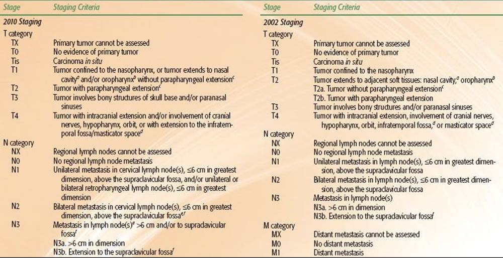

TABLE 41.7 FREQUENCY OF DIFFERENT HISTOLOGIC SUBTYPES OF NASOPHARYNGEAL CARCINOMA

PROGNOSTIC FACTORS
The extent of local invasion, regional lymphatic spread, and distant metastasis, as reflected by the TNM staging, is the most important prognostic factor. In general, advanced T-category is associated with worse local control and overall survival; advanced N-category predicts increased risk of distant metastasis and worse survival. Presence of distant metastasis (M1) upon presentation usually indicates poor prognosis, and treatment has conventionally been palliative in nature. A summary of the patterns of failure and survival rate for the different stages can be found in the Results of Treatment section.
The association of bone erosion, cranial nerve palsy, and lower nodal level with poorer survival is largely undisputed.86–87,88–89 However, the prognostic significance of parapharyngeal extension has been a topic of controversy. In a study of 364 patients, Chua et al.90 showed that greater tumor extension as defined by extension to the prestyloid space or extension to the anterior part of the masticator space was associated with a worse local failure-free rate (L-FFR; 72% vs. 86%) and lower distant failure-free survival rate (D-FFR; 68% vs. 87%) compared to tumors with no extension or extension only to the retrostyloid space. Other investigators reported similarly significant findings.75,76,88,91,92 Cheng et al.93 found parapharyngeal space extension to be the key factor in distant metastasis, even in N1 and N2 NPC.
However, Teo et al.87 did not find parapharyngeal space involvement to be an independent significant prognosticator in a study of 903 patients. Au et al.,94 using the AJCC/UICC definition of extension beyond the pharyngobasilar fascia in a study of 1,294 patients, also found that parapharyngeal extension was not a significant factor upon multivariate analysis.
These contradictory findings may be attributed to varying definitions of parapharyngeal space and incidence in the different series,75,87,88,91 prompting some to advocate for consideration of the degree of parapharyngeal space extension in future staging systems.76 In addition, suboptimal imaging by CT and conflation with retropharyngeal node enlargement likely contribute to the debate.95,96
Nevertheless, the most recent 2010 AJCC staging system (Table 41.6) downgraded involvement of oropharynx and/or nasal cavity without parapharyngeal extension from T2a to T1 while designating the presence of parapharyngeal extension to be the sole determinant of T2 classification. This was in light of recent multiple large retrospective studies that found no significant difference in disease failure hazard ratios between former AJCC 2002 T2a and T1.73,77,93,97
One recent topic of interest has been the prognostic significance of prevertebral space involvement (PSI). Recent MRI-based studies reported PSI to be an independent prognostic factor in cases of NPC treated with two-dimensional (2D) radiation therapy.98,99 In a study of 506 patients treated with intensity-modulated radiotherapy (IMRT), Zhou et al.100 found PSI to independently predict overall survival (OS) and distant metastasis-free survival (DMFS) rates similar to those of T4 disease and advocated for its inclusion as a T4 parameter in future AJCC staging systems.
Another prognostic factor to consider is the gross volume of the primary tumor (GTV-P). Although it is highly correlated to T-stage, considerable variability in tumor volume exists within the same T-stage, and evidence increasingly suggests that tumor volume as an independent significant factor can better predict prognosis than T-category as specified by both AJCC/UICC and Ho systems101,102–103,104,105 (Fig. 41.14).
In a study of 308 patients staged with MRI, Sze et al.103 showed that those with GTV-P of <15 cm3 had significantly higher L-FFR than those with a value of ≥15 cm3 (97% vs. 82% at 3 years; p < 0.01). Multivariate analysis confirmed GTV-P to be a strongly significant factor independent of T-category by the 1997 fifth edition of the AJCC/UICC; the risk of local failure increased by 1% for every 1-cm3 increase in volume. A similar observation between GTV and clinical outcomes was observed in other head and neck tumors, including oropharyngeal cancers.106 Further study is required to determine how tumor volume may be optimally incorporated into future staging systems.
Most series found significantly better prognosis for females and younger patients.86,94,107 In 759 patients, Sham and Choy86 showed a higher 5-year survival rate in females compared with males (45% vs. 28%) and in patients younger than 40 vs. older than 40 years of age (50% vs. 40%, p = .002). However, they did not find age to significantly affect the 10-year survival rate. Multivariate analysis of 1,294 patients by Au et al.94 also showed worse cancer-specific death rates in males (hazard ratio [HR] = 1.28, p = .02) and patients older than 50 years (HR = 1.79, p <0.001).
Although not all studies found histology to be an independent prognostic factor,27,108 many found nonkeratinizing and undifferentiated carcinomas (formerly known as lymphoepitheliomas) to be more radiosensitive and offer better prognosis than keratinizing squamous cell carcinoma.31,109,110 Of note, regarding ethnicity as a prognostic factor, a study by Corry et al.111 showed no prognostic difference between ethnic Asian and non-Asian patients with nonkeratinizing carcinoma.
FIGURE 41.14. The correlation between T-category and gross volume of primary tumor (GTV-P). UICC, International Union Against Cancer. (Modified from Sze W, Lee A, Yau T, et al. Primary tumor volume of nasopharyngeal carcinoma: Prognostic significance for local control. Int J Radiat Oncol Biol Phys 2004;59:21–27.)

EBV and Other Biomarkers
Because of the association of EBV with NPC, various anti-EBV antibodies have long been studied for their potential as biomarkers. While some studies showed that elevated level of serum anti-EBV antibodies could indicate presence60,112,113 of disease, others showed anti-EBV antibody titers to have little value for posttreatment surveillance.61,114,115 The prognostic value of such titers prior to treatment has also been controversial. While Xu et al.116found that high EBV DNase-specific neutralizing antibody at diagnosis predicted significantly worse event-free and overall survival, others found that a number of antibodies (VCA-IgG, VCA-IgA, EA-IgG, EA-IgA, EBNA-IgG, EBNA-IgA) could not predict prognosis.113,117,118
Circulating cell-free DNA of EBV in the plasma of NPC patients is a significant prognostic marker and has been found to be superior to serum anti-EBV antibodies.118 Lo et al.119 showed that plasma EBV DNA had high sensitivity (96%) and specificity (93%) for detecting NPC, while Ma et al.120 showed that circulating EBV DNA levels correlated significantly with tumor burden. Studies by Lo et al.121 and Lin et al.64 found that high pretreatment levels were associated with advanced stages and poor prognosis. Meanwhile, Leung et al.65 showed that pretreatment plasma EBV DNA load was an independent prognostic factor for OS in 376 patients and could be used to segregate early-stage patients into poor-risk and high-risk subgroups. Thus, pretreatment EBV DNA assays have the potential to complement TNM staging in guiding treatment.
Although Le et al.122 found no correlation between pretreatment EBV DNA levels and survival, they did find posttreatment levels to be a strongly significant predictor of outcome; patients with no detectable EBV DNA had a 2-year OS rate of 94% versus 55% for patients with detectable posttreatment levels (p <0.002).
Other studies also consistently reported that patients with elevated posttreatment EBV DNA load had higher risk of tumor recurrence.64,121,123–125 Using multivariate analysis to compare various prognostic factors for NPC, Lin et al.126 found that the combined EBV DNA load (pretreatment and 1-week posttreatment) was the most significant factor.
More recently, both Wang et al.127 and An et al.128 demonstrated that the clearance rate of plasma EBV DNA during the first month of salvage chemotherapy could predict tumor response and overall survival in patients with metastatic/recurrent NPC; undetectable levels after the first cycle indicated significantly better survival. These data suggest that early evaluation of plasma EBV DNA can offer oncologists timely insight for potential alterations in the therapeutic regimen for patients with a slow clearance rate. In addition, Wang et al.129 prospectively monitored the plasma EBV DNA of 245 NPC patients in clinical remission with assays every 3 to 6 months and found the plasma EBV DNA assay to have much greater sensitivity, specificity, and accuracy than FDG-PET in predicting relapse, suggesting its utility for posttreatment surveillance.
As with other head and neck squamous cell carcinomas, EGFR is commonly expressed in patients with NPC. Chua et al.130 found expression of EFGR in 89% of patients, in which overexpression was associated with significantly poorer disease-specific survival. Others reported similar findings of prognostic significance.24,131,132 Ma et al.131 performed multivariate analysis on several biomarkers in 78 patients, including microvessel density, Ki67 antigen, p53 oncoprotein, HER2, and EGFR, and found EGFR to be the only independent prognostic factor.
The study by Hui et al.133 showed that 58% of NPC patients had expression of hypoxia-inducible factor 1α (HIF-1α), 57% had carbonic anhydrase IX (CA IX), and 60% had vascular endothelial growth factor (VEGF). Those with positive hypoxic profile (high expression of HIF-1α and CA IX) had a worse progression-free survival (p = .04); those with both positive hypoxic and angiogenic profile (high VEGF) were strongly associated with worse progression-free survival (p = .0095). Multiple other studies have also showed that overexpression of these markers, particularly VEGF, is associated with poorer survival.134–136
Other biologic factors that might have prognostic significance include E-cadherin and β-catenin,137 c-erbB2,138 p53,139 NM23-HI,140,141 and interleukin-10.142 Further validation of these potential biomarkers is needed.
TREATMENT STRATEGY
Because of the anatomic location—proximity to critical structures—surgical exposure and tumor resection with sufficient margins have been very challenging.143 Primary surgical intervention was rare after the 1950s for these reasons, with surgical interventions employed mainly for biopsy to gain histologic confirmation and salvage therapy for persistent or recurrent cancer. Primary treatment since has typically employed radiotherapy (RT) alone and, more recently, in combination with chemotherapy.
Radiation Therapy
To achieve the best therapeutic ratio, every single step in the RT procedures (localization of gross tumor and target volumes, immobilization, optimization of dose fractionation, determination of treatment techniques, and precision in RT delivery) is important.
For planning, the patient should be set up in a supine position with head extended for adequate separation between the primary tumor/retropharyngeal nodes and the upper neck nodes. The tip of the uvula and the base of the occiput should be on a parallel plane to the beam axis. The patient is immobilized with a thermoplastic mask covering the head-to-shoulder region (Fig. 41.15). For patients to be treated by conventional 2D technique, a mouth bite is useful to minimize the dose to the oral cavity, with enlarged neck nodes to be marked with wire before imaging.
FIGURE 41.15. Immobilization of patient in a customized thermoplastic mask covering the whole head-to-shoulder region.

Dose, Time, and Fractionation
A significant dose–response relation was observed in the majority of retrospective studies, based on patients irradiated with 2D techniques. Marks et al.144 and Vikram et al.145 showed that local control was significantly improved in patients who received >67 Gy to the tumor target. Perez et al.27 observed that patients with T1-2 tumors had a local tumor control rate of 100% for those given >70 Gy, compared with 80% for those treated with 66 to 70 Gy. However, local control for patients with T3-4 tumors remained <55%, even with total dose >70 Gy. Similar findings were reported by Mesic et al.,146 where ≥70 Gy achieved better local control for T1-2 tumors than 60 Gy (94% vs. 76%), but higher doses or larger fields did not significantly improve outcomes in T3-4 tumors. These observations suggest that, besides consideration of the prescribed dose, the problem of sufficient coverage has to be overcome for advanced tumors.
Lee et al.147 reported a study of 1,008 patients with T1 tumors irradiated by four different fractionation schedules and demonstrated that total dose was the most important radiation factor (p = .01). Dose fraction did not affect local control; however, it was a significant risk factor for temporal lobe necrosis.148,149 Therefore, a fractional dose of >2 Gy should be avoided150 (see section Sequelae of Treatment).
The impact of the time factor is more contentious. A randomized study by Marcial et al.151 in which 62 patients were treated with split-course irradiation (30 Gy in 10 fractions over 2 weeks, then a 3-week rest period, followed by an additional 30 Gy in 10 fractions) and compared with 59 patients with 66 Gy in 33 fractions in 6.5 to 7 weeks demonstrated no significant difference in 5-year local control (86% vs. 80%), nodal control (86% vs. 78%), or disease-free survival (40% vs. 30%).
However, Vikram et al.145 observed that patients with interruption of RT for ≥21 days had significantly poorer local tumor control than patients without interruptions (34% vs. 67%). Similar findings have been subsequently reported,152,153 with the general consensus that prolongation is likely to be detrimental, even for nonkeratinizing NPC.
In general, the prescription recommended for NPC is to a total dose of about 70 Gy over 7 weeks to the gross tumor along with 50 to 60 Gy for elective treatment of potential risk sites.
TABLE 41.8 EXAMPLE OF GUIDELINE ON ANATOMIC STRUCTURES/BOUNDARIES FOR DELINEATING CLINICAL TARGET VOLUMES FOR INTENSITY-MODULATED RADIATION THERAPYA

Tumor Target Volumes
The GTV should encompass the primary nasopharyngeal tumor, gross retropharyngeal lymphadenopathy, and gross nodal disease as determined by clinical, endoscopic, and radiologic examinations. Presence of lymph nodes of ≥1 cm or with evidence of central necrosis is considered gross nodal disease. For patients given induction chemotherapy, it is recommended that the targets be determined by the prechemotherapy extent.
Prophylactic neck radiation is usually recommended in N0 patients because of the high incidence of occult neck node involvement. Lee et al.35 found that patients with a clinically negative neck who underwent elective neck irradiation had a significantly lower nodal recurrence rate than those who were untreated (11% vs. 40%). In addition, even with successful salvage by subsequent treatment, patients with nodal recurrence had a significantly greater incidence of distant metastases than those without recurrence (21% vs. 6%).
The clinical target volume (CTV) includes the GTV, regions of microscopic disease, and potential infiltrative spread. Different centers may have different philosophies in defining the margins and dose level. For example, Table 41.8 shows the delineation criteria for the various CTVs currently employed at Memorial Sloan-Kettering Cancer Center. A gross disease CTV (CTV70) is defined as the GTV plus an additional margin of 5 mm to 1 cm surrounding all gross disease. The margin may be decreased to as small as 1 mm in critical regions near the brainstem or spinal cord. The high-risk subclinical CTV (CTV59.4) encompasses the GTV including all potential areas of microscopic spread of disease. This volume should include at a minimum the entire nasopharynx; retropharyngeal lymph nodal regions; clivus; skull base; pterygoid fossae; parapharyngeal space; sphenoid sinus; posterior one-fourth to one-third of the nasal cavity; and posterior one-fourth to one-third of the maxillary sinuses. This CTV59.4 should also include lymph nodal groups that are at risk of potential microscopic disease spread: bilateral upper deep jugular (junctional, parapharyngeal), submandibular, subdigastric (jugulodigastric), midjugular, posterior cervical, and retropharyngeal lymph nodes. In patients with clinically N0 neck, it is not necessary to include level I nodal regions.
The planning target volume (PTV) is defined as the CTV including a circumferential margin of typically 3 to 5 mm to all the CTVs to account for setup errors and potential patient motion. The PTV margin may be decreased to as small as 1 mm in regions near critical normal structures such as the brainstem or spinal cord.
Conventional Two-Dimensional Treatment Techniques
One of the most common RT approaches employed is comprised of two phases.154 Phase I consists of large lateral opposing faciocervical fields that encompass the primary tumor and the upper neck nodes in one volume, with a matching lower anterior cervical field for the lower cervical lymphatics. Phase II is used after 40 Gy to limit the dose to the spinal cord. This three-field technique includes lateral opposing facial fields coupled with anterior facial field for the primary tumor. Typical treatment fields and radiologic landmarks are shown in Figure 41.16. Shrinking treatment fields by cone-down after 50 to 60 Gy should be done, when possible, to increase protection of critical structures.
The three-field technique in phase II allows the dose to be minimized to the temporomandibular joints and the bilateral temporal lobes. However, coverage may not be sufficient for tumors with extensive posterolateral extension to the parapharyngeal spaces or caudal extension to the oropharynx. To remedy this deficit, an additional dose is delivered by a posterolateral field with avoidance of neurologic structures.155
FIGURE 41.16. Conventional two-dimensional radiotherapy using Ho’s technique. A: Phase I, lateral-opposed faciocervical fields (I–II) and lower anterior cervical field (IVb). B: Phase II, sagittal view showing lateral-opposed facial fields and noncoplanar anterior facial field (III). C: Coronal view of anterior facial field (III). D: Anterior cervical field for whole neck (IV).

Three-Dimensional Conformal Treatment Techniques
Nasopharyngeal carcinoma presents most typically as a concave tumor, allowing for computerized three-dimensional (3D) treatment plans to be an important technical advance for improved radiation delivery. Several investigators designed multifield conformal plans, including the seven-field technique used at Memorial Sloan-Kettering Cancer Center (MSKCC) (New York, NY)156 and the “Boomerang” technique used at Peter MacCallum Cancer Institute (East Melbourne, Australia).157 When compared to conventional 2D plans, 3D planning demonstrated better tumor dose coverage while decreasing normal tissue dose in several studies.87,158,159
Leibel et al.160 from MSKCC demonstrated that the target volume underdosed at the 95% isodose level was lowered with 3D plans when compared with 2D plans (7% vs. 22%). On average, the mean tumor dose increased 13%, leading to an estimation that tumor control would increase by 15%. However, a subsequent study in which 68 patients received this technique for a boost of 19.8 to 25.2 Gy following phase I conventional 2D treatment for 50.4 Gy to a total dose of 70.2 to 75.6 Gy did not show significant improvement; the 5-year L-FFR was 77% and late toxicity grade ≥3 was 25%.156
More encouraging results were obtained by Jen et al.,161 who compared 72 patients treated with 3D conformal technique with 108 patients treated with 2D technique. A significant improvement in 3-year L-FFR for T4 (86% vs. 47%) and event-free survival for both stage III (80% vs. 56%) and stage IV (82% vs. 33%) was observed. Furthermore, the incidence of xerostomia at 3 years was significantly less with 3D conformal treatment (69.2% vs. 98.0%), although for most other late toxicities little difference was seen.
IMRT Techniques
IMRT has supplanted conventional radiotherapy in the treatment of NPC in an increasing number of institutions throughout the world. The intensity of the radiation beams can be modulated to deliver a high dose to the tumor with a superior target volume coverage while significantly limiting the dose to surrounding normal tissues.162–165,166,167 Following the initial publication168 and the subsequent update on IMRT for NPC from the University of California, San Francisco (UCSF),166 several other institutions have utilized IMRT with similar excellent treatment outcomes.169–170,171–175
Another point of interest is the possibility of biologic enhancement by simultaneous modulated accelerated-radiation therapy (SMART), also known as dose painting, as a new way of delivering an accelerated fractionation (AF) schedule, a concept that was first reported by Butler et al.176 for the treatment of other head and neck cancers with IMRT.
These differing methods and dose fractionation regimens for IMRT are being investigated by different groups. Table 41.9 summarizes the key features along with reported results. The majority of the patients in these series received additional chemotherapy and/or enhanced RT with boosts or AF. All reported encouraging early results, with local control in >90% at 2 to 4 years.
At UCSF patients were typically prescribed 70 Gy to the PTVgross disease and involved lymph nodes in 2.12- to 2.25-Gy fractions, while PTVhigh-risk subclinical patients were prescribed 59.4 Gy in 1.8 fractions, and a clinically negative neck (PTVlow-risk subclinical) patient received 54 Gy at 1.64-Gy fractions, all in conventional once-daily fractions.166,177 Bucci et al.177 reported the updated results of 118 patients and confirmed excellent locoregional control of 96%. Nonetheless, distant failure remained high (28%) despite broad use of concurrent-adjuvant CRT. OS was 74% at 4 years.
At MSKCC, Wolden et al.169 reported their experience with 74 patients: 59 were treated with AF using the concomitant boost method and 15 by the SMART method/dose painting. For the SMART cohort, a total dose of 70.2 Gy at 2.34 Gy/fraction was given to the gross disease, and the “microscopic” PTV received 54 Gy at 1.8 Gy/fraction. There was a trend, but no statistically significant improvement, in 3-year L-FFR than for patients treated by 3D conformal boost (91% vs. 79%, p = .11) Additional dose escalation by SMART boost in 50 patients with T3 to T4 tumors was reported by Kwong et al.178 from Queen Mary Hospital (Hong Kong). They sought to deliver a total dose of 76 Gy at 2.17 Gy/fraction to the gross tumor. The early result for locoregional control was excellent (96% at 2 years); however, serious late toxicities were observed, including 4% of patients having a life-threatening hemorrhage from carotid artery pseudoaneurysm, and another 4% developing temporal lobe necrosis with a median follow-up of 2.1 years.
Two different IMRT approaches are being utilized by different centers: (i) an extended-whole field (EWF) IMRT technique, in which the total target volume is encompassed in the IMRT plan, or (ii) a split-field (SF) IMRT technique, in which the target volumes superior to the vocal cords are treated with an IMRT plan and the lower neck nodes are treated with a conventional low anterior neck field.179–181 Discussions among practitioners on which IMRT technique is best have been persistent. Concerns of potential failures at the SF matchline due to potential underdosing, or even complications resulting from overdosing, have caused many centers to implement EWF IMRT even when no clinically involved neck nodes are evident in the matchline region. These concerns may be caused by the treatment delivery system that is used at centers where a perfect match between the IMRT fields and the low anterior neck field is not possible. However, with a EWF technique, an unnecessary dose of radiation is delivered to the normal glottic larynx,182 whereas with the SF IMRT technique, the dose to the vocal cords is minimal due to shielding by a midline Cerrobend block or the multileaf collimator (MLC). No IMRT matchline failures or complications have been reported with the SF IMRT technique.168
TABLE 41.9 INTENSITY-MODULATED RADIATION THERAPY FOR NASOPHARYNGEAL CARCINOMA: METHODS AND RESULTS BY DIFFERENT CENTERS

FIGURE 41.17. Intensity-modulated radiation therapy for a patient with T2N2M0 nasopharyngeal carcinoma treated at Memorial Sloan-Kettering Cancer Center, showing delineation of gross tumor target (GTV) and planning target volume (PTV) for 70 and 54 Gy, the dose distribution, and the dose volume histogram (DVH) for GTV, PTV70, and PTV59.4.

Figures 41.17 and 41.18 show examples of MSKCC IMRT plans delivered with the dynamic MLC system using a sliding-window technique to patients with early and advanced disease, respectively. A total dose of 70 Gy at 2.12 Gy/fraction to the PTVgross disease and 59.4 Gy to the PTVhigh-risk subclinical patients is given over 33 once-daily fractions (Table 41.8). For the low neck, if split-field IMRT is used, a dose of 50.4 Gy at 1.8 Gy/fraction/day is generally prescribed. However, if the low neck is included in the IMRT fields and is considered at low risk for nodal involvement, the PTVlow-risk subclinical patient typically receives 54 Gy at 1.64 Gy/fraction per day.
Inverse planning involves the appropriate specification of normal tissue dose constraints. It is important to note that overstringent use of normal tissue constraints might result in inadequate cover of tumor targets, and therefore optimal balance is essential. Different dose constraint guidelines have been suggested.183,184 An example of dose-constraint guidelines is provided in Table 41.10, which displays the guidelines used at Memorial Sloan-Kettering Cancer Center.
Dose Escalation
Excellent local tumor control has been reported by delivering an additional boost to patients with early disease treated by conventional 2D technique.
FIGURE 41.18. Intensity-modulated radiation therapy for a patient with T4, N2, M0 nasopharyngeal carcinoma treated at Memorial Sloan-Kettering Cancer Center, showing delineation of gross tumor volume (GTV) and planning target volume (PTV) for 70 and 54 Gy and the dose distribution.

TABLE 41.10 INTENSITY-MODULATED RADIATION THERAPY FOR NASOPHARYNGEAL CARCINOMA: AN EXAMPLE OF NORMAL TISSUE DOSE CONSTRAINTSA

Brachytherapy
The most commonly used method is brachytherapy. Intracavitary insertions185–193 or interstitial implants194–197 have been used in T1 to T3 nasopharyngeal carcinomas as a boost treatment following external beam irradiation (EBRT) or in the treatment of recurrent disease, either alone or in combination with EBRT. Brachytherapy is not suitable for treatment of tumors with intracranial extension because of the rapid reduction of dose as distance from the radioactive source increases. Since the advent of IMRT as primary radiotherapy for nasopharyngeal carcinoma and with its demonstration of excellent local control, the use of brachytherapy as a boost treatment following definitive IMRT has dramatically declined. Multiple applicators and techniques have been developed for the delivery of intracavitary brachytherapy.187,188,189,191,193,198 In the past, intracavitary brachytherapy was delivered using low–dose rate (LDR) techniques. However, at present, remote afterloading, fractionated high–dose rate (HDR) techniques are more commonly used (Fig. 41.19).198
Table 41.11 summarizes reports on the use of brachytherapy as a boost for dose escalation. Most studies demonstrated that local control of up to 90% to 95% could be achieved for T1-2 tumors without excessive late damages. A retrospective comparison by Wang188 from Massachusetts General Hospital (Boston, MA) reported that T1 to T2 patients who received a 10- to 15-Gy LDR brachytherapy boost after 60 to 64 Gy by EBRT had a 5-year L-FFR of 90% versus 54%, respectively, with p = .001 for patients receiving EBRT alone to 65 to 70 Gy. A similar study by Teo et al.199 in which delivery of 18 to 24 Gy in three fractions by HDR brachytherapy showed significant improvement of the 5-year L-FFR of 95% compared with 90% 5-year L-FFR for EBRT-only patients (p = .016).
However, a report by Ozyar et al.200 of patients with T1 to T4 tumors treated with HDR brachytherapy boost of 12 Gy in three fractions did not show improvement over EBRT alone (3-year L-FFR,86% vs. 94%; p = .23). More recently, a prospective trial by the International Atomic Energy Agency studied 275 patients with locoregionally advanced NPC disease (TNM stages III or M0 stage IV) who were all treated by induction chemotherapy followed by concurrent chemoradiotherapy to 70 Gy; one randomized arm then received a brachytherapy boost of 11-Gy LDR or three fractions of 3-Gy HDR. With a median follow-up of 29 months, the authors reported no additional benefit of brachytherapy boost compared with chemoradiotherapy alone toward 3-year OS (63.3% vs. 62.9%, p = .742, respectively), locoregional-FFR (54.4% vs. 60.5%, p = .647), or distant-metastasis–free survival (52.6% vs. 59.8%, p = .496).201
One major limitation of brachytherapy is that the dose delivered is adequate only for superficial nonbulky tumors. Furthermore, optimal positioning of the applicators depends both on the individual clinician’s skill and the patient’s anatomic features.
Stereotactic Radiosurgery
Stereotactic radiosurgery (SRT) or fractionated radiotherapy allows for precise delivery of highly conformal RT with a rapid dose falloff and provides an alternative for dose escalation. Hara et al.202 reported a study of 82 patients with T1 to T4 tumors showing excellent 5-year L-FFR of 98% after receiving a median SRT boost of 12 Gy (range, 7 to 15 Gy) following EBRT to 66 Gy. However, despite the addition of concurrent chemotherapy in 76% of the patients, the distant failure rate was 32% and OS was 69%. With a median follow-up of 40.7 months for living patients, 12.1% of patients developed radiographic temporal lobe necrosis (only 2.4% were symptomatic with seizures), and 3.6% developed retinopathy. The risk was especially high in patients with T4 tumors.
FIGURE 41.19. Endocavitary brachytherapy for nasopharyngeal carcinoma. A: The Rotterdam nasopharyngeal applicator. B: The simulator check-film showing the position of the radioactive sources and the dose distribution.

TABLE 41.11 ADJUVANT BRACHYTHERAPY BOOST FOR PRIMARY TREATMENT OF NASOPHARYNGEAL CARCINOMA

Altered Fractionation
Over the last few decades, studies have been conducted to explore the role of altered fractionation regimens in head and neck cancers, along with nasopharyngeal cancers. Hyperfractionation, accelerated fractionation, and a combination were explored in conjunction with concurrent chemotherapy.
Sanchiz et al.203 conducted a large randomized trial examining twice-daily (BID) versus once-daily (QD) irradiation for head and neck cancer, including tumors of the nasopharynx. A total of 859 patients with advanced head and neck cancers (T3-T4, N0-3, M0 by UICC staging), which included 92 patients with nasopharyngeal carcinoma, was randomly assigned to QD irradiation (group A), BID irradiation (group B), or QD irradiation with concurrent 5-fluorouracil (5-FU) chemotherapy (group C). Groups B and C showed a significant improvement in median duration of response and OS when compared to group A. No significant differences were seen between groups B and C.
The first randomized trial on accelerated fractionation (AF) for NPC by Teo et al.204 used an uncommon schedule of 2.5 Gy/fraction QD for 8 fractions before randomization to an experimental arm using 1.6 Gy BID for an additional 32 fractions versus a control arm treated with 2.5 Gy QD for another 16 fractions. The trial was terminated early because of excessive neurologic toxicities in the AF arm (49% vs. 23%). For this series of 159 patients (62% with T1-2 tumors), the AF arm did not achieve significant improvement in tumor control (5-year L-FFR, 89% vs. 85%). Jen et al.205 reported on a study of 222 patients in which 76 patients received hyperfractionated RT at 1.2 Gy/fraction BID and 12 patients received accelerated-hyperfractionated RT at 1.6 Gy BID, to a median dose of 80 Gy for these twice-daily RT groups. The remaining 134 patients treated by conventional QD fractionation to a median dose of 70 Gy. The patients treated by BID fractionation did not demonstrate a statistically significant difference in 5-year L-FFR when compared to QD fractionation (T1-3, 93% vs. 86%; T4, 44% vs. 37%, respectively). The 1.2-Gy/fraction regimen did not cause excessive toxicity; however, patients treated with 1.6 Gy/fraction had a 27% incidence of temporal lobe necrosis.206 See section Sequelae of Treatment for more details regarding the influence of dose fractionation on brain necrosis.
To minimize the risk of late damage, the more moderate AF schedule of the Danish Head and Neck Cancer Study Group 6–7 Trials using 2 Gy/fraction, six fractions per week207 of 1,476 patients with head and neck cancer, of which 435 had pharyngeal tumors, including nasopharynx, demonstrated that accelerated fractionation resulted in significantly improved 5-year L-FFR (76% vs. 64% for six and five fractions, respectively, p = .0001) and disease-specific survival (73% vs. 66%, for six fractions and five fractions, respectively, p = .01) but not OS. This same fractionation schedule was tested retrospectively for NPC by Lee et al.208 They reported that patients irradiated to a total dose of 66 Gy with 2D technique when on an AF schedule had significantly higher L-FFR than those treated with conventional five fractions per week. The benefit was significant particularly for T3-4 tumors (87% vs. 62%; p<0.01), and multivariate analyses confirmed that fractionation was an independent prognostic factor for overall progression (AF group: HR = 0.63, 95% confidence interval [CI], 0.41 to 0.98, p = .04). In addition, no significant increase in late toxicity was observed at 3 years (20% vs. 15%).
This schedule was then used in the subsequent NPC-9902 trial initiated by the Hong Kong Nasopharyngeal Carcinoma Study Group,209 which aimed to assess the therapeutic benefit of AF and/or concurrent-adjuvant chemoradiotherapy (CRT). It randomized 189 patients with locally advanced NPC (T3-T4, N0-1, M0) to four arms: (i) conventional fractionation (CF) alone, (ii) AF (six fractions/week) alone, (iii) CF with concurrent chemotherapy, and (iv) AF with concurrent chemotherapy. Preliminary results with a median follow-up of 2.9 years showed that AF per se did not demonstrate a significant improvement in event-free survival (EFS) when compared with CF (AF vs. CF: HR 0.68, 95% CI 0.37 to 1.25, p = .22). However, AF combined with CRT (arm 4) achieved a strongly significant improvement when compared with CF alone (EFS: 94% vs. 70%, p = .008) but without an improvement in OS. A significant increase in acute and late toxicity in the AF plus CRT arm was also noted.
From these and other experiences with altered fractionation schedules in the treatment of nasopharyngeal carcinoma, in addition to other head and neck cancers, it has been concluded that both total dose and overall treatment time are important factors in determining outcomes.210 With the increasing use of IMRT, dose escalation—to allow ample dose delivery to gross and subclinical disease—has become achievable without associated changes in rates of toxicity, considerably improving the therapeutic ratio of concurrent chemoradiation and causing the aforementioned fractionation schemes to fall out of favor.
Chemotherapy
Nasopharyngeal carcinoma is generally regarded to be a highly chemosensitive disease. While radiotherapy alone is the standard treatment for stage I NPC, concurrent CRT with or without adjuvant chemotherapy is the current standard for locally advanced disease (stage III–VB) based on multiple randomized, controlled trials and meta-analysis (Table 41.12). Although there is less evidence for CRT in intermediate stage (2010 AJCC stage II) disease, it is recommended that such patients be treated with CRT in light of pooled data from two phase III trials211,212 and a more recent phase III trial from Chen et al.213 In more detail, Chen et al.213 demonstrated that Chinese stage II NPC patients (equivalent to AJCC II-III; only 13% of the study’s patients are AJCC 2010 stage III) that received concurrent chemoradiotherapy resulted in a 5-year OS benefit compared to radiation alone (94.5% vs. 85.8%, p = .007), with improved distant control (94.8% vs. 83.9%, p = .007). Multivariate analyses found that number of chemotherapy cycles was the only independent factor associated with improved OS, progression-free survival, and distant control. With the addition of chemotherapy, an increase in acute side effects was observed, but no significant increase in late effects was reported.
TABLE 41.12 RANDOMIZED PROSPECTIVE TRIALS WITH CONCURRENT CHEMORADIOTHERAPY

Concurrent Chemoradiotherapy
The landmark Intergroup 0099 trial was the first to document a significant survival benefit for CRT versus RT alone.214 This trial randomized 147 patients with locally advanced NPC to either RT alone or CRT at centers located in the United States. Chemotherapy consisted of concurrent cisplatin (CDDP; 100 mg/m2 on days 1, 22, and 43), followed by three cycles of adjuvant CDDP (80 mg/m2 on day 1) and 5-FU (1000 mg/m2/d on days 1 to 4) every 4 weeks. Radiotherapy was delivered in 1.8- to 2-Gy fractions to a total dose of 70 Gy. The trial was closed early due to a significant overall survival benefit in favor of CRT (78% vs. 47% at 3 years). A 5-year update confirmed progression-free survival (58% vs. 29%) and overall survival (67% vs. 37%) in favor of CRT. Reactions to these findings were initially tempered by several limitations of the trial. Results achieved in the RT arm were much poorer than those generally obtained at centers treating endemic NPC. Outcomes in the CRT arm were in fact more consistent with outcomes obtained in endemic areas using RT alone. Another concern was that 24% of the enrolled patients had disease of keratinizing histology, and it was unknown whether the same benefit would be seen in endemic areas with predominantly undifferentiated disease.
Subsequent trials confirmed the benefit of concurrent CDDP-based chemotherapy in endemic populations. Wee et al.215 reported the results of 221 stage III-IVB patients from Singapore randomized to receive either RT alone or CRT. Chemotherapy consisted of a slightly modified version of the Intergroup regimen: CDDP (25 mg/m2 on days 1 to 4) for three cycles every 3 weeks, followed by adjuvant CDDP (20 mg/m2 on days 1 to 4) and 5-FU (1000 mg/m2 per day on days 1 to 4) for three cycles. Radiotherapy was delivered to a dose of 70 Gy in 2-Gy fractions. Three-year overall survival for the CRT and RT arms was 85% and 65%, respectively (p = .006). CRT reduced the incidence of distant metastasis by 17% at 2 years (p = .003).
Langendijk et al.216 performed a meta-analysis of 10 trials that randomized NPC patients to conventional RT or CRT. The 10 studies included 4 neoadjuvant trials,217–220 3 concurrent (with/without adjuvant) trials,214,221,222 2 adjuvant trials,223,224 and 1 neoadjuvant plus adjuvant trial.225 The authors found a pooled hazard ratio for death of 0.82, with an absolute survival benefit of 4% at 5 years. Subgroup analysis revealed that the overall survival benefit was only significant for those patients receiving concurrent chemotherapy, with a hazard ratio for death of 0.48 and absolute survival benefit of 20% at 5 years. Analysis of the neoadjuvant chemotherapy trials found a significant reduction in locoregional recurrence and distant metastasis but no overall survival benefit.
These results, in combination with results from a second meta-analysis226 and the Singapore trial reported by Wee et al.,215 confirmed CRT as the standard approach in stage III, IVA, and IVB NPC. At many centers, the standard course of chemotherapy has been based on the U.S. Intergroup regimen, which consisted of concurrent high-dose CDDP (100 mg/m2 for three cycles) and adjuvant CDDP/5-FU for three cycles. This regimen is associated with significant acute and late toxicities, and patient compliance is frequently difficult to achieve. In the Intergroup 0099 trial, for example, only 63% completed all three cycles of concurrent chemotherapy, and only 55% were able to receive all three courses of adjuvant therapy.214 As a result, weekly CDDP has been adopted by many institutions, especially for patients with poor nutritional status.227 In a phase III trial comparing CRT versus RT alone in 350 patients with locally advanced disease, Chan et al.228 demonstrated good efficacy and tolerability for a regimen consisting of weekly CDDP (40 mg/m2). Seventy-eight percent of patients in the CRT arm received at least four cycles of CDDP, and CRT was associated with a statistically significant survival benefit after adjusting for age and disease stage.
Other Chemotherapy Agents
Weekly oxaliplatin (70 mg/m2) has been demonstrated to have good tolerability and efficacy, albeit in the setting of a small phase III trial of 115 patients randomized to CRT or RT alone.229 Carboplatin has also been employed as a substitute to high-dose CDDP, with comparable efficacy, in a noninferiority trial reported by Chitapanarux et al.230 Two hundred and six patients were randomized to either concurrent high-dose CDDP and adjuvant CDDP/5-FU or concurrent weekly carboplatin and adjuvant carboplatin/5-FU. The trial had 80% power to detect a hazard ratio for death of 1.25 at 3 years. No significant difference in disease-free survival or overall survival was seen at median follow-up of 26 months. Disease-free survival was 59.6% and 64.7% (p = 0.522) for the carboplatin and CDDP arms, respectively.
Cetuximab to target epidermal growth factor receptor (EGFR; EGFR overexpression is observed in >80% of NPC patients) was examined in a phase II trial, with optimistic findings.231 The 2-year rates of OS, locoregional progression–free survival, distant metastasis–free survival, and progression-free survival were 89.9%, 93.0%, 82.8%, and 86.5%, respectively. Bevacizumab, to exploit the angiogenesis pathway (VEGF is overexpressed in about two-thirds of NPC patients), was studied in a phase II trial in which the agent was added to the standard chemoradiotherapy schedule and demonstrated promising results.232 Lee et al. reported, with a median follow-up of 2.5 years, 2-year OS, locoregional progression–free survival, distant metastasis–free survival, and progression-free survival of 90.9%, 83.7%, 90.8%, and 74.7%, respectively.
Adjuvant Chemotherapy
While good efficacy and tolerability have been shown for select alternative regimens, the greatest body of evidence for concurrent CRT has been with the CDDP-based U.S. Intergroup regimen of concurrent plus adjuvant chemotherapy. Nevertheless, it is unknown whether the adjuvant chemotherapy component of the U.S. Intergroup regimen contributed to its survival benefit.
Compliance with adjuvant chemotherapy can be especially difficult, as patients are recovering from the acute effects of CRT. Randomized trials comparing RT alone to RT plus adjuvant chemotherapy have all been negative.224,225Moreover, there are data to suggest a survival benefit for concurrent CRT without adjuvant chemotherapy.222,228
In 2011, Chen et al.233 reported a randomized trial that compared concurrent CRT to concurrent CRT plus adjuvant chemotherapy (CDDP/5-FU) in 508 patients. With a median follow-up of 38 months, there were fewer failures at any site in the concurrent chemoradiotherapy plus adjuvant chemotherapy group versus the chemoradiotherapy-only group (14% vs. 16%); however, this difference was not statistically significant (p = .13). It is important to note that this trial was not designed as a noninferiority trial against the standard. In addition, compliance was an issue in this study, in which about 18% of patients randomized to the adjuvant arm did not receive adjuvant chemotherapy.233Until further data emerge, adjuvant chemotherapy is considered by many to be optional in the setting of concurrent CRT, although it may have a role in patients with residual EBV DNA after CRT.
Neoadjuvant Chemotherapy
The effect of adding neoadjuvant chemotherapy to concurrent CRT is a topic of much current interest and the subject of two ongoing phase III randomized trials. Multiple phase II trials have shown excellent outcomes and tolerability with a variety of regimens.234–237 Figure 41.20 demonstrates the potential value of induction chemotherapy in tumor control. One recent randomized phase II trial demonstrated an overall survival benefit for docetaxel/CDDP followed by CDDP-RT when compared to concurrent CDDP-RT alone.238 Although the difference in 3-year progression-free survival did not reach statistical significance (88.2% vs. 59.2%, p = .12), 3-year overall survival was significantly improved in the neoadjuvant arm (94.1% v 67.7%, p = .012).
An ongoing phase III trial opened by the National Health Research Institute, Taiwan (http://clinicaltrials.gov/ct2/show/NCT00201396), is randomizing patients to concurrent CRT versus induction chemotherapy (CDDP, mitomycin, epirubicin, and leucovorin) plus concurrent CRT. The enrollment goal is 480 patients with an estimated completion date of December 2013. Another ongoing phase III trial, organized by the Hong Kong Nasopharyngeal Cancer Study Group Limited (http://clinicaltrials.gov/ct2/show/NCT00379262), is enrolling patients to one of three arms: (i) CDDP/5-FU induction chemotherapy plus concurrent CDDP-RT, (ii) concurrent CDDP-RT plus adjuvant CDDP/5-FU, or (iii) CDDP/capecitabine induction chemotherapy plus concurrent CDDP-RT. In addition, patients in each of these three arms will be randomized to either conventional or accelerated RT. The enrollment goal is 798 patients, with an estimated study completion date of April 2017.
FIGURE 41.20. Magnetic resonance imaging showing shrinkage of primary tumor by induction chemotherapy using cisplatin and 5-fluorouracil before proceeding to concurrent cisplatin and radiotherapy. (From Lee AW, Lau KY, Hung WM, et al. Potential improvement of tumor control probability by induction chemotherapy for advanced nasopharyngeal carcinoma. Radiother Oncol. 2008;87(2):204–210, with permission from Elsevier.)

PERSISTENT/RECURRENT NPC
Because long-term survival can be achieved for a substantial proportion of patients with early locoregional recurrence and useful palliation for those with extensive disease, aggressive salvage treatment is usually advocated. Several approaches can be used successfully, including surgery, brachytherapy, and EBRT. Chemotherapy is generally used in conjunction with local treatment in patients with advanced disease.
Early Detection and Diagnosis
While progress in surgical and reconstructive techniques and radiotherapy delivery methods has led to improvements in the control rate for primary treatment of NPC, local failure remains a problem for patients with advanced T-category disease. Distinction should be made between persistent disease (tumors that do not completely regress following primary treatment) and recurrent disease (tumors that reemerge after initial complete regression) because the prognoses and therapeutic considerations are different, with better survival and control rates for persistent disease.239
As tumors regress at different rates following RT, one difficult decision is when to consider residual tumors as genuine persistence and proceed with salvage treatment. In one prospective study by Kwong et al.,240 serial biopsies of the nasopharynx were performed on 803 patients after RT treatment to observe the time course of histologic remission for NPC and determine its prognostic significance. The 5-year L-FFR was 82% for patients who achieved early histologic remission (<5 weeks), 77% for those with delayed remission (5 to <12 weeks), but only 40% for those with persistent disease at 12 weeks, despite subsequent salvage treatment.240 Thus, while delayed histologic remission was not a poor prognostic factor, positive biopsies beyond 12 weeks did indicate poor prognosis. The optimal time for intervention remains uncertain, but because it is important to avoid both unnecessary overtreatment and excessive delay in treatment, the authors recommended an observation period of 10 weeks before additional treatment.240
Early detection of locoregional failure is crucial for a better chance of salvage, and regular follow-up after completion of primary treatment is recommended. Frequently used methods include manual palpation, rigid nasopharyngeal endoscopy and nasopharyngeal biopsies, imaging techniques (e.g., CT and MRI), and serologic tests (e.g., anti-EBV titers, plasma EBV DNA levels).
Nasopharyngoscopy is more sensitive than CT and MRI in detecting tumor persistence/recurrence and is the preferred method for initial screening.241,242 If a patient presents with suspicious endoscopic findings or elevated anti-EBV titers, a nasopharyngeal biopsy is performed to confirm diagnosis. CT or MRI is performed upon a confirmed diagnosis to delineate the tumor extent.243 Although MRI has limitations in separating tumor recurrence from radiation fibrosis,244 it is superior to CT in demonstrating extent of soft tissue tumors, as well as in identifying submucosal infiltration, marrow infiltration in the skull base, perineural invasion, and intracranial spread.241,245,246
Technetium-99m methoxyisobutylisonitrile single-photon emission computed tomography may be a useful tool for differentiating persistent or recurrent tumor from radiation fibrosis247 and was shown by Kostakoglu et al.248 to be superior to MRI performed at 3 to 6 months post-RT in diagnosing complete response. The advent of FDG-PET is another valuable development. FDG-PET and MRI were compared in 67 NPC patients 4 to 70 months after completion of RT, and FDG-PET was found to be superior to MRI in all aspects in detection of local recurrence, with increased sensitivity (100% vs. 62%) and specificity (93% vs. 44%).249 It may also contribute useful information to questionable findings on MRI.250,251
Paraneoplastic syndrome (PNS) can signal a silent neoplasm and may precede the clinical manifestation itself of persistent or recurrent NPC. PNS can follow the course of the tumor and can sometimes be used to diagnose recurrence and monitor its evolution, with the most common dermatologic manifestation being dermatomyositis and the syndrome of inappropriate secretion of antidiuretic hormone being a common endocrinologic presentation.252
Circulating cell-free DNA of EBV may be another useful tool for early detection of treatment failure. A longitudinal study by Lo et al.121 showed that elevation of EBV DNA levels was noted in patients with relapse up to 6 months before detectable clinical disease. In addition, EBV DNA copy number has been shown to predict margin status post salvage nasopharyngectomy. Wei et al.253 reported that in early recurrent NPC patients with elevated EBV DNA copies, surgical resection reduced the EBV DNA copy number postoperatively and that negative surgical margins are associated with zero EBV DNA copies postoperatively.
The incorporation plasma EBV DNA measurements as screening prior to PET in detecting posttreatment failures of NPC has been investigated. In a prospective study by Wang et al.,129 245 NPC patients in remission were monitored prospectively via plasma EBV DNA assay every 3 to 6 months, in which 36 patients with abnormal EBV DNA tests and 5 patients with clinically suggestion signs of recurrence but undetectable EBV DNA levels underwent FDG-PET scans. Elevated EBV DNA levels correctly predicted all 36 recurrences, while the 5 patients who presented with clinical signs suggestive of recurrent disease but with undetectable EBV DNA levels did not have recurrent disease. The authors concluded that plasma EBV DNA appears to be a useful biomarker for posttreatment surveillance in NPC.
In addition, the clearance rate of plasma EBV DNA during the first month of chemotherapy has also been found to predict tumor response and patient survival in 30 patients with recurrent NPC and may have potential as an early prognostic marker to help guide salvage treatment.127
Additional Radiation for Persistent Disease
Excellent results have been reported when using brachytherapy for locally persistent disease after a full course of EBRT (Table 41.13), with 5-year L-FFR in the range of 87% to 95% for patients with initial T1 tumors (AJCC 2002 T1-2a).199,239,254–256 Preliminary evidence suggests that patients with disease persisting from initial T2 tumors (AJCC 2002 T2b) could also be effectively treated by brachytherapy.257
Stereotactic RT is a valuable alternative for delivering additional EBRT. Yau et al.258 studied 755 patients with T1-4 tumors and found that 7% had positive biopsies 8 weeks after completion of primary RT. Twenty-one patients were treated with fractionated stereotactic RT to a median dose of 15 Gy and achieved a 3-year L-FFR of 82%, which was similar to the corresponding L-FFR of 86% in the complete responders and was significantly better than the corresponding L-FFR of 71% in 24 patients treated with high dose-rate brachytherapy to a median dose of 20 Gy.
TABLE 41.13 RESULTS OF LOCALLY PERSISTENT/RECURRENT NASOPHARYNGEAL CARCINOMA TREATED WITH BRACHYTHERAPY

TABLE 41.14 RESULTS ON REIRRADIATION FOR LOCAL RECURRENCE OF NASOPHARYNGEAL CARCINOMA

Reirradiation for Recurrent Disease
Various radiation therapy modalities are used to treat recurrent NPC, including intracavitary brachytherapy, external beam irradiation, interstitial implantation, particle beam radiotherapy, and stereotactic radiosurgery (Table 41.14).187,192,193,195,196,259 IMRT has also been used with excellent preliminary results, with control rates of up to 100% for rT1-3.260
The most important prognostic factors are the TNM stage of the tumor at the time of recurrence and reirradiation dose. Thorough restaging, including metastatic workup, is necessary. A study of 891 patients with local recurrence from 1976 to 1985 by Lee et al.261 showed that only 32% of reirradiated patients achieved local salvage, with 54% developing regional and/or distant failure. Most series using conventional 2D technique showed that doses ≥60 Gy were associated with better outcome.187,193,262,263 For IMRT, 60 to 70 Gy is recommended, taking into account factors such as previous radiation amount, overlap between previously treated area and target for reirradiation, interval between RT courses, tumor bulk, and whether concurrent chemotherapy will be given.264
Lee et al.265 retrospectively compared the symptomatic late toxicity rate in 487 patients with two courses of EBRT versus 3,635 patients with one course. They found that the major determinant of late complications was severity of damage during the initial course and that the sum of total biologic dose tolerated (BED-∑) was higher than expected with a single-course treatment (BED-1). This suggested partial recovery of normal tissue (especially in patients reirradiated after 2 or more years) and higher tolerance for reirradiation when primary treatment was given, with better sparing of normal tissues. Assuming α/β ratio of 3 Gy, it was found that the BED-∑ that incurred 20% toxicity at 5 years was 129% that of BED-1.
Brachytherapy has been widely used for treatment of recurrent NPC (Table 41.13) and can be used effectively on its own for early-stage recurrent NPC.239,255 Using interstitial implants with radioactive gold grains, Kwong et al.239reported a 5-year L-FFR of 63%; complications included headache (28%), palatal fistula (19%), and mucosal necrosis (16%). Law et al.255 achieved excellent local salvage up to 89% using iridium mold, but the complication rate was 53%.
The combination of brachytherapy and EBRT is useful, particularly when conventional 2D technique is used. Lee et al.262 showed that patients reirradiated by combined modalities had an improved 5-year L-FFR of 45% compared with 32% by EBRT alone and 29% by brachytherapy alone. The superiority of the combined method has been supported by other studies.187,193,266,267 A recent study from MSKCC found that combined-modality treatment (CMT), consisting of EBRT followed by brachytherapy, achieved similar L-FFR with previous historical series. CMT also demonstrated fewer late grade 3 or higher events compared with patients receiving EBRT alone (8% vs. 73%, respectively).268
Stereotactic radiosurgery or fractionated stereotactic radiotherapy is another useful tool for retreatment of local recurrence, as it allows for rapid fall-off of radiation dose outside tumor volume and near surrounding critical structures and can be used either alone for smaller lesions or in combination with EBRT for larger ones. Control rates ranging from 53% to 86% have been reported.269–272 For advanced recurrence with extension beyond the nasopharynx, this method offers better dose coverage than brachytherapy. A higher salvage rate from adding stereotactic radiation as a boost after EBRT has been reported.270,273,274 Although most series reported a low risk of complications, massive hemorrhage with potential fatal outcome has been described.270 Radiosurgery should thus be avoided when there is direct tumor encasement of the carotid artery or when a high cumulative dose has already been delivered. Fractionated stereotactic radiotherapy has shown improved late toxicity profile compared to 3D conformal RT275 and single-dose radiosurgery276 and may also give better local control rates compared with single-fraction radiosurgery.277
Advances in imaging technology and radiotherapy techniques have made it possible to reduce target volume without jeopardizing local control, reducing complication rates. Table 41.15 summarizes the treatment outcome and severe late complications by external beam reirradiation. Past series using 2D technique achieved 5-year survival rates in the range of 16% to 63%, and the incidence of temporal lobe necrosis ranged from 2% to 27%. The use of 3D conformal radiotherapy showed improving results. In a study by Chang et al.,273 none of the patients reirradiated by 3D technique developed temporal lobe necrosis, compared with 14% of those reirradiated by 2D technique. Zheng et al.278 reported a 5-year local salvage rate of 71% from 3D technique, but the actuarial rate of late toxicities (grade 4) was still as high as 49%.
The use of IMRT for reirradiation has shown very encouraging short-term results. Using IMRT to deliver 68 to 70 Gy, Lu et al.279 reported 100% salvage rate without any severe late complications in a series of 49 patients with a median follow-up of 9 months. Using IMRT to a median dose of 54 Gy in 31 patients (with or without induction chemotherapy and stereotactic boost), Chua et al.260 reported a 1-year control rate of 100% for rT1-T3 and 35% for rT4, with late complications (greater than grade 3) of 25% (at 1 year). Using a median dose of 70 Gy in 70 patients, Qiu et al.280 reported a locoregional salvage rate of 66% with similar toxicity rate at 2 years. While rT staging did not predict control rate, original T classification remained a significant adverse prognostic factor and may serve as a strong marker for the underlying locally aggressive biology of the original disease. Longer follow-up is needed to better evaluate treatment results and sequelae resulting from IMRT.
Chemoradiotherapy may also improve treatment outcome for recurrent NPC in certain patients, and most recent salvage radiation series have included cisplatin-based chemotherapy for advanced-stage disease.260,268,280,281 Using gemcitabine and cisplatin as induction chemotherapy followed by reirradiation with IMRT in 20 patients (95% rT3-4), Chua et al.260 reported a 1-year local salvage rate of 75%. In a study of 35 patients (66% rT3-4), Poon et al.281reported a 1-year EFS of 42% by concurrent cisplatin followed by adjuvant chemotherapy with cisplatin and 5-fluorouracil. However, while concomitant chemoirradiation is increasingly used to treat primary NPC, it remains uncertain whether concomitant chemoirradiation is appropriate for retreatment of purely local recurrences (rT1-2), due to high rates of toxicity and potential complications.
TABLE 41.15 INCIDENCE OF LATE TOXICITY FOLLOWING RADIATION WITH CONVENTIONAL TECHNIQUE (WITHOUT CONCURRENT CHEMOTHERAPY) FOR NASOPHARYNGEAL CARCINOMA

FIGURE 41.21. A: Computed tomography shows planned osteotomies of the maxilla and the posterior part of the nasal septum (broken line). B: The maxilla is swung laterally while still attached to the anterior cheek flap.

FIGURE 41.22. The left maxilla is swung laterally, exposing the nasopharynx (arrow).

Surgical Treatment
For patients with persistent nodal disease, radical neck dissection is the preferred treatment for persistent or recurrent lymph node involvement in the neck when there is no distant metastasis282 and may achieve a 5-year nodal control rate of 66% and disease-free survival rate of 37%.283 In cases with extension of disease beyond lymph nodes into nearby structures, additional afterloading brachytherapy to tumor bed may improve local control.284
Surgical management at the primary site is hampered by difficulties in obtaining adequate exposure and obtaining adequate surgical margins.143,285–287 Various approaches have been employed, including an infratemporal approach from the lateral aspect,288 transpalatal, transmaxillary, and transcervical approaches from the inferior aspect,287,289 and an anterolateral approach.290 More recently, minimally invasive techniques, such as a transnasal approach and an endoscopic approach, have also been employed successfully.291 Although controversial, salvage surgery by nasopharyngectomy can be a viable option in certain patients in whom disease is localized in the nasopharynx, with acceptable results reported for rT1-3 tumors.247 Wei et al.292 reported a 5-year control rate of 62% and 5-year disease-free survival rate of 49% in 60 patients who received curative resections. Typically, all patients with recurrence have undergone prior radical RT, with associated complications of trismus and palatal fistula being common; however, the mortalities associated with these surgical procedures are low.
Recurrent NPC is frequently located in the pharyngeal recess on the lateral wall. Access to this region is crucial for complete tumor extirpation. Wei and Sham293 advocated the anterolateral approach or maxillary swing approach for localized recurrence in the nasopharynx. After facial incisions and the necessary osteotomies, the maxilla bone is swung laterally while remaining attached to the anterior cheek flap as one osteocutaneous entity (Figs. 41.21 and 41.22). The nasopharynx with the tumor and its surrounding area, including the paranasopharyngeal region, are then widely exposed for resection. Upon completion of nasopharyngectomy, the maxilla is replaced and attached to the remainder of the facial skeleton with miniplates.
Wei et al.290 reported 161 patients with salvage nasopharyngectomy employing this approach performed at Queen Mary Hospital (Hong Kong) for recurrent NPC following primary treatment by radical RT. Twelve patients had prior brachytherapy as a salvage procedure. All patients were recurrent stage T1, with 78% of these patients achieving negative tumor resection margins, confirmed by frozen section, with the remaining patients demonstrating microscopic tumor at the internal carotid artery or the skull base during surgery, making further complete resection unattainable. All patients recovered from this anterolateral approach and were discharged. Regarding treatment-associated morbidities, trismus of varying grades was present in 60% and palatal fistula in 25% of patients. Recent modification of the palatal incision has eliminated the problem of palatal fistula.294
Satisfactory long-term results can be achieved when persistent/recurrent tumor are completely resected. Several recent surgical series reported locoregional control and OS rates of 40% to 72% and 30% to 54% respectively.291,295–297 Postoperative reirradiation is recommended for patients with positive surgical margins and/or advanced disease292,298,299 and may be beneficial even when surgical margins are negative.247
For recurrent tumors, margin status, adjuvant treatment type, and parapharyngeal space involvement were significant prognostic factors for local control, whereas dura or brain involvement, local recurrence, and adjuvant treatment type predicted survival.291 Preoperative EBV DNA levels and PET-CT can assist in predicting the outcome of salvage nasopharyngectomy, and PET-CT may predict presence of extracapsular spread of metastatic lymph nodes.300
TABLE 41.16 LOCAL TUMOR CONTROL AFTER CONVENTIONAL RADIOTHERAPY

TABLE 41.17 NODAL CONTROL AFTER CONVENTIONAL RADIOTHERAPY

TABLE 41.18 OVERALL SURVIVAL AFTER CONVENTIONAL RADIOTHERAPY

TABLE 41.19 OVERALL SURVIVAL BY T- AND N-STAGE AFTER CONVENTIONAL RADIOTHERAPY

RESULTS OF TREATMENT
Specific results of various treatments are mentioned in the previous respective sections. This section focuses on summarizing these results.
The local and regional control rates in select conventional radiotherapy series are listed in Tables 41.16 and 41.17. Control of the primary lesion using conventional radiotherapy varied with the T classification (Table 41.16), ranging from 64% to 97% for T1 lesions, 54% to 94% for T2 lesions, 34% to 100% for T3 lesions, and 40% to 71% for T4 lesions.27,31,32,145,146,301,302 Dose escalation with intracavitary brachytherapy if using non-IMRT treatment techniques188 has been shown to improve local control. Stereotactic radiosurgery boost after external beam radiotherapy (both conventional and IMRT) has also shown improved local control.202,303 Nonkeratinizing squamous cell carcinoma (both differentiated and undifferentiated subtypes) had improved local control rates when compared to keratinizing squamous cell carcinoma in T2 to T3 lesions but not in T1 or T4 lesions.27,214,216
Excellent nodal control rates in the neck have been demonstrated by conventional radiotherapy even after involvement of extensive cervical lymph node metastasis (Table 41.17). The neck nodal control rates ranged from 82% to 100% for N0, 86% to 92% for N1, and 78% to 89% for N2 to N3 disease.27,32,146 Five-year survival rates ranged from 36% to 58% (Table 41.18).27,31,32,146,301,302,304
The 5-year survival rate with conventional non-IMRT radiotherapy correlated with the T-stage, as well as with the N-stage (Table 41.19), being 60% to 76% for T1, 48% to 68% for T2, 27% to 55% for T3, and 0% to 29% for T4 lesions32,302 and 42% to 78% for N0, 27% to 70% for N1, and 32% to 52% for N2 to N3 disease. When nodes in the lower neck and/or the supraclavicular fossa are involved prognosis is poor.
Contemporary series with IMRT demonstrated excellent local and regional control achieved in 97% and 98% of the patients treated at UCSF, respectively.166 An update of the UCSF experience continued to show excellent local control of approximately 96%.177 Subsequently, several other institutions also recently published their results, which further demonstrate excellent local control rates ranging from 91% to 100% and regional control rates ranging from 91% to 98% (Table 41.20).150,169–170,171,178,231,232,305,306–310,311,312–313
The Radiation Therapy Oncology Group conducted a phase II trial using IMRT with/without chemotherapy in the treatment of nasopharyngeal carcinoma in which patients with ≥T2 (i.e., ≥T2b by AJCC 2002) and/or node-positive disease also received concurrent CDDP followed by adjuvant CDDP and 5-FU chemotherapies. The results showed that a multi-institutional setting can reproduce the excellent results (local control rate of 92.3%) observed from single-institution studies.305 This reproducibility of excellent locoregional control rates in a multi-institutional trial, along with several single-institution studies, with IMRT is encouraging; however, distant metastases remains a therapeutic challenge despite extensive use of chemotherapy. The distant recurrence rate ranges from 10% to 15% at 2 years,305,311 with 4-year rates as high as 34% (Table 41.20).166 Novel systemic therapies or regimens are needed for improved distant control and overall survival of this disease.
SEQUELAE OF TREATMENT
Overall Incidence and Types
Due to the anatomic proximity of the nasopharynx to critical structures and the need for high radiation doses and adequate field coverage, the risks of radiation-induced toxicities are substantial. The overall complication rate from conventional treatment ranged from 31% to 66%, with severe sequelae including temporal lobe necrosis, hearing loss, xerostomia, neck fibrosis, cranial nerve dysfunction, endocrine dysfunction, soft tissue necrosis, osteonecrosis, and transverse radiation myelitis.28,32,39 The diagnosis of irradiation injury can be difficult, as other possible causes (tumor recurrence in particular) must be excluded.
The toxicity results of five major series using conventional irradiation for NPC are summarized in Table 41.21.
The series of 378 patients treated at MDACC during 1954–1992 showed an actuarial frequency rate in grade ≥4 toxicity of 16%, 19%, and 29% at 5, 10, and 20 years, respectively.28 Despite the use of higher radiation doses, Sanguineti et al.28 showed a reduction in the 10-year actuarial rate of severe toxicity from 14% in 1954–1971 to 5% in 1983–1992. Other investigators reported similar findings.
The decreased rates of complications over time were likely due to the use of newer oblique and opposed lateral field techniques instead of single central field, custom blocking, and image-guided (CT) treatment planning.28
Due to the extremely narrow therapeutic treatment margin of NPC, maximum conformity and precision in RT delivery are crucial for minimizing the risk of late damage. The emergence of IMRT is a major advance for improving physical dose distribution and has increased the potential for protecting normal tissues. Studies on patients treated with IMRT thus far have shown substantial sparing of salivary function,166,170,171 while other benefits will require longer follow-up to confirm.
The improved conformity offered by IMRT has led to attempts at dose escalation in order to achieve better tumor coverage for patients with extensive locoregional infiltration. However, studies have shown that dose escalation together with concurrent chemotherapy may lead to severe toxicities.178,314 Furthermore, extensive use of concurrent CRT independent of incorporation of dose escalation has been shown to significantly increase toxicities (grade ≥3) compared to RT alone.209,214,215,315–318
TABLE 41.20 RESULTS FROM CONTEMPORARY IMRT SERIES WITH OR WITHOUT CHEMOTHERAPY

Temporal Lobe Necrosis
Temporal lobe necrosis (TLN) is perhaps the most troublesome complication. Studies on NPC patients treated with conventional 2D RT found that TLN accounted for up to 65% of all irradiation-induced deaths; large fractions (>2 Gy) and overacceleration of treatment schedule greatly increased risk, with incidence as high as 33%.39,148,149
Diagnosis of TLN was often difficult and thus delayed. In Lee et al.’s examination of 102 patients with late TLN following conventional 2D RT,319 only 31% presented with classic symptoms of TLN (hallucinations, absence attacks, déjà vu), while 14% had headaches, confusion, convulsions, or hemiparesis. Thirty-nine percent had vague symptoms of dizziness, poor memory, or sudden changes in behavior, while 16% were asymptomatic.
TLN remains a serious concern in patients treated with IMRT, with incidence of 3% to 4% being reported for schedules of 70 Gy at 2.12 Gy/fraction,177 66 to 74 Gy at 2 Gy/fraction,171 and 76 Gy at 2.17 Gy/fraction.178 Series using larger fractions (70.2 Gy at 2.34 Gy/fraction and 68 Gy at 2.27 Gy/fraction) had incidence rates as high as 12% to 14%.150,310 Bakst et al.,150 in a prospective trial of hypofractionated dose-painting IMRT using 2.34-Gy fractions to deliver a total dose of 70.2 Gy, had favorable disease control and survival outcomes; however, 12% of treated patients developed temporal lobe necrosis, and the conclusion was that large fractional doses should be avoided to prevent in-field brain radiation necrosis.
Cranial Neuropathy
Cranial nerves IX through XII, particularly XII, are the most frequently impaired by radiation.39,320,321 This is related to marked radiation fibrosis, especially in patients who receive an additional boost dose to parapharyngeal space. Common symptoms include slurring of speech, twitching of neck muscles, and/or dysphagia. In a study of 31 NPC patients with post-RT dysphagia, Wu et al.322 found that 77% aspirated after the act of swallowing, raising concerns of fatal aspiration pneumonia.
Cranial nerve VI is also frequently affected, particularly in patients with TLN, while isolated palsy of branches of cranial nerve V is less common.39 Optic neuropathy is rare with careful attention to the RT technique and should be considered when treating lesions with base-of-skull involvement.323 The possibility of intracranial recurrence may confound the diagnosis of radiation injury, and exclusion of recurrence is necessary.
TABLE 41.21 INCIDENCE OF LATE TOXICITY FOLLOWING RADIATION WITH CONVENTIONAL TECHNIQUE (WITHOUT CONCURRENT CHEMOTHERAPY) FOR NASOPHARYNGEAL CARCINOMA

Oral Complications
Xerostomia is an almost universal complication from treatment with conventional RT and may lead to dental caries. Jen et al.324 showed that the salivary flow dropped by half with a dose of 7.2 Gy, reached the nadir after 36 Gy, and then further dropped after completion of RT without recovery during the following 2 years.
However, Lee at al.166 reported marked recovery of salivary function in patients treated with parotid-sparing IMRT (mean parotid dose, 34 Gy); the rate of grade 2 xerostomia decreased from 64% at 3 months to 2.4% at 2 years. Randomized trials comparing 2D RT and IMRT in patients with T1-2 tumors confirmed IMRT’s advantage in this regard.325,326 Nevertheless, it is important to rule out tumor invasion of the parotid gland prior to parotid-sparing IMRT, as recurrences have been reported. Cannon and Lee327 concluded that PET alone may be insufficient for detection of intraparotid lymph node involvement in patients with multilevel nodal disease, including disease in level II nodes. Even with negative PET findings, these patients may require additional evaluation of any benign-appearing parotid nodules before parotid-sparing IMRT by fine-needle aspiration or CT-guided biopsy.
Dental sequelae frequently accompany xerostomia. In a series of 1,758 patients, 2.7% developed osteoradionecrosis at the maxilla and 1.7% at the mandible. Tong et al.328 reported a 29% complication rate in patients who had post-RT extraction of posterior maxillary teeth, with 10.5% developing osteonecrosis. Prophylactic fluoride treatment should be employed to prevent dental decay, and decayed teeth should be extracted prior to RT to reduce this risk.328,329
Aural Toxicity
Hearing loss has always been a common radiation sequela, and the increasing use of cisplatin-based concurrent CRT has resulted in deafness rates as high as 42%.330
Sensorineural hearing loss (SNHL), particularly in the high-frequency range, was found in at least 30% of patients assessed with audiograms following RT, with higher rates in patients treated by concurrent CRT.39,331,332,333 The primary determinant of high-frequency SNHL is mean cochlea dose, which should be kept below 48 Gy to minimize damage.39,331,332,333 Due to the location of the primary tumor in the nasopharynx, pharyngotympanic tube (Eustachian tube) damage resulting in otitis media is difficult to avoid. However, lowering the dose to the external auditory canal and mastoid air cells can reduce the incidence and severity of acute external otitis and chronic serous otitis media, respectively.
Carotid Artery Injury
Carotid stenosis is a potentially fatal complication reported in patients who undergo irradiation of the head and neck region. Interval from radiotherapy was a significant independent predictor for severe carotid stenosis. Some have advocated for routine duplex ultrasound screening for high-risk patients (age >60 years, smoking, hypertension, hypercholesterolemia, cerebrovascular symptoms).334,335 Severe cases may require carotid endarterectomy or endoplasty.
Massive bleeding from ruptured pseudoaneurysms at the petrous portion of the internal carotid has been reported following IMRT with dose escalation.178,336 Urgent diagnosis and intervention with endovascular occlusion or stenting may be needed to prevent fatal consequences. Other concerns include severe telangiectasia and hypervascularization in the internal maxillary artery territory, for which emergency embolization may be considered.
Endocrine Dysfunction
The most common endocrine sequelae are amenorrhea and/or galactorrhea from hyperprolactinemia in female patients, followed by hypothyroidism and hypoadrenalism.
Lee et al.39 observed symptomatic hypothalamic–pituitary dysfunction in 5% of patients, with a median latency of 5 years, while a longitudinal study by Lam et al.337 with detailed endocrine assessment found a 5-year incidence of 62%, with dysfunction detected as early as 1 year following RT.338 The deficiency of releasing or inhibitory factors indicated that the hypothalamus is the primary location of damage.339,340 As many of these dysfunctions may be corrected pharmacologically, routine evaluation of hypothalamic, pituitary, and thyroid function should be considered in the follow-up examination of long-term survivors.
Shielding may help lessen endocrine dysfunction when using 2D technique.341 The need for maximum conformity to protect normal tissues during radiotherapy is paramount.
Second Malignancies
Radiation-induced malignancy is rare, with an incidence of 0.04% and latency period of >10 years. The most common histologic types are maxillary osteosarcoma342 and soft tissue sarcoma.72 Surgery presents the only chance of cure, but the prognosis is often poor. While second primary head and neck cancer is relatively uncommon for NPC patients, Teo et al.343 reported an excessive incidence rate of tongue cancer at 0.13% per patient-year. The possibility of radiation carcinogenesis cannot be excluded.
REFERENCES
1. Lanier A, Bender T, Talbot M, et al. Nasopharyngeal carcinoma in Alaskan Eskimos Indians, and Aleuts: a review of cases and study of Epstein-Barr virus, HLA, and environmental risk factors. Cancer1980;46(9):2100–2106.
2. Parkin D, Muir C, Whelan S, et al. Cancer incidence in five continents. In: Parkin D, et al., eds. Epidemiology. Lyon, France: IARC Scientific Publications, 1992.
3. Curado M, Edwards B, Shin H, et al. Cancer incidence in five continents. Lyon, France: IARC Scientific Publications, 2007. IX(160).
4. Ferlay J, Shin H, Bray F, et al. GLOBOCAN 2008, Cancer incidence and mortality worldwide: IARC CancerBase No. 10. [Internet]. Lyon, France: International Agency for Research on Cancer, 2010.
5. Balakrishnan U. An additional younger-age peak for cancer of the nasopharynx. Int J Cancer 1975;15(4):651–657.
6. Singh W. Nasopharyngeal carcinoma in Caucasian children. A 25-year study. J Laryngol Otol 1987;101(12):1248–1253.
7. Ferlay J, Shin HR, Bray F, et al. Estimates of worldwide burden of cancer in 2008: GLOBOCAN 2008. Int J Cancer 2010;127(12):2893–2917.
8. Bei JX, Li Y, Jia WH, et al. A genome-wide association study of nasopharyngeal carcinoma identifies three new susceptibility loci. Nat Genet 2010;42(7):599–603.
9. Lu SJ, Day NE, Degos L, et al. Linkage of a nasopharyngeal carcinoma susceptibility locus to the HLA region. Nature 1990;346(6283):470–471.
10. Chan SH, Day NE, Kunaratnam N, et al. HLA and nasopharyngeal carcinoma in Chinese–a further study. Int J Cancer 1983;32(2):171–176.
11. Li X, Fasano R, Wang E, et al. HLA associations with nasopharyngeal carcinoma. Curr Mol Med 2009;9(6):751–765.
12. Tai TM. Descriptive epidemiology of nasopharyngeal cancer. Curr Opin Oncol 2001;8:114.
13. Ho JH. An epidemiologic and clinical study of nasopharyngeal carcinoma. Int J Radiat Oncol Biol Phys 1978;4(3–4):182–198.
14. Teo PM, Leung SF, Yu P, et al. A comparison of Ho’s, International Union Against Cancer, and American Joint Committee stage classifications for nasopharyngeal carcinoma. Cancer 1991;67(2):434–439.
15. Yu MC, Ho JH, Lai SH, et al. Cantonese-style salted fish as a cause of nasopharyngeal carcinoma: report of a case-control study in Hong Kong. Cancer Res 1986;46(2):956–961.
16. Henderson BE, Louie E, SooHoo Jing J, et al. Risk factors associated with nasopharyngeal carcinoma. N Engl J Med 1976;295(20):1101–1106.
17. Nam JM, McLaughlin JK, Blot WJ. Cigarette smoking, alcohol, and nasopharyngeal carcinoma: a case-control study among U.S. whites. J Natl Cancer Inst 1992;84(8):619–622.
18. Buell P. Race and place in the etiology of nasopharyngeal cancer: a study based on California death certificates. Int J Cancer 1973;11(2):268–272.
19. King H, Haenszel W. Cancer mortality among foreign- and native-born Chinese in the United States. J Chronic Dis 1973;26(10):623–646.
20. Dickson RI, Flores AD. Nasopharyngeal carcinoma: an evaluation of 134 patients treated between 1971–1980. Laryngoscope 1985;95(3):276–283.
21. Vasef MA, Ferlito A, Weiss LM. Nasopharyngeal carcinoma, with emphasis on its relationship to Epstein-Barr virus. Ann Otol Rhinol Laryngol 1997;106(4):348–356.
22. Gulley ML. Molecular diagnosis of Epstein-Barr virus-related diseases. J Mol Diagn 2001;3(1):1–10.
23. Brooks L, Yao QY, Rickinson AB, et al. Epstein-Barr virus latent gene transcription in nasopharyngeal carcinoma cells: coexpression of EBNA1, LMP1, and LMP2 transcripts. J Virol 1992;66(5):2689–2697.
24. Kung CP, Meckes DG Jr, Raab-Traub N. Epstein-Barr Virus LMP1 Activates EGFR, STAT3, and ERK through Effects on PKC{delta}. J Virol 2011;85(9):4399–4408.
25. Mainou BA, Raab-Traub N. LMP1 strain variants: biological and molecular properties. J Virol 2006;80(13):6458–6468.
26. Wang D, Liebowitz D, Kieff E. An EBV membrane protein expressed in immortalized lymphocytes transforms established rodent cells. Cell 1985;43(3 Pt 2):831–840.
27. Perez CA, Devineni VR, Marcial-Vega V, et al. Carcinoma of the nasopharynx: factors affecting prognosis. Int J Radiat Oncol Biol Phys 1992;23(2):271–280.
28. Sanguineti G, Geara FB, Garden AS, et al. Carcinoma of the nasopharynx treated by radiotherapy alone: determinants of local and regional control. Int J Radiat Oncol Biol Phys 1997;37(5):985–996.
29. Lee AW, Foo W, Law SC, et al. Nasopharyngeal carcinoma: presenting symptoms and duration before diagnosis. Hong Kong Med J 1997;3(4):355–361.
30. Pan WR, Suami H, Corlett RJ, et al. Lymphatic drainage of the nasal fossae and nasopharynx: preliminary anatomical and radiological study with clinical implications. Head Neck 2009;31(1):52–57.
31. Chu AM, Flynn MB, Achino E, et al. Irradiation of nasopharyngeal carcinoma: correlations with treatment factors and stage. Int J Radiat Oncol Biol Phys 1984;10(12):2241–2249.
32. Hoppe RT, Goffinet DR, Bagshaw MA. Carcinoma of the nasopharynx. Eighteen years’ experience with megavoltage radiation therapy. Cancer 1976;37(6):2605–2612.
33. McNeese MD, Fletcher GH. Retreatment of recurrent nasopharyngeal carcinoma. Radiology 1981;138(1):191–193.
34. Moench HC, Phillips TL. Carcinoma of the nasopharynx. Review of 146 patients with emphasis on radiation dose and time factors. Am J Surg 1972;124(4):515–518.
35. Lee AW, Poon YF, Foo W, et al. Retrospective analysis of 5037 patients with nasopharyngeal carcinoma treated during 1976–1985: overall survival and patterns of failure. Int J Radiat Oncol Biol Phys1992;23(2):261–270.
36. Frezza G, Barbieri E, Emiliani E, et al. Patterns of failure in nasopharyngeal cancer treated with megavoltage irradiation. Radiother Oncol 1986;5(4):287–294.
37. Johansen LV, Mestre M, Overgaard J. Carcinoma of the nasopharynx: analysis of treatment results in 167 consecutively admitted patients. Head Neck 1992;14(3):200–207.
38. Teo P, Shiu W, Leung SF, et al. Prognostic factors in nasopharyngeal carcinoma investigated by computer tomography–an analysis of 659 patients. Radiother Oncol 1992;23(2):79–93.
39. Lee AW, Law SC, Ng SH, et al. Retrospective analysis of nasopharyngeal carcinoma treated during 1976–1985: late complications following megavoltage irradiation. Br J Radiol 1992;65(778):918–928.
40. Hui EP, Leung SF, Au JS, et al. Lung metastasis alone in nasopharyngeal carcinoma: a relatively favorable prognostic group. A study by the Hong Kong Nasopharyngeal Carcinoma Study Group. Cancer2004;101(2):300–306.
41. Luk NM, Yu KH, Choi CL, et al. Skin metastasis from nasopharyngeal carcinoma in four Chinese patients. Clin Exp Dermatol 2004;29(1):28–31.
42. Ngan RK, Yiu HH, Cheng HK, et al. Central nervous system metastasis from nasopharyngeal carcinoma: a report of two patients and a review of the literature. Cancer 2002;94(2):398–405.
43. Li JC, Mayr NA, Yuh WT, et al. Cranial nerve involvement in nasopharyngeal carcinoma: response to radiotherapy and its clinical impact. Ann Otol Rhinol Laryngol 2006;115(5):340–345.
44. Lindberg R. Distribution of cervical lymph node metastases from squamous cell carcinoma of the upper respiratory and digestive tracts. Cancer 1972;29(6):1446–1449.
45. Poon PY, Tsang VH, Munk PL. Tumour extent and T stage of nasopharyngeal carcinoma: a comparison of magnetic resonance imaging and computed tomographic findings. Can Assoc Radiol J2000;51(5):287–295, quiz 286.
46. Lanzieri CF, Bangert B. Magnetic resonance imaging of the nasopharynx. Top Magn Reson Imaging 1990;2(4):39–47.
47. Mancuso AA, Bohman L, Hanafee W, et al. Computed tomography of the nasopharynx: normal and variants of normal. Radiology 1980;137(1 Pt 1):113–121.
48. Sievers KW, Greess H, Baum U, et al. Paranasal sinuses and nasopharynx CT and MRI. Eur J Radiol 2000;33(3):185–202.
49. Dillon WP, Harnsberger HR. The impact of radiologic imaging on staging of cancer of the head and neck. Semin Oncol 1991;18(2):64–79.
50. Glazer HS, Niemeyer JH, Balfe DM, et al. Neck neoplasms: MR imaging. Part II. Posttreatment evaluation. Radiology 1986;160(2):349–354.
51. Ng SH, Chang TC, Ko SF, et al. Nasopharyngeal carcinoma: MRI and CT assessment. Neuroradiology 1997;39(10):741–746.
52. Som PM. Detection of metastasis in cervical lymph nodes: CT and MR criteria and differential diagnosis. AJR Am J Roentgenol 1992;158(5):961–969.
53. van den Brekel MW, Stel HV, Castelijns JA, et al. Cervical lymph node metastasis: assessment of radiologic criteria. Radiology 1990;177(2):379–384.
54. Chang JT, Chan SC, Yen TC, et al. Nasopharyngeal carcinoma staging by (18)F-fluorodeoxyglucose positron emission tomography. Int J Radiat Oncol Biol Phys 2005;62(2):501–507.
55. Gordin A, Golz A, Daitzchman M, et al. Fluorine-18 fluorodeoxyglucose positron emission tomography/computed tomography imaging in patients with carcinoma of the nasopharynx: diagnostic accuracy and impact on clinical management. Int J Radiat Oncol Biol Phys 2007;68(2):370–376.
56. Liu FY, Lin CY, Chang JT, et al. 18F-FDG PET can replace conventional work-up in primary M staging of nonkeratinizing nasopharyngeal carcinoma. J Nucl Med 2007;48(10):1614–1619.
57. Neel HB 3rd. Nasopharyngeal carcinoma: diagnosis, staging, and management. Oncology (Williston Park), 1992;6(2):87–95; discussion 99–102.
58. Wara WM, Wara DW, Phillips TL, et al. Elevated IGA in carcinoma of the nasopharynx. Cancer 1975;35(5):1313–1315.
59. Ho HC, Ng MH, Kwan HC, et al. Epstein-Barr-virus-specific IgA and IgG serum antibodies in nasopharyngeal carcinoma. Br J Cancer 1976;34(6):655–660.
60. Henle W, Ho JH, Henle G, et al. Nasopharyngeal carcinoma: significance of changes in Epstein-Barr virus-related antibody patterns following therapy. Int J Cancer 1977;20(5):663–672.
61. Neel HB 3rd, Taylor WF. Epstein-Barr virus-related antibody. Changes in titers after therapy for nasopharyngeal carcinoma. Arch Otolaryngol Head Neck Surg 1990;116(11):1287–1290.
62. Zeng Y. Seroepidemiological studies on nasopharyngeal carcinoma in China. Adv Cancer Res 1985;44:121–138.
63. Zong YS, Sham JS, Ng MH, et al. Immunoglobulin A against viral capsid antigen of Epstein-Barr virus and indirect mirror examination of the nasopharynx in the detection of asymptomatic nasopharyngeal carcinoma. Cancer1992;69(1):3–7.
64. Lin JC, Wang WY, Chen KY, et al. Quantification of plasma Epstein-Barr virus DNA in patients with advanced nasopharyngeal carcinoma. N Engl J Med 2004;350(24):2461–2470.
65. Leung SF, Zee B, Ma BB, et al. Plasma Epstein-Barr viral deoxyribonucleic acid quantitation complements tumor-node-metastasis staging prognostication in nasopharyngeal carcinoma. J Clin Oncol2006;24(34):5414–5418.
66. Cammoun M, Ellouz R, Behi J, Attia RB. Histological types of nasopharyngeal carcinoma in an intermediate risk area. IARC Sci Publ 1978;20:13–26.
67. Shanmugaratnam K, Sobin LH. Histological typing of tumours of the upper respiratory tract and ear. 2nd ed. Heidelberg: Springer-Verlag, 1991.
68. AJCC: Pharynx. Manual for Staging of Cancer, ed. H.D. In Beahrs OH, Hulter RVP, Kennedy BJ, et al, eds. Vol. 31. Philadelphia: JB LIppincott Company, 1992.
69. UICC: Pharynx. TNM Atlas, ed. e.a. In Spiessl B, eds. Vol. 20. Berlin: Springer-Verlag, 1992.
70. Edge SB, American Joint Committee on Cancer. AJCC cancer staging manual, 7th ed. New York: Springer, 2010:xiv, 648.
71. Neel HB 3rd, Taylor WF. New staging system for nasopharyngeal carcinoma. Long-term outcome. Arch Otolaryngol Head Neck Surg 1989;115(11):1293–1303.
72. King AD, Ahuja AT, Leung SF, et al. Neck node metastases from nasopharyngeal carcinoma: MR imaging of patterns of disease. Head Neck 2000;22(3):275–281.
73. Liu MZ, Tang LL, Zong JF, et al. Evaluation of sixth edition of AJCC staging system for nasopharyngeal carcinoma and proposed improvement. Int J Radiat Oncol Biol Phys 2008;70(4):1115–1123.
74. Sham JS, Wei WI, Nicholls J, et al. Extent of nasopharyngeal carcinoma involvement inside the nasopharynx. Lack of prognostic value on local control. Cancer 1992;69(4):854–859.
75. Xiao GL, Gao L, Xu GZ. Prognostic influence of parapharyngeal space involvement in nasopharyngeal carcinoma. Int J Radiat Oncol Biol Phys 2002;52(4):957–963.
76. Ho HC, Lee MS, Hsiao SH, et al. Prognostic influence of parapharyngeal extension in nasopharyngeal carcinoma. Acta Otolaryngol 2008;128(7):790–798.
77. Mao YP, Xie FY, Liu LZ, et al. Re-evaluation of 6th edition of AJCC staging system for nasopharyngeal carcinoma and proposed improvement based on magnetic resonance imaging. Int J Radiat Oncol Biol Phys2009;73(5):1326–1334.
78. Huang SC. Nasopharyngeal cancer: a review of 1605 patients treated radically with cobalt 60. Int J Radiat Oncol Biol Phys 1980;6(4):401–407.
79. Sham JS, Cheung YK, Choy D, et al. Cranial nerve involvement and base of the skull erosion in nasopharyngeal carcinoma. Cancer 1991;68(2):422–426.
80. Roh JL, Sung MW, Kim KH, et al. Nasopharyngeal carcinoma with skull base invasion: a necessity of staging subdivision. Am J Otolaryngol 2004;25(1):26–32.
81. Batsakis JG, Solomon AR, Rice DH. The pathology of head and neck tumors: carcinoma of the nasopharynx, Part 11. Head Neck Surg 1981;3(6):511–524.
82. Chan J, Bray F, McCarron P, et al. Nasopharyngeal carcinoma. In: Pathology and genetics of head and neck tumours. World Health Organization classification of tumours. Lyon, France: IARC Press, 2005:85–97.
83. Ferlito A, Altavilla G, Rinaldo A, et al. Basaloid squamous cell carcinoma of the larynx and hypopharynx. Ann Otol Rhinol Laryngol 1997;106(12):1024–1035.
84. Niedobitek G. Epstein-Barr virus infection in the pathogenesis of nasopharyngeal carcinoma. Mol Pathol 2000;53(5):248–254.
85. Lo EJ, Bell D, Woo JS, et al. Human papillomavirus and WHO type I nasopharyngeal carcinoma. Laryngoscope 2010;120(10):1990–1997.
86. Sham JS, Choy D. Prognostic factors of nasopharyngeal carcinoma: a review of 759 patients. Br J Radiol 1990;63(745):51–58.
87. Teo P, Yu P, Lee WY, et al. Significant prognosticators after primary radiotherapy in 903 nondisseminated nasopharyngeal carcinoma evaluated by computer tomography. Int J Radiat Oncol Biol Phys1996;36(2):291–304.
88. Heng DM, Wee J, Fong KW, et al. Prognostic factors in 677 patients in Singapore with nondisseminated nasopharyngeal carcinoma. Cancer 1999;86(10):1912–1920.
89. Lee AW, Foo W, Law SC, et al. Staging of nasopharyngeal carcinoma: from Ho’s to the new UICC system. Int J Cancer 1999;84(2):179–187.
90. Chua DT, Sham JS, Kwong DL, et al. Prognostic value of paranasopharyngeal extension of nasopharyngeal carcinoma. A significant factor in local control and distant metastasis. Cancer 1996;78(2):202–210.
91. Sham JS, Choy D. Prognostic value of paranasopharyngeal extension of nasopharyngeal carcinoma on local control and short-term survival. Head Neck 1991;13(4):298–310.
92. Ma J, Mai HQ, Hong MH, et al. Is the 1997 AJCC staging system for nasopharyngeal carcinoma prognostically useful for Chinese patient populations? Int J Radiat Oncol Biol Phys 2001;50(5):1181–1189.
93. Cheng SH, Tsai SY, Yen KL, et al. Prognostic significance of parapharyngeal space venous plexus and marrow involvement: potential landmarks of dissemination for stage I-III nasopharyngeal carcinoma. Int J Radiat Oncol Biol Phys 2005;61(2):456–465.
94. Au JS, Law CK, Foo W, et al. In-depth evaluation of the AJCC/UICC 1997 staging system of nasopharyngeal carcinoma: prognostic homogeneity and proposed refinements. Int J Radiat Oncol Biol Phys2003;56(2):413–426.
95. King AD, Teo P, Lam WW, et al. Paranasopharyngeal space involvement in nasopharyngeal cancer: detection by CT and MRI. Clin Oncol (R Coll Radiol) 2000;12(6):397–402.
96. Ng WT, Chan SH, Lee AW, et al. Parapharyngeal extension of nasopharyngeal carcinoma: still a significant factor in era of modern radiotherapy? Int J Radiat Oncol Biol Phys 2008;72(4):1082–1089.
97. Lee AW, Au JS, Teo PM, et al. Staging of nasopharyngeal carcinoma: suggestions for improving the current UICC/AJCC Staging System. Clin Oncol (R Coll Radiol) 2004;16(4):269–276.
98. Feng AC, Wu MC, Tsai SY, et al. Prevertebral muscle involvement in nasopharyngeal carcinoma. Int J Radiat Oncol Biol Phys 2006;65(4):1026–1035.
99. Lee CC, Chu ST, Chou P, et al. The prognostic influence of prevertebral space involvement in nasopharyngeal carcinoma. Clin Otolaryngol 2008;33(5):442–449.
100. Zhou GQ, Mao YP, Chen L, et al. Prognostic value of prevertebral space involvement in nasopharyngeal carcinoma based on intensity-modulated radiotherapy. Int J Radiat Oncol Biol Phys2012;82(3):1090–1097.
101. Chua DT, Sham JS, Kwong DL, et al. Retropharyngeal lymphadenopathy in patients with nasopharyngeal carcinoma: a computed tomography-based study. Cancer 1997;79(5):869–877.
102. Chen MK, Chen TH, Liu JP, et al. Better prediction of prognosis for patients with nasopharyngeal carcinoma using primary tumor volume. Cancer 2004;100(10):2160–2166.
103. Sze WM, Lee AW, Yau TK, et al. Primary tumor volume of nasopharyngeal carcinoma: prognostic significance for local control. Int J Radiat Oncol Biol Phys 2004;59(1):21–27.
104. Chong VF, Zhou JY, Khoo JB, et al. Correlation between MR imaging-derived nasopharyngeal carcinoma tumor volume and TNM system. Int J Radiat Oncol Biol Phys 2006;64(1):72–76.
105. Zhou JY, Chong VF, Khoo JB, et al. The relationship between nasopharyngeal carcinoma tumor volume and TNM T-classification: a quantitative analysis. Eur Arch Otorhinolaryngol 2007;264(2):169–174.
106. Lok BH, Setton J, Caria N, et al. Intensity-modulated radiation therapy in oropharyngeal carcinoma: effect of tumor volume on clinical outcomes. Int J Radiat Oncol Biol Phys 2012;82(5):1851–1857.
107. Perez CA, Ackerman LV, Mill WB, et al. Cancer of the nasopharynx. Factors influencing prognosis. Cancer 1969;24(1):1–17.
108. Bohorquez J. Factors that modify the radio-response of cancer of the nasopharynx. AJR Am J Roentgenol 1976;126(4):863–876.
109. Applebaum EL, Mantravadi P, Haas R. Lymphoepithelioma of the nasopharynx. Laryngoscope 1982;92(5):510–514.
110. Marks JE, Phillips JL, Menck HR. The National Cancer Data Base report on the relationship of race and national origin to the histology of nasopharyngeal carcinoma. Cancer 1998;83(3):582–588.
111. Corry J, Fisher R, Rischin D, et al. Relapse patterns in WHO 2/3 nasopharyngeal cancer: is there a difference between ethnic Asian vs. non-Asian patients? Int J Radiat Oncol Biol Phys 2006;64(1):63–71.
112. Lynn TC, Tu SM, Kawamura A Jr. Long-term follow-up of IgG and IgA antibodies against viral capsid antigens of Epstein-Barr virus in nasopharyngeal carcinoma. J Laryngol Otol 1985;99(6):567–572.
113. de-Vathaire F, Sancho-Garnier H, de-The H, et al. Prognostic value of EBV markers in the clinical management of nasopharyngeal carcinoma (NPC): a multicenter follow-up study. Int J Cancer1988;42(2):176–181.
114. Fan H, Nicholls J, Chua D, et al. Laboratory markers of tumor burden in nasopharyngeal carcinoma: a comparison of viral load and serologic tests for Epstein-Barr virus. Int J Cancer 2004;112(6):1036–1041.
115. Shao JY, Li YH, Gao HY, et al. Comparison of plasma Epstein-Barr virus (EBV) DNA levels and serum EBV immunoglobulin A/virus capsid antigen antibody titers in patients with nasopharyngeal carcinoma. Cancer2004;100(6):1162–1170.
116. Xu J, Wan XB, Huang XF, et al. Serologic antienzyme rate of Epstein-Barr virus DNase-specific neutralizing antibody segregates TNM classification in nasopharyngeal carcinoma. J Clin Oncol2010;28(35):5202–5209.
117. Neel HB 3rd, Pearson GR, Taylor WF. Antibodies to Epstein-Barr virus in patients with nasopharyngeal carcinoma and in comparison groups. Ann Otol Rhinol Laryngol 1984;93(5 Pt 1):477–482.
118. Twu CW, Wang WY, Liang WM, et al. Comparison of the prognostic impact of serum anti-EBV antibody and plasma EBV DNA assays in nasopharyngeal carcinoma. Int J Radiat Oncol Biol Phys2007;67(1):130–137.
119. Lo YM, Chan LY, Lo KW, et al. Quantitative analysis of cell-free Epstein-Barr virus DNA in plasma of patients with nasopharyngeal carcinoma. Cancer Res 1999;59(6):1188–1191.
120. Ma BB, King A, Lo YM, et al. Relationship between pretreatment level of plasma Epstein-Barr virus DNA, tumor burden, and metabolic activity in advanced nasopharyngeal carcinoma. Int J Radiat Oncol Biol Phys2006;66(3):714–720.
121. Lo YM, Chan LY, Chan AT, et al. Quantitative and temporal correlation between circulating cell-free Epstein-Barr virus DNA and tumor recurrence in nasopharyngeal carcinoma. Cancer Res1999;59(21):5452–5455.
122. Le QT, Jones CD, Yau TK, et al. A comparison study of different PCR assays in measuring circulating plasma epstein-barr virus DNA levels in patients with nasopharyngeal carcinoma. Clin Cancer Res2005;11(16):5700–5707.
123. Lo YM, Chan AT, Chan LY, et al. Molecular prognostication of nasopharyngeal carcinoma by quantitative analysis of circulating Epstein-Barr virus DNA. Cancer Res 2000;60(24):6878–6881.
124. Chan AT, Lo YM, Zee B, et al. Plasma Epstein-Barr virus DNA and residual disease after radiotherapy for undifferentiated nasopharyngeal carcinoma. J Natl Cancer Inst 2002;94(21):1614–1619.
125. Tan EL, Selvaratnam G, Kananathan R, et al. Quantification of Epstein-Barr virus DNA load, interleukin-6, interleukin-10, transforming growth factor-beta1 and stem cell factor in plasma of patients with nasopharyngeal carcinoma. BMC Cancer 2006;6:227.
126. Lin JC, Wang WY, Liang WM, et al. Long-term prognostic effects of plasma epstein-barr virus DNA by minor groove binder-probe real-time quantitative PCR on nasopharyngeal carcinoma patients receiving concurrent chemoradiotherapy. Int J Radiat Oncol Biol Phys 2007;68(5):1342–1348.
127. Wang WY, Twu CW, Chen HH, et al. Plasma EBV DNA clearance rate as a novel prognostic marker for metastatic/recurrent nasopharyngeal carcinoma. Clin Cancer Res 2010;16(3):1016–1024.
128. An X, Wang FH, Ding PR, et al. Plasma Epstein-Barr virus DNA level strongly predicts survival in metastatic/recurrent nasopharyngeal carcinoma treated with palliative chemotherapy. Cancer2011;117(16):3750–3757.
129. Wang WY, Twu CW, Lin WY, et al. Plasma Epstein-Barr virus DNA screening followed by (1)F-fluoro-2-deoxy-D-glucose positron emission tomography in detecting posttreatment failures of nasopharyngeal carcinoma. Cancer2011;117(19):4452–4459.
130. Chua DT, Nicholls JM, Sham JS, et al. Prognostic value of epidermal growth factor receptor expression in patients with advanced stage nasopharyngeal carcinoma treated with induction chemotherapy and radiotherapy. Int J Radiat Oncol Biol Phys 2004;59(1):11–20.
131. Ma BB, Poon TC, To KF, et al. Prognostic significance of tumor angiogenesis, Ki 67, p53 oncoprotein, epidermal growth factor receptor and HER2 receptor protein expression in undifferentiated nasopharyngeal carcinoma–a prospective study. Head Neck 2003;25(10):864–872.
132. Lv X, Xiang YQ, Cao SM, et al. Prospective validation of the prognostic value of elevated serum vascular endothelial growth factor in patients with nasopharyngeal carcinoma: more distant metastases and shorter overall survival after treatment. Head Neck 2011;33(6):780–785.
133. Hui EP, Chan AT, Pezzella F, et al. Coexpression of hypoxia-inducible factors 1alpha and 2alpha, carbonic anhydrase IX, and vascular endothelial growth factor in nasopharyngeal carcinoma and relationship to survival. Clin Cancer Res 2002;8(8):2595–2604.
134. Qian CN, Zhang CQ, Guo X, et al. Elevation of serum vascular endothelial growth factor in male patients with metastatic nasopharyngeal carcinoma. Cancer 2000;88(2):255–261.
135. Krishna SM, James S, Balaram P. Expression of VEGF as prognosticator in primary nasopharyngeal cancer and its relation to EBV status. Virus Res 2006;115(1):85–90.
136. Xueguan L, Xiaoshen W, Yongsheng Z, et al. Hypoxia inducible factor-1 alpha and vascular endothelial growth factor expression are associated with a poor prognosis in patients with nasopharyngeal carcinoma receiving radiotherapy with carbogen and nicotinamide. Clin Oncol (R Coll Radiol) 2008;20(8):606–612.
137. Zheng Z, Pan J, Chu B, et al. Downregulation and abnormal expression of E-cadherin and beta-catenin in nasopharyngeal carcinoma: close association with advanced disease stage and lymph node metastasis. Hum Pathol1999;30(4):458–466.
138. Roychowdhury DF, Tseng A Jr, Fu KK, et al. New prognostic factors in nasopharyngeal carcinoma. Tumor angiogenesis and C-erbB2 expression. Cancer 1996;77(8):1419–1426.
139. Masuda M, Shinokuma A, Hirakawa N, et al. Expression of bcl-2-, p53, and Ki-67 and outcome of patients with primary nasopharyngeal carcinomas following DNA-damaging treatment. Head Neck1998;20(7):640–644.
140. Guo X, Min HQ, Zeng MS, et al. nm23-H1 expression in nasopharyngeal carcinoma: correlation with clinical outcome. Int J Cancer 1998;79(6):596–600.
141. Liu SJ, Sun YM, Tian DF, et al. Downregulated NM23-H1 expression is associated with intracranial invasion of nasopharyngeal carcinoma. Br J Cancer 2008;98(2):363–369.
142. Fujieda S, Lee K, Sunaga H, et al. Staining of interleukin-10 predicts clinical outcome in patients with nasopharyngeal carcinoma. Cancer 1999;85(7):1439–1445.
143. Wilson CP. The approach to the nasopharynx. Proc R Soc Med 1951;44(5):353–358.
144. Marks JE, Bedwinek JM, Lee F, et al. Dose-response analysis for nasopharyngeal carcinoma: an historical perspective. Cancer 1982;50(6):1042–1050.
145. Vikram B, Mishra UB, Strong EW, et al. Patterns of failure in carcinoma of the nasopharynx: I. Failure at the primary site. Int J Radiat Oncol Biol Phys 1985;11(8):1455–1459.
146. Mesic JB, Fletcher GH, Goepfert H. Megavoltage irradiation of epithelial tumors of the nasopharynx. Int J Radiat Oncol Biol Phys 1981;7(4):447–453.
147. Lee AW, Chan DK, Fowler JF, et al. Effect of time, dose and fractionation on local control of nasopharyngeal carcinoma. Radiother Oncol 1995;36(1):24–31.
148. Lee AW, Foo W, Chappell R, et al. Effect of time, dose, and fractionation on temporal lobe necrosis following radiotherapy for nasopharyngeal carcinoma. Int J Radiat Oncol Biol Phys 1998;40(1):35–42.
149. Lee AW, Kwong DL, Leung SF, et al. Factors affecting risk of symptomatic temporal lobe necrosis: significance of fractional dose and treatment time. Int J Radiat Oncol Biol Phys 2002;53(1):75–85.
150. Bakst RL, Lee N, Pfister DG, et al. Hypofractionated dose-painting intensity modulated radiation therapy with chemotherapy for nasopharyngeal carcinoma: a prospective trial. Int J Radiat Oncol Biol Phys 2011;80(1):148–153.
151. Marcial VA, Hanley JA, Chang C, et al. Split-course radiation therapy of carcinoma of the nasopharynx: results of a national collaborative clinical trial of the Radiation Therapy Oncology Group. Int J Radiat Oncol Biol Phys1980;6(4):409–414.
152. Luo RX, Tang QX, Guo KP, et al. Comparison of continuous and split-course radiotherapy for nasopharyngeal carcinoma–an analysis of 1446 cases with squamous cell carcinoma grade 3. Int J Radiat Oncol Biol Phys1994;30(5):1107–1109.
153. Kwong DL, Sham JS, Chua DT, et al. The effect of interruptions and prolonged treatment time in radiotherapy for nasopharyngeal carcinoma. Int J Radiat Oncol Biol Phys 1997;39(3):703–710.
154. Ho JHC. Nasopharynx. In: Halnan KE, ed. Treatment of cancer. New York: Igaku-Shoin, 1982:249–268.
155. Teo P, Tsao SY, Shiu W, et al. A clinical study of 407 cases of nasopharyngeal carcinoma in Hong Kong. Int J Radiat Oncol Biol Phys 1989;17(3):515–530.
156. Wolden SL, Zelefsky MJ, Hunt MA, et al. Failure of a 3D conformal boost to improve radiotherapy for nasopharyngeal carcinoma. Int J Radiat Oncol Biol Phys 2001;49(5):1229–1234.
157. Corry J, Hornby C, Fisher R, et al. ‘Boomerang’ technique: an improved method for conformal treatment of locally advanced nasopharyngeal cancer. Australas Radiol 2004;48(2):170–180.
158. Chau RM, Teo PM, Choi PH, et al. Three-dimensional dosimetric evaluation of a conventional radiotherapy technique for treatment of nasopharyngeal carcinoma. Radiother Oncol 2001;58(2):143–153.
159. Kutcher GJ, Fuks Z, Brenner H, et al. Three-dimensional photon treatment planning for carcinoma of the nasopharynx. Int J Radiat Oncol Biol Phys 1991;21(1):169–182.
160. Leibel SA, Kutcher GJ, Harrison LB, et al. Improved dose distributions for 3D conformal boost treatments in carcinoma of the nasopharynx. Int J Radiat Oncol Biol Phys 1991;20(4):823–833.
161. Jen YM, Shih R, Lin YS, et al. Parotid gland-sparing 3-dimensional conformal radiotherapy results in less severe dry mouth in nasopharyngeal cancer patients: a dosimetric and clinical comparison with conventional radiotherapy. Radiother Oncol 2005;75(2):204–209.
162. Nutting C, Dearnaley DP, Webb S. Intensity modulated radiation therapy: a clinical review. Br J Radiol 2000;73(869):459–469.
163. Xia P, Fu KK, Wong GW, et al. Comparison of treatment plans involving intensity-modulated radiotherapy for nasopharyngeal carcinoma. Int J Radiat Oncol Biol Phys 2000;48(2):329–337.
164. Eisbruch A, Ten Haken RK, Kim HM, et al. Dose, volume, and function relationships in parotid salivary glands following conformal and intensity-modulated irradiation of head and neck cancer. Int J Radiat Oncol Biol Phys1999;45(3):577–587.
165. Eisbruch A, Ship JA, Martel MK, et al. Parotid gland sparing in patients undergoing bilateral head and neck irradiation: techniques and early results. Int J Radiat Oncol Biol Phys 1996;36(2):469–480.
166. Lee N, Xia P, Quivey JM, et al. Intensity-modulated radiotherapy in the treatment of nasopharyngeal carcinoma: an update of the UCSF experience. Int J Radiat Oncol Biol Phys 2002;53(1):12–22.
167. Kam MK, Chau RM, Suen J, et al. Intensity-modulated radiotherapy in nasopharyngeal carcinoma: dosimetric advantage over conventional plans and feasibility of dose escalation. Int J Radiat Oncol Biol Phys 2003;56(1):145–157.
168. Sultanem K, Shu HK, Xia P, et al. Three-dimensional intensity-modulated radiotherapy in the treatment of nasopharyngeal carcinoma: the University of California-San Francisco experience. Int J Radiat Oncol Biol Phys2000;48(3):711–722.
169. Wolden SL, Chen WC, Pfister DG, et al. Intensity-modulated radiation therapy (IMRT) for nasopharynx cancer: update of the Memorial Sloan-Kettering experience. Int J Radiat Oncol Biol Phys2006;64(1):57–62.
170. Kwong DL, Pow EH, Sham JS, et al. Intensity-modulated radiotherapy for early-stage nasopharyngeal carcinoma: a prospective study on disease control and preservation of salivary function. Cancer2004;101(7):1584–1593.
171. Kam MK, Teo PM, Chau RM, et al. Treatment of nasopharyngeal carcinoma with intensity-modulated radiotherapy: the Hong Kong experience. Int J Radiat Oncol Biol Phys 2004;60(5):1440–1450.
172. Hsiung CY, Ting HM, Huang HY, et al. Parotid-sparing intensity-modulated radiotherapy (IMRT) for nasopharyngeal carcinoma: preserved parotid function after IMRT on quantitative salivary scintigraphy, and comparison with historical data after conventional radiotherapy. Int J Radiat Oncol Biol Phys 2006;66(2):454–461.
173. McMillan AS, Pow EH, Kwong DL, et al. Preservation of quality of life after intensity-modulated radiotherapy for early-stage nasopharyngeal carcinoma: results of a prospective longitudinal study. Head Neck 2006;28(8):712–722.
174. Chau RM, Teo PM, Kam MK, et al. Dosimetric comparison between 2-dimensional radiation therapy and intensity modulated radiation therapy in treatment of advanced T-stage nasopharyngeal carcinoma: to treat less or more in the planning organ-at-risk volume of the brainstem and spinal cord. Med Dosim 2007;32(4):263–270.
175. Taheri-Kadkhoda Z, Pettersson N, Bjork-Eriksson T, et al. Superiority of intensity-modulated radiotherapy over three-dimensional conformal radiotherapy combined with brachytherapy in nasopharyngeal carcinoma: a planning study. Br J Radiol 2008;81(965):397–405.
176. Butler EB, Teh BS, Grant WH 3rd, et al. Smart (simultaneous modulated accelerated radiation therapy) boost: a new accelerated fractionation schedule for the treatment of head and neck cancer with intensity modulated radiotherapy. Int J Radiat Oncol Biol Phys 1999;45(1):21–32.
177. Bucci M, Xia P, Lee N. Intensity-modulated radiation therapy for carcinoma of the nasopharynx: the update of the UCSFexperience [abstract]. Int J Radiat Oncol Biol Phys 2004;60:317–318.
178. Kwong DL, Sham JS, Leung LH, et al. Preliminary results of radiation dose escalation for locally advanced nasopharyngeal carcinoma. Int J Radiat Oncol Biol Phys 2006;64(2):374–381.
179. Amdur RJ, Liu C, Li J, et al. Matching intensity-modulated radiation therapy to an anterior low neck field. Int J Radiat Oncol Biol Phys 2007;69(2 Suppl):S46–S48.
180. Lee N, Mechalakos J, Puri DR, et al. Choosing an intensity-modulated radiation therapy technique in the treatment of head-and-neck cancer. Int J Radiat Oncol Biol Phys 2007;68(5):1299–1309.
181. Dabaja B, Salehpour MR, Rosen I, et al. Intensity-modulated radiation therapy (IMRT) of cancers of the head and neck: comparison of split-field and whole-field techniques. Int J Radiat Oncol Biol Phys2005;63(4):1000–1005.
182. Amdur RJ, Li JG, Liu C, et al. Unnecessary laryngeal irradiation in the IMRT era. Head Neck 2004;26(3):257–263; discussion 263–264.
183. Hunt MA, Zelefsky MJ, Wolden S, et al. Treatment planning and delivery of intensity-modulated radiation therapy for primary nasopharynx cancer. Int J Radiat Oncol Biol Phys 2001;49(3):623–632.
184. Xia P, Lee N, Liu YM, et al. A study of planning dose constraints for treatment of nasopharyngeal carcinoma using a commercial inverse treatment planning system. Int J Radiat Oncol Biol Phys2004;59(3):886–896.
185. Levendag PC, Schmitz PI, Jansen PP, et al. Fractionated high-dose-rate brachytherapy in primary carcinoma of the nasopharynx. J Clin Oncol 1998;16(6):2213–2220.
186. Levendag PC, Lagerwaard FJ, Noever I, et al. Role of endocavitary brachytherapy with or without chemotherapy in cancer of the nasopharynx. Int J Radiat Oncol Biol Phys 2002;52(3):755–768.
187. Wang CC. Re-irradiation of recurrent nasopharyngeal carcinoma–treatment techniques and results. Int J Radiat Oncol Biol Phys 1987;13(7):953–956.
188. Wang CC. Improved local control of nasopharyngeal carcinoma after intracavitary brachytherapy boost. Am J Clin Oncol 1991;14(1):5–8.
189. Zhang YW, Liu TF, Fi CX. Intracavitary radiation treatment of nasopharyngeal carcinoma by the high dose rate afterloading technique. Int J Radiat Oncol Biol Phys 1989;16(2):315–318.
190. Lee N, Hoffman R, Phillips TL, et al. Managing nasopharyngeal carcinoma with intracavitary brachytherapy: one institution’s 45-year experience. Brachytherapy 2002;1(2):74–82.
191. Teo P, Leung SF, Choi P, et al. Afterloading radiotherapy for local persistence of nasopharyngeal carcinoma. Br J Radiol 1994;67(794):181–185.
192. Hwang JM, Fu KK, Phillips TL. Results and prognostic factors in the retreatment of locally recurrent nasopharyngeal carcinoma. Int J Radiat Oncol Biol Phys 1998;41(5):1099–1111.
193. Pryzant RM, Wendt CD, Delclos L, et al. Re-treatment of nasopharyngeal carcinoma in 53 patients. Int J Radiat Oncol Biol Phys 1992;22(5):941–947.
194. Harrison LB, Sessions RB, Fass DE, et al. Nasopharyngeal brachytherapy with access via a transpalatal flap. Am J Surg 1992;164(2):173–175.
195. Vikram B. “Permanent iodine-125 (I-125) boost after teletherapy in primary cancers of the nasopharynx is safe and highly effective: long-term results”. Int J Radiat Oncol Biol Phys 1997;38(5):1140.
196. Choy D, Sham JS, Wei WI, et al. Transpalatal insertion of radioactive gold grain for the treatment of persistent and recurrent nasopharyngeal carcinoma. Int J Radiat Oncol Biol Phys 1993;25(3):505–512.
197. Syed AM, Puthawala AA, Damore SJ, et al. Brachytherapy for primary and recurrent nasopharyngeal carcinoma: 20 years’ experience at Long Beach Memorial. Int J Radiat Oncol Biol Phys2000;47(5):1311–1321.
198. Levendag PC, Peters R, Meeuwis CA, et al. A new applicator design for endocavitary brachytherapy of cancer in the nasopharynx. Radiother Oncol 1997;45(1):95–98.
199. Teo PM, Leung SF, Lee WY, et al. Intracavitary brachytherapy significantly enhances local control of early T-stage nasopharyngeal carcinoma: the existence of a dose-tumor-control relationship above conventional tumoricidal dose. Int J Radiat Oncol Biol Phys 2000;46(2):445–458.
200. Ozyar E, Yildz F, Akyol FH, et al. Adjuvant high-dose-rate brachytherapy after external beam radiotherapy in nasopharyngeal carcinoma. Int J Radiat Oncol Biol Phys 2002;52(1):101–108.
201. Rosenblatt E, El-Gantiry M, Elattar I, et al. Brachytherapy boost in locoregionally advanced nasopharyngeal carcinoma: a prospective randomized trial of the International Atomic Energy Agency [abstract]. 2011.
202. Hara W, Loo BW Jr, Goffinet DR, et al. Excellent local control with stereotactic radiotherapy boost after external beam radiotherapy in patients with nasopharyngeal carcinoma. Int J Radiat Oncol Biol Phys 2008;71(2):393–400.
203. Sanchiz F, Milla A, Torner J, et al. Single fraction per day versus two fractions per day versus radiochemotherapy in the treatment of head and neck cancer. Int J Radiat Oncol Biol Phys 1990;19(6):1347–1350.
204. Teo PM, Leung SF, Chan AT, et al. Final report of a randomized trial on altered-fractionated radiotherapy in nasopharyngeal carcinoma prematurely terminated by significant increase in neurologic complications. Int J Radiat Oncol Biol Phys 2000;48(5):1311–1322.
205. Jen YM, Lin YS, Su WF, et al. Dose escalation using twice-daily radiotherapy for nasopharyngeal carcinoma: does heavier dosing result in a happier ending? Int J Radiat Oncol Biol Phys 2002;54(1):14–22.
206. Jen YM, Hsu WL, Chen CY, et al. Different risks of symptomatic brain necrosis in NPC patients treated with different altered fractionated radiotherapy techniques. Int J Radiat Oncol Biol Phys2001;51(2):344–348.
207. Overgaard J, Hansen HS, Specht L, et al. Five compared with six fractions per week of conventional radiotherapy of squamous-cell carcinoma of head and neck: DAHANCA 6 and 7 randomised controlled trial. Lancet2003;362(9388):933–940.
208. Lee AW, Sze WM, Yau TK, et al. Retrospective analysis on treating nasopharyngeal carcinoma with accelerated fractionation (6 fractions per week) in comparison with conventional fractionation (5 fractions per week): report on 3-year tumor control and normal tissue toxicity. Radiother Oncol 2001;58(2):121–130.
209. Lee AW, Tung SY, Chan AT, et al. Preliminary results of a randomized study (NPC-9902 Trial) on therapeutic gain by concurrent chemotherapy and/or accelerated fractionation for locally advanced nasopharyngeal carcinoma. Int J Radiat Oncol Biol Phys 2006;66(1):142–151.
210. Fu KK, Pajak TF, Trotti A, et al. A Radiation Therapy Oncology Group (RTOG) phase III randomized study to compare hyperfractionation and two variants of accelerated fractionation to standard fractionation radiotherapy for head and neck squamous cell carcinomas: first report of RTOG 9003. Int J Radiat Oncol Biol Phys 2000;48(1):7–16.
211. Chan AT, Felip E. Nasopharyngeal cancer: ESMO clinical recommendations for diagnosis, treatment and follow-up. Ann Oncol 2009;20(Suppl 4):123–125.
212. Chua DT, Ma J, Sham JS, et al. Improvement of survival after addition of induction chemotherapy to radiotherapy in patients with early-stage nasopharyngeal carcinoma: subgroup analysis of two Phase III trials. Int J Radiat Oncol Biol Phys 2006;65(5):1300–1306.
213. Chen QY, Wen YF, Guo L, et al. Concurrent chemoradiotherapy vs radiotherapy alone in stage II nasopharyngeal carcinoma: phase III randomized trial. J Natl Cancer Inst 2011;103(23):1761–1770.
214. Al-Sarraf M, LeBlanc M, Giri PG, et al. Chemoradiotherapy versus radiotherapy in patients with advanced nasopharyngeal cancer: phase III randomized Intergroup study 0099. J Clin Oncol1998;16(4):1310–1317.
215. Wee J, Tan EH, Tai BC, et al. Randomized trial of radiotherapy versus concurrent chemoradiotherapy followed by adjuvant chemotherapy in patients with American Joint Committee on Cancer/International Union against cancer stage III and IV nasopharyngeal cancer of the endemic variety. J Clin Oncol 2005;23(27):6730–6738.
216. Langendijk JA, Leemans CR, Buter J, et al. The additional value of chemotherapy to radiotherapy in locally advanced nasopharyngeal carcinoma: a meta-analysis of the published literature. J Clin Oncol2004;22(22):4604–4612.
217. Chua DT, Sham JS, Choy D, et al. Preliminary report of the Asian-Oceanian Clinical Oncology Association randomized trial comparing cisplatin and epirubicin followed by radiotherapy versus radiotherapy alone in the treatment of patients with locoregionally advanced nasopharyngeal carcinoma. Asian-Oceanian Clinical Oncology Association Nasopharynx Cancer Study Group. Cancer1998;83(11):2270–2283.
218. Preliminary results of a randomized trial comparing neoadjuvant chemotherapy (cisplatin, epirubicin, bleomycin) plus radiotherapy vs. radiotherapy alone in stage IV (> or = N2, M0) undifferentiated nasopharyngeal carcinoma: a positive effect on progression-free survival. International Nasopharynx Cancer Study Group. VUMCA I trial. Int J Radiat Oncol Biol Phys 1996;35(3):463–469.
219. Hareyama M, Sakata K, Shirato H, et al. A prospective, randomized trial comparing neoadjuvant chemotherapy with radiotherapy alone in patients with advanced nasopharyngeal carcinoma. Cancer2002;94(8):2217–2223.
220. Ma J, Mai HQ, Hong MH, et al. Results of a prospective randomized trial comparing neoadjuvant chemotherapy plus radiotherapy with radiotherapy alone in patients with locoregionally advanced nasopharyngeal carcinoma. J Clin Oncol 2001;19(5):1350–1357.
221. Chan AT, Teo PM, Ngan RK, et al. Concurrent chemotherapy-radiotherapy compared with radiotherapy alone in locoregionally advanced nasopharyngeal carcinoma: progression-free survival analysis of a phase III randomized trial. J Clin Oncol 2002;20(8):2038–2044.
222. Lin JC, Jan JS, Hsu CY, et al. Phase III study of concurrent chemoradiotherapy versus radiotherapy alone for advanced nasopharyngeal carcinoma: positive effect on overall and progression-free survival. J Clin Oncol2003;21(4):631–637.
223. Chan AT, Teo PM, Leung TW, et al. A prospective randomized study of chemotherapy adjunctive to definitive radiotherapy in advanced nasopharyngeal carcinoma. Int J Radiat Oncol Biol Phys1995;33(3):569–577.
224. Chi KH, Chang YC, Guo WY, et al. A phase III study of adjuvant chemotherapy in advanced nasopharyngeal carcinoma patients. Int J Radiat Oncol Biol Phys 2002;52(5):1238–1244.
225. Rossi A, Molinari R, Boracchi P, et al. Adjuvant chemotherapy with vincristine, cyclophosphamide, and doxorubicin after radiotherapy in local-regional nasopharyngeal cancer: results of a 4-year multicenter randomized study. J Clin Oncol 1988;6(9):1401–1410.
226. Baujat B, Audry H, Bourhis J, et al. Chemotherapy in locally advanced nasopharyngeal carcinoma: an individual patient data meta-analysis of eight randomized trials and 1753 patients. Int J Radiat Oncol Biol Phys2006;64(1):47–56.
227. Chan AT. Nasopharyngeal carcinoma. Ann Oncol 2010;21(Suppl 7):vii308–vii312.
228. Chan AT, Leung SF, Ngan RK, et al. Overall survival after concurrent cisplatin-radiotherapy compared with radiotherapy alone in locoregionally advanced nasopharyngeal carcinoma. J Natl Cancer Inst2005;97(7):536–539.
229. Zhang L, Zhao C, Peng PJ, et al. Phase III study comparing standard radiotherapy with or without weekly oxaliplatin in treatment of locoregionally advanced nasopharyngeal carcinoma: preliminary results. J Clin Oncol2005;23(33):8461–8468.
230. Chitapanarux I, Lorvidhaya V, Kamnerdsupaphon P, et al. Chemoradiation comparing cisplatin versus carboplatin in locally advanced nasopharyngeal cancer: randomised, non-inferiority, open trial. Eur J Cancer2007;43(9):1399–1406.
231. Ma BB, Kam MK, Leung SF, et al. A phase II study of concurrent cetuximab-cisplatin and intensity-modulated radiotherapy in locoregionally advanced nasopharyngeal carcinoma. Ann Oncol2012;23(5):1287–1292.
232. Lee NY, Zhang Q, Pfister DG, et al. Addition of bevacizumab to standard chemoradiation for locoregionally advanced nasopharyngeal carcinoma (RTOG 0615): a phase 2 multi-institutional trial. Lancet Oncol 2012;13(3):172–180.
233. Chen L, Hu CS, Chen XZ, et al. Concurrent chemoradiotherapy plus adjuvant chemotherapy versus concurrent chemoradiotherapy alone in patients with locoregionally advanced nasopharyngeal carcinoma: a phase 3 multicentre randomised controlled trial. Lancet Oncol 2012;13(2):163–171.
234. Oh JL, Vokes EE, Kies MS, et al. Induction chemotherapy followed by concomitant chemoradiotherapy in the treatment of locoregionally advanced nasopharyngeal cancer. Ann Oncol 2003;14(4):564–569.
235. Chan AT, Ma BB, Lo YM, et al. Phase II study of neoadjuvant carboplatin and paclitaxel followed by radiotherapy and concurrent cisplatin in patients with locoregionally advanced nasopharyngeal carcinoma: therapeutic monitoring with plasma Epstein-Barr virus DNA. J Clin Oncol 2004;22(15):3053–3060.
236. Al-Amro A, Al-Rajhi N, Khafaga Y, et al. Neoadjuvant chemotherapy followed by concurrent chemo-radiation therapy in locally advanced nasopharyngeal carcinoma. Int J Radiat Oncol Biol Phys2005;62(2):508–513.
237. Yau TK, Lee AW, Wong DH, et al. Treatment of Stage IV (A-B) nasopharyngeal carcinoma by induction-concurrent chemoradiotherapy and accelerated fractionation: impact of chemotherapy schemes. Int J Radiat Oncol Biol Phys 2006;66(4):1004–1010.
238. Hui EP, Ma BB, Leung SF, et al. Randomized phase II trial of concurrent cisplatin-radiotherapy with or without neoadjuvant docetaxel and cisplatin in advanced nasopharyngeal carcinoma. J Clin Oncol2009;27(2):242–249.
239. Kwong DL, Wei WI, Cheng AC, et al. Long term results of radioactive gold grain implantation for the treatment of persistent and recurrent nasopharyngeal carcinoma. Cancer 2001;91(6):1105–1113.
240. Kwong DL, Nicholls J, Wei WI, et al. The time course of histologic remission after treatment of patients with nasopharyngeal carcinoma. Cancer 1999;85(7):1446–1453.
241. Chong VF, Fan YF. Detection of recurrent nasopharyngeal carcinoma: MR imaging versus CT. Radiology 1997;202(2):463–470.
242. Ragab SM, Erfan FA, Khalifa MA, et al. Detection of local failures after management of nasopharyngeal carcinoma: a prospective, controlled trial. J Laryngol Otol 2008;122(11):1230–1234.
243. Suarez C, Rodrigo JP, Rinaldo A, et al. Current treatment options for recurrent nasopharyngeal cancer. Eur Arch Otorhinolaryngol 2010;267(12):1811–1824.
244. Gong QY, Zheng GL, Zhu HY. MRI differentiation of recurrent nasopharyngeal carcinoma from postradiation fibrosis. Comput Med Imaging Graph 1991;15(6):423–429.
245. Chong VF, Fan YF. Skull base erosion in nasopharyngeal carcinoma: detection by CT and MRI. Clin Radiol 1996;51(9):625–631.
246. Chong VF, Fan YF, Khoo JB. Nasopharyngeal carcinoma with intracranial spread: CT and MR characteristics. J Comput Assist Tomogr 1996;20(4):563–569.
247. King WW, Ku PK, Mok CO, et al. Nasopharyngectomy in the treatment of recurrent nasopharyngeal carcinoma: a twelve-year experience. Head Neck 2000;22(3):215–222.
248. Kostakoglu L, Uysal U, Ozyar E, et al. Monitoring response to therapy with thallium-201 and technetium-99m-sestamibi SPECT in nasopharyngeal carcinoma. J Nucl Med 1997;38(7):1009–1014.
249. Yen RF, Hung RL, Pan MH, et al. 18-fluoro-2-deoxyglucose positron emission tomography in detecting residual/recurrent nasopharyngeal carcinomas and comparison with magnetic resonance imaging. Cancer 2003;98(2):283–287.
250. Ng SH, Joseph CT, Chan SC, et al. Clinical usefulness of 18F-FDG PET in nasopharyngeal carcinoma patients with questionable MRI findings for recurrence. J Nucl Med 2004;45(10):1669–1676.
251. Comoretto M, Balestreri L, Borsatti E, et al. Detection and restaging of residual and/or recurrent nasopharyngeal carcinoma after chemotherapy and radiation therapy: comparison of MR imaging and FDG PET/CT. Radiology2008;249(1):203–211.
252. Toro C, Rinaldo A, Silver CE, et al. Paraneoplastic syndromes in patients with nasopharyngeal cancer. Auris Nasus Larynx 2009;36(5):513–520.
253. Wei WI, Yuen AP, Ng RW, et al. Quantitative analysis of plasma cell-free Epstein-Barr virus DNA in nasopharyngeal carcinoma after salvage nasopharyngectomy: a prospective study. Head Neck2004;26(10):878–883.
254. Leung TW, Tung SY, Sze WK, et al. Salvage brachytherapy for patients with locally persistent nasopharyngeal carcinoma. Int J Radiat Oncol Biol Phys 2000;47(2):405–412.
255. Law SC, Lam WK, Ng MF, et al. Reirradiation of nasopharyngeal carcinoma with intracavitary mold brachytherapy: an effective means of local salvage. Int J Radiat Oncol Biol Phys 2002;54(4):1095–1113.
256. Zheng XK, Chen LH, Chen YQ, et al. Three-dimensional conformal radiotherapy versus intracavitary brachytherapy for salvage treatment of locally persistent nasopharyngeal carcinoma. Int J Radiat Oncol Biol Phys2004;60(1):165–170.
257. Leung TW, Tung SY, Wong VY, et al. Nasopharyngeal intracavitary brachytherapy: the controversy of T2b disease. Cancer 2005;104(8):1648–1655.
258. Yau TK, Sze WM, Lee WM, et al. Effectiveness of brachytherapy and fractionated stereotactic radiotherapy boost for persistent nasopharyngeal carcinoma. Head Neck 2004;26(12):1024–1030.
259. Lee N, Chan K, Bekelman JE, et al. Salvage re-irradiation for recurrent head and neck cancer. Int J Radiat Oncol Biol Phys 2007;68(3):731–740.
260. Chua DT, Sham JS, Leung LH, et al. Re-irradiation of nasopharyngeal carcinoma with intensity-modulated radiotherapy. Radiother Oncol 2005;77(3):290–294.
261. Lee AW, Law SC, Foo W, et al. Retrospective analysis of patients with nasopharyngeal carcinoma treated during 1976–1985: survival after local recurrence. Int J Radiat Oncol Biol Phys 1993;26(5):773–782.
262. Lee AW, Foo W, Law SC, et al. Reirradiation for recurrent nasopharyngeal carcinoma: factors affecting the therapeutic ratio and ways for improvement. Int J Radiat Oncol Biol Phys 1997;38(1):43–52.
263. Teo PM, Kwan WH, Chan AT, et al. How successful is high-dose (> or = 60 Gy) reirradiation using mainly external beams in salvaging local failures of nasopharyngeal carcinoma? Int J Radiat Oncol Biol Phys 1998;40(4):897–913.
264. Chen AM, Phillips TL, Lee NY. Practical considerations in the re-irradiation of recurrent and second primary head-and-neck cancer: who, why, how, and how much? Int J Radiat Oncol Biol Phys2011;81(5):1211–1219.
265. Lee AW, Foo W, Law SC, et al. Total biological effect on late reactive tissues following reirradiation for recurrent nasopharyngeal carcinoma. Int J Radiat Oncol Biol Phys 2000;46(4):865–872.
266. Fu KK, Newman H, Phillips TL. Treatment of locally recurrent carcinoma of the nasopharynx. Radiology 1975;117(2):425–431.
267. Leung TW, Tung SY, Sze WK, et al. Salvage radiation therapy for locally recurrent nasopharyngeal carcinoma. Int J Radiat Oncol Biol Phys 2000;48(5):1331–1338.
268. Koutcher L, Lee N, Zelefsky M, et al. Reirradiation of locally recurrent nasopharynx cancer with external beam radiotherapy with or without brachytherapy. Int J Radiat Oncol Biol Phys 2010;76(1):130–137.
269. Cmelak AJ, Cox RS, Adler JR, et al. Radiosurgery for skull base malignancies and nasopharyngeal carcinoma. Int J Radiat Oncol Biol Phys 1997;37(5):997–1003.
270. Chua DT, Sham JS, Hung KN, et al. Stereotactic radiosurgery as a salvage treatment for locally persistent and recurrent nasopharyngeal carcinoma. Head Neck 1999;21(7):620–626.
271. Chen HJ, Leung SW, Su CY. Linear accelerator based radiosurgery as a salvage treatment for skull base and intracranial invasion of recurrent nasopharyngeal carcinomas. Am J Clin Oncol2001;24(3):255–258.
272. Pai PC, Chuang CC, Wei KC, et al. Stereotactic radiosurgery for locally recurrent nasopharyngeal carcinoma. Head Neck 2002;24(8):748–753.
273. Chang JT, See LC, Liao CT, et al. Locally recurrent nasopharyngeal carcinoma. Radiother Oncol 2000;54(2):135–142.
274. Xiao J, Xu G, Miao Y. Fractionated stereotactic radiosurgery for 50 patients with recurrent or residual nasopharyngeal carcinoma. Int J Radiat Oncol Biol Phys 2001;51(1):164–170.
275. Ozyigit G, Cengiz M, Yazici G, et al. A retrospective comparison of robotic stereotactic body radiotherapy and three-dimensional conformal radiotherapy for the reirradiation of locally recurrent nasopharyngeal carcinoma. Int J Radiat Oncol Biol Phys 2011;81(4):e263–e268.
276. Wu SX, Chua DT, Deng ML, et al. Outcome of fractionated stereotactic radiotherapy for 90 patients with locally persistent and recurrent nasopharyngeal carcinoma. Int J Radiat Oncol Biol Phys2007;69(3):761–769.
277. Chua DT, Wu SX, Lee V, et al. Comparison of single versus fractionated dose of stereotactic radiotherapy for salvaging local failures of nasopharyngeal carcinoma: a matched-cohort analysis. Head Neck Oncol 2009;1:13.
278. Zheng XK, Ma J, Chen LH, et al. Dosimetric and clinical results of three-dimensional conformal radiotherapy for locally recurrent nasopharyngeal carcinoma. Radiother Oncol 2005;75(2):197–203.
279. Lu TX, Mai WY, Teh BS, et al. Initial experience using intensity-modulated radiotherapy for recurrent nasopharyngeal carcinoma. Int J Radiat Oncol Biol Phys 2004;58(3):682–687.
280. Qiu S, Lin S, Tham IW, et al. Intensity-modulated radiation therapy in the salvage of locally recurrent nasopharyngeal carcinoma. Int J Radiat Oncol Biol Phys 2012;83(2):676–683.
281. Poon D, Yap SP, Wong ZW, et al. Concurrent chemoradiotherapy in locoregionally recurrent nasopharyngeal carcinoma. Int J Radiat Oncol Biol Phys 2004;59(5):1312–1318.
282. Wei WI, Ho CM, Wong MP, et al. Pathological basis of surgery in the management of postradiotherapy cervical metastasis in nasopharyngeal carcinoma. Arch Otolaryngol Head Neck Surg1992;118(9):923–929; discussion 930.
283. Wei WI, Lam KH, Ho CM, et al. Efficacy of radical neck dissection for the control of cervical metastasis after radiotherapy for nasopharyngeal carcinoma. Am J Surg 1990;160(4):439–442.
284. Wei WI, Ho WK, Cheng AC, et al. Management of extensive cervical nodal metastasis in nasopharyngeal carcinoma after radiotherapy: a clinicopathological study. Arch Otolaryngol Head Neck Surg2001;127(12):1457–1462.
285. Hsu MM, Ko JY, Sheen TS, et al. Salvage surgery for recurrent nasopharyngeal carcinoma. Arch Otolaryngol Head Neck Surg 1997;123(3):305–309.
286. Fee WE Jr, Gilmer PA, Goffinet DR. Surgical management of recurrent nasopharyngeal carcinoma after radiation failure at the primary site. Laryngoscope 1988;98(11):1220–1226.
287. Fee WE Jr, Roberson JB Jr, Goffinet DR. Long-term survival after surgical resection for recurrent nasopharyngeal cancer after radiotherapy failure. Arch Otolaryngol Head Neck Surg 1991;117(11):1233–1236.
288. Fisch U. The infratemporal fossa approach for nasopharyngeal tumors. Laryngoscope 1983;93(1):36–44.
289. Morton RP, Liavaag PG, McLean M, et al. Transcervico-mandibulo-palatal approach for surgical salvage of recurrent nasopharyngeal cancer. Head Neck 1996;18(4):352–358.
290. Wei WI, Lam KH, Sham JS. New approach to the nasopharynx: the maxillary swing approach. Head Neck 1991;13(3):200–207.
291. Hao SP, Tsang NM, Chang KP, et al. Nasopharyngectomy for recurrent nasopharyngeal carcinoma: a review of 53 patients and prognostic factors. Acta Otolaryngol 2008;128(4):473–481.
292. Wei WI. Salvage surgery for recurrent primary nasopharyngeal carcinoma. Crit Rev Oncol Hematol 2000;33(2):91–98.
293. Wei WI, Sham JS. Nasopharyngeal carcinoma. Lancet 2005;365(9476):2041–2054.
294. Ng RW, Wei WI. Elimination of palatal fistula after the maxillary swing procedure. Head Neck 2005;27(7):608–612.
295. Chang KP, Hao SP, Tsang NM, et al. Salvage surgery for locally recurrent nasopharyngeal carcinoma-A 10-year experience. Otolaryngol Head Neck Surg 2004;131(4):497–502.
296. Danesi G, Zanoletti E, Mazzoni A. Salvage surgery for recurrent nasopharyngeal carcinoma. Skull Base 2007;17(3):173–180.
297. Wei WI. Cancer of the nasopharynx: functional surgical salvage. World J Surg 2003;27(7):844–848.
298. Wei WI, Ho CM, Yuen PW, et al. Maxillary swing approach for resection of tumors in and around the nasopharynx. Arch Otolaryngol Head Neck Surg 1995;121(6):638–642.
299. Shu CH, Cheng H, Lirng JF, et al. Salvage surgery for recurrent nasopharyngeal carcinoma. Laryngoscope 2000;110(9):1483–1488.
300. Chan JY, Chow VL, Mok VW, et al. Prediction of surgical outcome using plasma epstein-barr virus dna and (18) F-FDG PET-CT scan in recurrent nasopharyngeal carcinoma. Head Neck 2012;34(4):541–545.
301. Bailet JW, Mark RJ, Abemayor E, et al. Nasopharyngeal carcinoma: treatment results with primary radiation therapy. Laryngoscope 1992;102(9):965–972.
302. Wang C. Carcinoma of the nasopharynx. In: Wang CC, ed. Radiation Therapy for Head and Neck Neoplasms: Indications, Techniques, and Results. Chicago: Year Book Medical Publishers; 1990:261–283.
303. Le QT, Tate D, Koong A, et al. Improved local control with stereotactic radiosurgical boost in patients with nasopharyngeal carcinoma. Int J Radiat Oncol Biol Phys 2003;56(4):1046–1054.
304. Vikram B, Strong EW, Manolatos S, et al. Improved survival in carcinoma of the nasopharynx. Head Neck Surg 1984;7(2):123–128.
305. Lee N, Harris J, Garden AS, et al. Intensity-modulated radiation therapy with or without chemotherapy for nasopharyngeal carcinoma: radiation therapy oncology group phase II trial 0225. J Clin Oncol2009;27(22):3684–3690.
306. Lin S, Pan J, Han L, et al. Nasopharyngeal carcinoma treated with reduced-volume intensity-modulated radiation therapy: report on the 3-year outcome of a prospective series. Int J Radiat Oncol Biol Phys 2009;75(4):1071–1078.
307. Tham IW, Hee SW, Yeo RM, et al. Treatment of nasopharyngeal carcinoma using intensity-modulated radiotherapy-the national cancer centre singapore experience. Int J Radiat Oncol Biol Phys2009;75(5):1481–1486.
308. Kam M, Leung S, Yu K, et al. Long-term treatment outcome of nasopharyngeal carcinoma (NPC) using intensity-modulated radiotherapy (IMRT). J Clin Oncol 2010;28(15S):Abstract 5582.
309. Lin S, Lu JJ, Han L, et al. Sequential chemotherapy and intensity-modulated radiation therapy in the management of locoregionally advanced nasopharyngeal carcinoma: experience of 370 consecutive cases. BMC Cancer2010;10:39.
310. Xiao WW, Huang SM, Han F, et al. Local control, survival, and late toxicities of locally advanced nasopharyngeal carcinoma treated by simultaneous modulated accelerated radiotherapy combined with cisplatin concurrent chemotherapy: long-term results of a phase 2 study. Cancer 2010;117(9):1874–1883.
311. Ng WT, Lee MC, Hung WM, et al. Clinical outcomes and patterns of failure after intensity-modulated radiotherapy for nasopharyngeal carcinoma. Int J Radiat Oncol Biol Phys 2011;79(2):420–428.
312. Su SF, Han F, Zhao C, et al. Long-term outcomes of early-stage nasopharyngeal carcinoma patients treated with intensity-modulated radiotherapy alone. Int J Radiat Oncol Biol Phys 2012;82(1):327–333.
313. He X, Ou D, Ying H, et al. Experience with combination of cisplatin plus gemcitabine chemotherapy and intensity-modulated radiotherapy for locoregionally advanced nasopharyngeal carcinoma. Eur Arch Otorhinolaryngol2012;269(3):1027–1033.
314. Peters LJ, Harrison ML, Dimery IW, et al. Acute and late toxicity associated with sequential bleomycin-containing chemotherapy regimens and radiation therapy in the treatment of carcinoma of the nasopharynx. Int J Radiat Oncol Biol Phys 1988;14(4):623–633.
315. Lee AW, Yau TK, Wong DH, et al. Treatment of stage IV (A-B) nasopharyngeal carcinoma by induction-concurrent chemoradiotherapy and accelerated fractionation. Int J Radiat Oncol Biol Phys2005;63(5):1331–1338.
316. Chi KH, Chan WK, Cooper DL, et al. A phase II study of outpatient chemotherapy with cisplatin, 5-fluorouracil, and leucovorin in nasopharyngeal carcinoma. Cancer 1994;73(2):247–252.
317. Chen Y, Liu MZ, Liang SB, et al. Preliminary results of a prospective randomized trial comparing concurrent chemoradiotherapy plus adjuvant chemotherapy with radiotherapy alone in patients with locoregionally advanced nasopharyngeal carcinoma in endemic regions of china. Int J Radiat Oncol Biol Phys 2008;71(5):1356–1364.
318. Lee AW, Tung SY, Chua DT, et al. Randomized trial of radiotherapy plus concurrent-adjuvant chemotherapy vs radiotherapy alone for regionally advanced nasopharyngeal carcinoma. J Natl Cancer Inst2010;102(15):1188–1198.
319. Lee AW, Ng SH, Ho JH, et al. Clinical diagnosis of late temporal lobe necrosis following radiation therapy for nasopharyngeal carcinoma. Cancer 1988;61(8):1535–1542.
320. Leung TW, Tung SY, Sze WK, et al. Treatment results of 1070 patients with nasopharyngeal carcinoma: an analysis of survival and failure patterns. Head Neck 2005;27(7):555–565.
321. Lin YS, Jen YM, Lin JC. Radiation-related cranial nerve palsy in patients with nasopharyngeal carcinoma. Cancer 2002;95(2):404–409.
322. Wu CH, Hsiao TY, Ko JY, et al. Dysphagia after radiotherapy: endoscopic examination of swallowing in patients with nasopharyngeal carcinoma. Ann Otol Rhinol Laryngol 2000;109(3):320–325.
323. Parsons JT, Bova FJ, Fitzgerald CR, et al. Radiation optic neuropathy after megavoltage external-beam irradiation: analysis of time-dose factors. Int J Radiat Oncol Biol Phys 1994;30(4):755–763.
324. Jen YM, Lin YC, Wang YB, et al. Dramatic and prolonged decrease of whole salivary secretion in nasopharyngeal carcinoma patients treated with radiotherapy. Oral Surg Oral Med Oral Pathol Oral Radiol Endod2006;101(3):322–327.
325. Pow EH, Kwong DL, McMillan AS, et al. Xerostomia and quality of life after intensity-modulated radiotherapy vs. conventional radiotherapy for early-stage nasopharyngeal carcinoma: initial report on a randomized controlled clinical trial. Int J Radiat Oncol Biol Phys 2006;66(4):981–991.
326. Kam MK, Leung SF, Zee B, et al. Prospective randomized study of intensity-modulated radiotherapy on salivary gland function in early-stage nasopharyngeal carcinoma patients. J Clin Oncol2007;25(31):4873–4879.
327. Cannon DM, Lee NY. Recurrence in region of spared parotid gland after definitive intensity-modulated radiotherapy for head and neck cancer. Int J Radiat Oncol Biol Phys 2008;70(3):660–665.
328. Tong AC, Leung AC, Cheng JC, et al. Incidence of complicated healing and osteoradionecrosis following tooth extraction in patients receiving radiotherapy for treatment of nasopharyngeal carcinoma. Aust Dent J1999;44(3):187–194.
329. Bedwinek JM, Shukovsky LJ, Fletcher GH, et al. Osteonecrosis in patients treated with definitive radiotherapy for squamous cell carcinomas of the oral cavity and naso-and oropharynx. Radiology1976;119(3):665–667.
330. Lee AW, Ng WT, Hung WM, et al. Major late toxicities after conformal radiotherapy for nasopharyngeal carcinoma-patient- and treatment-related risk factors. Int J Radiat Oncol Biol Phys2009;73(4):1121–1128.
331. Grau C, Moller K, Overgaard M, et al. Sensori-neural hearing loss in patients treated with irradiation for nasopharyngeal carcinoma. Int J Radiat Oncol Biol Phys 1991;21(3):723–728.
332. Chen WC, Jackson A, Budnick AS, et al. Sensorineural hearing loss in combined modality treatment of nasopharyngeal carcinoma. Cancer 2006;106(4):820–829.
333. Chan SH, Ng WT, Kam KL, et al. Sensorineural hearing loss after treatment of nasopharyngeal carcinoma: a longitudinal analysis. Int J Radiat Oncol Biol Phys 2009;73(5):1335–1342.
334. Cheng SW, Ting AC, Lam LK, et al. Carotid stenosis after radiotherapy for nasopharyngeal carcinoma. Arch Otolaryngol Head Neck Surg 2000;126(4):517–521.
335. Lam WW, Leung SF, So NM, et al. Incidence of carotid stenosis in nasopharyngeal carcinoma patients after radiotherapy. Cancer 2001;92(9):2357–2363.
336. Cheng KM, Chan CM, Cheung YL, et al. Endovascular treatment of radiation-induced petrous internal carotid artery aneurysm presenting with acute haemorrhage. A report of two cases. Acta Neurochir (Wien), 2001;143(4):351–355; discussion 355–356.
337. Lam KS, Tse VK, Wang C, et al. Early effects of cranial irradiation on hypothalamic-pituitary function. J Clin Endocrinol Metab 1987;64(3):418–424.
338. Lam KS, Tse VK, Wang C, et al. Effects of cranial irradiation on hypothalamic-pituitary function–a 5-year longitudinal study in patients with nasopharyngeal carcinoma. Q J Med 1991;78(286):165–176.
339. Lam KS, Ho JH, Lee AW, et al. Symptomatic hypothalamic-pituitary dysfunction in nasopharyngeal carcinoma patients following radiation therapy: a retrospective study. Int J Radiat Oncol Biol Phys1987;13(9):1343–1350.
340. Lam KS, Wang C, Yeung RT, et al. Hypothalamic hypopituitarism following cranial irradiation for nasopharyngeal carcinoma. Clin Endocrinol (Oxf) 1986;24(6):643–651.
341. Sham J, Choy D, Kwong PW, et al. Radiotherapy for nasopharyngeal carcinoma: shielding the pituitary may improve therapeutic ratio. Int J Radiat Oncol Biol Phys 1994;29(4):699–704.
342. Dickens P, Wei WI, Sham JS. Osteosarcoma of the maxilla in Hong Kong Chinese postirradiation for nasopharyngeal carcinoma. A report of four cases. Cancer 1990;66(9):1924–1926.
343. Teo PM, Chan AT, Leung SF, et al. Increased incidence of tongue cancer after primary radiotherapy for nasopharyngeal carcinoma–the possibility of radiation carcinogenesis. Eur J Cancer1999;35(2):219–225.
344. Chong VFH, Mukherji SK, Ng SHH, et al. Nasopharyngeal carcinoma: review of how imaging affects staging. J Comput Assist Tomogr 1999;23(6):984–993.
345. Z, C. Improved local control with intensity modulated radiation therapy in patients with nasopharyngeal carcinoma. Int J Radiat Oncol Biol Phys 2004;60(1, Suppl):S317.
346. Su SF, Han F, Zhao C, et al. Treatment outcomes for different subgroups of nasopharyngeal carcinoma patients treated with intensity-modulated radiation therapy. Chin J Cancer 2011;30(8):565–573.
347. Chang JT, See LC, Tang SG, et al. The role of brachytherapy in early-stage nasopharyngeal carcinoma. Int J Radiat Oncol Biol Phys 1996;36(5):1019–1024.
348. Lu JJ, Shakespeare TP, Tan LK, et al. Adjuvant fractionated high-dose-rate intracavitary brachytherapy after external beam radiotherapy in Tl and T2 nasopharyngeal carcinoma. Head Neck2004;26(5):389–395.
349. Ng T, Richards GM, Emery RS, et al. Customized conformal high-dose-rate brachytherapy boost for limited-volume nasopharyngeal cancer. Int J Radiat Oncol Biol Phys 2005;61(3):754–761.
350. Ren YF, Gao YH, Cao XP, et al. 3D-CT implanted interstitial brachytherapy for T2b nasopharyngeal carcinoma. Radiat Oncol 2010;5:113.
351. Vikram B, Mishra S. Permanent iodine-125 (I-125) boost implants after external radiation therapy in nasopharyngeal cancer. Int J Radiat Oncol Biol Phys 1994;28(3):699–701.
352. Kwong DL, Sham JS, Au GK, et al. Concurrent and adjuvant chemotherapy for nasopharyngeal carcinoma: a factorial study. J Clin Oncol 2004;22(13):2643–2653.
353. Lee AW, Tung SY, Chan AT, et al. A randomized trial on addition of concurrent-adjuvant chemotherapy and/or accelerated fractionation for locally-advanced nasopharyngeal carcinoma. Radiother Oncol2011;98(1):15–22.
354. Fountzilas G, Ciuleanu E, Bobos M, et al. Induction chemotherapy followed by concomitant radiotherapy and weekly cisplatin versus the same concomitant chemoradiotherapy in patients with nasopharyngeal carcinoma: a randomized phase II study conducted by the Hellenic Cooperative Oncology Group (HeCOG) with biomarker evaluation. Ann Oncol 2012;23(2):427–435.
355. Chao KS, Perez CA. Nasopharynx. In: Halperin EC, Perez CA, Brady LW, eds. Principles and practice of radiation oncology. Philadelphia: Lippincott Williams & Wilkins, 1997:918–961.
356. Yeh SA, Tang Y, Lui CC, et al. Treatment outcomes and late complications of 849 patients with nasopharyngeal carcinoma treated with radiotherapy alone. Int J Radiat Oncol Biol Phys 2005;62(3):672–679.
357. Wong FC, Ng AW, Lee VH, et al. Whole-field simultaneous integrated-boost intensity-modulated radiotherapy for patients with nasopharyngeal carcinoma. Int J Radiat Oncol Biol Phys 2010;76(1):138–145.