
Roi Dagan and Robert J. Amdur
 BASIC THYROID ANATOMY AND PHYSIOLOGY
Gross Anatomy
The thyroid gland is located in the central anterior neck at the cervical–thoracic junction. The bulk of the gland is located immediately anterior and inferior to the thyroid cartilage. It has two lateral lobes connected by a central isthmus. Approximately 50% of individuals have a pyramidal lobe that extends superiorly from the central aspect of the gland, which is a remnant of the gland’s embryologic origin, the thyroglossal duct. The average adult thyroid gland measures approximately 5 × 5 cm and weighs 10 to 20 g.
The location of the gland in the central neck and its relation to critical structures explain the presenting symptoms of advanced neoplastic processes arising from the thyroid as well as potential complications of thyroid surgery and radiotherapy (Fig. 50.1). The lateral lobes extend superiorly to the level of the midthyroid cartilage overlying the larynx and inferiorly to the sixth tracheal ring. The gland wraps around 75% of the tracheal circumference, posteriorly encroaching on the esophagus. The lateral extent is just medial to the common carotid arteries. The recurrent laryngeal nerves, sympathetic trunks, vagus, and phrenic nerves are all found immediately posterior to the gland, and the gland is anteriorly bound by the strap muscles. Parathyroid glands are located posterior to the thyroid gland and vary in location and number.
Vasculature and Lymphatics
The arterial blood supply to the thyroid gland is provided by the paired superior thyroid arteries (branches of the external carotid arteries) and inferior thyroid arteries (branches of the thyrocervical trunk from the subclavian arteries). Venous blood drains via the paired superior and middle thyroid veins to the internal jugular veins and from the inferior thyroid veins to the subclavian and innominate veins. There is a dense lymphatic network draining the gland in multiple directions, and bilateral involvement of lymph node metastases is common. According to the American Joint Committee on Cancer’s (AJCC) seventh edition of the AJCC Cancer Staging Handbook, the first-echelon nodes for thyroid cancer metastases are located in level 6 (the central or “visceral” compartment) between the hyoid bone and the thoracic inlet.1 Specifically, these are the paralaryngeal, paratracheal, and prelaryngeal (Delphian) nodes. Second-echelon nodal spread is to the mid- and lower cervical nodes (levels 3 and 4), supraclavicular nodes, upper mediastinal nodes (level 7), and to a lesser extent the upper cervical nodes (level 2). Retropharyngeal node involvement is unusual but can be encountered in the setting of advanced nodal disease. Level 1 (submental and submandibular) lymph nodes are rarely involved.
FIGURE 50.1. Anatomy of the thyroid gland. A: Digitally reconstructed radiograph of a patient’s head and neck showing the relative position of the thyroid gland (blue) to the hyoid bone (green) and bottom of the cricoid cartilage (purple). B: Contrast-enhanced axial computed tomography slice at the level of the bottom of the cricoid cartilage (purple) showing the position of the thyroid gland (blue) relative to the esophagus (orange), carotid arteries (red), jugular vein (lavender) and the approximate position of the recurrent laryngeal nerves (yellow-green).

Microscopic Anatomy
Microscopically, the normal thyroid gland consists of numerous lobules comprising individual follicles forming the structural and functional unit of the gland. The supporting stoma and vasculature are intertwined around the follicles. Each follicle consists of a single layer of cuboidal surface epithelium comprising thyroid follicular cells surrounding a central lumen-containing colloid, a substance rich in thyroglobulin (Tg). Approximately 10% of the follicular epithelium contains parafollicular cells, or C cells, which are neural crest-derived cells containing granules of calcitonin. The gland is incompletely surrounded by a connective tissue capsule.
Physiology
The primary physiologic role of the thyroid gland is the production of thyroid hormone, which plays an important role in metabolic homeostasis. A secondary role is the production of calcitonin, a hormone involved in calcium homeostasis. The follicular cells of the thyroid gland synthesize and secrete Tg and thyroid hormone in two biologically active forms, thyroxine (3,5,3′,5′ iodothyronine or T4) and triiodothyronine (3,5,3′ iodothyronine or T3). It is useful to consider T4 as the storage and transport form of thyroid hormone and T3 as the metabolically active form. Most circulating thyroid hormone is bound to thyroxine-binding globulin. In peripheral tissues, where thyroid hormone executes its endocrine function, T4 is rapidly converted to the more active form T3 by the action of T4 monodeiodinase. T3 is transported to the nucleus where it binds to specific nuclear receptors and interacts with regulatory genes, influencing their expression. The gene products ultimately increase the cell’s basal metabolic rate, protein synthesis, catecholamine effects, and growth of long bones and play an essential role in metabolism of proteins, fats, and carbohydrates. Thyroid hormone exerts it endocrine function on virtually all cells in the body.
Iodine is a critical component of thyroid hormone and is essential for thyroid function. The recommended daily intake is 150 μg, and at least 50 μg of daily intake is necessary to prevent deficiency, resulting in a goiter. The follicular cells of the thyroid gland possess a unique ability to actively uptake and concentrate iodine. The sodium iodine symporter (NaIS) actively transports sodium and iodine against an electrochemical gradient across the cell membrane in an energy-dependent fashion. This transmembrane protein is stimulated by thyroid-stimulating hormone (TSH or thyrotropin), which is synthesized in the anterior pituitary. Functional NaIS is present on malignant follicular cells seen in multiple variants of differentiated thyroid cancer. The unique ability to concentrate iodine within these malignant cells makes radioactive iodine (RAI) a potent targeted therapy. The central role of TSH in driving the internalization of iodine within follicular thyroid cells is exploited in preparation for RAI. To increase the efficacy of iodine-131 (I-131) therapy, T4 deprivation, a low-iodine diet, and administration of recombinant human TSH (rhTSH) can be used to increases the effects of TSH on follicular thyroid cells (see the section on preparing patients for I-131). NaIS is also present in the parotid glands, breast tissues, gastric mucosa, and nasolacrimal ducts, placing them at risk for injury from RAI therapy.
Upon entering the follicular cells, iodine is transported across the apical membrane and oxidized to a form that binds to tyrosyl residues of Tg, a large (660 kDa) glycoprotein synthesized by the follicular cells. Iodinated Tg becomes the main component of the intraluminal colloid stores of the thyroid follicles. T3 and T4 are synthesized by the coupling of iodinated tyrosine. They remain attached to the Tg until leaving the gland. TSH stimulation results in endocytosis of colloid droplets from the lumen into the follicular cells where it is hydrolyzed and releases Tg, T4, and, to lesser degree, T3 into the circulation. Malignant differentiated follicular cells retain this function. Thus, serum Tg becomes a unique tumor marker after thyroidectomy and thyroid remnant ablation.2
A feedback loop between the thyroid, pituitary, and hypothalamus regulates thyroid hormone synthesis and secretion. Thyrotropin-releasing hormone (TRH) is synthesized in the hypothalamus, and its primary function is to increase the secretion of TSH in the pituitary. Both T3 and T4 in turn inhibit TRH release and TSH secretion.
CLASSIFICATIONS OF THYROID CANCER
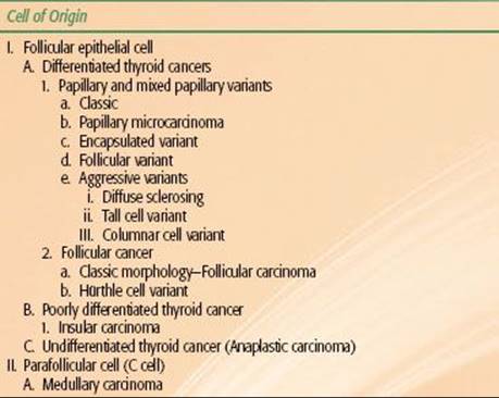
Correct pathological classification of thyroid carcinoma is fundamental to the appropriate clinical management of the malignancy. A system of classification is shown in Table 50.1 and discussed below. The cell of origin, cytological features, and growth morphology determine the pathologic classification. All carcinomas of the thyroid gland are derived from the follicular epithelium, except medullary thyroid carcinoma (MTC), which is derived from the parafollicular C cells. Capsular invasion is important in the pathologic diagnosis of certain thyroid carcinomas. The diagnosis of papillary thyroid carcinoma (PTC) is entirely predicated on the presence of diagnostic nuclear features and does not require invasive growth, while follicular carcinoma (FC) requires the presence of capsular or vascular invasion.3 Other rarely encountered malignant neoplasms of the thyroid gland include thyroid lymphoma and sarcomas. These two entities will not be discussed in this chapter and are better evaluated and managed under paradigms detailed in the chapters on lymphoma and sarcoma.
Differentiated (Follicular-Derived) Thyroid Carcinoma
The two major subgroups comprising differentiated thyroid carcinoma (DTC) are PTC and FC. They are distinguished by architectural and cytological features.
TABLE 50.1 PATHOLOGICAL CLASSIFICATION FOR THYROID CANCER
Papillary Thyroid Cancer
PTC is the most common histologic type, representing approximately 80% to 90% of all thyroid cancer diagnoses, and has the best prognosis of all thyroid malignancies.4 Malignant nuclear cytological characteristics are the distinguishing feature of PTC and include nuclear enlargement, hypochromasia, intranuclear cytoplasmic inclusions (nuclear pseudoinclusions), nuclear grooves, and distinct nucleoli. After formalin fixation, nuclei may appear pale, optically clear, and resemble “Orphan Annie’s eyes.” PTC is strongly lymphotropic, and multicentric disease within the parenchyma of the gland is often present secondary to early lymphatic spread. All PTC stain strongly for Tg.5–7
In its classic variant, PTC has a tumor architecture comprising branching papillae with fibrovascular cores. Papillary microcarcinoma is used to describe incidentally identified papillary carcinoma measuring <1.0 cm in diameter. The encapsulated variant of PTC (approximately 10% of PTCs) is surrounded by a thick fibrous capsule, unlike most PTCs that are nonencapsulated and infiltrate the surrounding thyroid parenchyma. Although not as favorable as microcarcinoma, encapsulated PTC has a favorable prognosis compared with classic PTC. The follicular variant of PTC differs from the classic variant by its follicular growth pattern; however, it retains the nuclear features diagnostic of PTC. Its prognosis roughly mirrors that of classic PTC. Other variants include diffuse sclerosing, tall cell, and columnar cell PTC, which have a much poorer prognosis.
Diffuse sclerosing is a rare form of PTC, with histologic features of extensive sclerosis, lymphocytic infiltrate, and psammoma bodies. A greater percentage of patients with diffuse sclerosing PTC present with extrathyroidal extension or distant metastases. Likewise, tall cell and columnar variants are commonly associated with poor prognostic features such as advanced age, large tumor size, aggressive histologic features with early invasion, and distant metastases. The distinguishing feature of tall cell PTC is that at least 70% of the carcinoma is composed of cells that are at least twice as tall as they are wide because of abundant cellular cytoplasm. Columnar cell variant is extremely rare. These carcinomas histologically show striking nuclear stratification, usually with papillary architecture. However, the usual nuclear features of PTCs may be absent; therefore, some pathologists prefer to classify columnar cell as poorly differentiated carcinomas.
Follicular Carcinoma
FC, like PTC, is a DTC of follicular cell origin. They comprise 15% to 30% of all thyroid carcinomas and share a similar prognosis to classic PTC. This malignant thyroid neoplasm lacks the cytological features of PTC. These carcinomas show evidence of thyroid follicle formation and are usually well circumscribed with a defined tumor capsule. They are grossly indistinguishable from follicular adenomas. The diagnosis of FC is dependent on the presence of one of two histologic features: (a) tumor invasion through the entire tumor capsule or (b) tumor invasion into a blood vessel located in the tumor capsule or immediately outside the tumor capsule. Minimally invasive forms of FC may present a pathologic challenge and often require serial sectioning to find evidence of capsular or vascular invasion, while widely invasive forms of FC are obviously invasive tumors both on gross and microscopic evaluation.
Hürthle Cell Carcinoma
This follicular epithelial-derived carcinoma, also known as oncocytic carcinoma, is characterized by large cells with abundant granular eosinophilic cytoplasms. Hürthle cells may be seen in both benign and other non-Hürthle cell malignant neoplastic lesions; however, at least 75% of the tumor must be comprised of Hürthle cells to designate it Hürthle cell carcinoma. Similar to FC, Hürthle cell carcinoma requires the presence of capsular or vascular invasion to classify it as a malignant neoplasm. Although Hürthle cell carcinoma is classically included under the subclassification FC, it is now apparent that there are both follicular and papillary types. Hürthle cell carcinoma has a less-favorable prognosis and usually presents with large tumor size, more invasive features, early regional and distant metastatic spread, and lower likelihood to concentrate RAI.
Poorly Differentiated Thyroid Carcinoma (Insular Carcinoma)
These follicular epithelial cell-derived tumors are intermediate between DTC and undifferentiated (anaplastic) thyroid cancers (ATC). Both their histologic appearance and biological behavior are more aggressive than DTC. They tend to be widely invasive and many will extend beyond the gland. Insular carcinoma is characterized by tumor cells that are arranged in discrete nests separated by a fibrous stroma. Microscopically, they are usually solid with some evidence of follicle formation. Mitoses and necrosis are often present. The presence of more differentiated areas of tumor admixed with insular carcinoma suggests that the latter represents “dedifferentiated” tumors.
Undifferentiated (Anaplastic) Thyroid Carcinoma
ATC comprise <5% of all malignant thyroid neoplasms. This is the most aggressive form of thyroid carcinoma, accounting for 14% to 50% of all thyroid cancer deaths. Most patients are diagnosed at the age of 65 years or older.8ATC originates from the thyroid follicular epithelium but shows little to no evidence of histologic differentiation. This tumor is defined as much by its clinical behavior as its histologic appearance. When diagnosed, most ATCs are widely invasive, replacing most if not all of the normal-appearing gland, and freely infiltrate the perithyroidal tissues. They are usually accompanied by bulky metastatic lymphadenopathy and distant metastatic spread. Gross pathologic evaluation is characterized by widely infiltrative disease with areas of necrosis and hemorrhage. Various microscopic patterns have been described, including small cell, spindle cell, giant cell, squamoid, or pleomorphic. They are hypothesized to be lesions that have dedifferentiated from more benign differentiated follicular neoplasms.
Medullary Thyroid Carcinoma
Medullary thyroid carcinoma (MTC) does not originate from the follicular epithelial cells, but from the parafollicular C cells, which are neural crest-derived cells whose function is to produce calcitonin. MTC comprises less that 5% to 10% of all thyroid cancers. Cases are seen sporadically (80%) or in association with familial multiple endocrine neoplasia (MEN IIa, MEN IIb, and pure familial MTC) syndromes. Multifocal and bilateral MTC are usually seen in patients with MEN, but, otherwise, familial and sporadic MTCs are indistinguishable. Grossly, these tumors are well circumscribed and nonencapsulated. Microscopically, tumors can have different appearances, including patterns that mimic other types of thyroid tumors. The most common pattern is of solid growth or nests similar to insular carcinoma. Amyloid, which is present in approximately 80% of cases, is a characteristic feature of MTC. Calcitonin stains are usually positive and specific for MTC, but up to 20% of cases may not stain for calcitonin; therefore, other neuroendocrine markers such as chromogrannin may be useful.
Classification Based on the Capacity to Concentrate Radioactive Iodine
Different pathologic classes of thyroid carcinoma concentrate RAI to various degrees. This characteristic is central to management decisions because the ability to concentrate RAI presents a useful therapeutic target. Table 50.2presents a classification system that categorizes cancers based on whether they always concentrate RAI, infrequently concentrate RAI, or never concentrate RAI. Papillary and follicular carcinomas (with the exception of the more aggressive variants), usually concentrate RAI. The follicular variant of papillary cancer has the same avidity for RAI as papillary cancer with classic morphology. Therefore, from the treatment standpoint, the distinction between these types of DTCs is not important. The aggressive variants of papillary carcinoma and Hürthle cell carcinoma can concentrate RAI, but to a lesser degree than the classic morphologies. In fact, there is often no measurable iodine uptake in metastases of these tumors. Anaplastic and medullary cancers never concentrate RAI to a clinically useful degree.
TABLE 50.2 A SYSTEM OF CLASSIFICATION FOR THYROID CANCER BASED ON THE ABILITY TO CONCENTRATE RADIOACTIVE IODINE

EPIDEMIOLOGY OF THYROID CANCER
Thyroid cancer is the most commonly diagnosed endocrine cancer. There will be an estimated 48,000 new thyroid cancer diagnoses and 1,740 thyroid cancer deaths in the United States by the end of 2011.9 The overall incidence is 7.7 per 100,000 persons per year.10 There is a strong female prevalence with women being three times more likely than men to be diagnosed with thyroid cancer. Thyroid cancer is the fifth most common cancer diagnosis in women, accounting for approximately 5% of all new female cancer diagnoses.9 DTC comprises the overwhelming majority (94%) of new thyroid cancer diagnoses. Approximately 5% are medullary thyroid carcinoma and 1% are anaplastic thyroid carcinoma.11 Per the National Cancer Institute’s Surveillance, Epidemiology, and End Results (SEER) database, the incidence of thyroid cancer has increased in the United States from 3.6 per 100,000 people in 1973 to 8.7 in 2002. The bulk of these cases result from an increase in detection of small primary cancer, with 49% and 87% of new cancers measuring <1 cm and 2 cm, respectively.12 According to another SEER database analysis, the most commonly diagnosed PTC in 2006 was papillary microcarcinoma (<1 cm).13 An even greater proportion of thyroid cancers remain clinically occult, and the prevalence of PTCs at autopsy has been reported as high as 35%.14
The incidence of thyroid cancer increases with age with a peak incidence at 40 to 44 years old for women and 65 to 69 years for men. The peak incidence for ATC is at 60 years for both sexes, and for MTC it can vary depending on whether it is familial or sporadic.15 The incidence of PTC varies significantly across race, with Asian women having the highest incidence of PTC at 10.96 per 100,000 woman-years compared with the lowest incidence for African American women of 4.9 per 100,000 woman-years.10
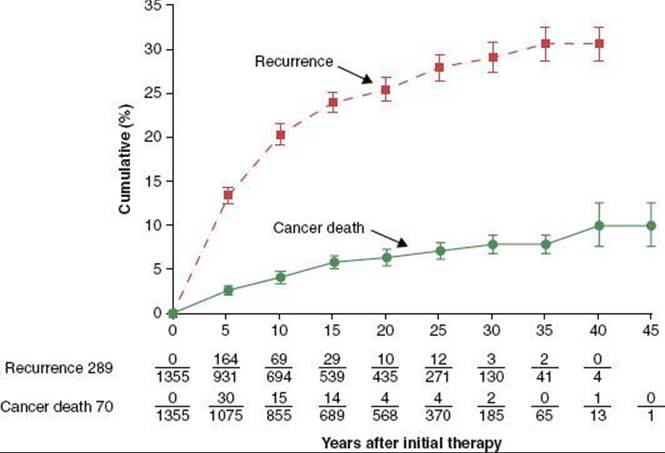
The prevalence of thyroid cancer in the U.S. population is much higher than the incidence of new diagnoses, a phenomenon attributed to the indolent nature of most DTCs. In 2001, there were an estimated 300,000 individuals living in the United States with thyroid cancer, a number similar to much more commonly diagnosed fatal cancers such as primary carcinomas of the bronchus and lung.15 Patients living with recurrent thyroid cancer comprise a substantial proportion of the prevalence of thyroid cancer. In a series of 1,355 patients with papillary and follicular thyroid cancers, the 30-year cumulative rate of recurrence was approximately 30%, while cancer-related mortality was <10% (Fig. 50.2).16 Studies reporting the outcomes of patients treated for thyroid cancer must be interpreted with consideration of the length of follow-up. Roughly 25% of all recurrences will present after 5 years of follow-up and as many as 16% will be detected after 15 years of follow-up.15
FIGURE 50.2. Tumor recurrence and cancer deaths in 1,355 patients treated for follicular and papillary thyroid carcinoma. (From Mazzaferri EL, Jhiang SM. Long-term impact of initial surgical and medical therapy on papillary and follicular thyroid cancer. Am J Med1994;97:418–28, with permission from Elsevier.)
Radiation-Induced Thyroid Cancer
Exposure to ionizing radiation is an established risk factor for thyroid nodules and cancer. Well-recognized data come from survivors of atomic bombs in Hiroshima and Nagasaki and the nuclear reactor accident in Chernobyl.17–18,19–20,21–23 A recent analysis of the 59,687 atomic bomb survivors in the Life Span Study demonstrated a total of 24.5% of cases of thyroid cancer in this cohort were attributed to radiation exposure with a strong dose response.21Women and young children appeared to be most susceptible. An estimated additional 1,000 cases of thyroid cancer throughout Europe have been attributed to the Chernobyl accident.23 An increased risk of thyroid cancer has also been attributed to exposure to therapeutic radiation in children treated for Hodgkin lymphoma, tinea capitis, enlarged tonsils, and enlarged thymus.24,25–26 Radiation-induced thyroid cancer is pathologically indistinguishable from spontaneous forms of the disease and is not associated with more aggressive biology or poorer prognosis.27,28–30
Familial Medullary Thyroid Carcinoma
Twenty-five percent of cases of MTCs are hereditary. All patients with MTC should be screened for familial disease, and genetic testing is considered the standard of care for all first-degree relatives of patients with newly diagnosed MTC. Heritable forms of MTC are associated with the familial MEN syndromes (MEN IIa, MEN IIb, and pure familial MTC) and have their own unique epidemiology. These disorders are inherited in an autosomal dominant fashion due to a germline-point mutation in the RET gene on chromosome 10q11.2. Men and women are affected equally given the pattern of inheritance. These patients typically present with bilateral and multifocal tumors compared to sporadic forms of MTC. The aggressiveness and age of onset of familial MTC differs depending on the specific genetic mutation. Prophylactic thyroidectomies are increasingly being performed on patients at risk for developing MTC.31
CLINICAL MANIFESTATIONS OF THYROID CANCER
The most common presentation of thyroid cancer is an asymptomatic thyroid nodule found incidentally by the patient, clinicians, or on an imaging study performed for other reasons. Palpable thyroid nodules occur in 3% to 4% of the normal population, and their incidence at autopsy has been reported as high as 50%.32,33–34 However, <1% of thyroid nodules represent a cancer. The differential diagnosis of thyroid nodules includes adenomas (macrofollicular, microfollicular, Hürthle cell, and atypical), cystic lesions (simple cysts or hemorrhagic cysts), colloid nodules, thyroiditis, granulomatous disease, and cancer. Positive findings on history and physical examination that raise suspicion that a thyroid nodule is malignant include rapid growth, firmness, fixation, vocal cord paralysis, cervical adenopathy, stridor, dysphagia, or neurological compromise. In cases where two or more of these symptoms or signs are present, the incidence of thyroid cancer is over 70%, and surgery should be recommended even in the presence of a nondiagnostic fine-needle aspiration (FNA).35
Clinically occult thyroid cancer and benign thyroid nodules will often present on imaging studies performed for other diagnoses. Studies utilizing computed tomography (CT), magnetic resonance imaging (MRI), or ultrasound (US) have reported the incidence of clinically occult thyroid nodules to be 19% to 67%.36 Fluorodeoxyglucose (FDG)-positron emission tomography (PET) studies have also reported the incidence of focal uptake within the thyroid gland at 2% to 3%, with only 25% to 50% ultimately representing cancer.37,38 Many incidentally detected thyroid cancers will ultimately prove to be clinically significant lesions with pathologically identified extrathyroidal extension, lymph node metastases, or both.39
Alternatively, locally advanced thyroid cancer presents with symptoms from involvement of the critical structures intimately associated with the thyroid gland, massive cervical lymphadenopathy, or metastatic disease. These patients present with symptoms such as hoarseness from tumor compression of the recurrent laryngeal nerve, airway compromise, dysphagia or respiratory symptoms, and pain and weight loss resulting from metastatic dissemination.
DIAGNOSTIC EVALUATION OF THYROID CANCER
Laboratory Studies
All patients with thyroid nodules should undergo a serum TSH in addition to basic metabolic, renal, and liver function tests and a complete blood count. Other lab tests, such as Tg, T3, and T4, are commonly obtained but are rarely useful in the initial management of thyroid cancer. Serum calcitonin can be measured during the initial evaluation of patients with MTC and is essential during follow-up for these patients.
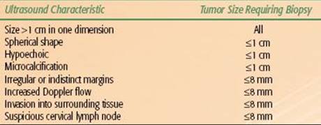
TABLE 50.3 ULTRASOUND FINDINGS USED TO SELECT THYROID NODULES FOR FINE-NEEDLE ASPIRATION
Ultrasound and Ultrasound-Guided Fine-Needle Aspirate
Neck US with Doppler and US-FNA are the standard diagnostic approaches for evaluating thyroid nodules, both palpable and incidentally detected on imaging. The ultrasonographic features used to select thyroid nodules for FNA are shown in Table 50.3. For a more-detailed discussion on the use of US in the evaluation of thyroid nodules, please refer to a consensus statement by the Society of Radiologists in Ultrasound on the management of thyroid nodules detected by US40 and the American Thyroid Association’s recently updated management guidelines for patients with thyroid nodules and differentiated thyroid cancer.41
US-FNA has several benefits in the evaluation of thyroid nodules over other means of obtaining a tissue diagnosis: (a) the technique is minimally invasive and is usually performed as an outpatient procedure; (b) the operator can visually verify that the biopsy needle is in the nodule(s) of suspicion; and (c) it allows for evaluation of nonpalpable nodules and, in the setting of cystic lesions, lowers the rate of inadequate specimens.
US-FNA of thyroid nodules yields cytology that can be broadly classified into four categories: (a) insufficient for diagnosis, (b) benign, (c) positive for cancer, and (d) indeterminate for diagnosis. Approximately 5% to 10% of US-FNA specimens are insufficient for diagnosis, and the rate of insufficient biopsies can be reduced by 50% with repeated biopsies or evaluation by personnel trained in cytological evaluation of thyroid nodules at the time of the procedure. Sixty-five percent to 75% of US-FNA cytology will be benign. Approximately 5% of all US-FNAs of thyroid lesions yield specimens with the diagnostic cytological features of PTC, anaplastic carcinoma, medullary carcinoma, or other poorly differentiated carcinomas. Finally, 15% to 20% of US-FNA will be classified as “suspicious” or indeterminate for diagnosis. Other synonyms for this cytological classification are follicular neoplasm or follicular tumor. The diagnostic evaluation of these lesions is aided by the use of laboratory evaluation of thyroid function and nuclear imaging with RAI uptake studies if the TSH is suppressed. About 20% of these will ultimately show FC or Hürthle cell carcinoma on surgical specimens.42
The overall diagnostic accuracy of US-FNA will depend on the skill and experience of the operator and the cytologist in interpreting the specimen. In a large series of 1,135 cases, there were 1,127 satisfactory samples: 6% cancer (or suspicious for cancer), 10% follicular neoplasms, 82% benign, and 0.7% indeterminate; the false-negative rate was 0% and only 1.5% were false positives. Surgery was performed on 169 patients, and a review of final pathology yielded a sensitivity of 100% and specificity of 67%. The corresponding positive and negative predictive values of US-FNA were 87% and 100%, respectively.43 Another study of 1,613 cases of FNA reported a sensitivity of 78%, specificity of 98%, and a predictive value of 100% for papillary carcinoma.44 The major limitation of this diagnostic tool is the inability to distinguish benign follicular adenomas from FC and follicular PTC.
Cervical US is also an important staging tool because it is effective in identifying lymph node metastases. Cervical lymph node size, presence of calcification, and irregular diffuse intranodular blood flow are the most important US features suggestive of lymph node metastases.45,46 Using criteria based on the ratio of largest to smallest lymph node diameter, nodule echogenicity, intranodal structure, and margin irregularity, the sensitivity and specificity of cervical US for identifying malignant cervical lesions were 90% and 82%, respectively, in a study of 112 patients followed for thyroid cancer by high-resolution US with Doppler.47 Preoperative cervical US identified lymph node metastases that were occult on physical examination in 33% to 39% of patients in a large retrospective series.48,49 Cervical US findings will alter the surgical approach in 14% to 24% of patients with PTC by identifying nonpalpable cervical lymph node metastases.45,49
Computed Tomography and Magnetic Resonance Imaging
CT and MRI commonly detect otherwise clinically occult thyroid nodules. Malignancy is likely with either imaging technique when there are findings of a mass with extrathyroidal extension, lymph node metastases, or both. In this setting, the studies are useful in presurgical planning by establishing the extent of extraglandular tumor and helping the surgeon determine the need for extended neck dissections (Fig. 50.3). MRI is superior to CT for establishing the local extent of a known cancer, because it will more clearly show esophageal or tracheal invasion (Fig. 50.4).50 MRI is indicated in the presence of hoarseness, stridor, dysphagia, or other clinical signs of locally extensive thyroid cancer that cannot be adequately assessed by US.
A major downside to CT is that useful imaging of the neck requires contrast enhancement, which can interfere with subsequent RAI therapy. Because of their high concentration of iodine, CT contrast agents can seriously compromise the effectiveness of therapeutic RAI. When patients receiving iodinated contrast agents require RAI, therapy should be delayed in order to achieve adequate efficacy. Given this drawback as well as the effectiveness of US in defining the local-regional extent of tumor, the authors strongly discourage the use of CT in the staging evaluation of known thyroid cancer. In selected cases, a noncontrasted CT of the chest may be indicated to evaluate the presence of mediastinal and lung metastases, but, generally speaking, it is not necessary to perform a CT before primary therapy of most DTCs.
Nuclear Medicine Studies
The four major nuclear medicine studies related to thyroid cancer evaluation with iodine isotopes are radioactive iodine uptake (RAIU), thyroid scan, diagnostic whole-body scan (DxWBS), and posttreatment whole-body scan (RxWBS). Table 50.4 summarizes each of these studies.
RAIU tests are used to quantify the RAI-concentrating ability of remnant thyroid tissue after thyroidectomy. This value is required by federal regulation for outpatient I-131 therapy.
Thyroid scans produce images of the thyroid’s ability to concentrate RAI and have a historical use in the evaluation of thyroid nodules, characterizing them as functional (“hot”) or nonfunctional (“cold”). The authors do not use thyroid scans in the workup or management of patients with thyroid cancer.
The typical DxWBS involves a total body image several days after administering approximately 3 mCi of I-131. The current value of a DxWBS in thyroid cancer management is controversial. Some experts use a DxWBS to determine the dose of I-131 in a patient being treated for thyroid cancer. Some experts use a DxWBS as part of the surveillance program following initial treatment. A comprehensive discussion of the issues related to DxWBS is beyond the scope of this chapter. The main arguments against doing DxWBS are low sensitivity, stunning of residual cancer cells, and unnecessary radiation exposure. The bottom line is that reasonable people will disagree about the role of DxWBS in thyroid cancer management. The authors do not use DxWBS to stage or monitor thyroid cancer patients and have not ordered a DxWBS in over 5 years.
FIGURE 50.3. A patient presenting with a neck mass. A: Computed tomography study showing the mass to be due to a primary papillary thyroid carcinoma coming into contact with a related level 4 metastatic lymph node (arrows). The lymph node at level 4 and in the tracheoesophageal groove is shown by the arrowheads. B: A section more superiorly shows level 3 adenopathy that is multicystic in a pattern typical but not necessarily diagnostic of papillary thyroid carcinoma. There is upper level 4 cystic adenopathy with an enhancing mural nodule (arrow)—a morphology suggesting of thyroid carcinoma. A contralateral enhancing positive node with a focal low-density defect (arrowhead) is also present. (From Mancuso AA, Mendenhall WM, Vaysberg M. Thyroid: nodules and malignant tumors. In: Mancuso AA, ed. Head and neck radiology. Philadelphia: Lippincott Williams & Wilkins, 2010:1457–1481, with permission.)

FIGURE 50.4. A patient demonstrating the value of magnetic resonance in the detection of invasion of the trachea and cervical esophagus, especially as it pertains to treatment planning. This patient has anaplastic carcinoma of the thyroid gland. In A: T1-weighted images show distortion of the trachea (arrow) and esophagus (arrowhead), but it is difficult to determine whether invasion is present. In B: the T2-weighted image shows obvious growth of tumor replacing the normal signal of cartilage of the trachea with growth on both sides of the cartilage (arrow) as well as replacement of the normal muscular wall signal of the cervical esophagus (arrowhead), indicating esophageal wall invasion. (From Mancuso AA, Mendenhall WM, Vaysberg M. Thyroid: nodules and malignant tumors. In: Mancuso AA, edr. Head and neck radiology. Philadelphia: Lippincott Williams & Wilkins, 2010:1457–1481, with permission.)

An RxWBS should be done in every patient who receives a therapeutic dose of RAI. The optimal interval between administration and scan in most patients is approximately 7 days. The primary purposes of an RxWBS is to detect gross residual disease in the regional lymphatics or in distant sites and to determine if known disease concentrates RAI.
FDG-PET is another nuclear medicine study that is commonly used to detect metastatic disease, but the predictive value of PET in thyroid cancer is not well defined. TSH stimulation increases the accuracy of FDG-PET in most types of DTC. A prospective trial comparing FDG-PET studies during thyroid hormone suppression to studies after administration of rhTSH showed increased detection of occult metastases in the stimulated studies.51 Some insurers will not reimburse for FDG-PET for thyroid cancer without prior documentation of follicular-derived thyroid cancer with a suppressed or stimulated Tg over 10 ng/mL and a negative DxWBS.41
When FDG-PET is performed for reasons other than thyroid cancer and reveals a hypermetabolic thyroid nodule, a biopsy is warranted in most cases as these lesions carry a 15% risk of malignancy.50 A recent study of 5,877 patients who received FDG-PET for cancer screening or staging or restaging purposes, 3.7% revealed hypermetabolic activity within the thyroid gland. Of the patients sent for US-FNA, 14% were diagnosed with thyroid malignancies.52 A larger review of pooling data from over 55,000 FDG-PET studies reported a 1% prevalence of focal thyroid gland abnormalities, and the rate of confirmed thyroid malignancies in this group was as high as 33%.53
TABLE 50.4 NUCLEAR IMAGING STUDIES USED IN THE MANAGEMENT OF DIFFERENTIATED THYROID CARCINOMAS

PROGNOSTIC FACTORS
The most important prognostic factor for disease recurrence and cancer mortality is the histologic classification. DTC, when diagnosed in an early stage, has a favorable prognosis. However, tall-cell variant can have a 10-year mortality of up to 25%.54 Hürthle cell carcinoma carries a relatively poor prognosis, with a 25% rate of metastatic disease55,56 and decreased survival at 10 years of 76%, compared with 93% and 85% for PTC and FC, respectively.57Columnar cell variant and diffuse-sclerosing variants likewise carry a poor prognosis relative to other forms of DTC. Contrarily, follicular-variant PTC and most FC share the same favorable prognosis as classic PTC relative to age and stage at diagnosis. ATC has an abysmal prognosis. All ATCs are considered stage IV under the current AJCC system and the overall 5-year survival is approximately 5%.1
Within the classification of DTC there are several important prognostic factors, including age, gender, family history, tumor size, extrathyroidal extension, vascular invasion, cervical and mediastinal lymph node metastases, degree of cytologic atypia and tumor necrosis, presence of distant metastases, and the degree to which the cancer can concentrate RAI. The prognostic variables are summarized in Table 50.5and several of these factors are discussed in detail below.
Age
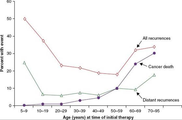
Age is a major prognostic variable that is accounted for in the current and previous AJCC staging systems (Fig. 50.5). The current AJCC system dichotomizes staging for DTC based on age under 45 years versus 45 years and older. In the current system, patients under 45 years of age with any T or N stage (based on tumor-node-metastasis [TNM] classification) are considered overall stage I, unless metastatic disease is present, which elevates the overall stage to stage II. Conversely, patients 45 years of age and older are given an overall stage I and II only if their T and N stages are T1N0 and T2N0, respectively. Thereby, this system considers age to be one of the most important prognostic variables. A limitation of this system is that it fails to account for the poor prognosis seen in patients diagnosed at a young age (<15 years old). Children present with more advanced tumors than young adults and have more recurrences and distant metastases.58,59 In one series of 56 children (ages 4 to 20 years) with PTC or FC, the 10-year progression-free survival was only 61%, although survival remained favorable at 98%.60 Another series of 21 children (ages 4 to 15 years) reported a 24% rate of distant metastases at presentation.61
Gender
Compared to women, men who develop DTC are diagnosed at an age approximately 20 years older, are twice as likely to develop distant metastases, and are 30% more likely to have regional metastases.62,63 It is unclear why there is a gender bias toward increasing age at diagnosis for men, but it may be attributed to delayed access to the U.S. health care system for men. Delaying the diagnosis for more than 12 months is associated with a doubling in the chance of thyroid cancer mortality.16
Primary Tumor Size
A linear relationship exists between primary tumor size and cancer-specific mortality.16,62,64,65–66 Very large tumors are associated with increased rates of extrathyroidal disease, lymph node and distant metastases, less-favorable histologies, and, in some cases, unresectable disease, all of which harbor a poor prognosis. Papillary microcarcinoma (<1 cm) have a disease course characterized by exceedingly favorable outcomes. These cancers, often found incidentally during surgery for benign thyroid conditions, are considered clinically insignificant malignancies and generally do not portend to decreased survival. Recurrences after total thyroidectomy in some series are nearly zero. Some small tumors, however, are associated with other poor prognostic factors that warrant more aggressive management. Lymph node metastases occur in as many as 20% to 60% of cases with small tumors; 30% to 40% can be multifocal, and 17% to 30% will have extrathyroidal extension.67–68,69 These outcomes have led some investigators to argue that treatment for papillary microcarcinoma should be no different from that for PTC larger than 1 cm in size, advocating for the routine use of RAI.70 The authors agree with other investigators that additional prognostic factors such as extrathyroidal extension, lymph node metastases,71 age, and multifocality should drive the decision to treat with adjuvant RAI when managing papillary microcarcinoma.
TABLE 50.5 PROGNOSTIC FACTORS IN DIFFERENTIATED THYROID CANCER
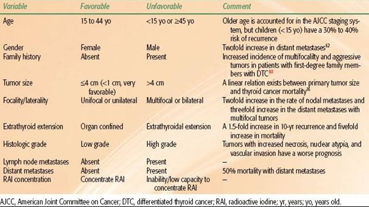
FIGURE 50.5. Effect of age on outcomes of patients with differentiated thyroid cancer. (From Mazzaferri EL, Kloos RT. Clinical review 128: current approaches to primary therapy for papillary and follicular thyroid cancer. J Clin Endocrinol Metab 2001;86:1447–1463. © 2001, The Endocrine Society.)
Multifocality and Bilaterality
Anywhere from 20% to 80% of patients undergoing thyroidectomy for unifocal tumors are diagnosed with multifocal disease and 44% will have disease in the contralateral lobe.6 This incidence of contralateral lobe disease is increased when multifocal disease is found in the incident lobe,72 underscoring the need for completion thyroidectomy after a thyroid cancer is found at the hemithyroidectomy. Patients with multifocal disease have double the rate of nodal metastases and triple the rate in distant metastases.73,74,75,76
Extrathyroidal Extension
Extrathyroidal extension (ETE) is a key risk factor for lymph node and distant metastases.77 The probability of primary tumor spread beyond the thyroid increases with tumor size,65 but can be seen even in small tumors.67–68,69 ETE predicts for increased lymph node recurrences and distant metastases.62,69 In its earliest form, ETE is clinically occult and seen only on pathologic evaluation. More advanced ETE presents with signs of local invasion of the muscles of the neck, blood vessels, recurrent laryngeal nerve, larynx, pharynx, esophagus, and even the spinal cord or brachial plexus. Gross ETE occurs in the majority (70%) of ATC.78
Lymph Node Metastases
Often a presenting symptom of a thyroid cancer, lymph node metastases are more common with PTC than FC (46% vs. 25%).16 The prognostic importance of lymph node metastases has historically been somewhat controversial, but several studies have shown this to be predictive of recurrence and decreased survival; bilateral and mediastinal lymphadenopathy in particular portend to a poor prognosis.16,79,80–81 Nevertheless, the absolute magnitude of negative impact on survival from lymph node metastases is marginal, decreasing 30-year survival from 94% to 90%.16
Distant Metastases
Although distant metastases are the leading cause of death in patients with DTC, they are not uniformly fatal. Roughly 10% of patients with PTC and 25% of those with FC develop distant metastases, with the lungs being the most common site of involvement. One-half of these metastases are present at diagnosis.82 Patients with DTC who develop distant metastases have an increased thyroid cancer mortality of 40% to 50% compared with 10% to 15% for those with local recurrence.62,74,82,83
STAGING
The seventh edition (2010) of the AJCC staging system for thyroid cancer is shown in Table 50.6. Staging is based on the anatomic extent of disease as well as the patient’s age and tumor histology. Both clinical and pathologic staging can be employed. Separate staging groups are recommended for differentiated, medullary, and anaplastic carcinomas.
SURGICAL MANAGEMENT OF THYROID CANCER
Surgery is the primary treatment of localized thyroid cancer of all histologies. A total thyroidectomy is the preferred oncologic procedure, because the gland is surgically accessible and its primary endocrine function can be replaced by exogenous therapy (levothyroxine). It is critical to understand that even a total thyroidectomy leaves residual thyroid tissue that will have major implications for subsequent therapy and disease monitoring. The clearest evidence of residual thyroid tissue after total thyroidectomy is seen on postoperative RAI thyroid studies, which nearly always show uptake in the region of the thyroid bed. The presence of residual thyroid tissue in this setting is not a sign of incomplete surgery, but a matter of practicality. The ligament connecting the posterior surface of the thyroid capsule to the trachea harbors microscopic nests of thyroid tissue and is rarely completely resected in order to reduce the risk of tracheal injury. Second, the recurrent laryngeal nerve is embedded in thyroid tissue at the point where the nerve enters the larynx, and it is not possible to remove all of this thyroid tissue without injuring the nerve and compromising voice quality and laryngeal function.
Several important complications can arise from thyroidectomy. The most common are recurrent laryngeal nerve injury (temporary 30% and permanent 2%) and hypoparathyroidism (temporary 5% and permanent 0.5%). These complications can be mitigated with the use of more conservative surgery.84,85 Other structures at risk for operative injury include the vagus, spinal accessory and superior laryngeal nerves, trachea, esophagus, thoracic duct, and carotid arteries. The operative morbidity is reduced by increasing the surgical experience, intraoperative monitoring of the recurrent laryngeal nerves, and autotransplantation of parathyroid glands.86
There are several critical issues regarding the surgical management of thyroid cancer and nodules that warrant detailed discussion:
1. When is surgery indicated for the evaluation of a thyroid nodule with nondiagnostic cytology?
2. When is it safe to consider hemithyroidectomy for thyroid carcinoma?
3. What is the appropriate extent of neck dissection?
These issues are discussed in further detail below.
Surgical Evaluation of Thyroid Nodules
The 2009 revision of the American Thyroid Association (ATA) “Guidelines for Management of Patients with Thyroid Nodules and Differentiated Thyroid Cancer” contains several recommendations addressing the role of surgery in the evaluation of thyroid nodules.41 Surgical evaluation of thyroid nodules is indicated for the following reasons: (a) cytology is suspicious for PTC; (b) cytology contains follicular cells with no concordant functioning nodule on an RAI scan, especially with a low- to normal-range serum TSH. (In these cases, lobectomy or total thyroidectomy should be considered.); (c) cytology contains Hürthle cell neoplasm, which does not warrant an RAI study and should be managed with either lobectomy or total thyroidectomy, depending on the lesion’s size and other risk factors; and (d) growing nodules, even in the face of benign cytology, may be considered for surgical removal. In cases when a lobectomy reveals a thyroid cancer, completion thyroidectomy is recommended for all patients for whom the procedure would have been recommended had the diagnosis been available before surgery. The use of I-131 ablation as an alternative to completion surgery is not recommended.
Lobectomy in the Management of Thyroid Cancers
Although total thyroidectomy is considered the standard surgical approach for nearly all resectable thyroid carcinomas, there are published clinical guidelines that consider a more conservative surgery appropriate in some clinical scenarios. The 2011 National Comprehensive Cancer Network (NCCN) guidelines find lobectomy acceptable in patients between the ages of 15 and 45 years with PTC tumors <4 cm and without prior radiotherapy, distant metastasis, cervical lymph node metastasis, extrathyroidal extension, or aggressive histologic variants.54 The 2009 ATA guidelines recommend considering lobectomy only in tumors with the above features that are <1 cm in size.41Advocates for lobectomy argue that operative morbidity is mitigated with the use of more conservative surgery.84
In a review of 52,173 patients from the National Cancer Database who underwent surgery for PTC, 82.9% underwent total thyroidectomy and 17.1% underwent lobectomy.87 On multivariate analysis, when lobectomy was used, there was a 57% increase in recurrences and 21% increase in death compared with total thyroidectomy. For PTC <1 cm, the surgical approach did not impact recurrence or survival. The benefit of total thyroidectomy was seen across all tumors >1 cm, even when patients with tumors 1 to 2 cm were analyzed separately.87 A previous SEER analysis of 4,402 patients with PTC failed to show a survival benefit to total thyroidectomy; however, patients undergoing total thyroidectomy in this analysis had larger tumors that were 10% more likely to have extrathyroidal extension and 20% more likely to have cervical lymph node metastases compared to patients who were not operated on.88 The AMES (age, distant metastasis, extent, and size) and MACIS (metastases, age, completeness of resection, invasion, size [Mayo Clinic]) prognostic scoring system has been proposed for selecting low-risk patients for more conservative initial resections89–90,91; however, most patients undergo total thyroidectomy even when classified as low risk,92 and, overall, the use of total thyroidectomy is increasing.93
In general, total thyroidectomy is the appropriate initial surgical approach for most patients with differentiated thyroid cancer because it increases the effectiveness of adjuvant I-131, removes all intrathyroidal cancer, reduces recurrences, and facilitates the use of surveillance scans and Tg. The surgeon’s experience with the operation is a large determinant of the ultimate outcome. In the hands of surgeons performing over 100 cases in a 5-year period, total thyroidectomy complication rates are comparable to less extensive surgeries and are two-thirds less likely than when performed by less experienced surgeons.86
TABLE 50.6 AMERICAN JOINT COMMITTEE ON CANCER (AJCC) 2010 TNM STAGING SYSTEM FOR THYROID CANCER

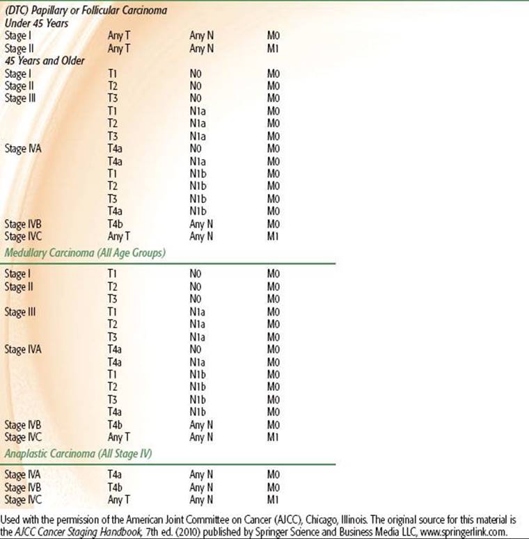
TABLE 50.7 PHYSICAL, BIOLOGICAL, AND EFFECTIVE HALF-LIVES (T½) OF I-131

Neck Dissection in Differentiated Thyroid Cancer
As with squamous cell carcinomas, a modified radical neck dissection is done for thyroid cancer when there is a visible or palpably positive node. However, elective neck dissection (removal of lymphatic regions at risk for subclinical disease) is generally not performed for DTC of follicular cell origin, unlike primary surgery for most other head and neck cancers. Despite a high incidence of subclinical lymph node involvement in even small PTCs, elective neck dissection is not included in the initial surgical approach because identifying microscopic regional metastases will not influence the recommendation for RAI nor will it affect prognosis.63
According to the 2009 ATA guidelines, central-compartment (level VI) dissection is recommended for all patients with clinically involved lymph nodes; however, for small T1 or T2 tumors without adverse clinical or pathologic features, a prophylactic central neck dissection can be omitted. In clinically N0 patients with other adverse features (T3 or T4), a prophylactic central neck dissection can be considered. A lateral level II to IV neck dissection should only be reserved for patients with biopsy-proven metastatic lateral cervical lymphadenopathy.41 When a lateral neck dissection is indicated, levels II to IV should be resected en bloc in lieu of removing only abnormal lymph nodes.94–95,96 Levels I, V, and VII should not be dissected unless clinically suspicious. Central and lateral neck dissections are part of the standard primary therapy for all patients with sporadic and hereditary forms of medullary thyroid carcinoma.97,98
Despite these ATA guidelines, there remains active controversy in the literature regarding the role of routine prophylactic central neck dissection in patients with DTC. Space limitation precludes a thorough discussion of this controversy, but details can be found in the articles referenced in the remainder of this paragraph. In general, the advocates for routine central neck dissection argue that it leads to more accurate staging, reduces central neck recurrences, avoids reoperations, and provides the optimal conditions for effective RAI therapy.99–104,105 Detractors of this approach point to a lack of clear recurrence and survival benefits with prophylactic central neck dissection with an associated increase in the rate of surgical complications.99,106,107–108 A recent meta-analysis with over 1,200 patients showed no improvement in recurrences with the addition of prophylactic central neck dissection.109
MANAGEMENT OF DIFFERENTIATED THYROID CANCER WITH RADIOACTIVE IODINE
Bioconcentration of Radioactive Iodine
RAI for DTC is arguably the most successful targeted therapy in all of oncology. The iodine-concentrating capacity of benign and neoplastic thyroid tissue makes an overwhelming majority of thyroid carcinomas amenable to RAI therapy. RAI is taken up by thyroid tissue, including DTC of follicular epithelial origin, at a rate 6.6 times more than most tissues in the body. The biological half-life in extrathyroidal tissue is merely 12 days compared to 80 days for thyroid tissue (Table 50.7).
Radioactive Decay of Iodine-131
I-131 is produced from the fission of uranium atoms during the operation of nuclear reactors or in the detonation of nuclear bombs. I-131 decays by negatron emission (beta-minus decay) to xenon-131 (Xe-131). A neutron from the I-131 nucleus converts to a proton and an electron (beta-particle) is emitted from the nucleus. This first transition results in a beta-particle with a range of energies from 250 to 800 keV. These beta-particles are core to I-131’s ability to deliver targeted cytotoxicity. Because electrons of this energy range will deposit their energy within a millimeter, only the cells taking up the I-131 are affected. In the second decay step, unstable Xe-131 decays to stable xenon, releasing a photon with the energy of 364 keV. This product is therapeutically undesirable, because the photon will travel far from the source where iodine is concentrated. It contributes very little cytotoxicity to thyroid cancer cells and increases the total body dose; however, it is this property that makes RAI useful for diagnostic imaging, forming the foundation for DxWBS and RxWBS.
Goals of Radioactive Iodine Therapy for Differentiated Thyroid Cancer
Broadly speaking, the two basic purposes of adjuvant RAI are (a) thyroid remnant ablation and (b) adjuvant therapy for residual microscopic disease. First, RAI provides potent cytotoxicity by targeting thyroid cancer cells remaining in the operative bed, occult lymph node metastases, and distant metastases. Second, RxWBS provides critical information including staging, prognosis, and determining which patients are likely to require additional treatments. Lastly, ablation of the remaining thyroid tissue facilitates the use of serum Tg as a very sensitive and specific marker for disease persistence after primary therapy. Serum Tg provides a powerful means of monitoring for early disease recurrences that are likely to be amenable to curative retreatment.
RAI therapy has been crucial in making DTC one of the most curable malignancies. There are multiple large retrospective series showing a significant reduction in recurrences and cause-specific mortality with the use of RAI.16,110–111,112,113 A summary of selected references is provided in Table 50.8, although this does not represent a complete list.
Patient Selection for Radioactive Iodine
Despite the evidence that RAI reduces thyroid cancer recurrences and potentially improves survival, there remain limitations on the indications for RAI for DTCs because these benefits have not been demonstrated in the setting of a randomized controlled trial. Moreover, several institutional series have failed to demonstrate a benefit for certain patients with the lowest risk of disease. Finally, the potential benefits in any individual patient must be weighed against toxicities. At our institution, all patients with DTC >1 cm or those with any size tumor with positive lymph nodes, distant metastases, or extrathyroidal extension are recommended RAI after surgery.
The 2009 ATA guideline recommends RAI for all patients with known distant metastases, gross extrathyroidal extension of the tumor regardless of tumor size, or primary tumor size >4 cm, even in the absence of other higher-risk features. RAI is recommended for selected patients with 1- to 4-cm thyroid cancers confined to the thyroid who have lymph node metastases or other high-risk features, such as age >45 years, intrathyroidal vascular invasion, multifocal disease, or aggressive histologic variants (tall cell, columnar cell, or insular carcinoma). Follicular and Hürthle cell variants are considered high risk, and these patients are nearly always recommended RAI, except for those with the smallest unifocal FCs manifesting as only capsular invasion (without vascular invasion). These so-called minimally invasive carcinomas are treated effectively with surgery alone and RAI can be avoided. RAI is not recommended for unifocal PTCs <1 cm and without high-risk features or when all the foci in multifocal disease are <1 cm.41 The 2011 NCCN guidelines recommend RAI for patients with persistent disease and for thyroid remnant ablation in selected patients without gross residual disease. For patients without residual disease or high-risk histology, RAI is not recommended when postoperative Tg is <1 ng/mL and anti-Tg antibodies and RAI imaging are negative.
TABLE 50.8 SELECTED RETROSPECTIVE SERIES DEMONSTRATING CLINICAL BENEFITS OF RAI

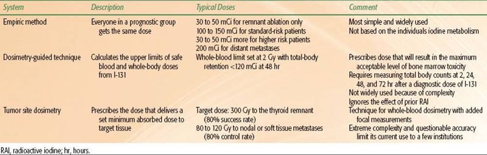
TABLE 50.9 IODINE-131 DOSIMETRY SYSTEMS
Selection of Iodine-131 Activity
Three methods of radioisotope dosing are used in the treatment of DTC with RAI (Table 50.9). There is no consensus on the optimal method of I-131 dosing, and an exhaustive body of literature exists on the matter, but this will not be discussed here because of space limitation. For more information the authors refer readers to Essentials of Thyroid Cancer Management.114 The authors use the empiric system at our institution for simplicity. In general, the dosimetric techniques require the expertise of dedicated physics’ support and nuclear medicine technologists with specialization in RAI administration. The current University of Florida empiric dosing regimen is summarized in the Table 50.10 for both initial therapy and retreatment settings.
Preparing Patients for Iodine-131
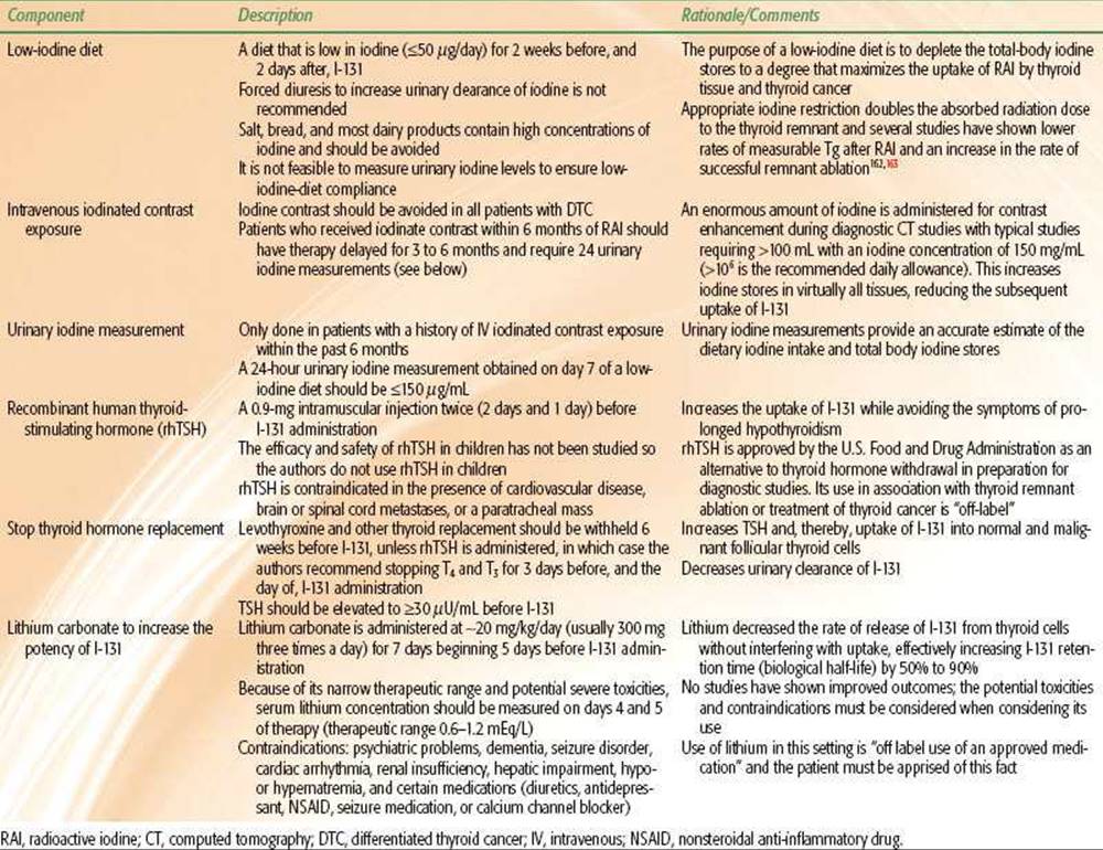
Appropriate preparation for RAI therapy is critical to obtaining the optimal desired effect of therapy, whether for remnant ablation or adjuvant therapy residual disease. There are several components (Table 50.11) comprising optimal preparation, including (a) a low-iodine diet, (b) avoidance of iodinated contrast agents (or adequate washout period if iodinated contrast has already been administered), (c) measuring of urinary iodine levels, (d) administration of rhTSH, (e) avoidance of thyroid replacement, and (f) administration of lithium for augmentation of I-131. With the exception of the latter, all of these steps will deplete the patient’s iodine stores and increase the level of TSH and thereby the activity of the NaIS, in an effort to increase the therapeutic efficacy of I-131. Low-iodine diets, avoidance and washout of iodinated contrast agent, and lithium for augmentation of I-131 biological half-life are useful in nearly all patients. Urinary iodine measurement requires 24-hour collection and is reserved for patients with documented iodine contrast administration within the 6 months preceding RAI. Otherwise, the authors approach patients with two general preparation regimens: T4 deprivation or rhTSH administration. The authors prefer using rhTSH to avoid the morbidity of hypothyroidism. Some experts believe that the cure rate will be higher when patients are prepared with hypothyroidism, but there are data suggesting equivalent efficacy with hypothyroidism versus rhTSH preparation.115–117 Both approaches also increase the diagnostic sensitivity of both DxWBS and RxWBS, although T4 deprivation may be superior in this regard.118,119
TABLE 50.10 UNIVERSITY OF FLORIDA EMPIRIC DOSING OF IODINE-131 FOR TREATMENT OF DIFFERENTIATED THYROID CANCER
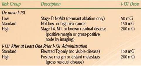
TABLE 50.11 PREPARATION OF PATIENTS FOR IODINE-131
Outpatient Management of Iodine-131
Grigsby et al.120 described the pattern of radiation exposure in 65 household members of 30 patients treated with RAI. The mean dose to family members was well below regulatory standards at 0.24 mSv and the maximum dose was 1.11 mSv. The Nuclear Regulatory Commission (NRC) has issued guidance on the release of patients following RAI treatment for thyroid cancer, and a copy of all material related to the issue can be found on the NRC website. Major recommendations were issued in 1997 (NRC Regulatory Guide 8.39) and have largely remained in place. Several minor subsequent recommendations have been issued, most recently in early 2011 under 10 CFR 35.75.121Overall, the recommendations are intended to keep the exposure to the public “as low as reasonably achievable (ALARA).” The 1997 regulations permitted patients to be released from the control of the licensee. The regulations state that:
• Providers are discouraged from recommending that patients stay in hotels immediately after treatment.
• Patients should avoid public transportation.
• Outpatient release following treatment is limited to patients when the radiation dose to third parties in not likely to exceed 5 mSv of total effective dose equivalent (TEDE). The TEDE is calculated using three factors: (a) activity of I-131 administered, (b) results of the thyroid uptake study, and (c) the occupancy factor, which takes into account the amount of time the patient will spend around other people for a few days after RAI.
• The patient must be capable of self-care, cannot live in a nursing home or communal living facility, and must prefer to be released after RAI.
• Patients must be given radiation safety instructions.
In our practice, the patient’s living condition is assessed with the assistance of a radiation safety officer to ensure that the radiation exposure to family members, caregivers, and the general public remains ALARA. This usually means that patients return to private residences, with their own room and bathroom available, where they will remain alone for 3 days subsequent to I-131. Family members and caregivers should spend time in those areas with the patient only when necessary. Young children are to avoid all contact. Specific instructions on cleaning clothing, linens, bathrooms, and eating utensils are provided.
In 2011, new ATA guidelines were issued on outpatient I-131 management that reiterated the goal of keeping radiation exposure to third parties ALARA.122,123 The guidelines were intended, in part, to describe the role of the radiation safety officer, standardize patient instructions, and describe best practices. The authors refer readers to that publication for specific recommendations.
Adverse Effects of Iodine-131
Side effects and complications of RAI therapy are shown in Table 50.12.124 Patients should be given a prescription for antiemetics and instructed to fill it before arriving for RAI administration. Parotid massage and nonsteroid anti-inflammatory drugs may be useful for acute neck swelling or parotitis. Second-malignancy risk warrants discussion with the patient.125,126 A well-recognized study estimated the excess absolute risk of second cancers from I-131 therapy to be 14.4 for solid cancers and 0.8 for leukemia per 27 mCi per 100,000 person-years of follow-up.127
TABLE 50.12 ADVERSE EFFECTS OF RADIOACTIVE IODINE THERAPY
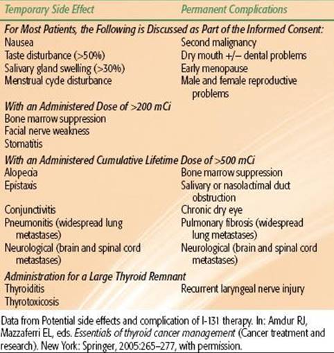
TABLE 50.13 INDICATIONS FOR EXTERNAL BEAM RADIOTHERAPY
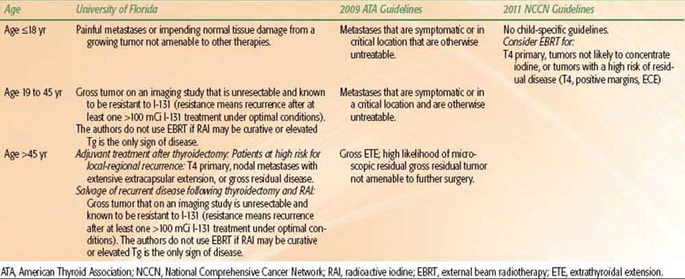
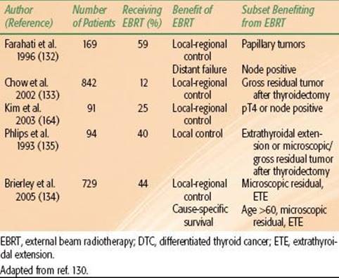
TABLE 50.14 RETROSPECTIVE SERIES SHOWING A BENEFIT FROM EXTERNAL BEAM RADIOTHERAPY IN PATIENTS WITH DIFFERENTIATED THYROID CANCER TREATED ROUTINELY WITH RADIOACTIVE IODINE
EXTERNAL BEAM RADIOTHERAPY FOR THYROID CANCER
Indications for External Beam Radiotherapy
There are no randomized control trials defining the indications for external beam radiotherapy (EBRT) in thyroid cancer. The European Multicentre Study on Differentiated Thyroid Cancer trial was planned as a prospective multicenter trial to evaluate the benefit of adjuvant EBRT in locally advanced DTC. Patients with pT4 disease with or without lymph node metastases and no known distant metastases who underwent total thyroidectomy, RAI, and TSH suppression were to be randomized to EBRT or observation. The trial closed prematurely, however, as only 16% of patients consented to be randomized to EBRT. The trial was converted to a prospective cohort study, and subsequent results failed to define the role of EBRT in this population.128–130 Therefore, recommendations for EBRT are based on institutional retrospective experiences and published clinical guidelines. Patients are approached differently with EBRT depending on whether the treatment plan is for primary, adjuvant, or palliative therapy. In general, only patients with unresectable tumor are treated with primary EBRT. Symptomatic metastatic tumors are well palliated with acceptable toxicity with plans utilizing simple beam arrangements to doses of 20 to 30 Gy in 5 to 10 fractions. For curative patients with nonmetastatic disease, RAI is always the preferred adjuvant therapy to EBRT, especially in young patients. EBRT has a niche role as adjuvant therapy after total thyroidectomy for certain patients at high risk for local recurrence who are not amenable to curative therapy with RAI. Table 50.13 summarizes the indications for EBRT from our institution, the 2009 ATA guidelines, and the 2011 NCCN guidelines.41,131 The authors find it useful to triage patients for EBRT based on the patient’s age, the potential to curatively treat with RAI, and the presence of high-risk clinical-pathologic features.
Powell et al.130 provide an excellent review of EBRT for DTC, including evidence supporting the indications for, and role of, EBRT. Table 50.14 summarizes the results of several retrospective analyses showing a benefit of EBRT in patients treated with thyroidectomy and adjuvant RAI. Even if prior I-131 has been given, at least one additional administration with >100 mCi with adequate preparation should be performed before considering EBRT.112,132–135
External Beam Radiotherapy Technique
Techniques for treating metastatic disease will vary by site, tumor burden, and clinical circumstances; therefore, this chapter will only discuss the technique used for treating the primary site and nodal regions. When both RAI and EBRT are indicated, the authors prefer to give RAI first because EBRT may result in thyroid stunning, decreasing the effectiveness of subsequent RAI.
CT simulation is used for treatment planning with patients positioned supine with arms at their side and the neck extended such that the mandible is at 90 degrees with respect to the treatment couch. An Aquaplast (WFR/Aquaplast Corporation, Avondale, PA) mask, custom head holder, and shoulder straps are used to immobilize the head and neck and to depress the level of the shoulders. Bolus material should be applied over scars in the neck. Intravenous contrast may be helpful in defining target volumes and normal-tissue structures, but this is contraindicated if RAI is being considered within the next 6 months. Axial CT images are acquired from above the skull base to the middle of the chest. Although not necessary, preoperative images sets may be fused to treatment planning CT scans to aid in target definitions. Historically, conventional anterior posterior/posterior anterior or lateral fields have been described to treat thyroid cancer, often requiring custom bolus materials to ensure homogenous dose distributions. Because the target volume straddles the level of the shoulder and, in nearly all cases, contains concavities with envaginated critical normal tissues, the authors find it useful to treat patients with intensity-modulated radiotherapy (IMRT).
Target Volumes and Dose
Target contours are delineated according to the definitions from the International Commission on Radiation Units. The gross tumor volume (GTV) will be any residual gross disease. The clinical target volume (CTVs) will define areas at risk for subclinical disease beyond any GTV. Schwartz et al.136 described an IMRT technique treating four separate CTVs with a single IMRT plan using a simultaneous integrated boost. For simplicity, the authors define two CTVs: the high-risk CTV corresponds to the region at highest risk for residual disease (positive margin, ETE, lymph node with extracapsular disease, or gross residual disease) and the standard-risk CTV, which is the region at moderate risk for residual disease (electively irradiated nodal stations).
Kim et al.137 analyzed the effect of treatment volume on patients treated with EBRT for nonanaplastic thyroid carcinoma. When limited fields were utilized treating only the primary (involved lobe) or recurrent tumor bed and the positive nodal area, 55% of patients developed local-regional failures compared with only 8% of patients treated with extended fields (including elective nodal irradiation). Another series analyzing patterns of failure after EBRT showed that several recurrences occur in superior mediastinal lymph nodes (level VII) when this area is not included in the treated volume.138 Therefore, the authors advocate for comprehensive irradiation including elective irradiations of cervical and upper mediastinal lymph node stations when indications for EBRT are present. When including the upper mediastinal lymph nodes in the standard risk CTV, the target volume extends inferiorly to the level of the carina. A contouring atlas showing selected CT slices is provided in Figure 50.6, which shows the high-risk CTV and standard-risk CTV. Normal-tissue organ at risks (OARs) include the spinal cord, brainstem, trachea, esophagus, parotid glands, oral cavity, cochleae, pharyngeal constrictors, brachial plexus, mandible, and lungs. Planning target volumes (PTVs) and planning OAR volume are created by three-dimensional symmetric expansions of the CTVs and OARs, respectively, to account for treatment setup uncertainty and organ motion based on institutional policies and generally range from 3 to 5 mm. Daily image guidance may be used to ensure target localization and reduce the setup uncertainties in the low neck. In general, 7 to 9 coplanar axially oriented intensity-modulated beams are used. The multileaf collimator sequencing, segment, and beam weighting are optimized by the inverse planning system based on parameters specified by the planner to meet specific goals for target coverage, limitations on dose heterogeneity, and normal tissue sparing. The authors prescribe 66 to 70 Gy to the high-risk PTV and 54 to 56 Gy to the standard-risk PTV in 33 to 35 fractions using a single IMRT plan with a simultaneous integrated boost.
External Beam Radiotherapy Outcomes
Several institutions have reported outcomes of patients treated with EBRT for thyroid cancer. When interpreting these data, it is important to consider that these series contain heterogeneous patient populations and are largely comprised of patients at very high risk for recurrence, either because of aggressive histologies, recurrent disease, poor or no response to RAI therapy, or gross unresectable tumor. Table 50.15 summarizes the findings of several recent series.
External Beam Radiotherapy Toxicity
One major reason for avoiding EBRT in patients with DTC is the toxicity associated with this therapeutic modality. The acute toxicities reported include mucositis, taste changes, xerostomia, pharyngitis, dysphagia, hoarseness, radiation dermatitis, weight loss, and malnutrition. One series reported an acute and subacute percutaneous feeding tube rate of 29%.139 Late complications include fibrosis and atrophy of the skin, lung apices, and neck musculature and tracheal and esophageal stenosis. The most commonly reported severe late complication appears to be esophageal stenosis.136 Late severe dysphagia requiring a permanent feeding tube is uncommon.136
FIGURE 50.6. Intensity-modulated radiation therapy clinical target volume contouring atlas for a patient with T2N0M0 insular thyroid carcinoma status post total thyroidectomy and radioactive iodine. Indications for external beam radiation therapy were insular carcinoma (poorly differentiated) and positive margin.
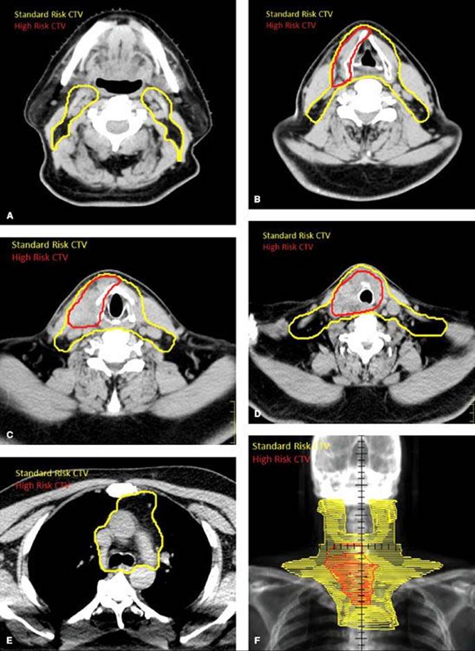
TABLE 50.15 RETROSPECTIVE SERIES REPORTING OUTCOMES OF PATIENTS TREATED WITH EXTERNAL BEAM RADIATION THERAPY FOR DIFFERENTIATED THYROID CANCER

THYROID-STIMULATING HORMONE SUPPRESSION FOR DIFFERENTIATED THYROID CARCINOMA
A common practice in management of DTC is administration of supratherapeutic doses of T4 in an effort to drive the TSH below detectable limits (<0.1 mIU/L), thereby decreasing stimulation of residual benign and malignant follicular-derived thyroid cells. There is in vitro evidence that TSH-receptor stimulation is sufficient to initiate thyroid tumorigenesis.140 A large retrospective series of patients with DTC with 30 years of follow-up showed that, compared with no adjunctive therapy, treatment with TSH suppression with T4 resulted in a 50% reduction in cancer deaths (6% vs. 12%).16 Another study showed that the degree of TSH suppression (TSH ≤0.05 mIU/L vs. ≥1 mIU/L) was associated with improved relapse-free survival.141
A major limitation to TSH suppression is the associated toxicity as these patients experience subclinical and even overt thyrotoxicosis. As such, these patients are prone to bone demineralization and cardiac abnormalities, including tachyarrhythmia, conduction abnormalities, increased contractility, ventricular hypertrophy, systolic and diastolic dysfunction, and even cardiac death.142 Nevertheless, a meta-analysis with over 4,000 patients with DTC demonstrated that the relative risk of major adverse clinical events was 0.73 (P <.05) favoring a “likely” or “questionable” benefit to TSH suppression in 15 of 17 trials.143 The authors concluded that TSH suppression is justified following initial therapy for DTC. The authors recommend TSH suppression to just below 0.1 mU/L for high-risk thyroid cancer patients, while maintaining the TSH at or slightly below the lower limit of normal (0.1–0.5 mU/L) in patients at low risk of recurring.
FOLLOW-UP OF A PATIENT WITH DIFFERENTIATED THYROID CARCINOMA
The follow-up of patients with DTC should be placed into the context of whether the patient has a low risk versus a high risk of recurrence. Low-risk patients are those who have no known distant metastases, removal of all macroscopic disease after total thyroidectomy, no evidence of ETE, no aggressive histologic variants, and no evidence of RAI uptake outside of the thyroid bed on an RxWBS. High-risk patients are patients who do not meet the criteria for low risk. All patients require surveillance for recurrent disease because many recurrences are amenable to curative treatment; however, high-risk patients warrant more vigorous follow up and many will require retreatment. The ATA recommends that serum Tg is measured and neck US is performed every 6 to 12 months following primary therapy for DTC.41 DxWBS and PET-CT scans are utilized only when clinically indicated.
Serum Tg is the most sensitive means of detecting persistent or recurrent tumor after surgery and RAI. Even as an isolated test, an undetectable serum Tg has a negative predictive value of 99%. However, it is important to realize that serum Tg measurement can reflect the level of both normal and malignant thyroid tissue remaining in the body, and that the value is very difficult to interpret in the setting of positive anti-Tg Ab, which is present in approximately 25% of patients with DTCs.
When Tg is elevated, DxWBS can be performed and, if negative, PET-CT may be warranted to assess the extent of disease. Often there is no clear evidence of overt disease on further diagnostic studies. The authors do not recommend proceeding to salvage therapy when an elevated serum Tg is the only evidence of disease.
CHEMOTHERAPY AND TARGETED AGENTS FOR DIFFERENTIATED THYROID CARCINOMA
Traditional systemic cytotoxic therapies have no significant role in the management of DTC because of poor response rates on the order of 25% to 40%.144 The most commonly utilized agent is doxorubicin, either alone or in combination with cisplatin. Even significant responses are rarely durable for more than a few months. Recent advances in targeted therapeutics have opened new doors for effective systemic therapy for recurrent or metastatic disease that is unresponsive to RAI. Most of the agents currently under investigation target specific pathways involved in cellular proliferation through inhibition of tyrosine kinase (TKI) receptors and angiogenesis through inhibition of vascular endothelial growth factor receptors (VEGFR). Axitinib, an oral TKI that effectively blocks VEGFRs, was studied in a phase II trial for advanced and metastatic thyroid carcinoma (including both follicular-derived and medullary histologies) and resulted in a 31% objective response rate; 38% of patients had stable disease for an average duration of over 16 weeks. Motesanib, another oral TKI, was studied in a similar phase II trial and demonstrated an 81% rate of disease control with a median progression-free survival of 9.3 months. Sorafenib and sunitinib have also been studied in a phase II trial in advanced and metastatic DTC unamenable to RAI with limited success.145
MANAGEMENT OF MEDULLARY THYROID CARCINOMA
All patients with MTC should be tested for RET mutations, including sporadic cases. Genetic screening and testing is also indicated. Similar to follicular epithelial-derived DTC, initial primary management of localized MTC is total thyroidectomy, which is the only completely effective therapy. Central neck dissection should be performed in all cases and compartment-oriented lateral neck dissection is indicated when clinically involved. There is no role for adjuvant RAI therapy. All patients should be followed with serum calcitonin levels as this presents a sensitive and specific marker for extent of residual disease. The indications for EBRT in a patient with MTC depend on the patient’s age. In children (<18 years old), EBRT is reserved for palliation of symptoms from tumors not amenable to other treatment or when tumor progression is likely to cause normal tissue damage. For adults, EBRT is indicated for treatment of unresectable gross disease or when there is a high risk of residual microscopic disease after total thyroidectomy based on pathologic evaluation revealing positive margins, T4 primary tumors, or nodal metastases with extensive extracapsular extension.
Schwartz et al.146 reported the outcomes of 34 consecutive patients treated at the M.D. Anderson Cancer Center with EBRT for MTC. Ten patients had recurrent disease, 15 had mediastinal involvement, and 10 had distant metastases; the preradiotherapy serum calcitonin was 556. The median EBRT dose was 60 Gy. The local-regional relapse-free survival, disease-specific survival, and overall survival at 5 years were 87%, 62%, and 56%, respectively, and 9% of patients developed significant late toxicity. Brierley et al.147 reported the outcomes of 73 consecutive patients treated at Princess Margaret Hospital (Toronto, Ontario, Canada), of which 46 patients were treated with EBRT to a median dose of 40 Gy (range, 20.0–75.5 Gy). EBRT was not associated with improved local-regional relapse-free survival (LRRFS) on multivariate analysis for the overall group, but when the analysis was limited to patients with a high risk of microscopic residual disease (ETE or positive lymph nodes), the 10-year LRRFS was 86% for patients receiving EBRT compared to 52% for patients not receiving EBRT.
Traditional cytotoxic systemic therapies have largely been ineffective in the management of metastatic or recurrent MTC; however, recent advances in therapies targeting the RET-tyrosine kinase receptors have shown promising preclinical and early clinical results.148–151,152
MANAGEMENT OF ANAPLASTIC THYROID CARCINOMA
Most ATC presents with extraglandular disease and it is not clear that any form of therapy improves outcomes. Complete surgical excision should be the goal of initial therapy, when feasible. However, surgery should be avoided when complete excision is not possible as debulking does not improve outcomes. There is no therapeutic role for RAI. EBRT is the standard of care for palliation of local symptoms from unresectable disease or as adjuvant therapy in the rare case of a completely resected tumor. Even when high doses are administered, however, immediate disease progression is commonly observed.153 Hyperfractionated therapy may improve outcomes.154,155 Despite aggressive treatment with concomitant chemoradiotherapy using a variety of agents, including docetaxel, paclitaxel, vincristine, cisplatin, or doxorubicin, outcomes remain grim.156–159
REFERENCES
1. American Joint Committee on Cancer. AJCC cancer staging handbook. New York: Springer, 2010.
2. Herle AJ, Uller RP. Elevated serum thyroglobulin. A marker of metastases in differentiated thyroid carcinomas. J Clin Invest 1975;56:272–277.
3. Ghossein R. Update to the College of American Pathologists reporting on thyroid carcinomas. Head Neck Pathol 2009;3:86–93.
4. Boone RT, Fan CY, Hanna EY. Well-differentiated carcinoma of the thyroid. Otolaryngol Clin North Am 2003;36:73–90.
5. Hay ID. Papillary thyroid carcinoma. Endocrinol Metab Clin North Am 1990;19:545–576.
6. Pacini F, Elisei R, Capezzone M, et al. Contralateral papillary thyroid cancer is frequent at completion thyroidectomy with no difference in low- and high-risk patients. Thyroid 2001;11:877–881.
7. Kawaura M, Pathak I, Gullane PJ, et al. Multicentricity in papillary thyroid carcinoma: analysis of predictive factors. J Otolaryngol 2001;30:102–105.
8. Nagaiah G, Hossain A, Mooney CJ, et al. Anaplastic thyroid cancer: a review of epidemiology, pathogenesis, and treatment. J Oncol 2011;2011:542358.
9. Siegel R, Ward E, Brawley O, et al. Cancer statistics, 2011: the impact of eliminating socioeconomic and racial disparities on premature cancer deaths. CA Cancer J Clin 2011;61:212–236.
10. Aschebrook-Kilfoy B, Ward MH, Sabra MM, et al. Thyroid cancer incidence patterns in the United States by histologic type, 1992–2006. Thyroid 2011;21:125–134.
11. Sherman SI. Thyroid carcinoma. Lancet 2003;361:501–511.
12. Davies L, Welch HG. Increasing incidence of thyroid cancer in the United States, 1973–2002. JAMA 2006;295:2164–2167.
13. Hughes DT, Haymart MR, Miller BS, et al. The most commonly occurring papillary thyroid cancer in the United States is now a microcarcinoma in a patient older than 45 years. Thyroid 2011;21:231–236.
14. Harach HR, Franssila KO, Wasenius VM. Occult papillary carcinoma of the thyroid. A “normal” finding in Finland. A systematic autopsy study. Cancer 1985;56:531–538.
15. Amdur RJ, Mazzaferri EL. Incidence, prevalence, recurrence, and mortality of differentiated thyroid cancer. In: Amdur RJ, Mazzaferri EL, eds. Essentials of thyroid cancer management (Cancer Treatment and Research). New York: Springer, 2005:123–130.
16. Mazzaferri EL, Jhiang SM. Long-term impact of initial surgical and medical therapy on papillary and follicular thyroid cancer. Am J Med 1994;97:418–428.
17. Wood JW, Tamagaki H, Neriishi S, et al. Thyroid carcinoma in atomic bomb survivors Hiroshima and Nagasaki. Am J Epidemiol 1969;89:4–14.
18. Parker LN, Belsky JL, Yamamoto T, et al. Thyroid carcinoma after exposure to atomic radiation. A continuing survey of a fixed population, Hiroshima and Nagasaki, 1958–1971. Ann Intern Med1974;80:600–604.
19. Sampson RJ, Key CR, Buncher CR, et al. Thyroid carcinoma in Hiroshima and Nagasaki. I. Prevalence of thyroid carcinoma at autopsy. JAMA 1969;209:65–70.
20. Richardson DB. Exposure to ionizing radiation in adulthood and thyroid cancer incidence. Epidemiology 2009;20:181–187.
21. Preston DL, Ron E, Tokuoka S, et al. Solid cancer incidence in atomic bomb survivors: 1958–1998. Radiat Res 2007;168:1–64.
22. Cardis E, Hatch M. The Chernobyl accident—an epidemiological perspective. Clin Oncol (R Coll Radiol) 2011;23:251–260.
23. Cardis E, Krewski D, Boniol M, et al. Estimates of the cancer burden in Europe from radioactive fallout from the Chernobyl accident. Int J Cancer 2006;119:1224–1235.
24. Ron E, Lubin JH, Shore RE, et al. Thyroid cancer after exposure to external radiation: a pooled analysis of seven studies. Radiat Res 1995;141:259–277.
25. Tucker MA, Jones PH, Boice JD Jr, et al. Therapeutic radiation at a young age is linked to secondary thyroid cancer. The Late Effects Study Group. Cancer Res 1991;51:2885–2888.
26. Sadetzki S, Chetrit A, Lubina A, et al. Risk of thyroid cancer after childhood exposure to ionizing radiation for tinea capitis. J Clin Endocrinol Metab 2006;91:4798–4804.
27. Naing S, Collins BJ, Schneider AB. Clinical behavior of radiation-induced thyroid cancer: factors related to recurrence. Thyroid 2009;19:479–485.
28. Schneider AB, Sarne DH. Long-term risks for thyroid cancer and other neoplasms after exposure to radiation. Nat Clin Pract Endocrinol Metab 2005;1:82–91.
29. Acharya S, Sarafoglou K, LaQuaglia M, et al. Thyroid neoplasms after therapeutic radiation for malignancies during childhood or adolescence. Cancer 2003;97:2397–2403.
30. Tuttle RM, Vaisman F, Tronko MD. Clinical presentation and clinical outcomes in Chernobyl-related paediatric thyroid cancers: what do we know now? What can we expect in the future? Clin Oncol (R Coll Radiol)2011;23:268–275.
31. Nose V. Familial thyroid cancer: a review. Mod Pathol 2011;24 (Suppl 2):S19–S33.
32. Mortensen JD, Woolner LB, Bennett WA. Gross and microscopic findings in clinically normal thyroid glands. J Clin Endocrinol Metab 1955;15:1270–1280.
33. Vander JB, Gaston EA, Dawber TR. The significance of nontoxic thyroid nodules. Final report of a 15-year study of the incidence of thyroid malignancy. Ann Intern Med 1968;69:537–540.
34. Nixon IJ, Ganly I, Hann LE, et al. Nomogram for predicting malignancy in thyroid nodules using clinical, biochemical, ultrasonographic, and cytologic features. Surgery 2010;148:1120–1127.
35. Hamming JF, Goslings BM, van Steenic GJ, et al. The value of fine-needle aspiration biopsy in patients with nodular thyroid disease divided into groups of suspicion of malignant neoplasms on clinical grounds. Arch Intern Med1990;150:113–116.
36. Tan GH, Gharib H. Thyroid incidentalomas: management approaches to nonpalpable nodules discovered incidentally on thyroid imaging. Ann Intern Med 1997;126:226–231.
37. Liu Y. Clinical significance of thyroid uptake on F18-fluorodeoxyglucose positron emission tomography. Ann Nucl Med 2009;23:17–23.
38. Wong C, Lin M, Chicco A, et al. The clinical significance and management of incidental focal FDG uptake in the thyroid gland on positron emission tomography/computed tomography (PET/CT) in patients with non-thyroidal malignancy. Acta Radiol 2011;52:899–904.
39. Nam-Goong IS, Kim HY, Gong G, et al. Ultrasonography-guided fine-needle aspiration of thyroid incidentaloma: correlation with pathological findings. Clin Endocrinol (Oxf) 2004;60:21–28.
40. Frates MC, Benson CB, Charboneau JW, et al. Management of thyroid nodules detected at US: Society of Radiologists in Ultrasound consensus conference statement. Radiology 2005;237:794–800.
41. Cooper DS, Doherty GM, Haugen BR, et al. Revised American Thyroid Association management guidelines for patients with thyroid nodules and differentiated thyroid cancer. Thyroid 2009;19:1167–1214.
42. Mazzaferri EL. The diagnosis of thyroid cancer. In: Amdur RJ, Mazzaferri EL, eds. Essentials of thyroid cancer management (Cancer Treatment and Research). New York: Springer, 2005:39–48.
43. Yang GC, Liebeskind D, Messina AV. Ultrasound-guided fine-needle aspiration of the thyroid assessed by ultrafast Papanicolaou stain: data from 1135 biopsies with a two- to six-year follow-up. Thyroid2001;11:581–589.
44. Ko HM, Jhu IK, Yang SH, et al. Clinicopathologic analysis of fine needle aspiration cytology of the thyroid. A review of 1,613 cases and correlation with histopathologic diagnoses. Acta Cytol2003;47:727–732.
45. Gomez NR, Kouniavsky G, Tsai HL, et al. Tumor size and presence of calcifications on ultrasonography are pre-operative predictors of lymph node metastases in patients with papillary thyroid cancer. J Surg Oncol2011;104:613–616.
46. Mazzaferri EL. Neck Ultrasonography in patients with thyroid cancer. In: Amdur RJ, Mazzaferri EL, eds. Essentials of thyroid cancer management (Cancer treatment and research). New York: Springer, 2005:101–120.
47. Gorges R, Eising EG, Fotescu D, et al. Diagnostic value of high-resolution B-mode and power-mode sonography in the follow-up of thyroid cancer. Eur J Ultrasound 2003;16:191–206.
48. Kouvaraki MA, Shapiro SE, Fornage BD, et al. Role of preoperative ultrasonography in the surgical management of patients with thyroid cancer. Surgery 2003;134:946–954.
49. Stulak JM, Grant CS, Farley DR, et al. Value of preoperative ultrasonography in the surgical management of initial and reoperative papillary thyroid cancer. Arch Surg 2006;141:489–494.
50. Mancuso AA, Mendenhall WM, Vaysberg M. Thyroid: nodules and malignant tumors. In: Mancuso AA, ed. Head and neck radiology. Philadelphia, PA: Lippincott Williams & Wilkins, 2010:1457–1481.
51. Chin BB, Patel P, Cohade C, et al. Recombinant human thyrotropin stimulation of fluoro-D-glucose positron emission tomography uptake in well-differentiated thyroid carcinoma. J Clin Endocrinol Metab 2004;89:91–95.
52. Ho TY, Liou MJ, Lin KJ, et al. Prevalence and significance of thyroid uptake detected by 18F-FDG PET. Endocrine 2011;40:297–302.
53. Shie P, Cardarelli R, Sprawls K, et al. Systematic review: prevalence of malignant incidental thyroid nodules identified on fluorine-18 fluorodeoxyglucose positron emission tomography. Nucl Med Commun 2009;30:742–748.
54. NCCN guidelines for thyroid carcinoma: National Comprehensive Cancer Network 2011.
55. Ruegemer JJ, Hay ID, Bergstralh EJ, et al. Distant metastases in differentiated thyroid carcinoma: a multivariate analysis of prognostic variables. J Clin Endocrinol Metab 1988;67:501–508.
56. Samaan NA, Schultz PN, Haynie TP, et al. Pulmonary metastasis of differentiated thyroid carcinoma: treatment results in 101 patients. J Clin Endocrinol Metab 1985;60:376–380.
57. Hundahl SA, Fleming ID, Fremgen AM, et al. A National Cancer Data Base report on 53,856 cases of thyroid carcinoma treated in the U.S., 1985–1995 [see comments]. Cancer 1998;83:2638–2648.
58. Vaisman F, Corbo R, Vaisman M. Thyroid carcinoma in children and adolescents-systematic review of the literature. J Thyroid Res 2011;2011:845362.
59. Chow SM, Law SC, Mendenhall WM, et al. Differentiated thyroid carcinoma in childhood and adolescence-clinical course and role of radioiodine. Pediatr Blood Cancer 2004;42:176–183.
60. Grigsby PW, Gal-or A, Michalski JM, et al. Childhood and adolescent thyroid carcinoma. Cancer 2002;95:724–729.
61. Okada T, Sasaki F, Takahashi H, et al. Management of childhood and adolescent thyroid carcinoma: long-term follow-up and clinical characteristics. Eur J Pediatr Surg 2006;16:8–13.
62. Shah JP, Loree TR, Dharker D, et al. Prognostic factors in differentiated carcinoma of the thyroid gland. Am J Surg 1992;164:658–661.
63. Villaret DB, Amdur RJ, Mazzaferri EL. Neck dissections to remove malignant lymph nodes. In: Amdur RJ, Mazzaferri EL, eds. Essentials of thyroid cancer management (Cancer treatment and research).New York: Springer, 2005:131–140.
64. Kramer JA, Schmid KW, Dralle H, et al. Primary tumour size is a prognostic parameter in patients suffering from differentiated thyroid carcinoma with extrathyroidal growth: results of the MSDS trial. Eur J Endocrinol2010;163:637–644.
65. Verburg FA, Mader U, Luster M, et al. Primary tumour diameter as a risk factor for advanced disease features of differentiated thyroid carcinoma. Clin Endocrinol (Oxf) 2009;71:291–297.
66. Machens A, Holzhausen HJ, Dralle H. The prognostic value of primary tumor size in papillary and follicular thyroid carcinoma. Cancer 2005;103:2269–2273.
67. Baudin E, Travagli JP, Ropers J, et al. Microcarcinoma of the thyroid gland: the Gustave-Roussy Institute experience. Cancer 1998;83:553–559.
68. Appetecchia M, Scarcello G, Pucci E, et al. Outcome after treatment of papillary thyroid microcarcinoma. J Exp Clin Cancer Res 2002;21:159–164.
69. Mercante G, Frasoldati A, Pedroni C, et al. Prognostic factors affecting neck lymph node recurrence and distant metastasis in papillary microcarcinoma of the thyroid: results of a study in 445 patients. Thyroid 2009;19:707–716.
70. Kucuk NO, Tari P, Tokmak E, et al. Treatment for microcarcinoma of the thyroid—clinical experience. Clin Nucl Med 2007;32:279–281.
71. Lombardi CP, Bellantone R, De Crea C, et al. Papillary thyroid microcarcinoma: extrathyroidal extension, lymph node metastases, and risk factors for recurrence in a high prevalence of goiter area. World J Surg 2010;34:1214–1221.
72. Kim ES, Kim TY, Koh JM, et al. Completion thyroidectomy in patients with thyroid cancer who initially underwent unilateral operation. Clin Endocrinol (Oxf) 2004;61:145–148.
73. Scheumann GF, Seeliger H, Musholt TJ, et al. Completion thyroidectomy in 131 patients with differentiated thyroid carcinoma. Eur J Surg 1996;162:677–684.
74. Mazzaferri EL, Kloos RT. Clinical review 128: current approaches to primary therapy for papillary and follicular thyroid cancer. J Clin Endocrinol Metab 2001;86:1447–1463.
75. Massin JP, Savoie JC, Garnier H, et al. Pulmonary metastases in differentiated thyroid carcinoma. Study of 58 cases with implications for the primary tumor treatment. Cancer 1984;53:982–992.
76. Pasieka JL, Thompson NW, McLeod MK, et al. The incidence of bilateral well-differentiated thyroid cancer found at completion thyroidectomy. World J Surg 1992;16:711–716.
77. Machens A, Holzhausen HJ, Lautenschlager C, et al. Enhancement of lymph node metastasis and distant metastasis of thyroid carcinoma. Cancer 2003;98:712–719.
78. Chiacchio S, Lorenzoni A, Boni G, et al. Anaplastic thyroid cancer: prevalence, diagnosis and treatment. Minerva Endocrinol 2008;33:341–357.
79. Sellers M, Beenken S, Blankenship A, et al. Prognostic significance of cervical lymph node metastases in differentiated thyroid cancer. Am J Surg 1992;164:578–581.
80. Clark OH. Thyroid cancer and lymph node metastases. J Surg Oncol 2011;103:615–618.
81. Shah PK, Shah KK, Karakousis GC, et al. Regional recurrence after lymphadenectomy for clinically evident lymph node metastases from papillary thyroid cancer: a cohort study : bulky nodal metastases in papillary thyroid cancer. Ann Surg Oncol 2011;19:1453–1459.
82. Mazzaferri EL. Management of a solitary thyroid nodule. N Engl J Med 1993;328:553–559.
83. Sampson E, Brierley JD, Le LW, et al. Clinical management and outcome of papillary and follicular (differentiated) thyroid cancer presenting with distant metastasis at diagnosis. Cancer 2007;110:1451–1456.
84. Udelsman R, Lakatos E, Ladenson P. Optimal surgery for papillary thyroid carcinoma. World J Surg 1996;20:88–93.
85. Mazzaferri EL. Potential complications of thyroid cancer surgery. In: Amdur RJ, Mazzaferri EL, eds. Essentials of thyroid cancer management (Cancer treatment and research). New York: Springer, 2005:153–160.
86. Sosa JA, Udelsman R. Total thyroidectomy for differentiated thyroid cancer. J Surg Oncol 2006;94:701–707.
87. Bilimoria KY, Zanocco K, Sturgeon C. Impact of surgical treatment on outcomes for papillary thyroid cancer. Adv Surg 2008;42:1–12.
88. Haigh PI, Urbach DR, Rotstein LE. Extent of thyroidectomy is not a major determinant of survival in low- or high-risk papillary thyroid cancer. Ann Surg Oncol 2005;12:81–89.
89. Van Nguyen K, Dilawari RA. Predictive value of AMES scoring system in selection of extent of surgery in well differentiated carcinoma of thyroid. Am Surg 1995;61:151–155.
90. Cross S, Wei JP, Kim S, et al. Selective surgery and adjuvant therapy based on risk classifications of well-differentiated thyroid cancer. J Surg Oncol 2006;94:678–682.
91. Hay ID, Grant CS, Taylor WF, et al. Ipsilateral lobectomy versus bilateral lobar resection in papillary thyroid carcinoma: a retrospective analysis of surgical outcome using a novel prognostic scoring system. Surgery1987;102:1088–1095.
92. Haigh PI, Urbach DR, Rotstein LE. AMES prognostic index and extent of thyroidectomy for well-differentiated thyroid cancer in the United States. Surgery 2004;136:609–616.
93. Bilimoria KY, Bentrem DJ, Linn JG, et al. Utilization of total thyroidectomy for papillary thyroid cancer in the United States. Surgery 2007;142:906–913.
94. Fritze D, Doherty GM. Surgical management of cervical lymph nodes in differentiated thyroid cancer. Otolaryngol Clin North Am 2010;43:285–300.
95. Bardet S, Malville E, Rame JP, et al. Macroscopic lymph-node involvement and neck dissection predict lymph-node recurrence in papillary thyroid carcinoma. Eur J Endocrinol 2008;158:551–560.
96. Noguchi S, Murakami N, Yamashita H, et al. Papillary thyroid carcinoma: modified radical neck dissection improves prognosis. Arch Surg 1998;133:276–280.
97. Moley JF, DeBenedetti MK. Patterns of nodal metastases in palpable medullary thyroid carcinoma: recommendations for extent of node dissection. Ann Surg 1999;229:880–887.
98. Oskam IM, Hoebers F, Balm AJ, et al. Neck management in medullary thyroid carcinoma. Eur J Surg Oncol 2008;34:71–76.
99. Dionigi G, Dionigi R, Bartalena L, et al. Surgery of lymph nodes in papillary thyroid cancer. Expert Rev Anticancer Ther 2006;6:1217–1229.
100. White ML, Doherty GM. Level VI lymph node dissection for papillary thyroid cancer. Minerva Chir 2007;62:383–393.
101. Sadowski BM, Snyder SK, Lairmore TC. Routine bilateral central lymph node clearance for papillary thyroid cancer. Surgery 2009;146:696–703.
102. Mulla M, Schulte KM. Central cervical lymph node metastases in papillary thyroid cancer: a systematic review of imaging-guided and prophylactic removal of the central compartment. Clin Endocrinol (Oxf) 2011;76:131–136.
103. Moo TA, McGill J, Allendorf J, et al. Impact of prophylactic central neck lymph node dissection on early recurrence in papillary thyroid carcinoma. World J Surg 2010;34:1187–1191.
104. Shindo M, Wu JC, Park EE, et al. The importance of central compartment elective lymph node excision in the staging and treatment of papillary thyroid cancer. Arch Otolaryngol Head Neck Surg2006;132:650–654.
105. Kutler DI, Crummey AD, Kuhel WI. Routine central compartment lymph node dissection for patients with papillary thyroid carcinoma. Head Neck 2011;34:260–263.
106. Shah MD, Hall FT, Eski SJ, et al. Clinical course of thyroid carcinoma after neck dissection. Laryngoscope 2003;113:2102–2107.
107. Zuniga S, Sanabria A. Prophylactic central neck dissection in stage N0 papillary thyroid carcinoma. Arch Otolaryngol Head Neck Surg 2009;135:1087–1091.
108. Rosenbaum MA, McHenry CR. Central neck dissection for papillary thyroid cancer. Arch Otolaryngol Head Neck Surg 2009;135:1092–1097.
109. Zetoune T, Keutgen X, Buitrago D, et al. Prophylactic central neck dissection and local recurrence in papillary thyroid cancer: a meta-analysis. Ann Surg Oncol 2010;17:3287–3293.
110. Samaan NA, Schultz PN, Hickey RC, et al. The results of various modalities of treatment of well differentiated thyroid carcinomas: a retrospective review of 1599 patients. J Clin Endocrinol Metab1992;75:714–720.
111. DeGroot LJ, Kaplan EL, McCormick M, et al. Natural history, treatment, and course of papillary thyroid carcinoma. J Clin Endocrinol Metab 1990;71:414–424.
112. Tsang RW, Brierley JD, Simpson WJ, et al. The effects of surgery, radioiodine, and external radiation therapy on the clinical outcome of patients with differentiated thyroid carcinoma. Cancer1998;82:375–388.
113. Taylor T, Specker B, Robbins J, et al. Outcome after treatment of high-risk papillary and non-Hurthle-cell follicular thyroid carcinoma. Ann Intern Med 1998;129:622–627.
114. Amdur RJ, Mazzaferri EL. Choosing the activity of I-131 for therapy. In: Amdur RJ, Mazzaferri EL, eds. Essentials of thyroid cancer management (Cancer treatment and research). New York: Springer, 2005:169–176.
115. Pacini F, Ladenson PW, Schlumberger M, et al. Radioiodine ablation of thyroid remnants after preparation with recombinant human thyrotropin in differentiated thyroid carcinoma: results of an international, randomized, controlled study. J Clin Endocrinol Metab 2006;91:926–932.
116. Elisei R, Schlumberger M, Driedger A, et al. Follow-up of low-risk differentiated thyroid cancer patients who underwent radioiodine ablation of postsurgical thyroid remnants after either recombinant human thyrotropin or thyroid hormone withdrawal. J Clin Endocrinol Metab 2009;94:4171–4179.
117. Lee J, Yun MJ, Nam KH, et al. Quality of life and effectiveness comparisons of thyroxine withdrawal, triiodothyronine withdrawal, and recombinant thyroid-stimulating hormone administration for low-dose radioiodine remnant ablation of differentiated thyroid carcinoma. Thyroid 2010;20:173–179.
118. Ladenson PW, Braverman LE, Mazzaferri EL, et al. Comparison of administration of recombinant human thyrotropin with withdrawal of thyroid hormone for radioactive iodine scanning in patients with thyroid carcinoma. N Engl J Med 1997;337:888–896.
119. Haugen BR, Pacini F, Reiners C, et al. A comparison of recombinant human thyrotropin and thyroid hormone withdrawal for the detection of thyroid remnant or cancer. J Clin Endocrinol Metab1999;84:3877–3885.
120. Grigsby PW, Siegel BA, Baker S, et al. Radiation exposure from outpatient radioactive iodine (131I) therapy for thyroid carcinoma. JAMA 2000;283:2272–2274.
121. NRC Regulatory Issue Summary 2011-01 NRC policy on release of iodine-131 therapy patients under 10 CFR 35.75 to locations other than private residences. U.S. Nuclear Regulatory Commission Office of Federal And State Materials and Environmental Management Programs, 2011.
122. Sisson JC, Freitas J, McDougall IR, et al. Radiation safety in the treatment of patients with thyroid diseases by radioiodine 131I: practice recommendations of the American Thyroid Association. Thyroid2011;21:335–346.
123. Kloos RT. Survey of radioiodine therapy safety practices highlights the need for user-friendly recommendations. Thyroid 2011;21:97–99.
124. Potential side effects and complication of I-131 therapy. In: Amdur RJ, Mazzaferri EL, eds. Essentials of thyroid cancer management (Cancer treatment and research). New York: Springer, 2005:265–277.
125. Brown AP, Chen J, Hitchcock YJ, et al. The risk of second primary malignancies up to three decades after the treatment of differentiated thyroid cancer. J Clin Endocrinol Metab 2008;93:504–515.
126. Sawka AM, Thabane L, Parlea L, et al. Second primary malignancy risk after radioactive iodine treatment for thyroid cancer: a systematic review and meta-analysis. Thyroid 2009;19:451–457.
127. Rubino C, de Vathaire F, Dottorini ME, et al. Second primary malignancies in thyroid cancer patients. Br J Cancer 2003;89:1638–1644.
128. Biermann M, Pixberg MK, Schuck A, et al. Multicenter study differentiated thyroid carcinoma (MSDS). Diminished acceptance of adjuvant external beam radiotherapy. Nuklearmedizin 2003;42:244–250.
129. Biermann M, Pixberg M, Riemann B, et al. Clinical outcomes of adjuvant external-beam radiotherapy for differentiated thyroid cancer—results after 874 patient-years of follow-up in the MSDS trial. Nuklearmedizin2009;48:89–98.
130. Powell C, Newbold K, Harrington KJ, et al. External beam radiotherapy for differentiated thyroid cancer. Clin Oncol (R Coll Radiol) 2010;22:456–463.
131. Amdur RJ, Mendenhall WM, Mazzaferri EL. Indications for external beam radiation therapy for differentiated thyroid cancer. In: Amdur RJ, Mazzaferri EL, eds. Essentials of thyroid cancer management (Cancer treatment and research). New York: Springer, 2005:407–412.
132. Farahati J, Reiners C, Stuschke M, et al. Differentiated thyroid cancer. Impact of adjuvant external radiotherapy in patients with perithyroidal tumor infiltration (stage pT4). Cancer 1996;77:172–180.
133. Chow SM, Law SC, Mendenhall WM, et al. Papillary thyroid carcinoma: prognostic factors and the role of radioiodine and external radiotherapy. Int J Radiat Oncol Biol Phys 2002;52:784–795.
134. Brierley J, Tsang R, Panzarella T, et al. Prognostic factors and the effect of treatment with radioactive iodine and external beam radiation on patients with differentiated thyroid cancer seen at a single institution over 40 years. Clin Endocrinol (Oxf) 2005;63:418–427.
135. Phlips P, Hanzen C, Andry G, et al. Postoperative irradiation for thyroid cancer. Eur J Surg Oncol 1993;19:399–404.
136. Schwartz DL, Lobo MJ, Ang KK, et al. Postoperative external beam radiotherapy for differentiated thyroid cancer: outcomes and morbidity with conformal treatment. Int J Radiat Oncol Biol Phys2009;74:1083–1091.
137. Kim TH, Chung KW, Lee YJ, et al. The effect of external beam radiotherapy volume on locoregional control in patients with locoregionally advanced or recurrent nonanaplastic thyroid cancer. Radiat Oncol 2010;5:69.
138. Azrif M, Slevin NJ, Sykes AJ, et al. Patterns of relapse following radiotherapy for differentiated thyroid cancer: implication for target volume delineation. Radiother Oncol 2008;89:105–113.
139. Terezakis SA, Lee KS, Ghossein RA, et al. Role of external beam radiotherapy in patients with advanced or recurrent nonanaplastic thyroid cancer: Memorial Sloan-Kettering Cancer Center experience. Int J Radiat Oncol Biol Phys 2009;73:795–801.
140. Ludgate M, Gire V, Crisp M, et al. Contrasting effects of activating mutations of G alpha S and the thyrotropin receptor on proliferation and differentiation of thyroid follicular cells. Oncogene1999;18:4798–4807.
141. Pujol P, Daures JP, Nsakala N, et al. Degree of thyrotropin suppression as a prognostic determinant in differentiated thyroid cancer. J Clin Endocrinol Metab 1996;81:4318–4323.
142. Suppression of TSH and the potential toxicity of I-131 therapy. In: Amdur RJ, Mazzaferri EL, eds. Essentials of thyroid cancer management (Cancer treatment and research). New York: Springer, 2005:262.
143. McGriff NJ, Csako G, Gourgiotis L, et al. Effects of thyroid hormone suppression therapy on adverse clinical outcomes in thyroid cancer. Ann Med 2002;34:554–564.
144. Haugen BR. Management of the patient with progressive radioiodine non-responsive disease. Semin Surg Oncol 1999;16:34–41.
145. Romagnoli S, Moretti S, Voce P, et al. Targeted molecular therapies in thyroid carcinoma. Arq Bras Endocrinol Metabol 2009;53:1061–1073.
146. Schwartz DL, Rana V, Shaw S, et al. Postoperative radiotherapy for advanced medullary thyroid cancer—local disease control in the modern era. Head Neck 2008;30:883–888.
147. Brierley J, Tsang R, Simpson WJ, et al. Medullary thyroid cancer: analyses of survival and prognostic factors and the role of radiation therapy in local control. Thyroid 1996;6:305–310.
148. Lam ET, Ringel MD, Kloos RT, et al. Phase II clinical trial of sorafenib in metastatic medullary thyroid cancer. J Clin Oncol 2010;28:2323–2330.
149. Wells SA, Jr., Gosnell JE, Gagel RF, et al. Vandetanib for the treatment of patients with locally advanced or metastatic hereditary medullary thyroid cancer. J Clin Oncol 2010;28:767–772.
150. Ye L, Santarpia L, Gagel RF. Targeted therapy for endocrine cancer: the medullary thyroid carcinoma paradigm. Endocr Pract 2009;15:597–604.
151. Torino F, Paragliola RM, Barnabei A, et al. Medullary thyroid cancer: a promising model for targeted therapy. Curr Mol Med 2010;10:608–625.
152. Schlumberger MJ, Elisei R, Bastholt L, et al. Phase II study of safety and efficacy of motesanib in patients with progressive or symptomatic, advanced or metastatic medullary thyroid cancer. J Clin Oncol2009;27:3794–3801.
153. Amdur RJ, Kim S, Li JG, et al. Conventional radiotherapy technique for treating thyroid cancer. In: Amdur RJ, Mazzaferri EL, eds. Essentials of thyroid cancer management (Cancer treatment and research). New York: Springer, 2005:419–424.
154. Tennvall J, Lundell G, Wahlberg P, et al. Anaplastic thyroid carcinoma: three protocols combining doxorubicin, hyperfractionated radiotherapy and surgery. Br J Cancer 2002;86:1848–1853.
155. De Crevoisier R, Baudin E, Bachelot A, et al. Combined treatment of anaplastic thyroid carcinoma with surgery, chemotherapy, and hyperfractionated accelerated external radiotherapy. Int J Radiat Oncol Biol Phys2004;60:1137–1143.
156. Haigh PI, Ituarte PH, Wu HS, et al. Completely resected anaplastic thyroid carcinoma combined with adjuvant chemotherapy and irradiation is associated with prolonged survival. Cancer 2001;91:2335–2342.
157. Sherman EJ, Lim SH, Ho AL, et al. Concurrent doxorubicin and radiotherapy for anaplastic thyroid cancer: a critical re-evaluation including uniform pathologic review. Radiother Oncol 2011;110:125–130.
158. Troch M, Koperek O, Scheuba C, et al. High efficacy of concomitant treatment of undifferentiated (anaplastic) thyroid cancer with radiation and docetaxel. J Clin Endocrinol Metab 2010;95:E54–E57.
159. Heron DE, Karimpour S, Grigsby PW. Anaplastic thyroid carcinoma: comparison of conventional radiotherapy and hyperfractionation chemoradiotherapy in two groups. Am J Clin Oncol 2002;25:442–446.
160. Mancuso AA. Thyroid and parathyroid glands: introduction. Head and neck radiology. Philadelphia: Lippincott Williams & Wilkins, 2011:1428–1433
161. Xu L, Li G, Wei Q, et al. Family history of cancer and risk of sporadic differentiated thyroid carcinoma. Cancer 2011;118:1228–1235.
162. Maxon HR, Thomas SR, Boehringer A, et al. Low iodine diet in I-131 ablation of thyroid remnants. Clin Nucl Med 1983;8:123–126.
163. Sawka AM, Ibrahim-Zada I, Galacgac P, et al. Dietary iodine restriction in preparation for radioactive iodine treatment or scanning in well-differentiated thyroid cancer: a systematic review. Thyroid2010;20:1129–1138.
164. Kim TH, Yang DS, Jung KY, et al. Value of external irradiation for locally advanced papillary thyroid cancer. Int J Radiat Oncol Biol Phys 2003;55:1006–1012.
165. Meadows KM, Amdur RJ, Morris CG, et al. External beam radiotherapy for differentiated thyroid cancer. Am J Otolaryngol 2006;27:24–28.