
Brian G. Czito, Albert S. DeNittis, Manisha Palta, and Christopher G. Willett
Less than 15% of patients diagnosed with esophageal cancer are cured, with approximately half of patients presenting with unresectable or metastatic disease. This chapter reviews the natural history and treatment of esophageal cancer, including anatomy, risk factors, patterns of spread and failure, staging, results of current therapeutic approaches, radiation planning techniques, toxicity data, and future treatment strategies.
 ANATOMY
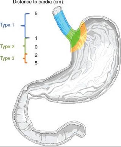
The esophagus is a thin-walled, hollow tube approximately 25 cm in length. It is lined with stratified keratinized squamous epithelium, extending from the cricopharyngeus muscle at the level of the cricoid cartilage superiorly to the gastroesophageal junction inferiorly. The lower one-third (5 to 10 cm) of the esophagus may contain glandular elements. Replacement of the stratified squamous epithelium with columnar epithelium is referred to as Barrett’s esophagus, often occurring in the lower one-third. The Z-line refers to the endoscopically visible junction of the squamous and glandular epithelium. The esophageal wall is composed of three layers: the mucosa, submucosa, and muscularis propria (Fig. 53.1). The mucosal layer contains the epithelium, lamina propria, and muscularis mucosae. The epithelium is separated from the lamina propria by a basement membrane. In the portion of esophagus containing columnar-type epithelium, the muscularis mucosae may consist of two layers. The mucosa may be divided into distinct layers, including M1 (epithelium), M2 (lamina propria), and M3 (muscularis mucosae). Similarly, the submucosal layer may be divided into inner (SM1), middle (SM2), and outer (SM3) layers. The muscularis propria consists of a circular inner layer and longitudinal outer layer. The adventitia (periesophageal connective tissue) lies directly on the muscularis propria.1 No serosa is present, facilitating extraesophageal spread of disease.
Although somewhat arbitrary, the esophagus is frequently divided into cervical and thoracic components. The most recent American Joint Committee on Cancer (AJCC) report divides the esophagus into four regions: cervical, upper thoracic, midthoracic, and lower thoracic (Fig. 53.2).1 The cervical esophagus begins at the cricopharyngeus muscle (approximately the C7 level or 15 cm from the incisors) and extends to the thoracic inlet (at approximately the T3 level or at approximately 20 cm from the incisors, at the level of the suprasternal notch), and therefore lies within the neck. The thoracic esophagus extends from approximately the level of T3 (beginning at about 20 cm) to T10 or T11.1,2 The upper thoracic esophagus is bordered superiorly by the thoracic inlet and inferiorly by the lower border of the azygos vein, extending from approximately 20 to 25 cm. Radiographically, tumors in this location would be located between the sternal notch and azygos vein. The middle thoracic esophagus extends from the lower border of the azygos vein to the inferior pulmonary veins, extending from approximately 25 to 30 cm. The lower thoracic esophagus extends from the inferior pulmonary veins and to the stomach and is inclusive of the gastroesophageal junction, typically extending from approximately 30 to 40 cm. Endoscopically, the gastroesophageal (GE) junction is often defined as the point where the first gastric fold is encountered, although this may be a “theoretical” landmark. The location of the GE junction can be accurately defined histologically as the squamocolumnar junction. In the most recent AJCC staging system, cancers with an epicenter in the lower thoracic esophagus, gastroesophageal junction, or within the proximal 5 cm of the stomach (i.e., cardia) and extending up to the GE junction or esophagus are staged as an adenocarcinoma of the esophagus. If the epicenter is >5 cm distal to the gastroesophageal junction or within 5 cm of the gastroesophageal junction but does not extend to the junction/esophagus, tumors are classified as stomach cancers. Useful landmarks in reference to endoscopy include the carina (∼25 cm from the incisors) and gastroesophageal junction (∼40 cm from the incisors).
Siewert et al.3,4 characterized cancer involving the gastroesophageal junction according to the location of the tumor (Fig. 53.3). If the tumor center is located from >1 cm up to 5 cm above the gastroesophageal junction (Z-line), the tumor is classified as a type I adenocarcinoma of the distal esophagus. If the tumor center is located within 1 cm cephalad to 2 cm caudad to the gastroesophageal junction, it is classified as type II. If the tumor center is located >2 cm below the gastroesophageal junction, the tumor is classified as type III. However, locally advanced/bulky tumors can make it difficult to accurately distinguish where tumors originated in relationship to the GE junction.
FIGURE 53.1. Diagram of esophageal wall. (Used with the permission of the American Joint Committee on Cancer, Chicago, Illinois. The original source for this material is American Joint Committee on Cancer. AJCC cancer staging handbook, 7th ed. New York: Springer, 2010, published by Springer Science and Business Media LLC, www.springerlink.com.)
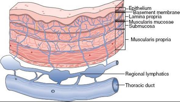
FIGURE 53.2. Anatomy of the esophagus. Note the lengths of various segments of the esophagus as measured from the upper central incisors. (Used with the permission of the American Joint Committee on Cancer, Chicago, Illinois. The original source for this material is American Joint Committee on Cancer. AJCC cancer staging handbook, 7th ed. New York: Springer, 2010, published by Springer Science and Business Media LLC, www.springerlink.com.)
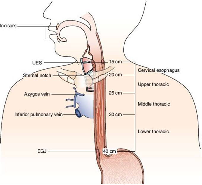
FIGURE 53.3. Siewert classification of the gastroesophageal junction cancers according to the location of the tumor. (From Matzinger O, Gerber E, Bernstein Z, et al. EORTC-ROG expert opinion: radiotherapy volume and treatment guidelines for neoadjuvant radiation of adenocarcinomas of the gastroesophageal junction and the stomach. Radiother Oncol 2009;92:164–175; copyright 2009, with permission from Elsevier.)
Lymphatic Drainage
The esophagus has an extensive, longitudinal interconnecting system of lymphatics. The esophageal lymphatic network is primarily located within the submucosa; however, channels are also present within the lamina propria, facilitating spread of even superficial cancers of the esophagus involving the mucosa. In addition to these longitudinal lymphatics, intramural lymphatics may traverse the muscularis propria, facilitating tumor spread to regional lymphatic channels and paraesophageal nodes. Supporting this, autopsy series have demonstrated a relatively high incidence of directly draining channels extending from the submucosa lymphatics into the thoracic duct (Fig. 53.1), facilitating systemic spread. Lymph can travel the entire length of the esophagus before draining into lymph nodes,2 and thus the entire esophagus is at potential risk for lymphatic involvement. Up to 8 cm or more of ‘normal’ tissue can exist between gross tumor and micrometastases “skip areas” secondary to this extensive lymphatic network.5 In addition, as many as 71% of frozen tissue sections scored as margin negative by conventional histopathology show involvement by lymphatic micrometastases with immunohistochemistry.6 Lymphatics of the esophagus drain into nodes that usually follow arteries, including the inferior thyroid artery, the bronchial and esophageal arteries, and the left gastric artery (celiac axis).7
Epidemiology and Risk Factors
Esophageal carcinoma accounts for approximately 6% of all gastrointestinal malignancies. In 2012, there will be an estimated 17,460 new patients diagnosed with esophageal cancer in the United States and 15,070 deaths. Most cases occur in males, at a rate of 4:1 relative to females.8,9
The incidence of esophageal carcinoma varies according to geography. The highest incidence occurs in Linxian, China, Russia, and the Caspian region of Iran. The incidence there is 100/100,000 persons. In addition, there are many differences within regions within those countries. In areas such as northern France, Kazakhstan, and South Africa incidence can be as high as 50 to 99/100,000. Although the reasons for the geographic discrepancy are unknown, some reports have linked the arid climate and alkaline soil with these high-risk areas, as well as the ingestion of nitrosamines, and inversely to the consumption of riboflavin, nicotinic acid, magnesium, and zinc.10,11 In the United States, the incidence rate among males is <5/100,000. Over the last 20 years, there has been an increase in the incidence of adenocarcinoma at a rate of 5% to 10% per year. This is a more rapid increase than that for any other cancer.12 In 1987, adenocarcinoma was reported to represent 34% and 12% of esophageal cancers in White men and women versus 3% and 1% for African American men and women, respectively.12 As of 1998, esophageal adenocarcinoma accounted for almost 55% of all diagnosed cases in White men. African American men are more frequently diagnosed with squamous cell carcinoma.12–14
In North America and Western Europe, alcohol and tobacco use are the major risk factors for squamous cell carcinoma, accounting for 80% to 90% of cases.15 Reports have described the relative risk of esophageal cancer by the amount of alcohol and tobacco consumed, including a relative risk of 155:1 when consuming >30 g/day of tobacco along with 121 g/day of alcohol.16
Diets of scant amounts of fruits, vegetables, and animal products are associated with increases in squamous cell carcinoma.17 Patients with Plummer-Vinson (Paterson-Kelly) syndrome, a condition characterized by iron-deficiency anemia and low riboflavin levels, are at an increased risk for oral cavity, hypopharyngeal, and esophageal cancer. In addition, dietary intake of nitrosamines, nitrosamides, and N-nitroso compounds has been implicated in esophageal carcinoma. Examples of nitrate-rich foods include pickled vegetables, alcoholic beverages, cured meats, and fish.18,19
Other risk factors associated with esophageal carcinoma include achalasia and tylosis. Achalasia of long duration (25 years) is associated with a 5% incidence of squamous cell carcinoma.20,21 Patients with tylosis (hyperkeratosis of the palms and soles and papilloma of the esophagus) have a reported 38% risk in developing esophageal cancer at a mean age of 45 years.22 In addition, carcinoma of the esophagus occurs in 2% to 4% of patients with head and neck cancer.
Risk factors leading to the development of adenocarcinoma of the esophagus are being increasingly understood. Most esophageal adenocarcinomas tend to arise from the metaplastic columnar-lined epithelium known as Barrett’s esophagus.23 Severe and long-standing gastroesophageal reflux disease has clearly been shown to be a significant risk factor for Barrett’s esophagus, which may lead to adenocarcinoma. It has been estimated that patients with long-standing severe reflux have a 44-fold risk of developing adenocarcinoma.24 Tobacco use is a more moderate risk factor for adenocarcinoma development. Smokers appear to have a twofold to threefold greater risk for developing esophageal adenocarcinoma than nonsmokers.25,26 The relative risk of esophageal adenocarcinoma persists to three decades following smoking cessation, in contrast to a significant decline in similar patients with squamous cell carcinoma.27 Obesity has also been linked to a threefold to fourfold risk of adenocarcinoma, possibly due to an increased risk of reflux.28 It has been estimated that a middle-aged patient with Barrett’s esophagus has a 10% to 15% risk of developing esophageal adenocarcinoma during his or her lifetime.29
Although many risk factors are associated with esophageal carcinoma, few studies have demonstrated a causal relationship leading to pathogenesis. Motesano et al.30 reported possible genetic abnormalities involved in the genesis of esophageal cancer. In addition, possible differences in mechanisms of pathogenesis for squamous cell carcinoma and adenocarcinoma were described. Genetic abnormalities in squamous cell carcinoma include p53 mutations and multiple allelic losses at 3p and 9q, with amplification of cyclin D1 and epidermal growth factor receptor (EGFR). These mutations lead to cell hyperplasia, low- and high-grade dysplasia, and, ultimately, squamous cell carcinoma. In contrast, genetic abnormalities in adenocarcinoma include overexpression of p53, multiple allelic losses at 17p, 5q, and 13q, and amplification and overexpression of EGFR and human epidermal growth factor receptor 2 (HER-2). These abnormalities may be involved in the stepwise development of Barrett’s esophagus, dysplasia, and, ultimately, adenocarcinoma. These differences suggest that squamous cell carcinoma and adenocarcinoma have different series of genetic mutations as etiologies, but these abnormalities occur in 23% to 94% of tumors studied.
TABLE 53.1 DISTRIBUTION OF METASTASES BY ANATOMIC SITE
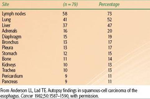
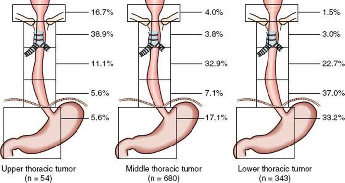
FIGURE 53.4. Positive lymph node distribution according to the location of the primary tumor, squamous cell carcinoma. (From Huang W, Li B, Gong H, et al. Pattern of lymph node metastases and its implication in radiotherapeutic clinical target volume in patients with thoracic esophageal squamous cell carcinoma: a report of 1077 cases. Radiother Oncol 2010;95:229–233; with permission from Elsevier.)
NATURAL HISTORY AND PATTERNS OF SPREAD
Squamous cell carcinoma is characterized by extensive local growth and proclivity to lymph node metastases. Because the esophagus has no covering serosa, direct invasion of contiguous structures may occur early. Lesions in the upper esophagus can impinge on or invade the recurrent laryngeal nerves, carotid arteries, and trachea. If extraesophageal extension occurs in the mediastinum, tracheoesophageal or bronchoesophageal fistula may occur. Tumors in the lower one-third of the esophagus can invade the aorta or pericardium, resulting in mediastinitis, massive hemorrhage, or empyema.
A review correlating the incidence of lymph node metastases with depth of penetration revealed that 18% of patients with spread to the submucosa had lymph node involvement.31 For T1 lesions, the reported incidence of nodal spread is 14% to 21%; for T2 lesions, this rises to 38% to 60%.32,33 The location of involved lymph nodes is influenced by the origin of the primary tumor. At autopsy, lymph node metastases are found in approximately 70% of patients.34,35–36 In patients with cervical lesions, lymph node metastases to the abdominal lymph nodes are rare. Distant metastasis can occur at almost any site (Table 53.1).37
A review of 1,077 patients with squamous cell carcinoma of the thoracic esophagus undergoing esophagectomy further characterized patterns of nodal spread. Primary disease was located in the upper (5%), middle (63%), and lower (32%) thoracic esophagus. In total, 47% of patients had lymph node metastases. On multivariate analysis, T stage, tumoral length, and degree of differentiation significantly correlated with incidence of lymph node metastases. In approximately 6% of cases, skip metastasis (distant lymph node metastases without regional lymph node metastasis) occurred, usually in patients with poorly differentiated, large and deeply invasive tumors. Of involved nodes, 37% were macroscopically involved versus 63% microscopically involved, indicating that most lymph node metastases would be below the resolution of detection using contemporary imaging tools. Lymph node involvement was grouped into five categories; cervical, thoracic upper mediastinum, thoracic middle mediastinum, thoracic lower mediastinum, and abdominal lymph nodes. Figure 53.4 shows the incidence of nodal metastases based on primary tumor location.38
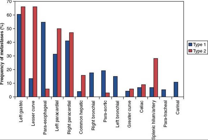
For lower esophageal and gastroesophageal junctional adenocarcinomas, approximately 70% of patients will have nodal metastases at presentation. This is influenced by tumoral depth of penetration, with nearly all T3 and T4 lesions exhibiting metastases in surgical series. Pathologic resection data demonstrated rates of lymphatic involvement for lower esophageal and GE junctional tumors of 45%, 85%, and 100% for T2, T3, and T4 tumors, respectively (Fig. 53.5).33 In patients with lower esophageal cancer, involvement of both mediastinal and abdominal lymph nodes is common (Fig. 53.6).33 The primary direction for lymphatic flow for the lower esophagus is toward the abdomen. According to the classification by Siewert, nodal metastases are often seen in the mediastinum and abdomen for type I tumors, whereas type III tumors metastasize almost exclusively inferiorly, toward the celiac axis. Type II tumors are intermediate, preferentially spreading inferiorly and less frequently into the mediastinum. The primary value in the Siewert classification is to the guidance of appropriate type surgery (i.e., type I tumors are generally treated with esophagectomy and mediastinal lymph node resection, with types II and III approached through the abdomen), although it may be useful in radiation field design as well.3 Generally speaking, the incidence of abdominal nodal involvement increases as one proceeds distally in the esophagus to the gastroesophageal junction. For patients with tumors arising from the gastroesophageal junction, mediastinal involvement is less common. Nodal metastases above the level of the carina are rare in lower esophageal and junctional tumors.39 In addition, histologic analyses of lower esophagus and gastroesophageal junction adenocarcinoma specimens suggest that many patients without nodal involvement on conventional histopathology actually have involvement when assessed by immunohistochemistry.40
Extensive nodal mapping has been performed by Japanese investigators, who have devised the Japanese Gastric Cancer Association Classification (Fig. 53.7).41 Using this system of nodal classification, investigators from Erlangen evaluated 326 patients with esophagogastric junction carcinoma undergoing primary resection. Tumors were stratified as AEG type I (distal esophagus), II (gastric cardia), and III (subcardia). Note there was significant overlap in the majority of tumors for all stages, with an overall incidence of lymph node metastasis of 71%. In T1 patients, only 17% exhibited lymph node metastasis, whereas 78%, 86%, and 90% of T2, T3, and T4, respectively, had involved nodes. Figure 53.8 shows varying patterns of nodal spread based on T stage as well as AEG location. Additional generalizations from this study include the following: (a) lymph vascular invasion was highly predictive of nodal spread and (b) proximal extension of type II and III tumors into the distal esophagus (particularly beyond the Z-line) predicted an increasing incidence of paraesophageal lymph node involvement.42
FIGURE 53.5. The incidence of nodal metastases related to depth of tumor invasion, adenocarcinoma. (From Dresner SM, Lamb PJ, Bennett MK, et al. The pattern of metastatic lymph node dissemination from adenocarcinoma of the esophagogastric junction. Surgery2001;129:103–109; with permission form Elsevier.)
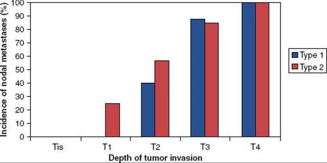
FIGURE 53.6. Distribution of nodal metastases by frequency of site involved for adenocarcinoma. (From Dresner SM, Lamb PJ, Bennett MK, et al. The pattern of metastatic lymph node dissemination from adenocarcinoma of the esophagogastric junction. Surgery 2001;129:103–109; with permission form Elsevier.)
LOCAL FAILURE
Aisner et al.43 and LePrise et al.44 reviewed the patterns of failure in esophageal cancer after radical irradiation, radical surgery, or a combination of both (Table 53.2). These data suggest that high rates of local recurrence occur when either radiation therapy or surgery alone is used. In a series at the University of Pennsylvania and Fox Chase Cancer Center of patients with adenocarcinoma of the esophagus and gastroesophageal junction treated with surgery alone, the local-regional recurrence rate was 77%.45 In contemporary randomized trials, local failure rates with surgery alone range from 32% to 45%.46–49 Similarly, data from recent randomized trials of esophageal cancer using “definitive” chemoradiation also indicates that local failure is a major cause of failure, with approximately 50% of patients failing locally (Table 53.3). In many trials, patterns of failure are reported as first site of recurrence. This fact, along with infrequent posttherapy imaging and inability to detect subclinical recurrences, likely leads to underestimates of local recurrence rates. These data clearly emphasize the need for improvements in local treatment modalities.
CLINICAL PRESENTATION
Symptoms of esophageal cancer often start 3 to 4 months before diagnosis. Location of the primary tumor in the esophagus may influence presenting symptoms. Dysphagia is seen in >90% of patients regardless of location. Odynophagia is present in up to 50% of patients.50 Weight loss is common, with 40% to 70% of patients reporting a loss of >5% of total body weight. This extent of weight loss has been associated with a worse prognosis. Less frequent symptoms may include vague chest pain, hoarseness, cough, and glossopharyngeal neuralgia.51
Advanced lesions can produce signs and symptoms from tumor invasion into local structures. Hematemesis, hemoptysis, melena, dyspnea, and persistent cough secondary to tracheoesophageal or bronchoesophageal fistula may occur. Compression or invasion of the left recurrent laryngeal nerve or the phrenic nerves can cause dysphonia or hemidiaphragm paralysis. Superior vena cava syndrome and Horner’s syndrome can also occur for very advanced lesions. Pleural effusion and exsanguination resulting from aortic communication may also be seen.50 Abdominal and back pain may occur with celiac axis nodal involvement with lower esophageal tumors.
FIGURE 53.7. Locations of lymph node stations. (From Matzinger O, Gerber E, Bernstein Z, et al. EORTC-ROG expert opinion: radiotherapy volume and treatment guidelines for neoadjuvant radiation of adenocarcinomas of the gastroesophageal junction and the stomach. Radiother Oncol 2009;92:164–175; with permission from Elsevier.)


DIAGNOSTIC WORKUP
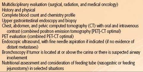
After a thorough history and physical examination, all patients with suspected esophageal cancer should have a workup similar to that outlined in Table 53.4. Attention should be paid to cervical and supraclavicular lymph nodes. Basic blood counts and a metabolic panel with liver function tests should be obtained.
TABLE 53.2 PATTERNS OF FAILURE IN ESOPHAGEAL CANCER

FIGURE 53.8. A: Frequency of nodal involvement for T2 tumors. B: Frequency of nodal involvement for T3-4 tumors. C: Frequency of involvement for AEG type I tumors. D: Frequency of nodal involvement for AEG type II/III tumors. (Based on data from Meier et al.42)

Although the esophagogram may be used to define lesion extent, endoscopy is the best tool to diagnose and define the extent of the lesion. During flexible endoscopy, biopsies and brushings should be taken of the primary site and suspicious areas harboring satellites or submucosal spread. In addition, accurate endoscopic measurement and characterization of tumor and gastroesophageal junction in relation to the incisors facilitates radiation treatment planning. Examination with panendoscopy of the oral cavity, pharynx, larynx, and tracheobronchial tree may also be performed at the time of esophagoscopy in patients with squamous cell carcinomas, given the high incidence of second tumors in the head and neck and upper airway.2 In addition, bronchoscopy should generally be performed in patients with proximal malignancies to evaluate for the presence of tracheal or carinal invasion, particularly for patients with tumors abutting these structures on computed tomography (CT). CT of the thorax and abdomen is critical to identify metastases to the liver, upper abdominal nodes, or adrenals. However, CT may not adequately assess periesophageal lymph node involvement or accurately define the true extent of the primary tumor.52,53 Conventional CT scan can accurately determine resectability in only 65% to 85% of cases. Furthermore, CT accurately predicts T stage in approximately 70% of cases and nodal involvement in only 50% to 70% of cases.54–56
To more accurately assess periesophageal and celiac lymph node involvement and transmural extent of disease, endoscopic ultrasonography (EUS) should be performed. EUS provides accuracy rates of 85% to 90% for tumor invasion (T stage) and 75% to 80% for lymph node metastases when matched to surgical pathology.57–60 However, the accuracy of endoscopic ultrasound following neoadjuvant therapy is significantly less, ranging from 27% to 48% for T staging and 38% to 71% for N staging. This is possibly due to the failure to discriminate tumor from postradiation inflammation and fibrosis.61–63
Surgical staging procedures, including thoracoscopy, mediastinoscopy, and laparoscopy, may provide additional staging information and are considered in selected patients at some institutions.64 Laparoscopy would primarily be used for tumors at the gastroesophageal junction.
Patients with significant obstruction with inability to maintain their weight may require placement of a feeding tube. If surgery is planned, gastric tube placement is generally avoided, given that the stomach will ultimately serve as the “neoesophagus” following resection.
More recently, positron emission tomography (PET) has proven to be a valuable staging tool in esophageal cancer patients. The addition of PET to standard staging studies such as CT can improve the accuracy of detecting stage III and stage IV disease by 23% and 18%, respectively.65,66 Overall, it is estimated that PET will detect distant metastatic disease in approximately 20% of patients who are considered to have local regional disease only by CT. However, PET also appears to have a lower accuracy in detecting local nodal disease compared to CT alone or in combination with endoscopic ultrasound. Of importance, emerging data suggest that PET can be used to predict response to therapy, with “PET responders” experiencing significantly improved outcomes compared to “nonresponders.”67 In addition, PET has been used to predict response to treatment early during the treatment course. This has led to ongoing investigation of early treatment response as measured by PET as a surrogate for therapeutic efficacy and clinical outcomes.68
TABLE 53.3 LOCAL FAILURE RATES FROM RANDOMIZED TRIALS EVALUATING CHEMORADIATION ALONE IN ESOPHAGEAL CANCER
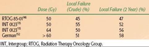
TABLE 53.4 DIAGNOSTIC WORKUP FOR ESOPHAGEAL CANCER
STAGING SYSTEMS
Esophageal staging can be based on pathologic or clinical criteria. Pathologic staging is performed after invasive procedures, including esophagectomy, mediastinotomy, or thoracotomy. Clinical staging is often employed with “definitive” and neoadjuvant chemoradiation approaches and is less accurate. With the combination of CT, PET, and EUS, clinical staging closely correlates with pathologic stage. Note that the most recent AJCC staging system takes into consideration tumor type and grade, as well as involved number of lymph nodes (Table 53.5).
PATHOLOGIC CLASSIFICATION
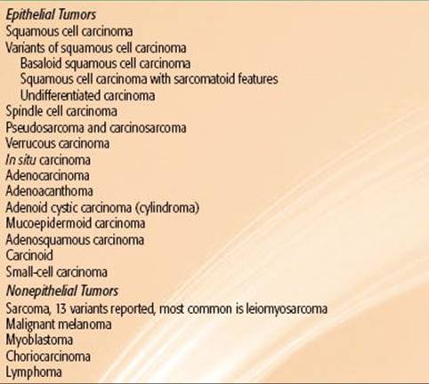
Squamous cell carcinoma and adenocarcinoma comprise 95% of all esophageal tumors, although other rare histologic subtypes are occasionally seen (Table 53.6).69
Given that there has been a rise in adenocarcinoma compared to squamous cell carcinoma, examinations of outcomes by histology have been performed. Some series have reported that patients with squamous cell carcinomas have improved survival,70 whereas others have suggested that patients with adenocarcinoma have an improved survival71 and others report no survival differences. Overall there can be no direct comparison until randomized studies are done with stratification based on histology.
Pseudosarcoma is a variant of a poorly differentiated squamous cell carcinoma with spindle-shaped cells in the stroma resembling fibroblasts. Verrucous carcinoma is a well-differentiated, papillary variant of squamous cell carcinoma.2 Squamous cell carcinoma in situ is rarely seen in the United States and should be distinguished from dysplasia.72–74
Adenocarcinoma may arise from foci of ectopic gastric mucosa or intrinsic esophageal glands. However, as described previously, it is believed the vast majority arise from Barrett’s esophagus. If a focus of squamous cell metaplasia is found in an adenocarcinoma, the tumor may be referred to as an adenoacanthoma.75
Adenoid cystic carcinomas are rare, with an incidence of 0.75%. Patients with this malignancy present around the sixth decade of life and have a poor median survival.76 Mucoepidermoid tumors (adenosquamous carcinomas) are more aggressive and carry a poor prognosis.77 The incidence of small-cell carcinoma is approximately 2%. Patients with these malignancies often present in the sixth to eighth decades of life, and the lesion is usually located in the middle to lower esophagus in males.78,79 These are believed to originate in the argyrophilic cells in the esophagus and may produce paraneoplastic syndromes, such as antidiuretic hormone secretion and hypercalcemia.80 The clinical course of small-cell carcinoma is similar to that of small-cell carcinoma of the lung and may be responsive to chemotherapy and radiation therapy.77,79
Nonepithelial tumors of the esophagus are rare. Among these, leiomyosarcomas are the most common. Twenty-five percent of patients with this tumor present with metastases.81–83 Histologically, these tumors have interlacing bundles of spindle-shaped cells. Less aggressive forms have fewer mitotic figures and less anaplasia. Prognosis has been reported to be more favorable than that of squamous cell carcinoma.77 In patients with Kaposi’s sarcoma, gastrointestinal involvement of the esophagus can be seen.84
Malignant melanoma is rare and can occur as a primary esophageal tumor or as a metastasis. These lesions are usually large and often covered by intact squamous mucosa with focal areas of ulceration. Spread is usually submucosal. Mean survival is approximately 7 months.85,86 Lymphoma comprises approximately 1% of esophageal malignancies. It is usually associated with direct extension from other organs, although primary esophageal lymphoma has been reported.87
TABLE 53.5 AMERICAN JOINT COMMITTEE ON CANCER 2010 ESOPHAGEAL CANCER STAGING SYSTEM


TABLE 53.6 PATHOLOGIC CLASSIFICATION OF MALIGNANT ESOPHAGEAL TUMORS
PROGNOSTIC FACTORS
According to the Union Internationale Contre le Cancer/American Joint Cancer Committee seventh-edition staging system, R-status, age, and histologic subtype were independent prognostic factors of survival, whereas tumor grade and site were not.88 It should be recalled that these data consider patients treated with surgery alone and as such do not apply to patients receiving chemotherapy or radiotherapy. A study published by Nomura et al.89 evaluated 301 patients treated with chemoradiotherapy showed that T stage, M stage, and gender were prognostic factors in multivariate analysis.
In addition to stage, other factors portend outcome. Tumor length/size may also impact outcome. In a study of 582 patients undergoing surgical resection as primary treatment, tumoral length adversely affected survival, with 5-year survival rates of 77%, 48%, 38%, and 23% for tumor lengths of 1, 2, 3, or >3 cm, respectively (p < .001).90 Of note, length was not prognostic if patients were N+ or M+. Similarly, in adenocarcinoma patients, 5-year survival rates have been shown to be significantly higher for patients with tumors ≤2 cm compared to those >2 cm.91
In another large study from the Mayo Clinic, clinicopathologic factors that affected prognosis included T and N status, tumor grade, age >76 years, extracapsular lymph node extension, and the absence of chemotherapy or radiotherapy; anatomic location did not influence survival.92 In addition, weight loss and low overall performance status also indicate poor prognosis.93 Deep ulceration of the tumor, sinus tract formation, and fistula formation are other poor prognostic factors.50
Obtaining uninvolved pathologic margins at resection is of significant importance with regard to long-term outcome. An Intergroup study (discussed later) evaluating chemotherapy preceding and following esophagectomy showed that outcomes were similar in patients undergoing R1 resection (positive microscopic margins) or R2 resection (gross residual disease) or patients not undergoing resection at all. Only patients undergoing R0 resection (uninvolved margins) had a substantial chance of long-term disease-free survival.47 A patterns-of-care survey examined the outcomes of patients with adenocarcinoma and squamous cell carcinoma of the esophagus between 1996 and 1999. Patients were treated with radiation therapy across 59 institutions. On multivariate analysis, significant improvements in survival were seen in patients treated at centers with ≥500 new cancer patients per year compared to centers seeing <500 (hazard ratio 1.32; p = 0.03).94
GENERAL MANAGEMENT
Treatment for esophageal carcinoma is characterized as curative or palliative. According to one historical study,95 only 20% patients present with cancer of the esophagus that is truly localized to the esophagus, indicating that at the time of diagnosis, approximately 80% patients have either locally advanced or distant disease.
Surgery with Curative Intent
Surgery of the thoracic esophagus usually requires a subtotal or total esophagectomy and is usually undertaken for lesions of the middle to lower one-third of the thoracic esophagus and gastroesophageal junction. Patients with stage I to III are often considered for potentially curative resection; however, aortic, tracheal, heart, or great vessel invasion may preclude resection. Esophagectomy may be accomplished by a number of techniques, including a transhiatal esophagectomy, right thoracotomy with laparotomy with intrathoracic anastomosis (Ivor-Lewis esophagogastrectomy), right thoracotomy with laparotomy with cervical anastomosis (McKeown esophagogastrectomy), left thoracotomy, or radical esophagectomy via open or laparoscopic approaches. Each technique has its advantages and disadvantages. In general, advantages of the transthoracic approach include better visualization with access and resection of the upper two-thirds of the esophagus and mediastinal lymph nodes. Alternatively, the transhiatal approach has less morbidity than thoracotomy (including respiratory compromise) with easier access to anastomotic leaks (neck vs. thorax). In any instance, achievement of negative margins at resection has been reported to be a significant prognostic factor and should be the goal of esophageal resection. Exemplifying this, in a study of 500 patients undergoing transthoracic resection, patients undergoing margin-negative resection had a 5-year survival rate of 29% versus no 5-year survivors in patients with involved margins.96
The Ivor-Lewis procedure is the classic approach to expose mid-esophageal lesions. A left thoracotomy procedure exposes lesions of the gastroesophageal junction. Transhiatal esophagectomy is performed without a thoracotomy and is useful in lower esophageal lesions, although direct visualization and dissection of varying mediastinal lymph nodes cannot be achieved. The optimal surgical approach is unknown. A randomized trial comparing transhiatal versus transthoracic approaches in patients with adenocarcinoma showed no significant survival advantage to the latter, although a possible trend was noted (5-year survival, 29% vs. 39%). However, perioperative mortality was also increased with the transthoracic approach.46
Laparotomy can be performed before or concurrently with esophagectomy to rule out any disease below the diaphragm. Multiple reconstruction options are available following definitive surgery; esophagogastrostomy is the most widely used, using the stomach as a conduit to replace the esophagus. Patients with significant obstruction and inability to maintain their weight often require placement of feeding jejunostomy. If possible surgery is planned, gastric tube placement is generally avoided, given that the stomach will ultimately serve as the “neoesophagus” following resection. Colon interposition, preferably with the left colon, can also be used; however, this approach is generally reserved for patients who have previously undergone gastric surgery or other procedures that have devascularized the stomach.
Squamous cell carcinoma of the cervical esophagus presents a difficult management situation. Proximal esophageal tumors <5 cm from the cricopharyngeus are generally treated with definitive chemoradiotherapy. If surgery is performed, resection of portions of the pharynx, the entire larynx, thyroid gland, and the proximal esophagus is often required. Radical neck dissections are also carried out.50 Because of the significant morbidity and loss of organ function with surgery, chemoradiation alone has been frequently employed. The survival probability with definitive chemoradiotherapy is similar, without the major functional impairments, morbidity, and mortality associated with surgery.97
Curative Combination Therapy
In the treatment of patients with esophageal cancer, an approach of radiation therapy with concurrent chemotherapy, with or without surgery, is frequently adopted. Multiagent chemotherapy with cisplatin and 5-fluorouracil (5-FU) is used most frequently, although combinations of other agents (e.g., paclitaxel and carboplatin) are being increasingly used. Additional taxanes, topoisomerase inhibitors, and anti-epidermal growth factor receptor inhibitors with radiation therapy are under investigation.
Palliative Treatment
Palliative treatment is frequently used for the relief of symptoms of esophageal carcinoma, especially dysphagia.98 Surgical palliation involves resection and reconstruction, if possible, removing the bulk of the disease, potentially preventing abscess and fistula formation, as well as bleeding. Substernal bypass with the colon or entire stomach has also been carried out.98 However, given the poor prognosis in patients with advanced disease and morbidity associated with resection, a surgical approach is not commonly adopted and should be avoided in patients who can be managed with nonsurgical modalities.
Endoscopic dilatation is a reasonable alternative. When the lumen of the esophagus is dilated to 15 mm, dysphagia is often no longer experienced. Repeat dilatation is often required.50 Esophageal stenting with either conventional plastic stents or metallic self-expanding stents can also be used to maintain patency.99
Palliative irradiation is frequently used to control the primary disease, as well as distant metastases. Resolution of symptoms, especially pain and dysphagia, can be accomplished in up to 80% of patients. Palliative treatment regimens range from 30 Gy over 2 weeks51 to 50 Gy over 5 weeks. Laser ablation with or without intraluminal brachytherapy can be used. The addition of 3 fractions of 7 Gy each can improve the stenosis-free interval and prevent obstruction.99
RADIATION THERAPY TECHNIQUES
Simulation
When patients are simulated, the radiation oncologist should know the extent of disease based on imaging (barium swallow, CT, PET), as well as on endoscopy. CT simulation is appropriate for treatment planning. During simulation, the patient is positioned, straightened, and immobilized on the simulation table. An immobilization device is used to minimize variation in daily setup. Arms are generally placed overhead and knee support underneath the legs. Palpable neck disease should be marked with a radiopaque wire. The administration of oral contrast to delineate the esophagus is generally used and helps to define the extent of mucosal irregularity. For GE junctional tumors (particularly with significant gastric involvement), it may be advisable to have the patient come in with an empty stomach. For cervical and upper thoracic lesions, an immobilization mask may assist in creating a reproducible position. Some authors have also advocated that mid-esophageal primaries be simulated in prone position to maximize distance between the target volumes and spinal cord. The patient is placed on the CT simulator in the treatment position, and a scan of the entire area of interest with margin is obtained. At minimum, 3- to 5-mm slices should be used, allowing accurate tumor characterization, as well as improved quality of digitally reconstructed radiographs. If patients lose >10% of their body weight during therapy, consideration should be given to repeat CT planning. Arterial phase intravenous contrast is generally used to delineate mediastinal and abdominal vascular nodal basins, including the celiac axis and to allow the radiation oncologist to discern normal vasculature from other adjacent normal structures, and potential adenopathy. The tumor and vital structures are then outlined on each slice on the treatment planning system, enabling a three-dimensional treatment plan to be generated. The use of respiratory gating or breath-hold techniques may help to reduce target motion with respiration and, therefore, avoid normal-tissue irradiation associated with larger margins used in free-breathing approaches, particularly for lower esophageal cancers. Additional techniques to minimize physiologic motion include abdominal compression devices. Four-dimensional CT scan may be appropriate to assess tumoral motion, facilitating appropriate margin placement on the target volumes, particularly in lower esophageal tumors and/or disease involving the stomach.
Treatment Planning
Target Design
In the design of radiation fields for esophageal cancer, it is important to define varying target volumes, including gross disease as well as potential areas of subclinical involvement (i.e., the gross tumor volumes [GTVs] and clinical target volumes [CTVs], respectively). Definition of gross tumor volume is based on multiple studies, including endoscopic descriptions (from both esophagogastroduodenoscopy [EGD] and EUS). The proximal and distal aspects of the tumor should be standardly defined by the gastroenterologist based on distance from the incisors, as well as relationship to varying landmarks as measured from the incisors (e.g., the GE junction). The radiation oncologist is able to use these measurements to help correlate with disease extent visualized on planning CT scan, using varying anatomic landmarks (e.g., the GE junction, which is frequently located ∼40 cm from the incisors in many patients), as well as carina (which is frequently located at ∼25 cm in most patients) to help more accurately define the GTV. Esophageal wall thickening correlating to the gross tumor volume can frequently be visualized on diagnostic and radiation planning CT. Similarly, EUS appears to be the most reliable test in detecting lymphadenopathy related to nodal spread. As in EGD, the endoscopist should be encouraged to accurately define not only the primary disease extent on EUS as measured by distances from the incisors, but also depth of penetration and potential involvement of adjacent structures, which can also be used to help guide GTV delineation. Similarly, EUS may detect lymph nodes that may not be appreciated on CT or PET imaging, and the endoscopist should describe the size as well as the location (e.g., distance from incisors, relationship to adjacent mediastinal structures, etc.) of these, further facilitating accurate definition of potentially involved lymph nodes, either adjacent to or well removed from the primary tumor itself. In addition, radiographic areas of lymphadenopathy should similarly be included in the GTV.
Although the utility of PET in esophageal cancer staging primarily lies in its ability to detect distant metastases not fully appreciated on CT imaging (and thereby alter treatment approach of these patients),100,101 diagnostic PET/CT has more recently been integrated into radiation planning of esophageal cancer patients and definition of GTV. Generally, gross disease as defined on PET is characterized by a standard uptake value of >2 to 2.5.102Generally speaking, PET-avid nodes should be included within the GTV volume. In a study from Fox Chase Cancer Center, the mean GTV length as determined by PET/CT closely correlated with endoscopy findings.59 Another study from Australian investigators reported that CT-alone–based definition of GTV excluded PET-avid disease in a majority of patients, resulting in a potential geographic miss of disease in a significant minority of patients.103Similarly, the fusion of CT-PET has been shown to prompt GTV and planning target volume (PTV) modification in a majority of patients.104 Recent analysis of PET/CT-based (as compared to CT alone) radiotherapy planning of esophageal cancer patients indicated a reduction of intraobserver and interobserver variability in GTV delineation.105
Finally, although the routine use of fluoroscopic barium swallow in esophageal cancer has decreased, this study may still be useful to the radiation oncologist for field design. A study by Chinese investigators suggested that, as compared to CT, endoscopy with barium swallow more accurately defines the true length of middle and lower esophageal tumors, although CT appeared to better determine this for tumors located at the GE junction.106
In summary, accurate definition of primary and nodal gross disease is paramount in radiation esophageal cancer planning. It is important to rely on all diagnostic studies, including barium swallow (when available), EGD, EUS, and CT, as well as PET scan.
The identification of potential direct and nodal pathways for spread of subclinical disease (i.e., CTV definition) in esophageal cancer is also of paramount importance. These areas vary significantly, depending on site of origin of disease, making esophageal cancer planning somewhat complex. In terms of direct disease extension along the esophagus itself, varying reports have assessed the histologic findings at primary esophagectomy to determine subclinical extent of disease. One prospective analysis of 66 resection specimens showed that placement of a 3-cm margin proximally and distally on the primary tumor would cover microscopic disease extension in 94% of squamous cell carcinomas. Similarly, in GE junctional carcinomas, a 3-cm proximal margin included subclinical disease extension in 100% of patients, and 5 cm distally covered 94% of subclinical spread.106
As described previously, the esophagus is characterized by a rich submucosal network of lymphatics that facilitates early lymph node spread of disease, even for superficial esophageal cancers. The appropriate cranial and caudal margins remain a matter of debate in the treatment of esophageal cancer. Historically, very large fields were treated, encompassing potential pathways and lymphatic spread from the thoracic inlet down to the celiac axis region; however, such fields are potentially fraught with treatment-related toxicity and may be difficult for patients to tolerate, particularly in the context of concurrent chemotherapy delivery. Most contemporary radiation trials used margins of 3 to 5 cm cranially and caudally on the GTV, along with an approximate 2-cm radial margin. With disease located at or above the carina (or middle/upper one-third of the esophagus in some instances), many of these trials recommended fields inclusive of the supraclavicular lymph node basins, whereas celiac axis nodal basin coverage was recommended for disease of the distal esophagus.107 Further description of lymph node basin coverage follows.
Field Design
Historically, the treatment of very proximal (cervical) esophageal cancers has been challenging. Because of the changing contour from the neck to the thoracic inlet, treatment of lesions in the upper one-third of the esophagus may present a difficult technical problem. Lesions in the upper cervical or postcricoid esophagus are treated from the laryngopharynx to the carina, depending on extent of disease. Supraclavicular and superior mediastinal nodes are irradiated electively. Using older/conventional methods, this was achieved with lateral parallel opposed or oblique portals to the primary tumor and a single anterior field for the supraclavicular and superior mediastinal nodes.93Another historical technique treated lesions in this region by means of a four-field box approach, using a wax bolus to build up the lack of tissue above the shoulders, acting as a compensator. A high-energy beam (>15 MV) is used, and both sides of the neck are treated prophylactically. Other methods of treating lesions at the thoracic inlet include 140-degree arc rotations, anterior wedged pairs, and three- or four-field techniques using posterior oblique portals combined with a single anterior portal or anteroposterior–posteroanterior (AP/PA) fields.93 Using three-dimensional (3D) approaches, varying techniques have been implemented, including treatment of the primary tumor and lymph nodes using an AP/PA approach to 39.6 to 41.4 Gy at 1.8 Gy per fraction, followed by a left or right opposed oblique pair to bring the total dose to 50.4 Gy, thereby limiting the spinal cord dose. This technique will generally exclude the supraclavicular fossa, and a separate electron field is often added, treating to a depth of 2 to 3 cm, depending upon individual anatomy. More recently, however, intensity-modulated radiation therapy (IMRT)–based planning has facilitated the treatment of upper esophageal lesions and is our preferred method for treating these tumors (Fig. 53.9) Strict normal tissue constraints, including normal lung and spinal cord, are important considerations in using these techniques (discussed later).
Based on the previously described and other pathologic patterns of spread data in squamous cell carcinoma of the esophagus, general guidelines in terms of field design can be made as follows: For cervical and upper thoracic squamous cell carcinoma, the CTV should generally include nodal basins extending from the lower cervical and supraclavicular region superiorly to the subcarinal lymph node basin inferiorly, inclusive of the upper paraesophageal lymph nodes (Fig. 53.9). For lower esophageal squamous cell carcinomas, lymph node basins from the subcarinal region superiorly to the left gastric and common hepatic artery/celiac lymph nodal basins inferiorly should generally be included (described further later in this chapter). For tumors of the middle esophagus, we recommend individual field design according to clinical scenarios, with a more complete coverage of paraesophageal mediastinal lymph nodes, particularly in patients with a good performance status (Fig. 53.10).37 These are general guidelines, and all plans should be individualized based on available imaging and endoscopic findings.
In field design, potential nodal involvement (and therefore target volumes) on nodal size is problematic, given that some reports demonstrated that <15% of metastatic nodes are >1 cm and that average size differences between involved and uninvolved nodes are frequently not significantly different.108 In addition, fluorodeoxyglucose-PET scanning has an estimated sensitivity of only 67% of detecting nodal metastases.8 Even endoscopic ultrasound, generally considered the most sensitive test for detecting lymph node metastases, is only able to detect such disease in approximately 75% of patients.109 Therefore, it is not appropriate to rely exclusively on varying imaging modalities to define areas of subclinical spread for esophageal cancer, realizing that patterns-of-spread data are important in determining radiation field design.
The design of radiation fields for the treatment of adenocarcinoma of the esophagus is similar to that of lower thoracic squamous cell carcinomas but deserves special mention. Periesophageal lymph nodes are generally included in all patients. Given that lymph node involvement is clearly associated with depth of tumor penetration (T stage) and the fact that most patients in the United States and Europe presenting with GE junctional carcinoma will have more advanced disease, inclusion of celiac lymph basins for adenocarcinoma of the distal esophagus/GE junction is usually indicated. Based on the previously described patterns of spread data from Erlangen and others, specific considerations include the following: Lymph vascular invasion is highly predictive of nodal spread. Proximal extension of tumors (particularly beyond the Z-line into the distal esophagus for type II and III tumors) predicts an increasing incidence of paraesophageal lymph node involvement. Based on an estimated nodal incidence cutoff of 20% for inclusion, specific considerations include the following: (a) The lower paraesophageal, paracardial, lesser curvature, and left gastric artery nodes should be included in the CTV. (b) The presence of lymph vascular invasion predicts a nodal positivity rate of >20% in the left and right gastroepiploic, greater curvature, celiac trunk, and splenic hilar regions. (c) In T3/4 disease, the gastroepiploic, greater curvature, celiac trunk, splenic hilar, splenic artery, and common hepatic artery should be included. (d) High-grade tumors should also include the left gastroepiploic, greater curvature, and celiac trunk nodes in CT design. (e) Larger and more deeply penetrating tumors should also include the splenic artery and splenic hilar nodes, as well as those along the greater curvature. (f ) Tumors extending above the diaphragm and those extending >1.5 cm beyond the Z-line should include the mid paraesophageal nodes, treating up to the carina. Of note, significant involvement of the distal esophagus by GE junctional tumors (>1.5 cm beyond the Z-line) should lead to inclusion of not only lower, but also middle paraesophageal nodes based on patterns of spread. However, treating these more extensive fields must be weighed against potential side effects of increased normal-tissue irradiation. In summary, middle and lower paraesophageal nodes should be included in patients with T2-T4 type I and T2-T4 type II tumors extending >1.5 cm above the Z-line and T3-T4 type II patients. The splenic hilar and artery nodes are considered “spareable” in T2 tumors, notably type I.42
FIGURE 53.9. A–C: Examples of planning target volume (PTV) in a 62-year-old woman with a cT3 N1 squamous cell carcinoma of cervical esophagus. Gross tumor volume (GTV), blue volume; PTV, red volume. D: Three-dimensional reconstruction of PTV from above the patient. Carina, yellow; GTV, dark blue; lungs, light blue; PTV, red; spinal cord, brown. E: Isodose curves of this patient treated with a nine-field intensity-modulated radiation therapy plan. Note that beams are primarily anteriorly oriented. (Courtesy of Rodney Hood, CMD.)
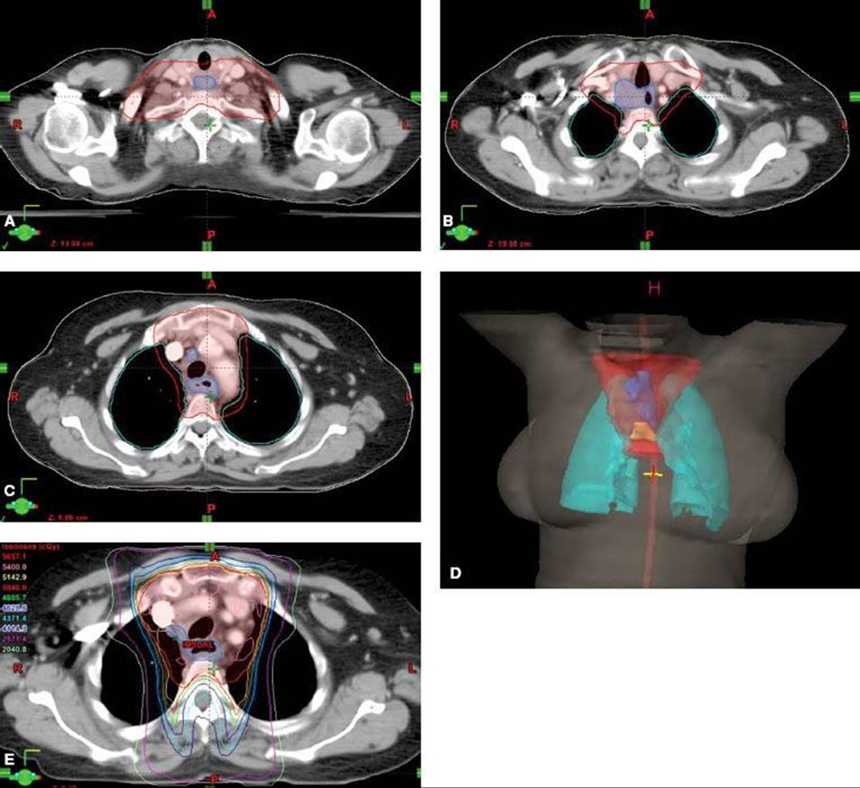
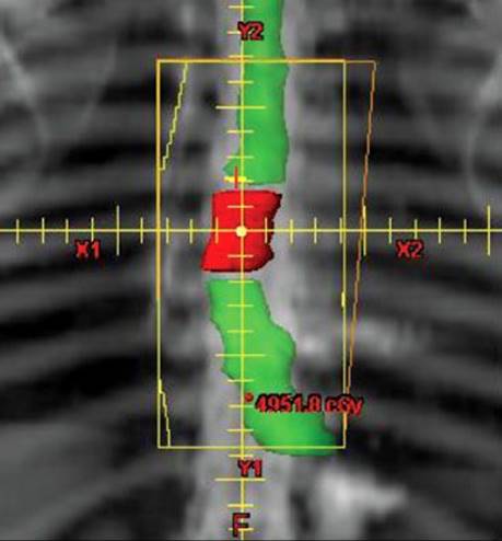
FIGURE 53.10. Example field for a midesophageal squamous cell carcinoma. Note that the field is inclusive of adjacent mediastinal/paraesophageal nodes, with approximately 5 cm margin superiorly on the gross tumor volume (GTV), as well as slightly larger margin on the inferior aspect of the GTV based on concern of previously unappreciated spread at time of endoscopy.
Although, historically, two-dimensional–based radiation planning has been carried out primarily using anatomic landmarks such as bone and carina, as well as fluoroscopic barium swallow, to determine field borders, contemporary treatment planning using CT-based planning allows improved visualization of both target and nontarget structures, along with three-dimensional reconstruction and creation of a “beam’s-eye” view of varying fields, allowing improved conformality around target structures and improvements in normal-tissue sparing. Because volumetric data can be obtained by CT scans, dose–volume histogram data can also be generated. A variety of three-dimensional techniques are being used and are described later.
Potential beam orientations for the treatment of thoracic esophageal and gastroesophageal junction tumors (again defined as tumors involving the gastroesophageal junction with an epicenter within 5 cm proximal or distal to the GE junction) include an AP/PA-alone approach, an initial AP/PA approach followed by AP/right posterior oblique (RPO)/left posterior oblique (LPO) fields with or without boost, an initial APPA approach followed by RAO/LPO fields with or without boost, and a three-field technique (AP/PA with left lateral or oblique field). One of our preferred approaches in lesions of the thoracic esophagus or GE junction is to use an initial AP/PA/RAO/LPO fields, with boost fields using laterally oriented beams. The inferior margin of the initial fields includes the gastroesophageal junction and, for lower or middle one-third lesions, the celiac axis nodal basins (generally located at the level of T12 and identifiable on CT), as well as gastrohepatic ligament. Initial fields are treated to a dose of 45 Gy, taking care to avoid as much of the heart as reasonably possible while continuing to minimize the kidney volume in the radiation field, inclusive of the above nodal basins. Reduced fields encompassing gross disease with an approximate 2-cm margin through oblique or lateral fields may then be used for an additional 5.4 Gy. Doses usually do not exceed 50 Gy (discussed later).
A margin of 5 cm above and below the GTV is generally recommended to cover subclinical submucosal/nodal disease, as well as an approximate 2.0- to 2.5-cm radial margin, although individual margins are case dependent. Because it is imperative to account for daily setup uncertainty as well as physiologic internal organ motion (secondary to respiration, peristalsis, cardiac motion, etc.), additional margin must be added to a clinical target volume, particularly to the more mobile distal esophagus. More recently, the internal target volume (ITV) has been used to account for physiologic motion of the target volume, which is included in the PTV. Varying reports analyzing esophageal motion have shown average anterior and posterior motion ranges from 0.1 to 4 mm, lateral motion from 0.3 to 4.2 mm, and superior-to-inferior motion from 3.7 to 10 mm.107 An analysis evaluating interfraction esophageal motion in the right–left and AP direction showed average right–left motion of 1.8 ± 5.1 mm (favoring leftward movement) and average AP motion of 0.6 ± 4.8 mm (favoring posterior movement), with an average absolute motion of 4.2 mm or less in the right–left and AP directions. The authors concluded that 12-mm left, 10-mm posterior, and 9-mm anterior margins are appropriate.110 Some authors have recommended defining 1-cm radial, 1.5-cm distal, and 1-cm proximal margins from CTV to ITV if target motion data are not available. Image-guided radiation therapy, including cone beam CT, may also be useful in localizing tumor and establishing physiologic variability between daily treatments.
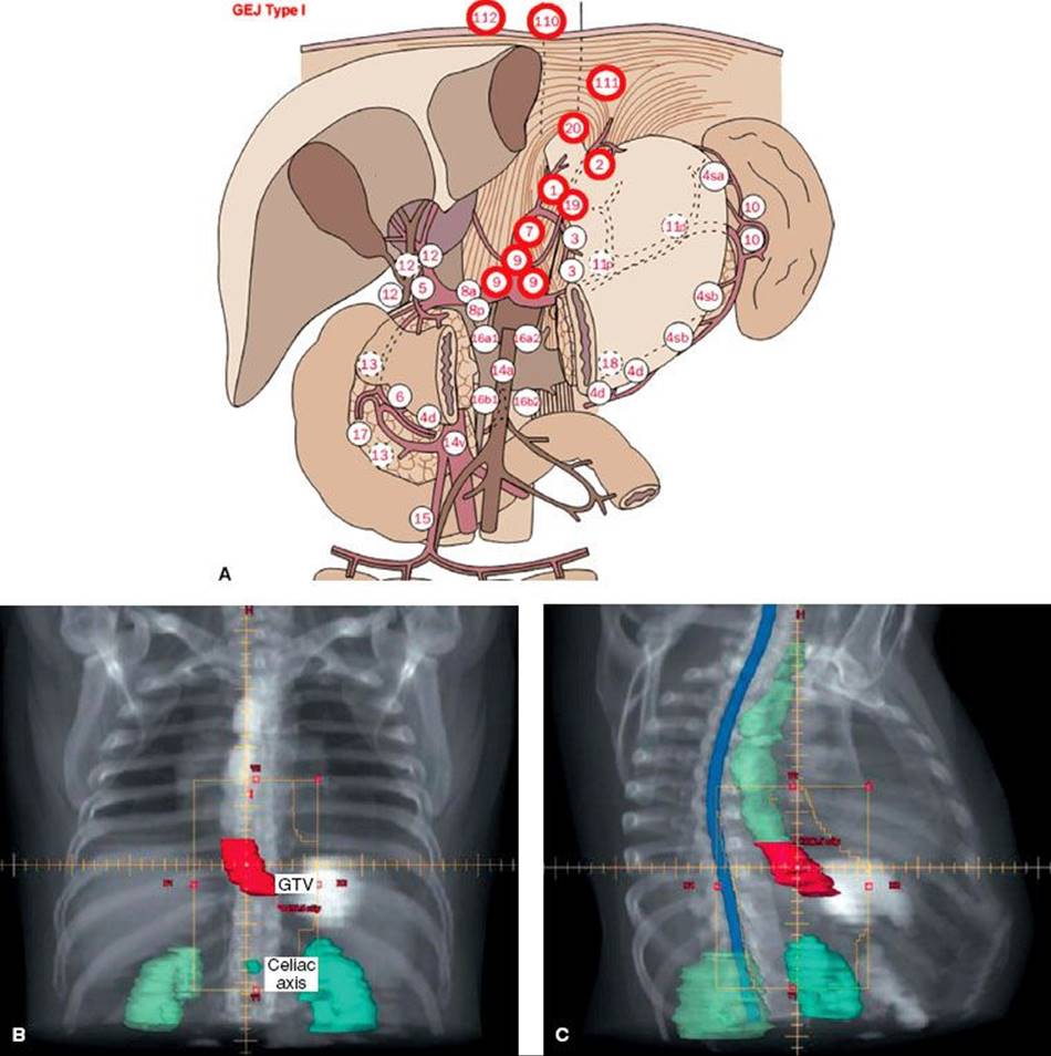
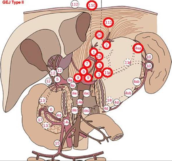
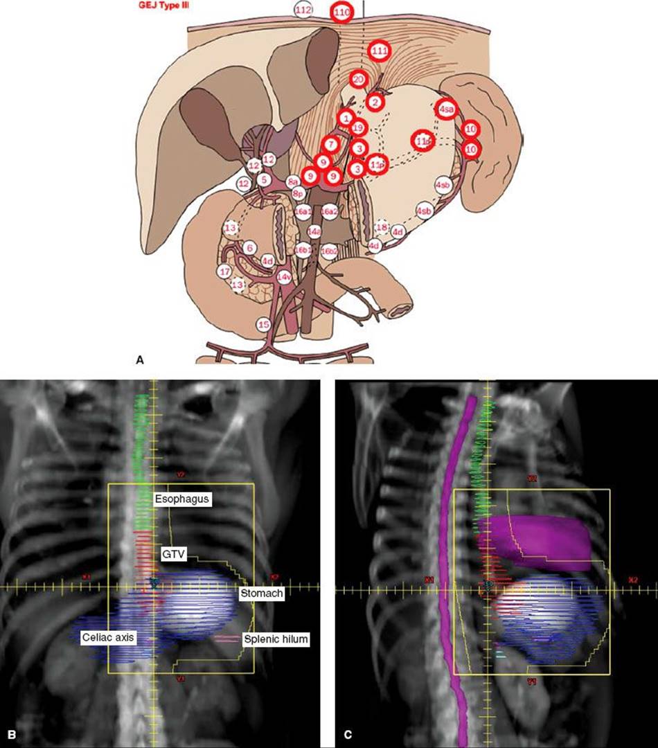
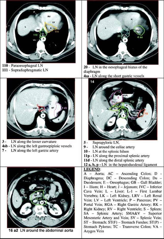
Figure 53.11 shows general recommendations for elective target nodal station coverage in a patient with a type I GE junctional tumor with an accompanying example field. Figure 53.12 shows general recommendations for elective target nodal station coverage in a patient with a type II GE junctional tumor. Nodal basins are similar to type I tumors with the exception of inclusion of nodal basins along the splenic artery course. Figure 53.13 shows general recommendations for elective target nodal station coverage in a patient with a type III GE junctional tumor with accompanying example fields. Note that in type III tumors there is less emphasis on more proximal (lower mediastinal) nodes and more comprehensive coverage of the splenic artery course, including splenic hilum. Figure 53.14 shows examples of varying nodal locations on axial CT images.
Another potential approach in the treatment of thoracic esophageal cancer is the use of IMRT. IMRT also uses CT-based planning, again allowing 3D reconstruction of varying structures. However, IMRT differs from 3D planning through the delivery of radiation dose by partitioning a radiation field into multiple smaller fields of varying shapes and sizes, varying the dose intensity between each area. This is carried out with either dynamic IMRT (in which collimating leaves move in and out of the radiation beam path during treatment) or “step-and-shoot” IMRT (in which the leaves change the radiation field shape while the beam is turned off). Either method is particularly effective at conforming radiation dose to the target structures while avoiding dose to normal tissue. Radiation oncologists must determine which structures are most critical and weight their importance during the treatment planning process. Of importance, the greater the number of “avoidance” normal structures, the more difficult it is to meet all dose constraints. IMRT use “inverse planning,” in which an intended prescription dose is placed on target volumes and dose constraints are placed on normal-tissue structures. Thereafter, computer software algorithms allow design of unconventional treatment fields that would not otherwise be possible with standard planning methods. Radiation oncologists and medical physicists critically evaluate numerous plans until dose constraints are satisfactorily met. The result should be a series of radiation doses that closely conform to the target volumes while minimizing dose to normal tissues. Dosimetric comparisons of IMRT versus 3D conformal therapy in cervical esophageal cancer have demonstrated superior target volume coverage and conformality with decreased normal-tissue dose.111 A potential disadvantage of IMRT is the possibility of delivering low doses of radiation therapy to normal tissue areas that might not normally be irradiated using 2D or 3D techniques. The influence of this on toxicity (e.g., low-dose pulmonary irradiation and development of postoperative pulmonary complications) remains uncertain. Another potential disadvantage to IMRT is possible dose inhomogeneity, leading to potential hot spots in normal organs. Because IMRT requires precise target definition, the potential for marginal miss increases, and careful target delineation is of paramount importance. With setup uncertainty, physiologic organ motion, and so on, care must be taken to ensure accurate and reproducible setup, including the use of immobilization devices, possible respiratory gating/breath-hold techniques, and so on. Generally, high-energy photons (4 to 18 MV) are recommended when using 3D conformal or IMRT therapy, potentially facilitating a reduction the integral lung dose.
FIGURE 53.11. A: Recommended nodal basins to be included for type I gastroesophageal (GE) junctional tumors based on Japanese Gastric Cancer Association lymph node stations. (From Matzinger O, Gerber E, Bernstein Z, et al. EORTC-ROG expert opinion: radiotherapy volume and treatment guidelines for neoadjuvant radiation of adenocarcinomas of the gastroesophageal junction and the stomach. Radiother Oncol 2009;92:164–175; with permission from Elsevier.) B: Example field for a type I GE junctional adenocarcinoma. C: Example oblique field for a type I GE junctional tumor, inclusive of the above nodal basins.
Dose Constraints
In radiation therapy planning of esophageal cancer, normal-tissue tolerance should always be considered. The spinal cord dose is generally limited to 45 Gy using 1.8-Gy fractions (and potentially less when delivered with novel systemic agents). Efforts to minimize radiation to the heart (in particular the left ventricle in lower esophageal/gastroesophageal junction lesions) should be made. Adopting an off-heart approach using oblique orientations (including right anterior and left posterior, described earlier) is one potential way of achieving this, although multiple approaches exist, including IMRT-based techniques. Similarly, efforts to minimize dose to normal pulmonary tissues should be made, based on data suggesting that the volume of irradiated lung may correspond to postoperative complications and worsened pulmonary function (described later). Frequently, the volume of irradiated lung can be minimized using a simple AP/PA approach. However, this sometimes results in significant cardiac dose, particularly in lower esophagus and gastroesophageal junction tumors. Therefore, oblique orientations are sometimes used, resulting in increased volumes of normal lung being irradiated. When giving concurrent chemotherapy, we generally limit these fields to 13 to 15 Gy.
Accurate delineation of adjacent organs, including lungs, liver, kidneys, heart, and spinal cord, is important. Varying dose–volume normal-organ constraints have been suggested, including achieving a lung V20 of <20%, limiting >2,300 cc of normal lung tissue to <5 Gy, and using mean lung dose of <18▒Gy. Similarly, a V10 of ≤60% has been proposed to reduce the incidence of postoperative pulmonary complications. Historically, heart dose constraints have included maintaining one-third, two-thirds, and total heart volumes of <45, 40, and 30 Gy, respectively. Recommended heart constraints include keeping <30% of the cardiac volume to a total dose of 40 Gy and <50% receiving 25 Gy, minimizing dose to the left ventricle. In the setting of potentially significant volumes of heart in the radiation field, consideration of 4D CT and/or breath-hold techniques can be made. For lower esophageal and gastroesophageal cancers, it is recommended that at least 70% of one physiologically functioning kidney receive a total dose of <20 Gy, and that, collectively, no more than 50% of the combined functional renal volume should receive >20 Gy.41 One should also consider the possibility of impaired kidney function in the context of varying comorbidities, using nuclear medicine renal studies to assess individual renal function in such situations or where significant volumes of kidneys are anticipated to be within the radiation field. Generally, 70% of the liver parenchyma should be kept to a dose of <30 Gy. Many of these constraints can be achieved through the use of three-dimensional planning with appropriate and careful design of shielding blocks/multileaf collimation and dose-volume histogram analysis, with the use of IMRT in select cases.
FIGURE 53.12. Recommended nodal basins to be included for type II gastroesophageal junctional tumors based on Japanese Gastric Cancer Association lymph node stations. (From Matzinger O, Gerber E, Bernstein Z, et al. EORTC-ROG expert opinion: radiotherapy volume and treatment guidelines for neoadjuvant radiation of adenocarcinomas of the gastroesophageal junction and the stomach. Radiother Oncol 2009;92:164–175; with permission from Elsevier.)
Doses of Radiation
Because local-regional failure is common after conventional chemoradiation, investigators have evaluated dose escalation techniques. Varying series using doses beyond 50 Gy have shown local control rates ranging from approximately 77% to 88%.112,113 However, Minsky et al.114,115 reported the results of a randomized trial in which 236 patients with clinical stage T1-4, N0/1, M0 squamous cell or adenocarcinoma of the esophagus were selected for nonsurgical therapy. Patients were randomized to receive 64.8 versus 50.4 Gy, both with concurrent 5-fluorouracil and cisplatin chemotherapy. Patients with cervical, mid, or distal esophageal cancer were eligible, with the exception of those with tumors within 2 cm of the gastroesophageal junction, with approximately 85% of patients with squamous cell histology. This study was closed after interim analysis showed no probability of superiority in the high-dose arm. No significant difference in median survival (13 vs. 18.1 months), 2-year survival (31% vs. 40%), or local-regional failure/persistence of disease (56% vs. 52%) was seen between the high-dose and standard-dose arms. Eleven treatment-related deaths occurred in the high-dose arm compared with 2 in the standard-dose arm, with 7 of the 11 high-dose arm deaths occurring in patients who received 50.4 Gy or less. The authors performed a separate survival analysis including only patients receiving the assigned radiation dose. Despite this, no survival advantage was noted in the high-dose arm. These authors concluded that higher radiation doses did not increase survival or local/regional control, and that the standard radiation dose for patients treated with concurrent 5-FU and cisplatin chemotherapy is 50.4 Gy. Based on these data, standard dose of radiation therapy for esophageal cancer is usually 50 to 50.4 Gy at 1.8 to 2 Gy per fraction, including delivery of similar doses in the both the definitive, adjuvant (45 to 50 Gy) or neoadjuvant (45 to 50 Gy) settings.
Brachytherapy
In addition to external-beam radiation therapy (EBRT), intracavitary therapy can be used with curative or palliative intent. Brachytherapy has also been used as a dose escalation tool in addition to EBRT. The advantage of brachytherapy centers on exploitation of the inverse-square law and quick dose fall-off, thus sparing surrounding tissues from radiation while providing focal dose escalation. The radioactive source of choice is usually 192Ir. High–dose-rate (HDR) techniques can deliver 100 to 400 Gy/hour, allowing treatment to be given in 5 to 10 minutes.
FIGURE 53.13. A: Recommended nodal basins to be included for type III gastroesophageal (GE) junctional tumors based on Japanese Gastric Cancer Association lymph node stations. (From Matzinger O, Gerber E, Bernstein Z, et al. EORTC-ROG expert opinion: radiotherapy volume and treatment guidelines for neoadjuvant radiation of adenocarcinomas of the gastroesophageal junction and the stomach. Radiother Oncol 2009;92:164–175; with permission from Elsevier.) B: Example field for a type III gastroesophageal junctional adenocarcinoma. C:Example oblique field for a type III GE junctional tumor, inclusive of the above nodal basins.
With brachytherapy, an afterloading catheter is introduced through the nose into the esophagus to the primary tumor site under fluoroscopic guidance. This is often performed with the patient on the simulation table. Contrast may be used to define the tumor site. CT scan can also be used to discern tumor location. After localization films are taken and dosimetry generated, the catheter is then attached to a remote afterloader through a guide cable and the 192Ir source inserted through remote control. Doses of 5 to 20 Gy are usually delivered to a depth of 1 cm from the center of the catheter. Dose can be shaped and modified through the use of dwell times.
FIGURE 53.14. Examples of lymph node stations on corresponding computed tomography slices. (From Matzinger O, Gerber E, Bernstein Z, et al. EORTC-ROG expert opinion: radiotherapy volume and treatment guidelines for neoadjuvant radiation of adenocarcinomas of the gastroesophageal junction and the stomach. Radiother Oncol 2009;92:164–175; with permission from Elsevier.)
RESULTS OF THERAPY
The best survival results have been reported in patients who have esophageal tumors that are truly localized. Survival rates range from 25% to >35% at 5 years, and these results have been attained using an array of treatment approaches. Problems arise in comparisons of various modalities, however, because of patient selection factors. A review of the Princess Margaret Hospital data116 supported the concept that extent of tumor, rather than therapy, is the most important factor influencing survival. They found a significant correlation between T stage and response to treatment: T1 lesions showed a 100% response rate, whereas T2 and T3 lesions had response rates of 68% and 58%, respectively. Not unexpectedly, they also found differences in survival according to T stage, M stage, and overall stage. Almost 20% of patients with stage I disease were alive at 3.5 years, whereas only 11% of stage II patients were alive after the same interval, and all patients with stage III died of disease by approximately 1.5 years following therapy.
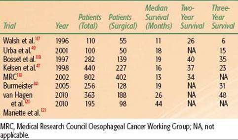
TABLE 53.7 COMPARISON OF SURGICAL ARMS IN RANDOMIZED STUDIES
Surgery Alone
Surgery remains a benchmark to which other modalities are compared. Surgery removes the tumor, a length of normal esophagus, and lymph nodes. Although multiple techniques exist for the resection of esophageal cancer, no one surgical approach has clearly been shown to be superior with regard to complications or outcomes. Proponents of more extended resection (including transthoracic approaches) have advocated that such procedures result in a superior nodal clearance and therefore offer a more complete “oncologic” resection. A trial from the Netherlands randomized 220 patients with esophageal adenocarcinoma to transhiatal esophagectomy alone or transthoracic esophagectomy with extended lymph node dissection. Patients undergoing transhiatal resection experienced significantly fewer pulmonary complications and chylous leaks, as well as significantly reduced ventilator dependence and intensive care unit and hospital stays. At a median 4.7-year follow-up, no significant difference in local-regional recurrence was seen between the two groups (32% transhiatal vs. 31% transthoracic). Furthermore, no significant differences were seen in median disease-free survival (1.4 vs. 1.7 years; p = .15) or median overall survival (1.8 vs. 2.0 years; p = .38). However, there did appear to be a nonsignificant trend favoring the transthoracic approach in improved disease-free survival (5-year, 27% vs. 39%) and overall survival (5-year, 29% vs. 39%) The authors concluded that a transhiatal approach was associated with less morbidity relative to transthoracic surgery, with no apparent survival advantage with either technique, although a trend toward improved survival with longer follow-up was seen.46 In summary, none of the surgical approaches to localized esophageal cancer has clearly been shown to be superior with regard to complications or outcomes, and no one standard surgical approach exists for esophageal cancer resection.
After resection alone, however, local-regional relapse is a common mode of failure. Contemporary randomized trials with surgery alone arms have reported local regional failure rates of 32% to 45%.46–49 It should be remembered that patterns of failure reports often describe first site of failure only and may include only patients undergoing R0 resection, potentially underreporting the true incidence of local-regional recurrence. These and other data suggest that even with modern surgical techniques, local-regional persistence of disease after resection remains a major problem. Prospective, randomized trials using surgery alone in the treatment of esophageal cancer have reported 3-year survival rates ranging from 6% to 48%, with more favorable rates likely reflecting inclusion of patients with earlier-stage disease117–121 (Table 53.7). Given these high rates of relapse and poor long-term survival, the integration of adjuvant or neoadjuvant chemoradiation approaches into the treatment of esophageal cancer is rational and indicated.
Radiation Therapy Alone
There are no randomized studies comparing surgery alone with radiation alone, and radiation therapy alone has been usually delivered when lesions are deemed inoperable because of tumor extent, medical contraindications, and/or palliative treatment is indicated. In general, patients receiving radiation as a sole treatment modality have a median survival of 6 to 12 months and 5-year survival of <10%.
A large review analyzing 49 series involving >8,400 patients treated primarily with radiation therapy alone found overall survival rates at 1, 2, and 5 years to be 18%, 8%, and 6%, respectively.122 Hancock and Glatstein97 reviewed 9,511 patients and found only 5.8% were alive at 5 years. Okawa et al.123 reported 5-year survival rates by stage. For patients with stage I disease, the 5-year survival rate was 20%; stage II, 10%; stage III, 3%; and stage IV, 0%. Overall, the 5-year survival rate was 9%. For cervical esophageal lesions treated with radiation alone, the cure rates are comparable with those in patients treated with surgery alone. Lederman124 treated 263 patients with radiation therapy alone and reported 3- and 5-year survival rates of 11% and 7%, respectively. In a more contemporary series, an Intergroup randomized study (discussed later) comparing combined chemotherapy with 5-FU and cisplatin with radiotherapy (50 Gy) versus radiotherapy only (64 Gy) showed that 3-year survival with radiotherapy alone was 0%. These and other data suggest that treatment with radiation therapy alone for esophageal cancer patients is palliative in the vast majority of patients.
TABLE 53.8 RANDOMIZED TRIALS OF PREOPERATIVE RADIATION THERAPY FOR ESOPHAGEAL CANCER

Preoperative Radiation Therapy
The use of preoperative radiation therapy has potential biologic and physical advantages, including increased resectability of tumors, increased tumor radioresponsiveness secondary to improved tumor oxygenation, a theoretical decreased likelihood of dissemination at the time of surgery, and avoidance of surgery in patients with rapidly progressive disease.
There are multiple, largely historical randomized studies comparing preoperative irradiation followed by surgery with surgery alone. These studies demonstrate no clinical benefit to the use of preoperative radiation therapy alone (Table 53.8). Launois et al.125 reported delivering 40 Gy over 8 to 12 days with surgery 8 days later versus surgery alone. Resection rates were similar—70% and 58% for preoperative irradiation and for surgery alone, respectively. The 5-year survival rate after resection was 11.5% for those treated with surgery alone, compared with 9.5% for those treated with irradiation and surgery. The second randomized study, published by the European Organisation for Research and Treatment of Cancer (EORTC), used 33 Gy over 12 days.126 There was no significant difference in survival between those receiving preoperative irradiation and those receiving surgery alone. Arnott et al.127 reported on 176 patients, 86 of whom were treated with esophagectomy alone versus 90 who were treated with preoperative radiation therapy. Preoperative radiation therapy was delivered with 4-MV photons using opposed fields, delivering 20 Gy at 2 Gy per fraction. Resectability and local failure were not reported. Patients receiving low-dose radiation therapy did not demonstrate a benefit in 5-year overall survival rates (17% vs. 9% for surgery and preoperative radiation, respectively; p = .4). Wang et al.128 randomized 206 patients to surgery alone versus 40 Gy in 2-Gy fractions delivered preoperatively. No significant survival advantage was seen for patients receiving radiation therapy (35% vs. 30%; p > .05).
A meta-analysis from the Oeosophageal Cancer Collaborative Group updated data from five randomized trials of >1,100 patients comparing preoperative radiotherapy alone versus surgery alone. The majority of patients had squamous cell carcinoma. At a median follow-up of 9 years, the hazard ratio was 0.89, suggestive of an overall reduction in the risk of death of 11% and absolute survival benefit of 4% at 5 years with the use of preoperative radiotherapy. However, this was not statistically significant (p = .06). The authors concluded that there was no clear evidence that preoperative radiotherapy improves survival of patients with potentially resectable esophageal cancer.4
In general, there were no differences in resectability rates or survival in almost all of the individual studies. Interpretation of these varying studies is complicated by differences in radiation techniques, suboptimal radiation doses, and inadequate radiation volumes. Nonetheless, although preoperative radiation therapy alone may improve local control, there is no convincing data that it results in improved survival in esophageal cancer patients.
Postoperative Radiation Therapy
The main advantage to adjuvant versus neoadjuvant approaches is knowledge of the pathologic staging for appropriately selected patients for therapy. Postoperative therapy may allow the radiation oncologist to treat areas at risk for recurrence while sparing otherwise normal radiosensitive structures, thereby decreasing toxicity. In addition, patients with pathologic T1, N0, M0 or metastatic disease may be spared treatment. Postoperative irradiation has historically been delivered to patients with esophageal cancer who have bulky tumors with gross residual disease or histologically proven microscopic residual disease. Potential disadvantages of postoperative radiation include limited tolerance of normal tissues after gastric pull-up or intestinal interposition and irradiation of a devascularized tumor bed, potentially larger fields compared to a preoperative approach, and potential delays in adjuvant treatment delivery.
Three randomized trials have assessed surgery alone versus surgery followed by postoperative radiation therapy.129–131 In a French trial, 221 patients with squamous cell carcinoma of the mid-lower esophagus undergoing esophagectomy were randomized to postoperative radiation therapy or no further treatment. Patients were stratified by extent of nodal involvement. Total dose was 45 to 55 Gy at 1.8 Gy per fraction, beginning within 3 months of surgery. Five-year survival in node-negative patients was 38% versus 7% with involved nodes. No significant survival difference was seen in patients receiving postoperative radiation versus surgery alone. Rates of local regional recurrence were lower in patients receiving radiation therapy (85% vs. 70%; p = NS). However, in patients without nodal involvement, local-regional recurrence was significantly improved in patients receiving postoperative therapy (90% vs. 65%; p < .2). The authors concluded that postoperative radiation therapy did not improve survival after resection for squamous cell carcinoma.129
Investigators from the University of Hong Kong reported the results of a randomized trial of 130 patients treated with postoperative radiation therapy versus surgery alone. Patients who underwent either curative or palliative resections were included in this trial. Radiation therapy was delivered to a total dose of 49 Gy (curative patients) or 52.5 Gy (palliative patients) using 3.5-Gy fractions. Most patients had squamous cell histology. Local recurrence was noted in 15% of patients receiving radiation and 31% of patients with surgery only (p = .06). In patients with squamous cell carcinoma, the local recurrence rate was 15% with radiation therapy versus 36% with surgery alone (p= .02). Median survival in patients was worse in patients receiving postoperative radiotherapy versus control patients (8.7 vs. 15.2 months; p = .02). Ten patients undergoing surgery alone had tracheal bronchial recurrence resulting in death versus 3 patients receiving adjuvant radiation therapy (p = .07). The authors concluded that postoperative radiation therapy was associated with increased morbidity and death caused by irradiation injury, as well as with the early appearance of metastatic disease and a reduced overall survival, although patients receiving radiation therapy were less likely to have a tracheobronchial recurrence. However, it should be noted the high rate of complications associated with radiation therapy in this study may possibly be related to the high dose per fraction and large total dose delivered.130
Last, a study conducted by Xiao et al.131 randomized 549 patients to radical resection versus radical resection followed by radiation therapy. All patients had squamous cell carcinoma. The radiation dose delivered was 60 Gy in 6 weeks. Patients were classified into three groups: group 1, no lymph node involvement; group 2, one to two lymph nodes involved; group 3, three or more lymph nodes involved. Results showed T stage, stage group, and the number of lymph nodes involved by tumor were highly predictive of survival. The 5-year survival for groups 1, 2, and 3 were 58.1%, 30.6%, and 14.4%, respectively. Local control and survival were improved in patients receiving postoperative irradiation. For patients with involved lymph nodes, 5-year survival for resection-only patients versus patients receiving resection and radiation therapy were 17.6% and 34.1%, respectively (p = .04).
In summary, postoperative radiation therapy may decrease local recurrence, particularly in the setting of involved margins, although the impact of this adjuvant treatment on overall survival remains less clear.
Postoperative Combined Chemoradiation
A large randomized Intergroup trial evaluating the role of adjuvant chemoradiation after surgery versus surgery alone for patients with adenocarcinoma of the stomach and GE junction was reported. In this study, patients with resected, margin-negative gastric or gastroesophageal junction adenocarcinoma were randomly assigned to surgery alone versus surgery with postoperative chemoradiotherapy. Approximately 20% of patients had lesions in the gastroesophageal junction. A significant survival advantage was seen in the adjuvantly treated group (median survival 27 vs. 36 months; p = .005). On subset analysis, this benefit was detected in patients with gastroesophageal cancer.132 Long-term results at >10-year median follow-up continued to show significant improvement in overall and disease-free survival in the chemoradiation group, benefiting all T- and N-stage patients included in the trial.133Therefore, in patients with resected-stage group Ib-IV, nonmetastatic GE junctional carcinoma, it is appropriate to advise adjuvant chemoradiotherapy in efforts to potentially improve upon local control and survival.
Preoperative Chemotherapy
Five randomized trials showed conflicting results with the use of neoadjuvant chemotherapy alone in the treatment of esophageal cancer, with two of these trials also including gastric cancers. Kelsen et al.47 reported the results of an Intergroup study randomizing 440 patients with squamous cell carcinoma and adenocarcinoma to receive either combined cisplatin or 5-FU chemotherapy for three cycles followed by resection, followed by a similar regimen of adjuvant chemotherapy, versus immediate resection with no chemotherapy. No apparent survival advantage (3-year survival of 23% vs. 26%) was seen in patients receiving chemotherapy. In addition, rates of local failure (32% vs. 31%) and distant metastasis development (41% vs. 50%) were not significantly different between the two groups. The authors concluded that neoadjuvant chemotherapy with cisplatin and 5-FU did not improve survival in patients with resectable esophageal cancer. Long-term results from a follow up analysis reported that only 59% and 63% of patients undergoing preoperative chemotherapy and surgery alone, respectively, underwent R0 resection, and patients undergoing less than R0 resection had an ominous prognosis, with only 5% of patients undergoing R1 resection surviving >5 years and no difference in median survival rates for patients with R1, R2, or no resection. In this follow-up study, patients with objective tumor regression after preoperative chemotherapy did have improved survival. An important caveat from this study is that all long-term R1 survivors received adjuvant RT, and the authors concluded that after R1 resection, postoperative chemoradiotherapy offers the possibility of long-term disease-free survival to a small percentage of patients.134
In contrast to the Intergroup study, a similar trial from the Medical Research Council (MRC) randomized 802 patients with squamous cell carcinoma or adenocarcinoma of the esophagus to either two cycles of combined cisplatin/5-FU chemotherapy or surgery alone. Patients receiving neoadjuvant chemotherapy had a statistically improved 2-year survival (43% vs. 34%).118 Follow-up analysis at a median follow-up of 6 years showed a persistent, significant survival benefit in the chemotherapy group (5-year survival 23% vs. 17%), with the effect maintained for both squamous cell carcinoma and adenocarcinoma patients.135 The reason for outcomes differences between these trials is not clear.
A smaller trial of 169 patients with squamous cell carcinoma of the esophagus randomized patients to receive preoperative chemotherapy alone with two to four cycles of etoposide and cisplatin–based chemotherapy versus surgery alone. Median and 5-year survival rates significantly favored the chemotherapy group at 16 versus 12 months and 26 versus 17%, respectively. The authors concluded that preoperative chemotherapy with a combination of etoposide and cisplatin improves survival in patients with squamous carcinoma of the esophagus.136
A more heterogeneous European study (the Medical Research Council Adjuvant Gastric Infusional Chemotherapy trial) randomly assigned patients with resectable adenocarcinoma of the stomach, gastroesophageal junction, or lower esophagus to preoperative and postoperative chemotherapy with epirubicin, cisplatin, and 5-FU versus surgery alone. Although originally designed to include patients with only tumors of the stomach, eligibility was later expanded to include tumors of the lower one-third of the esophagus. Approximately one-fourth of patients had adenocarcinoma involving the lower esophagus or gastroesophageal junction. No patient achieved pathologic complete response. However, patients receiving perioperative chemotherapy had a hazard ratio for death of 0.75, which was highly significant. Five-year survival in patients receiving chemotherapy was 36% versus 23% in patients undergoing surgery alone (p = .009). Subgroup analysis of patients with lower esophageal or gastroesophageal junction tumors showed benefit to the delivery of perioperative chemotherapy.137 A follow-up study is evaluating the role of the vascular endothelial growth factor inhibitor bevacizumab with a similar backbone regimen.
French investigators reported the results of a similar randomized trial of 224 patients assigned to perioperative chemotherapy (cisplatin and 5-FU) versus surgery alone. Although originally designed to include only patients with tumors of the lower one-third of the esophagus or gastroesophageal junction, eligibility was later expanded to include gastric cancers. Most patients (75%) had disease of the lower esophagus or gastroesophageal junction. Chemotherapy consisted of planned two or three preoperative and three or four postoperative cycles. This study was prematurely terminated because of low accrual. Patients receiving chemotherapy had improved overall survival (5-year, 38% vs. 24%; p = .02), disease-free survival (5-year, 34% vs. 19%, p = .003), and R0 resection rates (84% vs. 73%; p = .04). Only 50% of patients received postoperative chemotherapy. T0 disease (complete pathologic response at the primary site) was seen in 3% of neoadjuvantly treated patients. Total local regional recurrence rates were 24% and 26%, respectively in the chemotherapy versus surgery group, with distant recurrence rates of 42% versus 56%, respectively.138 In addition to the foregoing studies, two recent meta-analyses suggested that neoadjuvant chemotherapy resulted in absolute 2- and 5-year survival benefit of 7% and 4%, respectively.139,140
Preoperative Chemoradiation Versus Surgery Alone
Walsh et al.117 reported a randomized study to evaluate the role of concurrent preoperative chemoradiation combined with surgery. A total of 110 patients with adenocarcinoma of the esophagus were randomized to receive cisplatin, 5-FU, and concurrent radiation therapy followed by surgery versus surgery alone. Combined-modality patients received two courses of chemotherapy at weeks 1 and 6. Patients were treated using anteroposterior–posteroanterior fields (later changed to a three-field technique) to a total dose of 4,000 cGy in 15 fractions. Surgery was performed 4 to 6 weeks later, using five separate approaches. Median survival was 16 months with preoperative chemoradiation therapy compared to 11 months for the patients treated with surgery alone (p = .01). The 1-, 2-, and 3-year survival rates were 52%, 37%, and 32%, respectively, for patients who received multimodality therapy and 44%, 26%, and 6%, respectively, for those patients assigned to surgery. These results were significant at 3 years (p= .01). The authors concluded that neoadjuvant chemoradiation was superior to surgery alone in patients with resectable esophageal adenocarcinoma. This trial has been criticized for its poor surgery-alone results, short follow-up, and lack of prerandomization CT staging.
Urba et al.49 reported the results of 100 patients with nonmetastatic esophageal carcinoma (squamous and adenocarcinoma histology) randomized to receive preoperative chemoradiation followed by surgery versus transhiatal esophagectomy alone. Chemotherapy consisted of cisplatin, 5-FU, and vinblastine. Only 69% of the patients were able to receive the intended chemotherapy dose. Radiation was delivered at 1.5 Gy twice daily for 3 weeks to a total dose of 4,500 cGy. No elective nodal irradiation was performed. Surgery was performed on day 42. Tumors >5 cm, patient age >70 years, and squamous cell histology were associated with inferior survival. At median follow-up of 8 years, no significant difference in survival was seen between treatment arms, with a median survival of 17 months. However, 3-year survival rate was 16% in the surgery-alone arm versus 30% in the combined-modality arm (p = .15). A higher incidence of locoregional failure as first site of failure was seen in surgery-alone patients (42% vs. 19%, p= .02). In patients experiencing pathologic complete response, a median survival of 50 months and a 3-year survival rate of 64% was seen versus patients with residual tumor in the surgical specimen, where median survival was 12 months with a 3-year survival rate of 19% (p = .01). The investigators stated that “Although this is not statistically significant, this suggests a possible trend to the benefit of multimodality therapy, but the sample size was too small to detect a more subtle survival difference,” and that surgery should be continued as a standard of care.
Bosset et al.119 reported an EORTC trial randomizing 282 patients with squamous cell carcinoma of the esophagus to either immediate surgical resection or preoperative therapy using concurrent cisplatin chemotherapy with radiation therapy. Patients were treated with split-course radiotherapy with a 2-week interval, using 3.7 Gy per fraction to a total of 37 Gy. Postoperative mortality was significantly higher in patients receiving preoperative therapy (12% vs. 4%). Outcomes showed patients receiving neoadjuvant therapy experienced a significant improvement in disease-free survival, cancer-related mortality, margin-negative resection, and local control; however, no improvement in overall survival was seen versus patients undergoing surgery alone (median survival was 18.6 months for both groups). The authors concluded that neoadjuvant chemoradiation improved disease-free survival and local control in patients with squamous cell carcinoma of the esophagus but had no impact on overall survival. They judged that the increase in postoperative mortality in the combined group “could be due to deleterious effects of the high-dose of radiation per fraction,” among other factors, and believed that the dose of 3.7 Gy per fraction “probably had a detrimental effect.” This trial has also been criticized for the split-course treatment approach, as well as for potential delivery of suboptimal chemotherapy.
Burmeister et al.141 reported an Australian study randomizing 257 patients with adenocarcinoma and squamous cell carcinoma of the esophagus to surgery alone versus neoadjuvant therapy using concomitant 5-FU and cisplatin. Patient received 2.33 Gy per fraction to a total dose of 35 Gy. Patients undergoing neoadjuvant therapy had a 16% pathologic complete response rate at resection. Patients receiving neoadjuvant therapy were more likely to undergo curative resection and have negative lymph nodes on histologic examination. However, no significant improvement in median survival was seen (19 vs. 22 months; hazard ratio 0.89; p = .57). On subset analysis, there appeared to be a trend toward improved survival in patients with squamous cell carcinoma undergoing neoadjuvant therapy versus surgery alone (progression-free survival hazard ratio, 0.47; p = .01; overall survival hazard ratio, 0.69; p = .16). The authors concluded that neoadjuvant chemoradiation as delivered in their study provided no obvious survival benefit in patients with esophageal cancer, although further study was warranted in patients with squamous cell carcinoma. Potential criticisms of this trial include delivery of a single chemotherapy cycle, as well as delivery of lower radiation doses.
Results of a Cancer and Leukemia Group B study described 56 patients (75% with adenocarcinoma) randomized to either surgery alone or neoadjuvant chemoradiation followed by surgical resection. Patients in the neoadjuvant therapy arm received cisplatin/5-FU–based chemotherapy and 50.4 Gy of external beam radiation therapy at 1.8 Gy per fraction. This trial was closed prematurely due to poor accrual. In patients undergoing neoadjuvant therapy, pathologic complete response rate was 40%. A significant improvement in local control and survival was seen in patients receiving neoadjuvant combined-modality therapy (5-year survival, 39% vs. 16%). The authors concluded that neoadjuvant chemoradiation in patients with esophageal cancer significantly improves progression-free and overall survival.142
Results of the largest randomized trial (the Chemoradiotherapy for Oesophageal Cancer Followed by Surgery Study, or CROSS trial) assessing neoadjuvant chemoradiotherapy in the treatment of esophageal cancer showed a significant survival benefit in patients receiving preoperative therapy, with the majority of patients included in this trial having locally advanced adenocarcinoma.120 Patients with resectable T1N1 or T2-3 N0-1 tumors received preoperative chemoradiotherapy consisting of weekly paclitaxel and carboplatin with concurrent radiotherapy to a dose of 41.4 Gy versus surgery alone. R0 resection rates were 92% in patients receiving neoadjuvant chemoradiotherapy versus 69% for surgery alone, with a pathologic complete response rate of 29% in patients receiving preoperative therapy. No significant differences were seen in in-hospital mortality (4% vs. 4%). Median survival was 49 months in patients receiving chemoradiotherapy versus 24 months in surgery alone, with a significant improvement in 3-year survival (58% vs. 44%). The authors concluded that weekly administration of carboplatin and paclitaxel with concurrent radiotherapy improves overall survival compared to surgery alone and that this regimen can be considered the standard of care for patients with resectable esophageal or esophagogastric junction cancer.
In contrast, a preliminary report from France evaluating primarily early-stage squamous cell esophageal cancer patients randomized to receive preoperative chemoradiotherapy versus surgery alone showed no significant difference in outcomes.121 In this study, patients with early-stage disease (stage I-II) were randomized to receive surgery alone versus neoadjuvant chemoradiotherapy using concurrent 5-FU and cisplatin with 45 Gy. Thirty-day postoperative mortality rates were 1% in the surgery-alone group versus 7% in the neoadjuvant group (p = .054). Median survival was not significantly different between the groups (44 vs. 32 months; p = .66). The authors concluded that neoadjuvant chemoradiotherapy with cisplatin and 5-FU did not improve survival but increased postoperative mortality for patients with early-stage esophageal cancer. Outcomes of these trials are shown in Table 53.9.
Given these conflicting results of contemporary trials, multiple meta-analyses have been performed. Two of the largest and most contemporary of these demonstrated an absolute 2- and 5-year overall survival benefit of 13% and 6.5% with the use of neoadjuvant chemoradiotherapy, respectively, when compared to surgery-alone approaches, also suggesting a larger benefit of preoperative chemoradiotherapy as compared to a chemotherapy alone.139,140 It should be kept in mind that some of the included trials used sequential (vs. concurrent) chemotherapy, delivered what would be considered suboptimal doses of radiation therapy, used antiquated radiation therapy techniques, delivered (arguably) inadequate chemotherapy, and so on, potentially underestimating the effect of neoadjuvant combined-modality therapy. That being said, an updated meta-analysis including a total of 24 randomized trials evaluating the role of neoadjuvant chemoradiotherapy, neoadjuvant chemotherapy, or comparison of the two in patients with resectable esophageal cancer was recently reported,143 evaluating the outcomes of >4,000 patients. All-cause mortality for neoadjuvant chemoradiotherapy trials estimated an absolute survival benefit at 2 years of 8.7%, with survival benefits similar between squamous cell carcinoma and adenocarcinoma patients. By comparison, estimated absolute survival difference at 2 years was 5.1% in patients receiving neoadjuvant chemotherapy and was significant only for adenocarcinoma patients. The authors concluded that neoadjuvant chemoradiotherapy improves survival compared with surgery alone in operable patients, as does neoadjuvant chemotherapy, although the benefit of neoadjuvant chemotherapy was not as great as with neoadjuvant chemoradiotherapy, that a clear advantage is not established, and further randomized trials comparing these two strategies are warranted.
In summary, the above data suggest that neoadjuvant concurrent chemoradiation improves local control and modestly improves survival versus surgery alone in patients with resected esophageal cancer.
TABLE 53.9 RESULTS OF PREOPERATIVE COMBINED CHEMORADIATION VERSUS SURGERY ALONE—PHASE III TRIALS

Preoperative Chemoradiation Versus Preoperative Chemotherapy
A randomized trial comparing neoadjuvant chemotherapy alone versus neoadjuvant combined-modality therapy was conducted by German investigators (Preoperative Chemotherapy, or Radiochemotherapy in Esophagogastric Adenocarcinoma, or POET, Trial).144 Patients with advanced esophagogastric adenocarcinoma were randomized to receive (a) cisplatin/5-FU–based chemotherapy alone versus (b) a similar induction chemotherapy, followed by concurrent cisplatin/etoposide with 30 Gy of radiation therapy. Both groups went on to receive surgery. Although this study was closed early due to poor accrual, patients receiving preoperative chemoradiotherapy had significantly higher N0 rates (37% vs. 64%; p = .04) and pathologic complete response rates (2% vs. 16%; p = .03), as well as significant trends toward improved local control (59% vs. 76%; p = .06) and overall survival (3-year survival, 28% vs. 47%; p = .07; hazard ratio, .67). The authors concluded that preoperative combined modality improves overall survival as compared to chemotherapy alone in patients with locally advanced esophagogastric adenocarcinoma.
A smaller phase II study from Australian investigators randomized 75 patients to receive either preoperative chemotherapy with cisplatin and infusional 5-FU versus preoperative chemoradiotherapy with the same drugs with radiation therapy commencing day 21 of chemotherapy, delivering 35 Gy in 15 fractions.145 Histopathologic response rate and noncurative resection rates were significantly improved in the radiation-containing arm (8% vs. 31% and 11% vs. 0%, respectively). However, median overall survival was not significantly different between the groups (29 vs. 32 months). The authors concluded that despite their being no difference in survival, the potential advantage of achieving negative margins made preoperative chemoradiotherapy a reasonable option for bulky, locally advanced resectable adenocarcinoma of the esophagus.
Radiation Therapy Alone Versus Chemoradiation
There are multiple randomized studies comparing radiation therapy alone with concurrent radiation and chemotherapy146–148 as definitive therapy. However, many of these studies are handicapped by small patient numbers, substandard chemotherapy delivery, and the use of suboptimal radiotherapy techniques. This makes treatment results difficult to interpret. The landmark trial establishing the superiority of concurrent chemoradiation to radiation therapy alone was Radiation Therapy Oncology Group (RTOG) 85-01. Herskovic et al.149 reported results of this two-arm trial that treated 60 control patients with radiation alone to a total dose of 64 Gy versus 61 patients with 50 Gy of radiation therapy with concurrent 5-FU and cisplatin. The chemotherapy protocol consisted of four planned courses of infusional 5-FU and cisplatin. Although less radiation was delivered in the concurrent-therapy arm, the results demonstrated a significant advantage of the combined-modality arm over the radiation-alone arm. The median survival in patients treated by radiation alone was 8.9 months compared with 12.5 months for those treated with combined therapy, with 2-year survival rate 10% versus 38%; the incidence of local recurrence decreased from 24% to 16%, and the 2-year distant metastasis rate decreased from 26% to 12%. Because of this highly significant survival difference, the randomization was stopped, and 69 additional patients were treated on the chemoradiation arm. Updated trial results146,150 showed that at 5 years, survival rates were 26% and 0%, respectively, for chemoradiation and radiation therapy alone. Local recurrence rates were also decreased with the use of combined-modality therapy versus radiation alone (45% vs. 69%), and distant metastases were more frequent in the radiation-alone arm at 40% versus 12% for the combined-modality group. The incidence of acute toxicity, however, was higher for the combined-modality arm versus the radiation-alone arm (44% vs. 25%). Similarly, the incidence of life-threatening side effects, including hematologic toxicity and fistula formation, was increased from 3% to 20%. In conclusion, this study demonstrated a significant improvement in local control, median and overall survival, and distant metastasis development with the addition of chemotherapy, at the cost of increased side effects.
Comparison of outcomes data from “definitive” chemoradiation approaches suggests that survival with combined chemoradiation is similar to that achieved by surgery alone. In previously discussed studies, median survivals of 14 to 20 months and 5-year survival rates of 20% to 30% were achieved with chemoradiation alone; in comparison, in the MRC trial and Intergroup trial evaluating surgery alone, median survivals were 13 to 16 months, with 5-year survivals of approximately 20%. In addition, local failure rates appear similar. For example, in the RTOG/Intergroup studies using chemoradiation therapy alone, local failure rates as a first site of failure range from 39% to 45%. In comparison, local failure rate for the Intergroup study evaluating surgery alone was 31%. However, this analysis was limited to patients undergoing R0 resection only (59% of patients).134 This would undoubtedly be higher if all patients were considered. Therefore, local failure and survival rates appear similar between “definitive” chemoradiation and surgical approaches.
Chemoradiation Versus Chemoradiation Followed by Surgery
Two randomized trials examined whether surgery is necessary after combined-modality therapy. A report from French investigators randomized 445 patients with clinically resectable squamous cell or adenocarcinoma of the esophagus.151 All patients received concurrent 5-fluorouracil and cisplatin–based chemoradiation. Patients were treated with one of two radiation regimens: 46 Gy over 4.5 weeks (continuous) or 30 Gy at 15 Gy per week (split course). Two hundred fifty-nine patients who had at least a partial response were then randomized to either surgery or additional combined-modality therapy of 5-FU and cisplatin delivered concurrent with radiation (either an additional 20 Gy at 2 Gy per day or a split course of 15 Gy). No significant difference in 2-year survival (34% vs. 40%; p = .56) or median survival (18 vs. 19 months) was seen between the groups, although 2-year local control results favored the surgical arm (66% vs. 57%). The death rate at 3 months following treatment was 9% in the surgery group versus 1% in the combined-modality therapy–alone group. In addition, patients undergoing surgery were found to have a worse quality of life. However, the rate of stent and dilatation requirement was higher in the nonsurgical arm. The results of this trial suggest that surgery after chemoradiation in responding patients does not further enhance survival.
In another study, from Germany, patients with potentially resectable squamous cell carcinoma of the esophagus received induction chemotherapy with 5-FU, leucovorin, etoposide, and cisplatin for three cycles, followed by concurrent etoposide and cisplatin with 40 Gy of external-beam radiation therapy.152 Patients were then randomized to receive surgery versus continuing with combined chemoradiation (total radiation dose increased to 60 to 65 Gy, with or without brachytherapy). Local control was significantly improved in patients undergoing surgery (2-year local control, 64% vs. 41%; p < .05). Despite this, no significant difference in survival was seen (median survival, 16 vs. 15 months; 3-year survival; 31% vs. 24%; p = NS). The “severe” postoperative complication rate (including infection, leak) was 70%, and the hospital mortality rate was 11%. Overall treatment-related mortality was significantly higher in patients undergoing surgery (13% vs. 3.5%). In patients who did not respond to induction chemotherapy, 3-year survival was improved in patients undergoing surgery (18% vs. 9%). On regression analysis, only tumor response to induction chemotherapy was found to be a significant prognostic factor. An important caveat to this trial was that only approximately two-thirds of patients in the surgery arm actually had surgery. The authors concluded that (a) surgery after combined-modality therapy improves local control but had no impact on overall survival and (b) nonresponders to induction chemotherapy may benefit from surgery, and it may be appropriate to individualize therapy based on response to induction treatment. Although it has not been well studied, retrospective experience suggested that definitive chemoradiation in patients with adenocarcinoma of the esophagus results in median survival of 21 months and 2-, 3-, and 5-year survival rates of 44%, 33%, and 20% respectively.153
In summary, although surgery after combined chemoradiation for esophageal cancer appears to improve local control, its effect on ultimate survival is unclear.
Brachytherapy
Gaspar et al.154,155 reported the results of a prospective trial evaluating intraluminal brachytherapy in patients with nonoperable esophageal cancer. Patients initially received 50 Gy of external irradiation with concurrent chemotherapy, followed by a 2-week break and brachytherapy administration. Patients received either 15 Gy using HDR techniques over three consecutive weeks (5 Gy/fraction) or a single administration of 20 Gy using low–dose-rate (LDR) techniques. Dose was prescribed to 1 cm from the source axis. Treatments were accomplished by placement of a 10- to 12-French applicator inserted transnasally or transorally. The target length was defined as the pretreatment tumor length with 1-cm margin proximally and distally as determined by CT, barium swallow, and endoscopy. Both external irradiation and brachytherapy were given concurrently with 5-FU chemotherapy. After the development of fistulas in six patients, the HDR dose was reduced to 10 Gy in two fractions, and the LDR arm was ultimately closed because of poor accrual. Results showed a median survival of 11 months in all patients. Local disease persistence/recurrence was observed in 63% of 49 eligible patients receiving HDR therapy. Six patients developed esophageal fistulas, resulting in three deaths. These fistulas were deemed treatment related. The 1-year actuarial fistula development rate was 18%. The investigators concluded that esophageal brachytherapy, particularly in conjunction with chemotherapy, should be approached with caution. Review of other combined brachytherapy/EBRT series suggests that fistula formation rates range from 0% to 12%, with a possible trend toward a higher incidence in patients receiving concurrent chemotherapy with brachytherapy. The incidence of brachytherapy related mortality varies from 0% to 8%, with most series reporting rates of 4% or less.156
Other studies have suggested that HDR brachytherapy is effective for palliation of dysphagia in up to 90% of patients.156 Danish investigators reported the results of a randomized trial of 209 patients with dysphagia due to inoperable esophageal or gastroesophageal junctional tumors.157 Patients were randomized to either endoscopic stent placement or single-dose HDR brachytherapy. Patient exclusion criteria included tumors >12 cm, tumors within 3 cm of the upper esophageal sphincter, deeply ulcerated tumors, tracheoesophageal fistula/tracheal involvement, presence of a pacemaker, and previous radiation treatment or stent placement. Brachytherapy was delivered through a flexible 1-cm applicator delivering a dose of 12 Gy prescribed to 1 cm from the source axis. The treatment length was defined as gross disease plus 2 cm proximally and distally. Although trial results showed a more rapid improvement in dysphagia after stent insertion, long-term dysphagia relief was significantly improved in the group receiving brachytherapy. Patients undergoing brachytherapy experienced more days with low-grade or no dysphagia versus patients with stent placement. Complications rates were higher following stent placement (33% vs. 21%), primarily due to an increased incidence of late hemorrhage in the stent group. The authors concluded that single-dose brachytherapy is preferable to stent placement as the initial treatment for patients with progressive dysphagia due to inoperable esophageal or gastroesophageal junction carcinoma.
For patients treated with curative intent (unifocal thoracic tumors <10 cm, no distant metastases, no airway involvement or cervical esophageal location), the American Brachytherapy Society recommends a brachytherapy dose of 10 Gy in two weekly fractions of 5 Gy each (HDR) or 20 Gy in a single course at 0.4 to 1 Gy/hour (LDR). The dose is prescribed to 1 cm from mid source and delivered through a 6- to 10-mm applicator. The recommended active length is the visible mucosal tumor with a 1- to 2-cm proximal and distal margin. Ideally, brachytherapy is started 2 to 3 weeks after completion of concurrent external irradiation/chemotherapy to allow mucositis resolution. Concurrent chemotherapy with brachytherapy is not recommended. In palliative cases, a similar approach is recommended, with delivery of 10 to 14 Gy in one or two fractions (HDR) or 20 to 25 Gy in a single course (LDR). In previously untreated patients with a short life expectancy (<3 months), a dose of 15 to 20 Gy in two to four fractions (HDR) or of 25 to 40 Gy (LDR) without external irradiation is recommended (Tables 53.10 to 53.12).158 In summary, the use of brachytherapy in the curative approach to esophageal cancer does not appear to significantly improve results achieved with combined external-beam radiation therapy with chemotherapy alone.
Palliative Treatment
Although treatment advances have occurred in esophageal cancer over the last 20 years, the majority of patients diagnosed with esophageal cancer will die of their malignancy. Therefore, palliation remains an important goal. Dysphagia is a common presenting symptom, and may significantly impair patient’s quality of life. Many studies report a 60% to >80% rate of relief from dysphagia with radiation.159 Coia et al.159 reported that nearly half of patients with baseline dysphagia experienced an improvement in swallowing within 2 weeks of treatment initiation. By the completion of the sixth week, >80% experienced improvement. A median time to maximal improvement was approximately 1 month. Given the superior outcomes of patients receiving concurrent chemotherapy with radiation therapy in nonmetastatic disease, palliative chemoradiation is likely preferable to radiation alone for patients with advanced-stage esophageal carcinoma who have a good performance status. As described earlier, intraluminal brachytherapy has also been used for palliation of dysphagia. The previously described randomized trial from the Netherlands comparing intraluminal brachytherapy to stent placement showed that although patients undergoing stenting experienced a more rapid improvement in dysphagia, long-term palliation was significantly improved in patients treated with brachytherapy.157
The palliative management of patients with tracheoesophageal fistula presents a clinical dilemma. Fistulization usually precludes surgery. These patients are often treated effective with the placement of silicone-covered, self-expanding metal stents, often obviating palliative surgery. In addition, placement of feeding gastrostomy or jejunostomy may be appropriate. Although considered a “relative” contraindication to radiation therapy, limited data from a Mayo Clinic series suggests that radiation therapy may not increase fistula severity and can be administered safely in this setting; however, the presence of fistula is a poor prognostic factor.160
TABLE 53.10 SELECTION CRITERIA FOR BRACHYTHERAPY IN THE TREATMENT OF ESOPHAGEAL CANCER

TABLE 53.11 SUGGESTED SCHEMA FOR DEFINITIVE EXTERNAL BEAM RADIATION AND ESOPHAGEAL BRACHYTHERAPY

TABLE 53.12 SUGGESTED SCHEMA FOR EXTERNAL BEAM RADIATION AND BRACHYTHERAPY IN THE PALLIATIVE TREATMENT OF ESOPHAGEAL CANCER
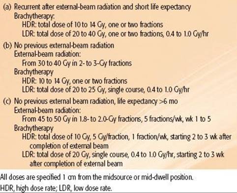
TREATMENT SEQUELAE
Advances in surgical technique, as well as improved preoperative and postoperative management, have decreased treatment-related mortality. Contemporary operative mortality rates are generally <10%.152,161 Complication rates can exceed 75%, including pulmonary and cardiac complications, anastomotic leak, and recurrent laryngeal nerve paralysis. Stricture formation can occur in 14% to 27% of patients. The addition of preoperative radiation therapy and chemotherapy may enhance surgical complication rates.
More than 75% of patients receiving radiation experience transient esophagitis and dysphagia, sometimes requiring some type of nutritional support. Chemotherapy-related leukopenia and thrombocytopenia are common. Additional acute toxicities of radiation therapy include esophagitis, epidermitis, fatigue, and weight loss in most patients. Nausea and vomiting are relatively common, particularly in patients with lower esophageal and gastroesophageal junction tumors. Many symptoms resolve within 1 to 2 weeks of treatment completion. A perforated esophagus is life-threatening and can be characterized by substernal chest pain, a high pulse rate, fever, and hemorrhage.93 The addition of chemotherapy can significantly increase acute complications. Moderate-to-severe and even life-threatening toxicities have been reported in 50% to 66% of patients.71,149,162 In the previously discussed RTOG study of chemoradiation alone, patients treated with combined therapy had a higher incidence of acute grade 3 (44% vs. 25%) and grade 4 toxicity (20% vs. 3%) compared to patients receiving radiation therapy alone.146Chemoradiation treatment–related mortality rates range from 0% to 3%.71,117,141,149,152
The most common late effects following radiation therapy are stenosis and stricture formation. Stenosis can occur in >60% of patients. Stricture requiring dilatation has been reported to occur at least 15% to 20% of the time. Dysphagia may be relieved with two to three dilatations.71 Long-term results from the RTOG study showed that late grade 3 or greater toxicity was similar in the combined versus radiation-alone arms (29% vs. 23%). However, grade 4 or greater toxicity was higher in patients receiving combined-modality therapy (10% vs. 2%).150 Other complications include clinically apparent damage to organs within the radiation therapy volume, although this is uncommon. Chemotherapy may further increase the risk of late treatment-related toxicities.
The effects of radiation on pulmonary and cardiac function deserve special mention. Pulmonary complications associated with either the definitive or neoadjuvant treatment of esophageal cancer patients can be broadly broken up into symptomatic pneumonitis following treatment completion and postoperative pulmonary complications in patients undergoing resection. Although they are still ill defined, various institutional series have described predictive factors for these complications (which are not mutually exclusive), which are described in the following.
Radiation pneumonitis is a relatively common complication in the treatment of thoracic (lung) malignancies, with can range from minimally symptomatic to fatal. Common symptoms include nonproductive cough, dyspnea, and, more uncommonly, respiratory distress. Generally, this occurs 2 to 6 months after radiation therapy completion. The ability to predict radiation pneumonitis has been a significant topic of investigation. However, most data come from patients with lung cancer, who may have more underlying intrinsic pulmonary/smoking-related disease. A variety of predictive parameters have been suggested, including V20 of >25%–30%, mean lung dose of >15 to 20 Gy, V5 of >42%, and absolute V5 of >3,000 cm3.107
In nonoperative patients, an analysis of esophageal cancer patients from Japan treated definitively to a dose of 60 Gy with concurrent 5-FU and cisplatin showed a radiation pneumonitis incidence of 27%.163 The authors concluded that an optimal V20 threshold to predict symptomatic pneumonitis was approximately 30%. In a study of 101 both operative and nonoperative patients (88% distal esophagus/GE junction) from the MD Anderson Cancer Center undergoing a mix of 3D and IMRT radiation therapy, 59%, 5%, and 1% of patients experienced grade 2, 3, and 5 radiation pneumonitis, respectively.164 An analysis from Japan using fields inclusive of supraclavicular, mediastinal, and celiac regions up to a dose of 60 Gy with concurrent cisplatin and 5-FU showed a 2-year cumulative incidence of late, high-grade cardiopulmonary toxicities for patients ≥75 years of 29% versus 3% in younger patients. They concluded that older patients may not tolerate extensive radiation fields.165 Other studies have shown that significant declines in diffusion capacity and total lung capacity may occur in patients irradiated for esophageal cancer.166
In operative patients, a study of 110 patients treated with preoperative chemoradiotherapy followed by resection at the MD Anderson Cancer Center showed that mean lung dose, effective dose, and absolute lung dose receiving ≤5 Gy were predictors of development of postoperative pulmonary complications.167 In a report on patients from the same institution describing complications in patients receiving neoadjuvant combined modality therapy,168 18% experienced pulmonary complications, with higher rates when the V10 was ≥40% (35% vs. 8%) and V15 was ≥30% (33% vs. 10%), leading the authors to conclude that minimization of lung volume irradiation was important in the preoperative planning of these patients. This increase in postoperative pulmonary complications (pneumonia, acute respiratory distress syndrome) when the V10 was >40% suggests that the volume of remaining/undamaged functional lung may determine postoperative pulmonary function, that is, patients with a small lung volume initially may be at higher risk of experiencing pulmonary complications, even if the relative V5 is low, and that patients with small lung volume with less functional reserve may be more susceptible to postoperative pulmonary complications. Therefore, it is important to consider not only the dose–volume histogram of the lung, but also the total lung volume.
A study from China evaluating patients receiving chemoradiotherapy followed by resection showed that the volume of lung spared from doses of ≥5 Gy was the only independent dosimetric factor on multivariate analysis in predicting postoperative pulmonary complications.169 Wang et al.170 similarly described that the relative V5 and all spared volumes from 5 to 35 Gy significantly correlated with the incidence of postoperative pulmonary complications, although on multivariate analysis, V5 was the only significant independent predictive factor, indicating that the volume of “unexposed” lung during induction therapy was predictive. Of note in this study was that the majority of patients were treated with induction chemotherapy alone initially (most paclitaxel), which has been shown to increase rates of pneumonitis in other disease sites. A significant association of induction chemotherapy alone prior to concurrent chemoradiotherapy was seen as a predictor of grade 2 or greater pneumonitis (49% vs. 14%; p= .003), leading the authors to conclude that induction chemotherapy alone may sensitize lung tissue to radiation damage. In contrast to the foregoing studies, however, another analysis of 98 patients receiving preoperative chemoradiotherapy with 5-FU and cisplatin showed no difference in pulmonary complications versus patients undergoing surgery alone, with no correlation of any lung dose–volume histogram findings with development of postoperative pulmonary complications seen, leading the authors to conclude that neoadjuvant chemoradiotherapy had no detrimental impact on postoperative course.171 Finally, an analysis from Taiwan of neoadjuvantly, IMRT-treated esophageal cancer patients undergoing resection suggested that preoperative (not prechemoradiation) forced expiratory volume in 1 second was an independent factor associated with postoperative pulmonary complications and that reducing the absolute volume of the right lung irradiated might decrease the risk of postoperative pulmonary complications.172
Radiation-induced cardiac toxicity is a broad term describing potential radiation injury to a number of cardiac structures, including pericardium (as manifested by effusion, pericarditis), coronary arteries, the heart muscle itself, and cardiac valves, as well as nerve/conduction injury. Radiation injury primarily consists of fibrosis and/or small-vessel injury. “Classic” radiation tolerance (TD5/5) of the heart is about 60 Gy when 25% or less of the heart is irradiated and 45 Gy if 65% of the heart is irradiated, assuming 2 Gy per fraction. The mechanism of radiation-induced cardiac injury is relatively poorly defined, particularly in the context of esophageal cancer. Historical data from the treatment of Hodgkin’s disease patients suggest that a dose of >40 Gy may increase the risk of cardiac death, as well as of pericarditis.173,174 Several studies of cardiac toxicity and esophageal cancer patient demonstrated that a V30 of >46% predicted a significant increase in pericardial effusion, and increasing fraction size (particularly ≥3.5 Gy) also predicted the same. In addition, some authors have shown a possible trend for decrease in ejection fraction in patients with increasing V20 of the left ventricle.107 In a study of 150 esophageal cancer patients receiving chemoradiotherapy (49 neoadjuvantly), the incidence of pericardial effusion was 28%, usually developing within 15 months of radiation therapy, with median onset time of approximately 5 months. The risk of pericardial effusion was associated with mean pericardial doses over an array of dose–volume points to the pericardium from 5 to 45 Gy. A matched-pair analysis of nonradiation patients receiving surgery versus those treated neoadjuvantly was performed, with 42% of patients in the radiation group demonstrating ischemia/scarring on single-photon emission computed tomography images versus 4% in the surgical group, with a median onset to abnormality of 3 months.175 In a Japanese study, long-term analysis of 139 patients treated with definitive chemoradiotherapy (cisplatin/5-FU with 60 Gy EBRT) for squamous cell carcinoma revealed grade 2, 3, and 4 late pericarditis occurring in 6%, 5%, and 1% of patients, respectively; grade 4 heart failure in 2 patients; grade 2, 3, and 4 pleural effusion development in 5%, 6%, and 0% of patients, respectively; and grade 2, 3, and 4 radiation pneumonitis development in 1%, 2%, and 0% of patients, respectively.176
FUTURE CONSIDERATIONS
Although modest improvements in survival have been achieved by combining neoadjuvant chemoradiation and surgery, patients treated with chemoradiation alone or with surgery have unacceptably high local-regional relapse rates and mortality rates. Ultimately, approximately 75% of patients succumb to metastatic disease. As described previously, efforts at radiation dose escalation have not resulted in significant gains for this disease. Given these data, clinical trials have turned to studies evaluating new and potentially more effective chemotherapeutic agents with radiation therapy. Agents such as irinotecan, oxaliplatin, capecitabine, epirubicin, gemcitabine, and docetaxel have been or are being investigated in the metastatic setting, as well as “curative” settings in combination with radiation therapy. Furthermore, there is ongoing investigation of the use of the vascular endothelial growth factor inhibitor bevacizumab, the HER2/neu receptor antagonist trastuzumab, and inhibitors of the epidermal growth factor receptors, including the antibodies cetuximab and panitumumab, in the treatment of esophageal cancer. All of these agents have radiosensitizing properties. The investigation of these agents with radiation therapy is the subject of ongoing and future trials.
SUMMARY
The prognosis for patients with carcinoma of the esophagus remains poor despite recent advances in combined-modality therapies. No firm recommendation can be made for managing locally advanced disease. The data suggest that neoadjuvant chemoradiation improves outcomes in patients who are candidates for surgery. Alternatively, randomized trials have also suggested that perioperative chemotherapy improves outcomes in these patients. However, many patients are not able to tolerate surgery, and combined chemoradiation may be more appropriate in selected patients because definitive chemoradiation has resulted in survival rates comparable to those from surgery alone. Locoregional failure remains a significant pattern of relapse. For patients with stage IV disease, palliation with single-modality therapy or several modalities should be used and tailored to the patient’s specific symptoms. Current unresolved issues include the following:
1. Which subsets of patients are more likely to benefit from the addition of surgery than others?
2. Which subsets of patients are more likely to benefit from the addition of neoadjuvant and/or perioperative therapies?
3. Can introduction of newer chemotherapy/targeted agents in the neoadjuvant or concurrent setting improve the results over “standard” chemoradiation with cisplatin and 5-FU?
4. Will new technologies such as 3D conformal therapy, PET-based planning, intensity-modulated radiation therapy, proton therapy, and image-guided radiation therapy decrease complication rates and influence cure rates?
5. Will PET scan allow early prediction of both response to treatment and ultimate outcomes and potentially allow avoidance of delivery of ineffective treatments early on during the course of therapy?
6. Will the identification of molecular prognostic markers allow “individualization” of treatments among patients?
7. In surgical patients, is there a superiority of a neoadjuvant chemoradiation versus a perioperative chemotherapy approach, and will some patients benefit from one particular treatment approach?
REFERENCES
1. American Joint Committee on Cancer. Esophagus. New York: Springer-Verlag, 2010.
2. Rosenberg JC, Franklin R, Steiger Z. Squamous cell carcinoma of the thoracic esophagus: an interdisciplinary approach. Curr Probl Cancer 1981;5:1–52.
3. Rudiger Siewert J, Feith M, Werner M, et al. Adenocarcinoma of the esophagogastric junction: results of surgical therapy based on anatomical/topographic classification in 1,002 consecutive patients. Ann Surg 2000;232:353–361.
4. Arnott SJ, Duncan W, Gignoux M, et al. Preoperative radiotherapy for esophageal carcinoma. Cochrane Database Syst Rev 2005;CD001799.
5. Goodner JT, Miller TP, Pack GT, et al. Torek esophagectomy; the case against segmental resection for esophageal cancer. J Thorac Surg 1956;32:347–359.
6. Hosch SB, Stoecklein NH, Pichlmeier U, et al. Esophageal cancer: the mode of lymphatic tumor cell spread and its prognostic significance. J Clin Oncol 2001;19:1970–1975.
7. Sharpiro A, Robillard G. The esophageal arteries. Ann Surg 1950;131:171.
8. Ott K, Weber W, Siewert JR. The importance of PET in the diagnosis and response evaluation of esophageal cancer. Dis Esophagus 2006;19:433–442.
9. Siegel R, Naishadham D, Jemal A. Cancer statistics, 2012. CA Cancer J Clin 2012;62(1):10–29. doi:10.3322/caac.20138. Epub 2012 Jan 4.
10. Cheng KK. The etiology of esophageal cancer in Chinese. Semin Oncol 1994;21:411–415.
11. Mahboubi M, Kmet J, Cook P. Esophageal cancer studies in the Caspian littoral of Iran: the Caspian Cancer Resgistry. Br J Cancer 1973;28:196.
12. Blot WJ, McLaughlin JK. The changing epidemiology of esophageal cancer. Semin Oncol 1999;26:2–8.
13. Denham JW, Burmeister BH, Lamb DS, et al. Factors influencing outcome following radio-chemotherapy for oesophageal cancer. The Trans Tasman Radiation Oncology Group (TROG). Radiother Oncol1996;40:31–43.
14. Pohl H, Welch HG. The role of overdiagnosis and reclassification in the marked increase of esophageal adenocarcinoma incidence. J Natl Cancer Inst 2005;97:142–146.
15. Schottenfeld D. Epidemiology of cancer of the esophagus. Semin Oncol 1984;11:92–100.
16. Blot WJ. Alcohol and cancer. Cancer Res 1992;52:2119s–2123s.
17. van Rensburg SJ. Epidemiologic and dietary evidence for a specific nutritional predisposition to esophageal cancer. J Natl Cancer Inst 1981;67:243–251.
18. Lijinsky W. Current concepts in the toxicology of nitrates, nitrites and nitrosoamines. Washington, DC: Hemisphere, 1979.
19. Miao C, Guo F, Zhang J. The relationship between fungi and nitrosamines and their precursors: II. The action of fungi isolated from grains in Linxian. Med Ref 1978;2:46.
20. Appelqvist P, Salmo M. Lye corrosion carcinoma of the esophagus: a review of 63 cases. Cancer 1980;45:2655–2658.
21. Hopkins RA, Postlethwait RW. Caustic burns and carcinoma of the esophagus. Ann Surg 1981;194:146–148.
22. Harper PS, Harper RM, Howel-Evans AW. Carcinoma of the oesophagus with tylosis. Q J Med 1970;39:317–333.
23. Spechler SJ. Barrett’s esophagus. Gastroenterologist 1994;2:273–284.
24. Lagergren J, Bergstrom R, Lindgren A, et al. Symptomatic gastroesophageal reflux as a risk factor for esophageal adenocarcinoma. N Engl J Med 1999;340:825–831.
25. Devesa SS, Blot WJ, Fraumeni JF Jr. Changing patterns in the incidence of esophageal and gastric carcinoma in the United States. Cancer 1998;83:2049–2053.
26. Zhang ZF, Kurtz RC, Sun M, et al. Adenocarcinomas of the esophagus and gastric cardia: medical conditions, tobacco, alcohol, and socioeconomic factors. Cancer Epidemiol Biomarkers Prev1996;5:761–768.
27. Heath EI, Limburg PJ, Hawk ET, et al. Adenocarcinoma of the esophagus: risk factors and prevention. Oncology (Williston Park) 2000;14:507–514; discussion 518–520, 522–503.
28. Brown LM, Swanson CA, Gridley G, et al. Adenocarcinoma of the esophagus: role of obesity and diet. J Natl Cancer Inst 1995;87:104–109.
29. DeMeester TR. Clinical biology of the Barrett’s metaplasia, dysplasia to carcinoma sequence. Surg Oncol 2001;10:91–102.
30. Montesano R, Hollstein M, Hainaut P. Genetic alterations in esophageal cancer and their relevance to etiology and pathogenesis: a review. Int J Cancer 1996;69:225–235.
31. Holdscher A, Bollschweiller E, Bumm R, et al. Prognostic factors of resected adenocarcinoma of the esophagus. Surgery 1995;118:845–855.
32. Collard JM, Otte JB, Fiasse R, et al. Skeletonizing en bloc esophagectomy for cancer. Ann Surg 2001;234:25–32.
33. Dresner SM, Lamb PJ, Bennett MK, et al. The pattern of metastatic lymph node dissemination from adenocarcinoma of the esophagogastric junction. Surgery 2001;129:103–109.
34. Akiyama H, Tsurumaru M, Kawamura T, et al. Principles of surgical treatment for carcinoma of the esophagus: analysis of lymph node involvement. Ann Surg 1981;194:438–446.
35. Bloedorn F, Kasdorf H. Radiotherapy in squamous cell carcinoma of the esophagus. Chicago: Year Book Medical, 1971.
36. Dormans E. Das Oesophaguscarcinoma: Ergebnisse der unter Mitarbet von 29 patholgischem Instituten Deutschlands Durchgefuhrten Erhebung uber das Oesophaguscarcinomon (1925–1933). Z Krebforsch 1939;49:86.
37. Anderson LL, Lad TE. Autopsy findings in squamous-cell carcinoma of the esophagus. Cancer 1982;50:1587–1590.
38. Huang W, Li B, Gong H, et al. Pattern of lymph node metastases and its implication in radiotherapeutic clinical target volume in patients with thoracic esophageal squamous cell carcinoma: a report of 1077 cases. Radiother Oncol 2010;95:229–233.
39. Monig SP, Baldus SE, Zirbes TK, et al. Topographical distribution of lymph node metastasis in adenocarcinoma of the gastroesophageal junction. Hepatogastroenterology 2002;49:419–422.
40. Schurr PG, Yekebas EF, Kaifi JT, et al. Lymphatic spread and microinvolvement in adenocarcinoma of the esophago-gastric junction. J Surg Oncol 2006;94:307–315.
41. Matzinger O, Gerber E, Bernstein Z, et al. EORTC-ROG expert opinion: radiotherapy volume and treatment guidelines for neoadjuvant radiation of adenocarcinomas of the gastroesophageal junction and the stomach. Radiother Oncol 2009;92:164–175.
42. Meier I, Merkel S, Papadopoulos T, et al. Adenocarcinoma of the esophagogastric junction: the pattern of metastatic lymph node dissemination as a rationale for elective lymphatic target volume definition. Int J Radiat Oncol Biol Phys 2008;70:1408–1417.
43. Aisner J, Forastiere A, Aroney R. Patterns of recurrence for cancer of the lung and esophagus. Cancer Treat Symp 1983;2:87–105.
44. LePrise E, Meunier B, Etienne P, et al. Sequential chemotherapy and radiotherapy for patients with squamous cell carcinoma of the esophagus. Cancer 1995;75:2.
45. Whittington R, Coia LR, Haller DG, et al. Adenocarcinoma of the esophagus and esophago-gastric junction: the effects of single and combined modalities on the survival and patterns of failure following treatment. Int J Radiat Oncol Biol Phys 1990;19:593–603.
46. Hulscher JB, van Sandick JW, de Boer AG, et al. Extended transthoracic resection compared with limited transhiatal resection for adenocarcinoma of the esophagus. N Engl J Med 2002;347:1662–1669.
47. Kelsen DP, Ginsberg R, Pajak TF, et al. Chemotherapy followed by surgery compared with surgery alone for localized esophageal cancer. N Engl J Med 1998;339:1979–1984.
48. Law SY, Fok M, Wong J. Pattern of recurrence after oesophageal resection for cancer: clinical implications. Br J Surg 1996;83:107–111.
49. Urba SG, Orringer MB, Turrisi A, et al. Randomized trial of preoperative chemoradiation versus surgery alone in patients with locoregional esophageal carcinoma. J Clin Oncol 2001;19:305–313.
50. Rosenberg J, Lichter A, Leichman L. Cancer of the esophagus. Philadelphia: JB Lippincott, 1989.
51. Moertel C. The esophagus. Philadelphia: Lea & Febiger, 1982.
52. Lea JWt, Prager RL, Bender HW Jr. The questionable role of computed tomography in preoperative staging of esophageal cancer. Ann Thorac Surg 1984;38:479–481.
53. Picus D, Balfe DM, Koehler RE, et al. Computed tomography in the staging of esophageal carcinoma. Radiology 1983;146:433–438.
54. Griffith JF, Chan AC, Chow LT, et al. Assessing chemotherapy response of squamous cell oesophageal carcinoma with spiral CT. Br J Radiol 1999;72:678–684.
55. Rankin SC, Taylor H, Cook GJ, et al. Computed tomography and positron emission tomography in the pre-operative staging of oesophageal carcinoma. Clin Radiol 1998;53:659–665.
56. Kole AC, Plukker JT, Nieweg OE, et al. Positron emission tomography for staging of oesophageal and gastroesophageal malignancy. Br J Cancer 1998;78:521–527.
57. Earlam R, Cunha-Melo JR. Oesophogeal squamous cell carcinoms: II. A critical view of radiotherapy. Br J Surg 1980;67:457–461.
58. Kelly S, Harris KM, Berry E, et al. A systematic review of the staging performance of endoscopic ultrasound in gastro-oesophageal carcinoma. Gut 2001;49:534–539.
59. Konski A, Doss M, Milestone B, et al. The integration of 18–fluoro-deoxy-glucose positron emission tomography and endoscopic ultrasound in the treatment-planning process for esophageal carcinoma. Int J Radiat Oncol Biol Phys 2005;61:1123–1128.
60. Rosch T. Endosonographic staging of esophageal cancer: a review of literature results. Gastrointest Endosc Clin North Am 1995;5:537–547.
61. Beseth BD, Bedford R, Isacoff WH, et al. Endoscopic ultrasound does not accurately assess pathologic stage of esophageal cancer after neoadjuvant chemoradiotherapy. Am Surg 2000;66:827–831.
62. Laterza E, de Manzoni G, Guglielmi A, et al. Endoscopic ultrasonography in the staging of esophageal carcinoma after preoperative radiotherapy and chemotherapy. Ann Thorac Surg 1999;67:1466–1469.
63. Zeccaro G, Rice T, Goldbloom J, et al. Endoscopic ultrasound can not determine suitability for esophagectomy after aggressive chemoradiotherapy for esophageal cancer. Am J Gasterinterol 1999;94:906–912.
64. Jaklitsch MT, Harpole DH Jr, Healey EA, et al. Current issues in the staging of esophageal cancer. Semin Radiat Oncol 1994;4:135–145.
65. Blackstock AW, Farmer MR, Lovato J, et al. A prospective evaluation of the impact of 18-F-fluoro-deoxy-D-glucose positron emission tomography staging on survival for patients with locally advanced esophageal cancer. Int J Radiat Oncol Biol Phys 2006;64:455–460.
66. Flamen P, Lerut A, Van Cutsem E, et al. Utility of positron emission tomography for the staging of patients with potentially operable esophageal carcinoma. J Clin Oncol 2000;18:3202–3210.
67. Monjazeb AM, Riedlinger G, Aklilu M, et al. Outcomes of patients with esophageal cancer staged with [18F]fluorodeoxyglucose positron emission tomography (FDG-PET): can postchemoradiotherapy FDG-PET predict the utility of resection? J Clin Oncol 28:4714–4721.
68. Weber WA, Ott K. Imaging of esophageal and gastric cancer. Semin Oncol 2004; 31:530–541.
69. Oota K, Shin L. Histological typing of gastric and oesophageal tumors. Geneva: World Health Organization, 1977.
70. Gill PG, Denham JW, Jamieson GG, et al. Patterns of treatment failure and prognostic factors associated with the treatment of esophageal carcinoma with chemotherapy and radiotherapy either as sole treatment or followed by surgery. J Clin Oncol 1992;10:1037–1043.
71. Coia LR, Engstrom PF, Paul AR, et al. Long-term results of infusional 5-FU, mitomycin-C and radiation as primary management of esophageal carcinoma. Int J Radiat Oncol Biol Phys 1991;20:29–36.
72. Burke EL, Sturm J, Williamson D. The diagnosis of microscopic carcinoma of the esophagus. Am J Dig Dis 1978;23:148–151.
73. Maimon HN, Dreskin RB, Cocco AE. Positive esophageal cytology without detectable neoplasm. Gastrointest Endosc 1974;20:156–159.
74. Smoron GL, O’Brien CA, Sullivan CA. Tumor localization and treatment technique for cancer of the esophagus. Radiology 1974;111:735–736.
75. Thompson WM. Esophageal cancer. Int J Radiat Oncol Biol Phys 1983;9:1533–1565.
76. Epstein JI, Sears DL, Tucker RS, et al. Carcinoma of the esophagus with adenoid cystic differentiation. Cancer 1984;53:1131–1136.
77. Turnbull AD, Rosen P, Goodner JT, et al. Primary malignant tumors of the esophagus other than typical epidermoid carcinoma. Ann Thorac Surg 1973;15:463–473.
78. Briggs JC, Ibrahim NB. Oat cell carcinomas of the oesophagus: a clinico-pathological study of 23 cases. Histopathology 1983;7:261–277.
79. Imai T, Sannohe Y, Okano H. Oat cell carcinoma (apudoma) of the esophagus: a case report. Cancer 1978;41:358–364.
80. Doherty MA, McIntyre M, Arnott SJ. Oat cell carcinoma of esophagus: a report of six British patients with a review of the literature. Int J Radiat Oncol Biol Phys 1984;10:147–152.
81. Gaede JT, Postlethwait RW, Shelburne JD, et al. Leiomyosarcoma of the esophagus: report of two cases, one with associated squamous cell carcinoma. J Thorac Cardiovasc Surg 1978;75:740–746.
82. Partyka EK, Sanowski RA, Kozarek RA. Endoscopic diagnosis of a giant esophageal leiomyosarcoma. Am J Gastroenterol 1981;75:132–134.
83. Postlethwait R, Sealy W. Surgery of the esophagus. New York: Appleton-Century-Crofts, 1979.
84. Gelb A, Miller S. AIDS and gastroenterology. Am J Gastroenterol 1986;81:619–622.
85. Ludwig ME, Shaw R, de Suto-Nagy G. Primary malignant melanoma of the esophagus. Cancer 1981;48:2528–2534.
86. Son YH. Primary mucosal malignant melanoma. Appraisal of role of radiation therapy. Acta Radiol Oncol 1980;19:177–181.
87. Orvidas LJ, McCaffrey TV, Lewis JE, et al. Lymphoma involving the esophagus. Ann Otol Rhinol Laryngol 1994;103:843–848.
88. Gertler R, Stein HJ, Langer R, et al. Long-term outcome of 2920 patients with cancers of the esophagus and esophagogastric junction: evaluation of the New Union Internationale Contre le Cancer/American Joint Cancer Committee staging system. Ann Surg 2011;253:689–698.
89. Nomura M, Shitara K, Kodaira T, et al. Prognostic impact of the 6th and 7th American Joint Committee on Cancer TNM staging systems on esophageal cancer patients treated with chemoradiotherapy. Int J Radiat Oncol Biol Phys 2012;82:946–952.
90. Wang BY, Goan YG, Hsu PK, et al. Tumor length as a prognostic factor in esophageal squamous cell carcinoma. Ann Thorac Surg 2011;91:887–893.
91. Gaur P, Sepesi B, Hofstetter WL, et al. Endoscopic esophageal tumor length: a prognostic factor for patients with esophageal cancer. Cancer 2011;117:63–69.
92. Yoon HH, Khan M, Shi Q, et al. The prognostic value of clinical and pathologic factors in esophageal adenocarcinoma: a Mayo cohort of 796 patients with extended follow-up after surgical resection. Mayo Clin Proc2010;85:1080–1089.
93. Hussey D, Barakley T, Bloedorn F. Carcinoma of the esophagus. Philadelphia: Lea & Febiger, 1980.
94. Suntharalingam M, Moughan J, Coia LR, et al. Outcome results of the 1996–1999 patterns of care survey of the national practice for patients receiving radiation therapy for carcinoma of the esophagus. J Clin Oncol2005;23:2325–2331.
95. Pearson JG. The present status and future potential of radiotherapy in the management of esophageal cancer. Cancer 1977;39:882–890.
96. Ellis FH, Jr. Standard resection for cancer of the esophagus and cardia. Surg Oncol Clin North Am 1999;8:279–294.
97. Hancock SL, Glatstein E. Radiation therapy of esophageal cancer. Semin Oncol 1984;11:144–158.
98. Skinner DB. En bloc resection for neoplasms of the esophagus and cardia. J Thorac Cardiovasc Surg 1983;85:59–71.
99. Sander R, Hagenmueller F, Sander C, et al. Laser versus laser plus afterloading with iridium-192 in the palliative treatment of malignant stenosis of the esophagus: a prospective, randomized, and controlled study. Gastrointest Endosc 1991;37:433–440.
100. Luketich JD, Friedman DM, Weigel TL, et al. Evaluation of distant metastases in esophageal cancer:100 consecutive positron emission tomography scans. Ann Thorac Surg 1999;68:1133–1136; discussion 1136–1137.
101. van Westreenen HL, Westerterp M, Bossuyt PM, et al. Systematic review of the staging performance of 18 F-fluorodeoxyglucose positron emission tomography in esophageal cancer. J Clin Oncol2004;22:3805–3812.
102. Zhong X, Yu J, Zhang B, et al. Optimal SUV threshold of gross tumor volume delineation validated by pathologic examination in patients with esophageal cancer. Int J Radiat Oncol Biol Phys2007;69:108–109.
103. Leong T, Everitt K, Yuen K, et al. A prospective study to evaluate the impact of coregisted PET/CT images in radiotherapy treatment planning for esophageal cancer. Int J Radiat Oncol Biol Phys2004;60:S139–S140.
104. Zobotto L, Toubouol E, Lerouge D, et al. Impact of Ct and 18F-deoxyglucose positron emission tomography image fusion for conformal radiotherapy in esophageal carcinoma. Int J Radiat Oncol Biol Phys 2005;63:340.
105. Vesprini D, Ung Y, Kamra J, et al. The addition of 18-fluoodeoxglucose positron emission tomography (FDG-PET) to CT based radiotherapy planning of carcinoma of the esophagus decreases both the intra- and interobserver variability of GTV dlineation. Int J Radiat Oncol Biol Phys 2006;66:S299–S300.
106. Gao XS, Qiao X, Wu F, et al. Pathological analysis of clinical target volume margin for radiotherapy in patients with esophageal and gastroesophageal junction carcinoma. Int J Radiat Oncol Biol Phys2007;67:389–396.
107. Hazard L, Yang G, McAleer M, et al. Principles and techniques of radiation therapy for esphageal and gastroesophageal junction cancers. J Natl Compr Canc Netw 2008;6:870–878.
108. Schroder W, Baldus SE, Monig SP, et al. Lymph node staging of esophageal squamous cell carcinoma in patients with and without neoadjuvant radiochemotherapy: histomorphologic analysis. World J Surg 2002;26:584–587.
109. Lightdale CJ, Kulkarni KG. Role of endoscopic ultrasonography in the staging and follow-up of esophageal cancer. J Clin Oncol 2005;23:4483–4489.
110. Cohen RJ, Paskalev K, Litwin S, et al. Esophageal motion during radiotherapy: quantification and margin implications. Dis Esophagus 2010;23:473–479.
111. Fenkell L, Kaminsky I, Breen S, et al. Dosimetric comparison of IMRT vs. 3D conformal radiotherapy in the treatment of cancer of the cervical esophagus. Radiother Oncol 2008;89:287–291.
112. Burmeister BH, Dickie G, Smithers BM, et al. Thirty-four patients with carcinoma of the cervical esophagus treated with chemoradiation therapy. Arch Otolaryngol Head Neck Surg 2000;126:205–208.
113. Murakami M, Kuroda Y, Okamoto Y, et al. Neoadjuvant concurrent chemoradiotherapy followed by definitive high-dose radiotherapy or surgery for operable thoracic esophageal carcinoma. Int J Radiat Oncol Biol Phys1998;40:1049–1059.
114. Minsky BD, Pajak TF, Ginsberg RJ, et al. INT 0123 (Radiation Therapy Oncology Group 94-05) phase III trial of combined-modality therapy for esophageal cancer: high-dose versus standard-dose radiation therapy. J Clin Oncol 2002;20:1167–1174.
115. Minsky BD, Neuberg D, Kelsen DP, et al. Final report of Intergroup Trial 0122 (ECOG PE-289, RTOG 90-12): phase II trial of neoadjuvant chemotherapy plus concurrent chemotherapy and high-dose radiation for squamous cell carcinoma of the esophagus. Int J Radiat Oncol Biol Phys 1999;43:517–523.
116. Beatty JD, DeBoer G, Rider WD. Carcinoma of the esophagus: pretreatment assessment, correlation of radiation treatment parameters with survival, and identification and management of radiation treatment failure. Cancer1979;43:2254–2267.
117. Walsh TN, Noonan N, Hollywood D, et al. A comparison of multimodal therapy and surgery for esophageal adenocarcinoma. N Engl J Med 1996;335:462–467.
118. Medical Research Council Oesophageal Cancer Working Group. Surgical resection with or without preoperative chemotherapy in oesophageal cancer: a randomized controlled trial. Lancet Oncol2002;359:1727–1733.
119. Bosset JF, Gignoux M, Triboulet JP, et al. Chemoradiotherapy followed by surgery compared with surgery alone in squamous-cell cancer of the esophagus. N Engl J Med 1997;337:161–167.
120. van Hagen P, Hulshof M, van Lanschot JJB, et al. Preoperative Chemoradiotherapy for Esophageal or Junctional Cancer. N Engl J Med 2012;366:2074–2084.
121. Mariette C, Seitz J, Maillard E, et al. Surgery alone versus chemoradiotherapy followed by surgery for localized esophageal cancer: analysis of a randomized controlled phase III trial FFCD 9901. J Clin Oncol 2010;28:15s.
122. Earlam R, Cunha-Melo JR. Oesophageal squamous cell carcinoma: I. A critical review of surgery. Br J Surg 1980;67:381–390.
123. Okawa T, Kita M, Tanaka M, et al. Results of radiotherapy for inoperable locally advanced esophageal cancer. Int J Radiat Oncol Biol Phys 1989;17:49–54.
124. Lederman M. Carcinoma of the oesophagus with special reference to the upper third: part I. Clinical considerations. Br J Cancer 1982;39:193.
125. Launois B, Delarue D, Campion JP, et al. Preoperative radiotherapy for carcinoma of the esophagus. Surg Gynecol Obstet 1981;153:690–692.
126. Gignoux M, Roussel A, Paillot B, et al. The value of preoperative radiotherapy in esophageal cancer: results of a study of the E.O.R.T.C. World J Surg 1987;11: 426–432.
127. Arnott SJ, Duncan W, Kerr GR, et al. Low dose preoperative radiotherapy for carcinoma of the oesophagus: results of a randomized clinical trial. Radiother Oncol 1992;24:108–113.
128. Wang M, Gu XZ, Yin WB, et al. Randomized clinical trial on the combination of preoperative irradiation and surgery in the treatment of esophageal carcinoma: report on 206 patients. Int J Radiat Oncol Biol Phys 1989;16:325–327.
129. Teniere P, Hay JM, Fingerhut A, et al. Postoperative radiation therapy does not increase survival after curative resection for squamous cell carcinoma of the middle and lower esophagus as shown by a multicenter controlled trial. French University Association for Surgical Research. Surg Gynecol Obstet 1991;173:123–130.
130. Fok M, Sham JS, Choy D, et al. Postoperative radiotherapy for carcinoma of the esophagus: a prospective, randomized controlled study. Surgery 1993;113:138–147.
131. Xiao ZF, Yang ZY, Miao YJ, et al. Influence of number of metastatic lymph nodes on survival of curative resected thoracic esophageal cancer patients and value of radiotherapy: report of 549 cases. Int J Radiat Oncol Biol Phys2005;62:82–90.
132. Macdonald JS, Smalley SR, Benedetti J, et al. Chemoradiotherapy after surgery compared with surgery alone for adenocarcinoma of the stomach or gastroesophageal junction. N Engl J Med2001;345:725–730.
133. Macdonald JS, Benedetti J, Smalley SR, et al. Chemoradiation of resected gastric cancer: a 10–year follow-up of the phase III trial INT0116 (SWOG 9008). J Clin Oncol 2009;27.
134. Kelsen DP, Winter KA, Gunderson LL, et al. Long-term results of RTOG trial 8911 (USA Intergroup 113): a random assignment trial comparison of chemotherapy followed by surgery compared with surgery alone for esophageal cancer. J Clin Oncol 2007;25:3719–3725.
135. Allum WH, Stenning SP, Bancewicz J, et al. Long-term results of a randomized trial of surgery with or without preoperative chemotherapy in esophageal cancer. J Clin Oncol 2009;27:5062–5067.
136. Boonstra JJ, Kok TC, Wijnhoven BP, et al. Chemotherapy followed by surgery versus surgery alone in patients with resectable oesophageal squamous cell carcinoma: long-term results of a randomized controlled trial. BMC Cancer 2011;11:181.
137. Cunningham D, Allum WH, Stenning SP, et al. Perioperative chemotherapy versus surgery alone for resectable gastroesophageal cancer. N Engl J Med 2006; 355:11–20.
138. Ychou M, Boige V, Pignon JP, et al. Perioperative chemotherapy compared with surgery alone for resectable gastroesophageal adenocarcinoma: an FNCLCC and FFCD multicenter phase III trial. J Clin Oncol 2011;29:1715–1721.
139. Gebski V, Burmeister B, Smithers BM, et al. Survival benefits from neoadjuvant chemoradiotherapy or chemotherapy in oesophageal carcinoma: a meta-analysis. Lancet Oncol 2007;8:226–234.
140. Thirion P, Michiels S, Le Maitre A, et al. Individual patient data-based meta-analysis assessing pre-operative chemotherapy in resectable oesophageal carcinoma. J Clin Oncol 2007;25:200s.
141. Burmeister BH, Smithers BM, Gebski V, et al. Surgery alone versus chemoradiotherapy followed by surgery for resectable cancer of the oesophagus: a randomised controlled phase III trial. Lancet Oncol2005;6:659–668.
142. Tepper J, Krasna MJ, Niedzwiecki D, et al. Phase III trial of trimodality therapy with cisplatin, fluorouracil, radiotherapy, and surgery compared with surgery alone for esophageal cancer: CALGB 9781. J Clin Oncol2008;26:1086–1092.
143. Sjoquist KM, Burmeister BH, Smithers BM, et al. Survival after neoadjuvant chemotherapy or chemoradiotherapy for resectable oesophageal carcinoma: an updated meta-analysis. Lancet Oncol2011;12:681–692.
144. Stahl M, Walz MK, Stuschke M, et al. Phase III comparison of preoperative chemotherapy compared with chemoradiotherapy in patients with locally advanced adenocarcinoma of the esophagogastric junction. J Clin Oncol2009;27:851–856.
145. Burmeister BH, Thomas JM, Burmeister EA, et al. Is concurrent radiation therapy required in patients receiving preoperative chemotherapy for adenocarcinoma of the oesophagus? A randomised phase II trial. Eur J Cancer2011;47:354–360.
146. al-Sarraf M, Martz K, Herskovic A, et al. Progress report of combined chemoradiotherapy versus radiotherapy alone in patients with esophageal cancer: an intergroup study. J Clin Oncol 1997;15:277–284.
147. Araujo CM, Souhami L, Gil RA, et al. A randomized trial comparing radiation therapy versus concomitant radiation therapy and chemotherapy in carcinoma of the thoracic esophagus. Cancer1991;67:2258–2261.
148. Roussell A, Jacob J, Haegele P, et al. Controlled clinical trial for the treatment of patients with inoperable esophageal carcinoma: a study of EORTC Gastrointestinal Tract Cancer Cooperative Group. Rec Results Cancer Res1988;110–121.
149. Herskovic A, Martz K, al-Sarraf M, et al. Combined chemotherapy and radiotherapy compared with radiotherapy alone in patients with cancer of the esophagus. N Engl J Med 1992;326:1593–1598.
150. Cooper JS, Guo MD, Herskovic A, et al. Chemoradiotherapy of locally advanced esophageal cancer: long-term follow-up of a prospective randomized trial (RTOG 85-01). Radiation Therapy Oncology Group. JAMA1999;281:1623–1627.
151. Bedenne L, Michel P, Bouche O, et al. Chemoradiation followed by surgery compared with chemoradiation alone in squamous cancer of the esophagus: FFCD 9102. J Clin Oncol 2007;25:1160–1168.
152. Stahl M, Stuschke M, Lehmann N, et al. Chemoradiation with and without surgery in patients with locally advanced squamous cell carcinoma of the esophagus. J Clin Oncol 2005;23:2310–2317.
153. Gwynne S, Hurt C, Evans M, et al. Definitive chemoradiation for oesophageal cancer—a standard of care in patients with non-metastatic oesophageal cancer. Clin Oncol (R Coll Radiol) 2011;23:182–188.
154. American Cancer Society. Cancer facts and figures—2005. Atlanta, GA: American Cancer Society, 2005.
155. Gaspar LE, Winter K, Kocha WI, et al. A phase I/II study of external beam radiation, brachytherapy, and concurrent chemotherapy for patients with localized carcinoma of the esophagus (Radiation Therapy Oncology Group Study 9207): final report. Cancer 2000;88:988–995.
156. Vuong T, Szego P, David M, et al. The safety and usefulness of high-dose-rate endoluminal brachytherapy as a boost in the treatment of patients with esophageal cancer with external beam radiation with or without chemotherapy. Int J Radiat Oncol Biol Phys 2005;63:758–764.
157. Homs MY, Steyerberg EW, Eijkenboom WM, et al. Single-dose brachytherapy versus metal stent placement for the palliation of dysphagia from oesophageal cancer: multicentre randomised trial. Lancet2004;364:1497–1504.
158. Gaspar LE, Nag S, Herskovic A, et al. American Brachytherapy Society (ABS) consensus guidelines for brachytherapy of esophageal cancer. Clinical Research Committee, American Brachytherapy Society, Philadelphia, PA. Int J Radiat Oncol Biol Phys 1997;38:127–132.
159. Coia LR, Soffen EM, Schultheiss TE, et al. Swallowing function in patients with esophageal cancer treated with concurrent radiation and chemotherapy. Cancer 1993;71:281–286.
160. Gschossmann JM, Bonner JA, Foote RL, et al. Malignant tracheoesophageal fistula in patients with esophageal cancer. Cancer 1993;72:1513–1521.
161. Tsutsui S, Moriguchi S, Morita M, et al. Multivariate analysis of postoperative complications after esophageal resection. Ann Thorac Surg 1992;53:1052–1056.
162. Baquet CR, Commiskey P, Mack K, et al. Esophageal cancer epidemiology in blacks and whites: racial and gender disparities in incidence, mortality, survival rates and histology. J Natl Med Assoc2005;97:1471–1478.
163. Asakura H, Hashimoto T, Zenda S, et al. Analysis of dose-volume histogram parameters for radiation pneumonitis after definitive concurrent chemoradiotherapy for esophageal cancer. Radiother Oncol95:240–244.
164. Hart JP, McCurdy MR, Ezhil M, et al. Radiation pneumonitis: correlation of toxicity with pulmonary metabolic radiation response. Int J Radiat Oncol Biol Phys 2008;71:967–971.
165. Morota M, Gomi K, Kozuka T, et al. Late toxicity after definitive concurrent chemoradiotherapy for thoracic esophageal carcinoma. Int J Radiat Oncol Biol Phys 2009;75:122–128.
166. Gergel TJ, Leichman L, Nava HR, et al. Effect of concurrent radiation therapy and chemotherapy on pulmonary function in patients with esophageal cancer: dose-volume histogram analysis. Cancer J2002;8:451–460.
167. Tucker SL, Liu HH, Wang S, et al. Dose-volume modeling of the risk of postoperative pulmonary complications among esophageal cancer patients treated with concurrent chemoradiotherapy followed by surgery. Int J Radiat Oncol Biol Phys 2006;66:754–761.
168. Lee HK, Vaporciyan AA, Cox JD, et al. Postoperative pulmonary complications after preoperative chemoradiation for esophageal carcinoma: correlation with pulmonary dose-volume histogram parameters. Int J Radiat Oncol Biol Phys 2003;57:1317–1322.
169. Wang SL, Liao Z, Vaporciyan AA, et al. Investigation of clinical and dosimetric factors associated with postoperative pulmonary complications in esophageal cancer patients treated with concurrent chemoradiotherapy followed by surgery. Int J Radiat Oncol Biol Phys 2006;64:692–699.
170. Wang S, Liao Z, Wei X, et al. Association between systemic chemotherapy before chemoradiation and increased risk of treatment-related pneumonitis in esophageal cancer patients treated with definitive chemoradiotherapy. J Thorac Oncol 2008;3:277–282.
171. Dahn D, Martell J, Vorwerk H, et al. Influence of irradiated lung volumes on perioperative morbidity and mortality in patients after neoadjuvant radiochemotherapy for esophageal cancer. Int J Radiat Oncol Biol Phys 77:44–52.
172. Hsu FM, Lee YC, Lee JM, et al. Association of clinical and dosimetric factors with postoperative pulmonary complications in esophageal cancer patients receiving intensity-modulated radiation therapy and concurrent chemotherapy followed by thoracic esophagectomy. Ann Surg Oncol 2009;16:1669–1677.
173. Cosset JM, Henry-Amar M, Pellae-Cosset B, et al. Pericarditis and myocardial infarctions after Hodgkin’s disease therapy. Int J Radiat Oncol Biol Phys 1991;21:447–449.
174. Hancock SL, Donaldson SS, Hoppe RT. Cardiac disease following treatment of Hodgkin’s disease in children and adolescents. J Clin Oncol 1993;11:1208–1215.
175. Gayed IW, Liu H, Yusuf SW, et al. The prevalence of myocardial ischemia after concurrent chemoradiation therapy as detected by gated myocardial perfusion imaging in patients with esophageal cancer. J Nucl Med2006;47:1756–1762.
176. Ishikura S, Nihei K, Ohtsu A, et al. Long-term toxicity after definitive chemoradiotherapy for squamous cell carcinoma of the thoracic esophagus. J Clin Oncol 2003;21:2697–2702.