
Sharad Goyal, Thomas A. Buchholz, and Bruce G. Haffty
Radiation therapy plays an essential and critical role in the management of breast cancer. In a general radiation oncology practice, breast cancer typically comprises approximately 25% of total patient caseload. This chapter will provide an overview of general concepts in breast cancer and will then focus on management of early-stage invasive disease. The conservative management of early-stage disease by lumpectomy with or without radiation is a major focus of this chapter. Postmastectomy radiation, as well as advanced invasive disease and local-regional recurrence, will be covered in the advanced disease chapter that follows. Management of ductal carcinoma in situ and lobular carcinoma in situ are the focus of the previous chapter.
 ANATOMY
The female breast lies on the anterior chest wall superficial to the pectoralis major muscle.1 The breast can extend from the midline to near the midaxillary line and cranial caudally from the second anterior rib to the sixth anterior rib. The upper-outer quadrant of the breast extends into the region of the low axilla and is frequently referred to as the axillary tail of Spence. This anatomical feature results in the upper outer quadrant of the breast containing a greater percentage of total breast tissue compared with the other quadrants, and, therefore, a greater percentage of breast cancers occur in this anatomical location.
The breast is made up of the mammary gland, fat, blood vessels, nerves, and lymphatics2 (Fig. 56.1). The surface of the breast has deep attachments of fibrous septa, called Cooper’s ligament, which run between the superficial fascia (attached to the skin) and the deep fascia (covering the pectoralis major and other muscles of the chest wall). Skin dimpling may be caused by tumors affecting these supporting structures. It is important to realize, from a staging perspective, that the chest wall includes the ribs, intercostal muscles, and the serratus anterior muscle, but not the pectoral muscles.
The breast parenchyma is composed of lobules and ducts. The function of the lobules is to produce milk and the function of the ducts is to transport lactation products to the nipple. The peripheral ducts converge into major lactiferous ducts, which then communicate with the nipple–areola complex. Most breast cancers develop at the interface between the ductal system and the lobules, a region called the terminal ductal lobular unit.
The breast parenchyma is intermixed with connective tissue, which has a rich vascular and lymphatic network. Mammary gland lymphatics begin in the interlobular or prelobular spaces, follow the ducts, and end in the subareolar network of lymphatics of the skin. The predominant lymphatic drainage of the breast is to axillary lymph nodes, which is commonly described in three levels, based on the relation of the lymph node regions to the pectoralis minor muscle (Fig. 56.2). The level I axilla is caudal and lateral to the muscle, level II is beneath the muscle, and level III (also known as the infraclavicular region) is cranial and medial to the muscle. A standard axillary lymph node dissection resects the tissue and lymph nodes within levels I and II. It is very unusual to have involvement of level III of the axilla without disease in level I or II. The axillary lymph nodes continue underneath the clavicle to become the supraclavicular lymph nodes, which can be involved in locally advanced breast cancers.
Lymphatics can also drain directly into the internal mammary lymph node chain (IMC), which are intrathoracic structures located in the parasternal space. Although these nodes are not usually visualized on computed tomography (CT), the anatomical region of the IMC can be determined by the internal mammary artery and vein, which are easily visualized by CT (Fig. 56.3) and usually lie 3 to 4 cm lateral to midline. When breast cancer involves the IMC, the majority of patients will have disease that is limited to lymph nodes in the first three interspaces. Regardless of location in the breast, the axilla is the most common site of lymphatic involvement. However, breast cancers that develop in the medial, central, or lower breast more commonly drain to the IMC (in addition to the axilla) than those occurring in the lateral and upper quadrants.
The use of lymphoscintigraphy, by injecting technicium-99 radiocolloid into the peritumoral region followed by scintillation scanning, is used now for sentinel lymph node imaging. This technique has helped to delineate primary lymphatic drainage patterns of breast cancer. The distribution of axillary and internal mammary drainage is summarized in Figure 56.4.3 Even in inner quadrant lesions, axillary drainage is more common than internal mammary drainage. However, internal mammary drainage was present in over 50% of lower inner quadrant lesions.
FIGURE 56.1. Anatomy of the breast and lymphatic drainage. (From Osborne MP. Breast development and anatomy. In: Harris JR, Hellman S, Henderson IC, et al., eds. Breast diseases. Philadelphia: JB Lippincott, 1987:1–14, with permission.)
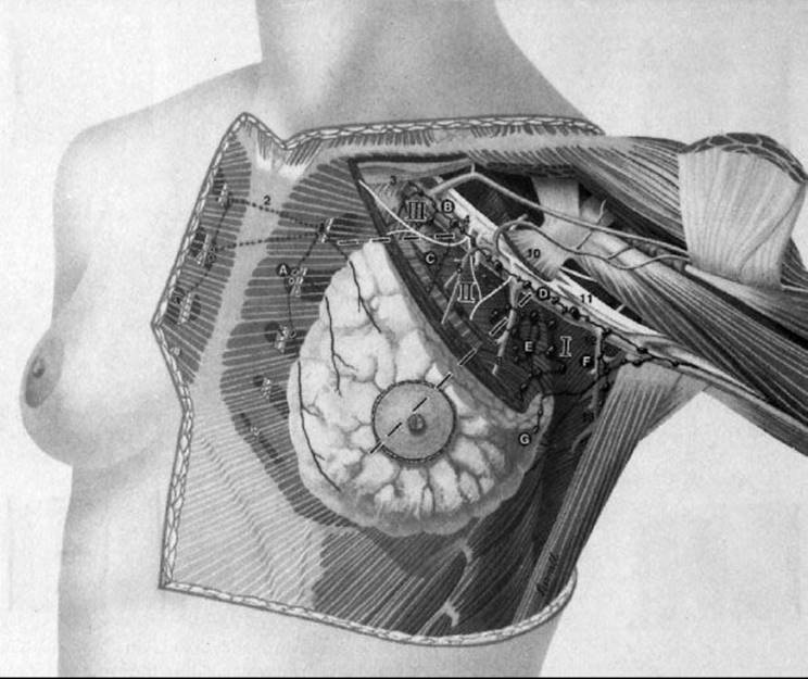
FIGURE 56.2. Location of the three levels of axillary lymph nodes. (Redrawn from Morrow M. Axillary node dissection: what role in managing BCa? Contemp Oncol 1994;8(4):16–27, copyright Medical Economics. Adapted from an illustration by John Daughterty, copyright 1994.)
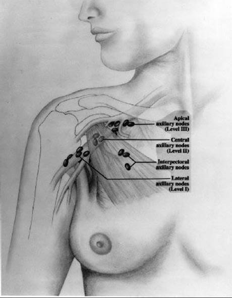
FIGURE 56.3. Treatment planning computed tomography scan of the chest demonstrating the location of the internal mammary vessels, which are typically located approximately 3 to 4 cm lateral to midline and approximately 3 cm deep to the surface. The internal mammary nodes are in close proximity to the vessels, with the most critical nodes being located in the first three intercostal spaces.
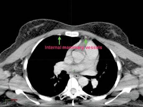
FIGURE 56.4. Distribution of lymphatic drainage of the breast to axillary and internal mammary chains according to the location within the breast. (Data extracted from study of 700 patients undergoing sentinel lymph node mapping by Estourgie et al. Lymphatic drainage patterns from the breast. Ann Surg2004;239(2):232–237.)
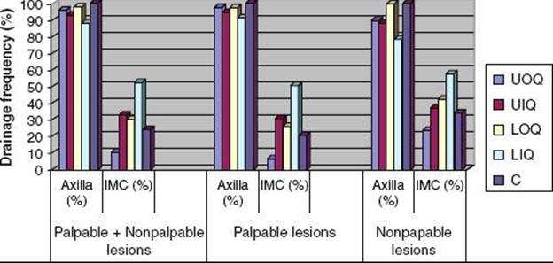
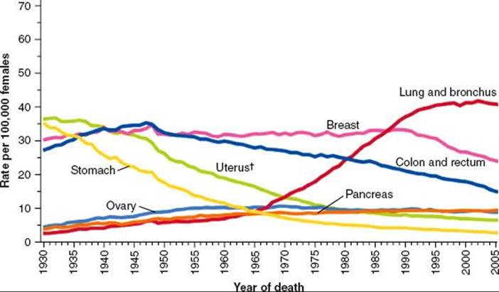
FIGURE 56.5. Age-adjusted cancer death rates for women. Statistics show a recent decrease in breast cancer mortality due to an increase in screening detected malignancies and improvements in treatment. (From Jemal A, Siegel R, Xu J, et al. Cancer statistics, 2010. CA Cancer J Clin 2010;60:277–300, with permission.)
EPIDEMIOLOGY
Breast cancer is the most frequently diagnosed cancer in women, and it is estimated that there will be 229,060 new cases of invasive breast cancer and 63,300 new cases of in situ breast cancers among women in the United States in 2012.4 Primarily due to increased utilization of screening mammography, breast cancer incidence rates increased rapidly in the 1980s. It is estimated that 39,920 breast cancer deaths will occur in 2012, with breast cancer ranking second among cancer deaths in women (after lung cancer). In contrast to the significant number of breast cancer cases in women, it is expected that 2,190 cases of breast cancer will be diagnosed in men in 2012, with approximately 410 breast cancer deaths in men. Due to a combination of early detection, increased awareness, and improvements in therapy, death rates from breast cancer actually declined by approximately 2.3% per year from 1990 to 2007. The decrease in breast cancer mortality is demonstrated in Figure 56.5.
There is considerable geographic, ethnic, and racial variability in breast cancer incidence. Ethnicity and national origin rank highly as predictors of risk for breast cancer, with up to a 10-fold variation throughout the world.5Compared with other well-established risk factors such as age of menarche and menopause, age at first childbirth, and family history, geographic and ethnic variability is quite significant. It is likely that a complex interaction of multiple factors, including genetic, environmental, and socioeconomic, contribute to the wide variability in age-adjusted incidence across populations.
The potential contribution of environmental factors and lifestyle is clearly demonstrated in the increasing incidence of breast cancers among Japanese American women and in trends of increasing incidence of breast cancer in Japan with recent changes in lifestyle. It is well recognized that the relatively low incidence of breast cancer in Asian immigrants to the United States has gradually increased as these immigrants have adapted to Western lifestyles.6In Japan, incidence rates have more than doubled from 1960 to 1990. This is likely a result of adaptation of Western lifestyles, including fewer children, later marriage, increasing rates of obesity, and possibly dietary influences.7
In the United States, the incidence of breast cancer in white women is higher than all other populations. Recent data from the National Cancer Institute’s Surveillance, Epidemiology, and End Results (SEER) program report incidence rates of 141 cases per 100,000 white women, compared with 122 in African American, 97 in Asian or Pacific Islanders, 90 in Hispanics, and 58 in Native Americans or Alaskan Natives.8
Although incidence is lower in African American women, the age of onset is younger and African American women are more likely to be diagnosed at a more advanced stage. Several studies have reported an earlier onset of breast cancer in African American, compared with white women, by approximately 10 years; other studies have indicated that after correcting for stage, African American women have more aggressive biology and a poorer overall prognosis.9,10
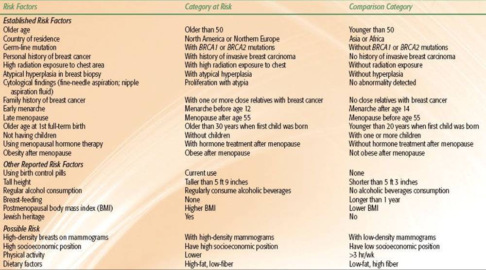
TABLE 56.1 RISK FACTORS FOR BREAST CANCER IN WOMEN
Risk Factors
Table 56.1 summarizes the major risk factors associated with development of breast cancer. With the exception of female gender, increasing age is the most consistent and significant risk factor, with most populations demonstrating increasing incidence rates with age. Other risk factors include personal history and family history of breast cancer, nulliparity or late age at first childbirth, early menarche and late menopause, prior breast biopsy with hyperplasia or atypical hyperplasia, high breast tissue density, radiation exposure at a young age, alcohol consumption, and use of postmenopausal hormone therapy. Some of the national origin or ethnicity variability discussed above may be explained in part by differences in established risk factors, such as age of menarche, parity, and age at first childbirth. However, these factors explain only part of the variability observed in national origin, indicating underlying genetic, environmental, and dietary factors are likely to contribute to the differences in the worldwide incidence of breast cancer.11 Breastfeeding, physical activity, and maintaining a healthy body weight have been demonstrated in various studies to be associated with a lower risk of breast cancer.11
Age
The risk of breast cancer increases exponentially up to the age of menopause, at which time the rate of increase in the risk slows significantly. After the age of 80, the incidence of breast cancer begins to show a slight decline. For women in their late 30s, the annual increase in risk of developing breast cancer is approximately 0.07% per year. This increases to 0.44% per year for women in their late 70s. Although these percentages may seem low, they represent only the risk for a given year; the lifetime risk is a summation of the annual breast cancer risks. Because younger women have a longer life expectancy than older women, younger women have a greater lifetime risk. Only 0.43% of women develop breast cancer before the age of 40, whereas 4% of women develop breast cancer between the ages of 40 and 59 and 6.88% of women develop breast cancer between the ages of 60 and 79.8
Child Bearing/Parity/Breastfeeding
The protective effect of child bearing at younger ages on breast cancer risk is well established. In a worldwide case control study, MacMahon et al.5,12 demonstrated a nearly linear relation between relative risk of breast cancer and age at first birth, with women aged 20 to 25 having nearly a 50% reduction in the relative risk of breast cancer compared with nulliparous women. Interestingly, for women whose first childbirth occurred over age 35, the risk appears greater than nulliparous women. Data on the effect of breastfeeding are not as strong as the data on age at first childbirth, but they do suggest a protective effect. The Oxford Collaborative Group conducted an analysis of 47 studies evaluating breastfeeding and breast cancer risk and reported a decrease in relative risk of breast cancer by 4.3% for each 12 months of breastfeeding.13
Ovarian Function
The relation between ovarian function and breast cancer risk has long been recognized, with long menstrual history (early menarche and late menopause) contributing significantly to breast cancer risk. In experimental models and observational studies, removal of the ovaries reduces the risk of breast cancer.5 Women with surgically induced menopause have been shown to have significantly reduced risks of breast cancer compared with women whose menopause occurred naturally. In comparison with women whose menopause occurs between the ages of 45 and 54 (relative risk = 1), women with early menopause before age 45 have a relative risk of breast cancer of 0.73 and women with late menopause at age 55 or older have a relative risk of 1.48. The data on early onset of menses and its association with breast cancer risk are also well established.5
Exogenous Hormone
The risk of breast cancer associated with hormonal therapy has been controversial. A collaborative meta-analysis from 51 epidemiological studies of over 150,000 women did show an increased relative risk of 1.35 for current or recent users of hormonal replacement therapy.14 The authors reported that postmenopausal hormone replacement therapy increased the annual relative risk of developing breast cancer by 2.3% for each year of hormonal therapy. A randomized trial of postmenopausal hormone therapy from the Women’s Health Initiative Study comparing estrogen and progestin with placebo was closed prematurely, demonstrating a 24% increase in breast cancer, coronary heart disease, stroke, and pulmonary emboli. This study of 46,000 women reported that the combined use of estrogen and progesterone increased the relative risk of breast cancer 8% compared with the risk in nonusers, whereas the use of estrogen alone increased the relative risk only 1%.15 After publication of this study, a dramatic decrease of almost 7% between 2002 to 2003 was primarily attributed to the reduction in the use of hormone replacement therapy following the publication of results from the Women’s Health Initiative in 2002. Since then, breast cancer incidence rates have been generally stable.4 Other studies have also demonstrated increased risks of breast cancer with long-term use of hormonal replacement therapy.16 However, short-term use of hormonal replacement, particularly in women with severe menopausal symptoms, has not been consistently associated with breast cancer risk. For women who have undergone hysterectomy, it seems that hormone replacement therapy with estrogen alone rather than estrogen and progesterone has a minimal effect on breast cancer risk. For women who have not undergone hysterectomy and who elect to be treated with hormone replacement therapy, combined estrogen and progesterone remains the standard for hormone replacement therapy to avoid the risk of endometrial cancer that is associated with unopposed estrogen replacement.16
The use of oral contraceptives has not been consistently shown to increase the risk of breast cancer. There is some evidence that use of oral contraceptives for more than 4 years prior to first pregnancy increases the risk of breast cancer. Other studies, however, have not demonstrated increased risks of breast cancer, even with long-term exposures of more than 15 years.17,18
Family History
The increased risk of breast cancer as a function of family history is well established. For women with a second-degree relative (aunt, grandmother) with breast cancer, the risk is about 1.5, and for women with a history in first-degree relatives (mother or sister), the risk is 1.7 to 2.5.19 This may be explained in part by inheritance of a genetic condition that predisposes an individual to breast cancer development (e.g., mutations in BRCA1 or BRCA2); shared lifestyle; and inheritance of genes that affect risk factors, such as body habitus and age at menarche. Between 20% and 25% of women diagnosed with breast cancer have a positive family history of the disease, and approximately 10% of women with breast cancer are from families who display an autosomal dominant pattern of breast cancer inheritance.20 The actual risk that family history conveys depends on the number of relatives affected and their age at diagnosis (having a first-degree relative with premenopausal breast cancer conveys a greater risk than does having a first-degree relative with postmenopausal cancer). Women with one first-degree relative affected by the disease have an increased relative risk of developing breast cancer two to three times that of women with no family history. Women with two or more first-degree relatives with a diagnosis of breast cancer have a still greater risk, four to six times that of women with no family history.19
Women with a strong family history, particularly those with multiple first- and second-degree relatives diagnosed with breast cancer in the premenopausal years, are at risk for carrying mutations in the breast cancer susceptibility genes, BRCA1 or BRCA2.Although these mutations are present in <1% of the population and account for approximately 5% to 10% of all breast cancer cases, women carrying these mutations have a lifetime risk of developing breast cancer of up to 70% to 80%.20 Genetic counseling or testing should be considered in women at risk for carrying these mutations. Recently, the National Comprehensive Cancer Network (NCCN) published guidelines for genetic testing.21,22 In the context of pre- and posttest counseling, the NCCN recommends that genetic testing be offered when:
1. The individual has a family history of a known BRCA1/BRCA2 mutation,
2. Personal history of breast cancer plus one of the following:
a. Diagnosed age 45 years or younger
b. Diagnosed age ≤50 years with one or more close blood relatives with breast cancer ≤50 years
c. Two breast primaries when first breast primary occurred before age 50
d. Diagnosed at any age, with two or more close blood relatives with breast and/or epithelial ovarian/fallopian tube/primary peritoneal cancer at any age
e. Close male relative with breast cancer
f. An individual of ethnicity associated with higher mutation frequency (e.g., Ashkenazi Jewish).
3. Personal history of epithelial ovarian/fallopian tube/primary peritoneal cancer, or
4. Personal history of male breast cancer.
Personal History of Breast Cancer and History of “Benign” Breast Biopsy
Women with a prior history of breast cancer are at an elevated risk to develop a second contralateral breast cancer.1 Studies with long-term follow-up have demonstrated a risk of breast cancer in the contralateral breast of approximately 10% to 15%, depending on the patient population and length of follow-up.23 Patients treated for invasive breast cancer or ductal carcinoma in situ (DCIS) have similar risks of developing a contralateral breast cancer, which does not appear to be effected by the type of local therapy for the initial lesion. A recent analysis of DCIS patients from the Connecticut Tumor Registry demonstrated a relative risk of developing contralateral breast cancer of 3.35 compared with women without a diagnosis of breast cancer.24 The risk of contralateral breast cancer as a function of prior radiation treatment is discussed in detail later.
Although women with a history of fibrocystic changes have been reported to have an elevated risk of breast cancer, recent evidence suggests that the majority of the elevated risk is due to the smaller proportion of women whose biopsy reveals atypical hyperplasia. Results from the Breast Cancer Detection Demonstration Project, which included over 280,000 women in 29 centers, demonstrated that women with atypical hyperplasia had 4.3 times the breast cancer risk of women without proliferative disease (95% confidence interval [CI], 1.7 to 11.0). In women with proliferative disease lacking atypical hyperplasia the relative risk was 1.3 (95% CI, 0.77 to 2.2). In that study the joint occurrence of family history and atypical hyperplasia had a strong synergistic effect on breast cancer risk.25
Radiation Exposure
Exposure to ionizing radiation during or after puberty increases the risk for development of carcinoma of the breast. Land et al.26,27 reviewed reports on three populations of patients exposed to ionizing radiation by atomic bombings, multiple fluoroscopic examinations for tuberculosis, and multiple examinations for mastitis. They concluded that the risk of radiation-induced cancer of the breast increased approximately linearly with increasing dose and was heavily dependent on age at exposure.
In a study of 31,710 women who had tuberculosis and were examined with repeated fluoroscopic studies, a substantial proportion (26.4%) received doses to the breast of ≥0.1 Gy; the breast cancer risk was greatest among women who had radiation exposure between the ages of 10 and 14 years (relative risk [RR] 4.5 per 0.01 Gy and an additive risk of 6.1 per 104 person-years per 0.01 Gy); there was substantially less excess risk with increasing age at first exposure.28
A high risk of solid tumors, especially breast cancer, has been described in women treated with radiation therapy at a young age for Hodgkin lymphoma. In a review of 1,380 women treated at 15 institutions before the age of 16 years, breast cancer developed in 17 women; 7 after radiation therapy alone and in 10 after irradiation and chemotherapy. Sixteen breast cancers appeared within or at the margin of the irradiation fields. The cumulative probability of breast cancer at 40 years of age was 35%. Women in this cohort of survivors had a risk of breast cancer 70 times higher than that of the general population.29
In a recent study, relative risks of breast cancer were defined by radiation dose to the chest (0, 20 ≤ 40 Gy, or ≥ 40 Gy). Estimates were from this case-control study conducted within an international population-based cohort of 3,817 female survivors of Hodgkin lymphoma diagnosed at age 30 years or younger. For a survivor who was treated at age 25 years with a chest radiation dose of at least 40 Gy without alkylating agents, estimated cumulative absolute risks of breast cancer by age 35, 45, and 55 years were 1.4% (95% CI, 0.9% to 2.1%), 11.1% (95% CI, 7.4% to 16.3%), and 29.0% (95% CI, 20.2% to 40.1%), respectively.30 A reduced volume of radiation fields has also been shown to reduce the breast cancer risk associated with treatment of Hodgkin lymphoma. The current practice of limiting radiation fields to involved nodal regions should help to further reduce the risk of radiation related breast cancers in Hodgkin’s survivors.31
Body Mass Index, Physical Activity, and Dietary Factors
The inherent complex interaction between body mass, physical activity, and diet complicates interpretation of epidemiologic studies correlating these factors with breast cancer risk. Body mass index (BMI) has been clearly associated with breast cancer risk in a number of studies, but it appears to influence breast cancer risk predominantly in postmenopausal women. In premenopausal women, most studies have not observed a strong relation between BMI and breast cancer risk. In postmenopausal women, a pooled analysis of prospective studies demonstrated the risk of breast cancer to be 30% higher in postmenopausal women with a BMI over 31 kg/m2 compared with women with a BMI of 20 kg/m2.32 The higher risk of breast cancer with increased BMI in postmenopausal women is likely due to higher estradiol levels associated with increased adipose tissue and increased aromatase, which is involved in the conversion of androgens to estradiol. In postmenopausal women, this is the primary source of estradiol, whereas in premenopausal women estradiol is predominantly from the ovaries so there is little association with BMI and estradiol levels.
Physical activity can have a significant impact on BMI, so it is sometimes difficult to separate these two effects in interpreting breast cancer risk. A majority of studies, however, have observed a lower risk of breast cancer among women who are more physically active compared with women who are sedentary.
Although it has been suggested that obesity and high intake of meat, dairy products, and fat may increases risk and fiber, fruits and vegetables, and phytoestrogens (soy products) may reduce risk, strong links between diet and breast cancer risk have not been clearly established.33 Accurate data regarding nutritional factors are difficult to evaluate in most epidemiologic studies. A pooled analysis of eight prospective studies did not conclude a relation between dietary fat intake and breast cancer risk. Similarly, large prospective studies have failed to demonstrate an association between dietary fiber intake and breast cancer risk.34 Phytoestrogens found in soy products and many cereals, tea, and vegetables may reduce the effects of estrogens. Given the lower incidence of breast cancer in Asian countries with high soy intake, one might hypothesize a relation between this dietary factor and breast cancer risk. Although animal studies suggest that high soy intake is protective, human studies have not been as conclusive.
Alcohol Consumption
In an analysis by the Oxford Group of 53 epidemiological studies, including 58,515 women with breast cancer and 95,067 women without breast cancer, women with daily consumption of four or more drinks a day had a 50% higher breast cancer risk.35 The average consumption of alcohol reported was 6.0 g per day (about half a unit or drink of alcohol per day). Compared with women who reported drinking no alcohol, the relative risk of breast cancer was 1.32 (1.19 to 1.45; P <.00001) for an intake of 35 to 44 g per day alcohol, and 1.46 (1.33 to 1.61; P <.00001) for ≥45 g per day or more of alcohol. The relative risk of breast cancer increased by 7.1% (95% CI, 5.5 to 8.7%; P <.00001) for each additional drink of alcohol consumed on a daily basis. Chen et al.36 reported on a prospective observational study of 105,986 women enrolled in the Nurses’ Health Study between 1980 and 2008. With 2.4 million person-years of follow-up, 7,690 cases of invasive breast cancer were diagnosed. They found that increasing alcohol consumption was associated with increased breast cancer risk that was statistically significant at levels as low as three to six drinks per week (RR 1.15; 95% CI, 1.06 to 1.24). Moreover, binge drinking, but not frequency of drinking, was associated with breast cancer risk after controlling for cumulative alcohol intake. They concluded that low levels of alcohol consumption were associated with a small increase in breast cancer risk, with the most consistent measure being cumulative alcohol intake throughout adult life.
Although the relation among physical activity, BMI, and dietary factors may be difficult to separate, it is apparent that maintaining a sound, varied diet, limiting alcohol intake, avoidance of obesity, and moderate physical activity are modifiable behaviors that can impact breast cancer risk (as well as other health-related issues) and should be encouraged.
Mammographic Density
There is a large body of evidence suggesting a correlation between mammographic breast density and breast cancer risk.37,38–39 The risk of breast cancer associated with the highest category of density has been estimated to be two to six times greater than in the lowest category. Although the causal link between mammographic density remains poorly understood, breast density is in part attributable to genetic factors.
Boyd et al.40 and Byrne et al.41 noted that women with 75% or greater breast density parenchymal patterns on the mammogram had a fivefold greater risk of breast cancer. This parameter was independent of other prognostic factors, such as family history, age at first birth, or alcohol consumption.
Determining an Individual’s Risk
It is important to consider the combination of risk factors when a generalized risk profile is determined. Gail et al.42 have used these epidemiologic risk factors to derive a model for predicting an individual’s annual and lifetime risks of breast cancer. In the Gail model, an individual’s annual risk of breast cancer is based on her present age, number of first-degree relatives with breast cancer, age at first birth, age at menarche, number of breast biopsies, and history of atypical ductal hyperplasia. The use of exogenous hormones is not considered in this model, and many of the other risk factors discussed above are not incorporated into this specific model.
TABLE 56.2 GENES ASSOCIATED WITH HEREDITARY BREAST CANCER
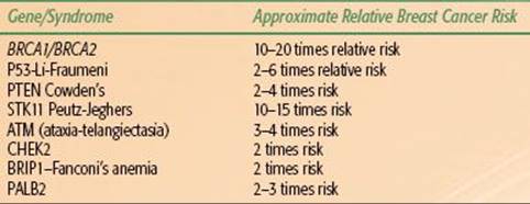
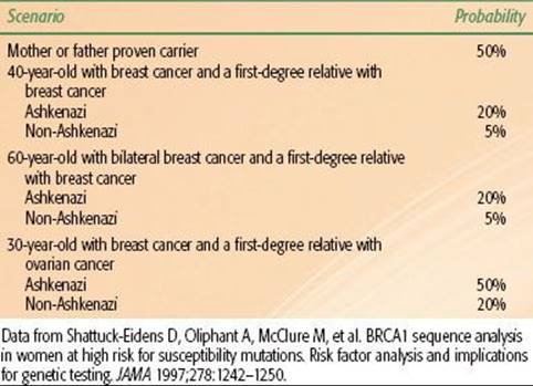
TABLE 56.3 PROBABILITY OF BRCA1 GERMLINE MUTATIONS IN VARIOUS CLINICAL SCENARIOS
PREVENTION AND GENETIC SCREENING
Approximately 10% of breast cancer patients have familial breast cancer, typically defined as breast cancer showing an autosomal dominant inheritance pattern.20 During the 1990s, germline mutations in three important tumor suppressor genes—p53, BRCA1,and BRCA2—were discovered in family members of individuals with familial breast cancer.43–45 All three genes have been shown unequivocally to predispose to breast cancer. A number of genetic conditions associated with increased risk of breast cancer are summarized in Table 56.2.
Mutations in p53
Germline mutations in the p53 gene are very rare and result in Li-Fraumeni syndrome, named after two investigators who made significant contributions to the understanding of this condition.45 The p53 gene is one of the most important tumor suppressor genes and has been called the “guardian of the genome” because of its critical role in cellular pathways that recognize and direct a response to DNA injury. One consequence of a germline mutation in p53is an increased risk for a variety of cancers, including childhood sarcomas, gynecologic tumors, and breast cancer. Breast cancer is the most common malignancy in patients with Li-Fraumeni syndrome; the lifetime risk is estimated to be 90%.45
Mutations in BRCA1 and BRCA2
Studies of patients with familial breast cancer led to the discovery of BRCA1 in 1995 and BRCA2 in 1996. Similar to p53, both BRCA1 and BRCA2 are tumor suppressor genes that contribute to the stability of the genome by mediating the effects of the cellular response to DNA injury. Individuals with a germline mutation in BRCA1 have a lifetime risk of breast cancer of 65% to 85%. In addition, these individuals have an elevated lifetime risk of ovarian cancer, which may approach 50%. Other types of cancer that develop more frequently in BRCA1 carriers include colon and prostate cancers. The lifetime risk of breast cancer for women with germline BRCA2 mutations mirrors that for women with BRCA1 mutations. BRCA2 mutation carriers are also at increased risk for ovarian cancer compared with the general population, but their risk is much less than the risk in women with BRCA1 mutations. BRCA2 is also associated with male breast cancer and pancreatic cancer. Genetic screening for germline mutations in BRCA1 and BRCA2 is now possible. Testing should be performed in centers equipped with genetic counseling programs designed to properly inform individuals of the social, economic, and legal consequences associated with genetic testing. Germline mutations in BRCA1and BRCA2 are rare, occurring in fewer than 7% of patients with breast cancer. Thus, only a minority of breast cancer patients with a family history of the disease would be predicted to carry a mutation in one of these genes. Table 56.3 contains data concerning the probability of carrying a BRCA1 mutation based on an individual’s age at cancer diagnosis, personal cancer history, and family cancer history and whether the individual is of Ashkenazi Jewish descent.46
No definitive data exist on which to base screening recommendations for individuals with a proven germline mutation in a gene predisposing to the development of breast cancer. The NCCN has published a guideline recommending that individuals with a genetic predisposition undergo annual clinical and self-breast examination prior to age 25 and annual mammography or magnetic resonance imaging (MRI) and semiannual clinical and self-breast examination after age 25.47 In addition, annual pelvic examinations with transvaginal sonography, color Doppler examinations of the ovaries, and measurement of serum cancer antigen (CA-125) levels are recommended beginning at age 25 to 35 years. For those women over the age of 35, a risk-reducing bilateral salpingo-oophorectomy is recommended, with possible short-term hormone replacement therapy.
Breast Cancer Prevention Strategies
Tamoxifen
Understanding of the role of estrogens and progesterones in breast cancer development has led to the development of pharmacologic strategies that could significantly decrease the incidence of breast cancer over the next two decades.
Several pharmaceuticals that affect the estrogenic pathways have been studied as chemopreventive agents, but the only agent for which mature data from clinical trials are available is tamoxifen.48,49–50,51–52,53 Interest in tamoxifen as a chemopreventive agent arose after a number of randomized trials designed to test the efficacy of hormonal therapy for invasive breast cancer reported that tamoxifen reduced the incidence of contralateral breast cancer. On the basis of these data, in 1992 the National Surgical Adjuvant Breast and Bowel Project (NSABP) began a randomized, placebo-controlled study (the P-1 trial) to test the efficacy of 5 years of tamoxifen in the prevention of breast cancer.53 Between 1992 and 1997, 13,388 women with a 1.67% or greater predicted risk of developing breast cancer within 5 years were enrolled in this trial. Risk was assessed using a modification of the Gail model, which permitted enrollment of any woman older than 60 years of age and selected women younger than 60 years with additional risk factors that increased their annual risk to at least that of a 60-year-old. In addition, women with a history of lobular carcinoma in situ (LCIS) were included. Women were not allowed to use estrogen replacement therapy during their participation in the trial. The results of the NSABP P-1 trial indicated that tamoxifen reduced the rates of invasive and noninvasive breast cancer by 49% and 50%, respectively. The benefit of tamoxifen was seen in all age groups (≤49 years, 50 to 59 years, ≥60 years). In addition, women with a history of atypical ductal hyperplasia had an 86% risk reduction, and women with a history of LCIS had a 56% risk reduction. Finally, the benefit was seen across all subgroups specified according to family history of breast cancer. Tamoxifen selectively reduced the incidence of estrogen receptor–positive tumors; estrogen receptor–negative tumors developed at an equal rate in the tamoxifen and placebo groups. No evidence was shown of a cardioprotective effect of tamoxifen in this trial, but the number of osteoporosis-related fractures was reduced in the tamoxifen-treated cohort. Tamoxifen increased the risk of developing stage I endometrial cancer (risk ratio of 2.53).
More recently, a landmark study was published that compared tamoxifen to raloxifene as a preventative agent in postmenopausal women with breast cancer. Raloxifene, a drug that is primarily used in prevention of osteoporosis, had been shown in prior studies to decrease the incidence of breast cancers. The NSABP study of tamoxifen and raloxifene was a prospective, double-blind, randomized clinical trial.54 There were 19,747 postmenopausal women of mean age 58.5 years with increased 5-year breast cancer risk. Patients were randomized to oral tamoxifen (20 mg per day) or raloxifene (60 mg per day) for 5 years. There were 163 cases of invasive breast cancer in women assigned to tamoxifen and 168 in those assigned to raloxifene, which was not significantly different between the two arms. The main benefit of raloxifene was in toxicity. There were 36 cases of uterine cancer with tamoxifen and 23 with raloxifene. No differences were found for other invasive cancer sites, for ischemic heart disease events, or for stroke. Thromboembolic events also occurred less often in the raloxifene group. The number of osteoporotic fractures in the groups was similar and there were fewer cataract surgeries with raloxifene. There was no difference in the total number of deaths (101 vs. 96 for tamoxifen vs. raloxifene) or in causes of death. It appears from this study that raloxifene is as effective as tamoxifen in reducing the risk of invasive breast cancer and has a lower risk of thromboembolic events. Of note there were slightly more noninvasive cancers in the raloxifene group, but that was not statistically significant
A comparison of the tamoxifen P-1, P-2, and other tamoxifen prevention trials is outlined in Table 56.4.48,49–50,51–52,55,56
Prophylactic Surgery
An alternative strategy used to prevent breast cancer development is prophylactic surgical intervention. Hartmann et al.57 analyzed outcomes in women with a family history of breast cancer who underwent bilateral prophylactic mastectomy at the Mayo Clinic between 1960 and 1993. With a median follow-up time of 14 years, only 4 of the 639 treated patients developed breast cancer. According to the Gail model, 37.4 cases of breast cancer would have been expected to develop in this population, so the prophylactic surgery resulted in an 89.5% risk reduction (P <.001).57
Breast cancer prevention strategies will continue to be a dynamic area of preclinical and clinical research.
TABLE 56.4 RESULTS OF CHEMO-PREVENTION TRIALS

NATURAL HISTORY AND ORIGINS
All forms of breast cancer are believed to develop as a consequence of unregulated cell growth and the development of phenotypic changes such as the ability to invade, recruit a new blood supply, and metastasize. These changes in phenotypes are secondary to the development of aberrations in genetic pathways. Some of these aberrations are inherited (germline mutations), whereas others develop during the life of a breast cell (somatic mutations). It is currently believed that most breast cancer is a consequence of a series of somatic mutations. As previously noted, only 20% to 25% of breast cancer patients have a history of breast cancer in a first-degree relative. However, it is possible that some women without a first-degree relative with breast cancer still inherit a genetic background that predisposes to breast cancer. These mutations may be insufficient to cause breast cancer unless accompanied by other mutations and, therefore, would be predicted to have a low penetrance. Historically, it has been much more difficult to discover low-penetrance mutations than to discover germline mutations that result in an autosomal dominant pattern of breast cancer development. However, with newer molecular techniques, such as DNA-array assays, the identification of low-penetrance predisposing mutations may be more feasible.
Left untreated, breast cancer can have a variable clinical course. A classic article by Bloom et al.58 outlined the natural history of breast cancer patients, seen between 1805 and 1933, not treated by surgery or irradiation, 250 of whom had a pathologic diagnosis of cancer. There were no patients with stage I disease, 2.4% with stage II, 23% with stage III, and 74% with stage IV. Survival in the untreated group was 3.6% compared with an overall survival of 34% in patients treated with radical or modified radical mastectomy with or without radiation.
Concepts regarding the natural history of breast cancer have undergone great evolution over the past 100 years, with a profound impact on the management of these patients. The Halsted59 model was based on an orderly progression to the regional lymph nodes and from there to distant metastatic sites. Later, Keynes60 and Crile et al.61 suggested that breast cancer is a systemic disease and that extensive surgery to achieve local tumor control was not as important as originally believed. This alternative hypothesis was fully demonstrated in both laboratory and clinical studies by Fisher,62 who advanced the concept that breast cancer, as a systemic process involving host–tumor interactions, would not show substantial effects on survival with variations in locoregional treatment. A third hypothesis put forward by Hellman63 considers breast cancer as a heterogeneous disease with a spectrum extending from a tumor that remains localized throughout its course to one that disseminates systemically, even when detected as a small lesion, suggesting that metastases are a function of tumor growth and progression factors.
The growth rate of a tumor in the breast is thought to be constant from the date of origin. Using estimates of doubling time, it would take an average of approximately 5 years for a tumor to reach palpable size, and those lesions with slower doubling time would have an even longer latent period.64
The most common site of origin of breast cancer is the upper outer quadrant (38.5%), followed by the central area (29%), the upper inner quadrant (14.2%), the lower outer quadrant (8.8%), and the lower inner quadrant (5%).64These rates correlate with the amount of breast tissue in the various quadrants. Cancer is somewhat more common in the left than in the right breast and may appear in both breasts simultaneously (1% to 2%). As noted above, women with a history of breast cancer have a 10% to 15% risk of developing a new primary in the contralateral breast.
As the cancer grows, it travels along the ducts, eventually breaking through the basement membrane of the duct, invading adjacent lobules, ducts, fascial strands, and the mammary fat, spreading through the breast lymphatics and into the peripheral lymphatics. The tumor can grow through the wall of blood vessels, spread into the deep lymphatics of the dermis, and eventually produce edema of the skin (peau d’orange), which usually indicates that the superficial as well as the deep lymphatics are involved. Skin dimpling can be caused by involvement of Cooper’s ligament. Ulceration and infiltration of overlying skin, which may develop late in the course of the disease, are usually preceded by fixation and localized redness of the skin over the tumor and are less frequently seen because of the current emphasis on screening and early diagnosis.64
TABLE 56.5 INTERNAL MAMMARY NODE INVOLVEMENT RELATED TO LOCATION OF PRIMARY TUMOR AND TO AXILLARY NODE INVOLVEMENTA

Axillary Spread
A common route of spread of breast carcinoma is first through the axillary lymph nodes, with the incidence increasing with larger tumors. Depending on mode of detection, tumor size, histology, and other clinical or pathological factors, between 10% and 40% of newly diagnosed stage T1 and T2 breast cancers have pathologic evidence of axillary nodal metastases. Voogd et al.65 assessed 7,680 patients with documented invasive breast cancer; of 5,125 patients known to have clinically negative lymph nodes who underwent axillary dissection, 1,748 (34%) had positive lymph nodes at pathologic examination. Univariate analysis showed that lymph node metastases were associated with tumors larger than 1 cm (P = .001), moderate or poorly differentiated nuclear grade (P= .005), high fraction of cells in the growth phase (S phase) of the cell cycle (P = .041), presence of lymphatic vascular invasion (P <.001), and age younger than 60 years (P = .01).
Multiple studies have shown a strong relation between primary tumor size and axillary nodal involvement.66–70 Even patients with T1a and T1b disease have significant nodal involvement. Mustafa et al.71 noted an overall frequency of axillary lymph node metastases in T1a and T1b lesions of 16%; integrating age, tumor size, and grade predicted the frequency of nodal metastases. Overall, patients with all three poor prognostic indicators had a 34% incidence of nodal involvement, and those with no poor prognostic factors had a ≤7% probability of nodal metastases. Gann et al.72 reviewed 18,025 patients with a diagnosis of breast carcinoma from the American College of Surgeons database. On multivariate analysis, the following factors were independently associated with a greater likelihood of one or more positive lymph nodes: larger tumor size, young age, African American or Hispanic race, outer-half tumor location, poor or moderate differentiation, aneuploidy, and infiltrating ductal histologic type.
Although up to 30% to 40% of T1 or T2 clinically node-negative breast cancers may have pathologically involved lymph nodes, data from NSABP-04 suggest that less than half of clinically negative but pathologically positive axilla will experience a clinical relapse in the axilla.73 In this study, operable breast cancer patients, who were primarily diagnosed with palpable breast tumors in the premammography era, were randomized to one of three arms: simple mastectomy without axillary dissection, simple mastectomy with axillary dissection, or simple mastectomy with comprehensive chest wall and regional nodal irradiation. In the arm undergoing axillary dissection, nodal positivity was approximately 40%. Nodal control was excellent (>97%) in this arm as well as in the arm treated with radiation. In the simple mastectomy arm, where no nodal treatment by radiation or dissection was administered, the axillary failure rate was approximately 20%. Assuming equal distribution among the arms, it is presumed the pathological involvement was approximately 40%, indicating that less than half of those with pathological involvement eventually failed clinically.
Internal Mammary Spread
Metastases to the internal mammary nodes (IMNs) are correlated with tumor size, are more frequent from medial half and central lesions, and occur more frequently when there is axillary node involvement (Table 56.5).74 A table with more detailed analysis of internal mammary involvement by tumor size, location and number of positive nodes is found in Chapter 57. Veronesi et al.75 found that, among women with tumors larger than 2 cm who were younger than 40 years of age and had positive axillary nodes, there was a 41% risk of having positive IMNs on IMN dissection; the corresponding risk for patients of that age with negative nodes was 16%. Sugg et al.76 reviewed 286 patients with breast cancer who underwent IMN dissection. Positive IMNs were associated with primary tumor size (P <.0001) and the number of positive axillary nodes (P <.0001), but not with age or primary tumor location. Patients who had positive IMNs (25% of all patients) had a significantly worse overall 20-year disease-free survival rate than did patients with negative IMNs (P <.0001). Clinical failure of the internal mammary nodes is extremely rare, despite the evidence of pathological involvement from these studies. Most studies looking at nodal failure patterns report failure in the internal mammary region of <1%.77–81
Supraclavicular Spread
Spread to supraclavicular lymph nodes usually follows involvement in the high axillary lymph nodes or IMNs depending on the location of the primary lesion. Chen et al.82 reviewed 2,658 patients with invasive breast cancer who underwent surgery and adjuvant therapy. With a median follow-up period of 39 months, supraclavicular lymph node metastasis developed in 113 (4.3%). Young age (≤40 years), tumor size >3 cm, angiolymphatic invasion, negative estrogen-receptor status, and DNA synthetic phase fraction >4% were significant for predicting supraclavicular metastasis on univariate analysis. Three predictive factors were significant after multivariate analysis: high histologic grade, more than four positive nodes, and axillary level II or III involved nodes. In patients with axillary level I involved nodes and four or fewer positive nodes, the incidence of supraclavicular lymph node metastasis was 4.4%, but if axillary level III was involved, it increased to 15.1%.
Clinical failure in the supraclavicular fossa is relatively rare in patients with early-stage breast cancer and is dependent on the degree of axillary involvement. For patients with no or minimal nodal involvement (less than three involved axillary nodes), supraclavicular failure is extremely rare. In an analysis of 691 patients with zero to three nodes involved undergoing breast-conserving surgery and radiation therapy to tangential fields only without regional nodal irradiation, Galper et al.78 reported failure in the supraclavicular fossa in 1.3% of patients.
Several studies have demonstrated that the failure rate in supraclavicular nodes, left untreated, may be as high as 20% in patients with advanced disease or more than four lymph nodes involved.83–86,87 In a cohort of 1,031 patients with operable breast cancer treated with mastectomy and level I or II node dissection plus adriamycin-based chemotherapy, but no radiation, Strom et al.87 reported failure in the supraclavicular fossa was 8% at 10 years. Predictors of supraclavicular failure included four or more involved nodes and gross extranodal extension. In these subgroups, supraclavicular failure ranged from 14% to 19%. Radiation to the supraclavicular fossa in these higher risk patients results in high local control rates, with isolated supraclavicular failures occurring in less that 1% of prophylactically treated nodes.
Systemic Spread
Using monoclonal antibodies to epithelial cytokeratins or tumor-associated cell membrane glycoproteins, carcinoma cells can be detected on cytologic bone marrow (or lymph node) preparations. Braun et al.88 combined patient data from nine studies involving 4,703 patients with stage I, II, or III breast cancer. Micrometastasis was detected in 30.6% of the patients. With a median follow-up of 5.2 years, patients with bone marrow micrometastasis had larger tumors and tumors with a higher histologic grade and more often had lymph node metastases and hormone receptor–negative tumors, compared with those without bone marrow micrometastasis. The presence of micrometastasis was a significant prognostic factor for poorer overall survival (RR 2.15; P <.001), breast cancer–specific survival (RR 2.44; P <.001), disease-free survival (RR 2.13), and distant disease-free survival (RR 2.33; P <.001 for all outcomes measures). In multivariable analysis, micrometastasis was an independent predictor of a poor outcome.
Local Control and Systemic Metastasis
Patients treated for breast cancer are at risk for local-regional failure, as well as systemic metastasis. It is evident from the available literature that optimizing local control can impact systemic metastasis and survival, and similarly, systemic therapy can impact local control.89–91,92 Integration of systemic therapy with radiation will be discussed in detail later, but numerous studies have clearly demonstrated a significant improvement in local control with the use of radiation therapy and systemic therapy (both cytotoxic and hormonal) compared with radiation therapy without the use of systemic therapy.92–97 Appropriate integration of both local and systemic treatments through a multidisciplinary approach is thus essential to optimize outcome. Although there is some overlap, prognostic factors for local-regional control and systemic metastasis often differ.
For patients with early-stage invasive breast cancer, even with appropriate systemic therapy, development of metastasis can vary from <5% in women with T1a disease and favorable histology, to >40% for women with T2 tumors and pathologically involved lymph nodes. Similarly, local-regional failure rates can vary from <5% to over 40% depending on local treatment and prognostic factors for local failure.93–95,96,98–100,101–102
The impact of local control on systemic metastasis in breast cancer as well as other malignancies has been the subject of considerable debate and controversy. Although the benefits of local control with respect to cosmesis and quality of life are apparent, the independent effect of local control on systemic disease and survival has been questioned. Several studies have identified local control as an independent predictor of disease-free or overall survival.99,102,103–106 Fisher et al.,103 in an analysis of patients treated in NSABP protocol B-06, concluded that ipsilateral breast tumor recurrence was a harbinger, but not a cause, of distant metastases. Although mastectomy or breast irradiation after lumpectomy prevented expression of the marker (breast relapse), neither lowered the risk of distant metastases, which was determined by a host of prognostic factors.
More recent meta-analyses, however, have demonstrated a small but significant impact of local control on systemic metastasis and overall survival.89,90 A meta-analysis of randomized trials by Vinh-Hung and Verschraegen90comparing breast-conserving surgery without radiation to breast-conserving surgery with radiation confirms an approximate threefold reduction in local relapse with radiation therapy and an 8.6% improvement in mortality in the radiated cohorts.
One of the most convincing and authoritative studies related to this subject is the recent analysis of the Early Breast Cancer Trialists Collaborative Group (EBCTCG).89 In this analysis, over 42,000 women were enrolled in 78 randomized trials that compared 24 types of local treatment (radiotherapy vs. no radiotherapy, more vs. less surgery, or more surgery vs. radiotherapy). The EBCTCG attempted to relate the effect on local control to breast cancer mortality by grouping studies into whether the 5-year local relapse risk difference between the two comparisons of local therapy exceeded 10%. In those comparisons in which the difference in 5-year local recurrence risk was <10%, there was no impact on 15-year breast cancer mortality. However, there were 25,000 women enrolled in trials in whom the comparisons involved >10% differences in local control. In those studies, the difference in local recurrence risks at 5 years were 7% versus 26%, and the 15-year mortality risks were 44.6% versus 49.5% (P <.00001). Figure 56.6 summarizes the results of this meta-analysis with respect to the impact of radiation on breast cancer mortality in both breast conservation and following mastectomy. An update from the EBCTCG focused on 10,801 women enrolled in 17 randomized trials of radiotherapy versus no radiotherapy after breast-conserving surgery and relates the absolute reduction in 15-year risk of breast cancer death to the absolute reduction in 10-year recurrence risk.107 Overall, radiotherapy reduced the 10-year risk of any first recurrence from 35.0% to 19.3% (P <00001) and reduced the 15-year risk of breast cancer death from 25.2% to 21.4% (P <0001). In women with pN+ disease (n = 1,050), radiotherapy reduced the 10-year recurrence risk from 63.7% to 42.5% (P <00001) and the 15-year risk of breast cancer death from 51.3% to 42.8% (P = 01). Overall, about one breast cancer death was avoided by year 15 for every four recurrences avoided by year 10.
FIGURE 56.6. Effect of radiotherapy on breast cancer mortality and on all-cause mortality after breast-conservation surgery or after mastectomy with axillary clearance: 15-year or 20-year probabilities. (From Clarke M, Collins R, Darby S, et al. Effects of radiotherapy and of differences in the extent of surgery for early breast cancer on local recurrence and 15-year survival: an overview of the randomised trials. Lancet 2005;366:2087–2106, with permission from Elsevier.)
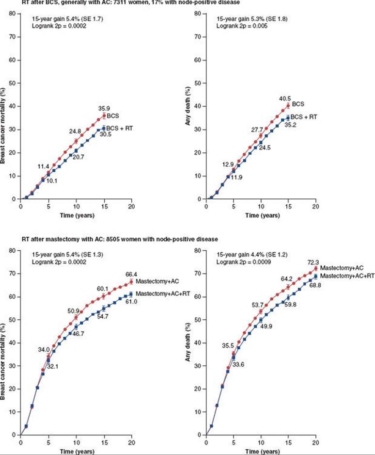
TABLE 56.6 BREAST CANCER SCREENING GUIDELINES

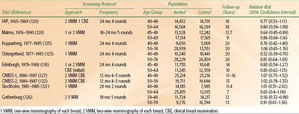
TABLE 56.7 RANDOMIZED CONTROLLED TRIALS OF BREAST CANCER SCREENING
CLINICAL PRESENTATION
The majority of patients with T1 or T2 breast cancers present with a painless or slightly tender breast mass or have an abnormal screening mammogram. Patients with more advanced tumors may have breast tenderness, skin changes, bloody nipple discharge, or occasionally change in the shape and size of the breast. Rarely, patients may present with axillary lymphadenopathy or even distant metastasis. As noted previously, however, depending on tumor size, method of detection, and pathologic factors associated with the primary tumor, up to 30% to 40% of women with a clinically negative axilla may harbor subclinical pathologically involved axillary nodes.
The impact of delays in evaluation or treatment on the survival of patients with breast cancer is controversial. Richards et al.,108 in a review of 2,964 patients, found 942 (32%) who had symptoms for 12 or more weeks before their first hospital visit. Locally advanced or metastatic disease was detected in 32% of patients with delays compared with 10% of patients with intervals of <12 weeks between the onset of symptoms and hospital referral (P <.0001). Multivariate analysis showed that a longer duration of symptoms had a highly significant adverse influence on survival, but this was no longer evident when tumor size and stage were included in the model. Olivotto et al.109 found that delays in diagnosis of 6 to 12 months led to increased risk of larger tumor size and more lymph node metastases compared with patients diagnosed within 4 to 12 weeks of an abnormal screening mammogram result.
SCREENING IN BREAST CANCER
Mammography
Screening mammography has resulted in a shift in both the incidence and stage of patients presenting with breast cancer. In a simplified model described by Harris et al.,110 for every 1,000 screening mammograms, 80 women (8%) will be recalled for additional diagnostic imaging, 10 (1%) will require tissue diagnosis, and of those undergoing biopsy only 3 (0.3%) will have a malignancy.
There is a large body of evidence that early detection by mammography, followed by appropriate local, regional, and systemic treatment, is associated with reduced breast cancer mortality rates for women 50 years of age and older.111–114 Although it remains an active area of debate, several authors agree that screening mammography in women 40 to 49 years of age may reduce mortality from breast cancer,25,112–118 and mammographic screening beginning at age 40 is encouraged in the majority of published guidelines (Table 56.6). The reader is referred elsewhere for an extensive discussion of screening mammography studies.112–114 Selected series will be briefly discussed here and a summary of the classic screening mammography trials is summarized in Table 56.7.70,119–124,125,126–127 Collectively, these studies demonstrate a decrease in mortality and migration of patients from later stages of disease to earlier stages of disease with the use of screening mammography.25,112–118,120,123,124
Sixteen-year results are available from the Health Insurance Plan study, which involved two systematically selected, randomly sampled groups of approximately 31,000 women aged 40 to 64 years who were offered screening examinations.128,129 Compared with the control group, which was observed and monitored, the mortality rate was reduced by approximately one-third in screened women 50 to 59 years of age. The survival difference between mammography-only and clinical examination–only cases appeared in years 7 to 10 after diagnosis. Although the greatest difference in mortality between screened and control group was detected in women 50 to 59 years of age when they entered the study, the differences are in favor of the study group at all ages.
Tabár et al.123–124,125 demonstrated the benefit from mammography screening in two Swedish counties. In the group of women 20 to 69 years of age, there were 6,807 diagnosed with breast carcinoma over a 29-year period and 1,863 breast carcinoma deaths. The mortality rate from breast carcinoma diagnosed in women 40 to 69 years of age who were screened during the screening period (1988–1996) declined by 63% (RR 0.37) compared with the breast carcinoma mortality rate during the period when no screening was available (1968–1977). The reduction in mortality rate observed during the service-screening period, adjusted for selection bias, was 48%. No significant change in breast carcinoma mortality rate was observed over the three periods in women who did not undergo screening. In a recent long-term update, there has remained a highly significant reduction in breast cancer mortality in women invited to screening (RR 0.69; 95% CI: 0.56 to 0.84; P = .001). At 29 years of follow-up there was a reduction in 1 death for every 414 women undergoing screening for 7 years. They conclude that the group invited to screening resulted in a highly significant decrease in breast cancer specific mortality.130
In contrast to the above, a Canadian study revealed that in women aged 50 to 59 years, the addition of annual mammography screening to physical examination had no effect on breast cancer mortality. Miller et al.122 reported a study of 39,405 women (aged 50 to 59 years) randomly assigned to one of two study groups. By December 31, 1993, 622 invasive and 71 in situ breast cancers were observed in the mammography plus physical examination group, compared with 610 and 16, respectively, in the physical examination–only group. At 13-year follow-up, the number of deaths from breast cancer was 107 in the mammography plus physical examination group and 105 in the physical examination–only group. The results of the Canadian study may be due to the unbalanced allocation of women with advanced cancers (large tumors, four or more positive nodes) to the screened group, the poor quality of the mammography in the trial, and an insufficient sample size.116,131
Screening in Women Under Age 50
Although the majority of studies clearly support the impact of screening mammography on mortality in women over 50 years of age, data on screening younger women are more conflicting. Frisell and Lidbrink121 presented updated data on breast cancer mortality for women younger than age 50 years from the Stockholm Mammographic Screening Trial. Approximately 40,000 women aged 40 to 64 years (14,842 aged 40 to 49 years) were randomized to a trial of breast cancer screening by single-view mammography alone; 20,000 women (7,103 aged 40 to 49) were randomized to a control group. In the 40- to 49-year age group, 24 and 12 breast cancer deaths were found in the study and control groups, respectively, after 11.4 years of follow-up. The relative risk of breast cancer death in screened versus nonscreened women was 1.08 (95% CI, 0.54 to 2.17).
A large trial was conducted in the United Kingdom involving 45,841 women aged 45 to 64 years who were offered annual screening by clinical examination and mammography; 63,636 were taught breast self-examination, and 127,117, for whom no extra services were provided, constituted a control population.132 After 16 years of follow-up, the breast cancer mortality rate was 27% lower in the two screening centers combined than in the four comparison centers. A 35% decrease in mortality rate was observed in mammographically screened women in all cohorts aged 45 to 64 years at entry. There was no evidence of less benefit in women aged 45 to 49 years at initial screening compared with a reduction of 25% in women aged 50 to 74 years.133
In a Swedish study evaluating women in the 40- to 49-year age group, Hellquist et al.134 reported 803 breast cancer deaths in a screening study group compared with 1,238 deaths in a control group after 16 years of follow-up. The estimated relative risk for women screened in this younger population was 0.74 (95% CI, 0.66 to 0.83), concluding that screening mammography reduced the breast cancer mortality rate in this younger population.
In the United States, screening mammography beginning at age 40 years is recommended for the general population.135 For some women at high risk for development of breast cancer, annual screening may be started at an earlier age. These women include those with a personal history of breast cancer, those who have had therapeutic radiation to the breast area especially for Hodgkin lymphoma, BRCA-positive women, women with a family history of a first-degree relative with breast cancer at a young age, and women with a biopsy diagnosis of LCIS or atypical ductal hyperplasia.
Despite some conflicting data in the large randomized trials and meta-analyses summarized above, there is general agreement that screening mammography can have a significant impact on stage of presentation of disease and breast cancer mortality. Given the incidence of breast cancer, promotion of screening for breast cancer is a major public health issue. The National Cancer Institute, American Cancer Society, and the American College of Radiology recommend a baseline mammogram at the age of 35 years (30 years in high-risk groups).135,136 Repeat examinations should be carried out every 2 years beginning at 40 years of age. In women older than 50 years, mammograms should be performed annually. Risk factor information could be used to determine the optimal frequency of screening. The U.S. Preventive Services Task Force (USPSTF) published a controversial recommendation statement on screening breast cancer in the general population.137 It recommended against routine screening mammography in women aged 40 to 49 years and recommended biennial screening mammography for women between the ages of 50 and 74 years. The group felt there was insufficient evidence to assess the additional benefits and harms of screening mammography in women 75 years or older. The Canadian Task Force on Preventive Health Care recently recommended against any screening in women aged 40 to 49 years and only every 2 or 3 years in women aged 50 to 74.138
This issue continues to be controversial and recommendations of the American Cancer Society and American College of Radiology have not been modified.
Digital Versus Screen Film Mammography
There has been increased utilization of digital mammography for screening. This technology utilizes a special detector capable of transforming x-ray images into electronic digital image. Advantages include no film processing, faster image acquisition, and less call-backs due to the ability to manipulate the image digitally. Results of a large-scale American College of Radiology Imaging Network (ACRIN) trial of 49,000 women revealed that digital mammography overall was at least as good as screen film mammography, but was superior in younger women and women with dense breasts.139 Given the potential efficiencies and advantages of digital mammography, most centers are moving in this direction.
Magnetic Resonance Imaging Screening
The role of MRI screening is rapidly evolving. MRI is unlikely to replace mammography for screening of the general population and is not recommended by the USPSTF in their statement on breast cancer screening.137,140,141However, its use in screening high-risk populations has recently been supported in several studies. In a prospective study by Lehman et al.,142 MRI detected otherwise occult contralateral breast cancers in 4% of women with a recent diagnosis of unilateral breast cancer. In 103 women with unilateral breast cancer, MRI detected four contralateral breast cancers, while mammography detected none. The increased yield was associated with 12% of women having an MRI recommended biopsy, resulting in a 33% positive predictive value.
For women at high risk for breast cancer due to strong family history or positive BRCA1/BRCA2 status, the standard screening techniques of breast self-examination, clinical breast examination, and mammography may be suboptimal. Nearly half of the cancers in this population are detected by physical examination between routine radiographic surveillance. In this population, increased breast density and rapid proliferative rates likely contribute to the relative insensitivity of mammography. Although MRI has not yet been shown to impact mortality, the sensitivity of MRI over mammography, clinical examination, and ultrasound in this high-risk population has been demonstrated in several studies.143–146,147 In a surveillance study, 236 women with BRCA1/BRCA2 mutations underwent one to three annual screenings with breast examination, mammography, MRI, and ultrasound. Of 22 cancers detected, 17 (77%) were detected by MRI, 8 (36%) by mammography, 7 (33%) by ultrasound, and 2 (9.1%) by breast examination. All four screening modalities combined had a sensitivity of 95%, which compared favorably to the 45% sensitivity for mammography and breast examination alone.145
In a landmark study by Kriege et al.,147 1,909 eligible women (cumulative lifetime risk of breast cancer of 15% or more) at high risk for familial breast cancer, including 358 carriers of germ-line mutations, were screened with an annual MRI and mammography. Within a median follow-up period of 2.9 years, the screening program yielded 51 tumors. The sensitivity of clinical breast examination, mammography, and MRI for detecting invasive breast cancer was 17.9%, 33.3%, and 79.5%, respectively, and the specificity was 98.1%, 95.0%, and 89.8%, respectively. The overall discriminating capacity of MRI was significantly better than that of mammography (P <.05). From this study it appears that MRI is more sensitive than mammography in detecting tumors in women at high risk for familial breast cancer.
In a multicenter cohort study of 649 high-risk women aged 35 to 49 with a strong family history and high probability of BRCA1/BRCA2 mutation, the Magnetic Resonance Imaging in Breast Screening Study detected a total of 35 cancers and found that contrast-enhanced MRI was more effective then mammography in detecting cancers, particularly for women with BRCA1 cancers.148
Ultrasound Screening
Ultrasound is a complementary tool to mammography for the diagnosis of breast cancer. As with MRI, it is unlikely to replace mammography for screening the general population. The NCCN recommends ultrasound for those women presenting with a dominant mass or asymmetric thickening or nodularity.47,149 In a randomized trial of ultrasound and mammography of 2,809 women with dense breasts from ACRIN, adding a single screening ultrasound yielded an additional 1.1 to 7.2 additional cancers found in high-risk women but substantially increased the number of false positives.150 The role of screening ultrasound and selection of patients remains controversial and will likely continue to evolve over the next decade.
Screening by Physical Examination
Two studies have evaluated the effectiveness of screening by breast self-examination alone, the United Kingdom and the Canadian trials. Using Breast Cancer Registry data, Constanza and Foster151,152 found fewer deaths from breast cancer (14% vs. 26%) and improved estimated 5-year survival rates (75% vs. 59%) among women who reported performing breast self-examination compared with those who did not. In the Breast Cancer Detection Demonstration Project, the estimated overall sensitivity of breast self-examination in detecting breast cancer was 26%, compared with 75% for the combination of clinical breast examination and mammography.153
Clinical breast examination and self-examination may be complementary to mammography, perhaps detecting interval cancers in the 10% to 12% of cancers not visualized by mammography. Although it is evident that clinical screening is not as sensitive as mammography, the combination of clinical examination and mammography appears to yield optimal results in early detection.
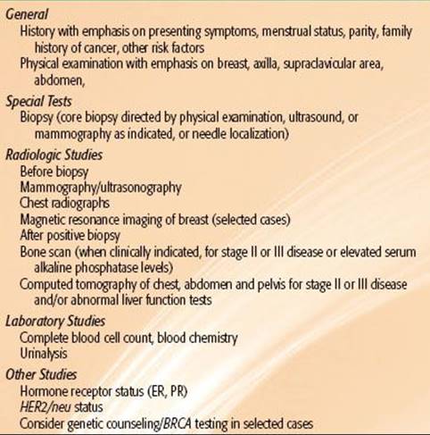
TABLE 56.8 DIAGNOSTIC WORKUP FOR CARCINOMA OF THE BREAST, STAGE T1 AND T2
DIAGNOSIS AND WORKUP
The workup of a patient with a breast mass, including complete clinical and family history, is summarized in Table 56.8. The patient should be examined both sitting up and lying down (to confirm masses felt on the sitting-up examination and to detect lesions deeper in the breast or against the chest wall). Careful inspection of both breasts should be made, including size, form, and symmetry, changes in pigmentation, scaling or discharge from the nipple, and dilated veins or edema of the skin in a nonpregnant patient. The location, size, consistency, tenderness, and mobility of the palpable tumor should be recorded. It is useful to draw and photograph the projection of any suspect or palpable masses on the skin of the breast or nodal areas.
In addition to examination of the breast, careful evaluation of the axilla and supraclavicular node areas is mandatory. The number, consistency, tenderness, mobility or fixation, and size of lymph nodes should be noted. Clinically node-negative patients have pathologic involvement in 10% to 40% of cases (depending on primary tumor size), whereas no pathologic evidence of tumor is found in 25% to 30% of patients with clinically palpable axillary nodes.
Examination of the abdomen for liver enlargement and evaluation for bony pain are also essential. Finally, a complete pelvic examination should be part of the overall evaluation of the patient, if not recently performed by the patient’s other physicians.
Laboratory studies include a complete blood count and chemistry profile, including liver function tests (e.g., aspartate aminotransferase, alanine aminotransferase, lactate dehydrogenase, bilirubin).
Imaging in Breast Cancer Diagnosis and Workup
Routine radiographic studies include chest radiography and bilateral mammograms. As clinically indicated, these may be supplemented by CT scanning, MRI, or positron emission tomography (PET) scans, bone scans, and plain radiographs of symptomaticbones, if clinically warranted.
TABLE 56.9 AMERICAN COLLEGE OF RADIOLOGY BIRADS ASSESSMENT CATEGORIES: MAMMOGRAPHY

Mammography
Mammography remains the most critical component of diagnostic imaging in breast cancer patients, and bilateral mammograms should be performed routinely in the workup of the breast cancer patient. The BIRADS (Breast Imaging Reporting and Data System) classification system, outlined in Table 56.9 has been widely adopted in classifying mammograms with respect to appropriate follow-up and intervention.154
The radiation oncologist should be familiar with the difference between diagnostic and screening mammography, because a majority of conservatively treated breast cancer patients will have undergone diagnostic mammography prior to treatment, as well as in follow-up. Screening mammography refers to routine mammographic images in asymptomatic women and consists of two views: craniocaudal and mediolateral oblique of each breast. Diagnostic mammography is used to characterize abnormalities detected at screening or in women with palpable masses, employs additional magnification views, and is generally done with the radiologist present to determine the need for additional views or follow-up studies. Following breast conservation, most patients will undergo diagnostic mammograms, as additional images are often needed to rule out suspicious findings in the previously radiated breast. Some mammographers recommend reverting to screening studies after several years of stable mammography in the conservatively managed breast cancer patient.
Kopans et al.155 reviewed the advantages and disadvantages of diagnostic imaging techniques for evaluation of patients with breast cancer. Classically, breast carcinoma is seen as an ill-defined mass that may have spiculated margins (Fig. 56.7), although rarely cancers may also be seen with a knobby, lobulated, or even a smooth contour (ultrasonography may distinguish them from cystic masses). Architectural distortion of the breast tissue may be present. The appearance of linear, radiated, or spiculated changes around a central focus should always be considered suspect for carcinoma. The tumor may be hidden by dense parenchyma; review of previous mammogram compression views and sometimes ultrasonograms is very important in detecting subtle interval changes in the appearance of the breast.110
Calcifications can be associated with either benign or malignant conditions of the breast. However, calcifications associated with malignant tumors are typically 100 to 300 μm in size and are rod like, tubular, branching, or punctate. Clusters of microcalcifications (more than five) are suggestive of intraductal disease, and in nonpalpable lesions needle localization aids in the diagnosis (Fig. 56.8). For patients undergoing biopsy of a suspicious mass or calcifications, about 30% will yield a diagnosis of malignancy.110 The average sensitivity of mammography is approximately 90% (60% to 95%) and the specificity is 94% (50% to 98%). The positive predictive value is approximately 8% to 14% for screened patients, but is significantly higher for patients with symptoms or palpable masses.155 If microcalcifications were initially present, radiographs of the surgical specimen and postlumpectomy mammography are important to rule out residual disease for patients considering breast-conservation therapy.156 Lally et al.157 reported on 114 patients with calcifications on mammography diagnosed with breast cancer. Of these cases, 75 breasts at risk had no residual suspicious calcifications and proceeded to radiotherapy without further surgery or mammography. Thirty-six breasts at risk proceeded to radiation with either known suspicious calcifications or with nondocumented removal of calcifications after another excision. Of the 36 breasts there were 7 local failures and 1 regional failure. Of 34 breasts that underwent re-excision after detection of suspicious calcifications by preirradiation mammography, 20 (59%) were found to have residual disease. Patients with documented removal of suspicious calcifications were found to have better local control than patients without documented removal. In addition, the presence of calcifications on a preirradiation mammogram was associated with a high probability of detecting residual disease.
FIGURE 56.7. Medial-lateral (A) and cephalad-caudal (B) views of mammogram depicting a 1-cm mass with stellate margins deeply located in the upper quadrant of the left breast, histologically proven to be an invasive ductal carcinoma. Example of ultrasonogram of the breast showing a hypoechoic mass with “shadowing” deeper to the lesion, characteristic of invasive carcinoma (C). T1-weighted magnetic resonance image of the breast demonstrating a mass that proved to be an invasive carcinoma (D).
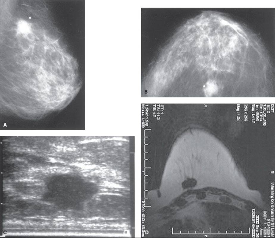
FIGURE 56.8. A: Mammogram demonstrating microcalcifications in the central portion of the breast with needle localization in place. B: Radiograph showing microcalcifications in the central portion of the wide excisional biopsy specimen. Pathologic diagnosis was intraductal carcinoma. Postlumpectomy mammogram showed no residual calcifications in the breast. Patient was treated with breast-conservation surgery and irradiation years ago and remains tumor free.
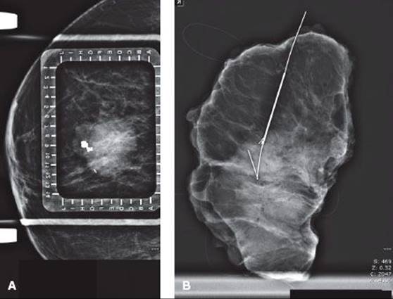
Ultrasound
Ultrasound can be a useful tool to complement physical examination and mammography in the diagnosis and treatment of breast cancer. Its use as a screening tool is limited and is the focus of ongoing investigations, as previously noted. Ultrasonography has a reported sensitivity of 73% and specificity of 95%. It is very helpful in differentiating cysts from solid tumors, and its primary use is the identification and characterization of palpable and nonpalpable abnormalities of the breast detected by physical examination or mammography.110,158 In the evaluation of a palpable mass in 420 patients, if both mammography and ultrasound are negative, Soo et al.159 reported the negative predictive value to be >99%. This was confirmed in a larger study of 3,516 patients from the Netherlands reported by Flobbe et al.160
In addition to complementing physical examination and mammography in diagnosis, ultrasound is often used as a guide for interventional procedures. Ultrasound-guided core biopsies are routinely performed in the diagnosis of breast cancer and have been shown to be more cost-effective than stereotactic biopsy. Ultrasound can also be used for fine-needle aspiration biopsies, cyst aspirations, presurgical localizations, and evaluation of breast tissue surrounding implants.
Magnetic Resonance Imaging
Although controversial for routine use, the use of MRI to supplement mammography in breast cancer diagnosis and treatment is rapidly increasing (see Fig. 56.7). In a review of MRI in the management of breast cancer, Hylton161summarized the potential for the current use of MRI: to complement mammography in screening; for differential diagnosis of questionable findings on physical examination, mammography, and ultrasound; and assessment of response in the neoadjuvant treatment of breast cancers. The NCCN recommends breast MRI for those women with early-stage disease whose breasts cannot be imaged adequately by mammography and ultrasound, those women who receive neoadjuvant chemotherapy to assess response to occult breast cancer, or women with genetic mutations leading to a higher risk of bilateral or contralateral breast cancer.47,162,163
In a retrospective study of MRI in the management of 441 women with breast cancer, Upponi and Warren164 reported the indications for MRI studies were diagnostic in 176, monitoring chemotherapy in 126, and study of MRI screening for breast cancer in 139. MRI results were confusing or incorrect in 6% of the diagnostic group, 13% of the chemotherapy group, and 9% of the screening group. The authors report that MRI resulted in an increase in confidence or change in clinical plan in 46% of the diagnostic group, 72% of the chemotherapy group, and 80% of the screening group. In 44 of 283 of these, MRI caused a beneficial change in the clinical plan based on conventional radiology.
Esserman et al.165 reported that MRI successfully detected cancer in 55 of 58 cases. There were two false-positive and two false-negative results, including a nonsignificant enhancement of one lesion. The anatomic extent of disease was correctly identified in 98% of cases by MRI but in only 55% by mammography. The utility of MRI as an adjunct to mammography in problematic cases was investigated by Lee et al.166 In 86 lesions with equivocal findings on mammography, positive findings were seen in 38 on MRI. Of these, 26 corresponded to areas of mammographic abnormalities. The remaining 12 sites were in areas where no abnormality had been suspected on mammography. Biopsies were performed on all 38 positive sites, and 10 (26%) were found to be malignant.
MRI has a clear role in the evaluation of patients who present with axillary metastasis with no evidence of a primary tumor in the breast by physical examination or mammography. In an analysis from Memorial Sloan-Kettering Cancer Center, Buchanan et al.167 reported on 55 patients who presented with axillary adenopathy without evidence of distant disease. MRI revealed suspicious lesions in 76% (42 of 55). In 62% (26 of 42), the MRI finding proved to be the occult primary tumor, of whom 58% (15 of 26) were candidates for breast conservation. MRI did not identify the primary tumor in 25 women. Of these 25, 12 underwent mastectomy, and cancer was found in 4 of these 12. The authors concluded that breast MRI detects mammographically occult cancer in half of women with axillary metastases and is a valuable tool for patients with occult primary breast cancer.
Computed Tomography
There is no established role for CT scans in routine staging of patients with early-stage breast cancer. The need for iodine contrast material to differentiate benign from malignant conditions, high radiation dose, cost per study, and inability to detect small lesions precludes the use of CT for initial evaluation, except under special circumstances. Most patients with node-negative breast cancer do not need to undergo routine CT scans for staging, because the yield is exceeding low. A small percentage of women with very high-risk node-negative disease or with node-positive disease may be upstaged by routine CT scans and, although the yield is low, it is common practice to CT stage high-risk node-negative and node-positive breast cancer patients. At present, the NCCN guidelines on breast cancer recommend an abdominopelvic CT if abnormal lab values or physical examination are present or if the patient is deemed as a stage IIIA (T3N1M0) or greater.47,168,169
Many women undergoing breast-conserving surgery and radiation do have CT scans as part of radiation therapy treatment planning. Without the use of contrast and specific diagnostic imaging protocols, these scans should not be considered as part of a staging procedure. In a study of 153 extended CT scans performed as part of radiation treatment, however, Mehta and Goffinet170 reported 11% revealed unexpected abnormalities, most of which were non-malignant. Only 2 of the 153 were found to have occult disease resulting in a change in stage.
Bone Scans
Routine bone scan at the time of initial treatment of stage I and II breast cancer is of limited value and should be reserved for patients with bone pain.171 In patients with stage I disease, the incidence of abnormalities on bone scan is approximately 2%, but a greater incidence of abnormalities is found in stages II (10%) and III (>20%).172 In a group of 7,604 patients who had bone scans, of over 20,000 women operated for breast cancer in Denmark, approximately 5% had abnormal study results.173 The incidence of abnormal scan results was greater in patients older than 60 years (8%) than in the younger group (3%), most likely because of the many benign bone and joint disorders frequently seen in older women.
Koizumi et al.174 reviewed records from 5,538 patients with breast cancer. The overall incidence of metastasis to bone was 2.13% (0% in patients with stage 0, 0.08% in stage I, 1.09% in stage II, 9.96% in stage III, and 34.04% in stage IV). Bone scans are more commonly recommended in patients with stage II larger tumors (>3 cm), aggressive histopathologic features, and in stage III or IV cancer.
Positron Emission Tomography Scanning
PET using 18F-labeled fluorodeoxyglucose (FDG) scanning, although not a routine component of staging, is being used more frequently in breast cancer. Its application in patients on initial presentation with early-stage disease has not been established. However, its potential role in patients with metastatic, advanced, and local-regional relapse of disease is rapidly evolving. At present, the NCCN guidelines recommend against routine PET scans in patients with stage 0 to IIIA disease but does state that it may be useful patients with locally advanced disease or in situations where standard imaging results are equivocal or suspicious.47,168,169
In an analysis of PET scanning in 165 patients from British Columbia, Weir et al.175 concluded that there are two clinical situations in which PET appears to be particularly valuable. The first is in the evaluation of patients who are suspected of having a tumor recurrence. The other is in identifying patients with multifocal or distant sites of malignancy who otherwise appear to have an isolated, potentially curable, local-regional recurrence.
Schirrmeister et al.176 also evaluated FDG-PET scanning and compared it prospectively with standard staging procedures within 2 weeks before surgery in 117 women who had palpable breast tumors or lesions suggestive of cancer on mammography or ultrasonography. On biopsy, 89 patients were determined to have breast cancer and 28 benign tumors. For interpreting results as being breast cancer, FDG-PET had a sensitivity of 93%, a specificity of 78%, an accuracy of 89%, a positive predictive value of 92%, and a negative predictive value of 96%. In detecting multifocal lesions, FDG-PET was twice as sensitive (63%) as the combination of mammography and ultrasonography (32%). Distant metastases in three patients were missed with the standard staging procedures but detected with FDG-PET. Because FDG-PET had a false-negative rate of 20% for detection of lymph node metastases, this imaging method cannot replace histologic evaluation of axillary nodes.
Summary of Imaging for Breast Cancer
All women should undergo history and physical examination, with mammography and liver function tests. Ultrasound or MRI may be useful in selected cases to complement mammography. For women who have operable disease with normal liver function tests, surgical staging of the breast and node sampling is performed. For low-risk patients, no further staging is required. For women with more advanced disease and those being considered for neoadjuvant chemotherapy, preoperative staging would routinely include a bone scan, chest x-ray and CT, or abdominal ultrasound. PET scanning may be considered in selected cases.
Pathologic Studies
Histopathologic diagnosis may be obtained by fine-needle aspiration of cystic or solid masses or biopsies of solid masses; any fluid aspirated from the breast should be examined for malignant cells. Fine-needle aspiration of the breast is a simple, low-cost, accurate diagnostic technique that has been used for many years in Europe and is gaining increasing acceptance in the United States.177,178 A potential limitation of fine-needle aspiration is that it provides cytology and no tissue architecture. Therefore, while the presence of malignant cells can be detected, cytology from fine-needle aspiration cannot conclusively differentiate invasive from noninvasive disease. However, for lesions that are palpable or easily visualized on ultrasound, this method results in rapid and efficient diagnosis.
Stereotactic core needle biopsy is increasingly used to obtain a histologic diagnosis with high accuracy, particularly in small breast lesions.179 In 6,152 lesions sampled at multiple institutions, 817 (13.3%) showed infiltrating breast cancer, 167 (2.7%) showed intermediate or high-grade DCIS, and 213 (3.5%) showed atypical hyperplasia or low-grade DCIS. Complete agreement between the core biopsy and subsequent histologic sections was reached in 89.7% of lesions and partial agreement in 9.2%. Clinically significant complications occurred in only 6 of 3,765 cases (0.2%) for which follow-up was available.
Breast biopsy of any suspicious mass is mandatory. The biopsy usually can be done using local anesthesia; the patient should be informed of the nature of the lesion to allow for her greater participation in therapeutic decisions. There has been no evidence that delay in treatment up to 2 weeks after biopsy worsens prognosis.180
In nonpalpable lesions, needle localization and radiographic techniques are necessary to identify the tissue to be removed. Failure to remove the mammographic abnormality has been reported in 2% to 8% of patients undergoing needle localization.181 The localizing wires should be left in place in the specimen and a radiograph obtained to ensure that the area of abnormality has been adequately excised. If the specimen radiograph does not document complete tumor removal, an immediate re-excision of the area at the tip of the wire should be carried out. However, specimen mammography may be of questionable benefit in the management or outcome of most patients undergoing image-guided, needle-localized breast biopsies. Bimston et al.182 reviewed 164 patients who underwent 165 needle/dye-localized breast biopsies for suspect mammographic abnormalities. In only three (1.8%) cases did the patient clearly benefit from specimen mammography; in no patient was a malignant neoplasm missed.
If there is any question, particularly in patients with microcalcifications, a postbiopsy mammogram should be obtained to determine the completeness of tumor excision.110 The surgeon should prepare (orient) the specimen accordingly, and the margins of the resected breast tissue should be identified and inked before processing. The pathologist should be made aware of the nature of the lesion for appropriate processing of the specimen.183 Radiation oncologists should be familiar with the implications of the diagnostic procedures for carcinoma of the breast as active participants in a breast preservation therapeutic approach.
In the United States, estrogen-receptor (ER) and progesterone-receptor (PR) assays are routinely done in the for patients with breast cancer; these parameters are correlated with prognosis and tumor response to chemotherapeutic and hormonal agents.184,185 Immunohistochemical techniques are commonly employed and correlate well with other hormonal-receptor assays. Cellular assays measure the growth fraction (S-phase fraction [SPF]) of tumors, either by thymidine-labeling index (TLI) or flow cytometry methods, and other tumor markers have prognostic implications, sometimes independent of tumor stage and hormone-receptor status.186,187 HER-2/neu assay is being done routinely because overexpression is associated with poor prognosis, and these patients are currently being offered adjuvant therapy directed at HER2/neu.188,189 HER2/neu analysis by fluorescent in situ hybridization techniques has recently evolved as the standard for determining response to therapy directed at the HER2/neuoncogene.188,189
Staging
Two staging systems are widely used for breast cancer: the American Joint Committee on Cancer (AJCC) (Table 56.10) and the Union Internationale Contre le Cancer systems.190,191 Major changes made in the seventh edition of the AJCC Cancer Staging Manual include:190
1. Identified specific imaging modalities that can be used to estimate clinical tumor size, including mammography, ultrasound, and magnetic resonance imaging (MRI).
2. Maintained that the term “inflammatory carcinoma” be restricted to cases with typical skin changes involving a third or more of the skin of the breast. While the histologic presence of invasive carcinoma invading dermal lymphatics is supportive of the diagnosis, it is not required, nor is dermal lymphatic invasion without typical clinical findings sufficient for a diagnosis of inflammatory breast cancer.
3. Classification of isolated tumor cell clusters and single cells is more stringent. Small clusters of cells not greater than 0.2 millimeters, or nonconfluent or nearly confluent clusters of cells not exceeding 200 cells in a single histologic lymph node cross section are classified as isolated tumor cells.
4. Stage I breast tumors have been subdivided into Stage IA and Stage IB; Stage IB includes small tumors (T1) with exclusively micrometastases in lymph nodes (N1mi).
5. Created new M0(i+) category, defined by presence of either disseminated tumor cells detectable in bone marrow or circulating tumor cells or found incidentally in other tissues (such as ovaries removed prophylactically) if not exceeding 0.2 millimeters. However, this category does not change the stage grouping. Assuming that they do not have clinically and/or radiographically detectable metastases, patients with M0(i+) are staged according to T and N.
The current AJCC staging for breast cancer does not include in the calculation of tumor size additional tumor found at the time of re-excision; this may result in understaging and undertreatment of patients with breast cancer.192The Columbia staging system is important both historically and because it clearly identifies prognostic factors affecting operability.64 Figure 56.9 depicts the various clinical stages according to tumor and nodal characteristics.
TABLE 56.10 AMERICAN JOINT COMMITTEE ON CANCER STAGING OF BREAST CANCER
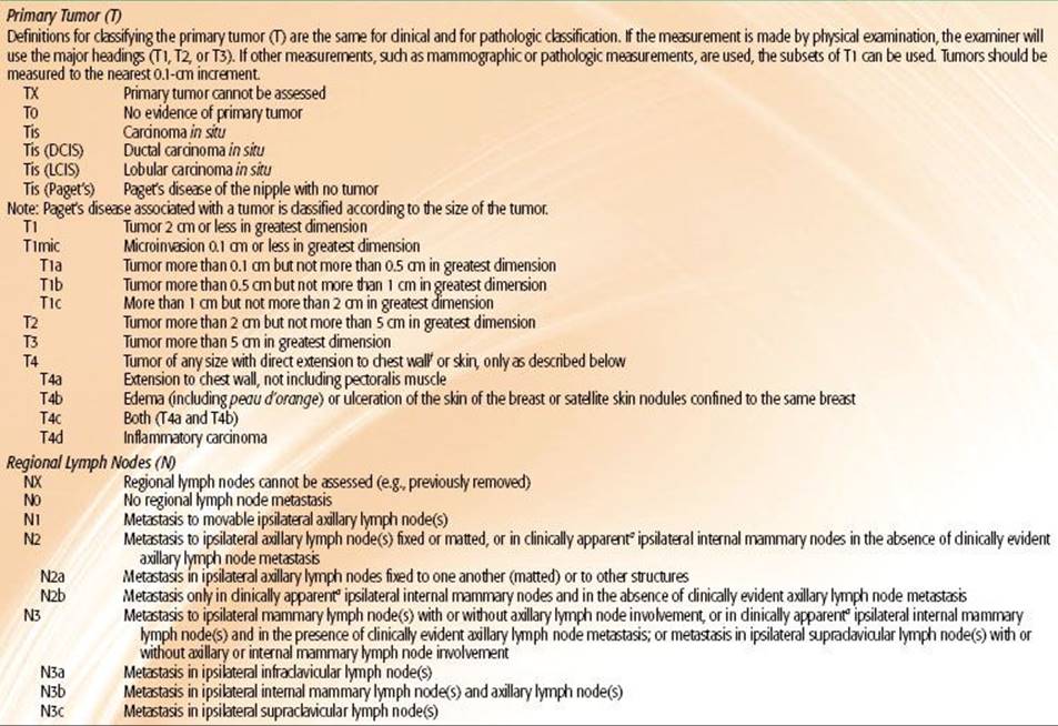
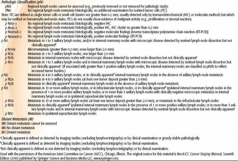
FIGURE 56.9. Clinical staging of carcinoma of the breast. Stages, in part, reflect curability by locoregional treatment modalities prognostically (surgery and radiation therapy). The equivalence of T and N categories are as follows: T2 = N1, T3 = N2, T4 = N3. The American Joint Committee on Cancer and Union Internationale Contre le Cancer classification systems use similar categories and stages. (From Langmuir VK, Poulter CA, Qazi R, et al. Breast cancer. In: Rubin P, ed. Clinical oncology: a multidisciplinary approach for physicians and students, 7th ed. Philadelphia: WB Saunders, 1993, with permission.)
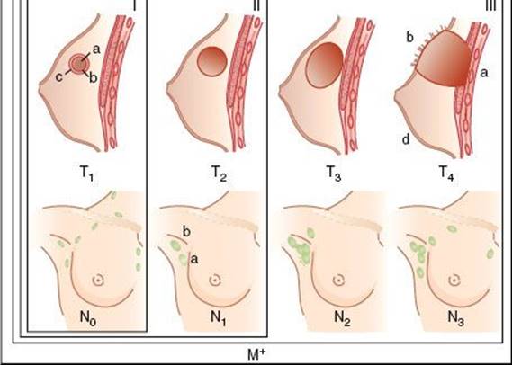
TABLE 56.11 AMERICAN JOINT COMMITTEE ON CANCER HISTOPATHOLOGIC CLASSIFICATION OF BREAST TUMORS

Pathologic Classification
The World Health Organization has classified proliferative conditions and tumors of the breast into the following categories: benign mammary dysplasias, benign or apparently benign tumors, carcinoma, sarcoma, carcinosarcoma, and unclassified tumors.193 The AJCC has developed the alternative system shown in Table 56.11.190
Numerous detailed reports and monographs describe the pathologic features and clinical implications of carcinoma of the breast, which are reviewed elsewhere.110,194–197 The radiation oncologist should be familiar with the histologic characteristics of breast cancer because many of them affect prognosis and may have important therapeutic implications. Brief descriptions of several types of carcinoma of the breast follow.
Microinvasive carcinoma is defined as “the extension of cancer cells beyond the basement membrane into the adjacent tissues with no focus more than 0.1 cm in greatest dimension.” Lesions that fulfill this definition are staged as T1mic, a subset of T1 breast cancer. The AJCC staging manual further states that “when there are multiple foci of microinvasion, the size of only the largest focus is used to classify the microinvasion” and that the sizes of the individual foci should not be added together. They also state that “the prognosis of microinvasive carcinoma is generally thought to be quite favorable, although the clinical impact of multifocal microinvasive disease is not well understood at this time.” Widely varying definitions of microinvasion have been used, and some differ substantially from that offered here.190
Invasive (infiltrating) ductal carcinoma is the most common type of breast cancer, comprising more than 50% of all cases. It appears as solid cords or groups of ductal tumor cells varying in size and cytoplasmic content and degree of differentiation.198 Necrosis is rare, but lymphatic invasion may be present. An associated in situ component is frequently seen.
Tubular carcinoma is composed of tubular structures typically lined by a single layer of well-differentiated epithelium. The tubular cells simulate those of normal ducts or ductules, are arranged in multiglandular cribriform or adenocystic configurations, and are frequently associated with other in situ carcinomas of the breast.199 Tubular carcinomas have a nonaggressive growth pattern, with an excellent prognosis. A meta-analysis of 680 women showed an overall frequency of nodal metastasis of 13.8%.200 In view of the low incidence of axillary node metastases at presentation (7%) in low-risk tubular carcinoma of the breast (≤1 cm), some have advocated that axillary dissection may be omitted. In a retrospective review of 73 cases of tubular carcinoma, Sullivan et al.201 reported treatment with conservative surgery (CS) plus radiation therapy (RT) in 67%, CS without RT in 18%, and mastectomy in 15%. The published literature of 529 conservatively treated tubular carcinomas was reviewed along with the 62 conservative cases from their series. No patients developed distant metastasis or died from disease. Local failure occurred in three (4%) of the cases. The literature review showed that adjuvant RT reduces local failure following CS for tubular carcinoma.
Medullary carcinoma is composed of cords and masses of large cells with reticular pleomorphic nuclei containing prominent nucleoli. There is a scant fibrous stroma, but lymphoid infiltrate is prominent. These tumors are microscopically and grossly well circumscribed. Prognosis, in general, is better than for other tumors. These tumors are more frequently seen in younger women and are commonly associated with patients with BRCA1 mutations.202
Lobular invasive carcinoma may be interspersed with; the cells appear singly or in small clusters in a targetoid or single-file pattern. Some scirrhous carcinomas probably are invasive lobular lesions; these tumors tend to be aggressive and multicentric and are prone to development of distant metastases. Du Toit et al.203 reported five subtypes of lobular carcinomas in 171 cases and observed a 12-year actuarial survival rate of 100% for the tubulolobular subtype but of only 47% for the solid variant. Two other characteristics of invasive lobular carcinoma (ILC) is that it is often “mammographically silent,” meaning its detection or the full appreciation of extent of disease is often not visualized mammographically. Invasive lobular cancers are much more commonly ER-positive than invasive ductal carcinoma. Infiltrating pleomorphic lobular carcinoma, an aggressive variant of ILC, was described in 38 cases; 29% of the specimens demonstrated signet ring cells.204
Mucinous carcinoma, also called mucoid or colloid carcinoma, has been observed in older women with relatively long duration of symptoms.205 It is more likely to be devoid of a cellular reaction; necrosis and lymphatic invasion are very rare. It is slowly growing with a pushing border and has a low frequency of axillary lymph node metastasis. Survival is appreciably better than with invasive ductal carcinoma.206 Anan et al.207 evaluated 76 patients with mucinous carcinoma (52 pure type and 24 mixed type). The incidence of lymphatic vessel invasion (4%) and nodal involvement (4%) was lower in pure mucinous carcinoma than in mixed carcinoma (P <.05). No nodal involvement occurred in patients with pure mucinous carcinoma <3 cm in diameter.
Adenocystic carcinoma is rarely found in the breast. Histologic features and clinical behavior are similar to its counterpart in the salivary gland and the upper respiratory tract.208 In 28 patients, only 1 had axillary node metastases; 22 were treated with mastectomy and 6 with local excision (with breast irradiation in 5). With a median follow-up of 7 years, there were no local recurrences; the 5-year disease-free survival rate was 95%.209
Invasive micropapillary carcinoma of the breast is characterized by growth of tumor cell clusters in prominent clear spaces resembling dilated angiolymphatic vessels. Nasser et al.210 reported on 83 invasive micropapillary carcinomas; the mean tumor size was 4 cm, 22% invaded skin, 58% were poorly differentiated, and 71% were ER-positive. Axillary node metastases were present in 77% of cases and were typically multiple (51% had three or more positive). Forty-six percent of the patients died from their disease (mean interval to death, 36 months). Skin involvement and nodal status were the only parameters predictive of poor survival (P = .01).
Metaplastic carcinoma is relatively rare. Park et al.211 noted axillary lymph node metastasis in 6 (40%) of 15 in whom axillary node dissection was performed. A recent analysis by Beatty et al.,212 of the Swedish Cancer Institute, identified 24 cases that were compared with typical breast cancer cases matched for age, date of diagnosis, stage, and ER, PR, and HER2 status. The mean metaplastic primary tumor diameter was 2.5 cm. The histologic or nuclear grade was high in 21 of 24 cases. ER or PR status was negative in all cases. HER2 was negative in 10 of 11 cases tested. Epidermal growth factor receptor (EGFR; HER1) was positive in 7 of 7 cases tested. Five-year survival was 83% (95% CI, 66% to 100%). Comparison with matched typical breast cancer cases revealed no significant difference in multidisciplinary treatment patterns, recurrence, or survival. The increased expression of EGFR (HER1) provides an opportunity for targeted tumor therapy in these tumors.
In an analysis of the MD Anderson Cancer Center experience and the SEER database, Hennessey et al.213 identified 100 patients with metaplastic sarcomatoid carcinoma, and 213 patients in the SEER database with similar histology. They conclude that these are aggressive tumors with poor response to therapy and poor outcomes, also suggesting that studies evaluating novel targeted therapy are needed for these patients.
Spindle cell carcinoma of the breast, a variant of metaplastic carcinoma, includes a wide spectrum of lesions with mildly atypical features that may resemble fasciitis, fibromatosis, or myofibroblastic tumors. Unlike spindle cell carcinomas in general, they have no propensity for distant metastasis and should be termed tumors rather than carcinomas. Sneige et al.214 studied 24 cases of fibromatosis-like spindle cell breast carcinoma. Treatment consisted of local excision (7 cases) or modified radical mastectomy (13 cases) and was not specified in 4 cases. In patients who underwent axillary nodal dissection, no lymph node metastases were found. Local recurrences developed in two of the six patients who underwent local excision only.
Primary neuroendocrine small cell carcinoma is uncommon. Francois et al.215 reported seven cases, and Shin et al.216 described nine cases. Immunohistochemical analysis showed consistent staining for cytokeratin markers but variable staining with neuroendocrine markers. The histologic type and prognosis are identical to those of lung cancer. It is important to distinguish these lesions from metastatic lung tumors or direct invasion of breast by Merkel cell carcinoma, lymphoma, or carcinoid tumor. It is reasonable to treat these patients with aggressive multiagent chemotherapy, excision of the primary tumor, and breast irradiation, although no data are available on the outcome of this approach.
Paget’s disease describes involvement of the nipple by tumor. Most investigators agree that it represents extension of neoplasms from subjacent ducts in the nipple or metastases from an underlying carcinoma.1 The tumor seems to travel linearly down the ducts and may appear to be multicentric. There may be an associated subareolar tumor. Breast-conserving surgery followed by radiation is effective in this disease.217,218
Cystosarcoma phyllodes is usually a benign lesion; in broad, fibrous beads that look “leaflike” are cystic clefts lined by a single layer of cells. These tumors are large; usually they are encapsulated, without invasion of the adjacent breast.64 The lesions frequently develop from pre-existing fibromas and have a long initial period of slow growth followed by a sudden, rapid increase in size. The grade (mitotic rate), surgical margins, and proliferative index have prognostic importance.219
Primary mammary lymphomas are rare. In a study of 35 cases, including 16 primary lymphomas, diffuse large cell lymphoma was present in 10 of 16 primary and 14 of 18 secondary cases.220 Lymphoepithelial lesions in ducts and lobules and frequent vascular involvement were found in both primary and secondary cases. Immunohistochemistry studies of 13 tumors showed that 12 were B cell in origin, and one was a primary T-cell lymphoma. Survival was related to stage and histologic characteristics. Half of the patients with primary lymphoma had recurrent disease. Although some local recurrences were observed, recurrence in other extranodal sites predominated.
Sarcoma
Primary breast sarcomas are occasionally seen. McGowan et al.221 described 78 cases of primary breast sarcoma without metastatic disease (76 women, 2 men); 32 patients had malignant cystosarcoma phyllodes, and the others had stromal sarcomas (14 patients), angiosarcomas (8 patients), fibrosarcomas (7 patients), carcinosarcomas (5 patients), liposarcomas (4 patients), or other lesions (8 patients). The cause-specific survival rate was 48%, the relapse-free rate was 42%, and the local relapse-free rate was 75% at 10 years. No statistically significant difference in outcome was noted between those treated with conservation surgery and those undergoing mastectomy. Patients with negative margins had a significantly better local relapse-free rate than did those with positive margins (80% vs. 33%; P = .009).
FIGURE 56.10. Five-year survival according to tumor size in node negative breast cancers (Data adapted from Carter CL, Allen C, Henson DE. Relation of tumor size, lymph node status and survival in 24,740 breast cancer cases. Cancer 1989;63:181–187.)

PROGNOSTIC FACTORS FOR SURVIVAL AND METASTASIS
Carcinoma of the breast represents a wide spectrum of tumors with a variety of clinical, biological, and genetic characteristics resulting in a considerable variation in prognosis. It is important to understand, particularly from the radiation oncologists’ perspective, that prognostic factors for systemic relapses and prognostic factors for local relapse differ significantly. Furthermore, prognostic factors for local relapse after mastectomy differ substantially from prognostic factors for local relapse after lumpectomy and radiation.222 For example, tumor size and nodal status are clearly among the strongest predictors of overall survival and metastasis and are also strong predictors of postmastectomy chest wall relapse when radiation is not used.110,223,224 However, these factors have not been consistently reported to be prognostic factors for in breast relapse after cancer surgery. On the other hand, margin status is a strong predictor of relapse in the conservatively treated breast but is not strongly correlated with distant metastasis.225–227 Young age is also a very strong predictor of local relapse after breast-conserving therapy, and although it has been shown to be predictive of systemic metastasis, the effect of young age on distant metastasis as an independent factor is clearly not as significant as it is for local relapse in the conservatively managed patient.228 Patients, as well as clinicians, often become confused regarding these issues, resulting in misconceptions regarding appropriate decision making and treatment. Below, prognostic factors for systemic relapse (i.e., distant metastasis and disease-free and overall survival) are discussed. A more in-depth discussion of prognostic factors for local relapse following lumpectomy and radiation is included later in the section on selection factors for the conservative management of breast cancer. Prognostic factors related to postmastectomy chest wall recurrence for patients with early-stage disease are discussed in Chapter 57.
The College of American Pathologists presented a consensus statement in 1999 summarizing prognostic factors in breast cancer.229 Factors were category I if they were proven to be of prognostic importance and useful in clinical patient management. Category II factors had been extensively studied biologically and clinically, but their importance remains to be validated in studies. Category III included all other factors not sufficiently studied to demonstrate their prognostic value.
Category I included tumor size, lymph node status, micrometastasis, histologic grade, mitotic count, and hormonal-receptor status. Category II included HER2/neu expression, p53 mutations, lymphovascular invasion, and DNA ploidy. Category III included tumor angiogenesis, EGFR, transforming growth factor, Bcl-2, and cathepsin D overexpression.
More recently, based on advances in molecular techniques allowing for genetic profiling of tumors, additional molecular prognostic factors have been identified, which can further refine clinical decision making and are already being applied clinically. This section will focus on the category I and II factors and discuss more recently introduced promising prognostic factors that may influence distant metastasis and clinical management.
Tumor Size
The size of the primary tumor ranks among the strongest predictors of distant metastasis and disease-free and overall survival. Although tumor size correlates strongly with the presence and number of involved axillary lymph nodes, it is clearly an independent prognostic factor. Among patients with documented node-negative disease, tumor size remains a strong and independent predictor of disease-free and overall survival. In a classic study with over 20 years of follow-up, Rosen et al.194 reported a recurrence-free survival of 88% for tumors <1 cm, 72% for tumors 1.1 to 3.0 cm, and 59% for tumors 3.1 to 5.0 cm. In an analysis of 826 women with node-negative breast cancer treated by mastectomy at the University of Chicago with a median follow-up of 13.5 years, Quiet et al.230 reported a 20-year disease-free survival of 79% for patients with tumors <2 cm, compared with 64% with tumors >2 cm. In multivariate analysis, the strongest predictor of outcome and time to relapse was pathologic tumor size. Survival as a function of primary tumor size in node-negative breast cancer patients is illustrated in Figure 56.10.
Axillary Nodal Status
Of all prognostic factors, nodal status continues to be the strongest predictor of disease-free and overall survival and is the primary factor that governs breast cancer staging. (Fig. 56.11).190 Although there is a direct relation between the number of axillary nodes involved and the risk of distant metastasis, the most commonly employed schema is to group patients into four prognostic categories (node negative, 1 to 3 involved nodes, 4 to 9 involved nodes, and more than 10 involved nodes). These nodal prognostic categories are employed in the N staging of the current AJCC staging system. Although outcomes will continually improve as systemic therapies advance, data from previous NSABP trials treated primarily with local-regional therapy alone revealed 5-year survival rates of 82.8% for node negative, 73% for 1 to 3 positive nodes, 45.7% for 4 to 12 positive nodes, and 28.4% for more than 13 positive nodes.73,180,231–233
FIGURE 56.11. Five-year survival according to tumor size and nodal status. (Data adapted from Carter CL, Allen C, Henson DE. Relation of tumor size, lymph node status and survival in 24,740 breast cancer cases. Cancer 1989;63:181–187.)
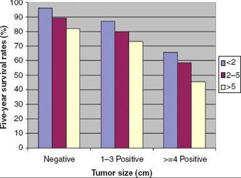
Micrometastasis
The influence of microscopic disease in the lymph nodes has been the subject of several recent analyses due to the increased detection of micrometastasis in the era of sentinel lymph node biopsy. Most recent studies have demonstrated that by using a combination of blue dye and radiolabeled colloid techniques the sentinel node can be identified in >95% of cases.234–235,236 In experienced hands, false-negative sentinel node rates are low (<10%), and prognosis of sentinel node-negative patients is similar to node-negative patients who have undergone a complete axillary dissection. Although the standard of care for sentinel node-positive patients has traditionally been completion axillary dissection, recent results of the American College of Surgeons Oncology Group (ACOSOG) Z0011 trial have challenged the need for completion axillary dissection in selected sentinel node-positive patients.237 The need for axillary dissection in node-positive patients will likely continue to be highly individualized and will be further evaluated in ongoing clinical trials.238
Hansen et al.239 evaluated the sentinel node in 696 women by hematoxylin and eosin staining and immunohistochemistry. With a median follow-up of 38 months the size of the sentinel node metastasis (<2 mm or >2 mm) was a significant prognostic factor. There was no difference in disease-free or overall survival, however, between true node-negative and immunohistochemistry-only -positive cases. Additional data from large databases will continue to emerge in the coming years to further refine the prognostic significance of micrometastasis and immunohistochemistry-only positive lymph node metastasis in breast cancer patients.
Tumor Type
The histologic subtype of invasive cancer has been shown to be of prognostic value in several studies. The tubular, mucinous, and medullary subtypes have been shown to have a more favorable prognosis, compared with invasive ductal.194,201,240,241 Invasive lobular tumors appear to have a prognosis similar to invasive ductal tumors.242 Poor prognostic categories include metaplastic, undifferentiated, and other rarer subtypes.110,195 In a classic analysis of 293 T2N0 breast cancers with over 20 years of follow-up treated by mastectomy, Rosen et al.194 reported more favorable relapse rates in medullary, mucinous, tubular, and papillary subtypes compared with invasive ductal and invasive lobular tumors.
Tumor Grade
Multiple tumor grading systems have been proposed in an effort to standardize and improve interobserver variability. The Scarff-Bloom-Richardson classification system utilizes mitotic index, differentiation, and pleomorphism, each with scores of 1 to 3. Scores of 3 to 5 are well differentiated, 6 to 7 moderately differentiated, and 8 to 9 poorly differentiated. This system is commonly employed and has been shown to be of independent prognostic significance.243
Elston and Ellis244 of the Nottingham group refined this methodology. The revised technique involves evaluation of three morphological features—the percentage of tubule formation, the degree of nuclear pleomorphism, and an accurate mitotic count using a defined field area. A numerical scoring system is used and the overall grade is derived from a summation of individual scores for the three variables: three grades of differentiation are used. Histologic grade, assessed in 1,831 patients, shows a very strong correlation with prognosis; patients with grade I tumors have a significantly better survival than those with grade II and III tumors (P <.0001). If the protocol is followed, reproducible and consistent results regarding prognosis can be obtained.
Estrogen and Progesterone Hormonal Receptors in Tumor Cells
Several studies have indicated that patients with hormonal receptors have a significantly higher survival rate.245,246 Crowe et al.247 studied 1,392 patients with carcinoma of the breast treated with modified radical mastectomy. ER-positive tumors (≥3 fmol/mg cytosol protein) were found in 1,063 patients (76.4%). Their 10-year overall survival rate of 65.9% was significantly better than the 56% rate in 329 patients with ER-negative tumors (P = .0001). However, this correlation is not consistent, with conflicting reports regarding the prognostic significance of hormonal receptor status.248 The apparent discrepancy in some of these reports may be explained by technical nuances. Esteban et al.249 noted that quantitative immunohistochemistry of ERs provides results with better predictive value than the biochemically procured ones.250
Tumors that express both ER and PR have the greatest benefit from hormonal therapy, but those containing only ER or PR still have significant responses. Two types of ERs—ER-α and ER-β—have now been identified. PR also exists in two forms, PRA and PRB.251 Patients with tumors negative for hormonal receptors have only a small probability of responding to hormonal therapy.185,251
Lymphatic and Vascular Invasion
Lymphatic and vascular invasion (LVI) in the peritumoral region has been clearly demonstrated to be of independent prognostic significance in several studies.252–256 In the study by Rosen et al.,194 recurrence rate for LVI-positive stage I patients was 38% compared with 22% for LVI-negative patients.
Proliferative Indices, S-Phase, and Thymidine Labeling Index
Various techniques for evaluating the proliferative rate of a tumor have been shown to correlate with distant metastasis and survival. The most common are the fraction of cells in SPF, TLI, mitotic index, or antibodies directed against proliferative markers such as Ki-67 and proliferating cell nuclear antigen.
Thymidine labeling represents the fraction of cells in S phase of the cell cycle and is based on the active incorporation of labeled thymidine into DNA; the TLI of primary breast cancers appears closely related to steroid receptor status and generally unrelated to pathologic stage. Retrospective analyses have shown that TLI is a prognostic indicator, independent of tumor size, steroid receptors, and p53, and Bcl-2 protein expression. Together with patient age and tumor size, TLI is able to identify patients at different levels of risk for locoregional or distant metastases.257
Wenger and Clark258 concluded that despite different techniques and cut points, a higher SPF is in general associated with worse tumor grade, absence of steroid receptors, larger tumors, and positive axillary lymph nodes. Higher SPF is usually associated with worse disease-free and overall survival rates in both univariate and multivariate analyses.
Bryant et al.,259 in over 4,000 patients from NSABP protocol B-14 who had ER-positive tumors and no axillary lymph node involvement, found a strong association between SPF and disease-free and overall survival rate.
DNA Ploidy Index
Most breast cancers exhibit a bimodal distribution of DNA values. DNA ploidy as measured by flow cytometry correlates with nuclear grade, with low-grade tumors being diploid and high-grade tumors being aneuploid.260 Ploidy was found to be associated with histologic type, tumor grade, and SPF values, but not with patient age, menopausal status, tumor size, axillary nodal status, ER status, or PR status.261 Diploid tumors tend to be ER-positive, whereas aneuploid tumors are frequently ER-negative. Older patients are more likely to have hyperdiploid tumors.262 Diploid tumors tend to have a better prognosis than those with an aneuploid DNA distribution.263,264 Toikkanen et al.,265 in 351 patients monitored for a minimum of 22 years, observed a 25-year survival rate of 28% for patients with nondiploid tumors, in contrast to 48% for those with a diploid DNA pattern. In a European Organisation for Research on Treatment of Cancer (EORTC) trial evaluation of DNA, proliferative compartment was the most important predicting factor for overall survival and metastasis-free survival in 281 premenopausal, lymph node–negative patients with invasive carcinoma of the breast.266
Studies by Ewers et al.267 and Fallenius et al.268 also confirm the prognostic significance of DNA ploidy. Keyhani-Rofagha et al.269 and Witzig et al.270 reported no statistically significant prognostic significance of DNA ploidy.
HER2/neu
The HER2/neu proto-oncogene (also called c-erbB-2) located on chromosome 17 codes for a transmembrane glycoprotein, p185, which has tyrosine kinase activity and is homologous to the EGFR.271 It is amplified or overexpressed in up to 30% of human breast carcinomas. Overexpression of the protein is associated with tumor aggressiveness and decreased disease-free survival in node-positive patients, with variable prognostic significance among node-negative patients. The conflicting reports regarding the prognostic significance of HER2/neu may be related to interobserver variability in interpretation of staining and uncertainty regarding the significance of intermediate staining.188,189,271 Staining for overexpression of HER2/neu is interpreted on a 0 to 3+ scale. The available data suggest that the majority of 0 to 1 staining is clearly negative and 3+ is clearly positive, while the classification of those patients with 2+ staining remains uncertain. Amplification of the oncogene identified using fluorescent in situhybridization (FISH) techniques has been found to be of more prognostic value.188,189,271,272
Variability in the prognostic value of HER2/neu may be related to variability in interpretation of protein expression levels. Birner et al.273 correlated results of the Hercep Test with HER-2/neu oncogene gene amplification assessed by FISH in 303 patients with lymph node–positive breast cancer.273 Results were compared with FISH analysis performed in all 2+ and 3+ specimens (103 cases) and 104 HER-2/neu-negative specimens; 3+ carcinomas were found in 8.9% to 15.7% of specimens. FISH revealed that almost exclusively 3+ positive cases had HER-2/neu gene amplification.
In univariate analysis, staining with the HercepTest revealed a worse prognosis in 3+ cases, which were significantly associated with lower ER levels and histologic grade III tumors. More critical than its prognostic significance, however, is its predictive value with respect to response to therapy and its value in identifying patients who may benefit from adjuvant targeted therapy directed at the protein.274 Several studies have demonstrated that HER-2/neustatus may be predictive of response to hormonal therapy, resistance to alkylating agent–based chemotherapy, and response to taxanes.275,276
p53 Gene
The p53 tumor suppressor gene encodes a nuclear phosphoprotein that is thought to be important to cell cycle regulation and DNA repair and that also may regulate induction of apoptosis by ionizing radiation.277,278 The p53 gene is most frequently mutated in sporadic breast cancer; alterations of this gene were identified in 43 of 192 tumors (22%).278 Mutations of p53 were found more often in tumors of younger women (P = .002) and African American women (P = .04) and in tumors lacking ER (P = .03), PR (P = .04), or both (P = .06). In 843 cases of breast cancer, p53 mutations were not found in low-grade carcinomas (tubular, mucinous, papillary, and invasive cribriform types), but were observed in 4.2% of ILCs (6 of 140 cases), 15.5% of high-grade IDCs (99 of 640 cases), and 50% of pure medullary carcinomas (5 of 10 cases).279 The overall survival rates were not significantly different in patients with mutant or wild-type p53 tumors. In another study of 156 patients with primary invasive breast cancer, overexpression of p53 protein emerged as a reliable and independent predictor for disease recurrence and reduced survival.280Jansen et al.,281 in a study of 345 patients with breast cancer with a median follow-up of more than 10 years, noted that Bcl-2 expression was not a prognostic factor, but p53 was an independent prognostic factor for overall survival (P = .005) and postrelapse survival (P = .006). However, p53 status was important only in the Bcl-2–positive subgroup.
Genetic Profiling
Recent developments in DNA microarray technologies allow for extensive profiling of tumors based on their gene expression signatures.282 Using this technology, investigators from the Netherlands Cancer Institute screened thousands of genes to develop a 70-gene prognostic signature that in 295 women demonstrated a 10-year disease-free survival of 50.6% in 180 poor prognosis signature patients compared with 85.2% in 115 women with a favorable signature. In a recent validation study, the profiling outperformed classic prognostic criteria, but the magnitude of the prognostic value was not as strong.283,284,285
Gene expression profiling has had a marked impact on our understanding of the biology of breast cancers and is routinely used in clinical decision making. Categorizing breast cancer into distinct clinical subtypes of luminal A, luminal B, HER2/neu, and basal-like has significant prognostic value and impacts on decisions regarding systemic therapy options.286,287 Although gene profiling forms the basis of this subtyping, most clinicians rely on common molecular markers of ER, PR, HER2/neu, Ki-67, and others as a surrogate to classify patients into these intrinsic subtypes of breast cancer for clinical decision making.288
An assay using gene profiling on paraffin-embedded specimens has been developed by Genomics Health Inc (Redwood City, CA, USA) The Oncotype DX assay is based on reverse transcriptase polymerase chain reaction assays to quantify expression of selected genes in paraffin embedded tissues. A panel of 16 cancer-related genes and 5 reference genes were employed to compute a recurrence score (0 to 100), which is used to estimate the odds of relapse over 10 years. Specimens from patients in NSABP trials with ER-positive tumors treated with tamoxifen alone were used to validate this scheme in which patients were categorized as low risk (<18 score), intermediate risk (18 to 30), or high risk (31 to 100). As shown in Figure 56.12, the likelihood of distant metastasis is low in those patients with favorable scores treated with tamoxifen alone.289 These data can be used to determine which patients with ER-positive tumors and otherwise favorable factors may avoid chemotherapy and aid the clinician and patient in clinical decision making.
FIGURE 56.12. Risk stratification in patients with receptor positive tumors treated with hormonal therapy broken as a function of recurrence score using the Oncotype DX gene profiling scheme.
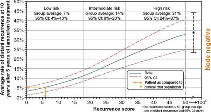
Age
Although young age has consistently been shown to be a predictor of local relapse following breast-conserving surgery, there are conflicting data regarding its prognostic significance for distant metastasis and overall survival.8,222,228,290–293 The available data are confounded by the fact that younger women often present with palpable disease and have larger tumors with a higher percentage of positive nodes. Several studies, however, have shown that when corrected for stage, young age remains a significant prognostic factor for distant metastasis. In an analysis of 1,751 patients with nonmetastatic breast cancer, Vanlemmens et al.294 demonstrated that younger women had a higher proportion of patients with ER-negative and high-grade tumors and lower disease-free and breast cancer–specific survival. In multivariate analysis, young age at diagnosis was an independent poor prognostic factor. Kolias et al.,295 however, in an analysis of 2,879 patients younger than 70 years old with operable disease in the Nottingham database, demonstrated that the association of young age at diagnosis with a worse prognosis was explained by a higher proportion of poorly differentiated cancers in young women and that young age itself had no influence on the prognosis of the individual. Although clearly not as valid as tumor size and nodal status, young age may be considered in combination with other prognostic factors in clinical decision making as a potential negative prognostic factor.
Race
African American women are commonly diagnosed with more advanced stages of breast cancer than white women.8 Simon and Severson,296 in a review of 10,502 women diagnosed with breast cancer (82% white and 18% African American), observed that African American women were more likely to present with regional or distant disease (45%) than white women (37%). White women had a better survival rate than African American women during the first 4 years after diagnosis (P <.0001), but there were no significant differences in survival by race in women who lived longer than 4 years (P = .64). Black women are more likely than whites to report that they have not had a mammogram within 3 years before diagnosis. However, history of mammographic screening accounted for <10% of the observed differences in stage at diagnosis.297
In 75 black and 615 white women with stage I and II breast cancer treated with breast-conservation therapy and CMF (cyclophosphamide, methotrexate, and fluorouracil), with or without prednisone and tamoxifen, the 5-year actuarial local-only first failure rates were 5% for black women and 6% for white women (P = .53); regional-only failure, 9% and 1% (P = .002); and regional recurrence as any component of first failure, 16% and 4%, respectively (P = .001).298 The 5-year overall survival rate for the black patients was 82% versus 91% for the white patients (P = .01); the disease-free survival rates were 64% and 83%, respectively (P = .0002).
Eley et al.299 reported on a study of 612 black and 518 white women aged 20 to 79 years with primary invasive breast cancer. After controlling for geographic site and age, the risk of dying was 2.2 times greater for blacks than whites. Adjustment for stage reduced risk from 2.2 to 1.7; further adjustment for sociodemographic variables had no effect. They concluded that approximately 75% of the racial difference in survival was explained by prognostic factors. Other studies have also indicated that black women more commonly develop breast cancers that are high grade and negative for ER, PR, and HER2/neu.300,301
Obesity and Body Mass Index
In a study of 923 women treated by mastectomy and axillary dissection, those who were obese (25% or more over optimal weight for height) at the time of primary breast cancer treatment 10 years after diagnosis were at significantly greater risk for recurrence (42%), compared with nonobese patients (32%; P <.01).302 On multivariate analysis, obesity remained a statistically significant prognostic factor after controlling for tumor size, number of positive axillary lymph nodes, age at diagnosis, and adjuvant chemotherapy. Recurrent disease developed in 32% of obese patients compared with 19% of nonobese women.
Daling et al.,303 in a study of 1,177 women 45 years of age and younger who had invasive ductal breast carcinoma, found that women with breast carcinoma who were in the highest quartile of BMI were 2.5 times more likely to die of their disease within 5 years of diagnosis compared with women in the lowest quartile of BMI.303
Smoking
High plasma levels of estrogens are associated with increased breast cancer risk. Manjer et al.304 in an analysis of 792 women in a mammographic screening trial with a mean follow-up of 12.1 years observed that 145 patients died of breast cancer. The RRs for smokers and ex-smokers, compared with those who had never smoked, were 1.44 and 1.13, respectively. The association with smoking remained significant after adjustment for age and stage at diagnosis and other potential confounders.
Pregnancy
Kroman et al.305 investigated the prognostic effect of age at first birth and total parity in 10,703 women with primary breast cancer. After adjusting for age and stage of tumor, the number of full-term pregnancies had no prognostic value. However, women with primary childbirth between 20 and 29 years experienced a significantly reduced risk of death compared with women with primary childbirth before the age of 20 years (20 to 24 years, RR 0.88; 25 to 29 years, RR 0.80). Psyrri and Burtness306 reviewed 117 articles and three abstracts referring to breast cancer in pregnancy. They concluded the prognosis of the pregnant breast cancer patient is similar to her stage-matched nonpregnant counterparts in most series. Management of breast cancer during and after pregnancy is discussed in detail later.
Although in the past it was thought that pregnancy after the diagnosis of breast cancer was associated with a worse prognosis, recent evidence suggests the opposite.307 Women with a history of breast cancer should be reassured that there is not strong evidence to suggest that subsequent pregnancy will increase the risk of recurrence.
Tumor Location
There is some evidence that medially located tumors have a poorer prognosis than laterally located tumors. An analysis of 45,880 patients from the SEER database by Gaffney et al.308 demonstrated that the hazard ratio for inner quadrant location compared with outer quadrant was 1.27 for breast cancer specific survival and 1.11 for overall survival. Both were significant on multivariate analysis. Lohrisch et al.,309 in an analysis of 6,781 patients, also demonstrated a twofold risk of relapse and breast cancer death associated with high-risk medial breast tumors compared with lateral tumors. They postulate that this may be due to occult spread to internal mammary nodes. On the other hand, Janni et al.310 in an analysis of 2,414 patients concluded that there is no sufficient evidence to support any independent prognostic significance of tumor location in early breast cancer. However, medial tumor location may lead to the underestimation of axillary lymph node involvement.
Selected Other Prognostic Factors
A wide variety of other prognostic factors have been extensively evaluated. Although some of these have been promising in initial reports, they have not been applied in routine clinical decision making. However, these markers may help to supplement information obtained with more established prognostic factors. Furthermore, with additional testing and validation, some of these markers may serve as targets for therapeutic interventions. Extensively evaluated and reported potentially useful prognostic factors include cathepsin-D, vascular endothelial growth factor, EGF, Bcl-2, carcinoembryonic antigen, prostate specific antigen, E-cadherin and others. The reader is referred elsewhere for an extensive review of molecular prognostic factors in breast cancer.311,312
MANAGEMENT OF BREAST CANCER
Management of invasive breast cancer should be based on the clinical extent and pathologic characteristics of the tumor, in addition to the age of the patient (menopausal status), some biologic prognostic factors, and the preference and psychological profile of the individual patient, optimally in a multidisciplinary setting. Although surgical, medical, and radiation oncology remain the primary therapeutic disciplines in the management of breast cancer, the patients management often is dependent on input from diagnostic radiology and pathology, the primary physician involved, and support services such as genetic counseling, social work, nursing, and others.
Chang et al.313 analyzed the records of 75 women with 77 breast lesions examined in a multidisciplinary breast cancer center; the panel disagreed with treatment recommendations from the outside physicians in 32 cases (43%), and agreed in 41 (55%). For the 32 patients with a disagreement, the treatment recommendations were breast-conservation treatment instead of mastectomy (n = 14; 41%) or re-excision (n = 2; 6%), and further workup instead of immediate definitive treatment in 10 (31%).
Patients with lesions smaller than 5 cm in diameter and some specific characteristics to be discussed later should be offered available options, with each modality thoroughly discussed. In some states, legislation has been enacted requiring treating physicians to comply with this practice.
Surgical Management of Breast Cancer
The surgical management of patients with early-stage operable breast cancer addresses both the primary tumor and regional lymphatics. The primary tumor may be managed by mastectomy or lumpectomy and the nodal regions may be surgically addressed by lymph node dissection or sentinel node biopsy. The radiation oncologist should be aware of the various surgical procedures, as it may influence the radiotherapeutic management. Procedures that remove the bulk of parenchymal breast tissue include the radical mastectomy, extended radical mastectomy, modified radical mastectomy, simple mastectomy (also referred to as total mastectomy), skin-sparing mastectomy, and nipple-sparing mastectomy. Partial mastectomy, lumpectomy, tylectomy, and quadrantectomy are collectively referred to as breast-conserving surgery.
Breast-conserving approaches, as well as the skin-sparing and nipple-sparing mastectomy, used in early-stage breast cancers, are briefly discussed here as they are often used in early-stage disease. Details of the mastectomy procedures are summarized in Chapter 57.
Skin-Sparing Mastectomy
Skin-sparing mastectomy is a standard mastectomy, with minimal skin sacrifice at the mastectomy site.110 This is often performed when immediate reconstruction is planned. This technique attempts to remove all breast tissue, but the preservation of skin provides cosmetic and reconstructive advantages. The procedure is oncologically sound, and patients undergoing skin-sparing mastectomy do not require postmastectomy radiation unless they have risk factors that place them at higher risk (i.e., positive nodes, positive margins, large primary tumors), as discussed in Chapter 57.
Nipple-Sparing Mastectomy
The nipple-sparing mastectomy is distinct from the skin-sparing mastectomy in that the nipple and/or nipple areola complex are conserved. This procedure is more controversial and is not routinely employed in cancer patients. However, there have been recent studies employing this technique in combination with intraoperative electrons in patients with operable breast cancer.314
Lumpectomy
Another treatment of breast cancer, initially described by Keynes in 1929 and 1937, combined breast-conserving surgery by wide local excision of the tumor followed by definitive irradiation to the intact breast.60,315–317 Various terms have been used to describe the surgical approach, including lumpectomy, wide local excision, breast-conserving surgery, tylectomy, tumorectomy, segmental mastectomy, partial mastectomy, and quadrantectomy. This approach is extensively discussed in this chapter. The NSABP recommends specific types of incisions depending on the location of the tumor (Fig. 56.13). The radiation oncologist may play an advisory role, as in many cases the patient may be evaluated by both the surgeon and the radiation oncologist before the definitive operation.
The optimal extent of breast resection for treatment of T1 and T2 breast cancer has not been defined. Increasing the size of the resection may lower the risk of local recurrence but also has an adverse impact on the cosmetic outcome. Because cosmesis is a critical reason for performing tumor excision and irradiation instead of mastectomy, wide local excision with microscopically negative margins is preferable to segmental mastectomy or quadrantectomy.
Surgical removal of additional breast tissue surrounding the original excision site is indicated when margins are positive and there is a substantial probability that the tumor cell burden exceeds what can be controlled by the usual doses of radiation. The percentage of patients with residual tumor at the time of re-excision varies widely (32% to 62%), depending on the criteria used for taking a patient back to the operating room for more breast surgery. If the initial margins of resection are positive, 55% to 69% of re-excision specimens contain cancer cells, compared with 49% in cases with unknown margins.318–321 Tumor size alone is not usually considered an indication for re-excision in the absence of other factors.
Some authors have advocated re-excision of the primary site if the biopsy was performed at an outside hospital and margins of resection were unknown. Of 210 patients having surgery at the MD Anderson Cancer Center, 67 underwent re-excision after biopsies performed at other institutions, and invasive carcinoma was identified in 57%.319 An 8.2% incidence of breast recurrence (12 of 135 patients) was noted when the tumor excision was performed before referral to the MD Anderson Cancer Center, but only 2% (4 of 210) when it was performed at that institution.
Other factors that may have an impact on the rate of positive re-excisions include extensive intraductal carcinoma (EIC) and residual calcifications on a postlumpectomy preirradiation mammogram. At Harvard University, when EIC was detected on the initial biopsy, 88% of the re-excisions were positive, compared with 48% when EIC was absent.320 At the University of Pennsylvania, when a posttylectomy mammogram detected residual microcalcifications, 86% of the re-excisions contained tumor.321 In a series from Yale, reported by Lally et al.,157 of 34 patients with a postlumpectomy mammogram that showed suspicious residual calcification who underwent re-excision, 20 (59%) were found to have residual disease. Patients whose initial tumor was associated with calcifications with questionable, close, or positive margins should be evaluated with a prelumpectomy mammogram with guided re-excision if suspicious residual calcifications are present.
Based on these findings, the authors recommend re-excision at the primary tumor site (a) when the surgical procedure was less than a complete lumpectomy, such as an initial incisional biopsy or core biopsy, (b) when pathologic margins on the initial excisional biopsy are shown to be involved by tumor, and (c) when there are residual suspicious microcalcifications on a postlumpectomy mammogram. As will be discussed later, although re-excision for involved margins is indicated, selected patients with a focally involved margin may be treated with radiation therapy, without re-excision, following lumpectomy with acceptable local control rates.
FIGURE 56.13. National Surgical Adjuvant Breast Project recommendations for the direction of incisions used for tumorectomy (A) and for axillary node dissection (B). (Courtesy of Bernard Fisher, MD, Chairman, National Surgical Adjuvant Breast Project. From Bedwinek JM. Treatment of stage I and II adenocarcinoma of the breast by tumor excision and irradiation. Int J Radiat Oncol Biol Phys 1981;7:1553, with permission from Elsevier.)
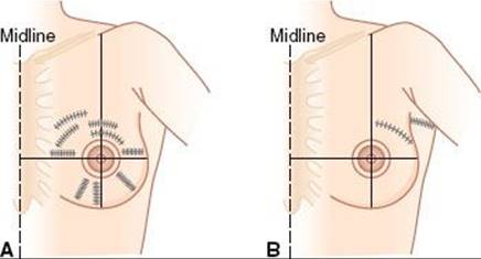
Choices Between Mastectomy and Breast-Conserving Surgery in Early-Stage Disease
For the majority of patients with early-stage breast cancer, breast-conserving surgery and mastectomy are both reasonable options, and patients are often conflicted regarding the choice. Patients should be reassured that provided they meet the criteria for breast-conserving surgery, all of the available medical evidence demonstrates equivalent long-term survival rates with both modalities.
A modified radical mastectomy may be preferable for some patients who wish to avoid radiation, for those in whom removal of clinical and radiographically apparent disease will result in a suboptimal cosmetic result, for those with diffusely positive margins which cannot be cleared with re-excision, and those with diffuse suspicious microcalcifications. Even patients who are ideally suited for breast-conserving therapy may have a personal preference for mastectomy, based on a number of factors.
Whelen et al.322 reported on the decision-making process in 82 consecutive node-negative patients presented with a decision board of therapeutic choices. Overall, 95% of women chose lumpectomy and breast irradiation. Kelemen et al.323 evaluated the choice between breast-conserving surgery and modified radical mastectomy in 7,815 women with early-stage breast cancer. There was a progressive increase from 16% to 47% in the use of breast-conserving therapy to treat tumors of all sizes over the 11 years of the study (P <.0001), and it was more frequently used for ≤2 cm tumors with an odds ratio of 2.46. Breast-conserving therapy was used at a slightly higher rate in medical centers than in community hospitals (31% vs. 28%; P <.0001); its use varied among geographic regions from a low of 24% in the southwestern United States to 36% in the northeast and 40% in hospitals outside the continental United States (P <.0001). Local availability of radiation therapy did not influence choice of treatment.
In response to a questionnaire mailed to 2,405 oncologists from all three disciplines and 60 oncology nurses in the United States, Tannock and Belanger324 noted that more than 60% thought that modified radical mastectomy and conservation surgery plus irradiation were equivalent options for patients with stage T1 and T2 carcinoma of the breast; 31% of the surgical oncologists favored the former and 35% of the radiation oncologists favored the latter approach. Medical oncologists were equally divided between the two procedures (14% and 18%, respectively).
In an analysis from Canada, Temple et al.325 prospectively evaluated participants with a first diagnosis of localized unilateral breast cancer who were candidates for breast-conserving therapy or mastectomy. Of 157 patients between 1992 and 1995, 71.3% anticipated having breast-conserving surgery and 28.7% anticipated modified radical mastectomy. The patient, physician, and significant other were perceived to play a role in the decision process. The two top-ranked items perceived to have influenced treatment choice were doctor’s advice and possibility of complete cure. Most women (60%) participated in treatment choice to the degree that they preferred, but only 13.6% received the preferred amount of information. The type of planned surgery was predicted by surgeon, contribution of doctor to choice of treatment, importance of breasts to sexuality, self-efficacy, and concerns about cancer recurrence from a multivariable logistic regression model. The authors concluded that both patient and surgeon factors are important predictors of type of planned surgery, but there is a gap between women’s preferences and actual experiences with regard to information provided.
Some patients with early-stage breast cancer will choose mastectomy to avoid the course of radiation, either due to the logistics of 6 weeks of therapy or due to fears of radiation therapy. It is likely that more mature results and selection factors for treating women with accelerated partial breast irradiation will become available. For those women in whom the time commitment of treatment is an issue, this option may further impact the choice between mastectomy and breast-conserving surgery.
Patients selecting mastectomy should also be made aware that this procedure does not totally eliminate the need for radiation treatment. For patients with early-stage operable breast cancer treated by modified radical mastectomy, postoperative irradiation of the chest wall and peripheral lymphatics may be indicated in selected patients with high-risk characteristics, positive nodes, or positive resection margins. Indications for postmastectomy radiation are discussed in Chapter 54.
Surgical Management of Axillary Lymph Nodes
An axillary node dissection or sentinel node biopsy are a standard component of the staging process for a majority of women with early-stage invasive breast cancer.190 Although the role of complete axillary dissection is evolving, currently most patients with positive sentinel nodes will undergo completion axillary dissection. Clearly the procedure is most important for women in whom axillary nodal status will influence subsequent management with respect to adjuvant systemic therapy. The role of axillary dissection and sentinel node sampling, however, continues to evolve and is currently being evaluated in several trials. It is not uncommon for clinicians to avoid axillary staging if it is not going to influence management.68,326,327 This is relevant in elderly women with receptor-positive tumors who will receive hormonal therapy, regardless of nodal status, and who are not thought to be candidates for cytotoxic chemotherapy. Recent data from the ACOSOG Z0011 trial may obviate the need for axillary dissection for selected woman of any age with a positive sentinel node biopsy.237,238 As will be discussed later, axillary radiation, as with axillary dissection, results in a high rate of regional control.68,326,327
Axillary Node Dissection
The axillary contents are divided into three levels: level I represents tissue between the axillary vein and the latissimus dorsi muscle and the lateral border of the pectoralis minor muscle; level II is located between the lateral and medial borders of the pectoralis minor muscle; and level III is between the medial border of the pectoralis minor and Halsted’s ligament (the apex of the axilla).1 Thorough dissection of levels I and II has traditionally been the most common axillary surgical procedure in patients with clinically node-negative breast cancer. Complete axillary dissection, including level III, may be performed in patients with clinically positive lymph nodes. A higher incidence of breast and arm edema has been noted with level III axillary node dissection, and the benefit of dissecting the level III lymph nodes has not been demonstrated.328 Pigott et al.329 reported on 146 patients treated with radical mastectomy (either modified or Halsted) for invasive ductal or lobular carcinoma of the breast. Eighty patients (55%) had histologically proven axillary lymph node metastases. If only the low (level I) axillary lymph nodes had been removed, 18 patients (25%) would have had metastases confined to levels II and III that would have gone undetected. However, only 1.4% of patients showed positive level III lymph nodes if levels I and II were negative.
Approximately 20% to 40% of patients with carcinoma of the breast and clinically negative lymph nodes have pathologic evidence of lymph node metastases.1,110,233,330,331 Yet, in patients with stage I or II breast cancer and clinically negative axillary lymph nodes, if an axillary dissection is not performed, axillary recurrence develops in only approximately 20%.73 In patients with clinically positive axilla, 20% to 30% have no histologic evidence of nodal metastatic disease.73
The necessity of axillary dissection has been questioned in selected patients with a low probability of nodal involvement by Silverstein et al.,68 who reported positive axillary lymph nodes in only 3 (3%) of 96 patients with tumors ≤5 mm in diameter and in 27 (17%) of 156 patients with tumors 6 to 10 mm in diameter. Iwasaki et al.,332 in a group of 823 patients with T1N0M0 invasive breast cancer, also identified a subgroup of patients who may not need to undergo axillary lymph node dissection. Certain tumor types (medullary, mucinous, and tubular carcinoma) had lower positive rates for lymph node involvement. With regard to the histologic grade, lymph node positivity increased significantly with high-grade tumors.
In a randomized trial, evaluating the necessity of axillary dissection in older women, Martelli et al.333 reported on 219 women, 65 to 80 years of age, with early breast cancer and clinically negative axillary nodes who were randomized to conservative breast surgery with or without axillary dissection. Tamoxifen was prescribed to all patients for 5 years. With a follow-up of 60 months, there were no significant differences in overall or breast cancer mortality or crude cumulative incidence of breast events between the two groups. Only two patients in the no axillary dissection arm (8 and 40 months after surgery) developed overt axillary involvement during follow-up. They conclude that older patients with T1N0 breast cancer can be treated by conservative breast surgery and no axillary dissection without adversely affecting breast cancer mortality or overall survival.
In light of the increased use of primary tumor-related factors, including molecular profiling, for decision making regarding systemic therapy, combined with sentinel node procedures and low rates of regional relapse with radiation therapy or observation in selected patients, it is likely that axillary dissection will be less frequently employed in the future.
Sentinel Lymph Node Biopsies
In recent years there has been a substantial increase in the use of sentinel lymph node biopsies to stage patients with breast cancer. Patients are injected around the tumor with technetium-99m (99mTc) sulfur colloid and vital blue dye, and a handheld γ -probe is used to identify areas of highest radioisotope uptake in the lymphatic system. The lymph nodes underlying this area (sentinel lymph nodes) are removed.235,334–336 The sentinel node procedure has been widely embraced as an acceptable standard for women with breast cancer and has a high degree of sensitivity and specificity.337
Although the current standard for patients with a positive sentinel node is to undergo completion axillary dissection, the necessity of this has been questioned and was the subject of the ACOSOG Z0011.237 This was a prospective trial examining survival of patients with sentinel node metastases detected by standard hematoxylin and eosin staining, who were randomized to undergo axillary lymph node dissection (ALND) after sentinel lymph node dissection (SLND) versus SLND alone; all patients received whole-breast irradiation. There were 446 patients randomized to SLND alone and 445 to SLND plus ALND. Patients were equally stratified according to multiple factors; those randomized to SLND plus ALND had a median of 17 axillary nodes removed compared with a median of only 2 sentinel nodes removed with SLND alone (P <.001). At a median follow-up time of 6.3 years, there were no statistically significant differences in local recurrence (P = .11) or regional recurrence (P = .45) between the two groups. Interestingly, ALND also removed more positive lymph nodes (P <.001) in 27% of the patients in this group. The patients in this trial all had whole-breast irradiation, but regional nodal irradiation was not allowed. The reason for the low regional relapse rates is likely a combination of factors, including a favorable subset of patients with a low likelihood of a high residual axillary burden of disease, the use of systemic therapy, and incidental radiation to the residual nodes in level I or II from the tangential breast irradiation. Several studies describe an approach to radiation field design based on probability of additional axillary nodal involvement in patients with positive sentinel nodes (Table 56.12).238,338–341
The ALMANAC (Axillary Lymphatic Mapping Against Nodal Axillary Clearance) trial is a multicenter randomized trial of 1,031 patients randomly assigned to sentinel node biopsy (n = 515) or standard axillary dissection (n = 516).335 Mansel et al.335 reported the primary outcome measures, which were arm and shoulder morbidity and quality of life. Drain usage, length of hospital stay, and resumption of normal activities after surgery were all highly significantly better in the sentinel lymph node group. In addition patient recorded quality of life and arm functioning scores were also significantly better, with no increase in anxiety levels in the sentinel node group. The authors conclude that sentinel node biopsy is the treatment of choice for patients who have early-stage breast cancer and clinically negative nodes.
Veronesi et al.342 also reported on a randomized trial of 516 patients with T1 tumors, randomized to either sentinel node biopsy or total axillary dissection. Axillary dissection was performed in the sentinel node group if the sentinel node contained metastases. In the axillary dissection group, the overall accuracy of the sentinel node status was 96.9%, the sensitivity 91.2%, and the specificity 100%. There was less pain and better arm mobility in the patients who underwent sentinel node biopsy only than in those who also underwent axillary dissection. There were 15 events associated with breast cancer in the axillary dissection group and 10 such events in the sentinel node group. Among the 167 patients who did not undergo axillary dissection, there were no cases of overt axillary metastasis during follow-up.
Weaver et al.343 evaluated 443 patients with breast cancer. After sentinel node biopsies, a complete axillary lymph node dissection was performed. Original pathologic material was reviewed for 431 patients enrolled in this study and for 214 patients with node-negative disease. Metastases were detected in 16% of the sentinel lymph nodes and in 4% of the nonsentinel nodes (odds ratio [OR] 4.3; P <.001). Occult metastases were detected in 4% of the sentinel lymph nodes and in 0.3% of the nonsentinel nodes. The probability of detecting metastases in nonsentinel nodes was more than 13 times greater in patients with positive sentinel lymph nodes than in patients with negative sentinel lymph nodes (P <.001).
The role of sentinel lymph node procedures in patients undergoing neoadjuvant chemotherapy is covered in Chapter 57.
TABLE 56.12 SUGGESTED APPROACH FOR RADIATION FIELD DESIGN IN SENTINEL NODE POSITIVE PATIENTS NOT UNDERGOING AXILLARY LYMPH NODE DISSECTION

SYSTEMIC MANAGEMENT OF BREAST CANCER
Systemic therapy is an essential component of both early-stage node-negative breast cancer as well as advanced-stage disease. Hormonal therapy, cytotoxic chemotherapy, and the more recently introduced biological therapies are routinely employed in the vast majority of patients with early-stage breast cancer. For patients with all stages of breast cancer, systemic therapy has been shown to decrease the relative risk of relapse and mortality. However, there are subsets of patients with a very favorable prognosis and extremely low rate of relapse, in whom the risk reduction results in only a very small absolute benefit. It is beyond the scope of this chapter to discuss all of the issues related to systemic management of breast cancer and the reader is referred to comprehensive reviews on the use of chemotherapy, endocrine therapies, and biologic therapies in the systemic management of breast cancer. These issues are also discussed in more detail in Chapter 57.344–346
In the recent 2011 St. Galen consensus meeting, the panel adopted the approach that systemic therapy should be made based on recognition of intrinsic subtypes, and that for practical reasons these intrinsic subtypes could be approximated based on more conventional parameters of estrogen receptor, progesterone receptor, HER2/neu, and Ki-67. Endocrine therapy alone is generally adequate for luminal A type cancers, while chemotherapy is typically indicated for luminal B and triple negative cancers, and trastuzumab is generally added for HER2-positive disease.347
RADIATION THERAPY IN THE MANAGEMENT OF EARLY-STAGE INVASIVE BREAST CANCER
The radiation oncologist plays a critical role in the management of early-stage breast cancer. As noted previously, the role of radiation therapy in postmastectomy radiation and in the neoadjuvant treatment of advanced breast cancers will be discussed in Chapter 57. The remainder of this chapter will primarily be focused on the role of radiation therapy in the conservative management of early-stage invasive breast cancer.
This will include an extensive discussion of the studies establishing breast-conserving surgery and radiation as the preferred standard of care for the majority of women with early-stage invasive disease and studies evaluating the avoidance of radiation therapy in selected patients. Integration of radiation therapy with systemic therapy will be discussed, as will selection of patients for breast-conserving therapy. Risk factors for local relapse and controversies in the management of patients with special circumstances will be discussed. Technical issues in the delivery of radiation therapy for early-stage breast cancer, including dosing and fractionation, matching techniques, and newer technical approaches, will be presented. The rapidly evolving area of partial breast irradiation will also be discussed. Follow-up of the breast cancer patient and sequelae of treatment will be presented. Management of local relapse in the conservatively treated breast and postmastectomy will be covered in Chapter 57. The role of radiation therapy in the management of DCIS and LCIS was discussed Chapter 55.
Breast-Conserving Therapy and Patient Selection
Breast-conserving surgery followed by radiation therapy to the intact breast is now clearly established as the most acceptable standard of care for the majority of women with early-stage invasive breast cancer. In 1992, the Journal of the National Cancer Institute published a monograph that stated that breast-conservation treatment is an appropriate method of primary therapy for most women with stage I or II breast cancer and is preferable because it provides survival equivalent to that of total mastectomy and axillary dissection while preserving the breast.348 Recommended techniques for breast-conservation treatment are wide local excision of the primary tumor, preferably with clear margins, axillary lymph node dissection, and breast irradiation (45 to 50 Gy), usually with a boost (10 to 20 Gy, depending on tumor size and status of the surgical margins). As will be discussed in detail under prognostic factors, while widely negative margins are desirable, patients with focally involved margins can be treated with radiation with excellent local control rates.
In addition to tumor control and survival, conservation of the breast with optimal cosmetic results is a crucial goal of this therapy, which is associated with improved psychoemotional adjustment of the patient to the diagnosis and treatment of carcinoma of the breast. It also enhances the acceptance by women of mammographic screening for early detection of this disease.
The widespread embracement of breast-conserving surgery followed by radiation therapy is based on numerous mature and well-documented studies, both prospectively designed randomized trials and large retrospective series of appropriately selected patients treated with breast conservation followed by radiation therapy.
It is important to select appropriate patients and tumors for breast-conservation therapy, with close consultation between the surgeon, medical oncologist, and the radiation oncologist and after thorough discussion of therapeutic alternatives with the patient. Risks and benefits of breast-conserving therapy compared with mastectomy should be discussed. Despite the clear evidence of equivalence, some patients will still prefer mastectomy, which remains an acceptable standard of care for all women with operable breast cancers. Although avoidance of radiation may be a primary rationale for some patients in choosing mastectomy, patients should realize that even with early-stage operable disease postmastectomy radiation may be indicated based on the pathologic findings at mastectomy. It is likely that over the next several years, long-term results and selection factors for accelerated partial breast irradiation will become available and may further influence patient choices regarding breast-conserving surgery with more rapid radiation compared with mastectomy.349,350–351
Ideally, patients electing breast-conserving surgery and radiation will have unicentric primary tumors that are <4 to 5 cm in diameter, as cosmesis is affected by the amount of tissue that must be removed in relation to the size of the breast.352 For patients in whom the size of the tumor, compared with the size of the breast, will result in an unacceptable cosmetic outcome, neoadjuvant chemotherapy followed by lumpectomy and radiation has been demonstrated to result excellent breast-conservation rates.353–355 This was discussed in detail in Chapter 54.
With careful attention to surgical margins, radiation technique, and the appropriate use of systemic therapy, local relapse rates in the majority of conservatively managed patients are low, and only a minority of patients with early-stage invasive breast cancer are not suitable for breast-conserving therapy. There are several perceived relative “contraindications” to breast-conserving therapy, including patients with collagen vascular disease, patients with germline mutations that predispose to breast cancer development, and those with positive margins, more advanced disease, multicentric disease, pregnancy, or who have had prior radiation. Although many of these factors require careful consideration and discussion between the treating physicians and the patient, as will be discussed in the section on special circumstances, selected patients faced with breast cancer in the setting of these controversial circumstances can be offered breast-conserving therapy with acceptable outcomes. Although some clinicians believe that patients at higher risk for development of local recurrence should not be treated with conservation surgery, there are relatively few absolute contraindications.
Perhaps with the exception of the patients with persistently positive diffuse margins or gross multicentric disease, where removal of clinically and radiographically apparent disease would result in an unacceptable cosmetic outcome, breast-conserving therapy followed by radiation can be offered to most women with early-stage breast cancer and may be offered to a high percentage of women with advanced cancers following neoadjuvant chemo- or hormonal therapy. The decision regarding breast-conserving therapy compared with mastectomy is often based on personal preference as the available medical and scientific evidence suggests equivalent overall and disease-free survival in all subsets of patients.
Early Reports of Breast-Conserving Therapy
In 1937, Keynes60 stated that “widespread operations based upon the permeation theory of lymphatics and fascial planes have no real justification and the idea of conservative treatment of cancer of the breast may become less repugnant to us [surgeons].” He treated 325 patients with local removal of the breast tumor and radium implantation at the site of local incision as well as in the axilla. In 250 patients, the 5-year survival rate was 71% for group 1 (disease confined to the breast), 29% for group 2 (disease apparently confined to breast and axilla), and 23.6% for group 3 (advanced disease or inoperable cancer). At the time, the results were comparable with those achieved with radical mastectomy. Other early reports316,356–359 paved the way for the development of randomized trials that have now clearly established breast-conserving surgery followed by radiation therapy as an accepted standard of care.
Randomized Studies Comparing Breast-Conserving Surgery Plus Radiation Therapy to Mastectomy
There have been numerous randomized trials that have now clearly established breast-conserving surgery followed by radiation therapy as equivalent to mastectomy for appropriately selected patients with early-stage breast cancer. Table 56.13 summarizes these randomized trials. In all of these trials, local tumor excision (tylectomy, lumpectomy), segmental mastectomy, or quadrantectomy combined with irradiation to the breast yielded survival and tumor control rates similar to those achieved with modified or classic radical mastectomy.93,101,102,360–361,362,363–364 Each of these randomized trials, as well as meta-analyses of the trials, clearly demonstrate equivalent mortality rates in conservatively treated patients compared with mastectomy.365–367
TABLE 56.13 PROSPECTIVE RANDOMIZED TRIALS COMPARING CONSERVATIVE SURGERY AND RADIATION WITH MASTECTOMY FOR EARLY-STAGE BREAST CANCER

FIGURE 56.14. Life-table analysis showing disease-free survival among patients in the three cohorts who were treated by total mastectomy, lumpectomy, or lumpectomy and breast irradiation. The number of events includes those that occurred after the 20-year follow-up period. (From Fisher B, Anderson S, Bryant J, et al. Twenty-year follow-up of a randomized trial comparing total mastectomy, lumpectomy, and lumpectomy plus irradiation for the treatment of invasive breast cancer. N Engl J Med 2002;347:1233–1241, 2002, copyright Massachusetts Medical Society.)
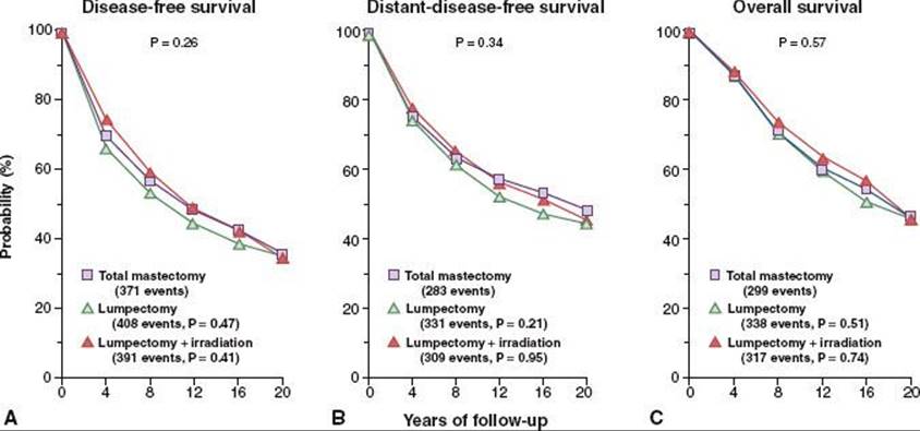
The earliest prospective, randomized trial comparing breast conservation with radical mastectomy was conducted at Guy’s Hospital in London. Three hundred and seventy women with stage I or II breast cancer were randomly assigned to receive either standard radical mastectomy or wide local excision plus irradiation.368 Although the rates of survival and distant metastasis were not significantly different for stage I disease, in stage II the recurrence rates in the breast and axilla were higher in the group treated with local excision and irradiation, and survival was significantly lower because of a higher rate of distant metastasis. Major weaknesses of this study were the low doses of irradiation used (35 to 38 Gy to the breast and 25 to 27 Gy to the axilla), probably patient selection, and surgical techniques.
Veronesi et al.363 reported on 701 patients with tumors <2 cm in diameter and without palpable axillary nodes of whom 352 were randomly assigned to treatment with either quadrantectomy and axillary dissection plus irradiation (50 Gy in 5 weeks to the breast and 10-Gy boost) and 349 to radical (Halsted) mastectomy. Women with positive axillary lymph nodes also received 12 cycles of adjuvant chemotherapy with CMF. Actuarial 20-year overall and disease-free survival rates were comparable in the two groups (58%). The death rates from breast cancer were 26.1% and 24.3%. The incidence of local failure was 2.3% with mastectomy and 8.8% with quadrantectomy and irradiation. There was no difference in the incidence of contralateral breast cancer (10.2% and 8.7%, respectively).102
Fisher et al.93,360 updated the results of the NSABP protocol B-06 in 1,843 women with clinical stage I or II carcinoma of the breast <4 cm in diameter. Patients were randomly assigned to be treated with total mastectomy or lumpectomy (segmental mastectomy), with or without irradiation. Irradiated patients received 50 Gy to the breast through tangential fields irradiation and a boost to the operative site was not given. With 20-year follow-up, there was no significant difference in survival among patients treated with mastectomy, lumpectomy alone, or lumpectomy combined with irradiation (Fig. 56.14). For the patients treated with lumpectomy alone, the ipsilateral breast relapse rates were approximately 40% if the nodes were negative and 50% if they were positive. For patients treated with lumpectomy and irradiation, the corresponding rates were 10% for all patients and those with negative nodes and 5% for those with positive nodes, illustrating the interaction of irradiation and adjuvant chemotherapy in local tumor control. Cumulate incidence of ipsilateral breast tumor relapse in the lumpectomy alone compared with the lumpectomy and radiation arm is shown in Figure 56.15.
The Institut Gustave-Roussy conducted a prospective, randomized trial comparing mastectomy with local excision plus irradiation for women with cancers measuring ≤2 cm.369,370 The 15-year disease-free survival rate was 55% for the tumorectomy group and 45% for the mastectomy group (P = .23). The 15-year local recurrence rate was 9% in the conservation surgery and irradiation group and 14% in the mastectomy group.
The EORTC Breast Cancer Cooperative Group conducted a randomized trial of women with stage I or II breast cancer comparing modified radical mastectomy (420 patients) with breast-conservation therapy (448 patients).362 The actuarial 8-year local tumor control rate was similar in both arms, 91% in the mastectomy group and 87% in the breast-conservation therapy group. There was one axillary recurrence in the mastectomy group and three in the conservation therapy group.
A Danish Cooperative Study carried out a similar randomized trial in 905 women (another 248 patients were treated with mastectomy or breast-conservation therapy according to preference without randomization).371 High-risk patients (tumor >5 cm, invasion to skin or deep fascia, metastatic axillary lymph nodes) who were treated with breast-conservation therapy received radiation therapy to the regional lymph nodes. Those who were treated with mastectomy also received irradiation of the same target volume and all high-risk patients received adjuvant CMF. At 6 years, the recurrence-free survival rate in 430 patients treated with breast-conservation therapy was 70%, compared with 66% in 429 patients treated with mastectomy. Overall survival rates were 79% and 82%, respectively. There were 12 breast relapses in the former group (3%) and 19 chest wall recurrences in the latter group (4%). In the breast-conservation therapy group, 31% of patients had excellent and 41% had satisfactory cosmesis.
The U.S. National Cancer Institute reported results of a randomized study in which 122 patients with T1–2N0M0 disease were treated with modified radical mastectomy and 125 with breast-conservation therapy (45 to 50 Gy to breast plus 15- to 20-Gy boost).372 Recently updated by Poggi et al.373 with a median follow-up of 18.4 years, there was no detectable difference with regard to overall survival between patients treated with mastectomy and those treated with breast-conserving therapy (58% vs. 54%; P= .67 overall). Twenty-seven women in the breast-conserving therapy arm (22%) experienced an in-breast event. After censoring in-breast events in the breast-conserving therapy arm that were salvaged successfully by mastectomy, disease-free survival also was found to be statistically similar (67% in the mastectomy arm vs. 63% in the breast-conserving therapy arm; P = .64 overall). There was no statistically significant difference in the incidence of contralateral breast carcinoma between the two treatment groups.
Van Dongen et al.101,362 reported on an EORTC trial comparing modified radical mastectomy with breast-conserving therapy (lumpectomy, axillary clearance, and irradiation to the breast, 50 Gy in 5 weeks and a 25-Gy boost with iridium implant) in 168 patients with stage I and 734 with stage II disease. Patients with microscopically incomplete excision of the tumor were included. Updated analysis from this trial with a median follow-up period of 13.4 years revealed locoregional recurrence rates were higher in the breast-conservation therapy group (10-year rate, 19.7%) than in the mastectomy group (10-year rate, 12.2%, P = .0097). The overall survival rate at 10 years was 66.1% for patients undergoing mastectomies and 65.2% for patients undergoing breast-conservation therapy (P = .11).
FIGURE 56.15. Cumulative incidence of ipsilateral breast recurrence after lumpectomy (open diamonds) or lumpectomy plus breast irradiation (solid diamonds) in 1,137 patients in the current-update cohort (cohort B) who had either negative or positive nodes and tumor-negative specimen margins. (From Fisher B, Anderson S, Bryant J, et al. Twenty-year follow-up of a randomized trial comparing total mastectomy, lumpectomy, and lumpectomy plus irradiation for the treatment of invasive breast cancer. N Engl J Med 2002;347:1233–1241, 2002, copyright Massachusetts Medical Society.)
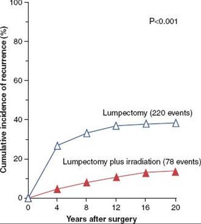
Additional Experiences with Breast-Conserving Surgery Plus Radiation Therapy
Although the acceptance of breast-conserving surgery plus radiation therapy as an alternative to mastectomy has been established based on the numerous randomized trials outlined above, large retrospective experiences with long-term follow-up have provided additional data regarding the selection of patients for breast-conserving therapy and radiation, prognostic factors for local-regional end points, impact of various treatment policies and techniques, complications, cosmesis, and long-term outcomes.
Mature Retrospective Series of Breast-Conserving Surgery Plus Radiation Therapy
There have been numerous reports of breast-conserving therapy and radiation, now with follow-up exceeding 10 to 15 years. These studies have provided valuable data regarding technical issues related to treatment and have provided additional data regarding outcomes in subsets of patients, prognostic factors for local control, cosmesis, and other sequelae. Lessons learned from these retrospective series have yielded valuable insight into the conservative management of breast cancer and provide the basis for further prospective studies. Selected publications are highlighted below and in the section on prognostic factors for local control. Long-term outcome from some of these experiences are summarized in Table 56.14.99,104,298,317,356,369,374–391 Collectively these studies clearly show long-term outcomes that are consistent with the randomized trials noted above. Local failure rates will depend on follow-up, selection factors, and treatment, as discussed later, but in general are expected to range between 0.5% and 1% per year.
PROGNOSTIC FACTORS FOR LOCAL RELAPSE FOLLOWING BREAST-CONSERVING SURGERY PLUS RADIATION THERAPY
The retrospective experiences outlined above together with the prospective randomized trials have provided additional data confirming acceptable long-term local control, cosmesis, and toxicities and have identified prognostic factors that lead to higher local relapse rates and impact treatment policies.
Local relapse in the conservatively managed breast is an active area of investigation, with numerous studies dedicated to the evaluation of factors that identify patients at increased risk for local relapse following lumpectomy and radiation therapy. Although prognostic factors for local relapse are not as well evaluated as they have been for systemic relapse, there have been numerous studies demonstrating the prognostic value of molecular and genetic markers for local relapse. Local relapse following breast-conserving surgery and radiation can be governed by a complex array of host, primary tumor, and treatment factors.
Given the complex interaction of these factors, it is sometimes difficult to separate out the independent significance of any one factor. There have, however, been several factors that have been consistently reported in influencing local relapse in the conservatively managed breast cancer patient. Table 56.15 summarizes some of the more commonly reported prognostic factors for local relapse. The factors that most consistently have been reported to influence local relapse, namely, young age, margin status, and the use of systemic therapy, are discussed first, followed by other commonly evaluated prognostic factors.
TABLE 56.14 CONSERVATION SURGERY AND IRRADIATION: NONRANDOMIZED STUDIES STAGE I AND II BREAST CANCER: RESULTS OF SELECTED STUDIES

TABLE 56.15 SUMMARY OF RISK FACTORS FOR LOCAL RELAPSE
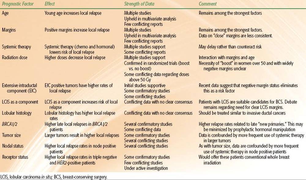
TABLE 56.16 IPSILATERAL BREAST RECURRENCE RATES BY AGE
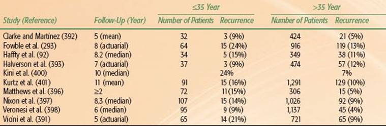
TABLE 56.17 IPSILATERAL BREAST RECURRENCE RATES BY MARGINS
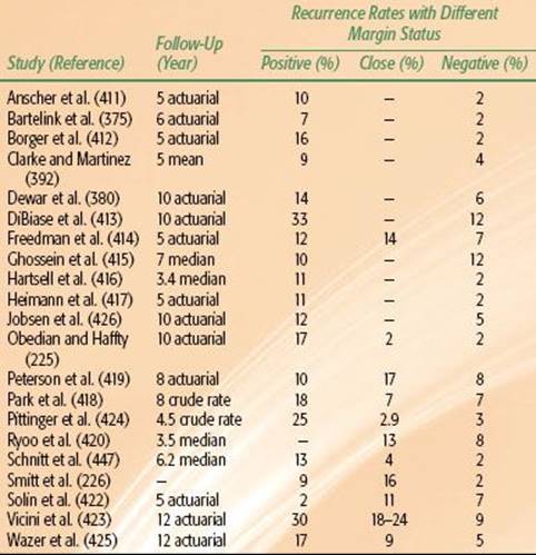
Age
Young age has been shown in numerous studies to be a risk factor for breast recurrence in conservation surgery and irradiation. These are summarized in Table 56.16.92,293,392–401
Different investigators have used various age cutoffs, such as 40, 35, and 30 years. Vilcoq et al.317 found a locoregional recurrence rate of 35% versus 4% in women younger or older than 30 years. Kurtz et al.395 reported a 19% incidence of local recurrence in 210 women younger than 40 years of age, compared with 9% in 1,172 older women. This observation correlated with EIC, high tumor grade, and a major mononuclear cell reaction. The Harvard Joint Center’s inferior results in younger women also correlated with the presence of EIC.402 These findings have also been reported by other authors.379,390,403
One of the most significant and powerful studies correlating young age with local relapse was the large EORTC boost versus no boost trial.404 Overall in this study young patient age was a significant predictor of local relapse. As shown in Figure 56.16, the use of a boost, as will be discussed later, was most effective in younger women, although the more recent update found an advantage to the boost in all age groups.405 In a review of 3,602 women who underwent surgery (breast conservation, 55%, or mastectomy, 45%) for early breast cancer and were rolled in EORTC studies, de Bock et al.406 clearly demonstrated the impact of young age on local relapse. The results of multivariate analysis showed that younger age and breast conservation were risk factors for isolated locoregional recurrence (breast cancer under 35 years of age vs. over 50 years of age: hazard ratio [HR] 2.80; 95% CI, 1.41 to 5.60; breast cancer age 35 to 50 years vs. over 50 years: HR 1.72; 95% CI, 1.17 to 2.54; breast conservation: HR 1.82; 95% CI, 1.17 to 2.86). After perioperative chemotherapy, less isolated locoregional recurrences were observed (HR 0.63; 95% CI, 0.44 to 0.91). It is concluded that young age and breast-conserving therapy are both independent predictors for isolated locoregional recurrence. The authors note that as an isolated locoregional recurrence is a potentially curable condition, women treated with breast conservation or diagnosed with breast cancer at a young age should be monitored closely to detect local recurrence at an early stage.
Numerous other studies have confirmed in univariate and multivariate analyses a significant correlation between younger age and local relapse rates. Although there is no apparent cutoff age where relapse rates significantly change, most of the studies have selected 35 years of age or 40 years of age to demonstrate the differences.290,292,293,394,397,407,408 However, one study that compared the outcome of breast conservation in patients under 40 found that those under 35 had a higher risk of recurrence compared with those aged 36 to 40.409
Margin Status
Positive margin status has been one of the most consistently reported factors associated with higher local relapse rates. Selected studies are summarized in Table 56.17.225,226,375,380,392,410–425 Due to the varying definitions of margin status and the complex interaction between margin status and use of systemic therapy, radiation dose, and patient age, there are conflicting conclusions regarding the influence of margin status on local relapse.
Definition of a negative margin is variable between institutions and in national studies. Although some consider no tumor cells seen on the inked margin definitive, as defined by the NSABP, others consider negative as margins of 1 or 2 mm beyond invasive cancer; of note, many of the studies evaluating margin status utilized pathological reports without a central re-review of the original pathology.
The degree of margin involvement (i.e., whether it is focally involved or more diffusely involved) is also a critical issue in determining the significance of margin involvement and how it should influence management. Although there are clearly conflicting reports, it is evident that obtaining a wide negative margin is desirable. However, a focally involved margin, particularly when re-excision is not technically feasible, as may be the case with a focally involved deep margin at the pectoralis facia, is not a contraindication to breast-conserving therapy.
FIGURE 56.16. Cumulative incidence of recurrence of tumor in the ipsilateral breast after whole-breast irradiation at 50 Gy, with or without an additional dose to the tumor bed as a function of age group. (From Bartelink H, Horiot JC, Poortmans PM, et al. Impact of a higher radiation dose on local control and survival in breast-conserving therapy of early breast cancer: 10-year results of the randomized boost versus no boost EORTC 22881–10882 trial. J Clin Oncol 2007;25(22):3259–3265, with permission.)
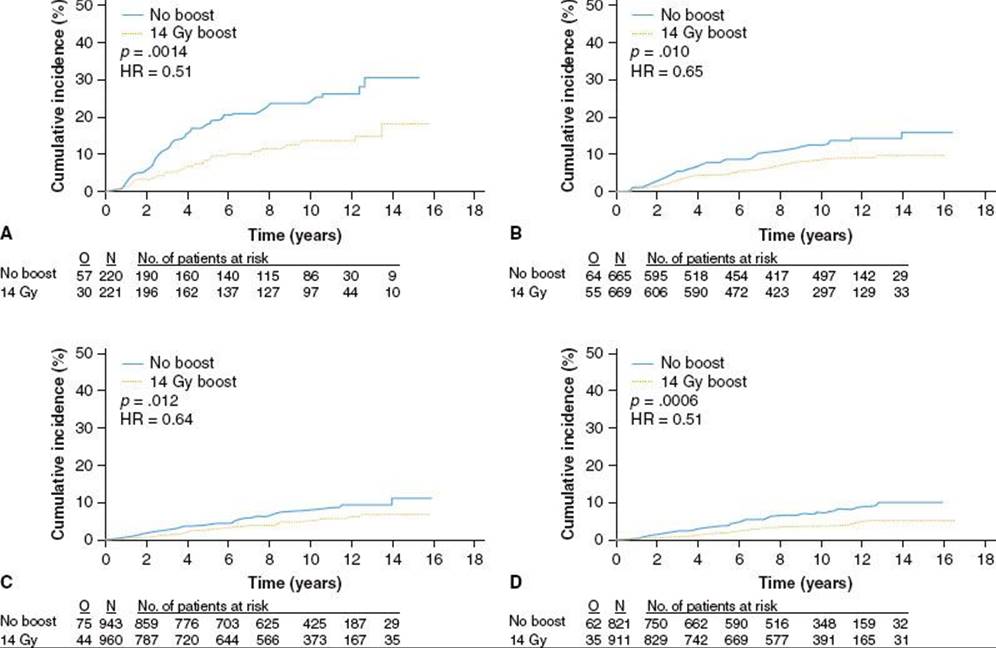
Interaction Between Margin Status and Other Variables
The complex interaction between margin status and other treatment-related factors is demonstrated in a recent study by Park et al.418 They demonstrated that local relapse was significantly higher in patients with diffuse margin involvement than in patients with negative margins. Patients with focally involved margins also had a higher risk of local relapse than patients with negative margins. However, in those women with focally involved margins who received systemic therapy, the local relapse rate was similar to those with negative margins. Whether systemic therapy delays or negates the effect of a focally involved margin is a matter of debate, but clearly there are confounding factors that complicate interpretation of the available data.
Freedman et al.414 studied 1,262 patients with clinical stage I or II breast cancer treated by breast-conserving surgery, axillary node dissection, and radiation therapy. The final margins were negative in 77%, positive in 12%, and close (≤2 mm) in 11%. The 5-year incidence of ipsilateral breast tumor recurrence was not significantly different amoung patients with negative (4%), positive (5%), or close (7%) margins. However, by 10 years, a significant difference in ipsilateral breast tumor recurrence became apparent (negative 7%, positive 12%, close 14%; P = .04). The 5-year cumulative ipsilateral breast tumor recurrence rate in patients with close or positive margins was 1% with adjuvant systemic therapy and 13% with no adjuvant therapy. However, by 10 years, the ipsilateral breast tumor recurrence rate was similar (18% vs. 14%), due to more late failures in the patients who received adjuvant systemic therapy.
A study by Jobsen et al.426 demonstrates some of the difficulties associated with interpretation of studies related to margin status and local relapse as it relates to other prognostic factors. In a study of 1,752 patients with known margin status and a median follow-up of 78 months, the 10-year local relapse rate was 5.6% and 12.2% for negative and positive margins, respectively. An interaction between age category and margin status was noted in relation to local relapse-free survival. The 5-year local relapse rate for women younger than 40 years of age was 8.4% for negative margins and 36.9% for positive margins (P = .005). On the other hand, the 5-year local relapse rate for women over 40 years old was 2.6% for negative and 2.2% for positive margins.
Although it is clear that margin status is a significant risk factor for local relapse, and negative margins are desirable, the available data suggest that patients with a focally involved margin are suitable candidates for breast-conserving therapy followed by radiation therapy to the intact breast.
TABLE 56.18 IPSILATERAL BREAST RECURRENCE RATES BY SYSTEMIC TREATMENT (SysTx)
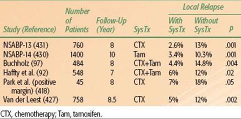
TABLE 56.19 CONSERVATION SURGERY AND IRRADIATION IN BREAST CANCER: LOCAL RECURRENCE AT 5 YEARS CORRELATED WITH INITIAL TUMOR STAGE
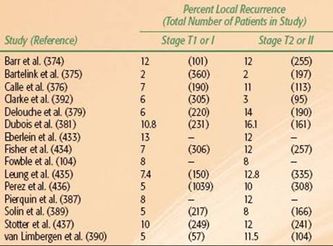
Effect of Systemic Therapy on Local Control
The use of systemic therapy, in the form of adjuvant tamoxifen or adjuvant chemotherapy, has been clearly shown to impact local control in numerous retrospective and prospective randomized trials. Although it has been clearly demonstrated in randomized trials that chemotherapy and tamoxifen are not appropriate substitutes for radiation therapy, in patients who are treated with radiation therapy, the use of systemic therapy improves local control.93,94,97,427As with the other critical prognostic factors, the degree to which systemic therapy influences local control is confounded by other factors.
In the NSABP B-06 trial, patients with lymph node–positive disease who were treated with radiation therapy and chemotherapy had an 8-year local recurrence rate of 5% compared with a local recurrence rate of 12% in lymph node–negative patients treated with surgery and radiation therapy alone.360 In the NSABP B-21 trial, tamoxifen similarly improved local control rates in patients with lymph node–negative breast tumors smaller than 1 cm. The crude rate of breast tumor recurrence was only 3% in women randomly assigned to undergo lumpectomy, radiation therapy, and tamoxifen compared with 7% in women treated with lumpectomy and radiation therapy alone.95 A retrospective analysis from the MD Anderson Cancer Center investigating the impact of systemic therapy on local control after breast-conserving therapy in patients with lymph node–negative breast cancer further confirmed these data.97 In this study, 277 patients treated with systemic therapy had improved 5-year (97.5% vs. 89.8%) and 10-year (95.6% vs. 85.2%) local control rates compared with 207 patients who received no systemic treatment. No statistically significant difference was evident in local control between patients treated with chemotherapy and those treated with tamoxifen alone (P = .219). In a Cox regression analysis, the use of systemic therapy was the most powerful clinical, pathologic, or treatment predictor of local control, producing a 3.3-fold reduction in the risk of local recurrence. Similar results have been reported in series from Yale as well as the Netherlands, where the risk of local relapse was lower among patients treated by breast-conserving surgery and radiation with adjuvant chemotherapy or adjuvant tamoxifen, compared with patients treated without adjuvant systemic therapy.427,428 Table 56.18summarizes several of these studies that have evaluated local control as a function of systemic therapy. It is evident that both hormonal therapy as well as cytotoxic chemotherapy influence local relapse rates.429–431
Trastuzumab (Herceptin) is a humanized monoclonal antibody against the human EGFR-2 (HER2), which is amplified or overexpressed in about 15% to 20% of invasive breast cancers; these tumors are known to be more aggressive and more susceptible to recurrence than HER2-negative tumors. Romond et al.274 reported on a combine analysis from two large cooperative group studies investigating the utility of trastuzumab in HER2-positive patients with operable breast cancer and found that trastuzumab improved disease-free survival by an absolute value of 12% at 3 years and was associated with a 33% reduction in the risk of death (P = .015). Interestingly, they also reported that ipsilateral breast tumor recurrence as a site of first failure was reduced from 57 patients to 27 patients with the use of trastuzumab. Recently, Kiess et al.432 reported on a series of 197 women with early-stage breast cancer; 70 women did not receive trastuzumab while 102 did receive trastuzumab. They found that the 3-year locoregional recurrence-free survival rate was 90% without trastuzumab and 99% with trastuzumab. Moreover, locoregional recurrences were reduced from 7 to 1 with the use of trastuzumab.
Tumor Size
Although tumor size is clearly a strong predictor of systemic relapse and overall survival, its prognostic value in local relapse has not been consistently reported (Table 56.19).98,363,374–376,379,381,387,389,390,392,433–437 Differences are probably related to the treatment techniques used (e.g., completeness of tumor excision, use of irradiation boost) and the complex interaction of other prognostic factors, as noted above.
Tumor Location
Location of the primary tumor within the breast is not known to be a contraindication to breast-conserving surgery, and any specific location is not associated with a higher local relapse rate. Haffty et al.,438 in a review of 1,014 patients with early breast cancer treated with breast-conservation therapy, identified 98 patients who had a central or subareolar tumor. Ten of 98 patients had the nipple–areola complex sacrificed at the time of surgery, whereas the remaining 88 patients had the entire area included in the boost cone–down field. The 10-year actuarial breast recurrence-free survival rate was 84%, the distant disease-free survival rate was 88%, and the overall survival rate was 79%, similar to patients with tumors in other locations. The nipple–areola complex could be preserved in most patients, and there were no significant complications. Thus, a subareolar breast cancer presentation was not a contraindication to breast-conserving therapy in early-stage disease.
However, Gajdos et al.439 reported on 95 women with tumors located within 2 cm of the border of the areola, considered to be subareolar carcinomas; 62 were treated with breast-conserving surgery and 33 with mastectomies. Radiation therapy was given to 87% of the breast-conserving surgery group and to 13% of the mastectomy group. The nipple–areola complex was removed in 11 women in the breast-conserving group. On univariate analysis, variables significantly related to pathologic involvement of the nipple–areola complex were clinical involvement of the nipple–areola complex (P = .001), mammographic calcifications or Paget’s disease (P <.001), pathologic tumor size (P = .019), and the presence of an EIC (P = .098). When radiation therapy was accounted for in multivariate analysis, the only variable significantly related to local recurrence in patients undergoing breast-conserving surgery was clinical involvement of the nipple–areola complex.
TABLE 56.20 INCIDENCE OF BREAST RELAPSE CORRELATED WITH EXTENSIVE INTRADUCTAL CARCINOMA COMPONENT IN PRIMARY BREAST TUMOR ADJACENT BREAST

Extensive Intraductal Carcinoma
According to the Harvard definition of EIC, 25% or more of the primary tumor is intraductal carcinoma, and intraductal carcinoma is seen outside (adjacent to) the infiltrating tumor border.440 Fourquet et al.383 reported a 20% incidence of EIC in 185 women younger than 45 years of age compared with 10.4% in 279 older women. EIC involving the primary tumor and adjacent tissues has been reported by some groups, particularly Harvard University and Marseilles, to be associated with a higher incidence of breast recurrences.395,441,442 In contrast, others have found no significant impact on local tumor control with EIC.196,232,378,390 This difference may be related to the definition of EIC, adequacy of tumor excision, doses of irradiation delivered to the boost volume, as well as interactions with other factors. It has been reported by some that a somewhat higher breast relapse rate in EIC-positive patients was seen only in women younger than 40 years of age. Table 56.20 summarizes reports of breast relapse correlated with presence of EIC in selected studies.98,196,198,375,398,402,433,442–444
Holland et al.445 stated that an EIC component is associated with subsequent breast recurrence because of the presence of residual intraductal carcinoma in these patients. In a series of 214 women who underwent mastectomy, 71% of those with EIC had residual intraductal carcinoma, compared with 28% of those without that pathologic feature. In particular, 44% of the EIC-positive patients had prominent residual tumor compared with 3% of those who were EIC negative (P <.00001).
The impact of EIC on local relapse, however, appears to be minimized if negative margins are achieved. Although negative margins are desirable in all patients undergoing breast-conserving surgery, attention to margins in patients with EIC is particularly relevant, as a negative margin may decrease or eliminate the significance of EIC with respect to local failure. In a study from the Harvard group, Gage et al.446 evaluated clinical stage I or II breast carcinoma treated with radiation therapy as part of breast-conserving therapy, of whom 343 had invasive ductal histology evaluable for an extensive intraductal component, had inked margins that were evaluable for an review of their pathology slides, and received ≥60 Gy to the tumor bed. The 5-year rate of ipsilateral breast recurrence (IBR) for patients with negative margins was 2%; for patients with positive margins, the rate was 16%. Among patients with negative margins, the 5-year rate of IBR was 2% for all patients with close margins (negative ≤1 mm) and 3% for those with negative margins >1 mm. For patients with close margins, the rates were 2% and 0% for EIC-negative and EIC-positive tumors, respectively; the corresponding rates for patients with negative margins greater than 1 mm were 1% and 14%. The 5-year rate of IBR for patients with focally positive margins was 9% (9% for EIC-negative and 7% for EIC-positive patients). The 5-year crude rate of IBR for patients with greater than focally positive margins was 28% (19% for EIC-negative and 42% for EIC-positive patients). The authors conclude that patients with negative margins of excision have a low rate of recurrence in the treated breast, whether the margin is >1 mm or ≤1 mm and whether the carcinoma is EIC-negative or EIC-positive. It appears from this study that although EIC may be a poor prognostic factor for local relapse, achievement of a negative margin eliminates EIC as a risk factor.
From a group of 885 patients treated for clinical stage I or II invasive breast cancer, Schnitt et al.447 limited their study to 181 patients with invasive ductal carcinoma (IDC) who received a radiation dose to the surgical site of 60 Gy or greater, whose final microscopic margins of resection were evaluable, and who had at least 5 years of follow-up. In 157 patients (87%), the tumor was evaluable for the presence or absence of EIC. The 5-year rates of recurrence among patients with negative, close, focally positive, and more than focally positive margins were 0%, 4%, 6%, and 21%, respectively. Among the 127 patients with EIC-negative tumors, the 5-year recurrence rate was <10% in all margin groups. Among the 30 patients with EIC-positive tumors, the 5-year recurrence rate was 0% when margins were negative or close but 50% when margins were more than focally positive. These results provide support for the use of breast-conserving therapy (including an irradiation boost to the primary site) for patients with EIC-positive tumors and negative margins.
Histology
In general, studies that have evaluated local relapse in relation to histologic subtypes of breast cancer have not demonstrated higher relapse rates associated with specific histologic patterns. Weiss et al.240 reported on 879 patients with stage I and II breast cancer treated with conservation surgery and irradiation. The patients were divided into 7 groups based on histologic subtype: 368 patients with infiltrating and intraductal ductal carcinoma, 389 with IDC, 41 with ILC, 23 with combined infiltrating ductal and lobular carcinoma, 28 with medullary carcinoma, 12 with colloid carcinoma, and 18 with tubular carcinoma. There were no significant differences in 5-year actuarial overall survival, cause-specific survival, or relapse-free survival rates among the histologic categories. There was, however, a difference among the seven groups in distant metastasis only at first failure, with IDCs having the highest rate.
Thurman et al.,241 in an analysis of the Harvard series, identified twenty clinical stage I and II patients with mucinous carcinoma, 27 with medullary carcinoma, 28 with tubular carcinoma, and 1,055 with IDC were identified. No significant difference was seen in the site of first failure among the four histologic types within the first 10 years after treatment. Local failure was significantly associated with age <50 years (P = .04), positive surgical margins (P = .007), lymphovascular invasion (P = .04), and presence of an extensive intraductal component (P <.001).
An analysis of medullary carcinomas treated conservatively was performed by the Yale group, who identified 46 cases of conservatively treated patients with medullary histology who were compared with 1,444 patients with infiltrating ductal carcinoma.202 The medullary cohort presented at a younger age with a higher percentage of patients in the 35 years or younger age group (26.1% vs. 6.6%; P <.00001). Twelve patients with medullary histology underwent genetic screening, and six patients were identified with deleterious mutations. This group showed greater association with BRCA1/2 mutations compared with screened patients in the control group (50.0% vs. 15.8%; P = .0035). The medullary cohort was also significantly associated with greater T stage and tumor size (37.0% vs. 17.2%; T2 mean size 3.2 vs. 2.5 cm; P = .00097) as well as negative ER (84.9% vs. 37.6%; P <.00001) and PR (87.5% vs. 48.1%; P = .00001) status. Breast relapse-free rates were not significantly different from the invasive ductal cancers (76.7% vs. 85.2%), however, 10-year distant relapse-free survival in the medullary cohort was significantly better than in the control group (94.9% vs. 77.5%; P = .028).
Tubular carcinomas treated with conservative surgery and radiation were reviewed by Sullivan et al.201 They reviewed 62 of their own cases from the Massachusetts General as well as 529 cases from the literature. They conclude that tubular carcinoma is associated with an excellent prognosis, but long-term follow-up is essential for detecting local failures. Adjuvant RT reduces the incidence of local failure following CS for tubular carcinoma. However, elderly women treated by CS may have a very low risk of local recurrence without adjuvant RT.
Infiltrating Lobular Carcinoma
A review of the literature strongly supports local tumor resection and breast irradiation as appropriate therapy for invasive lobular breast cancer, following the same guidelines used for invasive ductal tumors. Breast tumor control and survival after breast-conserving therapy are equivalent in patients with invasive ductal or lobular carcinoma. Due to the presumed multicentric nature of lobular carcinomas, several groups have attempted to assess whether these subtypes are more prone to local failure with breast-conserving approaches.242,448–453 Table 56.21 summarizes results of several selected series of patients treated with breast-conserving surgery and radiation comparing outcomes in lobular carcinoma to invasive ductal carcinomas. Although some of the studies show higher contralateral rates in lobular carcinomas, local relapse, disease-free, and overall survival appear to be comparable to invasive ductal carcinomas. The majority of studies show local-regional control, disease-free, and overall survival rates in lobular carcinomas that are comparable to patients with invasive ductal carcinomas.
TABLE 56.21 LOBULAR CARCINOMA TREATED WITH BREAST CONSERVING SURGERY AND RADIATION (SELECTED SERIES WITH 10-YEAR FOLLOW-UP)

TABLE 56.22 STUDIES EVALUATING LCIS AS A COMPONENT OF BREAST CANCER LOCAL RELAPSE IN BREAST-CONSERVING THERAPY PLUS RADIATION THERAPY

Lobular Carcinoma In Situ as a Component of Invasive Cancers
Several groups have evaluated whether patients with LCIS as a component of invasive cancer or DCIS was associated with higher local relapse rates. Conflicting results from these studies, as outlined in Table 56.22, preclude firm conclusions.454–458 Sasson et al.458 noted that LCIS was present in 65 of 1,274 patients (5%) with stage I or II breast cancer. LCIS was more likely to be associated with an ILC (30 of 59 patients; 51%) than with IDC (26 of 1,125 patients; 2%). The 10-year cumulative incidence rate of ipsilateral breast tumor recurrence was 6% in women without LCIS compared with 29% in women with LCIS (P = .0003). In both groups, the majority of recurrences were invasive. The 10-year cumulative incidence rate of ipsilateral breast tumor recurrence in patients who received tamoxifen was 8% when LCIS was present compared with 6% when LCIS was absent (P = .46). In a series of 56 patients with an LCIS component, Jolly et al.456 reported a higher risk of local relapse at 10 years (14%) compared with a rate of 7% in cases without an LCIS component. In multivariate analysis a component of LCIS was associated with a higher risk of local relapse.
However, studies from Yale, Harvard, Michigan, and a more recent study from Fox Chase failed to show a higher local relapse rate in patients with a component of LCIS. Abner et al.454 reviewed 1,181 patients with stage I or II infiltrating ductal, infiltrating lobular, or infiltrating carcinoma with mixed features who had received at least 60 Gy to the tumor bed and had a minimum follow-up of 8 years. Of the 1,181 patients, 137 had detectable LCIS in or adjacent to the tumor. The 8-year local recurrence rate was not significantly increased for patients with LCIS overall or for the subgroup of patients with LCIS in or adjacent to the tumor. The risk of contralateral disease and of distant treatment failure also was unaffected by the presence or extent of LCIS (5% to 10% in all groups). Similar results were reported by Moran and Haffty457 in an analysis of the Yale series where there was no statistically significant difference between patients with or without a component of LCIS in the 10-year overall survival (67% vs. 72%), distant disease-free survival (62% vs. 79%), or ipsilateral breast tumor recurrence-free survival (77% LCIS vs. 84% control). Ben-David et al.455 reported the results on 64 cases treated at University of Michigan and also found no association between local failure and presence of LCIS. The presence of LCIS at the margins and the size and presence of multifocal LCIS did not alter the rate of local control. Ciocca et al.,459 in an analysis of 290 patients with LCIS as a component compared with 2,604 without LCIS as a component, showed no difference in local control, even if LCIS was present at the final margin.
Other Histologic Features
Clemente et al.,460 in 506 cases of infiltrating ductal carcinoma (T1-2N0M0), described peritumoral lymphatic infiltration in 6.9% of routinely evaluated specimens, whereas in a randomly selected group of 234 cases the frequency was 20%. Patients with peritumoral lymphatic infiltration had worse disease-free and total survival rates than those without this feature (P = .0001 for each), as well as more local recurrences (P = .0001) and a higher incidence of distant metastases (P = .0576).
Wong et al.,461 in a study of 234 patients with clinical T1N0 breast cancer treated with breast-conservation surgery and radiation therapy, scored 180 patients as lymphatic vessel invasion negative and 54 as invasion positive (23 focal and 31 extensive). The local first failure rates were 14% and 22%, respectively. The percentages of regional distant failure (without local failure) were 12% and 21%, respectively. At 10 years, 60% of the lymphatic vessel invasion–negative patients remained free of any failure, compared with 50% of the lymphatic vessel invasion–positive patients.
Nodal Status
Although nodal status is the strongest predictor of distant metastasis and overall survival, most studies have not clearly demonstrated an effect of nodal status on local control in the conservatively managed breast cancer patient. This may be because a majority of node-positive patients receive chemotherapy or hormonal therapy, which may counteract any adverse effects on local relapse. There are some data, however, that suggest an effect of nodal status on local relapse in conservatively managed patients. The Primary Therapy of Breast Cancer Study Group and others noted lower survival and a greater incidence of local recurrences in patients with positive axillary nodes after partial mastectomy.233,462–464 At the Institut Gustave-Roussy, among 356 patients, local recurrence was noted in 26% of those with and 6.5% of those without nodal involvement; the greater the number of nodes involved, the more likely the occurrence of local failure and the lower the survival rate.242,465 Increased incidence of breast relapse was also observed by van Limbergen et al.390 in patients with N1b metastasis (8 of 42 patients, or 19%) and in those in whom three or more lymph nodes (4 of 14 patients, or 28.6%) compared with patients with N0 or N1a lymph nodes (14 of 187; 7.5%). However, more recent reports by several investigators noted lower survival rates but fewer breast relapses after breast-conservation therapy in patients with positive nodes. Again, this is likely a result of the interaction of irradiation to the breast with adjuvant chemotherapy. Because most node positive patients receive chemotherapy, which is synergistic with radiation in lowering the local relapse rate, any potential adverse effect of positive nodes on local relapse may be lost.
Molecular Factors and Local Relapse
In comparison with an explosion of data regarding molecular markers as risk factors for overall survival and distant metastasis in breast cancer, there are relatively few data relating molecular markers to local relapse in the conservatively managed breast. There have been several studies, however, that demonstrate the potential application of molecular markers in predicting local-regional relapse in breast cancer patients.466–468 Particularly exciting is the potential to not only use these markers to identify patients at risk for relapse, but also to consider the molecular markers as potential targets for therapeutic intervention and increasing radiation sensitivity. Several molecular markers have been shown in bench studies to be associated with radiation resistance, including p53, HER2/neu, insulin-like growth factor-1 receptor, and other markers associated with hypoxia.467–490 In general, molecular subtypes of breast cancer may be organized into the following general categories: luminal A (ER-positive or PR-positive and Ki-67 <14%), luminal B (ER-positive or PR -positive and Ki-67 ≥14%), luminal-HER2 (ER-positive or PR-positive and HER2-positive), HER2 enriched (ER-negative, PR-negative, and HER2-positive), and basal-like (ER-negative, PR-negative, HER2-negative, and EFGR-positive or CK5/6-positive).
Haffty et al.491 examined patients treated with breast-conserving therapy plus radiation and identified 482 patients with ER, PR, and HER2 available for analysis. Patients were then stratified into triple negative (TN) and non-TN status. They found that at 5 years, the TN cohort had a poorer distant metastasis-free rate compared with the other subtypes (67% vs. 82%, respectively; P = .002). TN subtype was an independent predictor of distant metastasis (HR 2.14; 95% CI, 1.31 to 3.53; P = .002) and cause-specific survival (HR 1.79; 95% CI, 1.03 to 3.22; P = .047). However, there was no significant difference in local control between the TN and other subtypes (83% vs. 83%, respectively). The authors concluded that although patients classified as TN have a poor prognosis, there was no evidence that these patients are at higher risk for local relapse after conservative surgery and radiation.
Nguyen et al.492 reported on 793 patients with invasive breast cancer who received breast-conserving therapy and radiation. With a median follow-up of 70 months, the 5-year rate of local recurrence was 1.8%; 0.8% (0.3, 2.2) for luminal A, 1.5% (0.2, 10) for luminal B, 8.4% (2.2, 30) for HER2, and 7.1% (3.0, 16) for basal. In addition, on multivariate analysis, HER2 and basal subtypes were associated with increased local recurrence as compared with luminal A (P <.01). Luminal B and basal subtypes were associated with increased distant metastases as compared with luminal A (P <.04).
Voduc et al.493 investigated the rate of local and regional relapse in 2,985 patients stratified by molecular subtype. With a median follow-up of 12 years, they found that after breast-conserving therapy and radiation, patients with luminal A tumors had the most favorable prognosis, with local relapse and regional relapse rates of only 8% and 3% at 10 years, respectively. HER2-enriched and basal-like groups exhibited the highest rates of LR (21% and 14%, respectively) and regional relapse (16% and 14%, respectively). After mastectomy, patients with luminal A tumors again had the best prognosis, with rates of local relapse and regional relapse (8% and 4%, respectively, at 10 years). All non–luminal A subtypes exhibited a greater risk of local relapse and regional relapse.
A recent analysis of triple negative breast cancers from a large Canadian database revealed an interesting observation. Abdulkarim et al.494 reported a higher rate of local relapse among T1/T2 node negative mastectomy patients with treated without radiation who had triple negative disease compared with a similar node negative cohort treated with breast-conserving surgery and radiation. These results emphasize the point that triple negative breast cancers do not necessarily fair better with mastectomy.495
Mamounas et al.496 investigated the risk of locoregional recurrence (LRR) in patients with early-stage, node-negative ER-positive breast cancer treated on the NSABP B14 and B20 based on the Oncotype DX recurrence score (RS). The RS was available in 355 placebo treated patients (B14), 424 chemotherapy plus tamoxifen patients (B20) and 895 tamoxifen patients (B14 and B20). In the tamoxifen-treated patients, the risk of LRR was 4.3% for patients with low RS (<18), 7.2% for those with intermediate RS (18 to 30), and 15.8% for those with high RS (>30). In placebo-treated patients, the risk of LRR was 10.8% for patients with low RS, 20.0% for those with intermediate RS, and 18.4% for those with high RS. In chemotherapy-treated patients, the risk of LRR was 1.6% for patients with low RS, 2.7% for those with intermediate RS, and 7.8% for those with high RS. The authors concluded that the Oncotype DX recurrence score was a significant predictor of LRR.
These analyses, while showing a consistent trend, should be considered exploratory and are not significant enough to base clinical decision making on. This is clearly an area that is an active area of investigation, ideally with molecular studies linked to large clinical trials, to help identify molecular markers predictive of local-regional outcomes and hopefully identify potential targets for improving outcomes. Table 56.23 summarizes selected studies evaluating molecular markers for local-regional relapse in conservatively managed breast cancers.490,497,498
TABLE 56.23 MOLECULAR MARKERS IN THE LOCAL MANAGEMENT OF EARLY BREAST CANCER TREATED WITH BREAST CONSERVATION SURGERY PLUS RADIATION THERAPY

BREAST-CONSERVING SURGERY WITHOUT RADIATION
Based on the mature data from the well-conducted randomized trials outlined above and on long-term follow-up from the several large retrospective series, it is apparent that breast-conserving surgery followed by radiation therapy is a safe and effective modality for the majority of women with early-stage invasive breast cancer.110 Whether subsets of patients can be treated with breast-conserving surgery alone without irradiation has been the subject of considerable debate and several randomized trials. With the possible exception of selected elderly women, which will be discussed in a later section, subsets of patients in whom radiation therapy can be safely avoided have yet to be clearly identified. Collectively, the randomized studies to date consistently demonstrate an approximately threefold greater local relapse rate in the unirradiated cohorts.90 Although the majority of these trials did not demonstrate an impact on survival, recent pooled analysis of these randomized trials demonstrates a small, but statistically significant impact on mortality as a result of the omission of radiation.
Vinh-Hung and Verschraegen90 conducted a pooled analysis of published randomized clinical trials that compared radiotherapy versus no radiotherapy after breast-conserving surgery. The outcomes studied were ipsilateral breast tumor recurrence and patient death from any cause. A search of the literature identified 15 trials with a pooled total of 9,422 patients available for analysis. The relative risk of ipsilateral breast tumor recurrence after breast-conserving surgery, comparing patients treated with no radiotherapy or radiotherapy, was 3.00 (95% CI, 2.65 to 3.40). Mortality data were available for 13 trials with a pooled total of 8,206 patients. The relative risk of mortality was 1.086 (95% CI, 1.003 to 1.175), corresponding to an estimated 8.6% (95% CI, 0.3% to 17.5%) relative excess mortality if radiotherapy was omitted (Fig. 56.17A,B).
In an analysis from the EBCTCG, similar conclusions were reached evaluating various forms of local therapy.89,107 Within this meta-analysis were 10,801 women treated with breast-conserving surgery in trials randomizing patients to radiation therapy versus no radiation therapy. The majority included patients with axillary clearance of node-negative disease and generally were treated with radiation to the conserved breast alone. The reduction in local recurrence (mainly in the conserved breast) in patients treated with radiotherapy was highly significant (P <.00001) in every separate trial. As seen in Figure 56.18, the recurrence rate ratio, comparing those allocated radiotherapy with those not, is about 0.3 in every trial, corresponding to a proportional reduction of 70%. Considering all 17 trials together, the 10-year risk of local recurrence is 19% among those allocated radiotherapy and 35% among those not, corresponding to an absolute reduction of 16% in this 10-year risk. The proportional risk reduction for breast cancer mortality is much less than that for local recurrence, and none of the trial-specific breast cancer mortality results are clearly significant on their own. However, collectively there is a significant impact on breast cancer mortality (breast cancer death rate ratio 0.83, standard error [SE] 0.05; 95% CI, 0.75 to 0.912; P =.0002), indicating a reduction of about one-sixth in the annual breast cancer mortality rate. The 15-year risk of death from breast cancer (in the hypothetical absence of other causes) is 30·5% among those allocated post–breast-conserving surgery radiotherapy and 35.9% among those not (corresponding to an absolute reduction of 5.4%; SE 1.7). Figure 56.19 highlights a recent update of the EBCTCG, demonstrating the impact of radiation therapy on outcome in early-stage breast cancer.107
FIGURE 56.17. Meta-analysis of survival and local control in randomized trials comparing breast conserving surgery with or without radiation. This meta-analysis demonstrated a threefold reduction in local relapse and a small but significant increase in survival with the use of radiation therapy following lumpectomy. (From Vinh-Hung V, Verschraegen C. Breast-conserving surgery with or without radiotherapy: pooled-analysis for risks of ipsilateral breast tumor recurrence and mortality. J Natl Cancer Inst 2004;96:115–121, with permission.)

Randomized Trials
Selected trials comparing breast-conserving surgery alone to breast-conserving surgery with radiation are summarized in Table 56.24.93,95,499,500–502 The Uppsala-Orebro Breast Cancer Study group reported on a trial in which women with stage I breast carcinoma were randomly assigned to be treated with either sector resection and axillary dissection plus 54 Gy breast irradiation (184 patients) or the same surgical procedure alone (197 patients).500 The actuarial local recurrence rates after a median follow-up of 63 to 65 months were 2.3% in the irradiated group and 18.4% with surgery only. The 5-year disease-free survival rates were 91% and 87%, and the 5-year overall survival rates were 91% and 90%, respectively.
Whelan et al.503 reported on a randomized study of women with stage I or II node-negative breast cancer; 403 had lumpectomy, axillary dissection, and breast irradiation and 396 had the same surgical procedure without irradiation. A dose of 40 Gy was given in 16 fractions to the whole breast, followed by a boost of 12.5 Gy in 5 fractions to the primary site. No patient received adjuvant systemic therapy. The 5-year ipsilateral breast relapse rate was 8% in patients receiving irradiation and 30% in the surgery-alone group (P <.0001). Survival rates at 5 years were 88% and 86%, respectively. Ipsilateral breast relapse correlated with increased incidence of distant metastases and greater mortality from cancer.
FIGURE 56.18. Meta-analysis of local control and survival from the Early Breast Cancer Trialists Collaborative Group demonstrating the impact of radiation therapy on both local control and survival in the management of breast cancer. (From Clarke M, Collins R, Darby S, et al. Effects of radiotherapy and of differences in the extent of surgery for early breast cancer on local recurrence and 15-year survival: an overview of the randomised trials. Lancet 2005;366:2087–2106, with permission from Elsevier.)

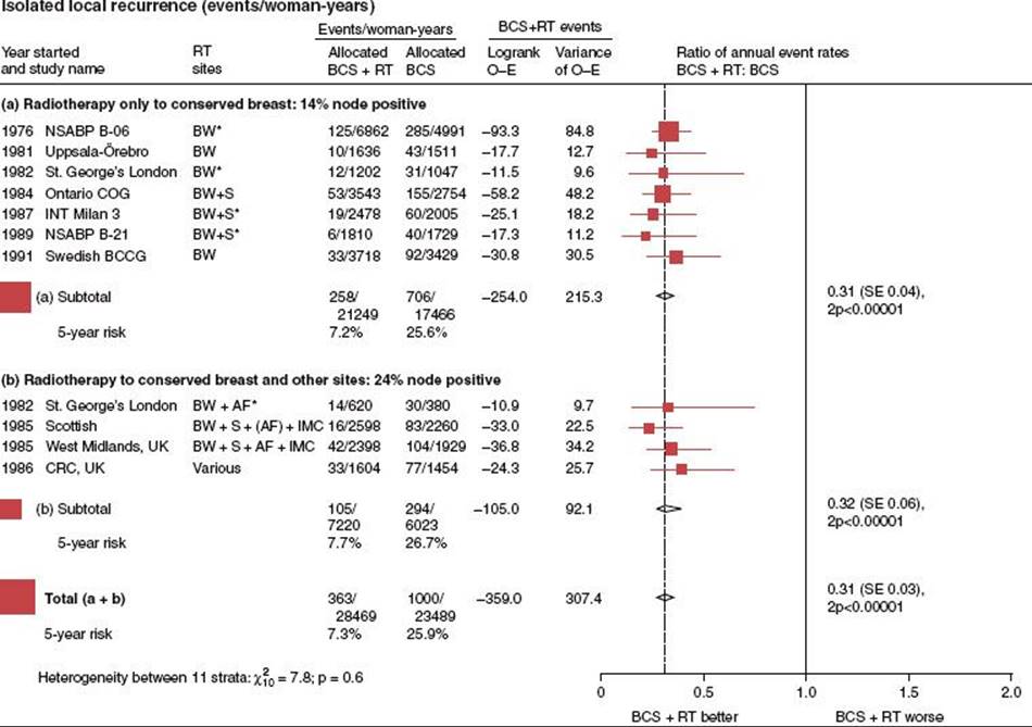
FIGURE 56.19. Updated meta-analysis from the Early Breast Cancer Trialists Collaborative Group comparing breast-conserving surgery alone to breast-conserving surgery with radiation therapy. (From Darby S, McGale P, Correa C, et al. Effect of radiotherapy after breast-conserving surgery on 10-year recurrence and 15-year breast cancer death: meta-analysis of individual patient data for 10,801 women in 17 randomised trials. Lancet 2011;378:1707–1716, with permission from Elsevier.)
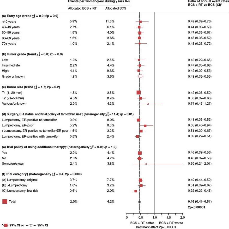
TABLE 56.24 RESULTS OF SELECTED RANDOMIZED TRIALS OF BREAST CONSERVING SURGERY WITH OR WITHOUT RADIATION

Clark et al.499 reported on a randomized study of 421 patients with tumors 4 cm or smaller and negative nodes who were treated with wide local excision and axillary dissection alone and 416 patients who were treated with the same surgery plus breast irradiation (40 Gy in 3 weeks, 16 fractions, and 12.5-Gy boost in 5 fractions). With 7.6 years’ median follow-up, breast recurrences were seen in 148 (35%) of the nonirradiated patients and in 147 (11%) of the irradiated patients (P <.0001); 99 patients (24%) in the former group and 87 (21%) in the latter group died during the study period.
Forrest et al.,504 after local excision of breast tumors <4 cm in diameter and axillary dissection, randomly assigned 291 patients to receive irradiation of 50 Gy to the breast plus a 10- to 15-Gy boost and 294 to receive no irradiation. Patients received either tamoxifen or 6 cycles of CMF. Overall survival was equivalent in the two groups. The rates of locoregional relapse were 6.1% (18 patients) in the irradiated group and 28.6% (84 patients) in the excision-alone group.
Renton et al.505 analyzed 418 patients treated by wide local excision and adjuvant chemotherapy (tamoxifen if ER-positive and CMF chemotherapy if ER-negative) who were randomized to have radiation therapy to the breast or not. At a minimum 5-year follow-up, the local recurrence rate in patients receiving irradiation was 13% compared with 35% in those not so treated. When histologically local excision was incomplete and patients received radiation therapy, the local recurrence rate was 17%.
One of the most significant trials addressing the issue of local relapse following lumpectomy alone was from the NSABP-06.93 This trial included three arms—modified radical mastectomy, lumpectomy with radiation, and lumpectomy without radiation—and included both node-positive and node-negative patients. In this trial, breast irradiation decreased the likelihood of a recurrence in the ipsilateral breast in the group of 1,137 lumpectomy-treated women whose surgical specimens had tumor-free margins. The cumulative incidence of a recurrence in the ipsilateral breast 20 years after surgery was 14.3% among the women who underwent irradiation after lumpectomy and 39.2% among those who underwent lumpectomy without irradiation (P <.001). The benefit of radiation therapy was independent of the nodal status. Among the women with negative nodes, 36.2% of those who did not receive radiation therapy and 17.0% of those who did had a recurrence in the ipsilateral breast within 20 years (P <.001). Among the women with positive nodes, 44.2% of those who did not undergo irradiation and 8.8% of those who did had a recurrence in the ipsilateral breast (P <.001). Among the lumpectomy-treated women whose surgical specimens had tumor-free margins, the hazard ratio for death among the women who underwent postoperative breast irradiation, as compared with those who did not, was 0.91 (95% CI, 0.77 to 1.06; P = .23). Radiation therapy was associated with a marginally significant decrease in deaths due to breast cancer. This decrease was partially offset by an increase in deaths from other causes.
Because of continued uncertainty regarding the need for radiation in more favorable tumors, the NSABP continued to investigate this issue of elimination of irradiation. In the B-21 trial, 1,009 women treated by lumpectomy were randomly assigned to tamoxifen (n = 336), radiation therapy and placebo (n = 336), or radiation therapy and tamoxifen (n = 337).94 End points were divided rates of breast relapse, distant recurrence, and contralateral breast cancer. Radiation and placebo resulted in a 49% lower hazard rate of local relapse as opposed to tamoxifen alone; radiation and tamoxifen resulted in a 63% lower rate as opposed to radiation and placebo. When compared with tamoxifen alone, radiation plus tamoxifen resulted in an 81% reduction in hazard rate of ipsilateral breast tumor recurrence (IBTR). Cumulative incidences of local relapse through 8 years were 16.5% with tamoxifen, 9.3% with radiation and placebo, and 2.8% with radiation and tamoxifen. The authors concluded that in women with tumors ≤1 cm, local relapse occurs with enough frequency after lumpectomy to justify considering radiation therapy regardless of ER status.
In another landmark trial, Veronesi et al.398,501 randomly assigned 567 women with small cancers of the breast (<2.5 cm in diameter) to quadrantectomy followed by radiation therapy or to quadrantectomy alone. All patients underwent total axillary dissection. The number of IBTRs was significantly higher in patients treated with surgery alone (59 cases of 273; 10-year crude cumulative incidence of 23.5%) than in patients treated with surgery plus radiotherapy (16 cases of 294; 10-year crude cumulative incidence of 5.8%). The difference in IBTR frequency between the two treatments was high in women up to 45 years of age, tending to decrease with increasing age up to no apparent difference in women older than 65 years. Overall survival curves for the two groups did not differ significantly (P = .326). However, a limited survival advantage was evident after radiotherapy for node-positive women.
Other Nonrandomized Studies of Lumpectomy Alone
Lim et al.506 recently updated a prospective single arm trial addressing omission of radiation for highly selected favorable patients from the Harvard group. Eighty-seven (of 90 planned) patients enrolled from 1986 until closure in 1992, when a predefined stopping boundary was crossed. Patients were required to have a unicentric, T1, pathologic node-negative invasive ductal, mucinous, or tubular carcinoma without an extensive intraductal component or lymphatic-vessel invasion. Surgery included local excision with margins of at least 1 cm or a negative re-excision. No RT or systemic therapy was given. Nineteen patients (23%) had local recurrence) as a first site of failure (average annual local recurrence: 3.5 per 100 patient-years of follow-up). The authors concluded that even in this highly selected cohort, a substantial risk of local recurrence occurred after breast-conserving therapy alone with margins of ≥1.0 cm.
McCready et al.507 reported on a postmenopausal group of 244 patients with breast cancer treated with lumpectomy alone. With a median follow-up of 9.1 years, the overall breast relapse rate was 24% (59 of 244). On univariate analysis, smaller tumor size, negative nodes, positive ER status, and no lymphovascular or perineural invasion were associated with significantly lower relapse rates (P <.05). On multivariate analyses, lymphovascular or perineural invasion, age, and amount of DCIS were all significantly associated with greater risk of local relapse. The authors defined a low-risk subgroup (node-negative, younger than 65 years of age, no comedo, ER positive, no emboli) with a crude 10-year local recurrence rate of 9%.
Conservative Surgery Alone in Elderly Women
The available evidence clearly establishes lumpectomy followed by radiation as the standard of care for the majority of women with early-stage invasive breast cancer. As noted in the numerous randomized trials reported above, lumpectomy alone results in a threefold increase in local relapse and compromised breast cancer–related survival to a lesser degree.90 Given the lower reported local relapse rates in elderly women, however, the absolute benefit of radiation therapy following breast-conserving surgery, however, may be less. The question of whether radiation can be eliminated following breast-conserving therapy has been addressed in both retrospective and, more recently, carefully designed prospective randomized trials.
There is evidence from several retrospective and prospective series that elderly women may be spared radiation. Cooke et al.508 identified 44 women treated with partial mastectomy, breast irradiation, and tamoxifen and compared them with 53 women treated in a similar fashion but without breast irradiation. At 39 months, the breast tumor recurrence rate was 5% with breast irradiation and 21% when irradiation was omitted. Of those not receiving irradiation, no breast relapses were seen in 22 patients older than 70 years of age at diagnosis, in contrast to 8 breast recurrences in 31 patients younger than 70 years.
In the trial of quadrantectomy versus quadrantectomy plus radiation reported by Veronesi et al.,501 although there was a clear benefit in local control overall, the benefit was significant and apparent only in younger women; for patients over age 65 there was no significant benefit.
Gajdos et al.509 noted that reported rates of local and distant recurrence for elderly patients were comparable with those for younger patients after both mastectomy and breast conservation. Ninety-eight of 920 patients older than 70 years of age were undertreated by conventional criteria. Undertreated elderly patients were significantly older (78 vs. 76 years; P = .003), were diagnosed with excisional biopsy more often (69% vs. 57%; P = .069) and had fine-needle aspiration less frequently (22% vs. 38%; P = .069), and were more likely to have breast-conservation therapy (90% vs. 73%; P = .004). Local and distant disease-free survival rates for both groups were comparable. Tamoxifen treatment significantly reduced the chances for development of distant metastasis in node-negative elderly patients with invasive tumors (P = .028). Omission of chemotherapy had no impact on disease control in the elderly. Therefore, elderly women with favorable prognostic factors may be candidates for treatment with tumor resection and tamoxifen without irradiation or chemotherapy and with close follow-up.
Given the apparent biological differences in breast cancers in the elderly, as well as the logistical issues in daily radiation treatment, two randomized trials have addressed the issue of the need for radiation therapy in elderly women with early-stage breast cancer.175,510 The first trial from the Cancer and Leukemia Group B (CALGB), published by Hughes et al.510 randomly assigned 636 women with clinical stage I, ER-positive breast carcinoma treated by lumpectomy to receive tamoxifen plus radiation therapy (317 women) or tamoxifen alone (319 women). The only significant difference between the two groups was the rate of local or regional recurrence at 5 years (1% in the group given tamoxifen plus irradiation and 4% in the group given tamoxifen alone; P<.001). There were no significant differences between the two groups with regard to the rates of mastectomy for local recurrence, distant metastases, or 5-year rates of overall survival. The authors concluded that lumpectomy plus adjuvant therapy with tamoxifen alone is a reasonable choice for the treatment of women 70 years of age or older who have early, ER-positive breast cancer.
The second trial was a Canadian study published by Fyles et al.511 of women 50 years of age or older who had T1 or T2 node-negative breast cancer. In this trial, 769 women with early breast cancer with a tumor diameter of ≤5 cm were randomly assigned to receive breast irradiation plus tamoxifen (386 women) or tamoxifen alone (383 women). With a median follow-up of 5.6 years, the rate of local relapse at 5 years was 7.7% in the tamoxifen group and 0.6% in the group given tamoxifen plus irradiation (HR 8.3; 95% CI, 3.3 to 21.2; P <.001). The corresponding 5-year disease-free survival rates were 84% and 91% (P = .004). A subgroup analysis of 611 women with T1, receptor-positive tumors, similar to the CALGB cohorts, also indicated a benefit from radiotherapy, with the 5-year rates of local relapse of 0.4% with tamoxifen plus radiotherapy and 5.9% with tamoxifen alone (P <.001). There was also a significant difference in the rate of axillary relapse at 5 years (2.5% in the tamoxifen group and 0.5% in the group given tamoxifen plus irradiation; P = .049), but there were no significant difference in the rates of distant relapse or overall survival. In both of these trials, women were not required to have surgical staging of the axilla, but they did have clinically negative axilla in both, and follow-up remains relatively short. Both studies show that even this favorable subgroup benefits from radiation with respect to local control, but the absolute benefit is small.
As a follow-up to the CALGB study, Smith et al.511 conducted a detailed analysis of women over age 70 from the SEER-Medicare database. They identified 8,724 women aged 70 years or older treated with conservative surgery for small, lymph node-negative, ER-positive (or unknown receptor status) breast cancer. Using a proportional hazards model, they tested whether radiation therapy was associated with a lower risk of a combined outcome, defined as a second ipsilateral breast cancer reported by SEER, or a subsequent mastectomy reported by Medicare claims. The results, summarized in Figure 56.20, were similar to those reported by the randomized studies above in that radiation therapy, compared with no radiation therapy, was associated with a lower risk of the combined outcome (HR 0.19; 95% CI, 0.14 to 0.28). Radiation therapy was associated with an absolute risk reduction of 4.0 events per 100 women at 5 years (from 5.1 events without radiation therapy to 1.1 with radiation therapy) and 5.7 events per 100 women at 8 years (from 8.0 events without radiation therapy to 2.3 with radiation therapy; P <.001).
Using a comorbidity analysis, radiation therapy was most likely to benefit those aged 70 to 79 years without comorbidity (number needed to treat to prevent one event, 21 to 22 patients) and was least likely to benefit those aged 80 years or older with moderate to severe comorbidity (number needed to treat, 61 to 125 patients). The authors conclude that for older women with early breast cancer, radiation therapy was associated with a lower risk of a second ipsilateral breast cancer and subsequent mastectomy. Patients aged 70 to 79 years with minimal comorbidity were the most likely to benefit, and older patients with substantial comorbidity were least likely to benefit.
Collectively, these studies indicate that the benefit of radiation therapy for elderly women is significant in terms of local control, but this absolute benefit is relatively small and must be weighed against comorbidities and other competing risks. The two randomized trials and the SEER-Medicare analysis are summarized in Table 56.25. For women with favorable T1N0 receptor-positive breast cancers, tamoxifen alone is a reasonable option that should be discussed. For patients with multiple comorbidities and shorter life expectancies, this option is often chosen. The authors own preference in patients with low comorbidity and long life expectancy is to offer radiation, even in those over age 70 with ER-positive tumors.
TABLE 56.25 FIVE-YEAR OUTCOME OF BREAST-CONSERVING SURGERY WITH OR WITHOUT RADIOTHERAPY IN ELDERLY WOMEN WITH BREAST CANCER

FIGURE 56.20. Association of radiation therapy with outcomes in elderly women. Patients were at risk for all outcomes beginning 9 months after diagnosis. A: Second ipsilateral breast cancer reported by Surveillance, Epidemiology, and End Results (SEER). This outcome was defined as a second ipsilateral, pathologically confirmed, invasive breast cancer. B: Subsequent mastectomy reported by Medicare claims. C: Second breast cancer event defined as a second ipsilateral, pathologically confirmed, invasive breast cancer reported by SEER data or as a subsequent mastectomy reported by Medicare claims. D:Repeat breast-conserving surgery as reported by Medicare claims. RT, radiation therapy. Error bars equal 95% confidence intervals. P values were calculated from a two-sided log-rank test. (From Smith BD, Gross CP, Smith GL, et al. Effectiveness of radiation therapy for older women with early breast cancer. J Natl Cancer Inst 2006;98:681–690, with permission.)
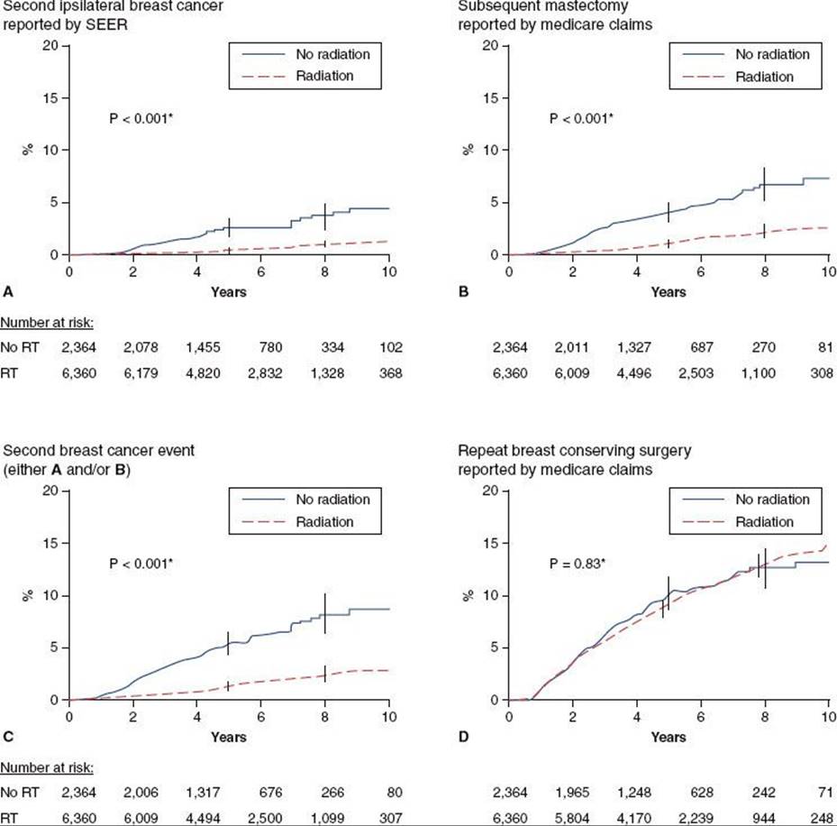
HYPOFRACTIONATED WHOLE-BREAST IRRADIATION
In breast cancer, the standard radiation therapy schedule treatment delivers 1.8 to 2.0 Gy per day for 25 to 28 days for a total dose of 45 to 50.4 Gy followed by a 5 to 8 fraction boost (10 to 16 Gy) for a total dose of 60 to 66 Gy delivered for 6 to 7.5 weeks. There has a been a growing trend toward hypofractionation, which involves delivering a higher dose per fraction for a shorter number of fractions for a biologically equivalent dose. This has been shown to be safe and effective as a standard treatment schedule in multiple randomized trials and will be discussed below.
The UK Standardisation of Breast Radiotherapy (START) Trial A randomized patients with early breast cancer (pT1-3a pN0-1 M0); these patients received either 50 Gy in 25 fractions of 2.0 Gy versus 41.6 Gy or 39 Gy in 13 fractions of 3.2 Gy or 3.0 Gy over 5 weeks after surgery.513 Thus, the overall treatment time was kept constant in all three arms. The trial did allow treatment of regional lymph nodes (supraclavicular and axillary) with additional radiation fields, and these were used in 20% of the patients. With the primary end point of locoregional tumor relapse, 749 women were assigned to the 50 Gy group, 750 to the 41.6 Gy group, and 737 to the 39 Gy group. With a median follow-up of 5.1 years, the rate of local-regional tumor relapse at 5 years was 3.6% (95% CI, 2.2 to 5.1) after 50 Gy, 3.5% (95% CI, 2.1 to 4.3) after 41.6 Gy, and 5.2% (95% CI, 3.5 to 6.9) after 39 Gy. The authors concluded that a lower total dose in a smaller number of fractions could offer similar rates of tumor control as standard fractionation. A major limitation of the study is the use of a conventionally fractionated boost of 14 Gy in 7 fractions. How this boost interacted with the altered fractionation effects is unclear.
The UK START Trial B randomized patients with early breast cancer (pT1-3a pN0-1 M0) at 23 centers in the UK who were assigned after primary surgery to receive 50 Gy in 25 fractions of 2.0 Gy over 5 weeks or 40 Gy in 15 fractions of 2.67 Gy over 3 weeks.514 In contrast to the START A trial, the overall treatment time was not consistent in both arms. With the primary end point of locoregional tumor relapse, 1,105 women were assigned to the 50 Gy group and 1,110 to the 40 Gy group. With a median follow-up of 6.0 years (interquartile range 5.0 to 6.2) the rate of local-regional tumor relapse at 5 years was 2.2% (95% CI, 1.3 to 3.1) in the 40 Gy group and 3.3% (95% CI, 2.2 to 4.5) in the 50 Gy group, representing an absolute difference of –0.7% (95% CI, –1.7% to 0.9%).
Whelan et al.515 reported on a study of women with invasive breast cancer who had undergone breast-conserving surgery and were randomized to whole-breast irradiation either at a standard dose of 50.0 Gy in 25 fractions over a period of 35 days (the control group) or at a dose of 42.5 Gy in 16 fractions over a period of 22 days (the hypofractionated radiation group). Notably, women with breast separations >25 cm were excluded. The risk of local recurrence at 10 years was 6.7% among the 612 women assigned to standard irradiation as compared with 6.2% among the 622 women assigned to the hypofractionated regimen (95% CI, –2.5 to 3.5). At 10 years, 71.3% of women in the control group as compared with 69.8% of the women in the hypofractionated radiation group had a good or excellent cosmetic outcome. The authors concluded that hypofractionated whole-breast irradiation was not inferior to standard radiation treatment in women who had undergone breast-conserving surgery for invasive breast cancer with clear surgical margins and negative axillary nodes.
A randomized trial from Hospital Necker in Paris compared 45 Gy in 25 fractions delivered in 5 weeks to 23 Gy delivered as 5 Gy on days 1 and 3 and 6.5 Gy on days 15 and 17. In patients treated by lumpectomy (56 in the conventional arm and 45 in the hypofractionation arm) the local-regional recurrence rate was similar (7% vs. 4%).516
In 2011, a task force authorized by the American Society for Radiation Oncology (ASTRO) weighed evidence from a systematic literature review and produced recommendations regarding the use of hypofractionated radiotherapy in patients with breast cancer.517 They stated that hypofractionated whole-breast irradiation was likely equivalent to conventional fractionation in patients who meet all of the following criteria: (a) age over 50 years, (b) pathologic state T1–2N0 treated with lumpectomy, (c) patient has not received systemic chemotherapy, and (d) the minimum and maximum dose along the central axis is not <93% and not >107% of the prescription dose, respectively. For patients not receiving a radiation boost, the task force favored a dose schedule of 42.5 Gy in 16 fractions when hypofractionated radiotherapy is planned; there was no conclusion regarding the use of a tumor bed boost in patients treated with hypofractionation. Lastly, the task force also recommended that the heart should be excluded from the primary treatment fields when hypofractionated whole-breast irradiation is used due to lingering uncertainty regarding late effects of this treatment on cardiac function.
In the authors institution, every effort is made to follow the ASTRO consensus guidelines. However, patient factors such as distance from the radiotherapy facility, age, and comorbidities all play a role in offering a patient hypofractionated whole-breast radiotherapy. At present, the RTOG-1005 is a phase III randomized trial comparing two fractionation schemes for whole-breast irradiation: hypofractionated radiation with concurrent boost versus standard whole-breast irradiation plus sequential boost for patients with early-stage breast cancer.
TABLE 56.26A TREATMENT POLICY FOR CONSERVATIVE MANAGEMENT OF EARLY STAGE INVASIVE BREAST CANCER

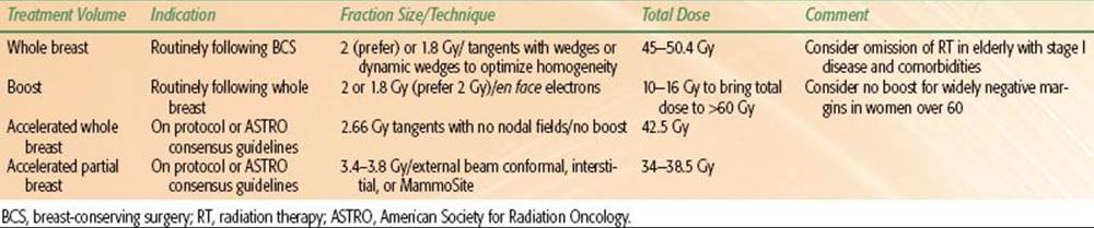
TABLE 56.26B TREATMENT POLICY FOR REGIONAL NODES
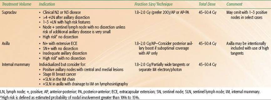
RADIATION MANAGEMENT OF THE REGIONAL LYMPHATICS
Radiation therapy of the regional lymphatics remains one of the most variable aspects of breast-conserving therapy.327,518,519 The role of radiation therapy in management of the regional lymphatics is influenced by the risk of subclinical microscopic disease in regional nodal basins and patterns of failure. This risk is in part determined by disease characteristics and in part determined by the extent of surgical evaluation of the axilla, which has become increasingly relevant as a result of increased use of sentinel node, and whether completion axillary dissections are performed for those patients with sentinel node–positive disease. Furthermore, clinicians differ significantly regarding their philosophy with respect to the treatment of subclinical microscopic disease, particularly as it relates to the internal mammary chain.
The issue of whether one treats the “axilla” in conservatively managed patients is further complicated by both uncertainty and misconceptions about the degree to which the axilla receives radiation from a standard tangential field. Although this will vary considerably, as will be discussed in the section on radiation techniques, tangential radiation ports will likely treat most level I nodes and a portion of level II nodes.520
All of this uncertainty, debate, and controversy is well founded as there are little in the way of randomized data to establish a clear standard. Studies are under way randomizing high-risk node-negative and node-positive patients to treatment to the breast or chest wall only compared with the breast or chest wall and regional lymphatics. In the interim, reliance on available retrospective data, calculated risks of subclinical disease, and patterns of failure form the basis for various treatment policies. Current treatment policies and guidelines at the authors institutions are outlined in Table 56.26.
Although in the earlier years of breast-conservation therapy, node-negative as well as node-positive patients often received regional nodal treatment, most authors currently agree that it is not necessary to irradiate the regional lymphatics if the nodes are pathologically negative and an adequate axillary dissection has been performed.79,327,521 This general practice has now been extended to those patients with a negative sentinel node, because available studies have demonstrated a low rate of pathologically involved nodes after a negative sentinel node procedure performed by an experienced surgeon.234,335,522
However, as noted previously, the clinically negative axilla harbors subclinical microscopic disease in up to 40% of patients with early-stage operable breast cancer,331 and both axillary dissection and axillary radiation result in high rates of regional nodal control.79,110,327,331,521
TABLE 56.27 AXILLARY RECURRENCE AFTER AXILLARY LYMPH NODE DISSECTIONOR AXILLARY RADIATION IN CONSERVATIVELY MANAGED PATIENTS

TABLE 56.28 AXILLARY FAILURE RATES IN PATIENTS IN RANDOMIZED TRIALS COMPARING AXILLARY TREATMENTS
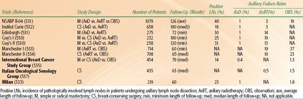
Radiation Compared with Axillary Surgery
Sentinel node sampling, with or without full axillary dissection, is now the most common method of axillary management in women with early-stage breast cancer. For patients in whom full axillary staging will not affect subsequent systemic management, who have not undergone any axillary staging procedure, or who have a positive sentinel node and did not undergo further axillary staging, axillary radiation has been shown to result in high rates of regional nodal control.79,110,327,521,523 Selected series demonstrating nodal control rates with radiation therapy in breast-conserving therapy are summarized in Table 56.27.81,379,386,435,524–528 Table 56.28 summarizes results of randomized trials comparing axillary surgery to radiation or observation.73,333,529–538
Retrospective Experiences
Haffty et al.79 reported actuarial nodal control rates of 97% and 96% at 10 years for two groups of patients, 245 receiving irradiation alone without axillary dissection and 187 treated with irradiation to the supraclavicular lymph nodes and IMNs after axillary dissection. Minimal morbidity was associated with this treatment policy. Recently Pejavar et al.539 updated the Yale experience, demonstrating a 98% 5-year regional nodal control rate in 582 patients with invasive breast cancer treated by regional nodal irradiation without dissection compared with 98% 5-year nodal control rate in 1,440 patients treated by axillary dissection. Within this experience were 16 patients with positive sentinel nodes who did not undergo completion axillary dissection and were treated with radiation therapy. None of those sentinel node–positive patients recurred.
Galper et al.525 estimated the efficacy of axillary radiation therapy after a positive sentinel node biopsy and evaluated the risk of regional nodal failure for patients with clinical stage I or II, clinically node-negative invasive breast cancer treated with either no dissection or a limited dissection (removal of five nodes or less) followed by axillary radiation therapy. Two hundred ninety-two patients had axillary radiation therapy instead of axillary dissection; 126 underwent axillary radiation therapy following limited node dissection. The median dose to the axilla was 46 Gy and to the supraclavicular fossa 45 Gy. Among patients found to have positive nodes on limited dissection, adjuvant chemotherapy and tamoxifen were administered to 81% and 7% of subjects, respectively. All patients had an 8-year follow-up. Six of the 418 patients (1.4%) had regional nodal failure within 8 years; four had simultaneous regional and distant recurrences; and two had isolated axillary failures. Three of the 292 patients (1%) with no axillary dissection, 0 of 84 patients with pathologically negative nodes, and 3 of 42 patients (7%) with pathologically involved nodes had regional node failure as a first site of failure.
A group of 511 patients with 519 stage I and II breast cancers treated with lumpectomy, with or without axillary dissection, and irradiation were reviewed by Halverson et al.540 Management of the axilla consisted of irradiation after axillary dissection in 74, irradiation alone in 75, and observation in 21 patients; the extent of nodal irradiation was at the discretion of the attending radiation oncologist. Overall, axillary recurrence was uncommon (1.2%) but was slightly more frequent after irradiation alone (2.7%) than after surgery alone (0.3%; P = .14). There was no benefit for supplemental axillary irradiation after an axillary dissection yielding negative nodes or one to three positive nodes. Among the 21 patients in whom the axilla was not treated, axillary recurrence was not observed. Supraclavicular failures were rare in women with negative or one to three positive axillary lymph nodes (0.5%) and were not significantly affected by elective irradiation. IMN recurrence was seen in only one patient and was not influenced by elective internal mammary irradiation.
Randomized Studies
Randomized studies evaluating axillary treatment (dissection vs. observation vs. radiation) are summarized in Table 56.28. One of the largest and earliest comparisons of axillary surgery to radiation was the NSABP-04 study, in which operable clinically node-negative patients were randomly assigned to radical mastectomy, simple mastectomy, or simple mastectomy with radiation to the chest wall and regional lymphatics.73 The nodal relapse rate approached 20% in those assigned to simple mastectomy without radiation. The nodal control rates in the radical mastectomy arm and simple mastectomy plus radiation arm were comparable, with <3% relapse rate in each of these arms.
A direct comparison of axillary treatment by dissection compared with radiation was recently reported by Louis-Sylvestre et al.,532 in which 658 patients with a breast carcinoma <3 cm in diameter and clinically uninvolved lymph nodes were randomly assigned to axillary dissection or axillary radiotherapy after breast-conserving surgery with radiation to the breast. Of the group undergoing dissection, 21% of the patients in the axillary dissection group were node positive. At 10 and 15 years, survival rates were identical in both groups (73.8% vs. 75.5% at 15 years). Recurrences in the axilla were less frequent in the axillary dissection group at 15 years (1% vs. 3%; P =.04). There was no difference in recurrence rates in the breast or supraclavicular region or distant metastases between the two groups.
Veronesi et al.537 carried out a study in which women older than 45 years of age with breast cancer up to 1.2 cm were randomized, 214 of whom were treated with breast-conservation surgery and irradiation without axillary treatment and 221 with conservation surgery plus breast and axillary radiation therapy (50 Gy in 5 weeks).After a median follow-up of 63 months, overt axillary metastases were fewer than expected: three cases in the no axillary treatment group (1.5%) and one in the RT group (0.5%). This study suggests that occult axillary metastases might never become clinically overt and axillary dissection might be avoided in patients with small carcinomas and a clinically negative axilla. Axillary RT seems to protect the patients from axillary recurrence almost completely. It is possible that systemic therapy, the undamaged immunocompetent tissue in the axillary lymph nodes, and axillary irradiation may all be factors contributing to the low incidence of axillary failures in these patients. Also, in the patients receiving no axillary irradiation, it is highly likely that the level I lymph nodes were included in the standard tangential fields.327,520,541
The European After Mapping of the Axilla Radiation or Surgery Trial is nearing completion. This trial randomizes patients after positive sentinel nodes to axillary dissection or axillary radiation and will help to address issues of local-regional control, survival, and morbidity using these two approaches.542
Irradiation of Lymphatics in Patients with Positive Axillary Lymph Nodes
Although there is general consensus that radiation to the regional lymph nodes is not necessary in patients with pathologically node-negative disease, there is considerable variability in radiation to the regional lymphatics in patients with pathologically node-positive disease.77–79,327,523,543 Many radiation oncologists favor irradiation of the regional lymphatics in addition to the breast in node-positive women, while others favor no nodal irradiation, particularly in women with one to three positive nodes. Based on the potential disease-free and overall survival advantage, as well as the risk of failure in the supraclavicular region, most radiation oncologists favor at least supraclavicular nodal irradiation in patients with four or more nodes. The recent presentation of preliminary results of the National Cancer Institute of Canada’s MA.20 trial demonstrates a distant metastasis, disease-free survival, and potential survival advantage to regional nodal irradiaton.544 The majority of patients in this trial had one to three positive nodes and were randomized after breast-conserving surgery to tangential breast irradiation alone or breast irradiation with regional nodal irradiation to the supraclavicular and internal mammary regions. Full publication of this trial is eagerly awaited. A similar trial of the EORTC, randomizing high-risk node-negative and node-positive patients to treatment to the breast or chest wall alone or breast or chest wall and regional lymphatics, may help to further clarify this important issue.
Acknowledging the lack of definitive data and clear consensus and allowing for flexibility depending on patient and physician preferences, the guidelines that the authors advocate are summarized in Table 56.26. In general, the authors favor treatment of the supraclavicular fossa in patients with positive nodes. Treatment of the axilla and internal mammary will vary, with attention to the indications outlined in the table. Until results of the ongoing MA.20 and EORTC randomized trials are fully available, regional lymphatic irradiation will likely continue to be highly individualized based on physician and patient preferences.
Sarrazin et al.370 carried out a randomized study comparing 88 patients treated with tumorectomy and irradiation and 91 patients treated with mastectomy. In a second randomization in the study, the patients with positive axillary lymph nodes in the first randomization were randomly assigned to receive or not receive nodal irradiation. There was no significant difference in overall survival between the two groups. Nevertheless, Yarnold521 advised elective irradiation of the axilla and the supraclavicular fossa in selected patients, such as those with four or more metastatic axillary lymph nodes, involvement of the apex of the axilla, or gross extracapsular tumor extension, even if the patients are to receive adjuvant chemotherapy. These recommendations are supported by reports that document the benefit of postmastectomy irradiation in patients receiving chemotherapy, which are reviewed in more detail in Chapter 54.
Treatment of the axilla varies significantly in patients with positive nodes. For those patients with negative nodes or with one to three positive nodes without extracapsular extension (ECE) who undergo adequate axillary dissection, there does not appear to be a benefit to targeting the full axilla.77–80,327,540 There is considerable variability regarding treatment of the full axilla, even in patients with multiple positive nodes.77,81,520,543 The risk of axillary recurrence after full dissection is low in patients with ECE, even without axillary lymph node irradiation. Whether to treat the full axilla following dissection for patients with multiple positive nodes or ECE generally includes consideration of the extent of dissection, the degree of nodal involvement and ECE, and the degree to which the patient and physician are willing to accept some increased risk of lymphedema with full axillary radiation following dissection.110,327
Hetelekidis et al.255 evaluated 368 patients with T1 or T2 breast cancer and pathologically positive lymph nodes treated with breast-conserving therapy. The median number of sampled lymph nodes was 10. Twenty percent of the patients were treated with supraclavicular radiation therapy, and 64% received both axillary and supraclavicular radiation therapy (45 Gy). One hundred twenty-two patients (33%) had ECE. There was no significant correlation of either disease-free or overall survival or local, regional nodal, or distant failure rates in patients with ECE compared with those without it.
Pierce et al.,545 in a review of 72 women with breast cancer treated with conservation surgery and irradiation, identified 27 patients (37.5%) who had evidence of ECE in the axilla. With a median follow-up of 14 months, 1 of 27 (4%) patients with ECE experienced an axillary failure, compared with 0 of 45 patients without ECE. Several authors concluded that ECE is associated with decreased survival but not with increased axillary failures and that radiation therapy may be omitted in a dissected axilla if the sole indication is extracapsular disease.
TABLE 56.29 STUDIES EVALUATING IMPACT OF INTERNAL MAMMARY TREATMENT (SURGERY OR RADIATION) ON 10 YEARS SURVIVAL
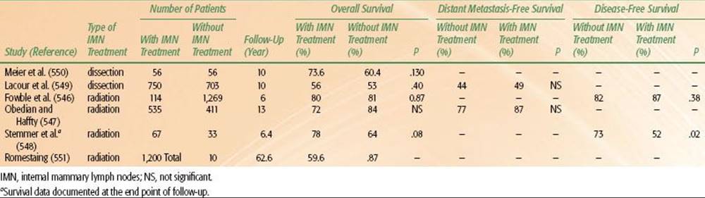
Internal Mammary Node Irradiation
The role of internal mammary nodal irradiation in node-positive breast cancer patients remains a controversial issue. Although, ongoing trials from the National Cancer Institute of Canada and EORTC may help to address this, currently there is no clear consensus on the role of internal mammary irradiation.518,519,546–548
Several surgical series comparing extended radical mastectomy and radical mastectomy, without adjuvant systemic therapy, have shown that extended radical mastectomy was associated with improved survival rates in patients with medial T1 or T2 tumors and positive axillary nodes.549,550 These surgical series and selected series evaluating internal mammary irradiation are summarized in Table 56.29.546–550 In a randomized trial of patients with node-positive breast cancer treated with postmastectomy radiation, randomized to internal mammary radiation or no internal mammary radiation, Romestaing551 showed no advantage to internal mammary irradiation. The full manuscript of this study has not as yet been published. The majority of randomized trials evaluating postoperative radiation therapy did include radiation to the internal mammary chain. However, it is difficult to distinguish whether the benefit derived from such treatment related specifically to radiation of the internal mammary chain or to the breast or chest wall, supraclavicular, or axillary treatment administered.
Freedman et al.552 examined data regarding patterns of failure after elective IMN treatment. Although controversial, data from the prospective, randomized trials of IMN treatment did not seem to support elective dissection or irradiation. IMN irradiation did not contribute to survival, yet it raised the risk of cardiac toxic effects. Sentinel lymph node mapping provided an opportunity to examine the IMN chain in early breast cancer. It is possible that biopsy of the “hot” nodes could be used to select patients who are most likely to benefit from additional regional therapy to these nodes.
Fowble et al.546 compared the outcome in 1,383 women with stage I or II breast cancer who underwent wide excision, axillary node dissection with 10 or more nodes removed, and breast irradiation. A total of 114 women had radiation to the IMNs with deep tangents and 1,269 did not. All axillary node-positive women received adjuvant chemotherapy or tamoxifen, or both. There were no significant differences in ipsilateral breast tumor recurrence, regional node recurrence, and initial or total distant metastases for the two groups. No IMN failures were observed among the 114 patients whose IMNs were treated, and only 4 IMN failures were found in the 1,269 other patients (2 of whom had distant metastases). Similarly, 5- and 10-year actuarial overall and cause-specific survival rates were not significantly different.
In a series from Yale, Obedian and Haffty547 found no difference in the 10-year disease-free survival rate after breast irradiation and excision, regardless of whether IMNs were irradiated. Of 984 patients with invasive breast cancer who were treated with conservative surgery and radiotherapy, patients were divided into two groups: those treated by intentionally targeting the internal mammary nodes (n = 535) and without intentionally targeting the internal mammary nodes (n = 411). The decision not to use a separate internal mammary field was a result of a change in treatment policy over time and generally not based on number of nodes or tumor location. There were no significant differences between the groups with respect to age, ER or PR status, or use of adjuvant chemotherapy or hormone therapy. There were more patients with T2 tumors, positive nodes, medial lesions, indeterminate margins, and slightly longer follow-up in the group treated to the internal mammary chain. There were no significant differences between the groups with respect to overall survival or distant metastasis-free survival.
In a study by Stemmer et al.548 of 100 node-positive patients scheduled to receive radiation to the internal mammary chain, 67 received the radiation and 33 did not due to technical difficulties. At a median follow-up of 77 months, disease-free survival was significantly prolonged in patients receiving internal mammary radiation compared with those without internal mammary radiation (73% vs. 52%; P =.02). A trend was seen for overall survival (78% vs. 64%; P =.08). Cox regression multivariate analysis found IMN radiotherapy to be significant both for disease-free and overall survival. There was no treatment-related mortality.
It is evident that data are conflicting, and opinions regarding the role of internal mammary radiation remain unresolved. Until more definitive data become available, it is likely that patient and physician biases will dictate practice. The authors approach is summarized in Table 56.26, acknowledging that uncertainties in the available data allow for substantial flexibility. The radiation oncologist, however, must be familiar with the various techniques to treat internal mammary nodes, which are summarized later in the section on techniques.
SEQUENCING CHEMOTHERAPY AND HORMONAL THERAPY
Sequencing Chemoradiation in the Conservative Management of Breast Cancer
With the increasing use of systemic therapy in patients with early-stage breast cancer, the integration of this treatment with surgery and radiation therapy has become an important clinical question. Initial retrospective series evaluating treatment sequencing suggested that a delay in the onset of radiation therapy to permit delivery of chemotherapy increased local recurrence rates.91,553,554 These data are summarized in a pooled analysis by Huang et al.91Ten retrospective studies involving 7,401 patients investigated the association between delay in initiating postoperative RT and local control in breast cancer (after lumpectomy in nine studies and lumpectomy or mastectomy in one study). Eight of these studies compared local control between patients who were treated more than 8 weeks after surgery and those treated within 8 weeks of surgery. The pooled random-effects odds ratio from the combined analysis was 1.62 (95% CI, 1.21 to 2.16), corresponding to an increase in the 5-year LRR from 5.8% in those patients treated within 8 weeks to 9.1% in those patients treated between 9 and 16 weeks after surgery. In a separate analysis exploring the optimum sequencing of adjuvant RT and systemic chemotherapy after surgery for breast cancer from 11 retrospective series, the pooled random-effects odds ratio in these 11 studies was 2.28 (95% CI, 1.45 to 3.57), corresponding to an increase in the 5-year LRR from 6.0% in the RT-first group to 16.0% in the chemotherapy-first group (Fig. 56.21).
Most of these data are subject to criticism due to their retrospective nature, the fact that patients were treated in an earlier era with different surgical techniques and lack of attention to margins, and inclusion of heterogenous groups of patients with these caveats, but there appears to be a trend toward higher local relapse rates with delays in radiation therapy, which appears to have been an appropriate concern with respect to integration of radiation therapy and chemotherapy in the conservatively managed patients.
Initial data regarding these concerns related to the delay in radiation while chemotherapy was being delivered led the Harvard group at Joint Center for Radiation Therapy (JCRT) to investigate the sequencing of radiation therapy and chemotherapy in a randomized prospective clinical trial.553,554 In this trial, women treated with breast-conserving surgery were randomly assigned to 4 cycles of doxorubicin-based combination chemotherapy, followed by radiation therapy or radiation therapy followed by 4 cycles of the same chemotherapy. In an update of this trial, Bellon et al.555 reported no statistically significant treatment difference in the rates of freedom from any event, including breast cancer recurrence, contralateral breast cancer, second malignancy, or death (Fig. 56.22). The 10-year rate of any event was 46% for patients in the chemotherapy-first arm compared with 51% in the radiation-first arm. The 10-year rates of distant metastasis were 35% and 36% in the two arms, respectively; and the 10-year rates of death were 28% and 33%, respectively. For the 123 patients with negative margins, the crude local recurrence rates for chemotherapy-first and radiation-first patients were 6% and 13%, respectively. Corresponding rates of distant and regional recurrences were 18% and 26%. Among women with close margins (n = 47), crude local recurrence rates were 32% and 4%, respectively; distant or regional recurrences were 37% and 43%. In the group with positive margins (n = 51), local recurrences occurred in 23% of chemotherapy-first and 20% of radiation-first patients.
Although the JCRT study provided important data concerning treatment sequencing, this study predominantly focused on patients with lymph node–positive disease. Investigators from the MD Anderson Cancer Center performed a retrospective analysis of sequencing of chemotherapy and radiation in 124 patients with lymph node–negative disease treated with breast-conserving therapy.556 In this series, 79% of the patients had negative margins. The 5-year actuarial rates of local control were 100% for the chemotherapy-first group (most commonly 6 cycles of doxorubicin-based chemotherapy) and 94% for the radiation-first group (P = .351). The 5-year recurrence-free survival rates for the chemotherapy-first and radiation-first groups were 92% and 77% (P = .083), respectively. These data again support an adjuvant-therapy schedule in which chemotherapy is delivered first. The median delays in radiation delivery were 6.7 months in the MD Anderson Cancer Center series and 16 weeks in the JCRT series.
More recently, with the addition of taxane-based chemotherapy to adriamycin-based regimens, concerns have arisen regarding the additional delays in initiating radiation therapy in conservatively managed patients. This was addressed in a study by Sartor et al.557 In this randomized CALGB study evaluating adriamycin and cytoxan versus adriamycin and cytoxan, followed by taxane, there were 345 conservatively managed patients. Although the sequencing of radiation was not randomized, patients in the adriamycin plus taxane arm had radiation delayed by an additional 84 days (4 21-day cycles of taxol). Despite this added delay, local-regional relapses were lower in the adriamycin plus taxane compared with the adriamycin arm (9.7% vs. 3.7%; P = .04). (The majority of patients in this randomized trial presumably had negative surgical margins.)
For the majority of patients undergoing breast-conserving surgery with negative margins, these data collectively indicate that administration of chemotherapy prior to radiation therapy does not result in excessive rates of local relapse, provided all modalities are given in a timely fashion without excessive delays. Whether patients with positive or close margins or other risk factors for local relapse would benefit from earlier administration of radiation remains an unresolved issue.
FIGURE 56.21. Associations between delay in postoperative radiotherapy (RT) and local recurrence rates (LRRs) in studies of the sequencing of adjuvant RT and chemotherapy for breast cancer. LRRs in patients who received delayed RT following initial chemotherapy are compared with the rates observed in those patients who received early RT by chemotherapy. Low-quality studies are indicated by an asterisk. (From Huang J, Barbera L, Brouwers M, et al. Does delay in starting treatment affect the outcomes of radiotherapy? A systematic review. J Clin Oncol 2003;21:555–563. Reused with permission. © 2012 American Society of Clinical Oncology. All rights reserved.)

FIGURE 56.22. Event-free survival (including breast cancer recurrence, contralateral breast cancer, second malignancy, or death) by sequencing of chemotherapy and radiation following breast-conserving therapy, from the Harvard randomized trial. (From Bellon JR, Come SE, Gelman RS, et al. Sequencing of chemotherapy and radiation therapy in early-stage breast cancer: updated results of a prospective randomized trial. J Clin Oncol 2005;23:1934–1940. Reprinted with permission. © 2005 American Society of Clinical Oncology.)

Concurrent Chemoradiation in Breast-Conserving Therapy
Although the concurrent use of chemoradiation therapy in conservatively managed breast cancer has fallen out of favor, there are data that suggest a high rate of local control in patients treated concurrently. These studies are summarized in Table 56.30.558–562 (The chemotherapy regimens used in these studies are no longer routinely employed because they are less effective.)
A randomized trial of concurrent versus sequential CMF chemotherapy was reported by Arcangeli et al.557 A total of 206 patients who had quadrantectomy and axillary dissection for breast cancer and were planned to receive adjuvant CMF chemotherapy were randomized to concurrent or sequential radiotherapy. Radiotherapy was delivered only to the whole breast through tangential fields to a dose of 50 Gy in 20 fractions over 4 weeks, followed by an electron boost of 10 to 15 Gy in 4 to 6 fractions to the tumor bed. No differences in 5-year breast recurrence-free, metastasis-free, disease-free, and overall survival were observed in the two treatment groups. All patients completed the planned radiotherapy. No evidence of an increased risk of toxicity was observed between the two arms. No difference in radiotherapy and in the chemotherapy dose intensity was observed in the two groups. The authors concluded that in patients with negative surgical margins receiving adjuvant chemotherapy, radiotherapy can be delayed to up to 7 months. However, concurrent administration of CMF chemotherapy and radiotherapy was safe, and the authors suggest that such an approach might be reserved for patients at high risk of local recurrence.
FIGURE 56.23. Risk of ipsilateral and contralateral breast tumor relapse as a function of BRCA1/BRCA2 mutation status in a cohort of conservatively managed breast cancer patients. (From Haffty BG, Harrold E, Khan AJ, et al. Outcome of conservatively managed early-onset breast cancer by BRCA1/2 status. Lancet 2002;359:1471–1477, with permission.)
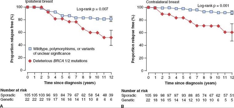
Another randomized study of concurrent versus sequential radiation therapy was reported by Rouesse et al.561 This study supports the concept that concurrent use of chemotherapy with radiation therapy improves local control in breast cancer. This trial compared concurrent chemoradiotherapy with FNC (5-fluourouracil [5-FU] 500 mg/m2, mitoxantrone 12 mg/m2, and cyclophosphamide 500 mg/m2), to sequential FEC (5-fluorouracil 500 mg/m2, epirubicin 60 mg/m2, and cyclophosphamide 500 mg/m2) followed by radiation in node-positive breast cancer in 650 women with operable breast cancer. All patients had node-positive disease and were randomized to sequential or concurrent chemoradiotherapy. Although there were no differences in disease-free or overall survival, local recurrences were significantly lower with concurrent therapy (3% vs. 7%). Of patients undergoing breast conservation, there were 6 local-regional relapses in the concurrent arm compared with 18 local-regional relapses in the sequential arm (P = .01). In multivariate Cox analysis, the sequential group had an increased risk of local-regional relapse compared with those in the concurrent group (RR 2.8; 95% CI, 1.1 to 7.2).
Toledano et al.563 recently reported the results of the ARCOSEIN sequential versus concurrent adjuvant chemotherapy with radiation therapy after breast-conserving surgery. After breast-conserving surgery, patients were treated either with sequential treatment with chemotherapy first followed by RT (arm A) or chemotherapy administered concurrently with RT (arm B). In all patients, the chemotherapy regimen consisted of mitoxantrone (12 mg/m2), 5-FU (500 mg/m2), and cyclophosphamide (500 mg/m2), 6 cycles (day 1 to day 21). Among the 214 evaluable patients, 107 were treated in each arm. Although local control was slightly superior in the concurrent arm, subcutaneous fibrosis, telangiectasia, skin pigmentation, and breast atrophy were significantly increased in arm B. No statistical difference was observed between the two arms of the study concerning grade 2 or greater pain, breast edema, or lymphedema.
Another randomized trial recently reported by Calais et al.,564 which is similar to the Rouesse et al.561 study, compared concurrent radiotherapy with mitoxantrone, 5-FU and cyclophosphamide to the same regimen followed by radiation therapy. Although toxicities were higher in the concurrent group, those patients with positive nodes had a significantly lower local relapse rate in the concurrent arm.
The concurrent use of chemotherapy and radiation following breast-conserving therapy has been reported in several nonrandomized studies. A prospective single-arm study by Bellon et al.559 also demonstrated favorable local control in a high-risk group of patients treated with concurrent CMF chemotherapy and reduced-dose radiation. Several other retrospective series, including one recently conducted from Yale, have demonstrated favorable local control rates and acceptable toxicity in patients at high risk for local relapse using concurrent chemoradiotherapy.428 In the Yale retrospective series, which compared 109 patients treated with concurrent chemoradiation to 426 patients treated with sequential chemoradiation, the concurrent group had a lower rate of local relapse, despite overall poorer prognostic factors for local control. However, the majority of these studies did not employ the most commonly used current chemotherapy agents such as adriamycin and taxanes. Therefore, it is difficult to extrapolate these results to current practice.
More recently, monoclonal antibodies such as trastuzumab and bevacizumab (Avastin) are being used commonly in the management of breast cancer patients. Romond et al.274 reported on a combine analysis from two large cooperative group studies investigating the utility of trastuzumab in HER2-positive patients with operable breast cancer and found that trastuzumab improved patient outcomes. There was no difference in acute locoregional toxicities, but it should be noted that the use of trastuzumab was associated with an increased risk of congestive heart failure or death from cardiac causes (4.1% vs. 0.8%). Goyal et al.565 reported on a series of 14 patients receiving bevacizumab in combination with whole-breast irradiation who were then matched to a group of patients who received whole-breast irradiation alone. No patient receiving bevacizumab plus RT experienced grade 3 or higher toxicity; however, three matched control patients experienced a grade 3 skin reaction. There was no difference in fatigue, radiation fibrosis, pneumonitis, or lymphedema between the two groups. Five patients (35%) developed reduction in ejection fraction in the bevacizumab arm; two with right-sided and three with left-sided treatment. Patients with left-sided treatment experienced a persistent reduction in ejection fraction compared with those receiving right-sided treatment. Thus, when treating patients with left-sided breast cancer, exclusion of the heart from the beam’s eye view should be undertaken such that no unnecessary radiation dose is delivered to the heart. One challenge moving forward is determining the toxicities of these novel therapies in combination with conventionally fractionated and hypofractionated radiotherapy regimens.
The critical issue that arises with conservatively managed breast cancer is whether the modest gain in local control outweighs the added toxicities and risks of concurrent chemoradiotherapy, using currently available agents. The benefit in local-regional control obtained in the Rouesse et al.561 study was statistically significant, but it remains debatable whether the added toxicity of the concurrent program is worth the added risk. Potential issues with concurrent chemoradiation, as pointed out in a study by Burstein et al.,566 include high rates of radiation pneumonitis in patients treated with radiation given in combination with weekly paclitaxel. Although they observed more favorable results with less frequent dosing, this study highlights the importance of prospective evaluation of radiation in combination with newer chemotherapeutic agents. Furthermore, this study highlights the importance of the dosing and scheduling of the chemotherapy agents given in combination with radiation.
The challenge over the next few years will be to identify those patients, who when treated by the traditional approach of surgery followed by chemotherapy followed by radiation therapy, remain at elevated risk of local relapse. Also at risk may be subsets of patients treated with neoadjuvant chemotherapy followed by surgery followed by radiation. Those patients who, when treated by these traditional sequencing approaches, are at high risk of local relapse are ideally suited for prospective evaluation of novel approaches using concurrent chemoradiation strategies. From such trials we can hopefully minimize local-regional relapse and optimize disease-free and overall survival, with acceptable treatment-related morbidity.
TABLE 56.30 SELECTED STUDIES EVALUATION CONCURRENT CHEMORADIATION IN BREAST-CONSERVING SURGERY WITH RADIATION
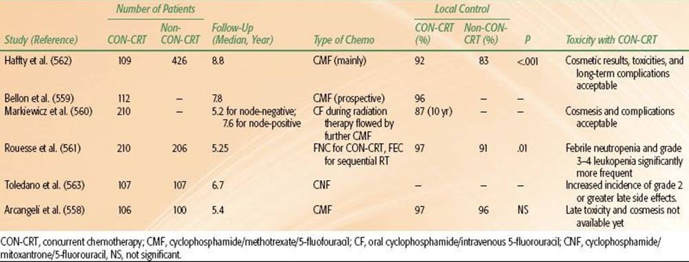
TABLE 56.31 OUTCOMES OF CONCURRENT OR SEQUENTIAL TAMOXIFEN WITH RADIATION THERAPY IN EARLY-STAGE BREAST CANCER
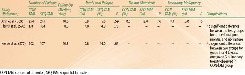
Sequencing Tamoxifen/Hormonal Therapy and Radiation Therapy in Conservatively Managed Patients
The question of optimal scheduling of hormonal therapy and radiation has been raised due to theoretical concerns that tamoxifen may decrease the radiation sensitivity of tumors. In cell culture studies, tamoxifen causes arrest of breast cancer cells in culture in the relatively radioresistant G0/G1 phases of the cell cycle. Although there are conflicting data, suggesting both no effect and increased radiation sensitivity, clinicians and patients have been in a quandary as to whether it is reasonable to begin tamoxifen during radiation. In addition, clinical studies have suggested increased pulmonary and breast fibrosis, possibly related to increased concentrations of transforming growth factor-β with the concurrent use of tamoxifen.567 In a retrospective case series, Wazer et al.568 reported a trend for an adverse cosmetic outcome associated with breast fibrosis in patients treated with tamoxifen and breast irradiation given either concurrently or sequentially (P = .06), although this was not confirmed in other series.569–570,571
A series of three separate retrospective series, however, performed independently but published simultaneously, reached similar conclusions that sequential or concurrent use of tamoxifen were both acceptable.569–570,571–572 These studies are summarized in Table 56.31. The largest study, by Ahn et al.569 from the Yale group compared 254 patients treated with concurrent tamoxifen and radiation therapy to 241 treated by radiation therapy followed by tamoxifen (n = 241). There were no significant differences in the risk of ipsilateral breast tumor recurrence, disease-free survival, or overall survival. The hazard ratio for ipsilateral breast tumor recurrence comparing sequential with concurrent tamoxifen and radiation therapy was 0.93 (95% CI, 0.42 to 2.05; P = .86). In this study morbidity outcomes were not reported.
The second study, by Harris et al.,570 from the group at the University of Pennsylvania compared 174 patients treated with concurrent tamoxifen and radiation therapy with 104 patients treated with radiation therapy followed sequentially by tamoxifen. Similar to the Yale study, patients were accrued throughout a long period between 1980 and 1995, and again no significant differences in ipsilateral breast tumor recurrence, disease-free survival, or overall survival were observed between groups. The hazard ratio for ipsilateral breast tumor recurrence (sequential vs. concurrent) was 1.23 (95% CI, 0.33 to 4.49; P = .78). In this study breast edema and arm edema as well as cosmetic outcome and pneumonitis were analyzed and no significant differences were observed.
The third study, by Pierce et al.,572 evaluated results from a randomized trial, in which patients were randomly assigned to cyclophosphamide, doxorubicin, and fluorouracil (CAF) followed by tamoxifen; CMF; or CMF followed by tamoxifen. Although the sequencing of tamoxifen was not randomized, 202 patients received concurrent tamoxifen and radiation therapy and 107 received radiation therapy followed sequentially by tamoxifen. In this study, no differences were noted in the risk of ipsilateral breast tumor recurrence, disease-free survival, or survival between radiation therapy followed sequentially by tamoxifen and concurrent tamoxifen and radiation therapy group. Patients who received concurrent tamoxifen and radiation therapy were more likely to receive radiation after chemotherapy, with less delay. The hazard ratio for risk of ipsilateral breast tumor recurrence (radiation therapy followed sequentially by tamoxifen vs. concurrent tamoxifen and radiation therapy) was 0.73 (95% CI, 0.26 to 2.04; P = .54).
Although these studies are limited by their retrospective design, they do offer some reassurance that the concurrent use of hormonal therapy with radiation therapy does not result in excessive rates of local relapse. Although a large randomized trial would be the appropriate next step in addressing this issue, it is unclear whether this issue will be addressed by such a trial in the near future. Given the lack of more definitive data, it appears that either the concurrent or the sequential use of tamoxifen is acceptable in the conservatively managed breast cancer patient. The majority of available data on the use of hormonal therapy and radiation are with tamoxifen. In a prospective phase II randomized trial evaluating concurrent or sequential letrozole, Azria et al.573 reported no difference in local relapse (one in each arm) at 26 months of follow-up. In addition, only two patients in each group had grade 2 or worse late effects. These authors conclude that letrozole can be safely delivered concomitantly with radiation, but longer follow-up is awaited from this trial.
BREAST-CONSERVING THERAPY: CONTROVERSIES AND SPECIAL CIRCUMSTANCES
The available evidence from all of the retrospective and prospective trials above suggests that although there are clearly cohorts of patients who are at increased risk of local relapse, there are relatively few contraindications to breast-conserving therapy and there is little evidence that treatment of patients at higher risk for local relapse with breast-conserving therapy compromises overall survival. Careful attention to patient selection, surgical technique, and radiation technique, with the appropriate integration of systemic therapy, should minimize the probability of local relapse. There are several areas of controversy and special circumstances in the selection of patients for breast-conserving therapy that warrant specific discussion.
TABLE 56.32 RATE OF IPSILATERAL BREAST TUMOR RELAPSE IN BRCA MUTATION CARRIERS COMPARED TO SPORADIC CONTROLS

Breast-Conserving Surgery and Radiation in Familial Breast Cancer and Carriers of BRCA1/2 Mutations
There is considerable controversy and uncertainty regarding the role of breast-conserving surgery and radiation in carriers of BRCA1/2 mutations.466,574,575 It has been just over a decade since these two major breast cancer predisposition genes were identified. Genetic linkage studies from families at high risk for predisposing germline mutations have led to the identification of the BRCA1/2 genes, and these two genes are thought to account for 5% to 10% of breast cancers.20,44,576–579 Patients with either mutation have up to an 80% lifetime risk of developing breast cancer depending on variable penetrance of the gene. Inherited mutation of BRCA1 also confers a 20% to 40% lifetime risk of ovarian cancer. Typical patient characteristics of BRCA-associated breast cancer include young age at onset and bilateral involvement. The median age at breast cancer diagnosis is 40 for BRCA1 carriers and 45 for BRCA2 carriers.20,576–579 Tumor characteristics of BRCA1 carriers have been well described. Histopathologic features are often more aggressive, with high nuclear grade, aneuploidy, and high proliferation indices; tumors with a medullary component are more common. Estrogen and progesterone receptors are more likely to be negative when compared with BRCA2 or sporadic counterparts. Although there are some conflicting data, BRCA1/2 carriers with breast cancer appear to have equivalent survival when compared with age and staged matched patients with sporadic disease.20,44,580,581–582
The loci for BRCA1 and BRCA2 are chromosome 17q21 and chromosome 13q12-13, respectively.20,576–579 Both function as tumor suppressor genes and are involved in DNA double-strand break repair. The role of BRCA1/2 in DNA repair suggests the possibility of hypersensitivity to radiation, as well as the potential for radiation-induced complications including second cancers. DNA double-strand breaks caused by ionizing radiation in BRCA1/2 carriers could theoretically result in increased cell kill secondary to deficient repair mechanisms.20,46,583–585
The risk of both contralateral primary breast cancer and ovarian cancer is substantially higher in patients with BRCA1/2 mutations than sporadic counterparts. An early publication by the Breast Cancer Linkage Consortium estimated a 64% risk of contralateral breast cancer by the age of 70 years in patients who have had BRCA1-associated breast cancer.44,586 The cumulative risk of ovarian cancer in these patients was 44% by age 70 years. Women with BRCA2 mutations have a risk of breast cancer similar to patients with BRCA1 mutations. There is a lesser risk of ovarian cancer, with a cumulative risk of <10% by age 70 years. These results have been interpreted with caution, as linkage studies are likely to overestimate the cancer risk associated with BRCA1/2 mutations. Several studies of known germline BRCA1/2 carriers have demonstrated a less pronounced increase in the rate of contralateral breast cancer compared with sporadic controls.20,44,586
Early study of familiar breast cancer used positive family history as a surrogate for genetic predisposition. Many of the patients included likely did not harbor germline mutations of the BRCA1/2 genes. Seynaeve et al.582 for the Dutch Cancer Society investigated local recurrence after breast-conservation therapy in patients with three or more first-degree relatives with breast or ovarian cancer or BRCA1/2 families. Local recurrence rates were initially similar, but with longer follow-up there was a higher rate of recurrence in the hereditary group when compared with age-matched sporadic patients. Other studies of breast-conservation therapy in patients with a family history of breast cancer have not shown an increase in ipsilateral breast recurrence.407,408,587 Disparate results are expected, as family history is not the sole factor in genetic predisposition.
The risk of local and contralateral breast cancers as a function of BRCA1 and BRCA2 status have been evaluated by numerous groups over the past 10 years.288,575,581,582,588,589,590–591,592–593,594–599 These are summarized in Table 56.32.
Robson et al.575 studied breast-conservation therapy in Ashkenazi women with the BRCA gene founder mutations (BRCA1 185delAG, BRCA1 5382insC, and BRCA2 617delT). Archival tissue samples were retrieved from 305 women, and 28 BRCA gene founder mutations were detected. BRCA1/2 carriers had a nonsignificant trend toward increased ipsilateral breast cancer recurrence and decreased overall survival at 5 and 10 years. This trend may be related to the greater likelihood of young age and axillary lymph node involvement in women with BRCA founder mutations. On univariate analysis, age but not BRCA mutation status was associated with ipsilateral breast tumor recurrence. The significance of age was maintained on multivariate analysis, with a relative risk of 2.5. The risk of contralateral breast cancer at 5 and 10 years was 14.8% and 27.0%, respectively.
FIGURE 56.24. Risk of ipsilateral breast tumor relapse in BRCA carriers as a function of whether carriers had undergone prophylactic oophorectomy. In BRCA carriers who did not undergo oophorectomy, the risk of late local relapses was significantly greater than sporadic controls. (From Pierce L, Levin AM, Rebbeck TR, et al. Ten-year multi-institutional results of breast-conserving surgery and radiotherapy in BRCA1/2-associated stage I/II breast cancer. J Clin Oncol 2006;24:2437–2443. Reprinted with permission. © 2006 American Society of Clinical Oncology.)

This series from Memorial Hospital was later combined with data from McGill University, yielding a total of 56 women with founder mutations. Again, BRCA1/2 carriers had an increased risk of contralateral breast cancer at a median follow-up of 9.7 years (27% vs. 8%; P <.001). Ipsilateral breast cancer recurrence for BRCA1/2 carriers was similar to noncarriers, and age <50 at diagnosis was the only significant predictor of metachronous ipsilateral disease (P = .002).581 BRCA1 mutations were an independent predictor of breast cancer mortality on multivariate analysis, but only for women who did not receive chemotherapy. BRCA2 mutations had no impact on breast cancer-specific survival.
Haffty et al.589 studied breast-conservation therapy in germline carriers with early-onset breast cancer. One hundred and twenty-seven women diagnosed with breast cancer at age 42 years or younger agreed to undergo genetic testing, and 22 were found to have BRCA1/2 mutations. Adjuvant tamoxifen or oophorectomy were not used in any of the carriers of BRCA1/2 mutations. Patients in the genetic group were younger than sporadic patients, and this difference was significant on multivariate analysis. Treatment outcomes were compared with results from patients with sporadic disease. With a median follow-up of 12.7 years, the genetic group had a higher rate of ipsilateral (49% vs. 21%; P = .007) and contralateral breast events (42% vs. 9%; P = .001). Nine of the 11 ipsilateral breast recurrences were classified as second primary tumors, based on a difference in tumor location (n = 7) or histology (n = 8). The rate of ipsilateral and contralateral events was much higher than those reported in earlier series and may be attributable to both the young age of the patients at diagnosis and longer duration of follow-up. The proportion of relapse free BRCA1/2 carriers was similar to noncarriers at 5 years and then progressively declined with time (Fig. 56.23). It is promising that all of the second events in BRCA1/2 carriers were successfully salvaged and remained disease free. Steinmann et al.600 confirmed the increased risk of developing ipsilateral second primaries in BRCA1/2 carriers and extended this concern to patients with bilateral breast cancer. Although the high rate of local relapses and contralateral events in these studies might be considered unacceptable, it is likely that the use of risk reduction strategies, such as tamoxifen or oophorectomy, would reduce these events to an acceptable level. Recent similar case control type studies were undertaken by and Garcia-Etienne et al.588 and Kirova et al.590 The study by Kirova et al. did not show a statistically higher local relapse rate in BRCA1/2 carriers. However, the study by Garcia-Etienne et al., comparing 54 genetic cases to 162 sporadic cases, reported a 15% local relapse rate in the genetic group compared with a 4% local relapse rate in the sporadic group (P = .03).
Although the data presented above have some apparent conflicting conclusions, a recent study by Pierce et al.592 helps to resolve some of these issues. In a large collaborative study these authors evaluated a total of 160 BRCA1/2mutation carriers with breast cancer matched to 445 controls with sporadic breast cancer (Fig. 56.24). Median follow-up was 7.9 years for mutation carriers and 6.7 years for controls. Although there was no significant difference in IBTR overall between carriers and controls (15-year estimates were 24% for carriers and 17% for controls; HR 1.37; P = .19), a subset analysis revealed higher rates of local relapse in those carriers who had not undergone prophylactic oophorectomy. Multivariate analyses for IBTR found BRCA1/2 mutation status to be an independent predictor of IBTR when carriers who had undergone oophorectomy were removed from analysis (HR 1.99; P = .04); the incidence of IBTR in carriers who had undergone oophorectomy was not significantly different from that in sporadic controls (P = .37). Contralateral breast cancers were significantly more frequent in carriers versus controls, with 10- and 15-year estimates of 26% and 39% for carriers and 3% and 7% for controls, respectively (HR 10.43; P <.0001). Tamoxifen use significantly reduced the risk of contralateral breast cancers in mutation carriers (HR 0.31; P = .05). Thus, it appears that this study confirms the findings of Haffty et al.589 that BRCA1/2 carriers have a high rate of both contralateral and ipsilateral breast events if they do not undergo specific measures to reduce the risk of subsequent breast cancers by undergoing oophorectomy or tamoxifen. Prophylactic mastectomy has been shown to significantly reduce the incidence of breast cancer in women with a family history of breast cancer and specifically women with BRCA1/2 mutations.57 This risk reduction strategy has complex emotional and psychological implications, and there are no data to suggest an improvement in survival when compared with close surveillance. Most preventative strategies have focused on primary prevention, but prophylactic strategies should also be considered at the time of breast cancer diagnosis. Oophorectomy and tamoxifen offer similar risk reduction for breast cancer patients with germline mutations. These agents have not been widely used in studies of conservatively managed breast cancer patients with BRCA1/2 mutations. Their potential benefits must be weighed against the possible complications of premature menopause following oophorectomy and the side effects of tamoxifen.
In a recent study of 655 women with BRCA1/2 mutations diagnosed with breast cancer and treated with breast-conserving therapy (n = 302) or mastectomy (n = 353) from a multicenter collaborative group, Pierce et al.596 reported on local failure, as first failure was significantly more likely in those treated with breast conservation (23.5 vs. 5.5%, respectively), at 15 years (P <.0001). Of note, the in-breast relapse rate appeared to be reduced in the breast-conservation group by chemotherapy, whereas the 15-year estimate in carriers treated with breast-conserving therapy and chemotherapy was 11.9% and did not significantly differ from those treated with mastectomy. There were no differences seen in regional or systemic recurrences between the breast-conserving therapy and mastectomy groups and no difference in overall survival, but contralateral breast cancers were common in both cohorts.
Women with BRCA1/2 mutations who underwent prophylactic oophorectomy to reduce the risk of ovarian cancer were found to have a decreased incidence of breast cancer. Rebbeck601 studied the risk of breast cancer in 43 BRCA1 carriers with no history of breast or ovarian cancer who underwent prophylactic bilateral oophorectomy. When these patients were compared with BRCA1 controls who did not undergo oophorectomy, there was a significant reduction in breast cancer risk (HR 0.53). A follow-up report from this author identified 551 women with BRCA1/2 germline mutations and reported the incidence of ovarian and breast cancer in women who had undergone prophylactic oophorectomy and matched controls.602,603 Six women who underwent prophylactic oophorectomy were diagnosed with stage I ovarian cancer at the time of the procedure. With a median follow-up of 8 years, two women developed papillary serous peritoneal carcinoma after oophorectomy and 58 controls were diagnosed with ovarian cancer. After the exclusion of women who were diagnosed with cancer at surgery, oophorectomy reduced the risk of ovarian cancer by 96%. Oophorectomy also reduced the incidence of breast cancer in the subgroup of 241 women with no history of breast cancer or prophylactic mastectomy. Twenty-one of the 99 (21.2%) women who underwent prophylactic oophorectomy developed breast cancer versus 60 of the 142 (42.3%) women in the control group (HR 0.47).
Kauff et al.604 conducted a prospective study of the risk of gynecologic cancer and breast cancer in 170 BRCA1/2 carriers who chose to undergo surveillance or prophylactic oophorectomy. In the 98 women who chose prophylactic oophorectomy, 3 were later diagnosed with breast cancer and peritoneal cancer was diagnosed in 1 patient. The surveillance group of 72 patients yielded 8 breast cancers, 4 ovarian cancers, and 1 peritoneal cancer. With a median follow-up of only 24 months, this prospective study supports an early reduction in breast and ovarian cancer risk with prophylactic oophorectomy.
In prospective trials, tamoxifen has been shown to reduce both the risk of breast cancer in high-risk women and the risk of contralateral breast cancer in patients with breast cancer.53,605 An analysis of the NSABP-P1 data from the tamoxifen versus placebo prevention trial identified 19 BRCA1/2 mutations in the 288 women who developed breast cancer.96 Five of the 8 women with BRCA1 mutations had taken tamoxifen versus 3 of 11 women with BRCA2mutations. This represented a 62% reduction in breast cancer incidence for BRCA2 carriers, but no benefit for tamoxifen in BRCA1 carriers. The dataset was small, however, with low power to detect a protective effect.
Narod et al.585 studied tamoxifen and the risk of contralateral breast cancer in BRCA1/2 carriers. This collaborative effort compared women with bilateral breast cancer and women with unilateral breast cancer in a case-control study. Sixty-four (13%) BRCA1mutation carriers used tamoxifen versus 39 (33%) BRCA2 carriers. This difference is expected as breast cancers associated with BRCA1 mutations are typically ER-negative and BRCA2-associated breast cancers are commonly ER-positive. Tamoxifen protected against contralateral breast cancer, with an odds ratio of 0.38 for BRCA1 carriers and 0.63 for BRCA2 carriers. The combined risk reduction for BRCA1 and BRCA2carriers was 50%. The benefit of tamoxifen in BRCA1 carriers was possibly detected due to the larger sample size. This study also noted a reduction in contralateral breast cancer in patients who received oophorectomy. The odds ratio was 0.42, which is similar to the reduction in contralateral breast cancer noted with tamoxifen.
TABLE 56.33 STUDIES EVALUATING COLLAGEN VASCULAR DISEASE IN BREAST CONSERVING THERAPY

Although the conservative management of breast cancer in patients with BRCA1/2 germline mutations warrants further study, the available evidence indicates that breast-conserving therapy followed by radiation therapy is an appropriate alternative to bilateral mastectomy in early-stage breast cancer in these women. Theoretical concerns for radiation-induced complications have not been demonstrated.606 Although development of second primary tumors in the ipsilateral and contralateral breast remains a concern, prophylactic oophorectomy and tamoxifen appear to significantly reduce the probability of these secondary events.20,585,592,601 Prophylactic oophorectomy is even more critical in the risk-reduction strategy for the development of primary tumors of the ovary. For those women considering breast-conserving surgery and radiation therapy, strategies to reduce secondary events, including prophylactic oophorectomy as soon as child-bearing issues have been addressed and resolved, with or without tamoxifen or other hormonal agents as indicated, appear to be rational and viable options. Despite some evidence that tamoxifen reduces the risk of secondary breast cancers in patients who are carriers of BRCA1/2 mutations, the use of tamoxifen in BRCA1 breast cancer patients who are ER-negative remains unresolved and controversial.
Collagen Vascular Disease
Increased acute and late effects of irradiation have been reported in patients with pre-existing collagen vascular disease (CVD). Selected studies addressing this issue are summarized in Table 56.33.607–614
Fleck et al.607 reported on five women in whom CVD developed 3 months to 10 years after radiation therapy and who had no complications. However, in three of four women with pre-existing CVD, severe complications developed, characterized by persistent moist desquamation, paresthesias in the ipsilateral arm, chest wall necrosis requiring surgical resection, and osteonecrosis of the clavicle, sternum, and rib cage. These authors concluded that a history of active CVD appeared to be a contraindication to breast-conservation surgery and irradiation.
On the other hand, Ross et al.613 evaluated a group of 61 patients with CVD who were compared with a matched control group of 61 patients without CVD. The CVD group included 39 patients with rheumatoid arthritis, 13 with systemic lupus erythematosus, 4 with scleroderma, 4 with dermatomyositis, and 4 with polymyositis. Overall, there was no significant difference between the CVD and control groups in terms of postirradiation acute complications (11% and 7%, respectively) or late complications (10% and 7%, respectively). This was also true when only patients who were treated definitively were considered. Three patients in the CVD group had fatal complications, compared with none in the control group. Rheumatoid arthritis was associated with a slight increase in late complications in definitively treated patients, whereas systemic lupus erythematosus was associated with a slight increase in acute reactions. No significant acute or late reactions were observed in the patients with scleroderma, dermatomyositis, or polymyositis.
Morris and Powell608 treated 96 patients with documented CVD with breast-conservation therapy (127 sites irradiated). Grade 3 or higher acute complications were seen in 15 of the 127 (11.8%) sites, and the actuarial rate of significant late complications was 24% at 10 years. There was a single in-field sarcoma. Patients with rheumatoid arthritis had less severe late effects than those with other CVD (6% vs. 37% at 5 years; P = .0001).
In a study specifically evaluating conservatively treated breast cancer patients, Chen et al.614 from the Yale group identified 36 patients with documented CVD conservatively treated for early-stage breast cancer between 1975 and 1998. All of these patients were treated with conventional radiation therapy to a median total dose of 64 Gy. Seventeen had rheumatoid arthritis; four, scleroderma; four, Reynaud’s phenomenon; five, lupus erythematosus; two, Sjögren disease; and four, polymyositis. Each of these patients was matched to two control patients without a history of CVD. Acute and late complications were assessed using a six-point scale from the toxicity criteria of the RTOG and the EORTC. No significant difference was detected between the CVD and control groups with respect to acute complications (14% vs. 8%). With respect to late complications, a significant difference was observed (17% vs. 3%) between the two groups. However, when patients in the CVD group were analyzed by specific disease, this significance disappeared in all but the scleroderma group.
Collectively these data suggest that with the exception of patients with scleroderma, there does not appear to be a significantly greater late complication rate associated with CVD. Nevertheless, when patients with CVD are irradiated, it is prudent to limit the whole-breast dose to 45 Gy with 1.8-Gy fractions, use 6-MV photons, optimize homogeneity of dose distribution, avoid concurrent chemoirradiation, and discuss with patients the potential increased risks of radiation sequelae. There are small, limited case series of treating breast cancer in patients with CVD with hypofractioned whole-breast or partial-breast radiotherapy.
Pregnancy
Breast Cancer During Pregnancy
Breast cancer is the most common cancer diagnosed during pregnancy and represents a significant therapeutic challenge. Recently, an expert international panel met and published general recommendations for breast cancer developing during pregnancy.615 The panel noted that the goal for the pregnant women with breast cancer is the same as that of the nonpregnant women: local control of disease and prevention of systemic metastasis. However, due to adverse effects on the fetus, certain treatment modalities, including radiation, must be avoided.
The incidence of breast cancer associated with pregnancy is estimated to be 1.5 to 2 in 10,000 pregnancies. Alberktsen et al.,616 in a study of 802,457 women from the Cancer Registry of Norway, observed a relative risk of 1.24 for breast cancer in the 3 to 4 years immediately after a pregnancy, followed by a decreased risk thereafter.
The prognosis of patients developing breast cancer during pregnancy, stage for stage, appears to be similar to age-matched controls, although delays in diagnosis may result in higher stages in pregnant women. Several studies have stated that poorer prognosis in breast cancer associated with pregnancy may be related to delay in diagnosis because pregnancy impedes early detection, and possibly to the biology of the tumor.617 Zemlickis et al.618 compared 118 women with breast cancer (119 pregnancies) with 269 nonpregnant control patients. The distribution of breast cancer stages among the 118 pregnant women was compared with that among 5,115 cases of breast cancer in nonpregnant women of reproductive age. Women having breast cancer in pregnancy were 2.5 times more likely to have metastatic disease (95% CI, 1.1 to 5.3) and had a significantly lower chance of having stage I disease (P = .015). However, stage for stage survival of pregnant women did not differ from that of the control patients. A number of authors have commented on the high percentage of pregnant patients with lymph node involvement, compared with nonpregnant patients. Others have also suggested that the poorer outcome relates to the young age of the patient and not necessarily to the pregnancy.306,619,620 A single-institution retrospective chart review was performed on 99 patients identified with pregnancy-associated breast cancer (PABC), where non-PABC controls were matched 2 to 1 to PABC cases by year of diagnosis and age.621 They found that PABC cases were more likely than controls to be negative for estrogen receptor (59% vs. 31%; P <.0001) and negative for progesterone receptor (72% vs. 40%; P <.0001). Cases were also more likely to have advanced T class (P = .03) and N class (P= .01) and higher grade tumors (P = .0115). With a median follow-up of 6.3 years for cases and 4.7 years for controls, overall survival did not differ between cases and controls (P = .08).
As will be discussed below, selected chemotherapy agents can be administered during the second and third trimesters. Although therapeutic abortion is not necessary, women with high-risk disease may find this preferable. They also note that in women with known deleterious mutations in BRCA1/2, early pregnancy is not known to decrease subsequent breast cancer risk. In addition, the available evidence suggests that in women with a history of breast cancer, subsequent pregnancy does not increase the risk of recurrence.
In addition to requiring close coordination among multidisciplinary cancer care givers, management of breast cancer during pregnancy also benefits from having obstetricians and pediatricians closely involved in therapeutic decision making. It is likely that as pregnancy in Western society is delayed to older ages, the incidence of breast cancer developing during pregnancy will increase.
Breast cancers during pregnancy are almost universally diagnosed after an abnormal physical examination finding.306 There frequently may be a delay in diagnosis because the breast mass is thought to represent obstructed milk ducts and inflammatory changes of the breast may be misdiagnosed as cellulitis. For patients who present with a breast mass, a careful history and physical examination should be performed. The overall goal of managing breast cancer during pregnancy requires attention to both the mother and the fetus. Certain diagnostic and therapeutic interventions are known to be teratogenic and therefore are best avoided. Although theoretically a chest radiograph may be safely performed because the maximum dose to the fetus is <0.005 Gy, radiographic and scintigraphic imaging for staging should be minimized or deferred.292 Mammography is somewhat controversial, although the irradiation dose to the fetus is minimal (<0.5 mrem).622 Ultrasound evaluations of the breast and lymph nodes can provide diagnostic information and serve as a method of guidance for core biopsy. Pathology of breast cancer during pregnancy is most frequently invasive ductal with high nuclear grade and lower rates of ER and PR positivity.306
The management of the patient and the risks of certain interventions are also highly dependent on the week of gestation. In general, potentially harmful interventions carry the greatest risk during the period of organogenesis (first trimester) and are safest during the final trimester. For patients with operable disease, data suggest that surgery can be safely performed after the 12th week of pregnancy. The type of surgical procedure is dependent on the extent of disease and the trimester of the pregnancy.306,616,618,619 Few data exist concerning the safety and efficacy of sentinel lymph node biopsy. Although the blue dye used in sentinel lymph node surgery is not approved for use in pregnant patients, the estimated radiation dose to the fetus from the radiocolloid tracer is low. Except for radiation, treatment should not be altered or delayed because of pregnancy. Either a modified radical mastectomy or lumpectomy with axillary dissection is acceptable local treatment. Immediate breast reconstruction should not be performed.
Systemic chemotherapy with FAC (5-fluorouracil, doxorubicin, cyclophosphamide) has been used in pregnancy. Investigators from MD Anderson Cancer Center reported a prospective series of 57 pregnant breast cancer patients who were treated on a single-arm, multidisciplinary, protocol with FAC in the adjuvant (n = 32) or neoadjuvant (n = 25) setting.623 Parents and guardians were surveyed by mail or telephone regarding outcomes of children exposed to chemotherapy in utero. All women who delivered had live births. One child has Down syndrome and two have congenital anomalies (club foot; congenital bilateral ureteral reflux). They conclude that breast cancer can be treated with FAC chemotherapy during the second and third trimesters without significant short-term complications for the majority of children exposed to chemotherapy in utero. Longer follow-up of the children is needed to evaluate possible late side effects such as impaired cardiac function and fertility. Administration of chemotherapy in the first trimester is associated with a high risk of birth defects (17%, 24 of 139), in terms of probability of intrauterine growth retardation, prematurity, fetal malformation, or death this risk is less in the second and third trimesters (1.3%, 2 of 150).624
As a general principle, hormonal therapy and radiation therapy should be avoided until after delivery. In one study the estimated dose to the fetus from breast or chest wall radiation to a dose of 0.5 Gy is 0.02 Gy in the first trimester, 0.022 to 0.246 Gy during the second trimester, and 0.02 to 0.586 Gy during the third trimester. Dose to the fetus in the range of 0.1 to 0.9 Gy during the first trimester have been associated with mental retardation.625
When the patient chooses breast-conserving therapy, irradiation should be deferred until the fetus is delivered because 50 Gy delivered to the breast, even with external shielding, exposes the fetus to 0.1 to 0.15 Gy if it is small and contained in the true pelvis. During later gestation, when the fetus is larger and high in the abdomen, some fetal areas may receive as much as 2 Gy.625 In another study using a phantom (film dosimetry), doses to the pelvis ranged from 0.043 Gy with 4-MV x-rays to 0.158 Gy with cobalt-60 (60Co) to the midpelvis.626
Although there is some question whether there is any safe dose of irradiation to the fetus, Brent,627 in an extensive review of the literature, defined 0.05 Gy as a relatively safe upper limit of fetal exposure. Hall628 suggested that 0.1 Gy in utero exposure be used as a dose beyond which a therapeutic abortion should be considered.
Some authors have suggested therapeutic abortion based on a study by Adair,629 who reported in the early 1950s a better outcome in patients who terminated their pregnancies. However, in 63 patients treated at Mayo Clinic, the 5-year survival rate was 59% in the women who carried to term and 43% in those who underwent a therapeutic abortion. The latter group had more advanced tumors.630 Petrek617 also noted that therapeutic abortion does not alter the outcome in patients with breast cancer. Contrary to popular belief, pregnancy does not appear to stimulate the growth of breast cancer. Therefore, no justification exists for therapeutic abortion, which may be relevant only in the patient who has rapidly progressing disease, such as inflammatory breast cancer or metastatic disease.
Pregnancy After Breast Cancer
Almost one-third of women of reproductive age in whom breast cancer later develops have one or more pregnancies, and 70% of these occur within 5 years of treatment.631 No data are available to suggest that subsequent pregnancy hastens or induces breast cancer recurrence. When matched by age and stage with nonpregnant patients, pregnant women with breast cancer do not have a worse outcome than nonpregnant patients with comparable stages. However, in patients receiving adjuvant chemotherapy, a minimum of 12 months between treatment and conception is advised. Breastfeeding is contraindicated in patients receiving chemotherapy because antineoplastic agents are excreted in the milk.306,617,619
Sutton et al.632 reviewed 227 women 35 years of age or younger at diagnosis who became pregnant after treatment with CAF adjuvant chemotherapy. Twenty-five patients had 33 pregnancies. The median interval between completion of chemotherapy and pregnancy was 12 months (range, 0 to 87 months). Ten pregnancies were terminated, 2 ended in spontaneous abortion, 2 patients were still pregnant at the time of the report, and 19 produced normal full-term infants. The incidence of recurrence was 46% in patients without pregnancy, compared with 28% in those who had subsequent pregnancies. Similarly, 38% of patients without subsequent pregnancy were dead at the time of the report, compared with 12% of patients who became pregnant after chemotherapy treatment for breast cancer.
A population-based matched survival study assessed the risk of death for patients with breast cancer in relation to whether they delivered a live child subsequent to their cancer diagnosis.633 Among 2,548 women younger than 40 years of age diagnosed with carcinoma of the breast, 91 experienced subsequent deliveries (10 months or longer after the diagnosis) and 471 control patients were matched for stage, age, and year of breast cancer diagnosis. The control subjects had to survive at least the interval between the cancer diagnosis and the delivery of their matched counterparts. The control subjects had a 4.8-fold greater risk of death compared with those who delivered after the diagnosis of breast cancer. These authors’ interpretation of this result was that there was a “healthy mother effect” (only women who felt healthy gave birth, and those who were affected by the disease did not). Nevertheless, 6 of 8 deaths among the 91 patients who did give birth were related to breast cancer.
Dow et al.634 evaluated treatment outcome and quality of life in 23 patients with subsequent pregnancies in a group of 1,624 patients treated with breast-conservation surgery and irradiation. This group was compared with 23 patients without subsequent pregnancy who were matched by age and stage at diagnosis and by time to pregnancy without recurrence. There were 32 pregnancies and 30 live births. Six of 23 (22%) women having children after breast cancer therapy had locally recurrent tumor, compared with 29% in the case-matched group. Contralateral breast cancer developed in one woman (4%) in the pregnancy group, compared with 11% in the case-matched group.
It may be helpful to suggest a waiting time (2 to 3 years) for the patient to regain health before attempting the physical stress of pregnancy and deferral of childbearing until after the period of greatest risk of recurrence of the tumor.631 The individual woman’s prognosis, well-being, desire for children, support from spouse or significant other, and other sociodemographic factors must be carefully considered in this difficult decision-making process.
Lactation After Breast-Conservation Therapy
Successful breastfeeding, from the untreated as well as the treated breast, is possible after conservation surgery and irradiation. Higgins and Haffty635 reviewed the records of 890 patients treated with radiation therapy for early-stage (stage I or II) breast cancer. This series was recently updated by Moran et al.636 Of over 3,000 patients treated from 1965 to 2003, a cohort of 21 premenopausal women who underwent breast-conserving therapy and subsequently sustained full-term pregnancies were identified. Lactation outcome parameters (breast swelling, ability to lactate, and volume of lactation in the treated and untreated breasts) were the main outcome measures. There were 28 pregnancies in 21 patients. One patient underwent bilateral breast treatment; therefore, a total of 22 breasts were irradiated. All patients interviewed reported little or no swelling of the treated breast during pregnancy. Of the patients studied, 4 (18.2%) elected pharmacologic suppression of lactation. Of the remaining 18 breasts, lactation occurred in 10 (55.6%), did not occur in 7 (38.9%), and was unknown for 1 (5.5%). The volume was reported as significantly diminished in 80% of breasts treated. Lactation in the contralateral breast occurred in all patients who did not undergo pharmacologic suppression. The authors confirm that successful lactation in the contralateral, untreated breast after breast-conserving therapy is expressed. In the treated breast, functional lactation is possible but is significantly diminished in the majority of patients. Tralins637 reported results of a survey describing 53 women who became pregnant after conservation therapy and breast irradiation. Eighteen exhibited some lactation and 13 (24.5%) were able to breastfeed from the involved breast. Pregnancy or lactation had no impact on prognosis; with a 5.4-year mean follow-up, the tumor-free survival rate was 82%.
Breast Irradiation in Patients Previously Irradiated for Hodgkin Lymphoma
There is an increased incidence of breast cancer in female patients who have previously undergone mantle irradiation for Hodgkin lymphoma. Numerous studies have demonstrated a significantly increased relative risk of breast cancer in women treated for Hodgkin lymphoma, with the risk significantly increasing with decreasing age of exposure. Table 56.34 summarizes the findings of several series.29,30,638–642,643,644–649,650,651–656
Travis et al.30 estimated that for a female Hodgkin lymphoma survivor who was treated at age 25 years with a chest radiation dose of at least 40 Gy without alkylating agents, the cumulative absolute risks of breast cancer by age 35, 45, and 55 years were 1.4% (95% CI, 0.9% to 2.1%), 11.1% (95% CI, 7.4% to 16.3%), and 29.0% (95% CI, 20.2% to 40.1%), respectively. In addition to radiation dose, treatment volumes may also play a role in determining a woman’s risk of developing breast cancer after radiotherapy for Hodgkin lymphoma. A study by De Bruin et al.657 reported on a cohort of 1,122 female patients with Hodgkin lymphoma treated between 1965 and 1995. The overall cumulative incidence 30 years after treatment was 19%; for those treated before age 21 years, it was 26%. Moreover, mantle field irradiation was associated with a 2.7-fold increased risk in developing breast cancer compared with similarly dosed (36 to 44 Gy) mediastinal irradiation alone.
TABLE 56.34 RISK OF SECONDARY BREAST CANCER DEVELOPMENT AFTER MANTLE RADIOTHERAPY FOR HODGKIN LYMPHOMA
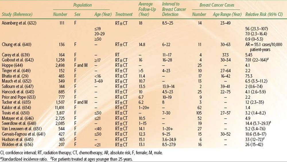
Mastectomy has been recommended as the preferred treatment option in these women. Lumpectomy followed by breast irradiation has been considered by some to be contraindicated due to the cumulative radiation dose to the breast. However, in selected patients using careful breast irradiation techniques that avoid significant overlap with the previous mantle port, anecdotal and retrospective reports in small numbers of patients suggest that it is possible to offer breast-conservation therapy. Therapeutic options and the potential increased risk of reirradiation sequelae should be thoroughly discussed with the patient. A second issue that should also be considered is the risk of the development of new primary. Similar to patients with BRCA mutations, patients who have a history of breast cancer development after irradiation for Hodgkin lymphoma are at risk in the development of subsequent new primaries and may benefit from prevention strategies such as mastectomy.
Elkin et al.658 reported on a retrospective multicenter, cohort study of 253 patients treated for Hodgkin lymphoma with radiation therapy who developed breast cancer and matched 3 to 1 with 741 patients with sporadic breast cancer. They found that patients had a median time from diagnosis of Hodgkin’s to breast cancer of 18 years, with a median age at breast cancer diagnosis of 42 years. Breast cancer after RT for Hodgkin lymphoma was more likely to be detected by screening, was more likely to be diagnosed at an earlier stage, and was more likely to be bilateral at diagnosis. Hodgkin lymphoma survivors had an increased risk of metachronous contralateral breast cancer (HR 4.3; 95% CI, 1.7 to 11.0) and death as a result of any cause (HR 1.9; 95% CI, 1.1 to 3.3).
Cutuli et al.659 reported on a retrospective multicenter analysis in which 117 women and 2 men treated for Hodgkin lymphoma subsequently developed 133 breast cancers. Hodgkin lymphoma treatment was radiation therapy alone in 74 patients and combined modality with chemotherapy in 43 patients. Breast cancer occurred after a median interval of 16 years. Tumors were treated by mastectomy without (n = 67) or with (n = 10) irradiation. Forty-four tumors were treated with lumpectomy without (n = 12) or with (n = 32) radiation therapy. Sixteen patients had isolated breast relapses, 39 had metastases, and 34 died. Young women treated for Hodgkin lymphoma should be carefully monitored in the long term by clinical examination, mammography, and ultrasonography. The authors suggested that a baseline mammography be performed 5 to 8 years after supradiaphragmatic irradiation (complete mantle or involved field) in patients treated before 30 years of age. Subsequently, mammography should be performed every 2 years or each year, depending on the characteristics of the breast tissue (e.g., density) and especially in the case of an association with other breast cancer risk factors.
Wolden et al.660 described 71 cases of breast cancer in 65 survivors of Hodgkin lymphoma. Median age at diagnosis was 24.6 years for Hodgkin lymphoma and 42.6 years for breast cancer; the relative risk for invasive breast cancer after Hodgkin lymphoma was 4.7 compared with an age-matched cohort. Cancers were detected by self-examination in 63%, mammography in 30%, and by physical examination alone in 7%. The majority were of invasive ductal histology and 27% had positive axillary nodes. The tumor was ER-positive in 63% of the cases, and 25% of patients had an associated family history. The majority of tumors were smaller than 4 cm. Ninety-five percent of cases were managed by mastectomy because of prior irradiation, and two women underwent excisional biopsy with breast irradiation. One of these patients had tissue necrosis in the region of overlap with the prior mantle field. The incidence of bilateral breast cancer was 10%. The 10-year disease-specific survival rate for DCIS was 100%, stage I, 88%, stage II, 55%, stage III, 60%, and stage IV, 0%.
One of the largest series using breast-conserving therapy plus radiation in patients previously treated for Hodgkin lymphoma is from Deutsch et al.661 In this retrospective review, 12 women treated with radiotherapy with or without chemotherapy for Hodgkin lymphoma (11 patients) and non-Hodgkin lymphoma (1 patient) in whom breast cancer developed 10 to 29 years later were treated with lumpectomy and breast irradiation. Patients were treated with whole-breast daily irradiation with a fractionation of 2 Gy to 50 Gy with boost to the operative area. Six also received adjuvant chemotherapy for breast cancer. Breast irradiation was well tolerated without any unusual acute or chronic sequelae. They conclude that this may be an option for previously radiated Hodgkin’s survivors who develop early-stage breast cancer.
This controversial area will continue to evolve. Given the development of partial breast irradiation programs over the past several years, it is likely that data regarding reirradiation with partial breast programs will be reported.662,663 Clearly, however, Hodgkin’s survivors and others who receive radiotherapy to breast at a young age are at high risk for developing breast cancers and should be carefully monitored. Women who develop breast cancer should be advised that mastectomy remains the treatment of choice. However, for patients highly motivated for breast preservation, anecdotal experiences using a variety of approaches have revealed acceptable toxicity and cosmesis.
TABLE 56.35 CONSERVATIVE SURGERY AND RADIATION IN THE TREATMENT OF MULTICENTRIC BREAST CANCER

Patients with More than One Invasive Carcinoma (Multicentric Disease)
Multicentricity of breast cancer has been considered by some a contraindication to breast-conserving surgery and suggest mastectomy as the preferred option.664 Conservation surgery and breast radiation therapy as an alternative to mastectomy is controversial in patients with two or more lesions in the same breast. Several studies have addressed this issue, and it appears that the risk of relapse in the conservatively treated patient is slightly higher than if there is one lesion, but the risk may be acceptable in patients with two or perhaps three lesions provided these lesions are surgically excised with negative margins and there are no residual areas of suspicion on physical examination, mammogram, or imaging studies. Selected studies evaluating the conservative management of breast cancer in patients with multicentric disease are summarized in Table 56.35.665–669
Kurtz et al.,669 in an analysis of 586 patients with unilateral stage I or II breast cancer treated with breast-conserving surgery and irradiation, found 61 patients who had two or more microscopic tumor nodules. After a median follow-up of 71 months, 15 patients (25%) had a recurrence in the treated breast, compared with 56 of 525 (11%) patients with single tumors (P <.005). Recurrence was noted more often in patients with multiple tumors diagnosed clinically or mammographically (8 of 22, 36%) than when multicentricity was apparent only on pathologic examination (7 of 39, 18%).
Wilson et al.665 reviewed their experience in 1,060 patients treated with conservation surgery and breast irradiation of whom 13 (1.2%) presented with synchronous multicentric ipsilateral breast cancer. With a median follow-up of 71 months, 3 of the 13 (23%) had an ipsilateral breast recurrence (72-month actuarial rate of 25%) compared with 12% in the single-lesion population. The use of conservation therapy in patients with more than one primary lesion should be considered with caution, and patients should be forewarned of the need for more extensive resections and the increased risk of breast relapse. Because resection of a larger volume of breast is required, cosmetic results may be compromised, and expectations may not be met. In such instances, a mastectomy may be the preferred approach.
Conservation Surgery and Irradiation After Breast Augmentation
A growing number of breast cancers occur in women with prior augmentation mammoplasty. The stage of breast cancer at diagnosis in women who have undergone augmentation mammoplasty has been examined with conflicting results. In a retrospective review, Clark et al.670 reported that 24% of 33 patients with augmented breasts and 42% of 1,735 patients with nonaugmented breasts had mammographically detected cancers (P = not significant). The incidence of DCIS in the two groups was similar (18% vs. 15%, respectively). Sizes of the mammographically detected tumors in the two groups were comparable. However, palpable tumors in the augmented group were significantly smaller than those in the nonaugmented group. Axillary lymph node involvement was detected in 19% of the augmented group and 41% of the nonaugmented group. Among those with mammographically detected tumors, there was no significant difference in axillary lymph node metastases between patients with augmented versus nonaugmented breasts (13% vs. 15%, respectively).
Patients with augmentations who have breast cancer are currently being treated with conservation therapy, but no study has investigated the complications and cosmetic results of radiation therapy specifically in this group of women.
Breast-conservation therapy in 17 augmented patients with breast cancer was reported by Handel et al.,671 where 15 patients were available for follow-up. In 10 patients (67%), significant capsular contracture occurred in the irradiated breast an average of 12 weeks after completion of treatment. Four patients underwent revision surgery to correct symptoms arising from contracture. These authors concluded that irradiation of the breast for cancer in augmented women results in a high incidence of scar tissue contracture and poor cosmetic results.
In contrast, Guenther et al.672 evaluated 20 women in whom breast cancer developed after augmentation mammoplasty (14 subcutaneous implants and 6 retromuscular implants). Patients were treated with wide local tumor excision and level I and II axillary lymph node dissection. Irradiation was delivered to the breast (45 to 50 Gy), and a boost (14 to 21 Gy) was given to the tumor excision site with either photons, electrons, or iridium-192 (192Ir) implant. With a median follow-up of 3.8 years (range, 6 months to 9.3 years), there were no local recurrences, although distant metastases developed in two patients. Seventeen patients (85%) had good or excellent cosmetic results.
Breast Conservation and Mammoplasty
Women with large, pendulous breasts have been documented to have poorer cosmetic outcomes when undergoing irradiation after breast-conservation surgery (thought to be caused by dose inhomogeneity) compared with women who have small or medium-size breasts. Smith et al.673 evaluated 10 women who had undergone bilateral reduction mammoplasty for breast malignancy followed by radiation therapy. A variety of reduction techniques were used to include the malignant lesions. Patients received 50 Gy in 25 fractions in 5 weeks after surgery. Radiation therapy was usually initiated within 4 weeks after surgery. With a follow-up of 37 months, no patients have had complications from the surgery or radiation therapy. No local recurrent malignancies have been detected. Cosmesis has been good to excellent in all patients.
Bilateral Carcinoma of the Breast
Among factors reported to be associated with an increased risk of bilateral breast carcinoma are younger age, family history of breast cancer, lobular carcinoma, multicentric disease, histologic differentiation of the primary tumor, parity, and PR-positive status.674,675,676–679 The appearance may be synchronous (1% to 2%) or metachronous (5% to 8%). Synchronous breast carcinoma was defined as a contralateral cancer diagnosed within 1 year of initial diagnosis.
Patients with bilateral carcinoma have been treated with total or modified radical mastectomy. However, the available evidence clearly supports lumpectomy followed by breast irradiation as an acceptable alternative for appropriately selected women. Solin et al.680 reported on 30 treated with breast-conservation therapy (11 with concurrent and 19 with metachronous carcinoma). A dose of 45 to 50 Gy was delivered to both breasts with tangential fields, in addition to a boost of 10 to 15 Gy with either iridium implant or electrons. The tangential fields were matched in the midline in 17 patients and overlapped by up to 3 cm in 10 patients. In the 60 treated breasts, the 5-year actuarial local failure rate was 6%. In 25 treated breasts with a minimum of 2 years of follow-up, 68% had excellent and 24% had good cosmetic results. The incidence of arm edema was 6%, similar to that reported in patients with unilateral disease.
Hungness et al.681 reviewed their experience with 51 patients with bilateral synchronous breast cancer (2.1% of 2,382 treated for breast cancer during the same period). The first cancer was detected by palpation in 81% and by mammography in 14%. The corresponding figures for the contralateral cancer were 24% and 54%, respectively. The histologic type of cancer was identical in the two breasts in 29 patients (57%) and was different in 22 patients (43%). The overall 10-year survival rate was 66%.
Heron et al.682 compared the outcomes in 1,315 patients (89.9%) with unilateral, 103 (7.1%) with metachronous, and 47 (3.0%) with synchronous breast carcinoma treated with either mastectomy or breast-conservation therapy. Patients with synchronous and metachronous bilateral carcinoma had a worse 8-year disease-free survival rate compared with patients who had unilateral breast carcinoma, as well as increased risk of distant metastasis. In multivariate analysis, differences in local tumor control and overall survival were not statistically significant for patients who had bilateral or metachronous cancer compared with those who had unilateral disease.
Kollias et al.,683 in 3,210 women age 70 years or younger treated for primary operable breast cancer, identified 106 who had bilateral breast cancer; in 26 (0.8%) the disease was synchronous and in 80 (24%) a contralateral breast cancer developed after treatment for an initial primary breast cancer. There was a significant difference in survival between women with unilateral breast cancer, synchronous bilateral breast cancers, and metachronous contralateral breast cancer, with survival rates at 16 years of 53.8%, 42.4%, and 60.1%, respectively (P <.0001) from the date of the diagnosis of the first primary tumor. There was no difference in survival among the three groups from the date of diagnosis of the second primary in cases of metachronous contralateral breast cancer (P = .31).
Ninety-five patients with bilateral carcinoma of the breast treated with mastectomy (60 patients), conservation of the breast (17 patients), or both (18 patients) were studied by Gustafsson et al.684 Cumulative 5-year local tumor control rates were 94% for the 138 mastectomy patients and 90% for the 52 patients treated with breast-conservation therapy. Twenty-eight percent of the first carcinomas were stage I, compared with 43% of second carcinomas (P<.05), probably reflecting the close follow-up after initial treatment. The 5-year distant disease-free survival rate from treatment for the second carcinoma was 74%. The 5-year distant recurrence-free survival rate when second carcinomas were diagnosed within 5 years was 58%, compared with 95% for patients diagnosed more than 5 years after the first carcinoma.
De la Rouchefordiere et al.685 reported on 149 patients with simultaneous bilateral breast cancer (diagnosed within 6 months). Of 298 tumors, 40% were T0 or T1, 45% T2, and 15% were T3 or T4. The majority (83%) were clinically node negative. Treatments were bilateral mastectomy in 43%, irradiation in 16%, and both in 41% of the patients. Fifty-one patients had bilateral breast-conserving therapy and 24 were treated exclusively with irradiation. The 5-year disease-free survival rates were 70% to 86%, respectively, similar to those observed at the same institution in patients with unilateral tumors. Cosmesis was assessed in 48 patients and was acceptable in 37 (77%). These authors advised special attention should be paid to any possible overlap of the supraclavicular and internal mammary fields over the spinal cord; in one patient, spinal cord myelopathy developed at T6.
Freedman et al.587 reviewed records of 116 patients with bilateral breast cancer and a breast cancer family history. The primary treatment was a breast-conserving procedure in 55 and a mastectomy in 61. Locoregional recurrences occurred in 4 of 46 cases treated with breast-conserving therapy, resulting in a 10-year actuarial locoregional tumor control rate of 83%. Of nine patients who did not receive radiation as a component of their breast-conserving therapy, locoregional recurrences developed in four (10-year locoregional control rate of 49%). The 10-year actuarial rates of locoregional control after mastectomy with and without radiation were 91% and 89%, respectively.
Fung et al.686 reported on 55 women with stage 0, I, or II concurrent (n = 12) or sequential (n = 43) bilateral breast cancers treated with irradiation after breast-conserving surgery. The tangential fields were matched with no overlap in 40 patients (73%); there was overlap on skin of up to 4 cm in 14 patients (25%). For the 110 treated breast cancers, the 10-year actuarial local failure rate was 15%. Complications included breast edema (28%), arm edema (8%), pneumonitis (4%), cellulitis (3%), rib fracture (1%), and brachial plexopathy (1%). No patient had match line fibrosis. For patients with a minimum of 3 years of relapse-free follow-up, the rate of excellent or good cosmetic outcome for 104 treated breasts was 85%.
FIGURE 56.25. Patient immobilized for breast irradiation on a slant board with custom mold to minimize day-to-day positioning errors.
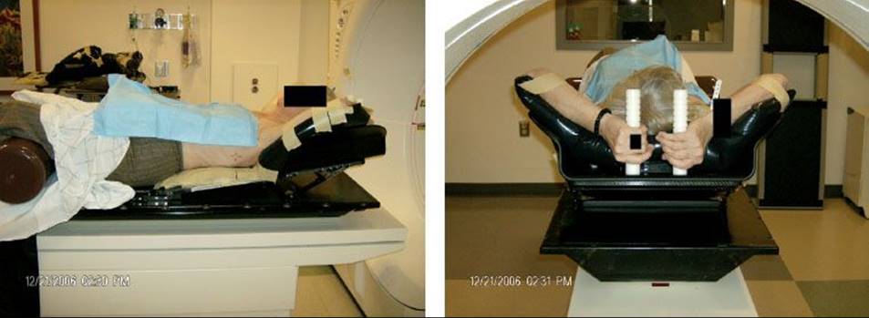
FIGURE 56.26. Prone breast board. A: Customized prone breast board with adjustable aperture and wedge for contralateral breast. B: Ipsilateral breast and anterior chest wall hang in dependent fashion away from thorax with ipsilateral arm placed above head. (From Goodman K, Hong L, Wagman R, et al. Dosimetric analysis of a simplified intensity modulation technique for prone breast radiotherapy. Int J Radiat Oncol Biol Phys 2004;60(1):95–102, with permission form Elsevier.)
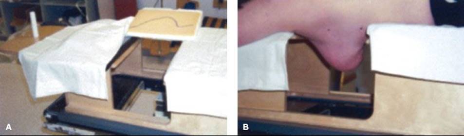
TECHNICAL ISSUES IN RADIATION MANAGEMENT OF EARLY-STAGE DISEASE
Treatment Position
Most patients are treated in the supine position, with the arm abducted (90 degrees or greater). Commercially available or custom made breast tilt boards with armrests that maintain the patient’s daily position with the slope of the chest wall parallel to the table, often in combination with immobilization devices (e.g., Alpha cradle, plastic molds) are typically used to reproduce daily positioning and minimize day-to-day setup errors (Fig. 56.25).
Other treatment positions have been used to improve the dosimetry in patients with large, pendulous breasts. A lateral decubitus position has been suggested by investigators at the Institut Curie.687,688
Irradiation in the prone position has been proposed by Merchant and McCormick,689 with reduction of dose in the high-dose region to 102% to 103% of the dose to the irradiated breast, as well as reduction of volume and dose to the underlying lung and heart and reduction of scattered dose to the contralateral breast. This technique is being increasingly employed and follow-up data appear to be promising with respect to toxicity and early outcomes.690 Patient positioning and a corresponding CT image are shown in Figures 56.26 and 56.27.
Treatment Volume
The entire breast and chest wall are included in the irradiated volume as shown in Figures 56.28 and 56.29. Radiopaque surgical clips placed at the margin of the tumor bed may assist in defining the target volume.691 The upper margin of the portals should be placed at the head of the clavicle to include the entire breast. The medial margin, if no internal mammary portal is used, should be at or 1 cm over the midline. If an internal mammary field is used, the medial tangential portal is located at the lateral margin of the internal mammary field. (See discussion later on regional nodal irradiation.) The lateral-posterior margin should be placed 2 cm beyond all palpable breast tissue, which is usually near the midaxillary line. The inferior margin is drawn 2 to 3 cm below the inframammary fold. Recently, the RTOG published their consensus definitions for breast cancer radiation therapy planning, which are listed in Table 56.36. An illustration of nodal volumes drawn on an axial planning CT scan is shown in Figure 56.30.
In patients treated with 6-MV or lower-energy photons with wide tangential fields in whom separation is >22 cm, there may be significant dose inhomogeneity in the breast; this may correlate with less satisfactory cosmetic results.352,692 This problem can be minimized by using higher-energy photons (10 t0o 18 MV) to deliver a portion of the breast radiation (approximately 50%) as determined with treatment planning to maintain the inhomogeneity throughout the entire breast to between 93 and 105%. If desired, the buildup of the beam may be modified with a “degrader.” “Simple intensity-modulated radiation therapy” techniques such as field-in-field or dynamic multileaf collimators (MLCs) to achieve electronic tissue compensation may be utilized to reduce dose inhomogeneity as well. Bolus should be avoided in conservatively managed patients. A variety of immobilizing devices or molds may be constructed to support the breast in the treatment position (Fig. 56.31). A polyvinyl chloride, ring-shaped device, held by a strap has been used around the breast to aid in positioning of patients with large, pendulous or flaccid breasts. Skin reactions where material is in contact with the skin should be closely monitored.693
FIGURE 56.27. Computed tomography simulation images used to determine arrangement of tangent beams in prone breast irradiation. (From Goodman K, Hong L, Wagman R, et al. Dosimetric analysis of a simplified intensity modulation technique for prone breast radiotherapy. Int J Radiat Oncol Biol Phys2004;60(1):95–102, with permission form Elsevier.)

FIGURE 56.28. Axial treatment planning computed tomography cut with contours of the level I, II, and III nodes.
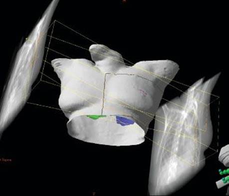
FIGURE 56.29. A tangential breast radiation field, demonstrating projection of tangential field on simulation film and patient surface.

Alignment of the Tangential Beam with the Chest Wall Contour
The anterior chest wall slopes downward from the midchest to the neck. To make the posterior edge of the tangential beam follow this downward-sloping contour, the collimator of the tangential beam may be rotated, or the patient may be placed on a slant so that the slope of the chest wall is parallel to the table. An alternative is to make the deep posterior edge of the tangential beam follow the chest wall contour by means of a rotating beam splitter mounted on a tray without rotation of the collimator or using multileaf collimation. In this way, the superior edge of the tangential beam remains in the true vertical and matches perfectly the vertical inferior edge of the supraclavicular field if used.
Usually up to 2 to 3 cm of underlying lung may be included in the tangential portals. The amount of lung included in the irradiated volume is greatly influenced by the portals used. Bornstein et al.694 determined the amount of lung irradiated in 40 patients with breast cancer using CT scans for treatment planning in the treatment position. Parameters measured from simulator films included the perpendicular distance from the posterior tangential field edge to the posterior part of the anterior chest wall at the center of the field (central lung distance [CLD]), the maximum perpendicular distance from the posterior tangential field edge to the posterior part of the anterior chest wall (maximum lung distance [MLD]), and the length of lung as measured at the posterior tangential field edge on the simulator film (Fig. 56.32). The best predictor of the percentage of ipsilateral lung volume treated by the tangential fields was the CLD. A CLD of 1.5 cm predicted that approximately 6% of the ipsilateral lung would be included in the tangential field, a CLD of 2.5 cm, approximately 16%, and a CLD of 3.5 cm, approximately 26% of the ipsilateral lung. A typical acceptable dose–volume histogram of a left-sided breast cancer treated with external-beam radiation is given in Figure 56.33.
When the CLD is >3 cm, in treatment of the left breast, a significant volume of heart will also be irradiated. To avoid this, a medial tangential breast port (3- to 5-cm wide), somewhat similar to an internal mammary port, may be designed. The beam is angled 10 to 15 degrees laterally to conform to the angle of the medial breast port. The dose is prescribed to the posterior border of the chest wall as determined by CT scanning.
Special attention should be paid to minimizing the volume of heart irradiated.695 As will be discussed later in the section on cardiac sequelae, even small amounts of heart in the field can affect cardiac function. Marks et al.696 have suggested the use of a cardiac block if the heart is in the tangential field, which can be supplemented by an electron field as shown in Figure 56.34. Use of short (10- to 15-second) treatments while the patient holds her breath is also feasible as a way of reducing cardiac radiation during left-sided breast cancer treatment.697
TABLE 56.36 RADIATION THERAPY ONCOLOGY GROUP CONSENSUS DEFINITIONS FOR BREAST CANCER RADIATION THERAPY PLANNING
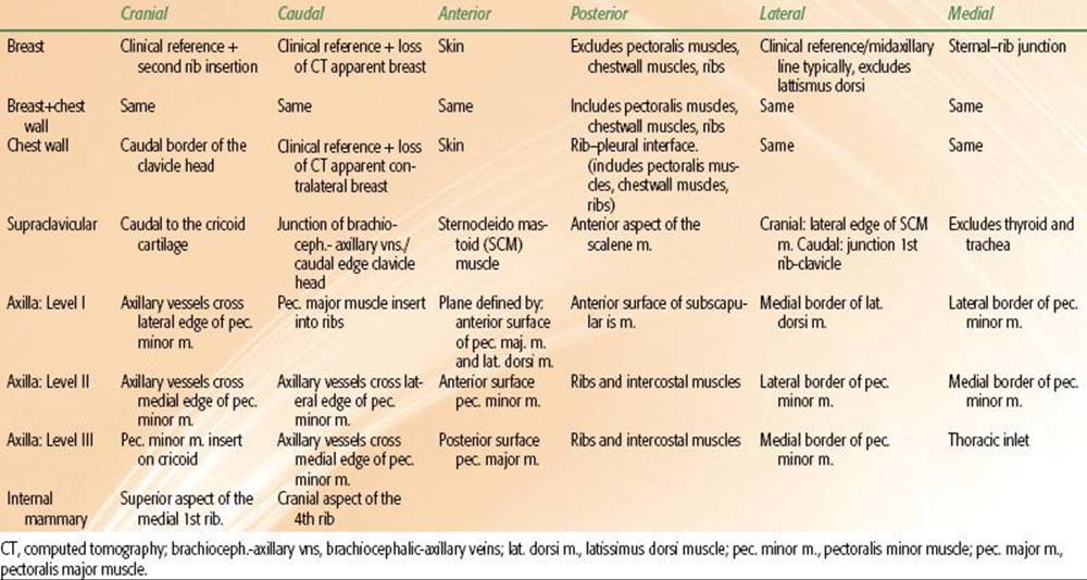
FIGURE 56.30. Nodal volumes drawn on axial planning CT scan.
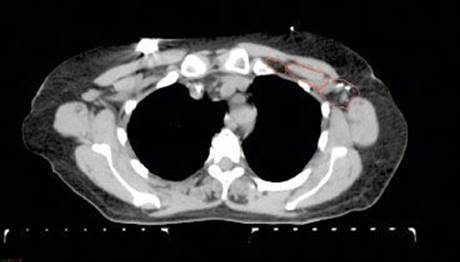
FIGURE 56.31. Immobilization material placed over breast tissue to help maintain day-to-day positioning of the breast.

FIGURE 56.32. Measurement of the radiographic parameters using virtual simulator. The contoured heart is shown in black, the lung in gray. The central lung distance (CLD) is the lung distance in the projection of the tangential fields at the level of the central axis. Lung length is the vertical lung distance included in the radiation port. The maximal heart distance (MHD) is the width of heart in the tangent fields at its maximal level, whereas the maximal heart length (MHL) is the maximal length in tangential fields referring to the heart contour in a digitally reconstructed radiograph (DRR). (From Kong F-M, Klein EE, Bradley JD, et al. The impact of central lung distance, maximal heart distance, and radiation technique on the volumetric dose of the lung and heart for intact breast radiation. Int J Radiat Oncol Biol Phys 2002;54:963–971, with permission from Elsevier.)

FIGURE 56.33. Dose-volume histogram of left-sided breast cancer treated with external beam radiation.
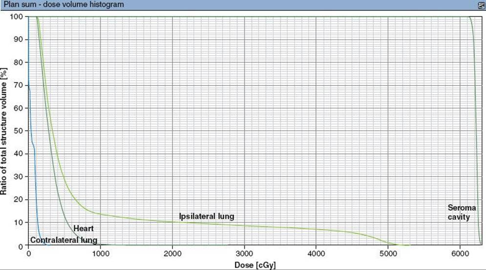
FIGURE 56.34. A and B: Left tangential breast field with heart block to shield left ventricle from radiation port. Projection of heart block on breast shields minimal amount of breast tissue. If necessary, a shadow electron field may be added to cover the portion of breast tissue shielded by heart block.

Doses of Radiation and Fractionation
Whole-Breast Dose
With whole-breast irradiation, tumor doses of approximately 45 to 50 Gy are delivered to the entire breast over 5 to 6 weeks (1.8- to 2-Gy tumor dose daily, 5 weekly fractions). Some authors have suggested daily fractions of 1.8 Gy for patients with large, pendulous breasts or when irradiation is combined with chemotherapy.559,560,698 The authors’ preference is to use 2 Gy fractions to 50 Gy because this is the scheme used in the vast majority of randomized trials using whole-breast radiation therapy following conservative surgery.
Alternative fractionation schemes have been employed and have been shown to be acceptable. These are discussed in greater detail in the hypofractionation section.
Radiation Beams
X-ray energies of 4 to 6 MV are preferred to treat the breast. Photon energies >6 MV underdose superficial tissues beneath the skin surface, but higher-energy photons may be helpful in large breasts to decrease the integral breast dose. In these patients the high-energy photon beam may be “degraded” to bring the maximum dose to more superficial tissues. It is not necessary to apply bolus to the breast because the skin is usually not at risk for recurrence after complete excision of a T1 or T2 lesion, as is the skin of the chest wall after a mastectomy. Use of bolus results in impaired cosmetic results.352
Wedges or compensating filters must be used for a portion of the treatment to achieve a uniform dose distribution in the breast (5% to 8% dose variance from the chest wall to the apex). Although conventional wedges improve dose homogeneity at the central axis of the breast, significant inhomogeneity can occur in the superior and inferior portions of the breast. Currently, with the use of MLCs and more sophisticated treatment planning techniques, optimization of homogeneity throughout the breast can be achieved through the use of a variety of techniques. Dose homogeneity in a breast plan uncompensated (without wedges), with standard wedges, and with electronic dynamic wedge technique to improve homogeneity in the superior-inferior plane are outlined in Figure 56.35. These techniques are discussed in more detail later.
Boost to Tumor Site
The need for a boost to the tumor bed following lumpectomy and whole-breast radiation remains an area of debate. In the earlier years of breast-conserving surgery, status of the surgical margins were not always assessed. Recent retrospective data suggest that patients with known negative margins have high local control rates with no boost following whole-breast irradiation.699 Fisher et al.670 have all raised the question of the need for a radiation dose boost at the excision site.
Most authors report that 65% to 80% of breast recurrences after conservation surgery and irradiation occur around the primary tumor site.104,106,196,298,376,388,671–703 These data provide a strong rationale for a tumor bed boost. Various series suggest that patients treated with higher doses have a greater probability of tumor control. Clark et al.377 noted in 1,504 patients a greater incidence of failures at 10 years of 17% in those to whom no boost was delivered, compared with 11% in those who received doses of 5 to 15 Gy at the primary excision site (P = .03). In other series of patients with unknown surgical margins, patients receiving a boost had roughly half the breast failure rate (6% to 11%) compared with those with no boost (9% to 20%).110,388,404,704
Others have advocated tailoring the need for a boost depending on margins. Arthur et al.699 reported on 205 patients who underwent re-excision prior to radiation. All patients in this cohort had no tumor on re-excision and were treated with whole-breast irradiation to a dose of 50 Gy without a boost. Five failures were documented, resulting in a 15-year local control rate of 92.4%. The authors advocate selective avoidance of the boost in these patients.
Randomized Data
The Lyon Breast Cancer Trial conducted a randomized study to assess the role of the boost in breast-conserving therapy in patients with stage I and II breast cancer (≤3 cm) who were treated with complete local tumor excision, axillary dissection, and 50 Gy to the breast in 20 fractions over 5 weeks and randomly assigned to receive or not a boost of 10 Gy with electrons to the tumor bed.705 With a median follow-up of 3.3 years, at 5 years 10 of 521 women who received a boost (3.6%) and 20 of 503 (4.5%) who received no further treatment experienced a local breast relapse (P = .044). Time to local recurrence is shown in Figure 49.52, with more patients failing after 7 years in the no-boost arm.
Bartelink et al.404 reported the results of the EORTC trial in which, after complete lumpectomy and axillary dissection, patients with stage I or II breast cancer received 50 Gy of radiation to the whole breast in 2-Gy fractions over a 5-week period and were randomly assigned to receive either no further local treatment (2,657 patients) or a boost of 16 Gy, usually given in 8 fractions by electron beam (2,661 patients). In the initial report, with a median follow-up of 5.1 years, local recurrences were observed in 182 of the 2,657 patients in the standard-treatment group and 109 of the 2,661 patients in the additional-radiation group. The 5-year actuarial rates of local recurrence were 7.3% and 4.3%, respectively (P <.001). Patients 40 years of age or younger benefited most; at 5 years, their rate of local recurrence was 19.5% with standard treatment and 10.2% with additional radiation (RR 0.46; 99% CI, 0.23 to 0.89; P = .002). In an update of this study published in 2007, with a median follow-up of 10.8 years, a significant benefit to the boost was noted in all age groups, with an overall hazard ratio of 0.59 in favor of the boost405 (see Fig. 56.16).
If a decision is made not to use a boost, careful assessment of lumpectomy margins is critical, as discussed in the following section.
FIGURE 56.35. Isodose distributions from three breast plans. A: Open fields demonstrating inhomogeneity. B: Standard wedges demonstrating improvement in central axis, but with a hot spot in the superior inferior plane. C: Dynamic wedge plan showing improved homogeneity in both the central axis as well as the superior–inferior plane.

Electron Versus Interstitial Boosts
Before the widespread availability of electron beam therapy, interstitial brachytherapy or cone-down photon boost was popular. Experiences with interstitial boosts have been reported by several groups, using both high dose rate after loading and low dose rate temporary implants.706–715 The reader is referred to these studies and the chapter on brachytherapy for a more extensive discussion of techniques related to interstitial tumor bed boosts. Currently, most institutions prefer electron beam boost because of its relative ease in setup, outpatient setting, lower cost, decreased time demands on the physician, and excellent results compared with 192Ir implants. The introduction of single lumen or multilumen balloon catheters, used primarily in partial breast irradiation, can also be considered an interstitial boost technique. Cosmetic results with either boost technique at various institutions are summarized in Table 56.37.
Electron Boosts
The patient is positioned with the arm toward the head to flatten the breast contour and may be rolled so the tumor bed is parallel to the table and the accelerator head can point straight down onto the target volume. An electron energy is selected that covers the target volume depth (usual range is 9 to 16 MeV electrons), based on review of the physical examination, mammogram, ultrasound, CT, or other imaging used to ascertain the location and depth of the tumor or metallic surgical clips. The 90% prescription isodose line is limited to the chest wall to decrease dose to the lung. The clinical setup for electron boost involves marking the projection of the postlumpectomy volume on the skin and adding 2 to 3 cm in all directions.
Accurate target volume definition is critical with any boost technique. Methods vary from simple and unsophisticated (as described in the previous paragraph) to complex and expensive, such as ultrasound and CT definition of the target volume.716,717 The accuracy of using the scar to define the lumpectomy cavity has been questioned. In a study by Oh et al.,716 30 women consecutively treated for 31 breast cancers had simulation CT scans performed before and after whole-breast irradiation. CT breast volumes were delineated using clinically defined borders, and excision cavity volumes were contoured based on surgical clips, the presence of a hematoma, or other surgical changes. Hypothetical electron boost plans were generated using the surgical scar with a 3-cm margin and analyzed for coverage. The volume reduction (R) in the excision cavity was inversely correlated with time elapsed since surgery (R = 0.46; P <.01) and body weight (R = 0.50; P <.01). The scar-guided hypothetical plans failed to cover the excision cavity adequately in 62% and 53.8% of cases using the pretreatment and postradiation CTs, respectively.
Surgical clips are ideal for the localization of the tumor bed.691,717,718 The surgical clip method requires the cooperation of the surgical team. Despite the fact that it would theoretically take an infinite number of clips to define every extension of a typical tylectomy cavity, in practice six clips suffice (superficial, deep, medial, lateral, cephalad, and caudal). In a study reported by Denham et al.,719 surgical hemoclips were left in situ in 27 patients to demarcate the limits of the excision cavity. The position of these clips varied widely in relation to the patient’s recollection of the position of the original lump, the surgical notes, and the surgical scar. Incomplete coverage of the excision cavity in the “coronal” (en face) plane using an electron field could have occurred in an estimated 10 of 24 (42%) cases had surgical clips not been left in situ. Depth of the surgical clips below the skin surface also varied markedly; in 19 of 26 (73%) cases, the clips were observed to be ≥3 cm below the skin surface, whereas in only 5 of 26 (19.2%) cases were the clips found to be ≤2 cm deep to the surface. Had a 9-MeV electron beam been used to treat all of the patients, a major underdose of the excision cavity would have been likely in 21 of 26 (81%) evaluable cases. Coverage would have improved to 11 of the 26 (42%) had a 12-MeV beam been used.
TABLE 56.37 EXCELLENT OR GOOD COSMETIC RESULTS WITH ELECTRON BEAM OR INTERSTITIAL BRACHYTHERAPY BOOST IN BREAST CONSERVATION THERAPY
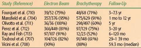
Fein et al.718 described a study in patients with stage I or II breast cancer treated with breast-conservation therapy; surgical clips were placed in the excision cavity in 556 patients, and no clips were placed in 808. After breast irradiation with tangential fields, the primary tumor incision site was boosted with electron beam (14 to 20 Gy). The actuarial breast recurrence rates at 10 years were 11% in patients with clips and 5% in patients without clips (P = .01). Increased rates of breast recurrence were noted for patients with clips who had some of the following: no adjuvant treatment, unknown surgical margins, no re-excision, pathologic negative nodes, and outer location of primary tumor. The higher incidence of breast relapse may be related to a specific surgeon who had a breast recurrence rate of 21%, compared with 6% for the remainder of the surgeons (P = .01); the status of the margins was unknown in 48% of these patients, compared with 10% overall (P = .001). The authors concluded that failure to ink the surgical specimen and inadequate assessment of margins cannot be compensated by placement of surgical clips or treatment planning using CT to delineate the surgical bed. On the other hand, this study failed to show any benefit from use of surgical clips at the tumor excision margins to design the boost volume.
Ultrasonography can provide the depth of the biopsy cavity, as well as the other dimensions, for use in designing electron portal borders and selection of electron energy.717,720 Ultrasonography was used in 30 patients to measure breast thickness for determination of the most appropriate electron beam energy for the boost. In most patients the depth was ≤4 cm, but in eight patients (32%), energy higher than 12 MeV should have been used to cover adequately the depth of the target volume.
CT-guided portal design should be done in the treatment position. This technique gives good definition of the depth of the chest wall and has been shown to be similar to ultrasound in delineating the lumpectomy cavity.717,720Delineation of the biopsy cavity becomes more difficult with increased interval from surgery. The combination of surgical clips with a treatment planning CT scan to define the lumpectomy site for electron boost is most ideal. In the absence of surgical clips, the CT scan evaluation of the biopsy cavity or postsurgical changes, in combination with clinical information including mammography findings, scar location, operative reports, and patient input, will provide accurate information regarding placement of the field and energy of the electron boost.
A recent multi-institutional study reported interobserver variabilities in the delineation of the seroma cavity and organs at risk in breast cancer patients undergoing breast-conserving surgery and whole-breast irradiation.721 Nine radiation oncologists specializing in breast radiotherapy from eight institutions independently delineated target volumes on the same three breast cancer patients. They concluded that “variations in delineating the targets and OAR’s [organs at risk] for breast RT by well-experienced observers from different institutions are substantial for all relevant structures.”
To reduce these variations, guidelines for contouring the seroma cavity have been established; the Seroma Clarity Scale (SCS), developed by the British Columbia Cancer Agency and the Cavity Visualization Score (CVS), developed by a Stanford group.717,722 Both scoring systems are remarkably similar with the distinction being the SCS is a scale from 0 to 5, with 0 being no visible seroma cavity and 1 being a scar or shadow; the CVS is a scale from 1 to 5, with 1 being no visible seroma cavity, and omits the presence of a scar or shadow on the scale. The scores of 2 to 5 are relatively consistent between the two classification methods.
It is pertinent to note that each of these classification systems were developed without noting the presence of surgical clips or fiducial markers with respect to the seroma cavity. However, the authors, along with other investigators, have previously published that the presence of surgical clips or fiducial markers in the surgical bed improve the interphysician accuracy in the delineation of the seroma cavity in early-stage breast cancer patients.723,724 In a study by Shaikh et al.,725 the presence of fiducial markers improved the mean CVS score (from 2.5 to 3.5) and accuracy of physician contours compared with patients without fiducial markers. The use of titanium clips in patients treated with accelerated partial breast irradiation (APBI) is a focus of the United Kingdom IMPORT LOW (Intensity Modulated and Partial Organ Radiotherapy) trial.726 An audit of the trial was performed to determine inter- and intraphysician variation for tumor bed localization for radiotherapy planning. Although no control group was used, clips were essential for the localization of the surgical cavity in 22 of 30 (73%) patients and led to modifications in radiotherapy field borders in 18 of 30 (60%) patients. A recent study by Dzhugashivili et al.724 found that the visualization of the lumpectomy cavity was greatly improved in treatment-planning CT scans when clips were present; this was found to be even more pronounced in patients with very dense mammary glands, regardless of the physician reading the CT image.
Irradiation of Regional Lymphatics
Radiation therapy to the breast or chest wall and regional lymphatics can be technically challenging and, as previously discussed, remains one of the more variable and controversial aspects in management. A wide variety of available techniques, in combination with difficulties associated with matching fields, anatomic variability between patients, and lack of clear evidence regarding the superiority of any single approach, has resulted in a lack of consensus in regional nodal management.
Anatomic variation was highlighted in a study by Mansur et al.727 They reported on 65 patients with breast cancer who had volumetric CT scanning in the treatment position. The IMNs and axillary lymph node regions were delineated according to a cross-sectional nodal atlas. The variable depths of IMNs at different intercostal spaces result in dose variations if the internal mammary port is treated with a single or inadequate electron beam energy. Axillary lymph nodes frequently overlapped the head of the humerus anteriorly when the arm was angled more than 90 degrees, but did not when the arm was angled 90 degrees or less. The larger the angle, the less head of the humerus could be spared in the supraclavicular port.
Arthur et al.728 evaluated treatment techniques for coverage of the intact breast and ipsilateral lymph node regions. Anatomic outlines were obtained from five randomly selected patients with CT scanning in treatment position (three with cancer of the left breast and two of the right). Three techniques used to treat ipsilateral breast and internal mammary and supraclavicular nodes (extended tangents, five-field, partially wide tangents) were configured and compared with a supraclavicular field matched to standard tangential fields. All of the treatment techniques covering IMNs included at least 10% more lung and heart volume than that covered by standard tangential fields. Because of increased chest wall thickness and depth of IMNs superiorly, complete coverage was not achieved with any technique if the IMN target extended superiorly into the medial supraclavicular field.
Goodman et al.729 examined the relation between tangential, anterior, and posterior radiation fields and regional lymph nodes, including level I to III axillary and supraclavicular lymph nodes in 55 patients who underwent CT scanning in the supine position. The mean depths of the level I to III axillary nodes were 4.6, 5.1, and 3.6 cm, respectively. The mean depth of the supraclavicular nodes was 3.9 cm. With the treatment using two tangential fields, level I axillary nodes appeared in the tangential portals in nine of nine patients, either alone or with other lymph node groups. In the three-field group, level I axillary nodes were in 16 of 16 tangential fields either alone or with level II nodes (8 patients). In eight patients, level III and the supraclavicular nodes were included in the anterior field, and in the other eight, levels II and III and the supraclavicular nodes were in the anterior field. There was considerable variation in the depth of supraclavicular and axillary lymph nodes in the fields in which these nodal groups appear and in the nodal group present in the posterior axillary boost field. To be certain that nodal groups to be treated are actually treated, as well as to minimize tissue irradiated, these authors recommended that before the placement of radiation fields, the nodal groups be outlined on a CT scan. From a practical standpoint, whether this results in better tumor control than standard techniques has not been demonstrated.
FIGURE 56.36. Supraclavicular and axillary field and field borders. Level I, II, and III nodes are demonstrated on the film. Note that level I nodes are included primarily in the tangential field in this case.
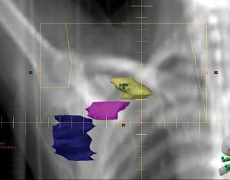
Supraclavicular Lymph Nodes
The inferior border of the supraclavicular field is matched to the tangential field usually just below the clavicular head. The medial border is 1 cm across the midline, extending upward, following the medial border of the sternocleidomastoid muscle to the thyrocricoid groove. The lateral border is a vertical line at the level of the coracoid process, just medial to the humeral head. This field is angled approximately 10 to 15 degrees laterally to spare the cervical spine (Fig. 56.36). The typical width of the supraclavicular field is 7 to 9 cm. The supraclavicular field is extended laterally to treat the full axilla, as clinically indicated. Figure 56.36 demonstrates a supraclavicular field with the supraclavicular and level I, II, and III nodes outlined. Level I as well as a portion of level II nodes will often be included in the tangential field; level III and supraclavicular nodes are covered in the supraclavicular field.
The total dose delivered to the supraclavicular field is 46 to 50.4 Gy at 1.8 to 2 Gy per day (calculated at a depth of 3 cm) in 5 fractions per week. For obese patients, an assessment of the depth of lymph nodes with ultrasound or CT treatment planning is useful to ensure adequate dose is delivered to the target. For patients in whom the target is deeper than 3 cm, higher energy photons should be considered.
Axillary Lymph Nodes
When the axilla is treated in patients with positive nodes, or in patients with inadequate or undissected axillae, the supraclavicular field is extended laterally to cover at least two-thirds of the humeral head, as demonstrated in Figure 56.36. The dose to the midplane of the axilla from the supraclavicular field is calculated at a point approximately 2 cm inferior to the midportion of the clavicle. Depending on the dose distribution and patient’s anatomy, a posterior axillary boost may be considered or anterior axillary boost, as suggested by Wang et al.,730 may be considered.
Posterior Axillary Boost
There is considerable debate regarding the necessity of a posterior axillary boost. Bentel et al.731 questioned its necessity in a majority of patients. In 49 patients undergoing treatment-planning CT scanning in the treatment position, the maximum depth of the supraclavicular and axillary lymph nodes was measured on CT images and the relation between the supraclavicular and axillary lymph node depth and patient diameter was determined. For an anterior field, the relative dose to the supraclavicular and axillary lymph nodes were calculated for a 6-MV photon beam. If an anterior 6-MV beam only is used to treat both supraclavicular and axillary lymph nodes, the dose to the axilla is within ± 5% of the supraclavicular dose in 53% (26 of 49) patients and is 90% or more of the dose delivered to the supraclavicular nodes in 90% (44 of 49) of patients. These authors concluded that higher energy beams or anterior-posterior/posterior-anterior supraclavicular axillary fields may be reasonable when the axillary and supraclavicular nodes are deep.
The posterior axillary boost has been employed to supplement axillary dose. At the end of the treatments to the supraclavicular field, the dose to the midplane of the axilla may be supplemented by a posterior axillary field, as shown in Figure 56.37. Alternatively, the axilla should be contoured in CT-treatment planning as variation of the depth of the axilla from the anterior-posterior skin surface varies from patient to patient and the supplement prescription point can be adjusted accordingly. If the dose is determined to be inadequate, a posterior boost or anterior boost, as suggested by Wang et al.,730 may be employed. When a posterior axillary boost is used, the borders are as follows: medially, the border is drawn to allow 1.5 to 2 cm of lung to show on the portal film; inferiorly, the border is at the same level as the inferior border of the supraclavicular field; laterally, the border just blocks fall-off across the posterior axillary fold; lastly, the superior border splits the clavicle and the superolateral border shields or splits the humeral head. Additional dose to the axilla midplane is usually administered to complete 46 to 50 Gy (2 Gy daily). When indicated, a boost of 10 to 15 Gy is delivered with reduced portals.
If the supraclavicular nodes are not felt to be at risk, a separate axillary field may not always be necessary to treat the axilla. In a study of 39 women with surgical clips in the axilla, Schlembach et al.520 demonstrated that with tangential fields, placing the caudal border of the field within 2 cm of the humeral head and 2 cm deep to the chest wall–lung interface includes the majority of level I and level II lymph nodes (Fig. 56.38).
Internal Mammary Lymph Nodes
The benefit of irradiation of the IMNs is an unresolved issue because clinical failures at this site are very rare and the majority of patients at risk receive adjuvant therapy.547,552 However, the IMNs are difficult to treat because their exact location is often uncertain and the radiation fields that include them irradiate more normal tissue.732,733 Several techniques are used, the most common of which is a direct anterior field matched to tangential fields, which was developed for postmastectomy radiation therapy and increases the volume of heart and lung tissue in the field. With this technique, the rising contour of the intact breast interferes with dosimetry, which may affect the traditional dose prescription point at depths of 4 to 5 cm and may prevent an easy match to the breast tangential fields. Including the IMNs in the tangential fields (wide or deep tangents) may significantly increase the volume of irradiated lung and heart tissue and often includes a portion of the contralateral breast as well.
FIGURE 56.37. Posterior axillary field used to supplement the dose at midplane of the axilla. Note the small amount of lung included and the shielding of the humeral head (whenever possible).
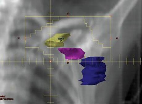
FIGURE 56.38. “High” tangential field shown coverage of level I and portion of level II nodes. With caudal edge of the field within 2 cm of the humeral head and leading edge approximately 2 cm from lung–chest wall interface, the majority of nodes in level I and level II will be covered by this technique.
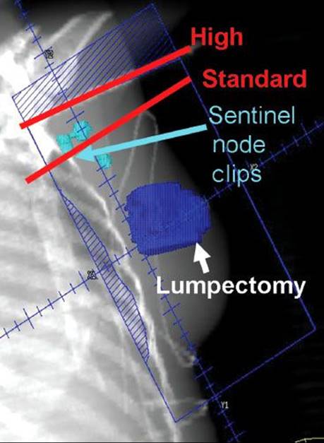
FIGURE 56.39. Irradiation of the breast: field configurations and isodose lines for 6-MV photons. A: “Standard tangents” technique. B: Deep tangents technique. C: En face internal mammary field (IMF) technique. D: Twenty-degree IMF technique. (From Roberson PL, Lichter AS, Bodner A, et al. Dose to lung in primary breast irradiation. Int J Radiat Oncol Biol Phys 1982;9:97–102, with permission from Elsevier.)
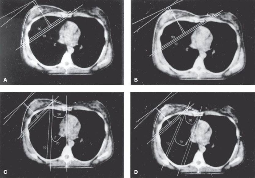
The medial border of the IMN field is the midline. The lateral border is usually 5 to 6 cm lateral to the midline. The superior border abuts the inferior border of the supraclavicular field and the inferior border is at the xiphoid or higher. If only the IMNs are to be treated, the superior border of the field is at the first intercostal space (superior border of the head of the clavicle). The field is set, as described previously, with an oblique incidence to match the medial tangential portal (Fig. 56.39).
The dose to the IMN field (45 to 50 Gy at 1.8 to 2 Gy per day) is calculated at a point 4 to 5 cm beneath the skin surface (depending on the thickness of anterior chest wall and ideally based on CT localization). Careful individualized planning and use of electrons of appropriate energy for all or a major portion of the IMN irradiation are necessary to minimize dose to the lung. To spare underlying lung, mediastinum, and spinal cord, electrons in the range of 12 to 16 MeV are preferred for a portion of the treatment, for example 14.4 to 16.2 Gy delivered with 4- to 6-MV photons and 30.6 to 32.4 Gy with electrons.
A solution that avoids matching of fields is the use of partially wide tangential fields to treat the internal mammary chain.734 Although IMNs can be imaged more clearly by radionuclide techniques, the nodes are typically located by identification of the internal mammary vessels, which can be seen on the CT simulator. The nodes in the first three intercostal spaces are thought to be most clinically significant. The medial border of the tangential field is moved 3 to 5 cm across the midline to cover the internal mammary nodes in the first three intercostal spaces. To minimize lung and cardiac exposure, a block is drawn in, as demonstrated in Figure 56.40, to block the inferior mediastinal nodes. The portal films should be inspected carefully to ensure that an excessive amount of lung or heart is not being irradiated. It is important to verify on the clinical setup that targeted breast tissue is not covered by the block.
Severin et al.734 compared the partially wide tangent technique (PWT) of breast and internal mammary chain irradiation with photon/electron (P/E) and standard tangent (ST) techniques in terms of dose homogeneity within the breast and the dose to critical structures such as the heart and lung in 16 patients who underwent CT simulation for left-sided breast cancers. The mean dose to the left breast with the ST, P/E, and PWT techniques was 94.7%, 98.4%, and 96.5%, respectively (P = .029). The left lung received the lowest mean dose with the ST technique (13.9%) compared with PWT (22.8%) and P/E (24.3%). The internal mammary chain volume was most consistently treated with the PWT (mean dose 99%) versus P/E (86%) and ST (38.4%) techniques, although this technique was associated with the greatest amount of contralateral breast (mean dose 5.8%) versus ST (3.2%) versus P/E (2.8%). The heart received the least dose with ST (mean dose 6.7%) versus PWT (10.3%) and P/E (19%). Pierce et al.,733 evaluating seven techniques of treating postmastectomy chest wall and lymphatics, also reported that the PWT technique was the most appropriate balance of target coverage and normal tissue sparing when irradiating the chest wall and internal mammary chain.
CT treatment planning is useful for irradiation of the IMN. Although the lymph nodes are most often not visible, the internal mammary vessels can be clearly seen and contoured on axial CT slices. This anatomic region can then be visualized in treatment field design and in dosimetry planning.
Matching the Tangential Fields with the Supraclavicular Field
A hot spot caused by divergence of the tangential beams into the supraclavicular field and of the supraclavicular beam into the tangential fields can exist just beneath the skin surface at the junction of the inferior border of the supraclavicular field and the superior border of the tangential fields.735 The sharp beam of a linear accelerator and the “horns” at the edge of this beam produce a marked increase in dose beneath the match line if these divergences are not corrected. This increased dose may result in severe match line fibrosis or even rib fracture.
There are numerous methods to adjust for divergence of the beams and minimize match line fibrosis. The divergence of the tangential fields can be eliminated by angling the foot of the treatment couch away from the radiation source to direct the tangential beams inferiorly so that the superior edges of these beams line up perfectly with the inferior border of the supraclavicular field (Fig. 56.41).736 In addition, the collimator may be rotated to geometrically eliminate overlap at this junction. Alternatively, the “hanging block” technique, in which a vertical block is affixed to the superior portion of the collimator to block off the nonvertical portion of the tangential beam, can be used (Fig. 56.42).
The inferior divergence of the supraclavicular beam can be eliminated by blocking the inferior half of the beam. This can be accomplished with a beam splitter or with multileaf collimation so that the central, nondiverging portion of the beam becomes the inferior border of this field (see Fig. 56.44B). The combination of the half-beam block supraclavicular field and the couch kick technique for the tangential field results in minimal overlap and has essentially eliminated the problem of match line fibrosis.
FIGURE 56.40. Partially wide tangential field covering internal mammary chain. The medial border of the tangential field is set 2 to 3 cm to the contralateral side to include the internal mammary chain. Below the fourth intercostals space, the field is blocked to minimize dose to the lungs and heart. Projection of the field on the patient’s surface demonstrates adequate coverage of the involved breast–chest wall with this technique. With this technique, there may be a small amount of overlap onto the superior contralateral breast.

FIGURE 56.41. A: Inferior angulation of the tangential beams eliminates their divergence into the supraclavicular field. B: Splitting the supraclavicular beam eliminates its divergence down into the tangential field. (A and B from Bedwinek JM. Treatment of stage I and II adenocarcinoma of the breast by tumor excision and irradiation. Int J Radiat Oncol Biol Phys 1981;7:1553, with permission from Elsevier.) C: Three-field treatment beam geometry in irradiation of the intact breast and supraclavicular fields illustrated in coronal, cross-sectional, and sagittal projections. The supraclavicular and tangential field blocks are shaded. (C from Svensson GK, Chin LM, Siddon RL, et al. Breast treatment techniques at the Joint Center for Radiation Therapy. In: Harris JR, Hellman S, Silen W, eds. Conservative management of breast cancer: new surgical and radiotherapeutic techniques. Philadelphia: JB Lippincott, 1983, with permission.)

The Single Isocenter Technique for Matching Supraclavicular and Tangential Fields
An alternative and attractive method for minimizing field matching problems between the supraclavicular and tangential fields is to use a technique that employs a single isocenter placed at the junction of the supraclavicular and tangential fields, as demonstrated in Figure 56.43. This single isocenter serves as the isocenter for both the supraclavicular/axillary axillary field and the tangential field, such that the nondivergent central axis single isocenter results in a perfect match of the supraclavicular and tangential fields.737 As demonstrated, when treating the supraclavicular field, the beam below the isocenter is completely blocked. The supraclavicular field is typically angled 5% to 10% away from the spinal cord. Without moving the isocenter or patient, the tangential field is treated by closing the field above the isocenter and angling the beam to treat the tangential fields. Using this technique, there is not an option for rotating the collimator. If the patient’s anatomy and positioning is ideal, the tangential field is closed down to the central axis, an acceptable amount of lung is exposed (<3 cm), and no block may be required. In some cases the tangential field needs to be opened up beyond the central axis and a block drawn to minimize lung exposure while maintaining coverage of the breast. Because much of the setup is performed on the CT simulator, it is critical when using this technique to clinically view the medial and lateral setups on the patient to ensure that the entire breast is covered, the medial border is not extending to the contralateral breast, and the blocks are not covering any of the targeted breast tissue.
FIGURE 56.42. The superior edge of the tangential beams can be made perfectly vertical by means of the “hanging block” technique (A) or by avoiding collimator rotation with the use of a rotating beam splitter (B). (From Bedwinek JM. Treatment of stage I and II adenocarcinoma of the breast by tumor excision and irradiation. Int J Radiat Oncol Biol Phys 1981;7:1553, with permission from Elsevier.)
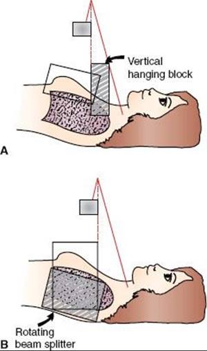
FIGURE 56.43. Diagrams showing several relationships between internal mammary and tangential fields. A: A significant cold region exists if the internal mammary (IM) tangential matchline overlies a large amount of breast tissue. B: The cold area may be negligible if the breast tissue beneath the matchline is thin. C: The lack of a separate IM field can result in irradiation of an excessive volume of lung, particularly in large-chested patients. (From Bedwinek JM. Treatment of stage I and II adenocarcinoma of the breast by tumor excision and irradiation. Int J Radiat Oncol Biol Phys 1981;7:1553, with permission from Elsevier.)
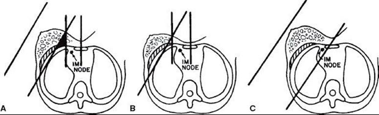
Matching the Tangential Fields with the Internal Mammary Field
When an internal mammary field is required, the match between it and the medial tangential field can be a problem if there is a significant amount of breast tissue beneath the match line. In this situation, a cold spot can exist (Fig. 56.44). The effect may be negligible if the breast tissue beneath this match line is thin (see Fig. 56.44B), or it can be avoided by including the internal mammary nodes in the tangential field, as described above (see Fig. 56.44C). Woudstra and van der Werf738 described a technique using an oblique incidence of the internal mammary portal to match the orientation of the adjacent medial tangential portal; this results in a more homogeneous dose distribution at the junction of the two fields (Fig. 56.45). One potential advantage of the partially wide tangential field in the conservatively managed patient is that it avoids the problem of matching over breast tissue.
Irradiation Dose to the Contralateral Breast
Irradiation dose to the contralateral breast is of concern due to the potential long-term carcinogenic effect of scattered radiation. The data on risk of the contralateral breast are extensively discussed later. Although this risk appears to be minimal with modern techniques, the goal must be to expose all normal tissues not within the target volume to as low a dose as is reasonably achievable. Fraass et al.739 measured the radiation dose to the contralateral breast in 16 women treated with 6-MV photon tangential fields and performed phantom measurements. For a typical treatment of 50 Gy, the contralateral breast received 0.5 to 2 Gy. Use of tangential fields only resulted in more dose delivered to the surface of the opposite breast, whereas use of the internal mammary field in addition to the tangential portals gave more dose deeper in the breast. Use of a 2.5-cm-thick lead shield over the contralateral breast during treatment with a medial tangential field reduced the dose to 35% of its original value. Similar shields used on the lateral tangential field had no protective effect. These authors recommended that wedges be used whenever possible on the lateral tangential fields rather than on the medial to decrease the dose to the contralateral breast.
A dosimetric study demonstrated that most of the scatter dose received by the opposite breast originates in the collimator and accessories of the accelerator, and it can be significantly decreased by increasing the distance between the source and the patient’s skin.740 Therefore, an isocentric source–skin distance technique may be desirable. The use of half-field blocks (beam splitter) or, even better, independent jaws combined with tailored beam splitters or a MLC following the contour of the chest wall of the patient is very helpful in decreasing the dose to the contralateral breast.
Kelly et al.741 reviewed the dose to the contralateral breast from breast irradiation with tangential fields using four different techniques. The highest dose was delivered with the use of Cerrobend half-beam blocks (regardless of the proportion of wedge used). Remaining techniques gave similar dose ranges, with the lowest total dose produced by the asymmetric jaw with no medial wedge.
The clinical significance of this inadvertent radiation dose to the opposite breast is uncertain, as various investigators have shown no increased risk of contralateral breast malignancy after treatment of the original breast by radiation therapy.23,93,678,742
Three-Dimensional Conformal or Intensity-Modulated Radiation Therapy
Standard opposed tangential fields with appropriate use of wedges to optimize dose homogeneity remains the most commonly employed method for delivery of whole-breast irradiation. A number of publications have explored the potential advantages of three-dimensional conformal radiation therapy (3D-CRT) or intensity-modulated radiation therapy (IMRT) to treat patients with breast cancer. Theoretically, 3D-CRT involves a reduction in the volume of normal tissues receiving a high dose, with an increase in dose to the target volume that includes the tumor and a limited amount of normal tissue. IMRT potentially can further improve the dose distribution between the target and nontarget tissue, but may also increase the volume of tissue exposed to lower doses of radiation. As suggested in a review by Hall and Wuu,628 this may increase the risk of second malignanciesthe potential gains and limitations of advanced planning techniques must be weighed carefully.
FIGURE 56.44. A and B: Monoisocentric matching technique. Single isocenter is set at the match between the supraclavicular and tangential fields. The inferior portion of the beam is blocked for the supraclavicular treatment and the superior blocked for the tangential field, with no movement of the isocenter, resulting in an ideal match. Blocks are drawn as indicated to shield lung and heart. The field should be viewed clinically to ensure that the blocks drawn to shield the heart and lungs to not block target tissue on the breast–chest wall. Projection of fields onto patient surface demonstrates perfect match of the supraclavicular and tangential fields.

Solin et al.743 devised 38 3D treatment plans in two patients using multiple CT scan sections and compared various dose distributions. Breast inhomogeneity doses ranged from 5% to 10%. Cobalt-60 produced greater inhomogeneities than 6-MV photons, with minimal improvement in tumor dose coverage. In contrast, 15-MV photons had significantly worse tumor coverage at shallow depths, although there was a slight reduction in hot spots. These authors were unable to identify any beam arrangement that improved dose distributions compared with standard tangential fields.
Vicini et al.744 reported on 281 patients treated with whole-breast IMRT using multiple static MLC segments. Figure 56.46 shows a representative dose distribution. The median volume of breast receiving 105% of the prescribed dose was 11% (range, 0% to 67.6%). The median breast volume receiving 110% of the prescribed dose was 0% (range, 0% to 39%), and the median breast volume receiving 115% of the prescribed dose was also 0%. Only three (1%) experienced grade III toxicity. The cosmetic results at 12 months (95 patients analyzable) were rated as excellent or good in 94 patients (99%). No skin telangiectasias, significant fibrosis, or persistent breast pain was noted. These authors concluded that the use of intensity modulation with a static MLC technique for tangential whole-breast RT is an efficient method for achieving a uniform and standardized dose throughout the whole breast, and widespread implementation of this technology can be achieved with minimal imposition on clinic resources and time constraints. As demonstrated in Figure 56.47, Dunst et al.745 compared optimized IMRT with 3D-CRT and reported a small reduction in does to the heart, lungs, and contralateral breast with IMRT.
In a trial of patients undergoing breast-conserving therapy, 358 patients undergoing whole-breast irradiation were randomized to radiation to the breast with standard wedges or with IMRT. Those randomized to IMRT experienced significantly lower degrees of moist desquamation due to the improved homogeneity with the IMRT techniques employed.746,747 In another randomized trial of IMRT in breast cancer, 306 women were randomized to standard treatment with tangential fields with wedge compensators compared with IMRT. The standard control patients were 1.7 times more likely to have a change in photographic appearance compared with those in the IMRT arm.748Further follow-up and additional studies relating to the technical delivery of radiation therapy in early-stage breast cancer will evolve rapidly in the next several years due to the rapid advances in technology.
It is important to recognize that the term intensity-modulated radiation therapy has been used in various ways to describe breast cancer treatment. In some studies, IMRT is described as a method of 3D dose compensation without a change in the gantry angles of predesigned tangential fields. In such instances, dose distribution has been improved but the fields are not more conformal. Accordingly, low dose to other organs is not an issue. For others, IMRT attempts to improve conformality of the high-dose region by using multiple field angles that increase the volume of normal tissues that receive low radiation doses.
FIGURE 56.45. A: An obliquely incident electron beam matched to the usual tangential beams. B: Isodose presentation of optimal matching of an obliquely incident electron beam to the tangential beams. The target volume is enclosed by the 90% isodose line (= 40.5 Gy). Electron beam, 16 MeV; photon beam, 6 MV. (From Woudstra E, van der Werf H. Obliquely incident electron beams for irradiation of the internal mammary lymph nodes. Radiother Oncol 1987;10:209–215, with permission from Elsevier.)

ACCELERATED PARTIAL BREAST IRRADIATION
The current standard of care for women with invasive breast cancer remains whole-breast irradiation following breast-conserving surgery.749 With the notable exception of selected elderly women, omission of radiation therapy has now been proven in numerous randomized trials and meta-analysis to compromise local control and to a lesser extent breast cancer–related mortality. For some women, the 6-week course of daily radiation with its associated time and travel issues is not feasible. In response to this, a wide variety of accelerated forms of treatment have been developed and have been proven safe and effective in short-term studies.663 These approaches include multicatheter interstitial implants placed around the excision cavity, a single balloon catheter that can be after loaded with a central radiation source (MammoSite) that is placed into the excision cavity, external-beam conformal partial breast irradiation, and intraoperative single-dose irradiation. Although these techniques vary considerably, they share the common strategy of delivering the radiation to a smaller volume of breast tissue around the lumpectomy site, using fewer larger fractions delivered over a shorter time. The rationale behind this approach is that the majority of breast relapses occur at or near the lumpectomy site. Pathological studies from mastectomy specimens have demonstrated a lower probability of subclinical microscopic disease with increasing distance from the primary tumor.102,103,222,298,391,663,750–752 Although the early results clearly demonstrate the feasibility and acceptable toxicity of accelerated partial breast irradiation, this approach has not yet been demonstrated in a randomized trial to be equivalent to whole-breast irradiation. There are several randomized ongoing trials that will attempt to answer the question of whether this approach is equivalent to whole-breast irradiation for selected patients. The current NSABP-39/RTOG-0413 is well under way with high enrollment.753 Although the mature results of this trial will not be available for several years, it should help to identify appropriate patients for this approach. Ongoing randomized trials investigating partial breast irradiation are summarized in Table 56.38. The NSABP-RTOG trial will randomize 4,300 women with early-stage breast cancer (including DCIS) to whole-breast versus accelerated partial breast irradiation. This trial allows for any one of the three techniques of partial breast irradiation, namely, interstitial brachytherapy, MammoSite, or 3D-CRT.663 The techniques are selected based on physician or patient preference prior to randomization. Fraction size is slightly higher for the external-beam conformal (3.85 Gy vs. 3.4 Gy) but all three employ twice daily radiation for a total of 10 treatments for a total dose of 38.5 or 34 Gy, respectively, over 5 days. Each of the various techniques of partial breast irradiation is discussed below.
FIGURE 56.46. A: Beam intensity profile over the breast (lighter) and the internal mammary nodes (darker) to minimize dose to the heart. (A from Cho BCJ, Hurkmans CW, Damen EMF, et al. Intensity modulated versus non-intensity modulated radiotherapy in the treatment of the left breast and upper internal mammary lymph node chain: a comparative planning study. Radiother Oncol 2002;62:127–136, with permission from Elsevier.) B: Dose distribution to the breast with intensity modulated radiation therapy. C: Dose distribution to the breast with three-dimensional conformal radiation therapy. (B and C from Keall PJ, Arnfield MR, Arthur DW, et al. An IMRT technique to reduce the heart and lung dose for early stage breast cancer. Int J Radiat Oncol Biol Phys 2001;51(Suppl 1):247(abstr), with permission from Elsevier.)

FIGURE 56.47. Partial breast irradiation demonstrating the MammoSite breast brachytherapy device. (From Arthur DW, Vicini FA. Accelerated partial breast irradiation as a part of breast conservation therapy. J Clin Oncol 2005;23:1726–1735. Reused with permission. © 2012 American Society of Clinical Oncology. All rights reserved.)
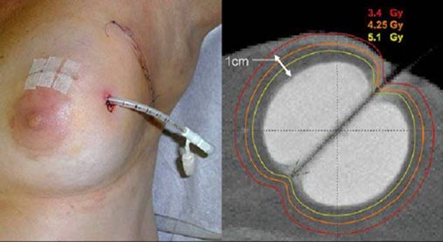
FIGURE 56.48. Partial breast irradiation demonstrating the with multiplane interstitial implant technique. (From Arthur DW, Vicini FA. Accelerated partial breast irradiation as a part of breast conservation therapy. J Clin Oncol 2005;23:1726–1735. Reused with permission. © 2012 American Society of Clinical Oncology. All rights reserved.)
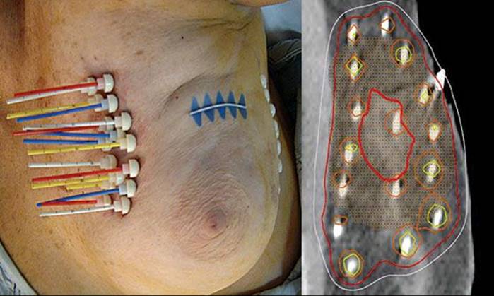
TABLE 56.38 RANDOMIZED TRIALS OF ACCELERATED PARTIAL BREAST IRRADIATION

Multicatheter Interstitial Techniques
Experience is greatest with the multicatherter interstitial technique, as it was initially developed (and is still employed) as a boost technique following whole-breast irradiation.754–756 As demonstrated in Figure 56.48, multiple catheters are generally position at 1.0- to 1.5-cm intervals, with the total number of catheters and planes employed dependent on the size, extent, and shape of the target. With refinements in image-guided techniques, this approach is generally quite adaptable to most cavities and locations within the breast. Due to its complexity, user dependence, and logistics, however, the use of this technique has been less widely embraced, compared with the MammoSite (described below) and external-beam techniques.
Kuske et al.757 recently reported the results of RTOG-95-17, a phase I and II study employing APBI using multiplane interstitial catheters in 99 women with early-stage breast cancer. The inclusion criteria for this study included invasive nonlobular tumors ≤3 cm after lumpectomy with negative surgical margins and axillary dissection with zero to three positive axillary nodes without extracapsular extension. The patients were treated with either low-dose rate (LDR) APBI (45 Gy in 3.5 to 5 days) or high-dose rate (HDR) APBI (34 Gy in 10 twice-daily fractions within 5 days). Chemotherapy and/or tamoxifen were administered at the discretion of the treating physicians. Of the 99 women, 33 were treated with LDR and 66 with HDR APBI. Of the 66 patients treated with HDR APBI, 2 (3%) had grade 3 or 4 toxicity. Of the 33 patients treated with LDR, 3 (9%) had grade 3 or 4 toxicity during brachytherapy. No patient experienced late grade 4 toxicity; the rate of grade 3 toxicity was 18% for the LDR and 4% for the HDR groups.
Vicini et al.227 reported on 133 cases of early-stage breast cancer managed with lumpectomy and axillary lymph node dissection followed by interstitial implant alone (99 cases using LDR and 34 with HDR implant) to the tumor bed, matched to a control group treated with external beam from the same institution. The number of catheters per patient ranged from 11 to 18 (median, 16). Tumor size ranged from 0.1 to 3 cm (median, 1.1 cm), with margins of excision >2 mm. Patients treated with LDR implants received 50 Gy over 96 hours as an inpatient procedure and those treated with HDR implants received 32 Gy in 8 fractions over 4 days (twice daily) as an outpatient procedure. The median follow-up for the external-beam radiation therapy group was 5.7 years versus 3.2 years for brachytherapy. No local or regional failures have been detected and only one patient failed distantly in the HDR group. No significant adverse sequelae were noted, and cosmetic results were judged to be good or excellent in 98% of patients. No statistically significant differences were noted in the 5-year actuarial rates of ipsilateral breast (3% vs. 0%; P =.17) or locoregional failure (4% vs. 0%; P = .37) between patients treated with external-beam radiation therapy and those treated with brachytherapy alone. In a recent update of this study with 12 years follow-up, there was no difference in the rate of local recurrence (3.8% vs. 5.0%; P = .40), regional recurrence (0% vs. 1.1%; P = .15), disease-free survival (87% vs. 91%; P = .30), cause-specific survival (93% vs. 95%; P = .28), or overall survival (78% vs. 71%; P = .06) between the WBI and APBI groups, respectively.758
Arthur et al.759 used HDR brachytherapy (34 Gy in 10 fractions twice a day over 5 days) in 26 patients or LDR (45 Gy given at a dose rate of 0.45 to 0.50 Gy per hour) in 18 patients. After a median follow-up of 31 months (range, 11 to 61 months), all patients remain locally controlled. Among patients receiving doxorubicin after brachytherapy, at a median follow-up of 12 months, recall reactions involving the skin overlying the implant site were observed in 42% of patients (6 of 14). On multivariate analysis, a recall reaction (P = .0007) and LDR brachytherapy (P = .04) were significant predictors of fibrosis and telangiectasis.
Wazer et al.760 reported the results of APBI using high-dose rate interstitial brachytherapy in a phase I or II single multi-institutional study in 33 women with early-stage breast cancer. Eligible patients included those with T1, T2, N0, N1 (≤3 nodes positive), and M0 tumors of nonlobular histologic features with negative surgical margins, no extracapsular lymph node extension, and a negative postexcision mammogram. High-activity 192Ir (3 to 10 Ci) was used to deliver 3.4 Gy per fraction, 2 fractions per day, for 5 consecutive days, to a total dose of 34 Gy to the target volume. The mean tumor size was 1.3 cm, and 55% had an extensive intraductal component. Three patients had positive axillary nodes. The RTOG late radiation morbidity scoring scheme was applied. Clinically evident fat necrosis occurred in eight patients at a median of 7.5 months after HDR brachytherapy completion. The only variables significantly associated with grade 3 or 4 toxicity were the number of source dwell positions and the volume of tissue encompassed by the prescription isodose shell. The global cosmetic scores after a minimum of 18 months’ follow-up were 0 cases with poor, 4 with fair, 5 with good, and 24 with excellent scores. One case of ipsilateral breast tumor recurrence was diagnosed.
MammoSite
MammoSite as an alternative method of delivering accelerated partial breast irradiation has been widely embraced due to its simplicity and less dependence on user experience.351,663,761,762 The technique employs a single balloon catheter introduced into the lumpectomy site either at the time of lumpectomy or percutaneously after the procedure. In the current NSABP/RTOG clinical trial, patients cannot be randomized until after the lumpectomy procedure when final margins and nodal status are known, and hence the device must be placed after the lumpectomy procedure. As shown in Figure 56.48, the catheter is located centrally within a distal balloon, which is inflated once the catheter is placed in the lumpectomy cavity. Adequacy of placement requires symmetry of the balloon, conformance of the balloon surface to the lumpectomy cavity, and a minimum distance between the surface of the balloon and skin of >5 mm (ideally >7 mm). Treatment is delivered via a high-dose rate remote after-loading system to a circumferential 1-cm distance from the balloon surface. This technique is one of the three methods employed in the ongoing randomized trial, with a dose prescription of 3.4 Gy delivered at 1 cm twice daily to a total dose of 34 Gy over 5 days.
Although early experiences with this technique are promising, the results of the ongoing randomized trial will help to identify suitable patients. The most extensive experience with this technique has been reported by the American Society of Breast Surgeons MammoSite Registry Trial, which included 1,419 patients treated in 87 institutions.351,763,764 This was a nonrandomized single-arm registration trial in which data were collected prospectively on clinical use of the MammoSite breast brachytherapy catheter for delivering breast irradiation. They reported on 1,237 patients (87% of enrolled patients) who received APBI (34 Gy to 1.0 cm in 10 fractions; 91% of the patients with invasive carcinoma (977 of 1,068 patients) had negative lymph node status, and 99% of all patients had negative margins. The median patient age was 65 years. Five hundred fifty-four catheters (45%) were placed with an open cavity at the time of lumpectomy, and 683 catheters (55%) were placed after lumpectomy. Skin spacing ranged from 2 to 75 mm (median, 10 mm). At 5 years, 37 cases (2.6%) developed an IBTR, for a 5-year actuarial rate of 3.80% (3.86% for inflammatory breast cancer [IBC] and 3.39% for DCIS). Negative ER status (P = .0011) was the only clinical, pathologic, or treatment-related variable associated with IBTR for patients with IBC and young age (<50 years; P =.0096) and positive margin status (P = .0126) in those with DCIS. The percentage of breasts with good or excellent cosmetic results at 60 months (n = 371) was 90.6%.The authors concluded that treatment efficacy, cosmesis, and toxicity 5 years after treatment with APBI using the MammoSite device are good and similar to those reported with other forms of APBI with similar follow-up.
External-Beam Conformal Radiation
Although external-beam conformal radiation has been developed only recently, it is the one that is most widely employed in the ongoing randomized trial.217,765,766 Recent data suggest that over 70% of patients in the randomized trial are opting for the 3D-CRT. Its widespread acceptance is likely because it is totally noninvasive and delivers a homogenous dose distribution. Although the ongoing trial mandates supine position, some authors have advocated prone accelerated breast irradiation.217,765,766
Three-dimensional-CRT, as shown in Figure 56.49, generally employs multiple conformal fields, although plans as simple as two opposing small conformal fields may be adequate. Challenges with this technique include daily positioning of the target, movement with breathing, and delivery of higher doses to surrounding normal breast tissue than with the brachytherapy. Nonetheless this approach has been widely embraced and has been shown to be reproducible. In the phase I and II RTOG-0319 trial of external-beam conformal radiation, Vicini et al.766 examined the use of 3D-CRT to deliver accelerated partial breast irradiation. Reproducibility, as measured by technical feasibility, was the primary end point. This study was designed such that if fewer than 5 cases in the first 42 patients evaluable were scored as unacceptable, the treatment would be considered reproducible. Patients received 38.5 Gy in 3.85 Gy per fraction delivered twice daily. The clinical target volume included the lumpectomy cavity plus a 10- to 15-mm margin bounded by 5 mm within the skin surface and the lung–chest wall interface. The planning target volume included the clinical target volume plus a 10-mm margin. A total of 58 patients were enrolled on this study over an 8-month period, 5 of whom were ineligible or did not receive protocol treatment. There were 4 cases with major variations and a total of 32 cases with minor variations in treatment plans. Based on this analysis, the authors concluded that accelerated partial breast irradiation using 3D conformal external-beam radiation therapy was technically feasible and reproducible in a multi-institutional trial using exceptionally strict dosimetric criteria. An update of this study found that the ipsilateral breast failure rate was 6%, in-field failure rate was 2%, and contralateral breast failure rate was 0%.767 Only two (4%) grade 3 toxicities were observed. There have, however, been some reports of increased toxicity with external-beam radiation using the fractionation schedule of 3.85 Gy per fraction, twice daily for 10 fractions.768,769 Longer follow-up and detailed outcomes from the ongoing NSABP/RTOG randomized trial will help to shed further light on this issue.
Again, the ongoing randomized trial will help to further define acceptability and reproducibility of 3D conformal external-beam radiation as an option for women with early-stage invasive breast cancer.
FIGURE 56.49. A and B: Partial breast irradiation demonstrating the external-beam conformal radiation technique.
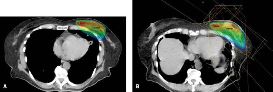
FIGURE 56.50. A: Linear electron beam accelerator in operating room during intraoperative radiation therapy. B: Proper placement of applicator in the breast. C: Before intraoperative radiation therapy delivery, an aluminum-lead disc (4 mm Al and 5 mm Pb thick) is placed between the deep face of residual breast and pectoralis muscle. (From Veronesi U, Oreechia R, Luini A, et al. A preliminary report on intraoperative radiotherapy (IORT) in limited-stage breast cancers that are conservatively treated. Eur J Cancer 2001;37:2178–2183, with permission.)

FIGURE 56.51. Probability of radiation pneumonitis versus dose. The relative lung volumes are 100%, 66%, and 33%. The curve parameters are D50 = 30 Gy, γ = 1.01, s = 0.01. The curve covers the probability range up to 100% (A), and up to 30% (i.e., within the interval of the clinical data) (B). (From Gagliardi G, Bjohle J, Lax I, et al. Radiation pneumonitis after breast cancer irradiation: analysis of the complication probability using the relative seriality model. Int J Radiat Oncol Biol Phys 2000;46:373–381, with permission from Elsevier.)

Intraoperative Accelerated Partial Breast Irradiation
APBI has been most widely employed outside of the United States.770,771 The radiation is delivered in a single intraoperative dose to the lumpectomy site at the time of surgery, using intraoperative electrons or intraoperative photons (Fig. 56.50). Vaidya et al.772 describe a preliminary report using a 50 kV spherical source to deliver a dose of 20 Gy at a depth of 1 cm, with acceptable toxicity. This group recently published the results of a phase III study that randomized over 2,200 women to either targeted intraoperative RT (IORT) or whole-breast irradiation.773 At 4 years, there were six local recurrences in the IORT cohort and five in the whole-breast irradiation cohort. The 4-year estimate of local recurrence in the conserved breast was 1.2% and 0.95% in the IORT and whole-breast irradiation cohorts, respectively. There were no differences in complications between the two groups.
Veronesi et al.770 developed an intraoperative radiation therapy (IORT) technique for a breast quadrant after the removal of the primary carcinoma using a mobile linear accelerator with a robotic arm to deliver electron beams with energies from 3 to 9 MeV. Through a Perspex applicator, the radiation is delivered directly to the mammary gland, and to spare the skin from radiation, the skin margins are stretched out of the radiation field (Fig. 56.51A,B). To protect the thoracic wall, an aluminum–lead disc is placed between the gland and the pectoralis muscle (see Fig. 56.52C). Different dose levels were tested from 10 to 21 Gy without important side effects. They estimated that a single fraction of 21 Gy is equivalent to 60 Gy delivered in 30 fractions at 2 Gy per fraction. Seventeen patients received an IORT dose of 10 to 15 Gy as a boost to external radiation therapy, whereas 86 patients received 17, 19, or 21 Gy intraoperatively as their whole treatment. The follow-up time of the 101 patients ranged from 1 to 17 months (mean, 8 months). The IORT treatment was very well accepted by all patients. The authors believe that single-dose IORT after breast resection for small mammary carcinomas may be an excellent alternative to the traditional postoperative radiation therapy. Based on these data the European Institute of Oncology has conducted a randomized trial comparing this option to whole-breast irradiation for selected patients, and results of this trial are eagerly awaited.771
In 2009, a task force of experts in the field of breast cancer developed a consensus statement on behalf of ASTRO on the use of APBI. They reported their recommendations on the suitability of patients receiving APBI outside the context of a clinical trial and divided patients into three categories: suitable, cautionary, and unsuitable (Table 56.39).774,775 In general, patients ≥60 years, with node-negative, invasive ductal tumors ≤2 cm, with negative margins, ER positivity, and no lymphovascular space involvement were deemed suitable for use of APBI off trial. The task force hoped their recommendations would provide guidance regarding the use of APBI outside a clinical trial and serve as a framework to promote additional research into the optimal role of APBI in the treatment of breast cancer.
TABLE 56.39 ASTRO CONSENSUS GUIDELINES FOR SUITABILITY OF PATIENTS TO RECEIVE ACCELERTATED PARTIAL BREAST IRRADIATION OFF TRIAL
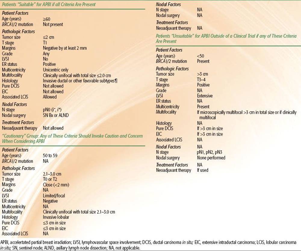
COSMETIC OUTCOMES AND SEQUELAE
Cosmesis
Surgical, radiotherapeutic, chemotherapeutic, and host factors may influence cosmetic outcome.352 Surgical factors to be considered include extent of surgical resection, re-excision, orientation and length of the scar, closure or not of the tylectomy cavity, separate or continuous axilla–tylectomy scars, extent of the axillary dissection, and whether an ellipse of skin over the tumor was removed. Radiation therapy factors are doses to the whole breast with tangential portals, homogeneity of dose throughout the breast (use of wedge or compensating filters), use of bolus, fractionation, overall duration of therapy including breaks, type and dose of boost, beam energy, and volume treated (whether peripheral lymphatic irradiation is administered). Chemotherapy issues include cytotoxic agents used, timing and sequence relative to radiation therapy, and doses and combinations of drugs. Host factors include size and shape of the breast, age, race, compliance with care and hygiene, concurrent medical illnesses (e.g., hypertension, diabetes, CVD), and intrinsic sensitivity to radiation.
Different methods have been used to evaluate breast cosmesis after breast-conservation therapy. Some are flawed because they do not establish strict guidelines or criteria for objectively judging cosmetic outcome. Pezner et al.776used scales and standard procedures for obtaining color slides to assess the cosmetic results of breast-conservation therapy and other scales designed by various investigators were given to patients for comparison. The study demonstrated that observer-based consensus of cosmetic results is difficult to obtain with two commonly used scales, but by changing the scale gradations from four to two (zero to one vs. two to three satisfactory results), consensus exceeding 85% of observers can be obtained.
FIGURE 56.52. A through D: Photographs of patients showing excellent cosmetic results obtained with conservation surgery and irradiation for patients with T1 and T2 carcinomas of the breast. The patient in (C) has minimal telangiectasia in the area treated with a boost (upper region of left breast).

A commonly employed simple scale, developed by the Harvard group, employs a 4-point scale: excellent, good, fair, and poor.777 At Washington University, questionnaires were completed by 458 patients and their radiation oncologists at regular 6-month intervals after treatment. Cosmetic outcome analysis of these patients was done for clinical and treatment-related factors.352 Approximately 80% of patients had excellent or good cosmesis (Fig. 56.52). Clinical factors at presentation were analyzed by age, menopausal status, race, and tumor-related parameters of size, palpable status, and location. Patients older than 60 years of age had lower excellent cosmetic scores compared with patients 60 years of age or younger. Tumor size significantly influenced cosmetic outcome, most likely related to the volume of breast removed and perhaps boost dose. Cosmetic outcome by race indicated 40% of whites had an excellent cosmetic rating, compared with only 18% for African Americans. Thirty percent of African American patients received concurrent chemotherapy or hormonal therapy with irradiation, compared with 23% of white patients. Of African American patients, 14% were obese, 17% were hypertensive, 25% had both obesity and hypertension, and 4% had diabetes.
Poorer cosmetic outcomes in African American women have also been reported by Pierce et al.490 and Tuomokuomo and Haffty.301 In the study by Tuomokomo and Haffty,301 a detailed cosmetic analysis was performed on a subset of 20 African American patients and 20 white patients from the Yale database. The two groups were intentionally matched by age, follow-up, adjuvant therapy, and breast size and were asked to participate in a detailed cosmetic evaluation. With respect to overall cosmetic outcome and all specific cosmetic measures (edema, fibrosis, and pigmentation), African American patients fared more poorly than white patients. Overall cosmesis was good to excellent in 55% of African Americans, compared with 90% of whites.
Cosmesis may be affected by multiple breast and axillary surgical factors.352 The type of breast surgery is important, with patients undergoing excisional biopsy having the highest rate of excellent cosmesis (56%) compared with wide excision (35%) or quadrantectomy (13%; P = .0001). Scar orientation compliance with NSABP guidelines was a significant factor, with a 44% excellent cosmetic rating compared with 27% for patients with noncompliant scar orientations (P = .0034). Re-excision of the primary site also resulted in a lower rate of excellent cosmesis (P = .0002). Breast tissue resection of >100 cm3 was associated with lower rates of excellent or good cosmesis, independent of breast size (P = .0001). Similarly, a resected skin area of >20 cm2 was correlated with a lower excellent cosmetic result (P = .045). Extent of axillary surgery did not significantly affect breast cosmesis.
Radiation factors affecting cosmesis included treatment volume (tangential breast fields only vs. three fields or more; P = .034), whole-breast dose >50 Gy (P = .024), total dose to the tumor site >65 Gy (P = .06), and optimum dose distribution created with use of compensating filters. Daily fraction size of 1.8 Gy versus 2 Gy, boost versus no boost, type of boost (brachytherapy vs. electrons), total irradiation dose, and use of bolus were not significant factors. Vrieling et al.486 published a report of the randomized EORTC trial in which 5,318 women with early-stage breast cancer after tumorectomy were randomized to a boost of 16 Gy to the tumor bed or no further treatment. Patients with microscopically incomplete excision were randomized to receive a boost of 10 Gy or 25 or 26 Gy with interstitial implant or external-beam radiation. Cosmetic results at 3 years were assessed in 731 women (364 with boost, 367 without boost) using digitizer measurements, and displacement of the nipple cosmesis in the boost group was excellent in 33%, good in 38%, fair in 26%, and poor in 3%. In the no-boost group, the results were 42%, 44%, 13%, and 1%, respectively. The position of the nipple was the only moderately representative parameter of the overall cosmetic outcome. Other measurements had no significant correlation with cosmesis. A global assessment of the appearance of the breast was thought to be a reliable method to assess cosmetic results. Factors associated with worse cosmesis were inferior tumor location, large excision volume, presence of postoperative complications, and radiation therapy boost.
TABLE 56.40 IMPACT OF ADJUVANT CHEMOTHERAPY ON COSMESIS IN BREAST CONSERVATION THERAPY

Impact of Adjuvant Chemotherapy on Cosmesis
Adjuvant chemotherapy may have a deleterious influence on excellent to good cosmetic results (Table 56.40).328,352,777–779 In several studies, the main effect was a switch from “excellent” results to the “good” category. In particular, concomitant administration of chemotherapy and irradiation appears to have a more pronounced affect on cosmesis. The majority of studies using concurrent chemotherapy, however, employed agents that are no longer routinely employed.
Rose et al.,777 reported on the Harvard cosmesis data and found that 68% of women not receiving chemotherapy had an excellent result at 3 years, compared with 37% who did receive chemotherapy. Conversely, 9% of patients who did not receive chemotherapy were judged to have fair or poor cosmetic results, compared with 24% of those who received chemotherapy. These differences were mostly the result of an increase in breast retraction and, to a lesser extent, development of telangiectasia.
Taylor et al.352 also reported impaired cosmetic outcome with concurrent administration of chemoirradiation. Excellent cosmetic outcome was observed in 43% of patients receiving sequential chemotherapy, in 25% receiving concomitant chemoirradiation, and in 41% receiving no adjuvant therapy (P = .02). The specific effect of methotrexate on cosmetic outcome was evaluated, and the proportion of excellent cosmetic outcomes with methotrexate omitted was 41% versus 16% with methotrexate included. Good results were obtained in 23% versus 58%, fair results in 23% versus 26%, and poor results in 12% versus 0%, respectively (P = .14). Similarly, studies by Markiewicz et al.560,779 and Danoff et al.780 report no compromise of cosmesis in patients receiving concurrent chemoradiation if methotrexate was held during the radiation.
In a randomized trial of concurrent versus sequential radiation therapy, using fluorouracil, Cytoxan, and mitoxantrone, Rouesse et al.561 also reported comparable and acceptable cosmetic outcomes, whether patients were treated with sequential or concurrent chemotherapy.
Breast Cosmetic Surgery After Irradiation
Breast deformities after conservation therapy may represent difficult reconstructive problems.781 Correction of a locally damaged breast is a surgical challenge that can result in a fully restored breast if selection of the surgical procedure is properly carried out. In 37 patients who underwent correction of deformities after breast-conservation surgery, which included simple submuscular placement of traditional or expandable implants, breast reshaping, transposition of a latissimus dorsi muscle or musculocutaneous flap, transverse rectus abdominis muscle flap, and reverse abdominoplasty, aesthetic outcome was judged to be good or excellent in 78% of patients.
When partial mastectomy, a term that encompasses a diversity of excisional techniques, follows radiation therapy, breast defects characterized by parenchymal loss, nipple–areola complex distortion, and cutaneous abnormalities can occur. Slavin et al.782 reported on eight patients who had reconstructive correction of an irradiated partial mastectomy deformity. Mammograms were obtained before and after the myocutaneous flap procedure. Six patients had reconstructions with latissimus dorsi flaps and two with rectus flaps. No patient underwent reconstruction sooner than 1 year after completion of radiation therapy for the entire group; a mean of 2.6 years elapsed from completion of radiation therapy to flap reconstruction of the breast. An aesthetic improvement of the partial mastectomy deformity was achieved in all eight patients. Complications consisted only of seroma formation in two patients after latissimus flap reconstruction. Mammographic evaluation revealed degeneration of the soft tissues of both types of flaps, a change that occurs as early as 6 months after operation and appears as a radiolucent area.
In a review of the MD Anderson Cancer Center experience, Kronowitz et al.783 evaluated results of 69 patients who underwent repair of a partial mastectomy defect after radiation. They concluded that immediate repair of partial mastectomy defects with local tissues results in a lower risk of complications and better aesthetic outcomes than immediate repair of partial mastectomy defects with a latissimus dorsi flap.
Follow-Up of Patients Treated with Breast-Conservation Surgery and Irradiation
It is important to closely monitor patients treated with conservation surgery and irradiation because early detection of a local recurrence may allow for another wide local excision or a total mastectomy, without significantly compromising the overall survival of the patient.103,222,348,750,784–786 Although the optimal interval for follow-up mammography has not been determined, a postradiation bilateral diagnostic mammogram should be obtained within the first year following radiation therapy.156,787–789
A careful history and physical examination are indicated every 3 to 6 months for 3 years and every 6 months for the following 2 years and annually thereafter. In patients who underwent breast-conservation therapy, a diagnostic mammogram every 6 to 12 months for the first 2 years and yearly thereafter is sufficient unless the radiologist recommends more frequent examinations.156,788,789 Monthly breast self-examination should be emphasized to every patient, including demonstration of the examination in the upright and supine positions. At least yearly evaluation is mandatory even 10 years after therapy because of the possibility of late breast relapses and occasional distant metastases. According to the American Society of Clinical Oncology’s surveillance guidelines, intensive follow-up should be limited to high-risk patients with breast cancer, especially those who enter randomized clinical trials.790
If there is strong evidence of suspect microcalcifications, masses, or architectural distortions of the breast after conservation surgery and irradiation, a biopsy should be obtained to rule out a recurrence. At times, these patients are difficult to evaluate. Posttreatment hematomas, fat necrosis, seromas, cysts, and scar tissue pose frequent dilemmas. Consultation with an experienced mammographer is essential.
Kollias et al.791 in the United Kingdom evaluated 5,102 contralateral screening mammograms performed biennially on 2,511 women aged ≤70 years after treatment for primary operable breast cancer. Sixty-five metachronous contralateral breast cancers were identified: 21 (32%) at routine clinical examination, 24 (37%) at mammography, and 20 (31%) by patients between routine follow-up appointments. The prognostic features of metachronous cancers were better than or similar to those of the first cancer in 59 of 65 (91%) cases. Mammography may have contributed to the long-term survival of 16 of 26 women in whom the histologic characteristics of the first cancer predicted a good prognosis. The cancer detection rate with mammography for these women was 6.5 per 1,000 contralateral mammograms at a cost of 3,852 pounds sterling (6,108 dollars) per cancer detected, suggesting that surveillance mammography of the contralateral breast is of value in women whose first cancer predicted a favorable prognosis.
Kramer et al.792 assessed the efficacy of contrast-enhanced dynamic MRI compared with palpation, mammography, and ultrasonography in 33 patients after breast-conservation therapy. The sensitivities for the diagnosis of local recurrences were 51% for palpation, 67% for mammography, 85% for ultrasonography, and 91% for MRI. All multicentric local recurrences were diagnosed by MRI. Mammography did not diagnose 11 local recurrences in radiodense breast, and ultrasonography was able to diagnose 8 of the 11, whereas MRI diagnosed 10 of the 11 recurrences. MRI may be useful as a complement to mammography and ultrasonography in the radiodense breast.788
It is important to define the cost–benefit ratio of follow-up procedures. In a controlled trial in Italy, 655 women were randomly assigned to be monitored with an intensive surveillance program including physician visits, bone scan, liver ultrasonography, chest radiography, and laboratory tests after initial treatment for breast cancer.793 A control group of 665 women was monitored by their physicians with physical examination and only the clinically indicated tests. Both groups received a yearly mammogram. Compliance in both protocols was more than 80%. With a median follow-up of 71 months, there was no difference in overall survival between the two groups. There were 132 deaths (20%) in the intensive surveillance group and 122 deaths (18%) in the control group. Time to detection of recurrence and parameters related to quality of life were similar in both groups. Therefore, unnecessary tests are discouraged in the follow-up of patients treated for breast cancer.
Radiographic Findings After Breast-Conservation Therapy
Dershaw156 summarized the most frequent mammographic findings: parenchymal distortion and fibrosis at the tumor excision site (secondary to surgical scar and irradiation); skin thickening, seen in 90% of patients, which may be diffuse or more prominent at the surgical excision site; and calcifications, due to fat necrosis, which are coarse and round and have radiolucent centers. Dershaw et al.789 retrospectively reviewed the mammograms of 22 patients with local tumor recurrence that were usually associated with 10 or more calcifications (17 patients, 77%). Recurrences commonly contained very suspect patterns of calcification, with linear forms in 15 cases (68%) and pleomorphic forms in 17 cases (77%). The distribution of calcifications was usually clustered (73%, 16 of 22) or segmental (18%, 4 of 22). Recurrences were characterized as obviously malignant in 77% of cases. The remainder were indeterminate, requiring biopsy. Therefore, women without worrisome mammographic patterns need not undergo breast biopsy. If the findings are stable, mammographic follow-up is sufficient. However, a change in number or characteristic pattern warrants a biopsy to rule out recurrent tumor. Mammographic findings were correlated with clinical observations in several studies.388,701,704,750,785,788,794–796 Most changes are observed in the first 12 months after therapy, with stabilization achieved at 12 to 36 months after completion of therapy. Breast edema is mammographically present in virtually all patients at completion of therapy, with a steady increase over 36 months and stabilization by 42 months.
Pretreatment and posttreatment mammograms were reviewed in 103 patients undergoing conservation therapy.797 The main posttreatment findings were a diffuse increase in parenchymal density with coarse stromal pattern, some parenchymal distortion, and thickening of the skin. Changes reached a peak at 9 months and slowly resolved over the next 2 years. At 31 to 33 months, 3 of 15 patients still had dense parenchyma and 6 had skin thickening. Sixty-nine patients had fibroadenosis. Scar with retraction in the surgical area was observed on the mammograms of 71 patients. Fat necrosis was noted in two patients. During the 3-year follow-up, recurrent cancer was noted in two treated breasts, and contralateral breast cancer developed in three women.
Orel et al.798 reported on 1,145 women with early breast cancer treated with lumpectomy and irradiation. One hundred two women with various mammographic and clinical findings later required biopsy at the treated site, and 58 had two sets of mammograms available for review (one within 3 months of the biopsy). Recurring cancer was documented in 38 (66%) of 58 patients. Thirteen (34%) of the recurrences were detected solely with mammography, and eight others were detected both mammographically and clinically. The positive predictive value for mammographic abnormalities was 72% (76% for soft tissue microcalcifications and 62% for other findings). Twenty-one recurrences (55%) were within the lumpectomy quadrant. Within the lumpectomy site, sensitivity was substantially better for physical examination (71%) than for mammography (43%). In the remaining breast outside the lumpectomy quadrant, mammography had a significantly higher sensitivity (71%) and positive predictive value (86%). The most common posttreatment findings reported by Orel et al.798,799 and Stomper et al.800 were calcifications alone (48%) or with a mass (29%), distortion of the breast parenchyma (20%), and inflammatory thickening of the breast skin.
Stomper et al.800 reported on 50 of 1,600 patients with stage I or II invasive breast cancer treated with conservation surgery and irradiation on whom biopsies were performed within 4 months of a mammogram for suspected recurrence in the irradiated breast. The tumor was suspected based on mammography in only eight patients (35%), on physical examination in nine (39%), and on both in six (26%). The most common radiographic findings were calcifications with or without a mass. Histologic evidence of recurrent cancer was found in 23 of 45 (51%) biopsy specimens. Sixty-five percent of patients had recurrences at the primary site and 22% in other sites; 13% were multifocal.
MRI is increasingly used in the evaluation of patients with equivocal mammographic findings. Viehweg et al.801 followed 207 patients with breast cancer treated with breast-conservation therapy; 40 patients were examined 0 to 12 months and 167 patients later than 12 months after radiation therapy. Suspect or indeterminate findings were suggested by clinical examination or conventional imaging in 80 studies. In 127 women, MRI was performed in breast tissue that was difficult to assess due to scarring or dense breast tissue. Recurrent carcinoma was confirmed in 27 patients by surgical biopsy. All 27 carcinomas, except for one with a slow signal increase, demonstrated early rise of signal intensity on dynamic T1-weighted, contrast-enhanced images. During the first year after therapy, the diagnostic accuracy was not improved by additional use of contrast-enhanced MRI because of strong and sometimes early and ill-circumscribed enhancement. Later than 12 months after therapy, enhancement decreased significantly and the false-positive calls could be reduced from 49 (conventional imaging) to 12 (conventional imaging plus MRI). A total of 12 of 26 recurrences and multifocality in four of five cases were diagnosed by MRI alone at this time.
Dao et al.802 evaluated 35 women with breast carcinoma treated with conservation therapy who underwent posttreatment MRI. Nine patients had recurrent tumors, and 26 had a benign fibrotic mass confirmed at biopsy. In all cases, a localized hypointense area was present on plain spin-echo T1-weighted images. In all recurrent tumors, dynamic gadolinium-enhanced T1-weighted images demonstrated early increased signal intensity of the lesion within 3 minutes after bolus injection.
Drew et al.803 also investigated MRI for screening for local recurrence after breast-conserving therapy. One hundred five patients were recruited for the study. Sixteen biopsies were performed and nine recurrences were confirmed histologically. The sensitivity for clinical examination, mammography, examination combined with mammography, and MRI alone for the detection of recurrent cancer were 89%, 67%, 100%, and 100%, respectively, and the specificity was 76%, 85%, 67%, and 93%. The authors concluded that when combined, clinical examination and mammography are as sensitive as MRI of the breast for the detection of locoregional recurrence, but MRI has greater specificity.
SEQUELAE OF IRRADIATION IN BREAST CANCER
The most frequent complications associated with conservation surgery plus irradiation are arm or breast edema, breast fibrosis, painful mastitis or myositis, pneumonitis, and rib fracture. Apical pulmonary fibrosis is occasionally noted when the regional lymph nodes are irradiated.1,110,318,352,804,805
Lymphedema and Breast Edema
Complications from axillary surgery, regardless of breast surgical procedure, have been reported by several authors. It should be emphasized that before the treatment of arm lymphedema after breast carcinoma, it is mandatory to differentiate between treatment-associated complications and tumor recurrence in the regional lymphatics.
An extensive review of the literature related to arm edema following breast surgery was conducted by Erickson et al.806 They found that arm edema is a common complication of breast cancer therapy that can result in substantial functional impairment and psychological morbidity. The risk of arm edema increases when axillary dissection and axillary radiation therapy are used. Preventive measures have not been well studied. Nonpharmacologic treatments, such as massage and exercise, have been shown to be effective therapies for lymphedema, but the effect of pharmacologic interventions remains uncertain. They conclude that as arm edema becomes more prevalent with the increasing survival of breast cancer patients, further research is needed to evaluate the efficacy of preventive strategies and therapeutic interventions.44
Maunsell et al.807 evaluated frequency of upper extremity problems from axillary surgery in 223 patients. At 3 months after surgery, 82% of patients reported at least one arm problem: swelling (24%), weakness (26%), some limitation in range of movement (32%), stiffness (40%), pain (55%), and numbness (58%). The severity of these problems changed little 15 months later. Regardless of the type of mastectomy, women who underwent axillary dissection had more problems.
Clarke et al.403 observed breast edema in approximately 20% of patients not undergoing axillary dissection, compared with 80% of those in whom this procedure was performed. The extent of the axillary dissection (medial or lateral to the tendon of the pectoralis minor) influences the incidence of breast or arm edema, with this complication being more frequent when more extensive axillary dissections are carried out (beyond level II—middle). On the other hand, Dewar et al.808 reported a greater incidence of upper limb sequelae in patients undergoing axillary surgery and irradiation (33.7%) or irradiation alone (26%) than in patients treated with axillary dissection only (7.2%). The most frequently noted complications were edema, impaired shoulder mobility, pain on movement, sensory or motor deficit, and pectoral muscle fibrosis.
Pain and discomfort after axillary lymph node dissection were significantly related to quality of life. Hack et al.,809 in 220 women with breast cancer who had undergone axillary lymph node dissection, noted that 73% had sensation of pain or discomfort or the point of maximum arm–shoulder movement was different between the affected and nonaffected side. Although more than half of the patients experienced pain-related discomfort and disability, patients in general reported a good quality of life and mental health. Younger women had significantly greater pain than older women. Patients with more than 13 lymph nodes dissected and patients receiving chemotherapy reported more pain.
Sentinel node sampling appears to be associated with a much lower degree of lymphedema.335 Sener et al.810 reported that 9 of 303 patients (3%) who underwent only sentinel lymphadenectomy had lymphedema, compared with 20 of 117 patients (17%) who underwent sentinel lymphadenectomy combined with axillary dissection (P <.0001). Among 303 patients who underwent sentinel lymphadenectomy only, lymphedema developed in 8 of 155 patients (5%) who had tumors in the upper-outer quadrant and in 1 of 148 patients (0.7%) whose tumors were in other locations. The ALMANAC randomized trial also confirms lower morbidity and improved quality of life following sentinel node biopsy compared with axillary dissection.335 Various treatment regimens have been used to treat lymphedema.811 The compression pump, along with skin care, exercise, and compression garments, is one. A second treatment is known as complex decongestive physiotherapy or complex physical therapy. Arm care, therapeutic exercises, manual lymph node drainage, and compression bandages or garments comprise this treatment regime. Decreases in lymphedema are noted if women are compliant with the prescribed treatment program.
Brorson et al.812 reported on 20 patients with arm lymphedema after breast cancer treatment who underwent liposuction combined with controlled compression therapy or controlled compression therapy alone. Liposuction combined with controlled compression therapy reduced arm edema volume by (median) 115% (range, 92% to 179%), whereas controlled compression therapy alone decreased arm edema volume by only 54% (range, 7% to 81%; P = .008).
In general the incidence of breast or arm edema after conservation therapy varies, and it is related to performance and technique of axillary dissection, whether the axillary lymph nodes were irradiated, and the dose of radiation delivered.
Skin and Breast Complications
A wide variety of symptoms may occur following radiation treatment to the conservatively treated breast. McCormick et al.318 showed that breast swelling was the most frequently noted symptom (31% of patients), followed by muscle pain (on motion), incision site pain, and general breast discomfort (approximately 20%). Rib pain was noted by 13%. Forty-eight percent of patients reported more breast discomfort in the treated breast compared with the untreated breast during sexual activity (64 sexually active patients).
In addition to host factors such as such as CVS and diabetes, underlying genetic factors may play a role in radiation complications. Iannuzzi et al.813 evaluated 46 patients with early-stage breast carcinoma who underwent limited surgery and breast irradiation. DNA was isolated from blood lymphocytes. Nine ataxia telangiectasis mutations were identified in six patients (eight novel and one rare). The median follow-up was 3.2 years (range, 1.3 to 19.3 years). All three of the patients (100%) who manifested grade 3 or 4 subcutaneous late sequelae possessed ataxia telangiectasis mutations, whereas only 3 of the 43 patients (7%) who did not have this form of severe toxicity harbored an ataxia telangiectasis mutation (P = .001).
Skin effects after postlumpectomy radiation therapy may be affected more significantly by the increase in the dose of radiation per fraction than by the total dose. Gorodetsky et al.814 studied 110 women with breast cancer who had been treated with lumpectomies and radiation therapy and normal controls using a viscoelasticity skin analyzer. With increasing age, the viscoelasticity of the skin decreased and anisotropy increased significantly. A small but significant increase in skin stiffness was noted with radiation therapy in the range of 45 to 50 Gy given in fractions of 1.8 Gy. A dose of 50 Gy given in fractions of 2.5 Gy produced a more pronounced effect.
Pseudosclerodermatosus panniculitis is an unusual variant of panniculitis seen as a complication of radiation therapy. Carrasco et al.815 described four women in whom this unusual entity developed on the anterior chest and abdominal skin after they received radiation therapy for either breast carcinoma or painful bone metastases from breast carcinoma. Histopathologically, the epidermis and dermis of the involved area showed little or no evidence of radiodermatitis. The main findings were in subcutaneous tissue and consisted of thickened sclerotic septa, composed of both thick and thin collagen bundles, and a lobular panniculitis characterized by lipophagic granulomas and scattered lymphocytes and plasma cells. This sequela should be distinguished from subcutaneous metastatic disease, cellulitis, or connective tissue diseases involving the subcutaneous fat.
Rayan et al.816 reported on a randomized clinical trial of breast-conserving surgery and tamoxifen with or without radiation therapy in women 50 years of age and older treated for stage T1 or T2, node-negative breast cancer. A companion study to assess breast pain was carried out during the past 2 years of accrual to the trial, in which 86 patients participated. Forty-one received radiation therapy and tamoxifen and 45 tamoxifen alone. The median age was 70 years. Baseline pain and quality-of-life scores were similar for the two groups. At 3 months, patients receiving radiation therapy experienced more breast pain compared with those receiving tamoxifen alone, but this did not reach statistical significance. At 3 months, the pain scores for the radiation therapy and tamoxifen and tamoxifen alone groups were 2.39 and 1.83, respectively (P = .47). At 12 months, pain scores were lower and fairly similar in both groups, with a difference of 0.20 (P = .71).
Tamoxifen has been shown to induce secretion of tumor growth factor-β, which has been implicated in pathogenesis of radiation fibrosis. Li et al.,719 in a study of 91 patients with T1 or T2 breast cancer, noted that tumor growth factor-β and the receptor–ligand complex appeared to be of clinical value in identifying patients at risk for development of postirradiation fibrosis of the breast. Wazer et al.568 showed a trend toward decreased cosmesis in patients receiving tamoxifen. In a randomized study, pulmonary fibrosis developed in 15 of 24 (63%) women treated with 36.6 Gy in 12 fractions and tamoxifen, compared with 10 of 30 (33%) receiving irradiation alone. Also, 5 of 14 (36%) women treated with 40.9 Gy in 22 fractions and tamoxifen had lung fibrosis, compared with 2 of 16 (13%) receiving irradiation alone.567 In contrast, Fowble et al.817 observed no difference in cosmetic results or complications in 154 patients who received tamoxifen in combination with breast-conservation therapy compared with 337 patients who did not receive tamoxifen. The incidences of radiation pneumonitis were 0.2% and 0.3%, respectively. The sequence of tamoxifen, given concurrently or following radiation, has not been clearly shown to correlate with complications or cosmesis.569–570,571
Markiewicz et al.779 analyzed complications in 1,053 women with stage I or II breast cancer treated with breast-conserving therapy. Of this group, 206 received chemotherapy alone, 141 had hormonal therapy alone, 94 had both, and 612 received no adjuvant therapy. The incidence of grade 4 or 5 arm edema (≥2 cm difference in arm circumference) was 2% without chemotherapy and 8% with chemotherapy (P = .00002). However, the incidence of arm edema was not affected by sequencing or type of chemotherapy; it occurred in 10% and 7% of patients with sequential or concurrent treatment, respectively, and in 8% and 18% of patients treated with CMF or CAF, respectively. The incidence of clinical pneumonitis and rib fracture was not influenced by use of chemotherapy, sequencing of drugs, or use of hormonal therapy. These authors concluded that some chemotherapy could be given concurrently with radiation therapy to the breast without significant compromise of cosmetic results or sequelae of treatment.
Hyperbaric oxygen therapy has been shown to be effective in the treatment of some late radiation sequelae. Carl et al.818 reported on 44 patients with persistent local symptoms after breast-conserving therapy. Hyperbaric oxygen therapy (100% oxygen at 240 kPa for 90-minute sessions) was administered to 32 patients for a median of 25 sessions (range, 7 to 60 sessions). The remaining 12 patients declined hyperbaric treatment and acted as control subjects. The patients given hyperbaric oxygen therapy demonstrated a significant reduction in pain, edema, and erythema scores compared with untreated control subjects (P <.001). Seven of the 32 women who were treated with hyperbaric oxygen therapy were free of symptoms after treatment, whereas all 12 patients in the control group had persistent complaints. However, hyperbaric oxygen therapy did not have a significant effect on fibrosis and telangiectasia in the irradiated breast.
Brachial Plexopathy
Brachial plexus dysfunction is a possible complication of regional nodal radiation therapy. In a review of 1,624 patients, brachial plexus sequelae were observed in 1.8% of patients.819 Pierce et al.819 found that the incidence of brachial plexopathy was significantly higher when the axillary dose was >50 Gy (P = .004). However, dose alone did not determine whether radiation damage would develop in a given patient. Treatment technique (two vs. three fields; P = .0009) and concomitant chemotherapy were also risk factors. Other investigators have found the incidence of this complication to be ≤1%.98,379 It is very important but difficult to distinguish between metastatic and radiation-induced brachial plexopathy.
Treatment for radiation brachial plexopathy consists of transdermal electrical nerve stimulation, dorsal column stimulators, neurolysis, and neurolysis with omentoplasty. Physical therapy, tricyclics, antiarrhythmics, anticonvulsives, nonsteroidal anti-inflammatory drugs, and steroids are helpful in therapy of both radiation-induced and metastatic brachial plexopathies.820
Pritchard et al.821 used hyperbaric oxygen in 34 volunteers with radiation-induced brachial plexopathy who were randomized to hyperbaric oxygen or a control group. The hyperbaric oxygen group breathed 100% oxygen for 100 minutes in a hyperbaric chamber (30 sessions over 6 weeks). The control group breathed a gas mixture equivalent to breathing 100% oxygen at surface pressure. Normalization of the warm sensory threshold was seen in two of the patients receiving hyperbaric oxygen therapy. Two cases with marked chronic arm lymphedema reported major improvement in arm volume. These authors concluded that there is no reliable evidence to support hyperbaric oxygen therapy to slow or reverse radiation-induced brachial plexopathy, although improvements in warm sensory threshold suggest a therapeutic effect improvement in long-standing arm lymphedema and justify further investigation.
Pulmonary Sequelae
Symptomatic pneumonitis is infrequent. This clinical syndrome is noted one to several months after irradiation.822 Patients present with dry cough (88%), shortness of breath (35%), or fever (53%), and on radiographic studies a pulmonary infiltrate is observed in the irradiated volume.804 The risk for development of radiation pneumonitis may be related to the volume of lung irradiated.804,823
The addition of regional nodal radiation therapy to breast irradiation significantly increase the incidence of symptomatic pneumonitis (1% without and 4% with regional node radiation therapy; P <.001). Combined axillary dissection and nodal irradiation result in a significantly higher incidence of arm edema compared with either alone (9.5% with axillary dissection, 6.1% with radiation therapy to the axilla and supraclavicular fossa, and 31% with combined modality therapy; P <.001).823
Lingos et al.804 reported on radiation pneumonitis in a retrospective review of 1,624 patients treated with conservation surgery and irradiation. Overall, pneumonitis developed in 1% of patients. No patient had late or persisting pulmonary symptoms. The incidence of radiation pneumonitis was correlated with the combined use of chemotherapy and a supraclavicular field (P = .0001). Fourteen of 17 patients who had radiation pneumonitis also had IMNs treated. When patients treated with the three-field technique received chemotherapy concurrently with irradiation, the incidence of radiation pneumonitis was 8.8% (8 of 92) compared with 1.3% (3 of 236) for those who received sequential chemotherapy and irradiation to the breast only and 0.5% (6 of 1,296) for those treated with irradiation to the breast only without chemotherapy (P = .002). In this study, the volume of lung irradiated did not correlate with the risk for development of radiation pneumonitis.
Taghian et al.,824 in 41 patients treated with radiation therapy and paclitaxel (21 concurrent, 20 sequential), also described a higher incidence of pneumonitis (14.6%) compared with control patients irradiated and not receiving chemotherapy (1.1%; P <.0001). Burstein et al.566 also recently reported that the concurrent use of weekly paclitaxel with radiation result in high rates of pneumonitis. However, an analysis of patients treated with radiation as a component of a randomized trial, in which 50% of the patients were treated with paclitaxel and 50% were treated with a nontaxane regimen, found that the rates of pneumonitis were very low and not significantly different between the two arms.825
The effect of tangential field technique on pulmonary function was reported by Lund et al.826 in 25 patients treated with conservation surgery and irradiation. Dynamic and static lung volumes, distribution of ventilation, and gas transfer were measured before irradiation and at varying intervals up to 1 year after completion of therapy. There was a small but statistically significant decrease in the forced vital capacity and forced expired volume in 1 second 3 months after irradiation (P <.05). These changes normalized within 1 year. The reduction in total lung capacity after 3 months almost achieved statistical significance (P = .06). These slight restrictive ventilatory changes are reversible and have no clinical importance.
Radiation pneumonitis was retrospectively assessed on the basis of clinical symptoms and radiologic findings using a serial organ model by Gagliardi et al.827 As demonstrated in Figure 56.52, a lung volume effect was relevant in the description of radiation pneumonitis. Lind et al.828 measured pulmonary function 5 months after radiation therapy in 144 patients with node-positive stage II breast cancer. No deterioration of pulmonary function was detected among the patients who were treated with local radiation therapy. Patients undergoing locoregional radiation therapy showed a 5% mean reduction in diffusion capacity (P <.001) and a 3% mean reduction in vital capacity (P = .001).
Cardiac Sequelae
The potential for excess cardiac morbidity associated with the use of radiation therapy in breast cancer has been extensively evaluated. It has been clearly demonstrated, based on data from randomized trials, overview, and meta-analyses, that when using older techniques, excess cardiac mortality from radiation offsets some of the benefits that radiation therapy clearly produced with respect to breast cancer mortality.70,365,829 Although the evidence from more modern trials, using techniques that minimize exposure to the normal cardiac and pulmonary structures, have reduced cardiac toxicity, the radiation oncologist must be cognizant of the potential for adverse cardiac effects of incidental irradiation, particularly in the setting of left-sided breast cancers in patients receiving other cardiotoxic therapies, including adriamycin, epirubicin, and trastuzumab.830,831
Earlier techniques of radiation therapy from large pools of randomized data have clearly been implicated in excess cardiac mortality. In an analysis of over 90,000 Swedish women, comparing left- to right-sided cancers, Darby et al.832 from the Oxford group reported excess ischemic heart disease mortality more than 10 years after initial treatment in the left-sided cancer group (HR 1.13; 95% CI, 1.03 to 1.25; P = .01). The majority of cardiovascular deaths were from earlier studies using techniques that are no longer used. However, for patients treated after 1980, although the ratio was still 1.11, the confidence intervals were much larger (0.95 to 1.29), so the hazard remains uncertain for the more modern techniques.
Gyenes et al.833 reported on the incidence of ischemic heart disease 15 to 20 years after adjuvant radiation therapy in 960 patients with breast cancer enrolled in the Stockholm Breast Cancer Trial. Of 37 long-term survivors, 20 received left-sided therapy and 17 received right-sided therapy or no therapy. Radiation therapy consisted of tangential fields for preoperative treatment and electron beam portals, which included the IMNs, for postoperative therapy (45 to 50 Gy). Evaluation consisted of echocardiography, exercise stress tests with 99mTc myocardial perfusion scan, and careful history for cardiac risk factors. Results showed that 5 of 20 (25%) patients treated with left-sided radiation had defects on 99mTc scan, compared with 0 of 17 control patients (P = .05).
Paszat et al.834 conducted a study of 25,570 cases of invasive female breast cancer that were linked to radiation therapy records from Ontario cancer centers. Postlumpectomy radiation therapy was administered to 1,555 patients on the left side and to 1,451 on the right side. Two percent of women with left-sided radiation therapy had a fatal myocardial infarction compared with 1% of women with right-sided radiation therapy (P = .02). Adjusting for age at diagnosis, the relative risk for fatal myocardial infarction with left-sided postlumpectomy radiation therapy was 2.10.
Rutqvist et al.835 followed 684 patients with breast cancer treated with breast-conserving surgery and radiation therapy using tangential photon fields (48 to 52 Gy in 4.5 to 5.5 weeks). The median follow-up was 9 years. In 88% of patients, the target volume involved the breast only; in the remaining patients, regional nodes were irradiated. A control group included 4,996 patients with breast cancer who underwent mastectomies without postoperative radiation therapy. Twelve patients (1.8%) in the irradiated group had myocardial infarctions and five patients (0.7%) died as a result of myocardial infarctions. The relative risk for a myocardial infarction between the irradiated group and the control group was 0.6, and the relative risk for death was 0.4. The study presents no evidence that the risk for myocardial infarction is increased with radiation therapy after breast-conserving surgery regardless of which side the tumor was located. However, because the number of myocardial infarctions in the study was small, there is no way to rule out the possibility of cardiac problems in patients with left breast carcinomas.
Shapiro et al.830 assessed the cardiac effects in 299 patients with breast cancer prospectively randomized to receive either 5 cycles or 10 cycles of cyclophosphamide and a doxorubicin intravenous bolus every 21 days. Of the 299 patients, 122 received radiation therapy. The risk of major cardiac events (congestive heart failure, acute myocardial infarction) was assessable in 276 patients, with a median follow-up of 6 years (range, 0.5 to 19.4 years). The estimated risk of cardiac events per 100 patient-years was significantly higher for 10 cycles than for 5 cycles of chemotherapy (1.7 vs. 0.5; P = .02). The risk of cardiac events in the 5-cycle patients, regardless of the cardiac radiation therapy dose volume, did not differ significantly from rates of cardiac events predicted for a general female population. For patients receiving 10 cycles, the incidence of cardiac events was significantly increased (RR 3.6; P <.00003) compared with the general population, particularly in groups that also received moderate- and high–dose-volume cardiac radiation therapy.
Cuzick et al.829 updated cardiac toxicity data from eight randomized trials initiated before 1975 in which radiotherapy was the randomized option and surgery was the same for both treatment arms. An initial analysis of these trials demonstrated an increased all-cause mortality rate in 10-year survivors associated with radiation, but in the update this was no longer present. The initial increase in mortality in the radiation arms was strongly influenced by the earliest trials, and more recent trials have found a nonsignificant net benefit in overall mortality associated with radiation therapy. However, an excess of cardiac deaths was apparent in both early and more recent trials (P <.001), but this was offset by a reduced number of deaths due to breast cancer, especially in more recent trials. Based on this it is clearly prudent to use techniques that minimize cardiac dose.
Although clinical evidence of cardiac morbidity has decreased with modern techniques, care should be taken to exclude heart from the tangential radiation field. In an analysis of 114 patients, Marks et al.696 assessed RT-induced left ventricular perfusion defects and whether these perfusion defects are related to changes in cardiac wall motion or alterations in ejection fraction. Patients were imaged 30 to 60 minutes after injection of 99mTc sestamibi or tetrofosmin. Post-RT perfusion scans were compared with the pre-RT studies to assess for RT-induced perfusion defects as well as functional changes in wall motion and ejection fraction. The incidence of new perfusion defects 6, 12, 18, and 24 months after RT was 27%, 29%, 38%, and 42%, respectively. New defects occurred in approximately 10% to 20% and 50% to 60% of patients with <5% and >5% of their left ventricle included within the RT fields, respectively. The rates of wall motion abnormalities in patients with and without perfusion defects were 12% to 40% versus 0% to 9%. These authors note that RT causes volume-dependent perfusion defects in approximately 40% of patients within 2 years of RT, and that these perfusion defects are associated with corresponding wall-motion abnormalities. However, additional study is necessary to determine if these defects are associated with functional consequences.
Given these findings, the authors suggest the use of a heart block if needed to reduce or eliminate cardiac irradiation. CT-based 3D treatment planning is used to design such cardiac blocks and select the optimal gantry angle to minimize the need for a heart block. There may be a small amount of breast tissue underdosed if the block overlies the medial inferior breast, but this tissue is typically <5%. In situations where the heart block may underdose the high-risk volume of the breast or chest wall, an “electron patch” can be used to treat the target tissue in the shadow of the heart block. A tangential field with a heart block is demonstrated in Figure 56.33.
Although it is clearly prudent to minimize exposure of the heart during radiation therapy, using modern techniques, the available evidence does not suggest a higher incidence of cardiac mortality in left-sided radiation therapy.
Analysis of the randomized postmastectomy Danish trials, with over 10 years of follow-up, showed no excess cardiac mortality with the use of postmastectomy radiation. Hojris et al.277 reported that the relative hazard of morbidity from ischemic heart disease among patients in the radiotherapy compared with the no-radiotherapy group was 0.86 (95% CI, 0.6 to 1.3), and that for death from ischemic heart disease was 0.84 (0.4 to 1.8). The hazard rate of morbidity from ischemic heart disease in the radiotherapy group compared with the no-radiotherapy group did not increase with time from treatment.
Patt et al.836 analyzed data from the SEER-Medicare database for women who were diagnosed with nonmetastatic breast cancer from 1986 to 1993, had known disease laterality, underwent breast surgery, and received adjuvant radiotherapy; 8,363 patients had left-sided breast cancer and 7,907 had right-sided breast cancer. With a mean follow-up of 9.5 years (range, 0 to 15 years), there were no significant differences in patients with left- versus right-sided cancers for hospitalization for ischemic heart disease (9.9% vs. 9.7%), valvular heart disease (2.9% vs. 2.8%), conduction abnormalities (9.7% vs. 9.6%), or heart failure (9.7% vs. 9.7%). The adjusted hazard ratio for left- versus right-sided breast cancer was 1.05 (95% CI, 0.94 to 1.16) for ischemic heart disease, 1.07 (95% CI, 0.89 to 1.30) for valvular heart disease, 1.07 (95% CI, 0.96 to 1.19) for conduction abnormalities, and 1.05 (95% CI, 0.95 to 1.17) for heart failure.
Similar conclusions were reached by Nixon et al.837 who reviewed 365 patients with 12-year follow-up who received irradiation to the left breast and 380 who received irradiation to the right breast as part of conservation therapy. Equivalent proportions from each group died of non–breast cancer causes (11%), including nine patients (2%) from each group who died from cardiac causes. Also, Vallis et al.,838 in a retrospective review of 2,128 women treated with lumpectomy and breast irradiation with a median follow-up of 10.2 years, noted that the incidence of myocardial infarction in the study cohort was comparable with that in an age-matched general population of women in Ontario.
The potential for cardiac vessel injury from radiation was recently explored in an elegant study by Nilsson et al.839 who analyzed radiation fields and subsequent coronary angiograms in a Swedish breast cancer cohort study. For right-sided compared with left-sided breast cancers, the odds ratio for grade 3 to 5 stenosis in arteries likely to be within the tangential radiation port was 4.38 (95% CI, 1.64 to 11.7) and was 7.22 (95% CI, 1.64 to 31.8) for grade 4 or 5 stenosis. They conclude there is an increased risk and direct link between radiation and the location of the coronary stenosis. In a related editorial Zagar and Marks840 emphasize the potential effects of radiation on the heart but point out that improved radiation techniques, including custom blocking of the heart and other techniques to manipulate the radiation dose, are likely to minimize these risks and improve the therapeutic ratio.
Collectively, these data suggest that although there may be excess cardiac morbidity using tangential fields to treat left-sided breast cancers, these effects can be minimized through careful treatment planning. It remains prudent to minimize cardiac exposure in all patients, and particularly in those receiving left-sided radiation in combination with other potentially cardiotoxic drugs.
TABLE 56.41 INCIDENCE OF CONTRALATERAL BREAST CANCER IN CARCINOMA OF THE BREAST TREATED WITH CONSERVATION SURGERY AND IRRADIATION OR MASTECTOMY

Risk of Stroke with Supraclavicular Radiation
For patients with node-positive disease undergoing supraclavicular radiation, there is a theoretical concern regarding the potential for development of accelerated carotid artery stenosis. In a study by Jagsi et al.,841 rates of stroke in 820 eligible early-stage breast cancer patients treated with radiation therapy were compared with expected rates. The relation between potential risk factors and actuarial rate of first stroke was analyzed. On multivariate analysis, only age (P <.001) and hypertension (P = .003) remained significant predictors of cardiovascular accident or transient ischemic attack. Age was the only significant predictor of cardiovascular accident alone (P <.001). This study found no significant association between supraclavicular RT and stroke after controlling for other factors. This study is in agreement with the findings of the EBCTCG who reported the causes of nonbreast cancer death in 32,800 patients treated in trials of surgery with and without RT. Although the incidence of heart disease was found to be significantly greater in those women who received RT, no significant excess mortality from RT was observed due to stroke. However, a recent nested case-control study by Nilsson et al.842 showed a significant increase in stroke (OR 1.8; 95% CI, 1.1 to 2.8) when comparing patients treated to the supraclavicular and internal mammary regions with those not treated to these regions. In addition, a study by Woodward et al.843 of the SEER database comparing 5,281 women presumably without supraclavicular radiation with negative nodes to 482 women presumably treated to the supraclavicular region with more than four nodes did not find any increased rate of hospitalization for stroke in the treated population.
Contralateral Breast Cancer and Irradiation
Although all patients with a diagnosis of breast cancer are at increased risk for developing a contralateral breast cancer, the additional risk contributed by radiation treatment appears to be minimal, particularly when one uses modern techniques and maintains a dose to the contralateral breast that is as low as is reasonably achievable. Although this issue is often a concern raised by patients, the available data using modern radiation techniques do not suggest a significant increase of risk for contralateral breast cancers in breast cancer patients who have been irradiated, in comparison to similar cohorts of breast cancer patients who have not undergone radiation.1,23,93,110,678,742,844,845,846,847Although there is some evidence suggesting a slight excess risk in women who are irradiated at a relatively young age (i.e., <45 years at diagnosis), the risk is extremely small and may be related to use of older techniques, and most experts would agree that the benefit of radiation far outweighs the risk.844 Nonetheless, it appears prudent to be aware of these potential risks and employ techniques that minimize scattered dose to the contralateral breast. As demonstrated in Table 56.41, the reported incidence of contralateral cancer in the majority of these studies of patients treated with conservative surgery and radiation do not appear to be elevated compared with those treated by mastectomy without radiation.
However, the EBCTCG overview analysis does suggest an elevated incidence of contralateral breast cancer in patients receiving radiation compared with those who did not receive radiation.89 This overview analysis of all randomized trials comparing radiation to surgery demonstrated an increased relative risk of contralateral breast cancers of 1.18 (=.002). Although the excess risk appears to be driven primarily by older trials using antiquated techniques, these data do demonstrate the potential long-term effects of radiation-related secondary cancers and highlight the need to maintain dose to the contralateral breast as low as possible.
A report by Hankey et al.,848 involving 27,175 women treated for breast cancer between 1960 and 1975, disclosed a relative risk of 1.2 to 1.4 for development of cancer in the contralateral breast in irradiated patients compared with those who did not receive irradiation. These authors, however, concluded that the data did not indicate a pattern of relative risk consistent with an increased incidence of carcinoma in the opposite breast.
Boice et al.844 evaluated the risk of second cancers associated with radiation therapy to the breast in 41,109 women with breast cancer who were registered in the Connecticut Tumor Registry between 1935 and 1982. They reviewed the records of 655 women in whom a second breast cancer developed 5 years or longer after initial treatment and compared the radiation exposure in these patients with the exposure in 1,189 matched control patients who did not have a second cancer. The average dose to the contralateral breast in women exposed to radiation was 2.82 Gy. The relative risk for development of a second breast cancer was 1.9 in the women who received radiation therapy, and among patients who survived for 10 years or longer, the relative risk was 1.33. Women <45 years of age had a relative risk of 1.59 for development of a second breast cancer, compared with 1.01 for older women. According to these authors, younger patients should be informed that, based on the results of this study, after 10 years the risk for development of a second cancer increases from 14% if they do not receive irradiation to 22% if they choose treatment involving irradiation.
On the other hand, Levitt and Mandell849 estimated the dose delivered to the contralateral breast to be between 1 and 4 Gy. Assuming that 20,000 women undergo radiation therapy after conservation surgery and using data on the risk for development of breast cancer after various doses of ionizing radiation, they concluded that fewer than one additional case of breast cancer would occur after 10 years. Storm et al.,850 in a case-controlled study of a registry-based cohort of patients with breast cancer in Denmark, also concluded there was little, if any, risk of radiation-induced breast cancer associated with exposure of adult breast tissue to low-dose irradiation.
Obedian et al.23 compared 1,029 breast cancer patients treated with conservative surgery and radiation to a cohort of 1,387 breast cancer patients who underwent surgical treatment by mastectomy and who did not receive postoperative radiation during the same time period. The median follow-up was 14.6 years for the conservatively treated group and 16 years for the mastectomy group. The 15-year risk of any second malignancy was nearly identical for both cohorts (17.5% vs. 19%, respectively). The second breast malignancy rate at 15 years was 10% for both groups. In the subset of patients ≤45 years of age at the time of treatment, the second breast and nonbreast malignancy rates at 15 years were 10% and 5% for patients undergoing breast-conserving therapy versus 7% and 4%,respectively, for patients undergoing mastectomy (probability not statistically significant).
To address whether the radiation administered may influence the development of breast cancers on the contralateral side, Khan and Haffty851 evaluated the location of contralateral breast cancers developing after radiation. There was not a preponderance of medial lesions developing in the contralateral group (where radiation dose would be higher), suggesting that there was no cause–effect relation with respect to the prior radiation. In a study by Hill-Kayser et al.852 the 20 year risk of contralateral breast cancer in 1801 patients treated with breast-conserving surgery and radiation was 15.4%. They also demonstrated that the distribution of location of the contralateral tumors did not appear to be influenced by the prior irradiation.
In a recent update of the Danish randomized trials of postmastectomy radiation, Nielsen et al.741 also reported no excess risk of second malignancies in the contralateral breast in patients randomized to receive radiation. In this long-term follow-up performed among the 3,083 patients from the Danish Breast Cancer Cooperative Group 82B and 82C, randomized to postmastectomy radiation or not, there was no significant difference in the risk of contralateral breast cancers (6% RT vs. 5% no RT) between the two groups.
Hooning et al.,846 in a study from Amsterdam of over 7,000 predominantly young women treated with or without radiation, did note an increase in contralateral breast cancer with decreasing age with tangential breast irradiation, particularly in young women with a strong family history. Of note, patients treated with mastectomy and chest wall electrons, where scattered dose to the contralateral breast was less, did not experience an increased contralateral breast cancer risk. The joint effects of tangential breast irradiation following lumpectomy and strong family history on contralateral breast cancer resulted in a hazard ratio of 3.52 (95% CI, 2.07 to 6.02; P = .043).
With modern technology scattered dose to the contralateral breast is lower and should minimize this issue, but care should be taken, particularly in younger women, to ensure that the dose to the contralateral breast is as low as possible.
Incidence of Other Second Malignancies
The incidence of secondary malignancies, as with the issue of contralateral breast cancer, appears to be very low, and it is evident that the appropriate use of radiation therapy far outweighs the risk of radiation induced malignancy. Nonetheless, there is some evidence, with very long-term follow-up, of higher rates of secondary cancers. Although this may be more prevalent with older techniques, it is an important component of treatment planning to minimize dose to nontarget normal tissues. In addition to the excess risk of contralateral breast cancers discussed previously, the EBCTCG overview analysis did demonstrate an excess risk of secondary cancers of the lung and esophagus as well as leukemia and sarcoma in all randomized trials of breast cancer that compared patients treated with and without radiation. The increased relative risk for each of these secondary malignancies as a function of radiation treatment for breast cancer was: lung cancer, 1.61 (± 0.18; P = .007); esophagus cancer, 2.06 (± 0.53; P = .05), leukemia, 1.71 (± 0.36; P = .03), and sarcoma, 2.34 (± 0.62; P = .03). The total relative risk for all secondary nonbreast malignancies was 1.20 (± 0.06; P = .001). Although the increased risk of secondary malignancies may be driven primarily by trials using older techniques, they highlight the importance of limiting dose to nontarget tissues.
Huang and Mackillop,853 in an analysis of 194,798 women from the SEER database who were diagnosed with invasive breast carcinoma (exclusive of those with distant metastasis) between 1973 and 1995, identified 54 women in a radiation therapy cohort and 81 women in a non–radiation therapy cohort in whom soft tissue sarcoma subsequently developed. In the radiation therapy cohort, the standardized incidence ratio was 26.2 for angiosarcoma and 2.5 for other sarcomas; in the non–radiation therapy cohort, the standardized incidence ratios were 2.1 and 1.3 (95% CI, 1.0 to 1.7), respectively. The largest increase was observed in the chest wall breast. The elevated relative risk was significant even within 5 years of radiation therapy, but it reached a maximum between 5 to 10 years.
Karlsson et al.854 quantified the risk of posttreatment sarcoma in 122,991 women with breast cancer in the Swedish Cancer Register. One-hundred and sixteen cases were found, giving a standardized incidence ratio of 1.9 per 104women. The absolute risk was 1.3 per 104 person-years. There were 40 angiosarcomas and 76 sarcomas of other types. The sarcomas were located in the breast region or on the ipsilateral arm in 63% (67 of 106). In a case-control study, angiosarcoma correlated significantly with lymphedema of the arm (OR 0.5), but no correlation with previous irradiation was observed. However, for other histologic types of sarcomas, the risk increased linearly with the integral dose to 150 to 200 J and stabilized at higher energies. The risk was 2.4 for an energy of 50 J, approximately corresponding to the radiation of the breast after breast-conserving surgery.
More contemporary retrospective series have not reported an excess risk of secondary malignancies. However, interpretation of these series is limited by follow-up periods of less than 20 years, which may not be adequate. A study by Fowble et al.855 evaluated nonbreast malignancies with approximately 9 years of follow-up and reported the 10-year risk of second malignancy was 16% for all cancers, 7% for contralateral breast cancer, and 8% for all second non–breast cancer malignancies. Obedian et al.23 also reported no increased risk of second non-breast malignancies in patients treated with conservative surgery and radiation compared with a cohort treated with mastectomy without radiation during the same time interval. The 15-year risk of a second nonbreast malignancy was 11% for the radiation group and 10% for the mastectomy group.
Galper et al.856 analyzed the risk for development of second nonbreast malignancies in 1,884 patients with clinical stage I or II breast cancer treated with excision and radiation therapy. By 8 years of follow-up, 147 (8%) had a second nonbreast malignancy compared with the 127.7 expected from SEER. This corresponds to an absolute excess of 1% of the study population and a relative increase of 15% greater than expected from SEER (P = .05). Lung as a second nonbreast malignancy was observed in 33 women, 50% more than the 21.67 predicted by SEER (P = .01), although most of the lung malignancies occurred <5 years after treatment. Of seven sarcomas, three developed in the radiation field. Second nonbreast malignancies occurred in a substantial minority (8%) of patients treated with conservation surgery and radiation therapy. However, the absolute excess risk compared with the general population was very small (1%) and only evident after 5 years.
Ahsan and Neugut857 reviewed SEER data in 220,806 women in whom breast cancer was diagnosed between January 1, 1973, and December 31, 1993. In women who had received radiation therapy for breast cancer, the relative risk for esophageal squamous cell carcinoma increased to 5.42 and the relative risk for esophageal adenocarcinoma increased to 4.22 10 years or more after radiation therapy. No increased risk was seen for either type of carcinoma among patients with breast cancer who did not receive radiation therapy.
The available evidence of the risk of lung cancer in patients undergoing radiation suggests that smoking and radiation may be synergistic in contributing to the risk of lung cancer. Ford et al.858 analyzed smoking, radiation, and both exposures on lung carcinoma development in women who were treated previously for breast carcinoma in a case-control study of 280 female patients with a diagnosis of breast cancer prior to lung cancer. Smoking increased the odds of lung carcinoma in women without radiation (odds ratio 6.0; 95% CI, 3.6 to 10.1), but radiation did not increase lung carcinoma risk in nonsmoking women (OR 0.5; 95% CI, 0.3 to 1.1). Overall, the odds ratio for both radiation and smoking, compared with no radiation or smoking, was 9.0 (95% CI, 5.1 to 15.9). The authors conclude that smoking is a significant independent risk factor for lung carcinoma after breast carcinoma, but radiation alone was not. Smoking and radiation combined enhanced the effect of either alone.
Deutsch et al.,859 in a long-term analysis of the NSABP-04 and NSABP-06 trials, suggest an excess risk of lung cancers associated with the extent of radiation. The records of all patients who developed a recurrence in the lung or a new primary lung tumor were reviewed to determine the incidence and laterality of confirmed and probable primary lung carcinoma. For the NSABP-04 trial, which employed more comprehensive radiation with larger lung volumes, there were a total of 23 subsequent confirmed and probable ipsilateral or contralateral primary lung carcinomas. In those patients who had received comprehensive postmastectomy radiotherapy, there was a statistically significant increase in the incidence of these new primary tumors (P = .029). With regard to the development of confirmed new primary ipsilateral lung carcinoma alone, the incidence was statistically significantly increased (P =.013) in those patients who had received radiotherapy as part of their treatment, and when confirmed and probable ipsilateral lung carcinomas were analyzed, there was a strong trend toward a statistically significant increase in those patients who had received radiotherapy (P = .066). For the NSABP-06 (mean follow-up of 19.0 years), there was a total of 30 second primary lung carcinomas but no increase in either ipsilateral or contralateral primary tumors of the lung in those patients who had received radiotherapy. They conclude that extensive postmastectomy irradiation of the chest wall and regional lymphatic node areas, with consequent exposure of a greater volume of lung to higher doses as administered in the NSABP-04 trial, compared with postlumpectomy breast irradiation in the NSABP-06 trial, was associated with an increased incidence of subsequent primary lung tumors, both ipsilateral and contralateral. Unfortunately, data regarding smoking were not available in this analysis.
Postirradiation Angiosarcoma of the Breast
Special attention should be paid to uncommon skin changes of the treated breast because clinical suspicion is the main clue to the diagnosis of postirradiation angiosarcoma. The primary therapy is simple mastectomy if wide tumor-free margins can be achieved. At this time, there is no clear indication for standard adjuvant chemotherapy or irradiation. Angiosarcomas arising in the field of radiation therapy are rare. Unlike other radiation-induced sarcomas, cutaneous angiosarcoma often occurs within a short time after irradiation. It is important to differentiate atypical vascular lesions from angiosarcoma, but currently there is no evidence that they represent a precursor to radiation-induced angiosarcoma. Deutsch and Rosenstein860 reported an angiosarcoma arising in the breast more than 7 years after lumpectomy and breast irradiation. The initial appearance was very similar to late radiation dermatitis, and the true nature of the malignant lesion was not known for 23 months. Fineberg and Rosen861 studied three patients with cutaneous angiosarcoma and four patients with atypical vascular lesions. All had breast-conserving surgery and axillary lymph node dissection, and six patients received conventional high-energy postoperative doses of external-beam radiation to the breast. Angiosarcoma was diagnosed 3.5, 3.7, and 5.25 years after radiation therapy. The three angiosarcomas were multifocal or diffuse and high grade, with solid cellular foci located mainly in the dermis. Two patients with angiosarcoma underwent mastectomy; one died 10 months after diagnosis with recurrent local angiosarcoma, and the other was alive and tumor free 2 months after diagnosis.
Feigenberg et al.862 reported results of hyperfractionated radiation therapy in conjunction with surgery for angiosarcoma occurring after breast-conserving therapy in three patients. All three patients were treated initially with radical surgery for the angiosarcoma, but extensive recurrences were noted within 1 to 2 months of surgery. Because of the extremely rapid growth before and after surgery, hyperfractionated radiation therapy was used. Two of the patients underwent resection of the recurrence after radiation therapy, and neither specimen demonstrated any evidence of high-grade angiosarcoma. All three patients were alive without any recurrent disease 22, 38, and 39 months, respectively, after treatment. For previously untreated angiosarcoma, the authors recommend hyperfractionated radiation therapy followed by surgery to enhance disease control and, in recurring tumors, removing as much reirradiated tissue as possible.
Thirty-six cases of angiosarcoma after irradiation had been reported in the literature, and Edeiken et al.863 presented two additional patients treated with breast-conserving treatment in whom angiosarcoma developed in the field of prior irradiation. Seven cases of angiosarcoma after radiation therapy for breast-conserving treatment of breast carcinoma had been reported, and the average time between the administration of radiation therapy and development of angiosarcoma was 8.6 years.
Marchal et al.864 reported on nine breast angiosarcomas identified in a review of 18,115 patients who underwent breast-conserving treatment for carcinomas at 11 French cancer centers over a 20-year period ending in 1997. The estimated prevalence of angiosarcomas after breast-conserving therapy for carcinomas was 5 per 10,000, which is approximately the same as for primary angiosarcomas in healthy breasts. The patients had a mean age of 62.5 years when the primary breast cancer was treated and 69 years when the angiosarcoma was diagnosed. Most angiosarcomas were stage T1N0M0 and were treated with radical mastectomy; two patients underwent reirradiation, and two patients were given adjuvant chemotherapy. The median time to the median survival after diagnosis of an angiosarcoma was 15.5 months. One patient was alive without progression of disease 32 months after a salvage mastectomy and the rest had died.
In a series of 3,295 patients treated with conservative surgery and irradiation for breast cancer, Zucali et al.865 observed three cases of soft tissue sarcoma in irradiated breasts. It appears from these collective experiences that the risk of a second primary tumor in the irradiated breast is too low to justify modification of current policies of conservation therapy of breast cancer.
A rare complication after radical mastectomy is development of lymphangiosarcoma. It is associated with the development of lymphedema in the affected extremity and occurs in approximately 5 of 1,000 patients who had radical mastectomy and survived 5 years.866
Cost Versus Benefit in Breast Cancer Treatment
Barlow et al.867 compared the total medical care costs from a regional nonprofit health management organization of breast-conservation therapy versus a mastectomy 5 years after diagnosis in 1,675 women with early-stage breast cancer who had initial diagnoses between 1990 and 1997 and were 35 years of age or older. These women were classified into four groups according to treatment: mastectomy only (group 1, n = 183), mastectomy plus adjuvant therapy (group 2, n = 417), breast-conservation therapy plus radiation therapy (group 3, n = 405), and breast-conservation therapy plus radiation therapy and adjuvant therapy (group 4, n = 670). At 6 months, the costs of the treatments differed significantly (P <.001). Breast-conservation therapy was more expensive than mastectomy. At 1 year, costs still differed significantly (P <.001) but were influenced more by the use of adjuvant therapy. By 5 years, the overall cost for breast-conservation therapy was lower than for a mastectomy, presumably due to costs of reconstruction or complications of mastectomy.
Warren et al.868 linked data of women with breast cancer from the SEER cancer registries with their Medicare claims from 1990 through 1998. Initial care costs for the 6 months after diagnosis for women who underwent breast-conservation therapy and irradiation were approximately $450 per month higher than for women with modified radical mastectomy in the continuing-care phase; costs for women undergoing breast-conserving surgery with radiation therapy were significantly less than for modified radical mastectomy cases. The two groups had similar costs in the terminal-care phase. Long-term costs for women undergoing breast-conserving therapy with radiation therapy were not statistically different from those for women undergoing modified radical mastectomy.
Liljegren et al.869 evaluated the cost-effectiveness of radiation therapy in a prospective, randomized trial of 381 women treated with sector resection plus axillary dissection with or without radiation therapy in stage I breast cancer. After a median follow-up of 5 years, 43 local recurrences, 6 of them in the radiation therapy group, had occurred (P <.0001). No differences in regional and distant metastases or survival rate were observed. Direct medical costs as well as indirect costs, in terms of production lost during the treatment period and travel expenses, were estimated from data in the medical records and the Swedish National Insurance Registry of each patient. Taking into account the cost of primary treatment, follow-up, cost of treatment of a local recurrence, travel expenses, and indirect costs (production lost) and excluding costs for treatment of regional and distant recurrence, the cost per avoided local recurrence at 5 years was $44,438. Adjustment of quality of life showed a cost for every gained quality-adjusted life-year (QALY) to be approximately $210,526. These results stress the importance of identifying risk factors for local recurrence, a better understanding of the impact on quality of life of a local recurrence, and adding cost evaluations to clinical trials in early breast cancer.
Hayman et al.870 performed a cost-utility analysis of electron beam boost using a Markov model. From a societal perspective, outcomes were measured in QALYs. On the basis of the Lyon trial, the electron-beam boost was assumed to reduce local recurrences by approximately 2% at 10 years but to have no impact on survival. Direct medical, time, and travel costs were considered. The electron-beam boost led to an additional cost of $2,008, an increase of 0.0065 QALY, and an incremental cost-effectiveness ratio of over $300,000 QALY. Even if patients do value a small cancer risk reduction, the mean cost-effectiveness ratio remains high, at $70,859 QALY, which is well above the commonly cited threshold for cost-effectiveness care ($50,000 QALY). The electron beam boost is cost-effective only if patients place an unexpectedly high value on the small absolute reduction in local tumor recurrences achievable with it.
The development of accelerated partial breast irradiation, with its associated reduced number of treatments, has recently been evaluated with respect to its costs by Suh et al.571 Treatment planning and delivery utilization data were modeled for eight different breast RT techniques: (a) whole-breast radiation: 60 Gy in 30 fractions; (b) WBRT: 50 Gy in 25 fractions; (c) accelerated whole-breast radiation: 42.5 Gy in 16 fractions; (d) whole-breast IMRT: 60 Gy in 30 fractions; (e) accelerated partial breast irradiation, MammoSite: 34 Gy in 10 twice-daily fractions; (f) accelerated partial breast HDR interstitial: 34 Gy in 10 twice-daily fractions; (g) accelerated partial breast 3D-CRT: 38.5 Gy in 10 twice-daily fractions; or (h) accelerated partial breast IMRT: 38.5 Gy in 10 twice-daily fractions. Costs incurred by payer and patient (i.e., direct nonmedical costs; time and travel) were estimated and total societal costs were then calculated. The least expensive partial breast-based RT approaches were the external-beam techniques (APBI 3D-CRT, APBI-IMRT). Any reduced cost to patients for the HDR brachytherapy-based APBI regimens were overshadowed by substantial increases in cost to payers, resulting in higher total societal costs; the cost of HDR treatment delivery was primarily responsible for the increased direct medical cost. For the whole-breast–based RT approaches, treating without a boost or with accelerated whole-breast irradiation reduced total costs. Overall, accelerated whole-breast irradiation was the least costly of all the regimens, in terms of costs to society; APBI approaches, in general, were favored over whole-breast techniques when only considering costs to patients.
Psychoemotional Aspects and Quality of Life in Patients with Breast Cancer
Approximately 25% to 35% of patients diagnosed with breast cancer have significant psychosocial distress manifested by anxiety or depression and some level of sexual dysfunction. These disruptive consequences of treatment remain bothersome for at least 2 years after initial therapy. Jensen,871 in a review of the literature studying psychosocial factors and their relation to breast cancer, revealed major methodologic problems in evaluation of the data, including small sample size, retrospective design, lack of cross-referencing for other important factors, cross-referencing studies instead of longitudinal studies, and insufficient statistical analysis. Regarding psychosocial factors, some of the most valid studies indicate that the risk of getting breast cancer may be connected with difficulties in expressing feelings, especially ones of aggression-coping strategy, amount of stress, and level of activity, which seem to be of possible influence on the prognosis. A possible connection between psyche and the immunologic system has been proposed, but there have been few data to support it.
The specific types, magnitude, and duration of emotional dysfunction of women undergoing breast-conservation therapy compared with those treated with mastectomy are highly variable, and although somewhat different, they require the attention and psychotherapeutic support of the treating physicians.872,873 Radical surgery produces more psychoemotional disruption in terms of feelings about body image, physical attractiveness, and sexuality, whereas lumpectomy and irradiation may interfere temporarily with the patient’s lifestyle and may cause worries about cancer and the perceived adverse effects of irradiation. However, at present this assumption is not supported by research findings; the fear of recurrence has been reported to be similar in women undergoing mastectomy or breast-conservation therapy.
A clinical decision analysis on the quality-adjusted life expectancy of patients with breast cancer, comparing a group treated with mastectomy and one treated with breast-conservation therapy, showed that breast-conservation therapy yields better quality-adjusted life expectancy than mastectomy. However, there are selected subgroups of patients who should preferably undergo mastectomy.874 Lasry and Margolese,875 in a comparison of psychological effects on some patients randomly assigned to NSABP protocol B-06, noted that patients who underwent more radical surgery did not express less fear of cancer recurrence than those treated with lumpectomy. The expected tradeoff between breast conservation and increased fear of cancer recurrence did not occur.
Body image, as a component of self-concept, was compared through mailed questionnaires sent to 257 patients treated with mastectomy, mastectomy with delayed reconstruction, mastectomy with immediate reconstruction, or conservation therapy.876 When analysis of covariance with age was used, body image in the conservation therapy group was significantly more positive than in either the mastectomy group or the mastectomy with immediate reconstruction group. No differences in self-concept were evident among the four groups.
The advantage of breast-conservation therapy is psychological because preservation of the configuration of the body maintains the sensation of female identity and body image to a better extent than mastectomy.371 Breast-conservation therapy does not, however, reduce the high frequency of anxiety phenomena, mental instability, and depression. Psychosocial adjustment, body image, and sexual function were retrospectively assessed in 72 women who had partial and 147 women who had total mastectomy and immediate breast reconstruction.877,878 Questionnaires completed at a mean of 4 years after surgery (44% of questionnaires returned) showed that fewer than 20% of women reported good adjustment in the areas measured. There was no significant difference between the two groups with regard to body image, sexual attractiveness, or marital happiness. Of 184 women who answered the question, 109 (59%) believed that cancer had brought them closer to their partner, 44 (24%) saw no significant impact, and 31 (17%) believed that cancer had interfered with their relationships. There was no significant differences between the two surgery groups with regard to frequency of sexual expression, desire for sex, or actual sexual activity. Pleasure with breast caressing had decreased since cancer treatment for 44% of women with partial mastectomy and for 83% of those with mastectomy and breast reconstruction. With regard to satisfaction with appearance of the breast, there was no significant difference between the surgical groups.
Schain et al.873 prospectively studied 142 women participating in clinical trials who were randomly assigned to undergo mastectomy or lumpectomy and radiation therapy. Baseline assessments were made before randomization and at 6, 12, and 24 months after treatment. At 6 months, patients receiving mastectomy reported significantly less control of events in their lives (P = .003) and more problems with sexual relations (P = .021) than did their conservatively treated counterparts. In addition, there were marked differences between patients receiving mastectomy and those undergoing lumpectomy or irradiation in the degree of distress over body image (P = .059 at 24 months). This study concluded that breast-conservation therapy protects a woman’s perception of her body but does not, over time, contribute to more positive sexual adjustment.
Despite numerous studies of partial mastectomy and psychological morbidity in the first 24 months after surgery, little is known about the long-term psychosocial repercussions. Dorval et al.879 assessed the effect of the type of mastectomy on psychological adjustment in 124 breast carcinoma survivors, 47 of whom underwent partial mastectomy and 77 total mastectomy, 8 years after initial treatment. Interviews were also conducted 3 and 18 months after surgery. Psychological distress was assessed using the Psychiatric Symptom Index. No statistically significant differences between partial and total mastectomy were observed with respect to long-term quality of life. Among women younger than 50 years of age, partial mastectomy appeared to be protective against distress compared with total mastectomy (P = .04). In contrast, among women 50 years of age or older, partial mastectomy was associated with higher psychological distress.
With the increasing use of adjuvant chemotherapy in younger women with early-stage breast cancer, the long-term impact on quality of life, effects of premature menopause, and changes in perceived sexual attractiveness must be given a high priority for research to improve posttreatment adjustment and satisfaction in these patients.877,880,881 Women who received chemotherapy were more likely to worry about breast cancer recurrence (P = .001), had sex less frequently (P = .013), tended to desire sex less frequently (P = .032), and had more vaginal dryness (P <.001) and dyspareunia (P <.001). Their ability to reach orgasm through intercourse tended to be reduced (P = .043), and their sexual satisfaction was significantly poorer (P = .001). The ability to have orgasm through noncoital caressing did not differ from that of other women. There was a significant correlation between the age of the patient and the frequency of sexual desire and activity.
In two large-scale clinical trials in Switzerland, adjuvant chemotherapy had a measurable effect on health-related quality of life, but this effect was transient and minor compared with the effect of patients’ adjustment and coping after diagnosis and surgery.882
Ganz et al.880 conducted a survey of 864 breast cancer survivors. RAND Health Survey scores were as good or better than those of healthy, age-matched women, and the frequency of depression was similar to general population samples. Marital or partner adjustment was similar to that in normal healthy samples, and sexual functioning mirrored that of healthy, age-matched postmenopausal women. However, these breast cancer survivors reported higher rates of physical symptom (e.g., joint pains, headaches, and hot flashes) than healthy women. Sexual dysfunction occurred more frequently in women who had received chemotherapy (all ages) and in younger women who were no longer menstruating. In women 50 years of age and older, tamoxifen therapy was unrelated to sexual functioning. Clinicians should inquire about common symptoms to provide symptomatic management or counseling for these women.
Ganz et al.881 also surveyed 1,096 women diagnosed with early-stage breast cancer between 1 and 5 years earlier in two large metropolitan centers in the United States; 356 had received tamoxifen alone, 180 chemotherapy alone, 395 chemotherapy and tamoxifen, and 265 received no adjuvant therapy. No significant differences in global quality-of-life or in depression scores were observed among the four treatment groups. The group receiving no adjuvant therapy had a physical functioning composite score that was at the mean for a normal population of healthy women, whereas those in the adjuvant treatment groups scored slightly lower. The mental health score was not significantly different among the four treatment groups and approximated scores from the normal population of healthy women. Overall, breast cancer survivors function at a high level, similar to healthy women without cancer. However, compared with survivors with no adjuvant therapy, those who received chemotherapy have significantly more sexual problems, and those treated with tamoxifen experience more vasomotor symptoms.
Nissen et al.883 carried out a quality-of-life study in women 30 to 85 years of age with newly diagnosed breast carcinoma who underwent breast-conserving surgery (n = 103), mastectomy alone (n = 55), or mastectomy with reconstruction (n = 40). Quality of life was assessed after diagnosis (baseline) and at 1, 3, 6, 12, 18, and 24 months. Women who underwent mastectomy with reconstruction had greater mood disturbance (P = .002) and poorer well-being (P = .002) after baseline than women who had mastectomy alone, and these differences remained 18 months after surgery. The breast-conserving surgery and mastectomy-only groups did not differ significantly regarding well-being.
 CONCLUSION
Radiation therapy plays an essential and critical role in the management of breast cancer, the most common cancer diagnosed in women. Early detection by mammography, followed by appropriate oncologic therapy, may be associated with reduced breast cancer mortality rates for women aged 50 years and older. After breast-conserving surgery, whole breast irradiation improves local control and survival rates in appropriately selected patients and therefore should be considered for all patients. The development of molecular signatures predictive for locoregional recurrence may revolutionize how we select patients for radiotherapy; significant progress has been made in this area with systemic therapy but similar work is needed with regard to locoregional therapies. Accelerated forms of treatment including hypofractionated whole breast irradiation and accelerated partial breast irradiation may increase the utilization rate of radiotherapy after breast-conserving surgery. Ongoing research will help to further define the safety and acceptability of these techniques in women with early stage breast cancer.
 REFERENCES
1. Wallgren A, Bernier J, Gelber RD, et al. Timing of radiotherapy and chemotherapy following breast-conserving surgery for patients with node-positive breast cancer. International Breast Cancer Study Group. Int J Radiat Oncol Biol Phys 1996;35(4):649–659.
2. Weinstein SP, Orel SG, Heller R, et al. MR imaging of the breast in patients with invasive lobular carcinoma. AJR Am J Roentgenol 2001;176(2):399–406.
3. Estourgie SH, Nieweg OE, Olmos RA, et al. Lymphatic drainage patterns from the breast. Ann Surg 2004;239(2):232–237.
4. Jemal A, Siegel R, Xu J, et al. Cancer statistics, 2010. CA Cancer J Clin 2010; 60(5):277–300.
5. MacMahon B. Epidemiology and the causes of breast cancer. Int J Cancer 2006;118(10):2373–2378.
6. Deapen D, Liu L, Perkins C, et al. Rapidly rising breast cancer incidence rates among Asian-American women. Int J Cancer 2002;99(5):747–750.
7. Chlebowski RT, Chen Z, Anderson GL, et al. Ethnicity and breast cancer: factors influencing differences in incidence and outcome. J Natl Cancer Inst 2005;97(6):439–448.
8. Jemal A, Siegel R, Ward E, et al. Cancer statistics, 2006. CA Cancer J Clin 2006;56(2):106–130.
9. Blaszyk H, Vaughn CB, Hartmann A, et al. Novel pattern of p53 gene mutations in an American black cohort with high mortality from breast cancer. Lancet 1994;343(8907):1195–1197.
10. Joslyn SA, West MM. Racial differences in breast carcinoma survival. Cancer 2000;88(1):114–123.
11. Colditz GA. Epidemiology and prevention of breast cancer. Cancer Epidemiol Biomarkers Prev 2005;14(4):768–772.
12. MacMahon B, Purde M, Cramer D, et al. Association of breast cancer risk with age at first and subsequent births: a study in the population of the Estonian Republic. J Natl Cancer Inst 1982;69(5):1035–1038.
13. Bonadonna G, Veronesi U, Brambilla C, et al. Primary chemotherapy for resectable breast cancer. Recent Results Cancer Res. 1993;127:113–117.
14. Breast cancer and hormone replacement therapy: collaborative reanalysis of data from 51 epidemiological studies of 52,705 women with breast cancer and 108,411 women without breast cancer. Collaborative Group on Hormonal Factors in Breast Cancer. Lancet 1997;350(9084):1047–1059.
15. Rossouw JE, Anderson GL, Prentice RL, et al. Risks and benefits of estrogen plus progestin in healthy postmenopausal women: principal results From the Women’s Health Initiative randomized controlled trial. JAMA2002;288(3):321–333.
16. Schairer C, Lubin J, Troisi R, et al. Menopausal estrogen and estrogen-progestin replacement therapy and breast cancer risk. JAMA 2000;283(4):485–491.
17. Grabrick DM, Hartmann LC, Cerhan JR, et al. Risk of breast cancer with oral contraceptive use in women with a family history of breast cancer. JAMA 2000;284(14):1791–1798.
18. Van Hoften C, Burger H, Peeters PH, et al. Long-term oral contraceptive use increases breast cancer risk in women over 55 years of age: the DOM cohort. Int J Cancer 2000;87(4):591–594.
19. Burke W, Daly M, Garber J, et al. Recommendations for follow-up care of individuals with an inherited predisposition to cancer. II. BRCA1 and BRCA2. Cancer Genetics Studies Consortium. JAMA 1997;277(12):997–1003.
20. Robson ME, Boyd J, Borgen PI, et at. Hereditary breast cancer. Curr Probl Surg 2001;38(6):387–480.
21. Ready K, Arun B. Clinical assessment of breast cancer risk based on family history. J Natl Compr Canc Netw 2010;8(10):1148–1155.
22. Ready K, Gutierrez-Barrera AM, Amos C, et al. Cancer risk management decisions of women with BRCA1 or BRCA2 variants of uncertain significance. Breast J 2011;17(2):210–212.
23. Obedian E, Fischer DB, Haffty BG. Second malignancies after treatment of early-stage breast cancer: lumpectomy and radiation therapy versus mastectomy. J Clin Oncol 2000;18(12):2406–2412.
24. Claus EB, Stowe M, Carter D, et al. The risk of a contralateral breast cancer among women diagnosed with ductal and lobular breast carcinoma in situ: data from the Connecticut Tumor Registry. Breast 2003;12(6):451–456.
25. Smart CR, Byrne C, Smith RA, et al. Twenty-year follow-up of the breast cancers diagnosed during the Breast Cancer Detection Demonstration Project. CA Cancer J Clin 1997;47(3):134–149.
26. Land CE, Boice JD Jr, Shore RE, et al. Breast cancer risk from low-dose exposures to ionizing radiation: results of parallel analysis of three exposed populations of women. J Natl Cancer Inst 1980;65(2):353–376.
27. Land CE. Studies of cancer and radiation dose among atomic bomb survivors. The example of breast cancer. JAMA 1995;274(5):402–407.
28. Miller AB, Howe GR, Sherman GJ, et al. Mortality from breast cancer after irradiation during fluoroscopic examinations in patients being treated for tuberculosis. N Engl J Med 1989;321(19):1285–1289.
29. Bhatia S, Robison LL, Oberlin O, et al. Breast cancer and other second neoplasms after childhood Hodgkin’s disease. N Engl J Med 1996;334(12):745–751.
30. Travis LB, Hill D, Dores GM, et al. Cumulative absolute breast cancer risk for young women treated for Hodgkin lymphoma. J Natl Cancer Inst 2005;97(19):1428–1437.
31. De Bruin ML, Sparidans J, van’t Veer MB, et al. Breast cancer risk in female survivors of Hodgkin’s lymphoma: lower risk after smaller radiation volumes. J Clin Oncol 2009;27(26):4239–4246.
32. van den Brandt PA, Spiegelman D, Yaun SS, et al. Pooled analysis of prospective cohort studies on height, weight, and breast cancer risk. Am J Epidemiol 2000;152(6):514–527.
33. Wu AH, Pike MC, Stram DO. Meta-analysis: dietary fat intake, serum estrogen levels, and the risk of breast cancer. J Natl Cancer Inst 1999;91(6):529–534.
34. Hunter DJ, Spiegelman D, Adami HO, et al. Non-dietary factors as risk factors for breast cancer, and as effect modifiers of the association of fat intake and risk of breast cancer. Cancer Causes Control 1997;8(1):49–56.
35. Hamajima N, Hirose K, Tajima K, et al. Alcohol, tobacco and breast cancer—collaborative reanalysis of individual data from 53 epidemiological studies, including 58,515 women with breast cancer and 95,067 women without the disease. Br J Cancer2002;87(11):1234–1245.
36. Chen WY, Rosner B, Hankinson SE, et al. Moderate alcohol consumption during adult life, drinking patterns, and breast cancer risk. JAMA 2011;306(17):1884–1890.
37. Boyd NF, Dite GS, Stone J, et al. Heritability of mammographic density, a risk factor for breast cancer. N Engl J Med 2002;347(12):886–894.
38. Boyd NF, Stone J, Vogt KN, et al. Dietary fat and breast cancer risk revisited: a meta-analysis of the published literature. Br J Cancer 2003;89(9):1672–1685.
39. Yaffe M, Boyd N. Mammographic breast density and cancer risk: the radiological view. Gynecol Endocrinol 2005;21(Suppl 1):6–11.
40. Boyd NF, Lockwood GA, Byng JW, et al. Mammographic densities and breast cancer risk. Cancer Epidemiol Biomarkers Prev 1998;7(12):1133–1144.
41. Byrne C, Schairer C, Wolfe J, et al. Mammographic features and breast cancer risk: effects with time, age, and menopause status. J Natl Cancer Inst 1995;87(21):1622–1629.
42. Gail MH, Brinton LA, Byar DP, et al. Projecting individualized probabilities of developing breast cancer for white females who are being examined annually. J Natl Cancer Inst 1989;81(24):1879–1886.
43. Malkin D, Li FP, Strong LC, et al. Germ line p53 mutations in a familial syndrome of breast cancer, sarcomas, and other neoplasms. Science 1990;250(4985):1233–1238.
44. Cancer risks in BRCA2 mutation carriers. The Breast Cancer Linkage Consortium. J Natl Cancer Inst 1999;91(15):1310–1316.
45. Hollstein M, Soussi T, Thomas G, et al. P53 gene alterations in human tumors: perspectives for cancer control. Recent Results Cancer Res 1997;143:369–389.
46. Shattuck-Eidens D, Oliphant A, McClure M, et al. BRCA1 sequence analysis in women at high risk for susceptibility mutations. Risk factor analysis and implications for genetic testing. JAMA 1997;278(15):1242–1250.
47. Bevers TB, Anderson BO, Bonaccio E, et al. NCCN clinical practice guidelines in oncology: breast cancer screening and diagnosis. J Natl Compr Canc Netw 2009;7(10):1060–1096.
48. Barrett-Connor E, Mosca L, Collins P, et al. Effects of raloxifene on cardiovascular events and breast cancer in postmenopausal women. N Engl J Med 2006;355(2):125–137.
49. Cummings SR, Eckert S, Krueger KA, et al. The effect of raloxifene on risk of breast cancer in postmenopausal women: results from the MORE randomized trial. Multiple Outcomes of Raloxifene Evaluation. JAMA1999;281(23):2189–2197.
50. Cuzick J, Forbes J, Edwards R, et al. First results from the International Breast Cancer Intervention Study (IBIS-I): a randomised prevention trial. Lancet 2002;360(9336):817–824.
51. Cuzick J, Powles T, Veronesi U, et al. Overview of the main outcomes in breast-cancer prevention trials. Lancet 2003;361(9354):296–300.
52. Veronesi U, Maisonneuve P, Rotmensz N, et al. Italian randomized trial among women with hysterectomy: tamoxifen and hormone-dependent breast cancer in high-risk women. J Natl Cancer Inst 2003;95(2):160–165.
53. Fisher B, Costantino JP, Wickerham DL, et al. Tamoxifen for prevention of breast cancer: report of the National Surgical Adjuvant Breast and Bowel Project P-1 Study. J Natl Cancer Inst 1998;90(18):1371–1388.
54. Vogel VG, Costantino JP, Wickerham DL, et al. Effects of tamoxifen vs raloxifene on the risk of developing invasive breast cancer and other disease outcomes: the NSABP Study of Tamoxifen and Raloxifene (STAR) P-2 trial. JAMA 2006;295(23):2727–2741.
55. Powles T, Eeles R, Ashley S, et al. Interim analysis of the incidence of breast cancer in the Royal Marsden Hospital tamoxifen randomised chemoprevention trial. Lancet 1998;352(9122):98–101.
56. Veronesi U, Maisonneuve P, Sacchini V, et al. Tamoxifen for breast cancer among hysterectomised women. Lancet 2002;359(9312):1122–1124.
57. Hartmann LC, Schaid DJ, Woods JE, et al. Efficacy of bilateral prophylactic mastectomy in women with a family history of breast cancer. N Engl J Med 1999;340(2):77–84.
58. Bloom HJ, Richardson WW, Harries EJ. Natural history of untreated breast cancer (1805–1933). Comparison of untreated and treated cases according to histological grade of malignancy. Br Med J 1962;5299:213–221.
59. Halsted WS. The results of operations for the cure of cancer of the breast performed at Johns Hopkins Hospital from June 1889 to January 1894. Johns Hopkins Hosp Bull 1894;4:297.
60. Keynes G. Carcinoma of the breast, the unorthodox view. Proc Cardiff Med Soc 1954;40.
61. Crile G Jr, Cooperman A, Esselstyn CB Jr, et al. Results of partial mastectomy in 173 patients followed for from five to ten years. Surg Gynecol Obstet 1980;150(4):563–566.
62. Fisher B. Laboratory and clinical research in breast cancer, a personal adventure. The David A Karnovsky Memorial Lecture. Cancer Res 1980;40:3863–3874.
63. Hellman S. Karnofsky Memorial Lecture. Natural history of small breast cancers. J Clin Oncol 1994;12(10):2229–2234.
64. Haagenson C. Diseases of the breast. Philadelphia: WB Saunders, 1986.
65. Voogd AC, Coebergh JW, Repelaer van Driel OJ, et al. The risk of nodal metastases in breast cancer patients with clinically negative lymph nodes: a population-based analysis. Breast Cancer Res Treat 2000;62(1):63–69.
66. Greco M, Crippa F, Agresti R, et al. Axillary lymph node staging in breast cancer by 2-fluoro-2-deoxy-D-glucose-positron emission tomography: clinical evaluation and alternative management. J Natl Cancer Inst2001;93(8):630–635.
67. Kambouris AA. Axillary node metastases in relation to size and location of breast cancers: analysis of 147 patients. Am Surg 1996;62(7):519–524.
68. Silverstein MJ, Gierson ED, Waisman JR, et al. Axillary lymph node dissection for T1a breast carcinoma. Is it indicated? Cancer 1994;73(3):664–667.
69. Tinnemans JG, Wobbes T, Holland R, et al. Treatment and survival of female patients with nonpalpable breast carcinoma. Ann Surg 1989;209(2):249–253.
70. Fein DA, Fowble BL, Hanlon AL, et al. Identification of women with T1–T2 breast cancer at low risk of positive axillary nodes. J Surg Oncol 1997;65(1):34–39.
71. Mustafa IA, Cole B, Wanebo HJ, et al. Prognostic analysis of survival in small breast cancers. J Am Coll Surg 1998;186(5):562–569.
72. Gann PH, Colilla SA, Gapstur SM, et al. Factors associated with axillary lymph node metastasis from breast carcinoma: descriptive and predictive analyses. Cancer 1999;86(8):1511–1519.
73. Fisher B, Montague E, Redmond C, et al. Findings from NSABP protocol no. B-04-comparison of radical mastectomy with alternative treatments for primary breast cancer. I. Radiation compliance and its relation to treatment outcome. Cancer1980;46(1):1–13.
74. Handley RS. Carcinoma of the breast. Ann R Coll Surg Engl 1975;57(2):59–66.
75. Veronesi U, Cascinelli N, Bufalino R, et al. Risk of internal mammary lymph node metastases and its relevance on prognosis of breast cancer patients. Ann Surg 1983;198(6):681–684.
76. Sugg SL, Ferguson DJ, Posner MC, et al. Should internal mammary nodes be sampled in the sentinel lymph node era? Ann Surg Oncol 2000;7(3):188–192.
77. Fowble B, Solin LJ, Schultz DJ, et al. Frequency, sites of relapse, and outcome of regional node failures following conservative surgery and radiation for early breast cancer. Int J Radiat Oncol Biol Phys 1989;17(4):703–710.
78. Galper S, Recht A, Silver B, et al. Factors associated with regional nodal failure in patients with early stage breast cancer with 0–3 positive axillary nodes following tangential irradiation alone. Int J Radiat Oncol Biol Phys1999;45(5):1157–1166.
79. Haffty BG, Fischer D, Fischer JJ. Regional nodal irradiation in the conservative treatment of breast cancer. Int J Radiat Oncol Biol Phys 1990;19(4):859–865.
80. Halverson KJ, Taylor ME, Perez CA, et al. Regional nodal management and patterns of failure following conservative surgery and radiation therapy for stage I and II breast cancer. Int J Radiat Oncol Biol Phys 1993;26(4):593–599.
81. Recht A, Pierce SM, Abner A, et al. Regional nodal failure after conservative surgery and radiotherapy for early-stage breast carcinoma. J Clin Oncol 1991;9(6):988–996.
82. Chen SC, Chen MF, Hwang TL, et al. Prediction of supraclavicular lymph node metastasis in breast carcinoma. Int J Radiat Oncol Biol Phys 2002;52(3):614–619.
83. Fisher B, Bauer M, Wickerham DL, et al. Relation of number of positive axillary nodes to the prognosis of patients with primary breast cancer. An NSABP update. Cancer 1983;52(9):1551–1557.
84. Fisher BJ, Perera FE, Cooke AL, et al. Extracapsular axillary node extension in patients receiving adjuvant systemic therapy: an indication for radiotherapy? Int J Radiat Oncol Biol Phys 1997;38(3):551–559.
85. Fisher BJ, Perera FE, Cooke AL, et al. Long-term follow-up of axillary node-positive breast cancer patients receiving adjuvant systemic therapy alone: patterns of recurrence. Int J Radiat Oncol Biol Phys 1997;38(3):541–550.
86. Vicini FA, Horwitz EM, Lacerna MD, et al. The role of regional nodal irradiation in the management of patients with early-stage breast cancer treated with breast-conserving therapy. Int J Radiat Oncol Biol Phys1997;39(5):1069–1076.
87. Strom EA, Woodward WA, Katz A, et al. Clinical investigation: regional nodal failure patterns in breast cancer patients treated with mastectomy without radiotherapy. Int J Radiat Oncol Biol Phys 2005;63(5):1508–1513.
88. Braun S, Vogl FD, Naume B, et al. A pooled analysis of bone marrow micrometastasis in breast cancer. N Engl J Med 2005;353(8):793–802.
89. Clarke M, Collins R, Darby S, et al. Effects of radiotherapy and of differences in the extent of surgery for early breast cancer on local recurrence and 15-year survival: an overview of the randomised trials. Lancet2005;366(9503):2087–2106.
90. Vinh-Hung V, Verschraegen C. Breast-conserving surgery with or without radiotherapy: pooled-analysis for risks of ipsilateral breast tumor recurrence and mortality. J Natl Cancer Inst 2004;96(2):115–121.
91. Huang J, Barbera L, Brouwers M, et al. Does delay in starting treatment affect the outcomes of radiotherapy? A systematic review. J Clin Oncol 2003;21(3):555–563.
92. Haffty BG, Fischer D, Rose M, et al. Prognostic factors for local recurrence in the conservatively treated breast cancer patient: a cautious interpretation of the data. J Clin Oncol 1991;9(6):997–1003.
93. Fisher B, Anderson S, Bryant J, et al. Twenty-year follow-up of a randomized trial comparing total mastectomy, lumpectomy, and lumpectomy plus irradiation for the treatment of invasive breast cancer. N Engl J Med2002;347(16):1233–1241.
94. Fisher B, Anderson S, Tan-Chiu E, et al. Tamoxifen and chemotherapy for axillary node-negative, estrogen receptor-negative breast cancer: findings from National Surgical Adjuvant Breast and Bowel Project B-23. J Clin Oncol 2001;19(4):931–942.
95. Fisher B, Bryant J, Dignam JJ, et al. Tamoxifen, radiation therapy, or both for prevention of ipsilateral breast tumor recurrence after lumpectomy in women with invasive breast cancers of one centimeter or less. J Clin Oncol2002;20(20):4141–4149.
96. King MC, Wieand S, Hale K, et al. Tamoxifen and breast cancer incidence among women with inherited mutations in BRCA1 and BRCA2: National Surgical Adjuvant Breast and Bowel Project (NSABP-P1) Breast Cancer Prevention Trial. JAMA2001;286(18):2251–2256.
97. Buchholz TA, Tucker SL, Erwin J, et al. Impact of systemic treatment on local control for patients with lymph node-negative breast cancer treated with breast-conservation therapy. J Clin Oncol 2001;19(8):2240–2246.
98. Fowble BL, Solin LJ, Schultz DJ, et al. Ten year results of conservative surgery and irradiation for stage I and II breast cancer. Int J Radiat Oncol Biol Phys 1991;21(2):269–277.
99. Haffty BG, Goldberg NB, Fischer D, et al. Conservative surgery and radiation therapy in breast carcinoma: local recurrence and prognostic implications. Int J Radiat Oncol Biol Phys 1989;17(4):727–732.
100. Recht A. Selection of patients with early stage invasive breast cancer for treatment with conservative surgery and radiation therapy. Semin Oncol 1996;23(1 Suppl 2):19–30.
101. van Dongen JA, Voogd AC, Fentiman IS, et al. Long-term results of a randomized trial comparing breast-conserving therapy with mastectomy: European Organization for Research and Treatment of Cancer 10801 trial. J Natl Cancer Inst2000;92(14):1143–1150.
102. Veronesi U, Cascinelli N, Mariani L, et al. Twenty-year follow-up of a randomized study comparing breast-conserving surgery with radical mastectomy for early breast cancer. N Engl J Med 2002;347(16):1227–1232.
103. Fisher B, Anderson S, Fisher ER, et al. Significance of ipsilateral breast tumour recurrence after lumpectomy. Lancet 1991;338(8763):327–331.
104. Fowble B, Solin LJ, Schultz DJ, et al. Breast recurrence following conservative surgery and radiation: patterns of failure, prognosis, and pathologic findings from mastectomy specimens with implications for treatment. Int J Radiat Oncol Biol Phys1990;19(4):833–842.
105. Haffty BG, Reiss M, Beinfield M, et al. Ipsilateral breast tumor recurrence as a predictor of distant disease: implications for systemic therapy at the time of local relapse. J Clin Oncol 1996;14(1):52–57.
106. Cheng JC, Chen CM, Liu MC, et al. Locoregional failure of postmastectomy patients with 1–3 positive axillary lymph nodes without adjuvant radiotherapy. Int J Radiat Oncol Biol Phys 2002;52(4):980–988.
107. Darby S, McGale P, Correa C, et al. Effect of radiotherapy after breast-conserving surgery on 10-year recurrence and 15-year breast cancer death: meta-analysis of individual patient data for 10,801 women in 17 randomised trials. Lancet2011;378(9804):1707–1716.
108. Richards MA, Smith P, Ramirez AJ, et al. The influence on survival of delay in the presentation and treatment of symptomatic breast cancer. Br J Cancer 1999;79(5–6):858–864.
109. Olivotto IA, Gomi A, Bancej C, et al. Influence of delay to diagnosis on prognostic indicators of screen-detected breast carcinoma. Cancer 2002;94(8):2143–2150.
110. Harris J, Lippman M, Morrow M, et al. Diseases of the breast. 3rd ed. Philadelphia: Lippincott Williams & Wilkins, 2004.
111. O’Malley MS, Fletcher SW. US Preventive Services Task Force. Screening for breast cancer with breast self-examination. A critical review. JAMA 1987;257(16):2196–2203.
112. Elmore JG, Armstrong K, Lehman CD, et al. Screening for breast cancer. JAMA 2005;293(10):1245–1256.
113. Elmore JG, Reisch LM, Barton MB, et al. Efficacy of breast cancer screening in the community according to risk level. J Natl Cancer Inst 2005;97(14):1035–1043.
114. Fenton JJ, Barton MB, Geiger AM, et al. Screening clinical breast examination: how often does it miss lethal breast cancer? J Natl Cancer Inst Monogr 2005(35):67–71.
115. Kopans DB. An overview of the breast cancer screening controversy. J Natl Cancer Inst Monogr 1997(22):1–3.
116. Kopans DB, Feig SA. The Canadian National Breast Screening Study: a critical review. AJR Am J Roentgenol 1993;161(4):755–760.
117. Smart CR, Hendrick RE, Rutledge JH 3rd, et al. Benefit of mammography screening in women ages 40 to 49 years. Current evidence from randomized controlled trials. Cancer 1995;75(7):1619–1626.
118. Tabar L, Vitak B, Chen HH, et al. Beyond randomized controlled trials: organized mammographic screening substantially reduces breast carcinoma mortality. Cancer 2001;91(9):1724–1731.
119. Alexander FE, Anderson TJ, Brown HK, et al. 14 years of follow-up from the Edinburgh randomised trial of breast-cancer screening. Lancet 1999;353(9168):1903–1908.
120. Andersson I, Aspegren K, Janzon L, et al. Mammographic screening and mortality from breast cancer: the Malmo mammographic screening trial. BMJ 1988;297(6654):943–948.
121. Frisell J, Lidbrink E. The Stockholm Mammographic Screening Trial: risks and benefits in age group 40–49 years. J Natl Cancer Inst Monogr 1997(22):49–51.
122. Miller AB, To T, Baines CJ, et al. Canadian National Breast Screening Study-2: 13-year results of a randomized trial in women aged 50–59 years. J Natl Cancer Inst 2000;92(18):1490–1499.
123. Tabár L, Duffy SW, Burhenne LW. New Swedish breast cancer detection results for women aged 40–49. Cancer 1993;72(4 Suppl):1437–1448.
124. Tabár L, Duffy SW, Vitak B, et al. The natural history of breast carcinoma: what have we learned from screening? Cancer 1999;86(3):449–462.
125. Tabár L, Vitak B, Chen HH, et al. The Swedish Two-County Trial twenty years later. Updated mortality results and new insights from long-term follow-up. Radiol Clin North Am 2000;38(4):625–651.
126. Bjurstam N, Bjorneld L, Warwick J, et al. The Gothenburg Breast Screening Trial. Cancer 2003;97(10):2387–2396.
127. Miller AB, To T, Baines CJ, et al. The Canadian National Breast Screening Study: update on breast cancer mortality. J Natl Cancer Inst Monogr 1997(22):37–41.
128. Shapiro S, Venet W, Strax P, et al. Ten- to fourteen-year effect of screening on breast cancer mortality. J Natl Cancer Inst 1982;69(2):349–355.
129. Shapiro S, Venet W, Strax P, et al. Selection, follow-up, and analysis in the Health Insurance Plan Study: a randomized trial with breast cancer screening. Natl Cancer Inst Monogr 1985;67:65–74.
130. Tabar L, Vitak B, Chen TH, et al. Swedish two-county trial: impact of mammographic screening on breast cancer mortality during 3 decades. Radiology 2011;260(3):658–663.
131. Tarone RE. The excess of patients with advanced breast cancer in young women screened with mammography in the Canadian National Breast Screening Study. Cancer 1995;75(4):997–1003.
132. UK Trial Group. 16-year mortality from breast cancer in the UK Trial of Early Detection of Breast Cancer. Lancet 1999;353(9168):1909–1914.
133. Wald NJ, Hackshaw A, Chamberlain J. The efficacy and safety of periodic mammographic breast cancer screening: summary of report of the European Society of Mastology. Clin Radiol 1994;49(9):592–593.
134. Hellquist BN, Duffy SW, Abdsaleh S, et al. Effectiveness of population-based service screening with mammography for women ages 40 to 49 years: evaluation of the Swedish Mammography Screening in Young Women (SCRY) cohort. Cancer2011;117(4):714–722.
135. American Cancer Society. Cancer facts & figures 2002. Atlanta: American Cancer Society, 2002.
136. Volkers N. NCI replaces guidelines with statement of evidence. J Natl Cancer Inst 1994;86(1):14–15.
137. Nelson HD, Tyne K, Naik A, et al. Screening for breast cancer: an update for the U.S. Preventive Services Task Force. Ann Intern Med 2009;151(10):727–737.
138. Tonelli M, Gorber SC, Joffres M, et al. Recommendations on screening for breast cancer in average-risk women aged 40–74 years. CMAJ 2011;183(17):1991–2001.
139. Pisano ED, Gatsonis C, Hendrick E, et al. Diagnostic performance of digital versus film mammography for breast-cancer screening. N Engl J Med 2005;353(17):1773–1783.
140. Screening for breast cancer: U.S. Preventive Services Task Force recommendation statement. Ann Intern Med 2009;151(10):716–726, W-236.
141. Summaries for patients. Screening for breast cancer: U.S. Preventive Services Task Force recommendations. Ann Intern Med 2009;151(10):I44.
142. Lehman CD, Blume JD, Thickman D, et al. Added cancer yield of MRI in screening the contralateral breast of women recently diagnosed with breast cancer: results from the International Breast Magnetic Resonance Consortium (IBMC) trial. J Surg Oncol2005;92(1):9–16.
143. Robson M. Breast cancer surveillance in women with hereditary risk due to BRCA1 or BRCA2 mutations. Clin Breast Cancer 2004;5(4):260–271.
144. Robson ME, Offit K. Breast MRI for women with hereditary cancer risk. JAMA 2004;292(11):1368–1370.
145. Warner E, Plewes DB, Hill KA, et al. Surveillance of BRCA1 and BRCA2 mutation carriers with magnetic resonance imaging, ultrasound, mammography, and clinical breast examination. JAMA 2004;292(11):1317–1325.
146. Warner E, Plewes DB, Shumak RS, et al. Comparison of breast magnetic resonance imaging, mammography, and ultrasound for surveillance of women at high risk for hereditary breast cancer. J Clin Oncol 2001;19(15):3524–3531.
147. Kriege M, Brekelmans CT, Boetes C, et al. Efficacy of MRI and mammography for breast-cancer screening in women with a familial or genetic predisposition. N Engl J Med 2004;351(5):427–437.
148. Leach MO, Boggis CR, Dixon AK, et al. Screening with magnetic resonance imaging and mammography of a UK population at high familial risk of breast cancer: a prospective multicentre cohort study (MARIBS). Lancet2005;365(9473):1769–1778.
149. Bevers TB. Ultrasound for the screening of breast cancer. Curr Oncol Rep 2008;10(6):527–528.
150. Berg WA, Blume JD, Cormack JB, et al. Combined screening with ultrasound and mammography vs mammography alone in women at elevated risk of breast cancer. JAMA 2008;299(18):2151–2163.
151. Costanza MC, Foster RS Jr. Relationship between breast self-examination and death from breast cancer by age groups. Cancer Detect Prev 1984;7(2):103–108.
152. Foster RS Jr, Costanza MC. Breast self-examination practices and breast cancer survival. Cancer 1984;53(4):999–1005.
153. Seidman H, Gelb SK, Silverberg E, et al. Survival experience in the Breast Cancer Detection Demonstration Project. CA Cancer J Clin 1987;37(5):258–290.
154. Orel SG, Kay N, Reynolds C, et al. BI-RADS categorization as a predictor of malignancy. Radiology 1999;211(3):845–850.
155. Kopans DB, Meyer JE, Sadowsky N. Breast imaging. N Engl J Med 1984;310(15):960–967.
156. Dershaw DD. Mammography in patients with breast cancer treated by breast conservation (lumpectomy with or without radiation). AJR Am J Roentgenol 1995;164(2):309–316.
157. Lally BE, Haffty BG, Moran MS, et al. Management of suspicious or indeterminate calcifications and impact on local control. Cancer 2005;103(11):2236–2240.
158. Smallwood JA, Guyer P, Dewbury K, et al. The accuracy of ultrasound in the diagnosis of breast disease. Ann R Coll Surg Engl 1986;68(1):19–22.
159. Soo MS, Rosen EL, Baker JA, et al. Negative predictive value of sonography with mammography in patients with palpable breast lesions. AJR Am J Roentgenol 2001;177(5):1167–1170.
160. Flobbe K, Bosch AM, Kessels AG, et al. The additional diagnostic value of ultrasonography in the diagnosis of breast cancer. Arch Intern Med 2003;163(10):1194–1199.
161. Hylton N. Magnetic resonance imaging of the breast: opportunities to improve breast cancer management. J Clin Oncol 2005;23(8):1678–1684.
162. Bevers TB, Anderson BO, Bonaccio E, et al. Breast cancer screening and diagnosis. J Natl Compr Canc Netw 2006;4(5):480–508.
163. Bevers TB, Armstrong DK, Arun B, et al. Breast cancer risk reduction. J Natl Compr Canc Netw 2010;8(10):1112–1146.
164. Upponi SS, Warren RM. The diagnostic impact of contrast-enhanced MRI in management of breast disease. Breast 2006;15(6):736–743.
165. Esserman L, Hylton N, Yassa L, et al. Utility of magnetic resonance imaging in the management of breast cancer: evidence for improved preoperative staging. J Clin Oncol 1999;17(1):110–119.
166. Lee CH, Smith RC, Levine JA, et al. Clinical usefulness of MR imaging of the breast in the evaluation of the problematic mammogram. AJR Am J Roentgenol 1999;173(5):1323–1329.
167. Buchanan CL, Morris EA, Dorn PL, et al. Utility of breast magnetic resonance imaging in patients with occult primary breast cancer. Ann Surg Oncol 2005;12(12):1045–1053.
168. Carlson RW, Allred DC, Anderson BO, et al. Invasive breast cancer. J Natl Compr Canc Netw 2011;9(2):136–222.
169. Carlson RW, Allred DC, Anderson BO, et al. Breast cancer. Clinical practice guidelines in oncology. J Natl Compr Canc Netw 2009;7(2):122–192.
170. Mehta VK, Goffinet DR. Unsuspected abnormalities noted on CT treatment-planning scans obtained for breast and chest wall irradiation. Int J Radiat Oncol Biol Phys 2001;49(3):723–725.
171. Thomsen HS, Lund JO, Munck O, et al. The value of pre-scheduled bone scintigraphies in breast cancer. Acta Oncol 1988;27(6A):617–619.
172. Butzelaar RM, van Dongen JA, van der Schoot JB, et al. Evaluation of routine pre-operative bone scintigraphy in patients with breast cancer. Eur J Cancer 1977;13(1):19–21.
173. Thomsen HS, Lund JO, Munck O, et al. Experience with 7,604 bone scintigraphies at time of operation for breast cancer 1977–1987. Dan Med Bull 1989;36(5):481–483.
174. Koizumi M, Yoshimoto M, Kasumi F, et al. What do breast cancer patients benefit from staging bone scintigraphy? Jpn J Clin Oncol 2001;31(6):263–269.
175. Weir L, Worsley D, Bernstein V. The value of FDG positron emission tomography in the management of patients with breast cancer. Breast J 2005;11(3):204–209.
176. Schirrmeister H, Kuhn T, Guhlmann A, et al. Fluorine-18 2-deoxy-2-fluoro-D-glucose PET in the preoperative staging of breast cancer: comparison with the standard staging procedures. Eur J Nucl Med 2001;28(3):351–358.
177. Winchester D, Bernstein J, Paige M. The early detection and diagnosis of breast cancer. Atlanta: American Cancer Society, 1988.
178. Zajdela A, Ghossein NA, Pilleron JP, et al. The value of aspiration cytology in the diagnosis of breast cancer: experience at the Fondation Curie. Cancer 1975;35(2):499–506.
179. Parker SH, Burbank F, Jackman RJ, et al. Percutaneous large-core breast biopsy: a multi-institutional study. Radiology 1994;193(2):359–364.
180. Fisher B. Some thoughts concerning the primary therapy of breast cancer. Berlin: Springer-Verlag, 1976.
181. Landercasper J, Gundersen SB Jr, Gundersen AL, et al. Needle localization and biopsy of nonpalpable lesions of the breast. Surg Gynecol Obstet 1987;164(5):399–403.
182. Bimston DN, Bebb GG, Wagman LD. Is specimen mammography beneficial? Arch Surg 2000;135(9):1083–1089.
183. Schnitt SJ, Connolly JL. Processing and evaluation of breast excision specimens. A clinically oriented approach. Am J Clin Pathol 1992;98(1):125–137.
184. Lippman ME, Allegra JC. Current concepts in cancer. Receptors in breast cancer. N Engl J Med 1978;299(17):930–933.
185. Wittliff JL. Steroid-hormone receptors in breast cancer. Cancer 1984;53(3 Suppl):630–643.
186. Meyer JS, Friedman E, McCrate MM, et al. Prediction of early course of breast carcinoma by thymidine labeling. Cancer 1983;51(10):1879–1886.
187. Meyer JS, Province MA. S-phase fraction and nuclear size in long term prognosis of patients with breast cancer. Cancer 1994;74(8):2287–2299.
188. Perez EA, Roche PC, Jenkins RB, et al. HER2 testing in patients with breast cancer: poor correlation between weak positivity by immunohistochemistry and gene amplification by fluorescence in situ hybridization. Mayo Clin Proc 2002;77(2):148–154.
189. Perez EA, Suman VJ, Davidson NE, et al. HER2 testing by local, central, and reference laboratories in specimens from the North Central Cancer Treatment Group N9831 intergroup adjuvant trial. J Clin Oncol2006;24(19):3032–3038.
190. American Joint Committee on Cancer. AJCC cancer staging manual, 7th ed. New York: Springer Science and Business Media LLC, 2010.
191. Union Internationale Contre le Cancer. Berlin: Springer-Verlag; 1982.
192. Brenin DR, Morrow M. Accuracy of AJCC staging for breast cancer patients undergoing re-excision for positive margins. American Joint Committee on Cancer. Ann Surg Oncol 1998;5(8):719–723.
193. Ambrosone CB, Freudenheim JL, Sinha R, et al. Breast cancer risk, meat consumption and N-acetyltransferase (NAT2) genetic polymorphisms. Int J Cancer 1998;75(6):825–830.
194. Rosen PR, Groshen S, Saigo PE, et al. A long-term follow-up study of survival in stage I (T1N0M0) and stage II (T1N1M0) breast carcinoma. J Clin Oncol 1989;7(3):355–366.
195. Fisher ER, Gregorio RM, Fisher B, et al. The pathology of invasive breast cancer. A syllabus derived from findings of the National Surgical Adjuvant Breast Project (protocol no. 4). Cancer 1975;36(1):1–85.
196. Fisher ER, Sass R, Fisher B, et al. Pathologic findings from the National Surgical Adjuvant Breast Project (protocol 6). II. Relation of local breast recurrence to multicentricity. Cancer 1986;57(9):1717–1724.
197. Rosen PP, Groshen S, Kinne DW, et al. Contralateral breast carcinoma: an assessment of risk and prognosis in stage I (T1N0M0) and stage II (T1N1M0) patients with 20-year follow-up. Surgery 1989;106(5):904–910.
198. Zafrani B, Vielh P, Fourquet A, et al. Conservative treatment of early breast cancer: prognostic value of the ductal in situ component and other pathological variables on local control and survival. Long-term results. Eur J Cancer Clin Oncol1989;25(11):1645–1650.
199. McDivitt RW, Stone KR, Craig RB, et al. A proposed classification of breast cancer based on kinetic information: derived from a comparison of risk factors in 168 primary operable breast cancers. Cancer 1986;57(2):269–276.
200. Papadatos G, Rangan AM, Psarianos T, et al. Probability of axillary node involvement in patients with tubular carcinoma of the breast. Br J Surg 2001;88(6):860–864.
201. Sullivan T, Raad RA, Goldberg S, et al. Tubular carcinoma of the breast: a retrospective analysis and review of the literature. Breast Cancer Res Treat 2005;93(3):199–205.
202. Vu-Nishino H, Tavassoli FA, Ahrens WA, et al. Clinicopathologic features and long-term outcome of patients with medullary breast carcinoma managed with breast-conserving therapy (BCT). Int J Radiat Oncol Biol Phys2005;62(4):1040–1047.
203. du Toit RS, Locker AP, Ellis IO, et al. Invasive lobular carcinomas of the breast—the prognosis of histopathological subtypes. Br J Cancer 1989;60(4):605–609.
204. Middleton LP, Palacios DM, Bryant BR, et al. Pleomorphic lobular carcinoma: morphology, immunohistochemistry, and molecular analysis. Am J Surg Pathol 2000;24(12):1650–1656.
205. Silverberg SG, Kay S, Chitale AR, et al. Colloid carcinoma of the breast. Am J Clin Pathol 1971;55(3):355–363.
206. Norris HJ, Taylor HB. Prognosis of mucinous (gelatinous) carcinoma of the breast. Cancer 1965;18:879–885.
207. Anan K, Mitsuyama S, Tamae K, et al. Pathological features of mucinous carcinoma of the breast are favourable for breast-conserving therapy. Eur J Surg Oncol 2001;27(5):459–463.
208. Santamaria G, Velasco M, Zanon G, et al. Adenoid cystic carcinoma of the breast: mammographic appearance and pathologic correlation. AJR Am J Roentgenol 1998;171(6):1679–1683.
209. Arpino G, Clark GM, Mohsin S, et al. Adenoid cystic carcinoma of the breast: molecular markers, treatment, and clinical outcome. Cancer 2002;94(8):2119–2127.
210. Nassar H, Wallis T, Andea A, et al. Clinicopathologic analysis of invasive micropapillary differentiation in breast carcinoma. Mod Pathol 2001;14(9):836–841.
211. Park JM, Han BK, Moon WK, et al. Metaplastic carcinoma of the breast: mammographic and sonographic findings. J Clin Ultrasound 2000;28(4):179–186.
212. Beatty JD, Atwood M, Tickman R, et al. Metaplastic breast cancer: clinical significance. Am J Surg 2006;191(5):657–664.
213. Hennessy BT, Giordano S, Broglio K, et al. Biphasic metaplastic sarcomatoid carcinoma of the breast. Ann Oncol 2006;17(4):605–613.
214. Sneige N, Yaziji H, Mandavilli SR, et al. Low-grade (fibromatosis-like) spindle cell carcinoma of the breast. Am J Surg Pathol 2001;25(8):1009–1016.
215. Francois A, Chatikhine VA, Chevallier B, et al. Neuroendocrine primary small cell carcinoma of the breast. Report of a case and review of the literature. Am J Clin Oncol 1995;18(2):133–138.
216. Shin SJ, DeLellis RA, Ying L, et al. Small cell carcinoma of the breast: a clinicopathologic and immunohistochemical study of nine patients. Am J Surg Pathol 2000;24(9):1231–1238.
217. Marshall JK, Griffith KA, Haffty BG, et al. Conservative management of Paget disease of the breast with radiotherapy: 10- and 15-year results. Cancer 2003;97(9):2142–2149.
218. Bijker N, Rutgers EJ, Duchateau L, et al. Breast-conserving therapy for Paget disease of the nipple: a prospective European Organization for Research and Treatment of Cancer study of 61 patients. Cancer 2001;91(3):472–477.
219. el-Naggar AK, Ro JY, McLemore D, et al. DNA content and proliferative activity of cystosarcoma phyllodes of the breast. Potential prognostic significance. Am J Clin Pathol 1990;93(4):480–485.
220. Ariad S, Lewis D, Cohen R, et al. Breast lymphoma. A clinical and pathological review and 10-year treatment results. S Afr Med J 1995;85(2):85–89.
221. McGowan TS, Cummings BJ, O’Sullivan B, et al. An analysis of 78 breast sarcoma patients without distant metastases at presentation. Int J Radiat Oncol Biol Phys 2000;46(2):383–390.
222. Veronesi U, Marubini E, Del Vecchio M, et al. Local recurrences and distant metastases after conservative breast cancer treatments: partly independent events. J Natl Cancer Inst 1995;87(1):19–27.
223. Wilson LD, Haffty BG. National Residency Matching Program (NRMP) results for radiation oncology, 2004 update. Int J Radiat Oncol Biol Phys 2004;60(2):689–690.
224. Recht A, Edge SB, Solin LJ, et al. Postmastectomy radiotherapy: clinical practice guidelines of the American Society of Clinical Oncology. J Clin Oncol 2001;19(5):1539–1569.
225. Obedian E, Haffty BG. Negative margin status improves local control in conservatively managed breast cancer patients. Cancer J Sci Am 2000;6(1):28–33.
226. Smitt MC, Nowels KW, Zdeblick MJ, et al. The importance of the lumpectomy surgical margin status in long-term results of breast conservation. Cancer 1995;76(2):259–267.
227. Vicini FA, Baglan KL, Kestin LL, et al. Accelerated treatment of breast cancer. J Clin Oncol 2001;19(7):1993–2001.
228. Zhou P, Gautam S, Recht A. Factors affecting outcome for young women with early stage invasive breast cancer treated with breast-conserving therapy. Breast Cancer Res Treat 2007;10(1):51–57.
229. Fitzgibbons PL, Page DL, Weaver D, et al. Prognostic factors in breast cancer. College of American Pathologists Consensus Statement 1999. Arch Pathol Lab Med 2000;124(7):966–978.
230. Quiet CA, Ferguson DJ, Weichselbaum RR, et al. Natural history of node-negative breast cancer: a study of 826 patients with long-term follow-up. J Clin Oncol 1995;13(5):1144–1151.
231. Fisher B. Laboratory and clinical research in breast cancer—a personal adventure: the David A. Karnofsky memorial lecture. Cancer Res 1980;40(11):3863–3874.
232. Fisher B, Anderson S. Conservative surgery for the management of invasive and noninvasive carcinoma of the breast: NSABP trials. National Surgical Adjuvant Breast and Bowel Project. World J Surg 1994;18(1):63–69.
233. Fisher B, Redmond C, Fisher ER. The contribution of recent NSABP clinical trials of primary breast cancer therapy to an understanding of tumor biology—an overview of findings. Cancer 1980;46(4 Suppl):1009–1025.
234. Krag D, Harlow S, Julian T. Breast cancer and the NSABP-B32 sentinel node trial. Breast Cancer 2004;11(3):221–226.
235. Krag DN, Harlow S, Weaver D, et al. Radiolabeled sentinel node biopsy: collaborative trial with the National Cancer Institute. World J Surg 2001;25(6):823–828.
236. Krag DN, Julian TB, Harlow SP, et al. NSABP-32: phase III, randomized trial comparing axillary resection with sentinel lymph node dissection: a description of the trial. Ann Surg Oncol 2004;11(3 Suppl):208S–210S.
237. Giuliano AE, Hunt KK, Ballman KV, et al. Axillary dissection vs no axillary dissection in women with invasive breast cancer and sentinel node metastasis: a randomized clinical trial. JAMA 9 2011;305(6):569–575.
238. Haffty BG, Hunt KK, Harris JR, et al. Positive sentinel nodes without axillary dissection: implications for the radiation oncologist. J Clin Oncol 2011;29(34):4479–4481.
239. Hansen NM, Ye X, Grube BJ, et al. Manipulation of the primary breast tumor and the incidence of sentinel node metastases from invasive breast cancer. Arch Surg 2004;139(6):634–640.
240. Weiss MC, Fowble BL, Solin LJ, et al. Outcome of conservative therapy for invasive breast cancer by histologic subtype. Int J Radiat Oncol Biol Phys 1992;23(5):941–947.
241. Thurman SA, Schnitt SJ, Connolly JL, et al. Outcome after breast-conserving therapy for patients with stage I or II mucinous, medullary, or tubular breast carcinoma. Int J Radiat Oncol Biol Phys 2004;59(1):152–159.
242. Sastre-Garau X, Jouve M, Asselain B, et al. Infiltrating lobular carcinoma of the breast. Clinicopathologic analysis of 975 cases with reference to data on conservative therapy and metastatic patterns. Cancer 1996;77(1):113–120.
243. Bloom HJ, Richardson WW. Histological grading and prognosis in breast cancer; a study of 1409 cases of which 359 have been followed for 15 years. Br J Cancer 1957;11(3):359–377.
244. Elston CW, Ellis IO. Pathological prognostic factors in breast cancer. I. The value of histological grade in breast cancer: experience from a large study with long-term follow-up. Histopathology 1991;19(5):403–410.
245. McGuire WL, Clark GM. Prognostic factors and treatment decisions in axillary-node-negative breast cancer. N Engl J Med 1992;326(26):1756–1761.
246. McGuire WL, Tandon AK, Allred DC, et al. How to use prognostic factors in axillary node-negative breast cancer patients. J Natl Cancer Inst 1990;82(12):1006–1015.
247. Crowe JP Jr, Gordon NH, Hubay CA, et al. Estrogen receptor determination and long term survival of patients with carcinoma of the breast. Surg Gynecol Obstet 1991;173(4):273–278.
248. Tsangaris TN, Knox SM, Cheek JH. Tumor hormone receptor status and recurrences in premenopausal patients with node-negative breast carcinoma. Cancer 1992;69(4):984–987.
249. Esteban JM, Felder B, Ahn C, et al. Prognostic relevance of carcinoembryonic antigen and estrogen receptor status in breast cancer patients. Cancer 1994;74(5):1575–1583.
250. Gaffney EV, Halpin DP, Blakemore WS. Relationship between low estrogen receptor values and other prognostic factors in primary breast tumors. Surgery 1995;117(3):241–246.
251. Osborne CK. Steroid hormone receptors in breast cancer management. Breast Cancer Res Treat 1998;51(3):227–238.
252. Epstein AH, Connolly JL, Gelman R, et al. The predictors of distant relapse following conservative surgery and radiotherapy for early breast cancer are similar to those following mastectomy. Int J Radiat Oncol Biol Phys1989;17(4):755–760.
253. Gruber G, Berclaz G, Altermatt HJ, et al. Can the addition of regional radiotherapy counterbalance important risk factors in breast cancer patients with extracapsular invasion of axillary lymph node metastases? Strahlenther Onkol 2003;179(10):661–666.
254. Hanrahan EO, Valero V, Gonzalez-Angulo AM, et al. Prognosis and management of patients with node-negative invasive breast carcinoma that is 1 cm or smaller in size (stage 1; T1a,bN0M0): a review of the literature. J Clin Oncol 2006;24(13):2113–2122.
255. Hetelekidis S, Schnitt SJ, Silver B, et al. The significance of extracapsular extension of axillary lymph node metastases in early-stage breast cancer. Int J Radiat Oncol Biol Phys 2000;46(1):31–34.
256. Truong PT, Yong CM, Abnousi F, et al. Lymphovascular invasion is associated with reduced locoregional control and survival in women with node-negative breast cancer treated with mastectomy and systemic therapy. J Am Coll Surg 2005;200(6):912–921.
257. Amadori D, Silvestrini R. Prognostic and predictive value of thymidine labelling index in breast cancer. Breast Cancer Res Treat 1998;51(3):267–281.
258. Wenger CR, Clark GM. S-phase fraction and breast cancer—a decade of experience. Breast Cancer Res Treat 1998;51(3):255–265.
259. Bryant J, Fisher B, Gunduz N, et al. S-phase fraction combined with other patient and tumor characteristics for the prognosis of node-negative, estrogen-receptor-positive breast cancer. Breast Cancer Res Treat 1998;51(3):239–253.
260. Patek E, Johannisson E, Krauer F, et al. Microfluorometric grading of mammary tumors. A pilot study. Anal Quant Cytol 1980;2(4):264–271.
261. Frierson HF Jr. Ploidy analysis and S-phase fraction determination by flow cytometry of invasive adenocarcinomas of the breast. Am J Surg Pathol 1991;15(4):358–367.
262. Taylor IW, Musgrove EA, Friedlander ML, et al. The influence of age on the DNA ploidy levels of breast tumours. Eur J Cancer Clin Oncol 1983;19(5):623–628.
263. Ellis CN, Frey ES, Burnette JJ, et al. The content of tumor DNA as an indicator of prognosis in patients with T1N0M0 and T2N0M0 carcinoma of the breast. Surgery 1989;106(2):133–138.
264. Kallioniemi OP, Blanco G, Alavaikko M, et al. Tumour DNA ploidy as an independent prognostic factor in breast cancer. Br J Cancer 1987;56(5):637–642.
265. Toikkanen S, Joensuu H, Klemi P. The prognostic significance of nuclear DNA content in invasive breast cancer—a study with long-term follow-up. Br J Cancer 1989;60(5):693–700.
266. Mandard AM, Denoux Y, Herlin P, et al. Prognostic value of DNA cytometry in 281 premenopausal patients with lymph node negative breast carcinoma randomized in a control trial: multivariate analysis with Ki-67 index, mitotic count, and microvessel density. Cancer 2000;89(8):1748–1757.
267. Ewers SB, Langstrom E, Baldetorp B, et al. Flow-cytometric DNA analysis in primary breast carcinomas and clinicopathological correlations. Cytometry 1984;5(4):408–419.
268. Fallenius AG, Franzen SA, Auer GU. Predictive value of nuclear DNA content in breast cancer in relation to clinical and morphologic factors. A retrospective study of 227 consecutive cases. Cancer 1988;62(3):521–530.
269. Keyhani-Rofagha S, O’Toole RV, Farrar WB, et al. Is DNA ploidy an independent prognostic indicator in infiltrative node-negative breast adenocarcinoma? Cancer 1990;65(7):1577–1582.
270. Witzig TE, Ingle JN, Cha SS, et al. DNA ploidy and the percentage of cells in S-phase as prognostic factors for women with lymph node negative breast cancer. Cancer 1994;74(6):1752–1761.
271. Slamon DJ, Clark GM, Wong SG, et al. Human breast cancer: correlation of relapse and survival with amplification of the HER-2/neu oncogene. Science 1987;235(4785):177–182.
272. Hoang MP, Sahin AA, Ordonez NG, et al. HER-2/neu gene amplification compared with HER-2/neu protein overexpression and interobserver reproducibility in invasive breast carcinoma. Am J Clin Pathol 2000;113(6):852–859.
273. Birner P, Oberhuber G, Stani J, et al. Evaluation of the United States Food and Drug Administration-approved scoring and test system of HER-2 protein expression in breast cancer. Clin Cancer Res 2001;7(6):1669–1675.
274. Romond EH, Perez EA, Bryant J, et al. Trastuzumab plus adjuvant chemotherapy for operable HER2-positive breast cancer. N Engl J Med 2005;353(16):1673–1684.
275. Menard S, Valagussa P, Pilotti S, et al. Response to cyclophosphamide, methotrexate, and fluorouracil in lymph node-positive breast cancer according to HER2 overexpression and other tumor biologic variables. J Clin Oncol2001;19(2):329–335.
276. Slamon DJ, Romond EH, Perez EA. Advances in adjuvant therapy for breast cancer. Clin Adv Hematol Oncol 2006;4(3 Suppl 1):4–9.
277. Hojris I, Andersen J, Overgaard M, et al. Late treatment-related morbidity in breast cancer patients randomized to postmastectomy radiotherapy and systemic treatment versus systemic treatment alone. Acta Oncol2000;39(3):355–372.
278. Caleffi M, Teague MW, Jensen RA, et al. p53 gene mutations and steroid receptor status in breast cancer. Clinicopathologic correlations and prognostic assessment. Cancer 1994;73(8):2147–2156.
279. Martinazzi M, Crivelli F, Zampatti C, et al. Relationship between p53 expression and other prognostic factors in human breast carcinoma. An immunohistochemical study. Am J Clin Pathol 1993;100(3):213–217.
280. Friedrichs K, Gluba S, Eidtmann H, et al. Overexpression of p53 and prognosis in breast cancer. Cancer 1993;72(12):3641–3647.
281. Jansen RL, Joosten-Achjanie SR, Volovics A, et al. Relevance of the expression of bcl-2 in combination with p53 as a prognostic factor in breast cancer. Anticancer Res 1998;18(6A):4455–4462.
282. Hayes DF. Prognostic and predictive factors revisited. Breast 2005;14(6):493–499.
283. Buyse M, Loi S, van’t Veer L, et al. Validation and clinical utility of a 70-gene prognostic signature for women with node-negative breast cancer. J Natl Cancer Inst 2006;98(17):1183–1192.
284. van’t Veer LJ, Paik S, Hayes DF. Gene expression profiling of breast cancer: a new tumor marker. J Clin Oncol 2005;23(8):1631–1635.
285. van de Vijver MJ, He YD, van’t Veer LJ, et al. A gene-expression signature as a predictor of survival in breast cancer. N Engl J Med 2002;347(25):1999–2009.
286. Perou CM, Sorlie T, Eisen MB, et al. Molecular portraits of human breast tumours. Nature 2000;406(6797):747–752.
287. Sorlie T, Perou CM, Tibshirani R, et al. Gene expression patterns of breast carcinomas distinguish tumor subclasses with clinical implications. Proc Natl Acad Sci USA 2001;98(19):10869–10874.
288. Morris SR, Carey LA. Molecular profiling in breast cancer. Rev Endocr Metab Disord 2007;8(3):185–198.
289. Paik S, Shak S, Tang G, et al. A multigene assay to predict recurrence of tamoxifen-treated, node-negative breast cancer. N Engl J Med 2004;351(27):2817–2826.
290. Chabner E, Nixon A, Gelman R, et al. Family history and treatment outcome in young women after breast-conserving surgery and radiation therapy for early-stage breast cancer. J Clin Oncol 1998;16(6):2045–2051.
291. Chung M, Chang HR, Bland KI, et al. Younger women with breast carcinoma have a poorer prognosis than older women. Cancer 1996;77(1):97–103.
292. de la Rochefordiere A, Asselain B, Campana F, et al. Age as prognostic factor in premenopausal breast carcinoma. Lancet 1993;341(8852):1039–1043.
293. Fowble BL, Schultz DJ, Overmoyer B, et al. The influence of young age on outcome in early stage breast cancer. Int J Radiat Oncol Biol Phys 1994;30(1):23–33.
294. Vanlemmens L, Hebbar M, Peyrat JP, et al. Age as a prognostic factor in breast cancer. Anticancer Res 1998;18(3B):1891–1896.
295. Kollias J, Murphy CA, Elston CW, et al. The prognosis of small primary breast cancers. Eur J Cancer 1999;35(6):908–912.
296. Simon MS, Severson RK. Racial differences in survival of female breast cancer in the Detroit metropolitan area. Cancer 1996;77(2):308–314.
297. Jones BA, Kasl SV, Curnen MG, et al. Can mammography screening explain the race difference in stage at diagnosis of breast cancer? Cancer 1995;75(8):2103–2113.
298. Kurtz JM, Amalric R, Brandone H, et al. Local recurrence after breast-conserving surgery and radiotherapy. Frequency, time course, and prognosis. Cancer 1989;63(10):1912–1917.
299. Eley JW, Hill HA, Chen VW, et al. Racial differences in survival from breast cancer. Results of the National Cancer Institute Black/White Cancer Survival Study. JAMA 1994;272(12):947–954.
300. Jones BA, Kasl SV, Howe CL, et al. African-American/white differences in breast carcinoma: p53 alterations and other tumor characteristics. Cancer 2004;101(6):1293–1301.
301. Tuamokumo NL, Haffty BG. Clinical outcome and cosmesis in African-American patients treated with conservative surgery and radiation therapy. Cancer J 2003;9(4):313–320.
302. Senie RT, Rosen PP, Rhodes P, et at. Obesity at diagnosis of breast carcinoma influences duration of disease-free survival. Ann Intern Med 1992;116(1):26–32.
303. Daling JR, Malone KE, Doody DR, et al. Relation of body mass index to tumor markers and survival among young women with invasive ductal breast carcinoma. Cancer 2001;92(4):720–729.
304. Manjer J, Berglund G, Bondesson L, et al. Breast cancer incidence in relation to smoking cessation. Breast Cancer Res Treat 2000;61(2):121–129.
305. Kroman N, Wohlfahrt J, Andersen KW, et al. Parity, age at first childbirth and the prognosis of primary breast cancer. Br J Cancer 1998;78(11):1529–1533.
306. Psyrri A, Burtness B. Pregnancy-associated breast cancer. Cancer J 2005;11(2):83–95.
307. Hornstein E, Skornick Y, Rozin R. The management of breast carcinoma in pregnancy and lactation. J Surg Oncol 1982;21(3):179–182.
308. Gaffney DK, Tsodikov A, Wiggins CL. Diminished survival in patients with inner versus outer quadrant breast cancers. J Clin Oncol 2003;21(3):467–472.
309. Lohrisch C, Jackson J, Jones A, et al. Relationship between tumor location and relapse in 6,781 women with early invasive breast cancer. J Clin Oncol 2000;18(15):2828–2835.
310. Janni W, Rack B, Sommer H, et al. Intra-mammary tumor location does not influence prognosis but influences the prevalence of axillary lymph-node metastases. J Cancer Res Clin Oncol 2003;129(9):503–510.
311. Ross JS, Linette GP, Stec J, et al. Breast cancer biomarkers and molecular medicine. Expert Rev Mol Diagn 2003;3(5):573–585.
312. Ross JS, Linette GP, Stec J, et al. Breast cancer biomarkers and molecular medicine: part II. Expert Rev Mol Diagn 2004;4(2):169–188.
313. Chang JH, Vines E, Bertsch H, et al. The impact of a multidisciplinary breast cancer center on recommendations for patient management: the University of Pennsylvania experience. Cancer 2001;91(7):1231–1237.
314. Petit JY, Veronesi U, Orecchia R, et al. Nipple-sparing mastectomy in association with intra operative radiotherapy (ELIOT): a new type of mastectomy for breast cancer treatment. Breast Cancer Res Treat 2006;96(1):47–51.
315. Calle R, Pilleron JP, Schlienger P, et al. Conservative management of operable breast cancer: ten years experience at the Foundation Curie. Cancer 1978;42(4):2045–2053.
316. Peters MV. Wedge resection with or without radiation in early breast cancer. Int J Radiat Oncol Biol Phys 1977;2(11–12):1151–1156.
317. Vilcoq JR, Calle R, Stacey P, et al. The outcome of treatment by tumorectomy and radiotherapy of patients with operable breast cancer. Int J Radiat Oncol Biol Phys 1981;7(10):1327–1332.
318. McCormick B, Yahalom J, Cox L, et al. The patients perception of her breast following radiation and limited surgery. Int J Radiat Oncol Biol Phys 1989;17(6):1299–1302.
319. Montague ED. Conservation surgery and radiation therapy in the treatment of operable breast cancer. Cancer 1984;53(3 Suppl):700–704.
320. Schnitt SJ, Connolly JL, Khettry U, et al. Pathologic findings on re-excision of the primary site in breast cancer patients considered for treatment by primary radiation therapy. Cancer 1987;59(4):675–681.
321. Solin LJ, Fowble B, Martz K, et al. Results of re-excisional biopsy of the primary tumor in preparation for definitive irradiation of patients with early stage breast cancer. Int J Radiat Oncol Biol Phys 1986;12(5):721–725.
322. Whelan TJ, Levine MN, Gafni A, et al. Breast irradiation postlumpectomy: development and evaluation of a decision instrument. J Clin Oncol 1995;13(4):847–853.
323. Kelemen JJ 3rd, Poulton T, Swartz MT, et al. Surgical treatment of early-stage breast cancer in the Department of Defense Healthcare System. J Am Coll Surg 2001;192(3):293–297.
324. Tannock IF, Belanger D. Use of a physician-directed questionnaire to define a consensus about management of breast cancer: implications for assessing costs and benefits of treatment. J Natl Cancer Inst Monogr 1992(11):137–142.
325. Temple WJ, Russell ML, Parsons LL, et al. Conservation surgery for breast cancer as the preferred choice: a prospective analysis. J Clin Oncol 2006;24(21):3367–3373.
326. Haffty BG, McKhann C, Beinfield M, et al. Breast conservation therapy without axillary dissection. A rational treatment strategy in selected patients. Arch Surg 1993;128(12):1315–1319.
327. Recht A, Houlihan MJ. Axillary lymph nodes and breast cancer: a review. Cancer 1995;76(9):1491–1512.
328. Ray GR, Fish VJ, Marmor JB, et al. Impact of adjuvant chemotherapy on cosmesis and complications in stages I and II carcinoma of the breast treated by biopsy and radiation therapy. Int J Radiat Oncol Biol Phys1984;10(6):837–841.
329. Pigott J, Nichols R, Maddox WA, et al. Metastases to the upper levels of the axillary nodes in carcinoma of the breast and its implications for nodal sampling procedures. Surg Gynecol Obstet 1984;158(3):255–259.
330. Danforth DN Jr, Findlay PA, McDonald HD, et al. Complete axillary lymph node dissection for stage I–II carcinoma of the breast. J Clin Oncol 1986;4(5):655–662.
331. Fisher B, Jeong JH, Anderson S, et al. Twenty-five-year follow-up of a randomized trial comparing radical mastectomy, total mastectomy, and total mastectomy followed by irradiation. N Engl J Med 2002;347(8):567–575.
332. Iwasaki Y, Fukutomi T, Akashi-Tanaka S, et al. Axillary node metastasis from T1N0M0 breast cancer: possible avoidance of dissection in a subgroup. Jpn J Clin Oncol 1998;28(10):601–603.
333. Martelli G, Boracchi P, De Palo M, et al. A randomized trial comparing axillary dissection to no axillary dissection in older patients with T1N0 breast cancer: results after 5 years of follow-up. Ann Surg 2005;242(1):1–9.
334. Clarke D, Khonji NI, Mansel RE. Sentinel node biopsy in breast cancer: ALMANAC trial. World J Surg 2001;25(6):819–822.
335. Mansel RE, Fallowfield L, Kissin M, et al. Randomized multicenter trial of sentinel node biopsy versus standard axillary treatment in operable breast cancer: the ALMANAC Trial. J Natl Cancer Inst 2006;98(9):599–609.
336. Mansel RE, Goyal A, Newcombe RG. Internal mammary node drainage and its role in sentinel lymph node biopsy: the initial ALMANAC experience. Clin Breast Cancer 2004;5(4):279–286.
337. Krag D, Weaver D, Ashikaga T, et al. The sentinel node in breast cancer—a multicenter validation study. N Engl J Med 1998;339(14):941–946.
338. Van Zee KJ, Manasseh DM, Bevilacqua JL, et al. A nomogram for predicting the likelihood of additional nodal metastases in breast cancer patients with a positive sentinel node biopsy. Ann Surg Oncol 2003;10(10):1140–1151.
339. Katz A, Smith BL, Golshan M, et al. Nomogram for the prediction of having four or more involved nodes for sentinel lymph node-positive breast cancer. J Clin Oncol 2008;26(13):2093–2098.
340. Lambert LA, Ayers GD, Hwang RF, et al. Validation of a breast cancer nomogram for predicting nonsentinel lymph node metastases after a positive sentinel node biopsy. Ann Surg Oncol 2006;13(3):310–320.
341. Van Zee KJ, Kattan MW. Validating a predictive model for presence of additional disease in the non-sentinel lymph nodes of a woman with sentinel node positive breast cancer. Ann Surg Oncol 2007;14(8):2177–2178.
342. Veronesi U, Paganelli G, Viale G, et al. A randomized comparison of sentinel-node biopsy with routine axillary dissection in breast cancer. N Engl J Med 2003;349(6):546–553.
343. Weaver DL, Krag DN, Ashikaga T, et al. Pathologic analysis of sentinel and nonsentinel lymph nodes in breast carcinoma: a multicenter study. Cancer 2000;88(5):1099–1107.
344. Hortobagyi GN, Buzdar AU. Current status of adjuvant systemic therapy for primary breast cancer: progress and controversy. CA Cancer J Clin 1995;45(4):199–226.
345. Hudis CA, Gianni L. Triple-negative breast cancer: an unmet medical need. Oncologist 2011;16(Suppl 1):1–11.
346. Arteaga CL, Sliwkowski MX, Osborne CK, et al. Treatment of HER2-positive breast cancer: current status and future perspectives. Nat Rev Clin Oncol 2011;9(1):16–32.
347. Goldhirsch A, Wood WC, Coates AS, et al. Strategies for subtypes—dealing with the diversity of breast cancer: highlights of the St. Gallen International Expert Consensus on the Primary Therapy of Early Breast Cancer 2011. Ann Oncol 2011;22(8):1736–1747.
348. Veronesi U. NIH consensus meeting on early breast cancer. Eur J Cancer 1990;26(7):843–844.
349. Arthur DW, Vicini FA, Kuske RR, et al. Accelerated partial breast irradiation: an updated report from the American Brachytherapy Society. Brachytherapy 2003;2(2):124–130.
350. Haffty BG. Accelerated partial breast irradiation: where do we go from here? Breast J 2005;11(5):303–305.
351. Vicini FA, Beitsch PD, Quiet CA, et al. First analysis of patient demographics, technical reproducibility, cosmesis, and early toxicity: results of the American Society of Breast Surgeons MammoSite breast brachytherapy trial. Cancer 2005;104(6):1138–1148.
352. Taylor ME, Perez CA, Halverson KJ, et al. Factors influencing cosmetic results after conservation therapy for breast cancer. Int J Radiat Oncol Biol Phys 1995;31(4):753–764.
353. Buchholz TA, Tu X, Ang KK, et al. Epidermal growth factor receptor expression correlates with poor survival in patients who have breast carcinoma treated with doxorubicin-based neoadjuvant chemotherapy. Cancer2005;104(4):676–681.
354. Chen AM, Meric-Bernstam F, Hunt KK, et al. Breast conservation after neoadjuvant chemotherapy: the MD Anderson cancer center experience. J Clin Oncol 2004;22(12):2303–2312.
355. Chen AM, Meric-Bernstam F, Hunt KK, et al. Breast conservation after neoadjuvant chemotherapy. Cancer 2005;103(4):689–695.
356. Amalric R, Santamaria F, Robert F, et al. Radiation therapy with or without primary limited surgery for operable breast cancer: a 20-year experience at the Marseilles Cancer Institute. Cancer 1982;49(1):30–34.
357. Janjan NA, Murray KJ, Conway P, et al. Prognosis for breast cancer surgery and radiation therapy compared with mastectomy alone. A retrospective analysis of 759 patients with stage I/II breast cancer. Cancer1992;69(11):2842–2848.
358. Montague ED, Paulus DD, Schell SR. Selection and follow-up of patients for conservation surgery and irradiation. Front Radiat Ther Oncol 1983;17:124–130.
359. Rissanen PM. A comparison of conservative and radical surgery combined with radiotherapy in the treatment of stage I carcinoma of the breast. Br J Radiol 1969;42(498):423–426.
360. Fisher B, Anderson S, Redmond CK, et al. Reanalysis and results after 12 years of follow-up in a randomized clinical trial comparing total mastectomy with lumpectomy with or without irradiation in the treatment of breast cancer. N Engl J Med1995;333(22):1456–1461.
361. Lichter AS, Fraass BA, Yanke B. Treatment techniques in the conservative management of breast cancer. Semin Radiat Oncol 1992;2(2):94–106.
362. van Dongen JA, Bartelink H, Fentiman IS, et al. Randomized clinical trial to assess the value of breast-conserving therapy in stage I and II breast cancer, EORTC 10801 trial. J Natl Cancer Inst Monogr 1992(11):15–18.
363. Veronesi U, Zucali R, Luini A. Local control and survival in early breast cancer: the Milan trial. Int J Radiat Oncol Biol Phys 1986;12(5):717–720.
364. Blichert-Toft M, Rose C, Andersen JA, et al. Danish randomized trial comparing breast conservation therapy with mastectomy: six years of life-table analysis. Danish Breast Cancer Cooperative Group. J Natl Cancer Inst Monogr 1992(11):19–25.
365. Fowble B. Postmastectomy radiation in patients with one to three positive axillary nodes receiving adjuvant chemotherapy: an unresolved issue. Semin Radiat Oncol 1999;9(3):230–240.
366. Jatoi I, Proschan MA. Randomized trials of breast-conserving therapy versus mastectomy for primary breast cancer: a pooled analysis of updated results. Am J Clin Oncol 2005;28(3):289–294.
367. Morris AD, Morris RD, Wilson JF, et al. Breast-conserving therapy vs mastectomy in early-stage breast cancer: a meta-analysis of 10-year survival. Cancer J Sci Am 1997;3(1):6–12.
368. Atkins H, Hayward JL, Klugman DJ, et al. Treatment of early breast cancer: a report after ten years of a clinical trial. Br Med J 1972;2(811):423–429.
369. Sarrazin D, Le M, Rouesse J, et al. Conservative treatment versus mastectomy in breast cancer tumors with macroscopic diameter of 20 millimeters or less. The experience of the Institut Gustave-Roussy. Cancer1984;53(5):1209–1213.
370. Sarrazin D, Le MG, Arriagada R, et al. Ten-year results of a randomized trial comparing a conservative treatment to mastectomy in early breast cancer. Radiother Oncol 1989;14(3):177–184.
371. Blichert-Toft M. Breast-conserving therapy for mammary carcinoma: psychosocial aspects, indications and limitations. Ann Med 1992;24(6):445–451.
372. Jacobson JA, Danforth DN, Cowan KH, et al. Ten-year results of a comparison of conservation with mastectomy in the treatment of stage I and II breast cancer. N Engl J Med 1995;332(14):907–911.
373. Poggi MM, Danforth DN, Sciuto LC, et al. Eighteen-year results in the treatment of early breast carcinoma with mastectomy versus breast conservation therapy: the National Cancer Institute Randomized Trial. Cancer2003;98(4):697–702.
374. Barr LC, Brunt AM, Goodman AG, et al. Uncontrolled local recurrence after treatment of breast cancer with breast conservation. Cancer 1989;64(6):1203–1207.
375. Bartelink H, Borger JH, van Dongen JA, et al. The impact of tumor size and histology on local control after breast-conserving therapy. Radiother Oncol 1988;11(4):297–303.
376. Calle R, Vilcoq JR, Zafrani B, et al. Local control and survival of breast cancer treated by limited surgery followed by irradiation. Int J Radiat Oncol Biol Phys 1986;12(6):873–878.
377. Clark RM, Wilkinson RH, Miceli PN, et al. Breast cancer. Experiences with conservation therapy. Am J Clin Oncol 1987;10(6):461–468.
378. Clarke DH, Le MG, Sarrazin D, et al. Analysis of local-regional relapses in patients with early breast cancers treated by excision and radiotherapy: experience of the Institut Gustave-Roussy. Int J Radiat Oncol Biol Phys1985;11(1):137–145.
379. Delouche G, Bachelot F, Premont M, et al. Conservation treatment of early breast cancer: long term results and complications. Int J Radiat Oncol Biol Phys 1987;13(1):29–34.
380. Dewar JA, Arriagada R, Benhamou S, et al. Local relapse and contralateral tumor rates in patients with breast cancer treated with conservative surgery and radiotherapy (Institut Gustave Roussy 1970–1982). IGR Breast Cancer Group. Cancer1995;76(11):2260–2265.
381. Dubois JB, Gary-Bobo J, Pourquier H, et al. Tumorectomy and radiotherapy in early breast cancer: a report on 392 patients. Int J Radiat Oncol Biol Phys 1988;15(6):1275–1282.
382. Fagundes MA, Fagundes HM, Brito CS, et al. Breast-conserving surgery and definitive radiation: a comparison between quadrantectomy and local excision with special focus on local-regional control and cosmesis. Int J Radiat Oncol Biol Phys1993;27(3):553–560.
383. Fourquet A, Campana F, Zafrani B, et al. Prognostic factors of breast recurrence in the conservative management of early breast cancer: a 25-year follow-up. Int J Radiat Oncol Biol Phys 1989;17(4):719–725.
384. Gage I, Recht A, Gelman R, et al. Long-term outcome following breast-conserving surgery and radiation therapy. Int J Radiat Oncol Biol Phys 1995;33(2):245–251.
385. Leborgne F, Leborgne JH, Ortega B, et al. Breast conservation treatment of early stage breast cancer: patterns of failure. Int J Radiat Oncol Biol Phys 1995;31(4):765–775.
386. Osborne MP, Ormiston N, Harmer CL, et al. Breast conservation in the treatment of early breast cancer. A 20-year follow-up. Cancer 1984;53(2):349–355.
387. Pierquin B, Huart J, Raynal M, et al. Conservative treatment for breast cancer: long-term results (15 years). Radiother Oncol 1991;20(1):16–23.
388. Recht A, Silen W, Schnitt SJ, et al. Time-course of local recurrence following conservative surgery and radiotherapy for early stage breast cancer. Int J Radiat Oncol Biol Phys 1988;15(2):255–261.
389. Solin LJ, Fowble B, Martz KL, et al. Definitive irradiation for early stage breast cancer: the University of Pennsylvania experience. Int J Radiat Oncol Biol Phys 1988;14(2):235–242.
390. van Limbergen E, van den Bogaert W, van der Schueren E, et al. Tumor excision and radiotherapy as primary treatment of breast cancer. Analysis of patient and treatment parameters and local control. Radiother Oncol1987;8(1):1–9.
391. Vicini FA, Recht A, Abner A, et al. Recurrence in the breast following conservative surgery and radiation therapy for early-stage breast cancer. J Natl Cancer Inst Monogr 1992(11):33–39.
392. Clarke DH, Martinez AA. Identification of patients who are at high risk for locoregional breast cancer recurrence after conservative surgery and radiotherapy: a review article for surgeons, pathologists, and radiation and medical oncologists. J Clin Oncol1992;10(3):474–483.
393. Halverson KJ, Perez CA, Taylor ME, et al. Age as a prognostic factor for breast and regional nodal recurrence following breast conserving surgery and irradiation in stage I and II breast cancer. Int J Radiat Oncol Biol Phys1993;27(5):1045–1050.
394. Kini VR, Vicini FA, Frazier R, et al. Mammographic, pathologic, and treatment-related factors associated with local recurrence in patients with early-stage breast cancer treated with breast conserving therapy. Int J Radiat Oncol Biol Phys 1999;43(2):341–346.
395. Kurtz JM, Spitalier JM, Amalric R, et al. Mammary recurrences in women younger than forty. Int J Radiat Oncol Biol Phys 1988;15(2):271–276.
396. Matthews RH, McNeese MD, Montague ED, Oswald MJ. Prognostic implications of age in breast cancer patients treated with tumorectomy and irradiation or with mastectomy. Int J Radiat Oncol Biol Phys 1988;14(4):659–663.
397. Nixon AJ, Neuberg D, Hayes DF, et al. Relationship of patient age to pathologic features of the tumor and prognosis for patients with stage I or II breast cancer. J Clin Oncol 1994;12(5):888–894.
398. Veronesi U, Salvadori B, Luini A, et al. Conservative treatment of early breast cancer. Long-term results of 1232 cases treated with quadrantectomy, axillary dissection, and radiotherapy. Ann Surg 1990;211(3):250–259.
399. Curtis RE, Boice JD Jr, Stovall M, et al. Risk of leukemia after chemotherapy and radiation treatment for breast cancer. N Engl J Med 1992;326(26):1745–1751.
400. Kini VR, White JR, Horwitz EM, et al. Long term results with breast-conserving therapy for patients with early stage breast carcinoma in a community hospital setting. Cancer 1998;82(1):127–133.
401. Kurtz JM, Jacquemier J, Amalric R, et al. Why are local recurrences after breast-conserving therapy more frequent in younger patients? J Clin Oncol 1990;8(4):591–598.
402. Recht A, Connolly JL, Schnitt SJ, et al. The effect of young age on tumor recurrence in the treated breast after conservative surgery and radiotherapy. Int J Radiat Oncol Biol Phys 1988;14(1):3–10.
403. Clarke D, Martinez A, Cox RS, et al. Breast edema following staging axillary node dissection in patients with breast carcinoma treated by radical radiotherapy. Cancer 1982;49(11):2295–2299.
404. Bartelink H, Horiot JC, Poortmans P, et al. Recurrence rates after treatment of breast cancer with standard radiotherapy with or without additional radiation. N Engl J Med 2001;345(19):1378–1387.
405. Bartelink H, Horiot JC, Poortmans PM, et al. Impact of a higher radiation dose on local control and survival in breast-conserving therapy of early breast cancer: 10-year results of the randomized boost versus no boost EORTC 22881–10882 trial. J Clin Oncol 2007;25(22):3259–3265.
406. de Bock GH, van der Hage JA, Putter H, et al. Isolated loco-regional recurrence of breast cancer is more common in young patients and following breast conserving therapy: long-term results of European Organisation for Research and Treatment of Cancer studies. Eur J Cancer 2006;42(3):351–356.
407. Haas JA, Schultz DJ, Peterson ME, et al. An analysis of age and family history on outcome after breast-conservation treatment: the University of Pennsylvania experience. Cancer J Sci Am 1998;4(5):308–315.
408. Harrold EV, Turner BC, Matloff ET, et al. Local recurrence in the conservatively treated breast cancer patient: a correlation with age and family history. Cancer J Sci Am 1998;4(5):302–307.
409. Oh JL, Bonnen M, Outlaw ED, et al. The impact of young age on locoregional recurrence after doxorubicin-based breast conservation therapy in patients 40 years old or younger: How young is “young”? Int J Radiat Oncol Biol Phys 2006;65(5):1345–1352.
410. Abner AL, Recht A, Eberlein T, et al. Prognosis following salvage mastectomy for recurrence in the breast after conservative surgery and radiation therapy for early-stage breast cancer. J Clin Oncol 1993;11(1):44–48.
411. Anscher MS, Jones P, Prosnitz LR, et al. Local failure and margin status in early-stage breast carcinoma treated with conservation surgery and radiation therapy. Ann Surg 1993;218(1):22–28.
412. Borger J, Kemperman H, Hart A, et al. Risk factors in breast-conservation therapy. J Clin Oncol 1994;12(4):653–660.
413. DiBiase SJ, Komarnicky LT, Heron DE, et al. Influence of radiation dose on positive surgical margins in women undergoing breast conservation therapy. Int J Radiat Oncol Biol Phys 2002;53(3):680–686.
414. Freedman G, Fowble B, Hanlon A, et al. Patients with early stage invasive cancer with close or positive margins treated with conservative surgery and radiation have an increased risk of breast recurrence that is delayed by adjuvant systemic therapy. Int J Radiat Oncol Biol Phys 1999;44(5):1005–1015.
415. Ghossein NA, Vilcoq J, Stacey P, et al. Is it necessary to irradiate the breast after conservative surgery for localized cancer? Arch Surg 1987;122(8):913–917.
416. Hartsell WF, Recine DC, Griem KL, et al. Delaying the initiation of intact breast irradiation for patients with lymph node positive breast cancer increases the risk of local recurrence. Cancer 1995;76(12):2497–2503.
417. Heimann R, Powers C, Halpem HJ, et al. Breast preservation in stage I and II carcinoma of the breast. The University of Chicago experience. Cancer 1996;78(8):1722–1730.
418. Park CC, Mitsumori M, Nixon A, et al. Outcome at 8 years after breast-conserving surgery and radiation therapy for invasive breast cancer: influence of margin status and systemic therapy on local recurrence. J Clin Oncol2000;18(8):1668–1675.
419. Peterson ME, Schultz DJ, Reynolds C, et al. Outcomes in breast cancer patients relative to margin status after treatment with breast-conserving surgery and radiation therapy: the University of Pennsylvania experience. Int J Radiat Oncol Biol Phys1999;43(5):1029–1035.
420. Ryoo MC, Kagan AR, Wollin M, et al. Prognostic factors for recurrence and cosmesis in 393 patients after radiation therapy for early mammary carcinoma. Radiology 1989;172(2):555–559.
421. Slotman BJ, Meyer OW, Njo KH, et al. Importance of timing of radiotherapy in breast conserving treatment for early stage breast cancer. Radiother Oncol 1994;30(3):206–212.
422. Solin LJ, Fowble BL, Schultz DJ, et al. The significance of the pathology margins of the tumor excision on the outcome of patients treated with definitive irradiation for early stage breast cancer. Int J Radiat Oncol Biol Phys1991;21(2):279–287.
423. Vicini FA, Goldstein NS, Pass H, et al. Use of pathologic factors to assist in establishing adequacy of excision before radiotherapy in patients treated with breast-conserving therapy. Int J Radiat Oncol Biol Phys 2004;60(1):86–94.
424. Pittinger TP, Maronian NC, Poulter CA, et al. Importance of margin status in outcome of breast-conserving surgery for carcinoma. Surgery 1994;116(4):605–609.
425. Wazer DE, Jabro G, Ruthazer R, et al. Extent of margin positivity as a predictor for local recurrence after breast conserving irradiation. Radiat Oncol Investig 1999;7(2):111–117.
426. Jobsen JJ, van der Palen J, Ong F, et al. The value of a positive margin for invasive carcinoma in breast-conservative treatment in relation to local recurrence is limited to young women only. Int J Radiat Oncol Biol Phys2003;57(3):724–731.
427. van der Leest M, Evers L, van der Sangen MJ, et al. The safety of breast-conserving therapy in patients with breast cancer aged < or = 40 years. Cancer 2007;109(10):1957–1964.
428. Haffty BG, Wilmarth L, Wilson L, et al. Adjuvant systemic chemotherapy and hormonal therapy. Effect on local recurrence in the conservatively treated breast cancer patient. Cancer 1994;73(10):2543–2548.
429. Fisher B, Costantino J, Redmond C, et al. A randomized clinical trial evaluating tamoxifen in the treatment of patients with node-negative breast cancer who have estrogen-receptor-positive tumors. N Engl J Med1989;320(8):479–484.
430. Fisher B, Dignam J, Wolmark N, et al. Tamoxifen and chemotherapy for lymph node-negative, estrogen receptor-positive breast cancer. J Natl Cancer Inst 1997;89(22):1673–1682.
431. Fisher B, Redmond C, Dimitrov NV, et al. A randomized clinical trial evaluating sequential methotrexate and fluorouracil in the treatment of patients with node-negative breast cancer who have estrogen-receptor-negative tumors. N Engl J Med1989;320(8):473–478.
432. Kiess AP, McArthur HL, Mahoney K, et al. Adjuvant trastuzumab reduces locoregional recurrence in women who receive breast-conservation therapy for lymph node-negative, human epidermal growth factor receptor 2-positive breast cancer. Cancer2012;118(8):1982–1988.
433. Eberlein TJ, Connolly JL, Schnitt SJ, et al. Predictors of local recurrence following conservative breast surgery and radiation therapy. The influence of tumor size. Arch Surg 1990;125(6):771–777.
434. Fisher B, Wickerham DL, Deutsch M, et al. Breast tumor recurrence following lumpectomy with and without breast irradiation: an overview of recent NSABP findings. Semin Surg Oncol 1992;8(3):153–160.
435. Leung S, Otmezguine Y, Calitchi E, et al. Locoregional recurrences following radical external beam irradiation and interstitial implantation for operable breast cancer—a twenty three year experience. Radiother Oncol1986;5(1):1–10.
436. Perez CA, Garcia DM, Kuske RR, et al. Organ preservation therapy in stage T1 and T2 carcinoma of the breast. Front Radiat Ther Oncol 1993;27:62–88.
437. Stotter AT, McNeese MD, Ames FC, et al. Predicting the rate and extent of locoregional failure after breast conservation therapy for early breast cancer. Cancer 1989;64(11):2217–2225.
438. Haffty BG, Wilson LD, Smith R, et al. Subareolar breast cancer: long-term results with conservative surgery and radiation therapy. Int J Radiat Oncol Biol Phys 1995;33(1):53–57.
439. Gajdos C, Tartter PI, Bleiweiss IJ. Subareolar breast cancers. Am J Surg 2000;180(3):167–170.
440. Harris JR, Connolly JL, Schnitt SJ, et al. The use of pathologic features in selecting the extent of surgical resection necessary for breast cancer patients treated by primary radiation therapy. Ann Surg 1985;201(2):164–169.
441. Harris JR, Botnick L, Bloomer WD, et al. Primary radiation therapy for early breast cancer: the experience at the Joint Center for Radiation Therapy. Int J Radiat Oncol Biol Phys 1981;7(11):1549–1552.
442. Kurtz JM, Jacquemier J, Amalric R, et al. Risk factors for breast recurrence in premenopausal and postmenopausal patients with ductal cancers treated by conservation therapy. Cancer 1990;65(8):1867–1878.
443. Boyages J, Recht A, Connolly J, et al. Factors associated with local recurrence as a first site of failure following the conservative treatment of early breast cancer. Recent Results Cancer Res 1989;115:92–102.
444. Schnitt SJ, Connolly JL, Harris JR, et al. Pathologic predictors of early local recurrence in stage I and II breast cancer treated by primary radiation therapy. Cancer 1984;53(5):1049–1057.
445. Holland R, Connolly JL, Gelman R, et al. The presence of an extensive intraductal component following a limited excision correlates with prominent residual disease in the remainder of the breast. J Clin Oncol 1990;8(1):113–118.
446. Gage I, Schnitt SJ, Nixon AJ, et al. Pathologic margin involvement and the risk of recurrence in patients treated with breast-conserving therapy. Cancer 1996;78(9):1921–1928.
447. Schnitt SJ, Abner A, Gelman R, et al. The relationship between microscopic margins of resection and the risk of local recurrence in patients with breast cancer treated with breast-conserving surgery and radiation therapy. Cancer 1994;74(6):1746–1751.
448. Schnitt SJ, Connolly JL, Recht A, et al. Influence of infiltrating lobular histology on local tumor control in breast cancer patients treated with conservative surgery and radiotherapy. Cancer 1989;64(2):448–454.
449. Santiago RJ, Harris EE, Qin L, et al. Similar long-term results of breast-conservation treatment for stage I and II invasive lobular carcinoma compared with invasive ductal carcinoma of the breast: the University of Pennsylvania experience. Cancer2005;103(12):2447–2454.
450. Vo TN, Meric-Bernstam F, Yi M, et al. Outcomes of breast-conservation therapy for invasive lobular carcinoma are equivalent to those for invasive ductal carcinoma. Am J Surg 2006;192(4):552–555.
451. Peiro G, Bornstein BA, Connolly JL, et al. The influence of infiltrating lobular carcinoma on the outcome of patients treated with breast-conserving surgery and radiation therapy. Breast Cancer Res Treat 2000;59(1):49–54.
452. Salvadori B, Biganzoli E, Veronesi P, et al. Conservative surgery for infiltrating lobular breast carcinoma. Br J Surg 1997;84(1):106–109.
453. Moran MS, Yang Q, Haffty BG. The Yale University experience of early-stage invasive lobular carcinoma (ILC) and invasive ductal carcinoma (IDC) treated with breast conservation treatment (BCT): analysis of clinical-pathologic features, long-term outcomes, and molecular expression of COX-2, Bcl-2, and p53 as a function of histology. Breast J 2009;15(6):571–578.
454. Abner AL, Connolly JL, Recht A, et al. The relation between the presence and extent of lobular carcinoma in situ and the risk of local recurrence for patients with infiltrating carcinoma of the breast treated with conservative surgery and radiation therapy. Cancer 2000;88(5):1072–1077.
455. Ben-David MA, Kleer CG, Paramagul C, et al. Is lobular carcinoma in situ as a component of breast carcinoma a risk factor for local failure after breast-conserving therapy? Results of a matched pair analysis. Cancer2006;106(1):28–34.
456. Jolly S, Kestin LL, Goldstein NS, et al. The impact of lobular carcinoma in situ in association with invasive breast cancer on the rate of local recurrence in patients with early-stage breast cancer treated with breast-conserving therapy. Int J Radiat Oncol Biol Phys 2006;66(2):365–371.
457. Moran M, Haffty BG. Lobular carcinoma in situ as a component of breast cancer: the long-term outcome in patients treated with breast-conservation therapy. Int J Radiat Oncol Biol Phys 1998;40(2):353–358.
458. Sasson AR, Fowble B, Hanlon AL, et al. Lobular carcinoma in situ increases the risk of local recurrence in selected patients with stages I and II breast carcinoma treated with conservative surgery and radiation. Cancer2001;91(10):1862–1869.
459. Ciocca RM, Li T, Freedman GM, et al. Presence of lobular carcinoma in situ does not increase local recurrence in patients treated with breast-conserving therapy. Ann Surg Oncol 2008;15(8):2263–2271.
460. Clemente CG, Boracchi P, Andreola S, et al. Peritumoral lymphatic invasion in patients with node-negative mammary duct carcinoma. Cancer 1992;69(6):1396–1403.
461. Wong JS, O’Neill A, Recht A, et al. The relationship between lymphatic vessel invasion, tumor size, and pathologic nodal status: can we predict who can avoid a third field in the absence of axillary dissection? Int J Radiat Oncol Biol Phys 2000;48(1):133–137.
462. Lash TL, Silliman RA, Guadagnoli E, et al. The effect of less than definitive care on breast carcinoma recurrence and mortality. Cancer 2000;89(8):1739–1747.
463. Identification of breast cancer patients with high risk of early recurrence after radical mastectomy. II. Clinical and pathological correlations. A report of the Primary Therapy of Breast Cancer Study Group. Cancer1978;42(6):2809–2826.
464. Ege GN, Clark RM. Internal mammary lymphoscintigraphy in the conservative management of breast carcinoma: an update and recommendations for a new TNM staging. Clin Radiol 1985;36(5):469–472.
465. Donegan WL, Perez-Mesa CM, Watson FR. A biostatistical study of locally recurrent breast carcinoma. Surg Gynecol Obstet 1966;122(3):529–540.
466. Haffty BG. Molecular and genetic markers in the local-regional management of breast cancer. Semin Radiat Oncol 2002;12(4):329–340.
467. Moran MS, Haffty BG. Local-regional breast cancer recurrence: prognostic groups based on patterns of failure. Breast J 2002;8(2):81–87.
468. Haffty BG, Glazer PM. Molecular markers in clinical radiation oncology. Oncogene 2003;22(37):5915–5925.
469. Haffty BG, Brown F, Carter D, et al. Evaluation of HER-2 neu oncoprotein expression as a prognostic indicator of local recurrence in conservatively treated breast cancer: a case-control study. Int J Radiat Oncol Biol Phys1996;35(4):751–757.
470. Turner BC, Gumbs AA, Carbone CJ, et al. Mutant p53 protein overexpression in women with ipsilateral breast tumor recurrence following lumpectomy and radiation therapy. Cancer 2000;88(5):1091–1098.
471. Elkhuizen PH, Hermans J, Leer JW, et al. Isolated late local recurrences with high mitotic count and early local recurrences following breast-conserving therapy are associated with increased risk on distant metastasis. Int J Radiat Oncol Biol Phys2001;50(2):387–396.
472. Elkhuizen PH, Voogd AC, van den Broek LC, et al. Risk factors for local recurrence after breast-conserving therapy for invasive carcinomas: a case-control study of histological factors and alterations in oncogene expression. Int J Radiat Oncol Biol Phys1999;45(1):73–83.
473. Linderholm B, Tavelin B, Grankvist K, et al. Does vascular endothelial growth factor (VEGF) predict local relapse and survival in radiotherapy-treated node-negative breast cancer? Br J Cancer 1999;81(4):727–732.
474. Pierce LJ, Merino MJ, D’Angelo T, et al. Is c-erb B-2 a predictor for recurrent disease in early stage breast cancer? Int J Radiat Oncol Biol Phys 1994;28(2):395–403.
475. Ringberg A, Anagnostaki L, Anderson H, et al. Cell biological factors in ductal carcinoma in situ (DCIS) of the breast-relationship to ipsilateral local recurrence and histopathological characteristics. Eur J Cancer2001;37(12):1514–1522.
476. Ringberg A, Idvall I, Ferno M, et al. Ipsilateral local recurrence in relation to therapy and morphological characteristics in patients with ductal carcinoma in situ of the breast. Eur J Surg Oncol 2000;26(5):444–451.
477. Santiago RJ, Wu L, Harris E, et al. Fifteen-year results of breast-conserving surgery and definitive irradiation for stage I and II breast carcinoma: the University of Pennsylvania experience. Int J Radiat Oncol Biol Phys2004;58(1):233–240.
478. Silvestrini R, Benini E, Veneroni S, et al. p53 and bcl-2 expression correlates with clinical outcome in a series of node-positive breast cancer patients. J Clin Oncol 1996;14(5):1604–1610.
479. Stal O, Sullivan S, Wingren S, et al. c-erbB-2 expression and benefit from adjuvant chemotherapy and radiotherapy of breast cancer. Eur J Cancer 1995;31A(13–14):2185–2190.
480. Zellars RC, Hilsenbeck SG, Clark GM, et al. Prognostic value of p53 for local failure in mastectomy-treated breast cancer patients. J Clin Oncol 2000;18(9):1906–1913.
481. Silvestrini R, Daidone MG, Luisi A, et al. Cell proliferation in 3,800 node-negative breast cancers: consistency over time of biological and clinical information provided by 3H-thymidine labeling index. Int J Cancer1997;74(1):122–127.
482. Silvestrini R, Veneroni S, Benini E, et al. Expression of p53, glutathione S-transferase-pi, and Bcl-2 proteins and benefit from adjuvant radiotherapy in breast cancer. J Natl Cancer Inst 1997;89(9):639–645.
483. Choi DH, Kim S, Rimm DL, et al. Immunohistochemical biomarkers in patients with early-onset breast carcinoma by tissue microarray. Cancer J 2005;11(5):404–411.
484. Kim S, Rimm D, Carter D, et al. BRCA status, molecular markers, and clinical variables in early, conservatively managed breast cancer. Breast J 2003;9(3):167–174.
485. Silvestrini R, Daidone MG, Luisi A, et al. Biologic and clinicopathologic factors as indicators of specific relapse types in node-negative breast cancer. J Clin Oncol 1995;13(3):697–704.
486. Vrieling C, Collette L, Fourquet A, et al. The influence of patient, tumor and treatment factors on the cosmetic results after breast-conserving therapy in the EORTC “boost vs. no boost” trial. EORTC Radiotherapy and Breast Cancer Cooperative Groups. Radiother Oncol 2000;55(3):219–232.
487. Amornmarn R, Bui MM, Prempree TB, et al. Molecular predictive factors for local recurrence and distant metastasis of breast cancer after lumpectomy with postoperative radiation therapy. Ann Clin Lab Sci 2000;30(1):33–40.
488. Harris EE, Hwang WT, Lee EA, et al. The impact of HER-2 status on local recurrence in women with stage I–II breast cancer treated with breast-conserving therapy. Breast J 2006;12(5):431–436.
489. Meyer A, John E, Dork T, et al. Breast cancer in female carriers of ATM gene alterations: outcome of adjuvant radiotherapy. Radiother Oncol 2004;72(3):319–323.
490. Pierce L, Fowble B, Solin LJ, et al. Conservative surgery and radiation therapy in black women with early stage breast cancer. Patterns of failure and analysis of outcome. Cancer 1992;69(11):2831–2841.
491. Haffty BG, Yang Q, Reiss M, et al. Locoregional relapse and distant metastasis in conservatively managed triple negative early-stage breast cancer. J Clin Oncol 2006;24(36):5652–5657.
492. Nguyen PL, Taghian AG, Katz MS, et al. Breast cancer subtype approximated by estrogen receptor, progesterone receptor, and HER-2 is associated with local and distant recurrence after breast-conserving therapy. J Clin Oncol2008;26(14):2373–2378.
493. Voduc KD, Cheang MC, Tyldesley S, et al. Breast cancer subtypes and the risk of local and regional relapse. J Clin Oncol 2010;28(10):1684–1691.
494. Abdulkarim BS, Cuartero J, Hanson J, et al. Increased risk of locoregional recurrence for women with T1–2N0 triple-negative breast cancer treated with modified radical mastectomy without adjuvant radiation therapy compared with breast-conserving therapy. J Clin Oncol 2011;29(21):2852–2858.
495. Pignol JP, Rakovitch E, Olivotto IA. Is breast conservation therapy superior to mastectomy for women with triple-negative breast cancers? J Clin Oncol 2011;29(21):2841–2843.
496. Mamounas EP, Tang G, Fisher B, et al. Association Between the 21-gene recurrence score assay and risk of locoregional recurrence in node-negative, estrogen receptor-positive breast cancer: results from NSABP B-14 and NSABP B-20. J Clin Oncol2010;28(10):1677–1683.
497. Grills IS, Kestin LL, Goldstein N, et al. Risk factors for regional nodal failure after breast-conserving therapy: regional nodal irradiation reduces rate of axillary failure in patients with four or more positive lymph nodes. Int J Radiat Oncol Biol Phys2003;56(3):658–670.
498. Vrieling C, Collette L, Fourquet A, et al. Can patient-, treatment- and pathology-related characteristics explain the high local recurrence rate following breast-conserving therapy in young patients? Eur J Cancer 2003;39(7):932–944.
499. Clark RM, Whelan T, Levine M, et al. Randomized clinical trial of breast irradiation following lumpectomy and axillary dissection for node-negative breast cancer: an update. Ontario Clinical Oncology Group. J Natl Cancer Inst 1996;88(22):1659–1664.
500. Liljegren G, Holmberg L, Adami HO, et al. Sector resection with or without postoperative radiotherapy for stage I breast cancer: five-year results of a randomized trial. Uppsala-Orebro Breast Cancer Study Group. J Natl Cancer Inst 1994;86(9):717–722.
501. Veronesi U, Marubini E, Mariani L, et al. Radiotherapy after breast-conserving surgery in small breast carcinoma: long-term results of a randomized trial. Ann Oncol 2001;12(7):997–1003.
502. Winzer KJ, Sauer R, Sauerbrei W, et al. Radiation therapy after breast-conserving surgery; first results of a randomised clinical trial in patients with low risk of recurrence. Eur J Cancer 2004;40(7):998–1005.
503. Whelan T, Clark R, Roberts R, et al. Ipsilateral breast tumor recurrence postlumpectomy is predictive of subsequent mortality: results from a randomized trial. Investigators of the Ontario Clinical Oncology Group. Int J Radiat Oncol Biol Phys1994;30(1):11–16.
504. Forrest AP, Stewart HJ, Everington D, et al. Randomised controlled trial of conservation therapy for breast cancer: 6-year analysis of the Scottish trial. Scottish Cancer Trials Breast Group. Lancet 1996;348(9029):708–713.
505. Renton SC, Gazet JC, Ford HT, et al. The importance of the resection margin in conservative surgery for breast cancer. Eur J Surg Oncol 1996;22(1):17–22.
506. Lim M, Bellon JR, Gelman R, et al. A prospective study of conservative surgery without radiation therapy in select patients with stage I breast cancer. Int J Radiat Oncol Biol Phys 2006;65(4):1149–1154.
507. McCready DR, Chapman JA, Hanna WM, et al. Factors associated with local breast cancer recurrence after lumpectomy alone: postmenopausal patients. Ann Surg Oncol 2000;7(8):562–567.
508. Cooke AL, Perera F, Fisher B, et al. Tamoxifen with and without radiation after partial mastectomy in patients with involved nodes. Int J Radiat Oncol Biol Phys 1995;31(4):777–781.
509. Gajdos C, Tartter PI, Bleiweiss IJ, et al. The consequence of undertreating breast cancer in the elderly. J Am Coll Surg 2001;192(6):698–707.
510. Hughes KS, Schnaper LA, Berry D, et al. Lumpectomy plus tamoxifen with or without irradiation in women 70 years of age or older with early breast cancer. N Engl J Med 2004;351(10):971–977.
511. Fyles AW, McCready DR, Manchul LA, et al. Tamoxifen with or without breast irradiation in women 50 years of age or older with early breast cancer. N Engl J Med 2004;351(10):963–970.
512. Smith BD, Gross CP, Smith GL, et al. Effectiveness of radiation therapy for older women with early breast cancer. J Natl Cancer Inst 2006;98(10):681–690.
513. Bentzen SM, Agrawal RK, Aird EG, et al. The UK Standardisation of Breast Radiotherapy (START) Trial A of radiotherapy hypofractionation for treatment of early breast cancer: a randomised trial. Lancet Oncol2008;9(4):331–341.
514. Bentzen SM, Agrawal RK, Aird EG, et al. The UK Standardisation of Breast Radiotherapy (START) Trial B of radiotherapy hypofractionation for treatment of early breast cancer: a randomised trial. Lancet2008;371(9618):1098–1107.
515. Whelan TJ, Pignol JP, Levine MN, et al. Long-term results of hypofractionated radiation therapy for breast cancer. N Engl J Med 2010;362(6):513–520.
516. Jacquillat C, Weil M, Baillet F, et al. Results of neoadjuvant chemotherapy and radiation therapy in the breast-conserving treatment of 250 patients with all stages of infiltrative breast cancer. Cancer 1990;66(1):119–129.
517. Smith BD, Bentzen S, Correa C, et al. Fractionation for whole breast irradiation: an American Society for Radiation Oncology (ASTRO) evidence-based guideline. Int J Radiation Oncol Biol Phys 2011;81(1):59–68.
518. Ceilley E, Jagsi R, Goldberg S, et al. Radiotherapy for invasive breast cancer in North America and Europe: results of a survey. Int J Radiat Oncol Biol Phys 2005;61(2):365–373.
519. Jagsi R, Makris A, Goldberg S, et al. Intra-European differences in the radiotherapeutic management of breast cancer: a survey study. Clin Oncol (R Coll Radiol) 2006;18(5):369–375.
520. Schlembach PJ, Buchholz TA, Ross MI, et al. Relationship of sentinel and axillary level I–II lymph nodes to tangential fields used in breast irradiation. Int J Radiat Oncol Biol Phys 2001;51(3):671–678.
521. Yarnold JR. Selective avoidance of lymphatic irradiation in the conservative management of breast cancer. Radiother Oncol 1984;2(2):79–92.
522. Giuliano AE, Haigh PI, Brennan MB, et al. Prospective observational study of sentinel lymphadenectomy without further axillary dissection in patients with sentinel node-negative breast cancer. J Clin Oncol 2000;18(13):2553–2559.
523. Recht A. Should irradiation replace dissection for patients with breast cancer with clinically negative axillary lymph nodes? J Surg Oncol 1999;72(4):184–192.
524. Cabanes PA, Salmon RJ, Vilcoq JR, et al. Value of axillary dissection in addition to lumpectomy and radiotherapy in early breast cancer. The Breast Carcinoma Collaborative Group of the Institut Curie. Lancet1992;339(8804):1245–1248.
525. Galper S, Recht A, Silver B, et al. Is radiation alone adequate treatment to the axilla for patients with limited axillary surgery? Implications for treatment after a positive sentinel node biopsy. Int J Radiat Oncol Biol Phys2000;48(1):125–132.
526. Wazer DE, Erban JK, Robert NJ, et al. Breast conservation in elderly women for clinically negative axillary lymph nodes without axillary dissection. Cancer 1994;74(3):878–883.
527. Pierquin B, Mazeron JJ, Glaubiger D. Conservative treatment of breast cancer in Europe: report of the Groupe Europeen de Curietherapie. Radiother Oncol 1986;6(3):187–198.
528. Baeza MR, Sole J, Leon A, et al. Conservative treatment of early breast cancer. Int J Radiat Oncol Biol Phys 1988;14(4):669–676.
529. Hamilton T, Langlands AO, Prescott RJ. The treatment of operable cancer of the breast: a clinical trial in the South-East region of Scotland. Br J Surg 1974;61(10):758–761.
530. Hayward J, Caleffi M. The significance of local control in the primary treatment of breast cancer. Lucy Wortham James Clinical Research Award. Arch Surg 1987;122(11):1244–1247.
531. Langlands AO, Prescott RJ, Hamilton T. A clinical trial in the management of operable cancer of the breast. Br J Surg 1980;67(3):170–174.
532. Louis-Sylvestre C, Clough K, Asselain B, et al. Axillary treatment in conservative management of operable breast cancer: dissection or radiotherapy? Results of a randomized study with 15 years of follow-up. J Clin Oncol2004;22(1):97–101.
533. Lythgoe JP, Palmer MK. Manchester regional breast study—5 and 10 year results. Br J Surg 1982;69(12):693–696.
534. Ribeiro GG, Magee B, Swindell R, et al. The Christie Hospital breast conservation trial: an update at 8 years from inception. Clin Oncol (R Coll Radiol) 1993;5(5):278–283.
535. Rudenstam CM, Zahrieh D, Forbes JF, et al. Randomized trial comparing axillary clearance versus no axillary clearance in older patients with breast cancer: first results of International Breast Cancer Study Group Trial 10–93. J Clin Oncol 2006;24(3):337–344.
536. Sanuki-Fujimoto N. Benefits of axillary radiotherapy unclear in women with early stage breast cancer undergoing conservative breast surgery without axillary dissection. Cancer Treat Rev 2005;31(6):496–500.
537. Veronesi U, Orecchia R, Zurrida S, et al. Avoiding axillary dissection in breast cancer surgery: a randomized trial to assess the role of axillary radiotherapy. Ann Oncol 2005;16(3):383–388.
538. Zurrida S, Orecchia R, Galimberti V, et al. Axillary radiotherapy instead of axillary dissection: a randomized trial. Italian Oncological Senology Group. Ann Surg Oncol 2002;9(2):156–160.
539. Pejavar S, Wilson LD, Haffty BG. Regional nodal recurrence in breast cancer patients treated with conservative surgery and radiation therapy (BCS+RT). Int J Radiat Oncol Biol Phys 2006;66(5):1320–1327.
540. Halverson KJ, Taylor ME, Perez CA, et al. Management of the axilla in patients with breast cancers one centimeter or smaller. Am J Clin Oncol 1994;17(6):461–466.
541. Wong JS, Recht A, Beard CJ, et al. Treatment outcome after tangential radiation therapy without axillary dissection in patients with early-stage breast cancer and clinically negative axillary nodes. Int J Radiat Oncol Biol Phys1997;39(4):915–920.
542. Straver ME, Meijnen P, van Tienhoven G, et al. Sentinel node identification rate and nodal involvement in the EORTC 10981–22023 AMAROS trial. Ann Surg Oncol 2010;17(7):1854–1861.
543. Mehta K, Haffty BG. Long-term outcome in patients with four or more positive lymph nodes treated with conservative surgery and radiation therapy. Int J Radiat Oncol Biol Phys 1996;35(4):679–685.
544. Whelan T. NCIC CTG MA.20: an intergroup trial of regional nodal irradiation in early breast cancer. J Clin Oncol 2011;9(Suppl):(abstr LBA1003).
545. Pierce LJ, Oberman HA, Strawderman MH, et al. Microscopic extracapsular extension in the axilla: is this an indication for axillary radiotherapy? Int J Radiat Oncol Biol Phys 1995;33(2):253–259.
546. Fowble B, Hanlon A, Freedman G, et al. Internal mammary node irradiation neither decreases distant metastases nor improves survival in stage I and II breast cancer. Int J Radiat Oncol Biol Phys 2000;47(4):883–894.
547. Obedian E, Haffty BG. Internal mammary nodal irradiation in conservatively-managed breast cancer patients: is there a benefit? Int J Radiat Oncol Biol Phys 1999;44(5):997–1003.
548. Stemmer SM, Rizel S, Hardan I, et al. The role of irradiation of the internal mammary lymph nodes in high-risk stage II to IIIA breast cancer patients after high-dose chemotherapy: a prospective sequential nonrandomized study. J Clin Oncol2003;21(14):2713–2718.
549. Lacour J, Bucalossi P, Cacers E, et al. Radical mastectomy versus radical mastectomy plus internal mammary dissection. Five-year results of an international cooperative study. Cancer 1976;37(1):206–214.
550. Meier P, Ferguson DJ, Karrison T. A controlled trial of extended radical versus radical mastectomy. Ten-year results. Cancer 1989;63(1):188–195.
551. Romestaing P. Ten-year results of a randomized trial of internal mammary chain irradiation after mastectomy. Paper presented at Proceedings of the 51st Annual ASTRO Meeting, 2009.
552. Freedman GM, Fowble BL, Nicolaou N, et al. Should internal mammary lymph nodes in breast cancer be a target for the radiation oncologist? Int J Radiat Oncol Biol Phys 2000;46(4):805–814.
553. Recht A. Integration of systemic therapy and radiation therapy for patients with early-stage breast cancer treated with conservative surgery. Clin Breast Cancer 2003;4(2):104–113.
554. Recht A, Come SE, Henderson IC, et al. The sequencing of chemotherapy and radiation therapy after conservative surgery for early-stage breast cancer. N Engl J Med 1996;334(21):1356–1361.
555. Bellon JR, Come SE, Gelman RS, et al. Sequencing of chemotherapy and radiation therapy in early-stage breast cancer: updated results of a prospective randomized trial. J Clin Oncol 2005;23(9):1934–1940.
556. Buchholz TA, Hunt KK, Amosson CM, et al. Sequencing of chemotherapy and radiation in lymph node-negative breast cancer. Cancer J Sci Am 1999;5(3):159–164.
557. Sartor CI, Peterson BL, Woolf S, et al. Effect of addition of adjuvant paclitaxel on radiotherapy delivery and locoregional control of node-positive breast cancer: cancer and leukemia group B 9344. J Clin Oncol 2005;23(1):30–40.
558. Arcangeli G, Pinnaro P, Rambone R, et al. A phase III randomized study on the sequencing of radiotherapy and chemotherapy in the conservative management of early-stage breast cancer. Int J Radiat Oncol Biol Phys2006;64(1):161–167.
559. Bellon JR, Shulman LN, Come SE, et al. A prospective study of concurrent cyclophosphamide/methotrexate/5-fluorouracil and reduced-dose radiotherapy in patients with early-stage breast carcinoma. Cancer2004;100(7):1358–1364.
560. Markiewicz DA, Fox KR, Schultz DJ, et al. Concurrent chemotherapy and radiation for breast conservation treatment of early-stage breast cancer. Cancer J Sci Am 1998;4(3):185–193.
561. Rouesse J, de la Lande B, Bertheault-Cvitkovic F, et al. A phase III randomized trial comparing adjuvant concomitant chemoradiotherapy versus standard adjuvant chemotherapy followed by radiotherapy in operable node-positive breast cancer: final results. Int J Radiat Oncol Biol Phys 2006;64(4):1072–1080.
562. Haffty BG, Kim JH, Yang Q, et al. Concurrent chemo-radiation in the conservative management of breast cancer. Int J Radiat Oncol Biol Phys 2006;66(5):1306–1312.
563. Toledano A, Garaud P, Serin D, et al. Concurrent administration of adjuvant chemotherapy and radiotherapy after breast-conserving surgery enhances late toxicities: long-term results of the ARCOSEIN multicenter randomized study. Int J Radiat Oncol Biol Phys 2006;65(2):324–332.
564. Calais G, Berger C, Descamps P, et al. Conservative treatment feasibility with induction chemotherapy, surgery, and radiotherapy for patients with breast carcinoma larger than 3 cm. Cancer 1994;74(4):1283–1288.
565. Goyal S, Rao MS, Khan A, et al. Evaluation of acute locoregional toxicity in patients with breast cancer treated with adjuvant radiotherapy in combination with bevacizumab. Int J Radiat Oncol Biol Phys 2011;79(2):408–413.
566. Burstein HJ, Bellon JR, Galper S, et al. Prospective evaluation of concurrent paclitaxel and radiation therapy after adjuvant doxorubicin and cyclophosphamide chemotherapy for stage II or III breast cancer. Int J Radiat Oncol Biol Phys 2006;64(2):496–504.
567. Bentzen SM, Skoczylas JZ, Overgaard M, et al. Radiotherapy-related lung fibrosis enhanced by tamoxifen. J Natl Cancer Inst 1996;88(13):918–922.
568. Wazer DE, DiPetrillo T, Schmidt-Ullrich R, et al. Factors influencing cosmetic outcome and complication risk after conservative surgery and radiotherapy for early-stage breast carcinoma. J Clin Oncol 1992;10(3):356–363.
569. Ahn PH, Vu HT, Lannin D, et al. Sequence of radiotherapy with tamoxifen in conservatively managed breast cancer does not affect local relapse rates. J Clin Oncol 2005;23(1):17–23.
570. Harris EE, Christensen VJ, Hwang WT, et al. Impact of concurrent versus sequential tamoxifen with radiation therapy in early-stage breast cancer patients undergoing breast conservation treatment. J Clin Oncol 2005;23(1):11–16.
571. Suh WW, Pierce LJ, Vicini FA, et al. A cost comparison analysis of partial versus whole-breast irradiation after breast-conserving surgery for early-stage breast cancer. Int J Radiat Oncol Biol Phys 2005;62(3):790–796.
572. Pierce LJ, Hutchins LF, Green SR, et al. Sequencing of tamoxifen and radiotherapy after breast-conserving surgery in early-stage breast cancer. J Clin Oncol 2005;23(1):24–29.
573. Azria D, Belkacemi Y, Romieu G, et al. Concurrent or sequential adjuvant letrozole and radiotherapy after conservative surgery for early-stage breast cancer (CO-HO-RT): a phase 2 randomised trial. Lancet Oncol2010;11(3):258–265.
574. Alpert TE, Haffty BG. Conservative management of breast cancer in BRCA1/2 mutation carriers. Clin Breast Cancer 2004;5(1):37–42.
575. Robson M, Svahn T, McCormick B, et al. Appropriateness of breast-conserving treatment of breast carcinoma in women with germline mutations in BRCA1 or BRCA2: a clinic-based series. Cancer 2005;103(1):44–51.
576. Easton DF. How many more breast cancer predisposition genes are there? Breast Cancer Res 1999;1(1):14–17.
577. Easton DF, Steele L, Fields P, et al. Cancer risks in two large breast cancer families linked to BRCA2 on chromosome 13q12–13. Am J Hum Genet 1997;61(1):120–128.
578. Ford D, Easton DF, Peto J. Estimates of the gene frequency of BRCA1 and its contribution to breast and ovarian cancer incidence. Am J Hum Genet 1995;57(6):1457–1462.
579. Ford D, Easton DF, Stratton M, et al. Genetic heterogeneity and penetrance analysis of the BRCA1 and BRCA2 genes in breast cancer families. The Breast Cancer Linkage Consortium. Am J Hum Genet 1998;62(3):676–689.
580. Ansquer Y, Gautier C, Fourquet A, et al. Survival in early-onset BRCA1 breast-cancer patients. Institut Curie Breast Cancer Group. Lancet 1998;352(9127):541.
581. Robson ME, Chappuis PO, Satagopan J, et al. A combined analysis of outcome following breast cancer: differences in survival based on BRCA1/BRCA2 mutation status and administration of adjuvant treatment. Breast Cancer Res 2004;6(1):R8–R17.
582. Seynaeve C, Verhoog LC, Van De Bosch LM, et al. Ipsilateral breast tumour recurrence in hereditary breast cancer following breast-conserving therapy. Eur J Cancer 2004;40(8):1150–1158.
583. Moynahan ME, Pierce AJ, Jasin M. BRCA2 is required for homology-directed repair of chromosomal breaks. Mol Cell 2001;7(2):263–272.
584. Noguchi S, Kasugai T, Miki Y, et al. Clinicopathologic analysis of BRCA1- or BRCA2-associated hereditary breast carcinoma in Japanese women. Cancer 1999;85(10):2200–2205.
585. Narod SA, Brunet JS, Ghadirian P, et al. Tamoxifen and risk of contralateral breast cancer in BRCA1 and BRCA2 mutation carriers: a case-control study. Hereditary Breast Cancer Clinical Study Group. Lancet2000;356(9245):1876–1881.
586. Prevalence and penetrance of BRCA1 and BRCA2 mutations in a population-based series of breast cancer cases. Anglian Breast Cancer Study Group. Br J Cancer 2000;83(10):1301–1308.
587. Freedman LM, Buchholz TA, Thames HD, et al. Local-regional control in breast cancer patients with a possible genetic predisposition. Int J Radiat Oncol Biol Phys 2000;48(4):951–957.
588. Garcia-Etienne CA, Barile M, Gentilini OD, et al. Breast-conserving surgery in BRCA1/2 mutation carriers: are we approaching an answer? Ann Surg Oncol 2009;16(12):3380–3387.
589. Haffty BG, Harrold E, Khan AJ, et al. Outcome of conservatively managed early-onset breast cancer by BRCA1/2 status. Lancet 2002;359(9316):1471–1477.
590. Kirova YM, Savignoni A, Sigal-Zafrani B, et al. Is the breast-conserving treatment with radiotherapy appropriate in BRCA1/2 mutation carriers? Long-term results and review of the literature. Breast Cancer Res Treat2010;120(1):119–126.
591. Kirova YM, Stoppa-Lyonnet D, Savignoni A, et al. Risk of breast cancer recurrence and contralateral breast cancer in relation to BRCA1 and BRCA2 mutation status following breast-conserving surgery and radiotherapy. Eur J Cancer 2005;41(15):2304–2311.
592. Pierce LJ, Levin AM, Rebbeck TR, et al. Ten-year multi-institutional results of breast-conserving surgery and radiotherapy in BRCA1/2-associated stage I/II breast cancer. J Clin Oncol 2006;24(16):2437–2443.
593. Pierce LJ, Phillips KA, Griffith KA, et al. Local therapy in BRCA1 and BRCA2 mutation carriers with operable breast cancer: comparison of breast conservation and mastectomy. Breast Cancer Res Treat 2010;121(2):389–398.
594. Turner BC, Harrold E, Matloff E, et al. BRCA1/BRCA2 germline mutations in locally recurrent breast cancer patients after lumpectomy and radiation therapy: implications for breast-conserving management in patients with BRCA1/BRCA2 mutations. J Clin Oncol 1999;17(10):3017–3024.
595. Pierce LJ, Haffty BG. Radiotherapy in the treatment of hereditary breast cancer. Semin Radiat Oncol 2011;21(1):43–50.
596. Pierce LJ, Phillips KA, Griffith KA, et al. Local therapy in BRCA1 and BRCA2 mutation carriers with operable breast cancer: comparison of breast conservation and mastectomy. Breast Cancer Res Treat 2010;121(2):389–398.
597. Robson M. Are BRCA1- and BRCA2-associated breast cancers different? Prognosis of BRCA1-associated breast cancer. J Clin Oncol 2000;18(21 Suppl):113S–118S.
598. Robson M, Levin D, Federici M, et al. Breast conservation therapy for invasive breast cancer in Ashkenazi women with BRCA gene founder mutations. J Natl Cancer Inst 1999;91(24):2112–2117.
599. Pierce LJ. Postmastectomy radiotherapy: future directions. Semin Radiat Oncol 1999;9(3):300–304.
600. Steinmann D, Bremer M, Rades D, et al. Mutations of the BRCA1 and BRCA2 genes in patients with bilateral breast cancer. Br J Cancer 2001;85(6):850–858.
601. Rebbeck TR. Prophylactic oophorectomy in BRCA1 and BRCA2 mutation carriers. J Clin Oncol 2000;18(21 Suppl):100S–103S.
602. Rebbeck TR, Friebel T, Lynch HT, et al. Bilateral prophylactic mastectomy reduces breast cancer risk in BRCA1 and BRCA2 mutation carriers: the PROSE Study Group. J Clin Oncol 2004;22(6):1055–1062.
603. Rebbeck TR, Friebel T, Wagner T, et al. Effect of short-term hormone replacement therapy on breast cancer risk reduction after bilateral prophylactic oophorectomy in BRCA1 and BRCA2 mutation carriers: the PROSE Study Group. J Clin Oncol2005;23(31):7804–7810.
604. Kauff ND, Satagopan JM, Robson ME, et al. Risk-reducing salpingo-oophorectomy in women with a BRCA1 or BRCA2 mutation. N Engl J Med 2002;346(21):1609–1615.
605. Fowble B, Hanlon AL, Patchefsky A, et al. The presence of proliferative breast disease with atypia does not significantly influence outcome in early-stage invasive breast cancer treated with conservative surgery and radiation. Int J Radiat Oncol Biol Phys1998;42(1):105–115.
606. Pierce LJ, Strawderman M, Narod SA, et al. Effect of radiotherapy after breast-conserving treatment in women with breast cancer and germline BRCA1/2 mutations. J Clin Oncol 2000;18(19):3360–3369.
607. Fleck R, McNeese MD, Ellerbroek NA, et al. Consequences of breast irradiation in patients with pre-existing collagen vascular diseases. Int J Radiat Oncol Biol Phys 1989;17(4):829–833.
608. Morris MM, Powell SN. Irradiation in the setting of collagen vascular disease: acute and late complications. J Clin Oncol 1997;15(7):2728–2735.
609. De Naeyer B, De Meerleer G, Braems S, et al. Collagen vascular diseases and radiation therapy: a critical review. Int J Radiat Oncol Biol Phys 1999;44(5):975–980.
610. Phan C, Mindrum M, Silverman C, et al. Matched-control retrospective study of the acute and late complications in patients with collagen vascular diseases treated with radiation therapy. Cancer J 2003;9(6):461–466.
611. Rakfal SM, Deutsch M. Radiotherapy for malignancies associated with lupus: case reports of acute and late reactions. Am J Clin Oncol 1998;21(1):54–57.
612. Robertson JM, Clarke DH, Pevzner MM, et al. Breast conservation therapy. Severe breast fibrosis after radiation therapy in patients with collagen vascular disease. Cancer 1991;68(3):502–508.
613. Ross JG, Hussey DH, Mayr NA, et al. Acute and late reactions to radiation therapy in patients with collagen vascular diseases. Cancer 1993;71(11):3744–3752.
614. Chen AM, Obedian E, Haffty BG. Breast-conserving therapy in the setting of collagen vascular disease. Cancer J 2001;7(6):480–491.
615. Loibl S, von Minckwitz G, Gwyn K, et al. Breast carcinoma during pregnancy. International recommendations from an expert meeting. Cancer 2006;106(2):237–246.
616. Albrektsen G, Heuch I, Kvale G. The short-term and long-term effect of a pregnancy on breast cancer risk: a prospective study of 802,457 parous Norwegian women. Br J Cancer 1995;72(2):480–484.
617. Petrek JA. Breast cancer during pregnancy. Cancer 1994;74(1 Suppl):518–527.
618. Zemlickis D, Lishner M, Degendorfer P, et al. Maternal and fetal outcome after breast cancer in pregnancy. Am J Obstet Gynecol 1992;166(3):781–787.
619. Nugent P, O’Connell TX. Breast cancer and pregnancy. Arch Surg 1985;120(11):1221–1224.
620. Adami HO, Malker B, Holmberg L, et al. The relation between survival and age at diagnosis in breast cancer. N Engl J Med 1986;315(9):559–563.
621. Murphy CG, Mallam D, Stein S, et al. Current or recent pregnancy is associated with adverse pathologic features but not impaired survival in early breast cancer. Cancer 2012;118(13):2354–2359.
622. Liberman L, Giess CS, Dershaw DD, et al. Imaging of pregnancy-associated breast cancer. Radiology 1994;191(1):245–248.
623. Hahn KM, Johnson PH, Gordon N, et al. Treatment of pregnant breast cancer patients and outcomes of children exposed to chemotherapy in utero. Cancer 2006;107(6):1219–1226.
624. Doll DC, Ringenberg QS, Yarbro JW. Antineoplastic agents and pregnancy. Semin Oncol 1989;16(5):337–346.
625. Wolmark N, Dunn BK. The role of tamoxifen in breast cancer prevention: issues sparked by the NSABP Breast Cancer Prevention Trial (P-1). Ann NY Acad Sci 2001;949:99–108.
626. Diallo I, Lamon A, Shamsaldin A, et al. Estimation of the radiation dose delivered to any point outside the target volume per patient treated with external beam radiotherapy. Radiother Oncol 1996;38(3):269–271.
627. Brent RL. The effect of embryonic and fetal exposure to x-ray, microwaves, and ultrasound: counseling the pregnant and nonpregnant patient about these risks. Semin Oncol 1989;16(5):347–368.
628. Hall EJ, Wuu CS. Radiation-induced second cancers: the impact of 3D-CRT and IMRT. Int J Radiat Oncol Biol Phys 2003;56(1):83–88.
629. Adair FE. Cancer of the breast. Surg Clin North Am 1953:313–327.
630. King RM, Welch JS, Martin JK Jr, et al. Carcinoma of the breast associated with pregnancy. Surg Gynecol Obstet 1985;160(3):228–232.
631. Danforth DN Jr. How subsequent pregnancy affects outcome in women with a prior breast cancer. Oncology (Williston Park) 1991;5(11):23–31, 35.
632. Sutton R, Buzdar AU, Hortobagyi GN. Pregnancy and offspring after adjuvant chemotherapy in breast cancer patients. Cancer 1990;65(4):847–850.
633. Sankila R, Heinavaara S, Hakulinen T. Survival of breast cancer patients after subsequent term pregnancy: “healthy mother effect.” Am J Obstet Gynecol 1994;170(3):818–823.
634. Dow KH, Harris JR, Roy C. Pregnancy after breast-conserving surgery and radiation therapy for breast cancer. J Natl Cancer Inst Monogr 1994(16):131–137.
635. Higgins S, Haffty BG. Pregnancy and lactation after breast-conserving therapy for early stage breast cancer. Cancer 1994;73(8):2175–2180.
636. Moran MS, Colasanto JM, Haffty BG, et al. Effects of breast-conserving therapy on lactation after pregnancy. Cancer J 2005;11(5):399–403.
637. Tralins AH. Lactation after conservative breast surgery combined with radiation therapy. Am J Clin Oncol 1995;18(1):40–43.
638. Aisenberg AC, Finkelstein DM, Doppke KP, et al. High risk of breast carcinoma after irradiation of young women with Hodgkin’s disease. Cancer 1997;79(6):1203–1210.
639. Carey RW, Linggood RM, Wood W, et al. Breast cancer developing in four women cured of Hodgkin’s disease. Cancer 1984;54(10):2234–2236.
640. Chung CT, Bogart JA, Adams JF, et al. Increased risk of breast cancer in splenectomized patients undergoing radiation therapy for Hodgkin’s disease. Int J Radiat Oncol Biol Phys 1997;37(2):405–409.
641. Gervais-Fagnou DD, Girouard C, Laperriere N, et al. Breast cancer in women following supradiaphragmatic irradiation for Hodgkin’s disease. Oncology 1999;57(3):224–231.
642. Guibout C, Adjadj E, Rubino C, et al. Malignant breast tumors after radiotherapy for a first cancer during childhood. J Clin Oncol 2005;23(1):197–204.
643. Hancock SL, Tucker MA, Hoppe RT. Breast cancer after treatment of Hodgkin’s disease. J Natl Cancer Inst 1993;85(1):25–31.
644. Hoppe RT. Hodgkin’s disease: complications of therapy and excess mortality. Ann Oncol 1997;8(Suppl 1):115–118.
645. Hudson MM, Poquette CA, Lee J, et al. Increased mortality after successful treatment for Hodgkin’s disease. J Clin Oncol 1998;16(11):3592–3600.
646. Metayer C, Lynch CF, Clarke EA, et al. Second cancers among long-term survivors of Hodgkin’s disease diagnosed in childhood and adolescence. J Clin Oncol 2000;18(12):2435–2443.
647. Salloum E, Doria R, Schubert W, et al. Second solid tumors in patients with Hodgkin’s disease cured after radiation or chemotherapy plus adjuvant low-dose radiation. J Clin Oncol 1996;14(9):2435–2443.
648. Swerdlow AJ, Barber JA, Hudson GV, et al. Risk of second malignancy after Hodgkin’s disease in a collaborative British cohort: the relation to age at treatment. J Clin Oncol 2000;18(3):498–509.
649. Tinger A, Wasserman TH, Klein EE, et al. The incidence of breast cancer following mantle field radiation therapy as a function of dose and technique. Int J Radiat Oncol Biol Phys 1997;37(4):865–870.
650. Travis LB, Hill DA, Dores GM, et al. Breast cancer following radiotherapy and chemotherapy among young women with Hodgkin disease. JAMA 2003;290(4):465–475.
651. van Leeuwen FE, Klokman WJ, Veer MB, et al. Long-term risk of second malignancy in survivors of Hodgkin’s disease treated during adolescence or young adulthood. J Clin Oncol 2000;18(3):487–497.
652. Mauch PM, Kalish LA, Marcus KC, et al. Second malignancies after treatment for laparotomy staged IA-IIIB Hodgkin’s disease: long-term analysis of risk factors and outcome. Blood 1996;87(9):3625–3632.
653. Prior P, Pope DJ. Hodgkin’s disease: subsequent primary cancers in relation to treatment. Br J Cancer 1988;58(4):512–517.
654. Kaldor JM, Day NE, Band P, et al. Second malignancies following testicular cancer, ovarian cancer and Hodgkin’s disease: an international collaborative study among cancer registries. Int J Cancer 1987;39(5):571–585.
655. Tucker MA, Coleman CN, Cox RS, et al. Risk of second cancers after treatment for Hodgkin’s disease. N Engl J Med 1988;318(2):76–81.
656. Wolden SL, Lamborn KR, Cleary SF, et al. Second cancers following pediatric Hodgkin’s disease. J Clin Oncol 1998;16(2):536–544.
657. De Bruin ML, Sparidans J, van’t Veer MB, et al. Breast cancer risk in female survivors of Hodgkin’s lymphoma: lower risk after smaller radiation volumes. J Clin Oncol 2009;27(26):4239–4246.
658. Elkin EB, Klem ML, Gonzales AM, et al. Characteristics and outcomes of breast cancer in women with and without a history of radiation for Hodgkin’s lymphoma: a multi-institutional, matched cohort study. J Clin Oncol2011;29(18):2466–2473.
659. Cutuli B, Borel C, Dhermain F, et al. Breast cancer occurred after treatment for Hodgkin’s disease: analysis of 133 cases. Radiother Oncol 2001;59(3):247–255.
660. Wolden SL, Hancock SL, Carlson RW, et al. Management of breast cancer after Hodgkin’s disease. J Clin Oncol 2000;18(4):765–772.
661. Deutsch M, Gerszten K, Bloomer WD, et al. Lumpectomy and breast irradiation for breast cancer arising after previous radiotherapy for Hodgkin’s disease or lymphoma. Am J Clin Oncol 2001;24(1):33–34.
662. Alpert TE, Kuerer HM, Arthur DW, et al. Ipsilateral breast tumor recurrence after breast conservation therapy: outcomes of salvage mastectomy vs. salvage breast-conserving surgery and prognostic factors for salvage breast preservation. Int J Radiat Oncol Biol Phys 2005;63(3):845–851.
663. Arthur DW, Vicini FA. Accelerated partial breast irradiation as a part of breast conservation therapy. J Clin Oncol 2005;23(8):1726–1735.
664. Morgenstern L, Kaufman PA, Friedman NB. The case against tylectomy for carcinoma of the breast. The factor of multicentricity. Am J Surg 1975;130(2):251–258.
665. Wilson LD, Beinfield M, McKhann CF, et al. Conservative surgery and radiation in the treatment of synchronous ipsilateral breast cancers. Cancer 1993;72(1):137–142.
666. Leopold KA, Recht A, Schnitt SJ, et al. Results of conservative surgery and radiation therapy for multiple synchronous cancers of one breast. Int J Radiat Oncol Biol Phys 1989;16(1):11–16.
667. Hartsell WF, Recine DC, Griem KL, et al. Should multicentric disease be an absolute contraindication to the use of breast-conserving therapy? Int J Radiat Oncol Biol Phys 1994;30(1):49–53.
668. Kaplan J, Giron G, Tartter PI, et al. Breast conservation in patients with multiple ipsilateral synchronous cancers. J Am Coll Surg 2003;197(5):726–729.
669. Kurtz JM, Jacquemier J, Amalric R, et al. Breast-conserving therapy for macroscopically multiple cancers. Ann Surg 1990;212(1):38–44.
670. Clark CP 3rd, Peters GN, O’Brien KM. Cancer in the augmented breast. Diagnosis and prognosis. Cancer 1993;72(7):2170–2174.
671. Handel N, Lewinsky B, Silverstein MJ, et al. Conservation therapy for breast cancer following augmentation mammaplasty. Plast Reconstr Surg 1991;87(5):873–878.
672. Guenther JM, Tokita KM, Giuliano AE. Breast-conserving surgery and radiation after augmentation mammoplasty. Cancer 1994;73(10):2613–2618.
673. Smith ML, Evans GR, Gurlek A, et al. Reduction mammaplasty: its role in breast conservation surgery for early-stage breast cancer. Ann Plast Surg 1998;41(3):234–239.
674. Horn PL, Thompson WD. Risk of contralateral breast cancer. Associations with histologic, clinical, and therapeutic factors. Cancer 1988;62(2):412–424.
675. Recht A. Radiotherapy and surgery in early breast cancer. N Engl J Med 1996;334(15):989.
676. Hislop TG, Elwood JM, Coldman AJ, et al. Second primary cancers of the breast: incidence and risk factors. Br J Cancer 1984;49(1):79–85.
677. Lewis TR, Casey J, Buerk CA, et al. Incidence of lobular carcinoma in bilateral breast cancer. Am J Surg 1982;144(6):635–638.
678. Nielsen M, Christensen L, Andersen J. Contralateral cancerous breast lesions in women with clinical invasive breast carcinoma. Cancer 1986;57(5):897–903.
679. Sears HF, Janus C, McDermott A, et al. Bilateral breast carcinoma: prospective evaluation of factors assisting diagnosis. J Surg Oncol 1986;32(4):203–207.
680. Solin LJ, Fowble BL, Schultz DJ, et al. Bilateral breast carcinoma treated with definitive irradiation. Int J Radiat Oncol Biol Phys 1989;17(2):263–271.
681. Hungness ES, Safa M, Shaughnessy EA, et al. Bilateral synchronous breast cancer: mode of detection and comparison of histologic features between the 2 breasts. Surgery 2000;128(4):702–707.
682. Heron DE, Komarnicky LT, Hyslop T, et al. Bilateral breast carcinoma: risk factors and outcomes for patients with synchronous and metachronous disease. Cancer 2000;88(12):2739–2750.
683. Kollias J, Evans AJ, Wilson AR, et al. Value of contralateral surveillance mammography for primary breast cancer follow-up. World J Surg 2000;24(8):983–989.
684. Gustafsson A, Tartter PI, Brower ST, et al. Prognosis of patients with bilateral carcinoma of the breast. J Am Coll Surg 1994;178(2):111–116.
685. de la Rochefordiere A, Asselain B, Scholl S, et al. Simultaneous bilateral breast carcinomas: a retrospective review of 149 cases. Int J Radiat Oncol Biol Phys 1994;30(1):35–41.
686. Fung MC, Schultz DJ, Solin LJ. Early-stage bilateral breast cancer treated with breast-conserving surgery and definitive irradiation: the University of Pennsylvania experience. Int J Radiat Oncol Biol Phys 1997;38(5):959–967.
687. Fourquet A, Campana F, Rosenwald JC, et al. Breast irradiation in the lateral decubitus position: technique of the Institut Curie. Radiother Oncol 1991;22(4):261–265.
688. Cross MA, Elson HR, Aron BS. Breast conservation radiation therapy technique for women with large breasts. Int J Radiat Oncol Biol Phys 1989;17(1):199–203.
689. Merchant TE, McCormick B. Prone position breast irradiation. Int J Radiat Oncol Biol Phys 1994;30(1):197–203.
690. Stegman LD, Beal KP, Hunt MA, et al. Long-term clinical outcomes of whole-breast irradiation delivered in the prone position. Int J Radiat Oncol Biol Phys 2007;68(1):73–81.
691. Solin LJ, Danoff BF, Schwartz GF, et al. A practical technique for the localization of the tumor volume in definitive irradiation of the breast. Int J Radiat Oncol Biol Phys 1985;11(6):1215–1220.
692. Moody AM, Mayles WP, Bliss JM, et al. The influence of breast size on late radiation effects and association with radiotherapy dose inhomogeneity. Radiother Oncol 1994;33(2):106–112.
693. Bentel GC, Marks LB. A simple device to position large/flaccid breasts during tangential breast irradiation. Int J Radiat Oncol Biol Phys 1994;29(4):879–882.
694. Bornstein BA, Cheng CW, Rhodes LM, et al. Can simulation measurements be used to predict the irradiated lung volume in the tangential fields in patients treated for breast cancer? Int J Radiat Oncol Biol Phys1990;18(1):181–187.
695. Kong FM, Klein EE, Bradley JD, et al. The impact of central lung distance, maximal heart distance, and radiation technique on the volumetric dose of the lung and heart for intact breast radiation. Int J Radiat Oncol Biol Phys2002;54(3):963–971.
696. Marks LB, Yu X, Prosnitz RG, et al. The incidence and functional consequences of RT-associated cardiac perfusion defects. Int J Radiat Oncol Biol Phys 2005;63(1):214–223.
697. Lu HM, Cash E, Chen MH, et al. Reduction of cardiac volume in left-breast treatment fields by respiratory maneuvers: a CT study. Int J Radiat Oncol Biol Phys 2000;47(4):895–904.
698. Recht A, Come SE, Gelman RS, et al. Integration of conservative surgery, radiotherapy, and chemotherapy for the treatment of early-stage, node-positive breast cancer: sequencing, timing, and outcome. J Clin Oncol1991;9(9):1662–1667.
699. Arthur DW, Cuttino LW, Neuschatz AC, et al. Tumor bed boost omission after negative re-excision in breast-conservation treatment. Ann Surg Oncol 2006;13(6):794–801.
700. Fisher B, Bauer M, Margolese R, et al. Five-year results of a randomized clinical trial comparing total mastectomy and segmental mastectomy with or without radiation in the treatment of breast cancer. N Engl J Med1985;312(11):665–673.
701. Haffty BG, Fischer D, Beinfield M, et al. Prognosis following local recurrence in the conservatively treated breast cancer patient. Int J Radiat Oncol Biol Phys 1991;21(2):293–298.
702. Harris JR, Recht A, Amalric R, et al. Time course and prognosis of local recurrence following primary radiation therapy for early breast cancer. J Clin Oncol 1984;2(1):37–41.
703. Smith TE, Lee D, Turner BC, et al. True recurrence vs. new primary ipsilateral breast tumor relapse: an analysis of clinical and pathologic differences and their implications in natural history, prognoses, and therapeutic management. Int J Radiat Oncol Biol Phys 2000;48(5):1281–1289.
704. Recht A, Silver B, Schnitt S, et al. Breast relapse following primary radiation therapy for early breast cancer. I. Classification, frequency and salvage. Int J Radiat Oncol Biol Phys 1985;11(7):1271–1276.
705. Romestaing P, Lehingue Y, Carrie C, et al. Role of a 10-Gy boost in the conservative treatment of early breast cancer: results of a randomized clinical trial in Lyon, France. J Clin Oncol 1997;15(3):963–968.
706. Mansfield CM, Komarnicky LT, Schwartz GF, et al. Perioperative implantation of iridium-192 as the boost technique for stage I and II breast cancer: results of a 10-year study of 655 patients. Radiology 1994;192(1):33–36.
707. Mansfield CM, Komarnicky LT, Schwartz GF, et al. Ten-year results in 1070 patients with stages I and II breast cancer treated by conservative surgery and radiation therapy. Cancer 1995;75(9):2328–2336.
708. Mariani L, Salvadori B, Marubini E, et al. Ten year results of a randomised trial comparing two conservative treatment strategies for small size breast cancer. Eur J Cancer 1998;34(8):1156–1162.
709. Touboul E, Belkacemi Y, Lefranc JP, et al. Early breast cancer: influence of type of boost (electrons vs iridium-192 implant) on local control and cosmesis after conservative surgery and radiation therapy. Radiother Oncol1995;34(2):105–113.
710. Vicini F, White J, Gustafson G, et al. The use of iodine-125 seeds as a substitute for iridium-192 seeds in temporary interstitial breast implants. Int J Radiat Oncol Biol Phys 1993;27(3):561–566.
711. Vicini FA, Kestin LL, Edmundson GK, et al. Dose-volume analysis for quality assurance of interstitial brachytherapy for breast cancer. Int J Radiat Oncol Biol Phys 1999;45(3):803–810.
712. Fourquet A, Campana F, Mosseri V, et al. Iridium-192 versus cobalt-60 boost in 3–7 cm breast cancer treated by irradiation alone: final results of a randomized trial. Radiother Oncol 1995;34(2):114–120.
713. Olivotto IA, Rose MA, Osteen RT, et al. Late cosmetic outcome after conservative surgery and radiotherapy: analysis of causes of cosmetic failure. Int J Radiat Oncol Biol Phys 1989;17(4):747–753.
714. Perez CA, Taylor ME, Halverson K, et al. Brachytherapy or electron beam boost in conservation therapy of carcinoma of the breast: a nonrandomized comparison. Int J Radiat Oncol Biol Phys 1996;34(5):995–1007.
715. Ray GR, Fish VJ. Biopsy and definitive radiation therapy in stage I and II adenocarcinoma of the female breast: analysis of cosmesis and the role of electron beam supplementation. Int J Radiat Oncol Biol Phys 1983;9(6):813–818.
716. Oh KS, Kong FM, Griffith KA, et al. Planning the breast tumor bed boost: changes in the excision cavity volume and surgical scar location after breast-conserving surgery and whole-breast irradiation. Int J Radiat Oncol Biol Phys 2006;66(3):680–686.
717. Smitt MC, Birdwell RL, Goffinet DR. Breast electron boost planning: comparison of CT and US. Radiology 2001;219(1):203–206.
718. Fein DA, Fowble BL, Hanlon AL, et al. Does the placement of surgical clips within the excision cavity influence local control for patients treated with breast-conserving surgery and irradiation. Int J Radiat Oncol Biol Phys1996;34(5):1009–1017.
719. Denham JW, Sillar RW, Clarke D. Boost dosage to the excision site following conservative surgery for breast cancer: it’s easy to miss! Clin Oncol (R Coll Radiol) 1991;3(5):257–261.
720. Ringash J, Whelan T, Elliott E, et al. Accuracy of ultrasound in localization of breast boost field. Radiother Oncol 2004;72(1):61–66.
721. Li XA, Tai A, Arthur DW, et al. Variability of target and normal structure delineation for breast cancer radiotherapy: an RTOG multi-institutional and multiobserver study. Int J Radiat Oncol Biol Phys 2009;73(3):944–951.
722. Petersen RP, Truong PT, Kader HA, et al. Target volume delineation for partial breast radiotherapy planning: clinical characteristics associated with low interobserver concordance. Int J Radiat Oncol Biol Phys 2007;69(1):41–48.
723. Coles CE, Wishart G, et al. The IMPORT Gold Seed Study: evaluation of tumour bed localisation and image-guided radiotherapy for breast cancer. Clin Oncol (R Coll Radiol) 2007;19(3 Suppl):S26–S27.
724. Dzhugashvili M, Pichenot C, Dunant A, et al. Surgical clips assist in the visualization of the lumpectomy cavity in three-dimensional conformal accelerated partial-breast irradiation. Int J Radiat Oncol Biol Phys2010;76(5):1320–1324.
725. Shaikh T, Chen T, Khan A, et al. Improvement in interobserver accuracy in delineation of the lumpectomy cavity using fiducial markers. Int J Radiat Oncol Biol Phys 2010;78(4):1127–1134.
726. Coles CE, Wilson CB, Cumming J, et al. Titanium clip placement to allow accurate tumour bed localisation following breast conserving surgery: audit on behalf of the IMPORT Trial Management Group. Eur J Surg Oncol2009;35(6):578–582.
727. Mansur DB, El Naqa I, Kong F, et al. Localization of internal mammary lymph nodes by CT simulation: implications for breast radiation therapy planning. Radiother Oncol 2004;73(3):355–357.
728. Arthur DW, Arnfield MR, Warwicke LA, et al. Internal mammary node coverage: an investigation of presently accepted techniques. Int J Radiat Oncol Biol Phys 2000;48(1):139–146.
729. Goodman RL, Grann A, Saracco P, et al. The relationship between radiation fields and regional lymph nodes in carcinoma of the breast. Int J Radiat Oncol Biol Phys 2001;50(1):99–105.
730. Wang X, Yu TK, Salehpour M, et al. Breast cancer regional radiation fields for supraclavicular and axillary lymph node treatment: is a posterior axillary boost field technique optimal? Int J Radiat Oncol Biol Phys2009;74(1):86–91.
731. Bentel GC, Marks LB, Hardenbergh PH, et al. Variability of the depth of supraclavicular and axillary lymph nodes in patients with breast cancer: is a posterior axillary boost field necessary? Int J Radiat Oncol Biol Phys2000;47(3):755–758.
732. Bentel G, Marks LB, Hardenbergh P, et al. Variability of the location of internal mammary vessels and glandular breast tissue in breast cancer patients undergoing routine CT-based treatment planning. Int J Radiat Oncol Biol Phys 1999;44(5):1017–1025.
733. Pierce LJ, Butler JB, Martel MK, et al. Postmastectomy radiotherapy of the chest wall: dosimetric comparison of common techniques. Int J Radiat Oncol Biol Phys 2002;52(5):1220–1230.
734. Severin D, Connors S, Thompson H, et al. Breast radiotherapy with inclusion of internal mammary nodes: a comparison of techniques with three-dimensional planning. Int J Radiat Oncol Biol Phys 2003;55(3):633–644.
735. Bedwinek JM, Brady L, Perez CA, et al. Irradiation as the primary management of stage I and II adenocarcinoma of the breast: analysis of the RTOG breast registry. Cancer Clin Trials 1980;3(1):11–18.
736. Broët P, de la Rochefordiere A, Scholl SM, et al. Contralateral breast cancer: annual incidence and risk parameters. J Clin Oncol 1995;13(7):1578–1583.
737. Hartsell WF, Kelly CA, Schneider L, et al. A single isocenter three-field breast irradiation technique using an empiric simulation and asymmetric collimator. Med Dosim 1994;19(3):169–173.
738. Woudstra E, van der Werf H. Obliquely incident electron beams for irradiation of the internal mammary lymph nodes. Radiother Oncol 1987;10(3):209–215.
739. Fraass BA, Roberson PL, Lichter AS. Dose to the contralateral breast due to primary breast irradiation. Int J Radiat Oncol Biol Phys 1985;11(3):485–497.
740. Muller-Runkel R, Kalokhe UP. Scatter dose from tangential breast irradiation to the uninvolved breast. Radiology 1990;175(3):873–876.
741. Kelly CA, Wang XY, Chu JC, et al. Dose to contralateral breast: a comparison of four primary breast irradiation techniques. Int J Radiat Oncol Biol Phys 1996;34(3):727–732.
742. Nielsen HM, Overgaard M, Grau C, et al. Study of failure pattern among high-risk breast cancer patients with or without postmastectomy radiotherapy in addition to adjuvant systemic therapy: long-term results from the Danish Breast Cancer Cooperative Group DBCG 82 B and C randomized studies. J Clin Oncol 2006;24(15):2268–2275.
743. Solin LJ, Chu JC, Sontag MR, et al. Three-dimensional photon treatment planning of the intact breast. Int J Radiat Oncol Biol Phys 1991;21(1):193–203.
744. Vicini FA, Sharpe M, Kestin L, et al. Optimizing breast cancer treatment efficacy with intensity-modulated radiotherapy. Int J Radiat Oncol Biol Phys 2002;54(5):1336–1344.
745. Dunst J, Steil B, Furch S, et al. Prognostic significance of local recurrence in breast cancer after postmastectomy radiotherapy. Strahlenther Onkol 2001;177(10):504–510.
746. Pignol J. A multicentre randomized trial of breast IMRT to reduce acute radiation dermatitis. J Clin Oncol 2008;26(13):2085–2092.
747. Pignol J, Olivotto I, Rakovitch E, et al. Phase III randomized study of intensity modulated radiation therapy versus standard wedging technique for adjuvant breast radiotherapy. Paper presented at Proceedings of the ASTRO 48th annual meeting 2006; Philadelphia, PA.
748. Donovan E, Bleakley N, Denholm E, et al. Randomised trial of standard 2D radiotherapy (RT) versus intensity modulated radiotherapy (IMRT) in patients prescribed breast radiotherapy. Radiother Oncol 2007;82(3):254–264.
749. National Institutes of Health Consensus Development Conference Statement: Adjuvant therapy for breast cancer, November 1–3, 2000. J Natl Cancer Inst Monogr 2001;2001(30):5–15.
750. Fowble B. Ipsilateral breast tumor recurrence following breast-conserving surgery for early-stage invasive cancer. Acta Oncol 1999;38(Suppl 13):9–17.
751. Freedman GM, Anderson PR, Hanlon AL, et al. Pattern of local recurrence after conservative surgery and whole-breast irradiation. Int J Radiat Oncol Biol Phys 2005;61(5):1328–1336.
752. Haffty BG, Carter D, Flynn SD, et al. Local recurrence versus new primary: clinical analysis of 82 breast relapses and potential applications for genetic fingerprinting. Int J Radiat Oncol Biol Phys 1993;27(3):575–583.
753. NSABP-B39, RTOG-0413. A randomized phase III study of conventional whole breast irradiation versus partial breast irradiation for women with stage 0, I, or II breast cancer. Available at: http://www.rtog.org/members/protocols/0413/0413.pdf.
754. Kuske RR Jr. Breast brachytherapy. Hematol Oncol Clin North Am 1999;13(3):543–558, vi–vii.
755. Vicini FA, Arthur DW. Breast brachytherapy: North American experience. Semin Radiat Oncol 2005;15(2):108–115.
756. Wazer DE, Lowther D, Boyle T, et al. Clinically evident fat necrosis in women treated with high-dose-rate brachytherapy alone for early-stage breast cancer. Int J Radiat Oncol Biol Phys 2001;50(1):107–111.
757. Kuske RR, Winter K, Arthur DW, et al. Phase II trial of brachytherapy alone after lumpectomy for select breast cancer: toxicity analysis of RTOG 95–17. Int J Radiat Oncol Biol Phys 2006;65(1):45–51.
758. Shah C, Antonucci JV, Wilkinson JB, et al. Twelve-year clinical outcomes and patterns of failure with accelerated partial breast irradiation versus whole-breast irradiation: results of a matched-pair analysis. Radiother Oncol2011;100(2):210–214.
759. Arthur DW, Koo D, Zwicker RD, et al. Partial breast brachytherapy after lumpectomy: low-dose-rate and high-dose-rate experience. Int J Radiat Oncol Biol Phys 2003;56(3):681–689.
760. Wazer DE, Berle L, Graham R, et al. Preliminary results of a phase I/II study of HDR brachytherapy alone for T1/T2 breast cancer. Int J Radiat Oncol Biol Phys 2002;53(4):889–897.
761. Arthur DW, Vicini FA. MammoSite RTS: the reporting of initial experiences and how to interpret. Ann Surg Oncol 2004;11(8):723–724.
762. Keisch M, Vicini F, Kuske RR, et al. Initial clinical experience with the MammoSite breast brachytherapy applicator in women with early-stage breast cancer treated with breast-conserving therapy. Int J Radiat Oncol Biol Phys2003;55(2):289–293.
763. Vicini F, Beitsch P, Quiet C, et al. Five-year analysis of treatment efficacy and cosmesis by the American Society of Breast Surgeons MammoSite Breast Brachytherapy Registry Trial in patients treated with accelerated partial breast irradiation. Int J Radiat Oncol Biol Phys 2011;79(3):808–817.
764. Vicini F, Beitsch PD, Quiet CA, et al. Three-year analysis of treatment efficacy, cosmesis, and toxicity by the American Society of Breast Surgeons MammoSite Breast Brachytherapy Registry Trial in patients treated with accelerated partial breast irradiation (APBI). Cancer 2008;112(4):758–766.
765. Formenti SC. External-beam partial-breast irradiation. Semin Radiat Oncol 2005;15(2):92–99.
766. Vicini F, Winter K, Straube W, et al. A phase I/II trial to evaluate three-dimensional conformal radiation therapy confined to the region of the lumpectomy cavity for stage I/II breast carcinoma: initial report of feasibility and reproducibility of Radiation Therapy Oncology Group (RTOG) Study 0319. Int J Radiat Oncol Biol Phys 2005;63(5):1531–1537.
767. Vicini F, Winter K, Wong J, et al. Initial efficacy results of RTOG 0319: three-dimensional conformal radiation therapy (3D-CRT) confined to the region of the lumpectomy cavity for stage I/II breast carcinoma. Int J Radiat Oncol Biol Phys2010;77(4):1120–1127.
768. Jagsi R, Ben-David MA, Moran JM, et al. Unacceptable cosmesis in a protocol investigating intensity-modulated radiotherapy with active breathing control for accelerated partial-breast irradiation. Int J Radiat Oncol Biol Phys2010;76(1):71–78.
769. Hepel JT, Tokita M, MacAusland SG, et al. Toxicity of three-dimensional conformal radiotherapy for accelerated partial breast irradiation. Int J Radiat Oncol Biol Phys 2009;75(5):1290–1296.
770. Veronesi U, Gatti G, Luini A, et al. Full-dose intraoperative radiotherapy with electrons during breast-conserving surgery. Arch Surg 2003;138(11):1253–1256.
771. Orecchia R, Veronesi U. Intraoperative electrons. Semin Radiat Oncol 2005;15(2):76–83.
772. Vaidya JS, Baum M, Tobias JS, et al. The novel technique of delivering targeted intraoperative radiotherapy (Targit) for early breast cancer. Eur J Surg Oncol 2002;28(4):447–454.
773. Vaidya JS, Joseph DJ, Tobias JS, et al. Targeted intraoperative radiotherapy versus whole breast radiotherapy for breast cancer (TARGIT-A trial): an international, prospective, randomised, non-inferiority phase 3 trial. Lancet2010;376(9735):91–102.
774. Smith BD, Arthur DW, Buchholz TA, et al. Accelerated partial breast irradiation consensus statement from the American Society for Radiation Oncology (ASTRO). J Am Coll Surg 2009;209(2):269–277.
775. Smith BD, Arthur DW, Buchholz TA, et al. Accelerated partial breast irradiation consensus statement from the American Society for Radiation Oncology (ASTRO). Int J Radiat Oncol Biol Phys 2009;74(4):987–1001.
776. Pezner RD, Lipsett JA, Vora NL, et al. Limited usefulness of observer-based cosmesis scales employed to evaluate patients treated conservatively for breast cancer. Int J Radiat Oncol Biol Phys 1985;11(6):1117–1119.
777. Rose MA, Olivotto I, Cady B, et al. Conservative surgery and radiation therapy for early breast cancer. Long-term cosmetic results. Arch Surg 1989;124(2):153–157.
778. Beadle GF, Come S, Henderson IC, et al. The effect of adjuvant chemotherapy on the cosmetic results after primary radiation treatment for early stage breast cancer. Int J Radiat Oncol Biol Phys 1984;10(11):2131–2137.
779. Markiewicz DA, Schultz DJ, Haas JA, et al. The effects of sequence and type of chemotherapy and radiation therapy on cosmesis and complications after breast conservation therapy. Int J Radiat Oncol Biol Phys1996;35(4):661–668.
780. Danoff BF, Goodman RL, Glick JH, et al. The effect of adjuvant chemotherapy on cosmesis and complications in patients with breast cancer treated by definitive irradiation. Int J Radiat Oncol Biol Phys 1983;9(11):1625–1630.
781. Berrino P, Campora E, Leone S, et al. Correction of type II breast deformities following conservative cancer surgery. Plast Reconstr Surg 1992;90(5):846–853.
782. Slavin SA, Love SM, Sadowsky NL. Reconstruction of the radiated partial mastectomy defect with autogenous tissues. Plast Reconstr Surg 1992;90(5):854–869.
783. Kronowitz SJ, Feledy JA, Hunt KK, et al. Determining the optimal approach to breast reconstruction after partial mastectomy. Plast Reconstr Surg 2006;117(1):1–14.
784. Freedman GM, Fowble BL. Local recurrence after mastectomy or breast-conserving surgery and radiation. Oncology (Williston Park) 2000;14(11):1561–1584.
785. Haffty BG. Follow-up and salvage therapy for the conservatively treated breast cancer patient. Semin Radiat Oncol 1992;2(2):132–139.
786. Veronesi U, Banfi A, Salvadori B, et al. Breast conservation is the treatment of choice in small breast cancer: long-term results of a randomized trial. Eur J Cancer 1990;26(6):668–670.
787. Weight SC, Windle R, Stotter AT. Optimizing surveillance mammography following breast conservation surgery. Eur J Surg Oncol 2002;28(1):11–13.
788. Dershaw DD. Breast imaging and the conservative treatment of breast cancer. Radiol Clin North Am 2002;40(3):501–516.
789. Dershaw DD, Giess CS, McCormick B, et al. Patterns of mammographically detected calcifications after breast-conserving therapy associated with tumor recurrence. Cancer 1997;79(7):1355–1361.
790. American Society of Clinical Oncology. Recommended breast cancer surveillance guidelines. J Clin Oncol 1997;15:2149–2156.
791. Kollias J, Ellis IO, Elston CW, et al. Prognostic significance of synchronous and metachronous bilateral breast cancer. World J Surg 2001;25(9):1117–1124.
792. Kramer S, Schulz-Wendtland R, Hagedorn K, et al. Magnetic resonance imaging in the diagnosis of local recurrences in breast cancer. Anticancer Res 1998;18(3C):2159–2161.
793. Verhoog LC, Brekelmans CT, Seynaeve C, et al. Survival and tumour characteristics of breast-cancer patients with germline mutations of BRCA1. Lancet 1998;351(9099):316–321.
794. Mendelson EB. Evaluation of the postoperative breast. Radiol Clin North Am 1992;30(1):107–138.
795. Solin LJ, Fowble BL, Troupin RH, Goodman RL. Biopsy results of new calcifications in the postirradiated breast. Cancer 1989;63(10):1956–1961.
796. Samuels JR, Haffty BG, Lee CH, Fischer DB. Breast conservation therapy in patients with mammographically undetected breast cancer. Radiology 1992;185(2):425–427.
797. Braw M, Erlandsson I, Ewers SB, et al. Mammographic follow-up after breast conserving surgery and postoperative radiotherapy without boost irradiation for mammary carcinoma. Acta Radiol 1991;32(5):398–402.
798. Orel SG, Fowble BL, Solin LJ, et al. Breast cancer recurrence after lumpectomy and radiation therapy for early-stage disease: prognostic significance of detection method. Radiology 1993;188(1):189–194.
799. Orel SG, Troupin RH, Patterson EA, et al. Breast cancer recurrence after lumpectomy and irradiation: role of mammography in detection. Radiology 1992;183(1):201–206.
800. Stomper PC, Recht A, Berenberg AL, et al. Mammographic detection of recurrent cancer in the irradiated breast. AJR Am J Roentgenol 1987;148(1):39–43.
801. Viehweg P, Heinig A, Lampe D, et al. Retrospective analysis for evaluation of the value of contrast-enhanced MRI in patients treated with breast conservative therapy. Magma 1998;7(3):141–152.
802. Dao TH, Rahmouni A, Campana F, et al. Tumor recurrence versus fibrosis in the irradiated breast: differentiation with dynamic gadolinium-enhanced MR imaging. Radiology 1993;187(3):751–755.
803. Drew PJ, Kerin MJ, Turnbull LW, et al. Routine screening for local recurrence following breast-conserving therapy for cancer with dynamic contrast-enhanced magnetic resonance imaging of the breast. Ann Surg Oncol1998;5(3):265–270.
804. Lingos TI, Recht A, Vicini F, et al. Radiation pneumonitis in breast cancer patients treated with conservative surgery and radiation therapy. Int J Radiat Oncol Biol Phys 1991;21(2):355–360.
805. Taylor PJ, Cooper GG, Sarkar TK. Upper-limb arterial disease in women treated for breast cancer. Br J Surg 1995;82(8):1089–1091.
806. Erickson VS, Pearson ML, Ganz PA, et al. Arm edema in breast cancer patients. J Natl Cancer Inst 2001;93(2):96–111.
807. Maunsell E, Brisson J, Deschenes L. Arm problems and psychological distress after surgery for breast cancer. Can J Surg 1993;36(4):315–320.
808. Dewar JA, Sarrazin D, Benhamou E, et al. Management of the axilla in conservatively treated breast cancer: 592 patients treated at Institut Gustave-Roussy. Int J Radiat Oncol Biol Phys 1987;13(4):475–481.
809. Hack TF, Cohen L, Katz J, et al. Physical and psychological morbidity after axillary lymph node dissection for breast cancer. J Clin Oncol 1999;17(1):143–149.
810. Sener SF, Winchester DJ, Martz CH, et al. Lymphedema after sentinel lymphadenectomy for breast carcinoma. Cancer 2001;92(4):748–752.
811. Morrell RM, Halyard MY, Schild SE, et al. Breast cancer-related lymphedema. Mayo Clin Proc 2005;80(11):1480–1484.
812. Brorson H, Svensson H, Norrgren K, et al. Liposuction reduces arm lymphedema without significantly altering the already impaired lymph transport. Lymphology 1998;31(4):156–172.
813. Iannuzzi CM, Atencio DP, Green S, et al. ATM mutations in female breast cancer patients predict for an increase in radiation-induced late effects. Int J Radiat Oncol Biol Phys 2002;52(3):606–613.
814. Gorodetsky R, Lotan C, Piggot K, et al. Late effects of dose fractionation on the mechanical properties of breast skin following post-lumpectomy radiotherapy. Int J Radiat Oncol Biol Phys 1999;45(4):893–900.
815. Carrasco L, Moreno C, Pastor MA, et al. Postirradiation pseudosclerodermatous panniculitis. Am J Dermatopathol 2001;23(4):283–287.
816. Rayan G, Dawson LA, Bezjak A, et al. Prospective comparison of breast pain in patients participating in a randomized trial of breast-conserving surgery and tamoxifen with or without radiotherapy. Int J Radiat Oncol Biol Phys2003;55(1):154–161.
817. Fowble B, Fein DA, Hanlon AL, et al. The impact of tamoxifen on breast recurrence, cosmesis, complications, and survival in estrogen receptor-positive early-stage breast cancer. Int J Radiat Oncol Biol Phys 1996;35(4):669–677.
818. Carl UM, Feldmeier JJ, Schmitt G, et al. Hyperbaric oxygen therapy for late sequelae in women receiving radiation after breast-conserving surgery. Int J Radiat Oncol Biol Phys 2001;49(4):1029–1031.
819. Pierce SM, Recht A, Lingos TI, et al. Long-term radiation complications following conservative surgery (CS) and radiation therapy (RT) in patients with early stage breast cancer. Int J Radiat Oncol Biol Phys 1992;23(5):915–923.
820. Kori SH. Diagnosis and management of brachial plexus lesions in cancer patients. Oncology (Williston Park) 1995;9(8):756–760; discussion 765.
821. Pritchard J, Anand P, Broome J, et al. Double-blind randomized phase II study of hyperbaric oxygen in patients with radiation-induced brachial plexopathy. Radiother Oncol 2001;58(3):279–286.
822. Kaufman J, Gunn W, Hartz AJ, et al. The pathophysiologic and roentgenologic effects of chest irradiation in breast carcinoma. Int J Radiat Oncol Biol Phys 1986;12(6):887–893.
823. Rothwell RI, Kelly SA, Joslin CA. Radiation pneumonitis in patients treated for breast cancer. Radiother Oncol 1985;4(1):9–14.
824. Taghian AG, Assaad SI, Niemierko A, et al. Risk of pneumonitis in breast cancer patients treated with radiation therapy and combination chemotherapy with paclitaxel. J Natl Cancer Inst 2001;93(23):1806–1811.
825. Yu TK, Whitman GJ, Thames HD, et al. Clinically relevant pneumonitis after sequential paclitaxel-based chemotherapy and radiotherapy in breast cancer patients. J Natl Cancer Inst 2004;96(22):1676–1681.
826. Lund MB, Myhre KI, Melsom H, et al. The effect on pulmonary function of tangential field technique in radiotherapy for carcinoma of the breast. Br J Radiol 1991;64(762):520–523.
827. Gagliardi G, Bjohle J, Lax I, et al. Radiation pneumonitis after breast cancer irradiation: analysis of the complication probability using the relative seriality model. Int J Radiat Oncol Biol Phys 2000;46(2):373–381.
828. Lind PA, Rosfors S, Wennberg B, et al. Pulmonary function following adjuvant chemotherapy and radiotherapy for breast cancer and the issue of three-dimensional treatment planning. Radiother Oncol 1998;49(3):245–254.
829. Cuzick J, Stewart H, Peto R, et al. Overview of randomized trials of postoperative adjuvant radiotherapy in breast cancer. Cancer Treat Rep 1987;71(1):15–29.
830. Shapiro CL, Hardenbergh PH, Gelman R, et al. Cardiac effects of adjuvant doxorubicin and radiation therapy in breast cancer patients. J Clin Oncol 1998;16(11):3493–3501.
831. Shapiro CL, Recht A. Side effects of adjuvant treatment of breast cancer. N Engl J Med 2001;344(26):1997–2008.
832. Darby SC, McGale P, Taylor CW, Peto R. Long-term mortality from heart disease and lung cancer after radiotherapy for early breast cancer: prospective cohort study of about 300,000 women in US SEER cancer registries. Lancet Oncol 2005;6(8):557–565.
833. Gyenes G, Fornander T, Carlens P, et al. Morbidity of ischemic heart disease in early breast cancer 15–20 years after adjuvant radiotherapy. Int J Radiat Oncol Biol Phys 1994;28(5):1235–1241.
834. Paszat LF, Mackillop WJ, Groome PA, et al. Mortality from myocardial infarction following postlumpectomy radiotherapy for breast cancer: a population-based study in Ontario, Canada. Int J Radiat Oncol Biol Phys1999;43(4):755–762.
835. Rutqvist LE, Liedberg A, Hammar N, et al. Myocardial infarction among women with early-stage breast cancer treated with conservative surgery and breast irradiation. Int J Radiat Oncol Biol Phys 1998;40(2):359–363.
836. Patt DA, Goodwin JS, Kuo YF, et al. Cardiac morbidity of adjuvant radiotherapy for breast cancer. J Clin Oncol 2005;23(30):7475–7482.
837. Nixon AJ, Manola J, Gelman R, et al. No long-term increase in cardiac-related mortality after breast-conserving surgery and radiation therapy using modern techniques. J Clin Oncol 1998;16(4):1374–1379.
838. Vallis KA, Pintilie M, Chong N, et al. Assessment of coronary heart disease morbidity and mortality after radiation therapy for early breast cancer. J Clin Oncol 2002;20(4):1036–1042.
839. Nilsson G, Holmberg L, Garmo H, et al. Distribution of coronary artery stenosis after radiation for breast cancer. J Clin Oncol 2012;30(4):380–386.
840. Zagar TM, Marks LB. Breast cancer radiotherapy and coronary artery stenosis: location, location, location. J Clin Oncol 2012;30(4):350–352.
841. Jagsi R, Griffith KA, Koelling T, et al. Stroke rates and risk factors in patients treated with radiation therapy for early-stage breast cancer. J Clin Oncol 2006;24(18):2779–2785.
842. Nilsson G, Holmberg L, Garmo H, et al. Radiation to supraclavicular and internal mammary lymph nodes in breast cancer increases the risk of stroke. Br J Cancer 2009;100(5):811–816.
843. Woodward WA, Giordano SH, Duan Z, et al. Supraclavicular radiation for breast cancer does not increase the 10-year risk of stroke. Cancer 2006;106(12):2556–2562.
844. Boice JD Jr, Harvey EB, Blettner M, et al. Cancer in the contralateral breast after radiotherapy for breast cancer. N Engl J Med 1992;326(12):781–785.
845. Arriagada R, Le MG, Rochard F, et al. Conservative treatment versus mastectomy in early breast cancer: patterns of failure with 15 years of follow-up data. Institut Gustave-Roussy Breast Cancer Group. J Clin Oncol1996;14(5):1558–1564.
846. Hooning MJ, Aleman BM, Hauptmann M, et al. Roles of radiotherapy and chemotherapy in the development of contralateral breast cancer. J Clin Oncol 2008;26(34):5561–5568.
847. Nielsen HM, Overgaard M, Grau C, et al. Loco-regional recurrence after mastectomy in high-risk breast cancer—risk and prognosis. An analysis of patients from the DBCG 82 B& C randomization trials. Radiother Oncol2006;79(2):147–155.
848. Hankey BF, Curtis RE, Naughton MD, et al. A retrospective cohort analysis of second breast cancer risk for primary breast cancer patients with an assessment of the effect of radiation therapy. J Natl Cancer Inst1983;70(5):797–804.
849. Levitt SH, Mandel J. Benefits versus risks in conservation surgery with irradiation for breast cancer. Am J Med 1984;77(1):93–100.
850. Storm HH, Andersson M, Boice JD Jr, et al. Adjuvant radiotherapy and risk of contralateral breast cancer. J Natl Cancer Inst 1992;84(16):1245–1250.
851. Khan AJ, Haffty BG. The location of contralateral breast cancers after radiation therapy. Breast J 2001;7(5):331–336.
852. Hill-Kayser CE, Harris EE, Hwang WT, et al. Twenty-year incidence and patterns of contralateral breast cancer after breast conservation treatment with radiation. Int J Radiat Oncol Biol Phys 2006;66(5):1313–1319.
853. Huang J, Mackillop WJ. Increased risk of soft tissue sarcoma after radiotherapy in women with breast carcinoma. Cancer 2001;92(1):172–180.
854. Karlsson P, Holmberg E, Samuelsson A, et al. Soft tissue sarcoma after treatment for breast cancer—a Swedish population-based study. Eur J Cancer 1998;34(13):2068–2075.
855. Fowble B, Hanlon A, Freedman G, et al. Second cancers after conservative surgery and radiation for stages I–II breast cancer: identifying a subset of women at increased risk. Int J Radiat Oncol Biol Phys 2001;51(3):679–690.
856. Galper S, Gelman R, Recht A, et al. Second nonbreast malignancies after conservative surgery and radiation therapy for early-stage breast cancer. Int J Radiat Oncol Biol Phys 2002;52(2):406–414.
857. Ahsan H, Neugut AI. Radiation therapy for breast cancer and increased risk for esophageal carcinoma. Ann Intern Med 1998;128(2):114–117.
858. Ford MB, Sigurdson AJ, Petrulis ES, et al. Effects of smoking and radiotherapy on lung carcinoma in breast carcinoma survivors. Cancer 2003;98(7):1457–1464.
859. Deutsch M, Land SR, Begovic M, et al. The incidence of lung carcinoma after surgery for breast carcinoma with and without postoperative radiotherapy. Results of National Surgical Adjuvant Breast and Bowel Project (NSABP) clinical trials B-04 and B-06. Cancer 2003;98(7):1362–1368.
860. Deutsch M, Rosenstein MM. Angiosarcoma of the breast mimicking radiation dermatitis arising after lumpectomy and breast irradiation: a case report. Am J Clin Oncol 1998;21(6):608–609.
861. Fineberg S, Rosen PP. Cutaneous angiosarcoma and atypical vascular lesions of the skin and breast after radiation therapy for breast carcinoma. Am J Clin Pathol 1994;102(6):757–763.
862. Feigenberg SJ, Mendenhall NP, Reith JD, et al. Angiosarcoma after breast-conserving therapy: experience with hyperfractionated radiotherapy. Int J Radiat Oncol Biol Phys 2002;52(3):620–626.
863. Edeiken S, Russo DP, Knecht J, et al. Angiosarcoma after tylectomy and radiation therapy for carcinoma of the breast. Cancer 1992;70(3):644–647.
864. Marchal C, Weber B, de Lafontan B, et al. Nine breast angiosarcomas after conservative treatment for breast carcinoma: a survey from French comprehensive Cancer Centers. Int J Radiat Oncol Biol Phys 1999;44(1):113–119.
865. Zucali R, Merson M, Placucci M, et al. Soft tissue sarcoma of the breast after conservative surgery and irradiation for early mammary cancer. Radiother Oncol 1994;30(3):271–273.
866. Martin MB, Kon ND, Kawamoto EH, et al. Postmastectomy angiosarcoma. Am Surg 1984;50(10):541–545.
867. Barlow WE, Taplin SH, Yoshida CK, et al. Cost comparison of mastectomy versus breast-conserving therapy for early-stage breast cancer. J Natl Cancer Inst 2001;93(6):447–455.
868. Warren JL, Brown ML, Fay MP, et al. Costs of treatment for elderly women with early-stage breast cancer in fee-for-service settings. J Clin Oncol 2002;20(1):307–316.
869. Liljegren G, Karlsson G, Bergh J, et al. The cost-effectiveness of routine postoperative radiotherapy after sector resection and axillary dissection for breast cancer stage I. Results from a randomized trial. Ann Oncol1997;8(8):757–763.
870. Hayman JA, Hillner BE, Harris JR, et al. Cost-effectiveness of adding an electron-beam boost to tangential radiation therapy in patients with negative margins after conservative surgery for early-stage breast cancer. J Clin Oncol2000;18(2):287–295.
871. Jensen AB. Psychosocial factors in breast cancer and their possible impact upon prognosis. Cancer Treat Rev 1991;18(3):191–210.
872. Schain W, Edwards BK, Gorrell CR, et al. Psychosocial and physical outcomes of primary breast cancer therapy: mastectomy vs excisional biopsy and irradiation. Breast Cancer Res Treat 1983;3(4):377–382.
873. Schain WS, d’Angelo TM, Dunn ME, et al. Mastectomy versus conservative surgery and radiation therapy. Psychosocial consequences. Cancer 1994;73(4):1221–1228.
874. Verhoef LC, Stalpers LJ, Verbeek AL, et al. Breast-conserving treatment or mastectomy in early breast cancer: a clinical decision analysis with special reference to the risk of local recurrence. Eur J Cancer 1991;27(9):1132–1137.
875. Lasry JC, Margolese RG. Fear of recurrence, breast-conserving surgery, and the trade-off hypothesis. Cancer 1992;69(8):2111–2115.
876. Mock V. Body image in women treated for breast cancer. Nurs Res 1993;42(3):153–157.
877. Schover LR. Sexuality and body image in younger women with breast cancer. J Natl Cancer Inst Monogr 1994(16):177–182.
878. Schover LR, Yetman RJ, Tuason LJ, et al. Partial mastectomy and breast reconstruction. A comparison of their effects on psychosocial adjustment, body image, and sexuality. Cancer 1995;75(1):54–64.
879. Dorval M, Maunsell E, Deschenes L, et al. Type of mastectomy and quality of life for long term breast carcinoma survivors. Cancer 1998;83(10):2130–2138.
880. Ganz PA, Rowland JH, Desmond K, et al. Life after breast cancer: understanding women’s health-related quality of life and sexual functioning. J Clin Oncol 1998;16(2):501–514.
881. Ganz PA, Rowland JH, Meyerowitz BE, et al. Impact of different adjuvant therapy strategies on quality of life in breast cancer survivors. Recent Results Cancer Res 1998;152:396–411.
882. Hurny C, Bernhard J, Coates A. Quality of life assessment in the International Breast Cancer Study Group: past, present, and future. Recent Results Cancer Res 1998;152:390–395.
883. Nissen MJ, Swenson KK, Ritz LJ, et al. Quality of life after breast carcinoma surgery: a comparison of three surgical procedures. Cancer 2001;91(7):1238–1246.