
Nicholas James, Richard T. Bryan, Richard Viney, Prashant Patel, and Syed A. Hussain
The basic management of bladder tumors has remained essentially unchanged for over 50 years, and in spite of its importance in terms of incidence, prognosis, and cost, bladder cancer research remains significantly underfunded.1Every aspect of the perceptions and management of this disease requires change; departing from the use of the term “superficial” bladder cancer is just the first step,2 because the term is both inaccurate and implies an inappropriate lack of importance. In particular, given the very high costs to health care systems from long-term surveillance and treatment of the disease,3 it is particularly surprising that there has not been more emphasis on the disease from health policymakers and pharmaceutical companies.
In addition, a rethinking of the view that cystectomy is the gold standard for invasive bladder cancer is long overdue. Comparisons of large surgical4 and radiotherapy5 series suggest very similar long-term survival rates, and population-based studies do not appear to show any survival differences linked to the mode of treatment.6 Furthermore, most large surgical series have median ages in the mid-60s,4,7 well below the (rising) disease population median, suggesting the results may not be applicable to many or even most patients with invasive bladder cancer. Use of bladder preservation varies worldwide from around 10% in the United States8 to 25% in Scandinavia9 to around 50% in the United Kingdom.10 Moreover, there is good evidence that older or less fit patients in low-volume centers are less likely to be referred for surgery, despite the likelihood of them being fit for radiotherapy.8,9
In contrast, recent large randomized radiotherapy series from the United Kingdom suggest that radical radiotherapy with sensitization, either with low-dose chemotherapy11 or with hypoxia-targeting agents,12 is effective and well tolerated by elderly patients (median age in both studies was 72 to 73 years). Long-term functional outcomes with radiotherapy are excellent,11–13 making it particularly suitable for less-fit patients who may struggle with major surgery or a urinary diversion.
This chapter outlines the evidence base for the current therapeutic approaches to bladder cancer and in particular will examine the proposition that bladder preservation for muscle-invasive disease is an approach that merits re-examination.
 ANATOMY OF THE BLADDER
The bladder is a hollow, muscular organ situated in the pelvis when empty but able to extend up into the abdomen when full, particularly in situations where bladder emptying is impeded. At birth, the pelvis is relatively small in comparison to the abdomen, and thus the bladder has a larger abdominal component at birth and becomes more “pelvic” as growth and maturity proceed. By puberty, the bladder has migrated to the confines of the deepened true pelvis.
The bladder is described as having an apex, a superior surface, two inferolateral surfaces, a base or posterior surface, a trigone, and a neck. The apex reaches a short distance cephalad above the pubic bone and ends as a fibrous cord, the remnant of the fetal urachus, which connects the bladder to the allantois. The urachus lies anterior to the peritoneal cavity and is important as tumors can arise in the urachal remnant. The superior surface is covered by the peritoneum, again an important anatomical feature as it means that there is bowel lying superiorly to the bladder, which is potentially a critical site to consider when planning radiotherapy, particularly in men. In women it is associated with the uterus and ileum. The base of the bladder is posterior and is separated from the rectum by the vas deferens, seminal vesicles, and ureters in the male, and by the uterus and vagina in the female. The seminal vesicles form a V-shaped structure at the base of the bladder, with the vas deferens entering the middle of the “V.” The ureters enter into the bladder slightly superior and lateral to the seminal vesicles, with the vas deferens coursing above and in a caudal direction to the ureters. Again these relations are critical as enlarging tumors either at the base or in the prostate can involve the ureters with consequent hydronephrosis. Inferiorly and laterally to the bladder lie the various pelvic bones and muscles: pubis, the levator ani, and obturator internus muscles. Within the pelvis, the lateral parts of the bladder are surrounded by loose connective tissue. Anteriorly the bladder is separated from the pubic bone by the retropubic space. The inferior part of the bladder is described as the neck and is in continuity with the urethra and, very importantly, the prostate gland in males. The neck of the bladder is anchored in the pelvis, and the superior portions distend and expand upward as the bladder fills.
The mucosal lining of the bladder comprises a transitional epithelium that extends from the renal pelvis to the urethra. The most common tumors arising in the urinary system are transition cell (or urothelial) carcinomas (TCC or UC). These tumors can arise from anywhere within the urothelium, so diagnosis, treatment, and surveillance protocols must take account of this important biological feature. As a distensible organ, the macroscopic appearance of the urothelium varies with distension from smooth and flat to folded when empty. A ridge called the interureteral fold lies between the ureteric orifices.
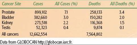
TABLE 64.1 2008 UROLOGICAL CANCER INCIDENCE AND MORTALITY WORLDWIDE
EPIDEMIOLOGY
Bladder cancer, with over 385,000 new cases reported worldwide in 2008,14 is a major cause of cancer morbidity and mortality (Table 64.1). Median age at diagnosis is above 70 years and, as the tumor is often smoking related, many patients have significant comorbidity, posing risks for radical surgical approaches. Survival rates are poor, with around 45% of muscle-invasive cancer patients surviving 5 years irrespective of treatment modality.4,5,7Demographically, the industrializing nations will contribute to a significant rise in the global incidence of bladder UC,15 with particularly large numbers likely in China given the rapid improvement in standards of living and the high prevalence of smoking. However, despite the decreasing incidence in developed nations, there remain specific challenges, mainly due to the aging population and increased life-expectancy. Within two large cohorts separated by 15 years (1991 to 1992 and 2005 to 2010), researchers have recently demonstrated an increase in the median age at presentation of 4 years, with an increase from 13% to 24% in the proportion of patients over 80 years old.16 Overall around 75% to 80% of patients with bladder cancer are male, mostly reflecting historic trends in cigarette smoking.
There are well-known associations of squamous cell bladder carcinomas with bilharzia caused by Schistosoma haematobium infection in Africa, particularly in Egypt.17 Aromatic amines, polycyclic aromatic and chlorinated hydrocarbons, arsenic-laced drinking water, aristolochic acid, cyclophosphamide exposure, and a range of industrial chemicals have been implicated in urothelial carcinogenesis. Importantly, as with most carcinogens, there are variations in individual susceptibility, and the basis of some of these polymorphisms regulating varied detoxification mechanisms has been identified.18 With increasing awareness of these industrial associations, regulation of these processes means that these cases are becoming increasingly rare in the developed world. Their principal importance now is that those with industrially linked tumors may be entitled to compensation payments. In Egypt there have been successful public health approaches to the control of S. haematobium infections, leading to a substantial decline in incidence and mortality from squamous carcinomas of the bladder.19
Within the developed world, the overwhelming majority of bladder tumors are now TCCs, and the main known causative factor is tobacco (particularly cigarette) smoking,20,21–22,23–25 explaining approximately half of the cases in men and one-third of the cases in women in Europe (discussed in detail below). The relation between smoking and other prognostic factors is interesting, as it could give insight into biological mechanisms of disease and, perhaps more importantly, have clinical implications by increasing our ability to identify patients at risk of more malignant disease.
NATURAL HISTORY
Non-muscle-Invasive Bladder Cancer
Most cases (70% to 80%) present with non-muscle-invasive bladder cancer (NMIBC, stage Ta, T1, and carcinoma in situ [Tis]), which is rarely lethal, but shows a high recurrence rate of 50% to 70% after treatment by transurethral resection of the bladder tumor (TURBT).26 In about 10% to 20% of patients with NMIBC, the disease progresses to muscle invasion (≥T2 lesions), which can lead to metastasis and death.26 However, the majority of patients with NMIBC will die of other causes, given the typically advanced age at presentation and the strong association with cigarette smoking,20,21–22,23–25 although it is worth noting that up to 21% of patients with Ta tumors and 49% of patients with T1 tumors will die from bladder cancer.27 For patients with NMIBC, it has been observed that tumor grade and stage, and also tumor number, size, presence of carcinoma in situ (CIS), recurrence rate, and age at diagnosis are risk factors of progression.28–29,30 The risk of both recurrence and progression necessitates lifelong follow-up for patients with bladder tumors. However, there are factors that predict a higher risk of progression (to invasion) as opposed to recurrence in lower risk tumors, and the European Organisation for Research and Treatment of Cancer (EORTC) “bladder cancer calculator” quantifies the risk depending on the tumor characteristics imputed.30
Invasive Disease
Muscle-invasive bladder cancer has a poor prognosis due to a very high rate of occult metastatic disease at the time of diagnosis. Evidence for this comes from the high rate of death from metastasis after apparently successful surgery. Furthermore, reported 5-year survival rates with radiotherapy or surgery are remarkably similar at around 45% to 50%,4,5 despite a higher rate of pelvic recurrence after radiotherapy versus surgery, suggesting that prognosis is driven by the presence or otherwise of metastases at the time of diagnosis, driven by tumor-related factors such as stage and grade.6
Metastatic Disease
A minority of patients (probably <10%) present with metastatic disease; most patients with metastatic disease have had prior treatment for apparently localized disease. Metastatic disease carries a poor prognosis. Overall survival from diagnosis of metastasis is difficult to ascertain as many patients receive only palliative treatment. A minority of patients are fit for systemic chemotherapy, and there are good data on outcomes with chemotherapy. In essence, extensive randomized studies, mostly carried out in the 1980s and 1990s, have demonstrated the superiority of cisplatinum-based combinations over those containing other drugs or cisplatinum alone.31 Of the platinum-based combinations, methotrexate/vinblastine/doxorubicin (Adriamycin)/cisplatinum (MVAC)32 and gemcitabine/cisplatinum (GC)33 have proven to be superior to other combinations and broadly similar in efficacy to each other34,35 (as reviewed by Hussain and James36). Median survival is 12 to 18 months, depending on the extent of disease and fitness of the patient. Intriguingly, however, a small minority of patients do appear to survive long term after chemotherapy for metastatic disease, but this percentage has sadly proved very hard to increase from that originally observed in the MVAC trials.
ETIOLOGY
The link between occupational exposure and an increased risk of urothelial cancer of the bladder was established more than a century ago when Rehn37 reported on three cases of bladder cancer in a German chemical dye works in 1895.38 During the following 40 years, similar reports appeared from around the world.39 In 1938, Hueper et al.39 demonstrated that when naphthylamine, an industrial arylamine used in the synthetic dye industry, was fed to dogs, it caused bladder carcinomas identical to the human disease. The link between industrial arylamines and bladder cancer was thus established and later confirmed by Case et al.40 in 1954. These authors also identified an excess of bladder cancer in the tire industry, attributable to the use of 2-naphthylamine in the manufacture of rubber.38,40 Around this time the “o-aminophenol hypothesis” of arylamine-induced human bladder cancer was proposed, suggesting that conjugation of aromatic amines by the liver and excretion in the urine with subsequent urinary reactions would liberate the carcinogen o-aminophenol.41 Other workers later added to this hypothesis: arylamines are hydroxylated in the liver and conjugated with glucuronic acid, followed by excretion into urine and reliberalization of the active carcinogenic metabolite into the bladder lumen by urinary glucuronidases.38,42,43 Slow acetylation by N-acetyltransferase, an enzyme involved in the metabolism of arylamines, has been shown to be a contributory risk factor for bladder carcinogenesis.20,44,45
Due to the widespread use of arylamines in textile dyes, hair dyes, and paint pigments, a number of high-risk occupations have been identified, including chemical, dye, textile, and rubber workers and painters and hairstylists.21,46–48 In addition, the presence of various arylamines in tobacco smoke means that a significant proportion of bladder cancer cases can be attributed to cigarette smoking.20,21–22,23–25 In fact, abandonment of the manufacture of many of these arylamines in the latter half of the 20th century means that smoking is currently the single most important cause of urothelial cancer.24,25,49–51 Tobacco (particularly cigarette) smoking now explains approximately half of bladder cancer cases in men and one-third of cases in women in Europe. It has been demonstrated that an increased smoking frequency and duration and a lower age at initiation are associated with an increased risk of bladder cancer, while cessation seems to reduce the risk.49 The relation between smoking and other prognostic factors is interesting, as it could give insight into biological mechanisms of disease and, perhaps more importantly, have clinical implications by increasing the ability to identify patients at risk of more malignant disease. Cigarette smoking also appears to be a risk factor for disease recurrence following a diagnosis of bladder UC,49,52 although due to a lack of conclusive evidence, there is currently a low rate of physicians providing smoking cessation assistance.49
Studies also demonstrate a relation between N-acetyltransferase-2 slow acetylators and cigarette smoking, resulting in a further increase in the risk of bladder cancer, especially in those individuals with a high smoking intensity.53–55 A similar relation has also been demonstrated with arylamine exposure.56,57 A number of other susceptibility loci have also been identified, although such markers do not yet have sufficient discriminatory ability to be utilized for risk prediction in the general population or for prediction or prognostication in patients diagnosed with bladder cancer.54,58,59
CHRONIC INFLAMMATION AND BLADDER CANCER
Squamous metaplasia is considered a precursor of squamous cell carcinoma of the bladder and is a relatively common occurrence, especially on the trigone of the female bladder where a prevalence of up to 50% is reported.60,61Experimental evidence suggests that this does not occur by direct transformation of the superficial apical umbrella cells of the urothelium or by their dedifferentiation and redifferentiation; it is postulated that basal cells (probable stem cells) are selectively activated.60 The normal urothelium is slowly proliferating, but urothelium undergoing squamous metaplasia becomes hyperplastic, and it may be that the hyperplasia component of urothelial squamous metaplasia is a major contributor to an enhanced risk of cancer formation.60,61
Squamous cell carcinoma and adenocarcinoma of the bladder often occur in the presence of chronic inflammation. In Africa and the Middle East, where these tumors are much more prevalent, the chronic inflammation occurs as a result of infestation with the parasite S. haematobium (bilharziasis), with a bladder carcinoma incidence of 2 to 4 per 100,000 in S. haematobium endemic areas.62,63–67 This infestation can lead to malignancy through local tissue damage, mechanical irritation, bilharzial toxins, secondary bacterial infection, and the production of nitrosamines.62,68,69 With liver involvement and subsequent liver dysfunction, tryptophan metabolism may be disturbed, resulting in excretion of carcinogenic metabolites.62 In S. haematobium infected individuals, the prevalence of squamous metaplasia rises significantly during the first 10 to 15 years of life, with a plateau at roughly constant levels thereafter (30% to 40% in males and 40% to 50% in females) when the active infection may have subsided.61 It is suggested that proliferative changes in the bladder urothelium may become independent of ongoing infection after long periods of chronic S. haematobium–induced inflammation.68,69 Severe metaplasia of the bladder may represent a precancerous transformation in some individuals, but in others it may only represent a marker for the prolonged inflammation that is associated with a high cancer risk.68,69 This sort of proliferative growth combined with the increased excretion or local formation of mutagens in the S. haematobium-inflamed bladder may significantly contribute to the onset of cancer formation, possibly involving mutations of the p53 and CDKN2 tumor-suppressor genes.68
In Europe and North America, the stimulus of chronic bladder inflammation is usually chronic bacterial infection, bladder calculi, or long-term indwelling catheters.70–73 A number of metaplastic conditions (squamous metaplasia, von Brunn’s nests, cystitis cystica, and cystitis glandularis) may occur prior to frank malignant change, although the premalignant nature of some of these lesions is still unclear.70–79
Field Cancerization and Clonality
A fundamental characteristic of neoplasia is monoclonality, in which one transformed cell gives rise to daughter cells that all exhibit the same genetic changes that provided the initial growth advantages to the originally transformed parent cell.80 Further genetic changes accumulate in subsequent daughter cells and provide additional growth advantages.80 However, TCC behaves as a multifocal disease, often with multiple primary tumors and frequent recurrences that can occur anywhere in the urinary tract from the renal pelvis to the urethra. These observations gave rise to the idea of a “field defect” or “field cancerization,” suggesting that the whole urothelium is exposed to the same urinary carcinogens, leading to the transformation of many independent separate urothelial cells and resulting in multiple tumors developing independently in multiple sites. Such tumors are thus genetically unrelated. An alternative explanation is that the multifocality of TCC arises as a result of a single carcinogenic insult to a single cell or group of cells. The progeny or clones of these cells spread throughout the bladder, either through intraepithelial migration or through cell shedding and reimplantation, leading to multiple synchronous and metachronous tumors.80 These tumors are thus topographically distinct but are genetically related. This is the hypothesis of clonality.
Utilizing the relatively rudimentary technique of X-inactivation,80,81–82 the early studies in this field appeared to show that the urothelium is derived from a small number of cells (200 to 300), which subsequently develop into larger patches; each patch is clonally related and possesses different predispositions to tumorigenesis.83 Such stem cell–derived clonal units actively replenish the urothelium during aging.84 Multiple synchronous and metachronous TCCs in the same patient appear to be clonally related when studied by X-inactivation.80,82,85–88 However, these studies only provide a 50% probability that one particular TCC is related to the primary TCC, although that probability improves when multiple TCCs are analyzed. Clonal patch size also needs to be taken into consideration, and because of the large patch size of the urothelium (120 mm2), X-inactivation studies are heavily biased toward demonstrating monoclonality.81 Ideally, these studies should have taken into account the relation of the tumors to patch boundaries.81 In addition, DNA methylation patterns change as a natural consequence of aging.89 Therefore, although X-inactivation studies provided an early insight into the relative importance of the processes of clonality and field cancerization, they cannot be considered as entirely accurate and reliable. Similarly, using immunohistochemistry to study specific p53 and pRb mutations cannot be considered to be wholly reliable for demonstrating monoclonality.
The accuracy of these investigations has been improved by utilizing newer and more sensitive techniques such as comparative genomic hybridization, fluorescence in situ hybridization, and loss of heterozygosity studies. These experiments revealed both monoclonality and oligoclonality in synchronous and metachronous TCCs. More recently, research has suggested that a genetic expression profile is established early in bladder tumor development, and that this profile is stable and maintained in recurring tumors.90–91,92 Majewski et al.91 matched the clonal allelic losses in distinct chromosomal regions to specific phases of bladder neoplasia: these genetic changes mapped to six regions or “forerunner genes” involved in the early phases of bladder cancer development, representing critical hits driving bladder carcinogenesis. It is suggested that the clonal expansion, over vast expanses of the bladder mucosa, of urothelial cell populations containing losses of forerunner genes may represent the earliest molecular change in bladder carcinogenesis. A further wave of genetic “hits” within subregions of these clonally expanded cells leads to the first microscopically recognizable features of dysplasia, and a third and final wave is associated with the fully transformed phenotype of severe dysplasia or CIS.91,93 The genetic changes map to six chromosomal regions that are suggested to represent the critical hits driving the development of bladder cancer.91,93 In addition, Knowles et al.94 have demonstrated that deletions of chromosome 9 occur in over half of bladder tumors of all grades and stages (9p, 51%; 9q, 57%). Loss of heterozygosity also occurs on 17p (32%), 11p (32%), 8p (23%), 4p (22%), and 13q (15%), and loss of heterozygosity of 5p, 8p, and 21q are significantly associated with worse grade and stage.94 Genomic copy number alterations are also frequent in bladder TCC, with the most frequent changes involving complete or partial loss of 4q (83%) and gain of 20q (78%).95 Other frequent losses are of 18q (65%), 8p (65%), 2q (61%), 6q (61%), 3p (56%), 13q (56%), 4p (52%), 6p (52%), 10p (52%), 10q (52%), and 5p (43%).94
Taken together, the studies described above show that multifocal TCCs are frequently monoclonal, whereas others show oligoclonality. The evidence for both theories is compelling (as reviewed by Duggan et al.96), with evidence supporting both the clonality and field cancerization theories. In reality, these theories are equally valid, with both processes seemingly often occurring simultaneously in the same patient.86,87 In addition, many of these studies have demonstrated that deletions on chromosome 9p occur most frequently and early in transitional cell carcinogenesis with 17p13 losses (p53 gene mutations) occurring in more advanced TCCs, shedding some light on the molecular pathology of bladder TCC.
Pathways to Muscle-Invasive and Nonmuscle-Invasive Bladder Cancer
A number of different approaches can be taken to describe the molecular alterations involved in bladder tumorigenesis (TCC). Some authors 97,98 have previously described such pathways in detail based on the six original “hallmarks of cancer” described by Hanahan and Weinberg99 in 2000. In 2011, Hanahan and Weinberg100 updated their original landmark review, describing genome instability and inflammation as underlying these hallmark changes and proposed “reprogramming of energy metabolism” and “evading immune destruction” as two emerging hallmarks with potential for generality. In addition, they reported that tumors exhibit another dimension of complexity by containing a repertoire of recruited, ostensibly normal cells that contribute to the acquisition of hallmark traits by creating the “tumor microenvironment.”100 The particular timing and sequence of hallmark events can vary widely between tumors of the same type and within the same tumor, but ultimately these hallmark capabilities of cancer will be reached.99 In their 2011 update, Hanahan and Weinberg100 also introduce the concept of “cancer stem cells,” a concept that has existed for quite some time for hematopoietic malignancies.101,102 Cancer stem cells are a subset of tumor cells that have the ability to self-renew and to generate all of the heterogeneous cells that comprise a tumor (properties that are analogous to a stem cell, the original cell of an organ, and responsible for organogenesis and organ maintenance).101,103–106 It is proposed that these cells are responsible for tumorigenesis, tumor differentiation, tumor maintenance, tumor spread, and tumor relapse.101,103–106 In the setting of bladder cancer, cancer stem cells appear to play a role in a subset of tumors, but their true significance has yet to be clarified.104
A number of other authors have also reviewed this field in detail,107–112 and there is general consensus on a divergent pathway for the development of Ta/T1 disease and Tcis/T2+ disease, as illustrated in Figure 64.1. Significant contributions to work in this field have been made by Knowles et al.110,113–119 (Leeds, UK); in their 2010 review, Goebell and Knowles113 propose a third hypothetical pathway for the development of high-grade papillary tumors.
A detailed examination of these pathways and related biomarkers is beyond the scope of this chapter. In addition, this is a rapidly changing field and new developments appear frequently with the advent of high-throughput experimental platforms, including “deep sequencing,”120 proteomics,121,122 and metabolomics,123 so readers are directed to the reviews cited above or to the latest work in this field.
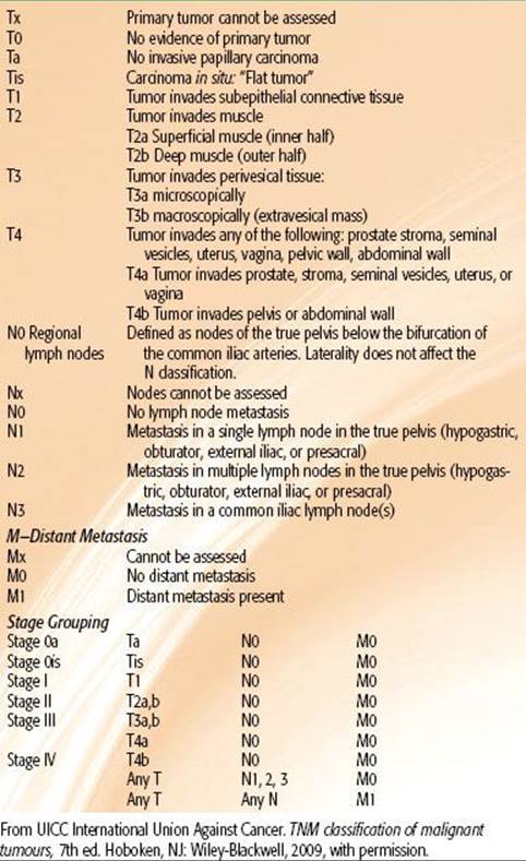
TABLE 64.2 TNM CLASSIFICATION OF TUMORS (2009)
SYMPTOMS AND SIGNS
The typical presenting symptoms of bladder cancer include painless, visible hematuria, infection, and storage symptoms. As the first of these is typically transient and the latter two are also attributable to prostate problems, patients often go undiagnosed for considerable periods.27 Given the intermittent nature of the hematuria associated with bladder cancer, patients presenting with a convincing episode of hematuria require urgent assessment. Similar considerations apply to male patients with a urinary tract infection. Female patients present a more difficult problem due to the higher incidence of urinary infection and the lower risk of bladder cancer. Hematuria is also more likely to be misinterpreted in women of childbearing age.
 INVESTIGATION OF PATIENTS WITH BLADDER CANCER
This section is divided into the investigation of patients with suspected bladder cancer and the subsequent staging of those with an established diagnosis.
Suspected Bladder Cancer
In developed countries, most patients will be referred to some sort of rapid access hematuria or suspected bladder cancer clinic. A minority may present via other routes (e.g., gynecological clinics) or with metastatic symptoms (<10%). Hematuria clinics will generally include a clinical assessment, full blood count and biochemical profile, prostate-specific antigen (if indicated), urine cytological examination, flexible cystoscopy, and some sort of imaging of the urothelium (e.g., ultrasound, intravenous urogram, or computed tomography [CT] urogram), which will vary with local practice and facilities.124,125 Patients identified as having a bladder tumor on flexible cystoscopy will then require further investigations to stage the disease.
Staging Bladder Cancer
The next stage for most patients will be an examination under anesthetic coupled with TURBT. The presence or absence of a mass after TURBT is also an important prognostic factor as it potentially indicates either unsuccessful clearance of tumor, extravesical extension, or both. This serves as both definitive staging of the bladder lesion as well as a substantial proportion of the initial treatment. Pathological review of the resected specimen will ascertain whether muscle invasion is present or not and whether there is CIS; these are the key determinants of further investigation and treatment. Patients with NMIBC, including CIS, do not usually require detailed further imaging, and treatment and surveillance are primarily by intravesical means. The main exception to this is patients with either extensive CIS or grade 3 lesions for whom additional cross-sectional imaging may be warranted. Patients with muscle-invasive bladder cancer (MIBC) will require detailed cross-sectional staging with CT or magnetic resonance imaging (MRI) of the chest, abdomen, and pelvis. If there are any features suggestive of bone metastasis (e.g., raised alkaline phosphatase, bone pain), an isotope bone scan will be indicated in addition.
A major confounding factor with imaging in bladder cancer is the effect of TURBT on the interpretation of the extent of the primary bladder tumor. Recent TURBT will cause perivesical changes that may be interpreted as extravesical spread. For similar reasons, enlarged pelvic nodes may be related to reactive rather than metastatic effects. On the other hand, changes such as infiltration of adjacent organs or hydronephrosis are likely to be reliable indicators of tumor stage and poor prognosis.
STAGING SYSTEM
The 2010 version of the UICC International Union Against Cancer’s TNM system is the current, internationally used staging system,126 and it is based on the size and extent of the primary tumor (T stage), presence or absence of nodal (N), and metastatic (M) spread (Table 64.2). The TNM stage must be used in conjunction with the pathological assessment of tumor removed at TURBT to decide on optimal therapy. Disappointingly, despite a substantial volume of research, no biomarkers have yet established themselves in clinical practice as either prognostic or predictive markers as has happened, for example, with estrogen-receptor or HER2 status in breast cancer. It is hoped that the wider availability of high throughput systems such as proteomics, in-depth sequencing, and others to be developed will allow the development of such markers in the future.
PATHOLOGY
In excess of 90% of bladder cancers are transitional cell carcinomas. Of the remainder, around 5% are squamous, although it should be noted that squamous differentiation is often present in poorly differentiated TCCs, so the extent to which these are genuinely distinct is open to question. As already noted, infestation with S. haematobium can lead to squamous cell carcinoma, but this is rapidly decreasing due to eradication programs. Small cell carcinoma is rare but important as it is generally very chemosensitive and treatment tends to follow schedules adapted from treating small cell lung cancer. Other tumor types include melanoma, carcinosarcoma, and adenocarcinoma (particularly in the urachal remnant). Typically bladder TCCs will be graded using the standard TNM system of Gx (cannot be assessed) then G1 through to G3 (well through to poorly differentiated).127 CIS will frequently be found in addition to a tumor mass and critically affects treatment choices.
TREATMENT OF BLADDER CANCER
Non-muscle-Invasive Bladder Cancer
Around 80% of patients with bladder cancer present with nonmuscle-invasive disease. These are classified as Tis, Ta, and T1 by the TMN classification system. The gold standard for diagnosis is the resection of the lesion with adequate sampling of the detrusor muscle deep to the lesion (TURBT). This will give tissue for accurate staging of the bladder lesion as well as definitive treatment for the lesion. Recurrences after TURBT are found in up to 70% of patients undergoing surveillance, and, more importantly, up to 15% of patients on surveillance will progress to muscle-invasive bladder cancer. At the time of diagnosis, the upper urinary tract also needs assessment. This can be done with intravenous urography, CT urography or ultrasound. The incidence of upper tract tumors at the time of presentation with hematuria is only 1.8%, calling into question the use of contrast imaging for the group as a whole.128Ultrasound is now being used more frequently, with contrast imaging modalities being used for the higher risk disease.
Prognostication and management strategy are based on accurate initial staging and grading of the disease. There can be variation in the interpretation of pathological specimens, so review of pathology is recommended (European Association of Urology [EAU] guidelines129). If there is uncertainty over the pathology, a further early re-resection is indicated. The risk of residual tumor can be as high as 53% in T1 tumors.130 If the disease is defined as NMIBC, it can be characterized as low, intermediate, or high risk, and this will dictate how the disease is managed (see below). The EORTC bladder cancer calculator provides a valuable online tool for doing this and determining follow-up frequency.131
Following resection of the tumor, there is good evidence that a single dose of mitomycin-C administrated into the bladder for 1 hour within 24 hours of surgery will reduce the relative risk of recurrence by 24.2% but will not impact disease progression and disease survival.132 Recurrences after TURBT are found in up to 70% of patients undergoing surveillance, and up to 15% of patients on surveillance will progress to muscle-invasive bladder cancer. Emerging endoscopic techniques employing photodynamic therapy aimed at improving diagnostic yield and thereby ultimately reducing the rates of recurrence and progression have demonstrated promising results.133
Low-risk tumors are single tumors that are <3 cm in diameter are graded as G1 disease and staged as Ta with no evidence of CIS. These tumors have a 15% probability of recurrence and a 0.2% risk of progression at 1 year.30These patients should undergo a flexible cystoscopy 3 months after the initial resection, and if this is negative, a flexible cystoscopy should be undertaken 9 months later and then annually thereafter.
Intermediate- and high-risk tumors are defined using a scoring system based on a number of clinical and pathological factors:
a. Number of tumors
b. Tumor size
c. Prior recurrence rate
d. T category
e. Presence of concurrent CIS
f. Tumor grade131
The high-risk tumors should be followed up with 3 monthly flexible cystoscopy for 2 years and 6 monthly for a further 5 years and then annually thereafter. Intermediate risk tumors should be followed up using a surveillance regime somewhere between that used for low- and high-risk disease, which is adapted according to personal and subjective factors. The intermediate-risk tumors have up to a 38% probability of recurrence and a 5% risk of progression at 1 year. The high-risk tumors have a 61% probability of recurrence and a 17% risk of progression at 1 year.30
The use of flexible cystoscopy with urine cytology is the standard of bladder surveillance. There is ongoing research into improving the sensitivity and specificity of flexible cystoscopy using variations in the wavelength of the light source used (e.g., narrow band imaging). There are a wide number of urinary biomarkers available such as NMP22, UroVysion (Abbott Laboratories, Abbott Park, Illinois), and ImmunoCyt (Scimedx, Denville, New Jersey). These agents suffer from high false-positive rates and variable sensitivity and are costly (as reviewed by Vrooman and Witjes134).
The presence of CIS in the bladder carries a 54% risk of disease progression without treatment.135 Patients with high-risk tumors or CIS should be offered intravesical immunotherapy using bacille Calmette-Guérin (BCG) (EAU and American Urology Association guidelines).129,136 There are a variety of treatment schedules in the literature, but the authors recommend an intravesical treatment once a week for 6 weeks followed by a subsequent 3 weeks as an induction treatment. If there is no cystoscopic evidence of recurrence, the patient should then be offered ongoing maintenance BCG with 6-week courses of BCG every 3 to 6 months with regular cystoscopic surveillance. In a recent meta-analysis of trials with BCG maintenance, a 32% reduction in the risk of recurrence was seen for BCG compared with mitomycin-C (P <.0001), whereas there was a 28% increase in the risk of recurrence (P = .006) for patients treated with BCG in the trials without BCG maintenance.137 BCG is a very effective treatment, but not all patients with NMIBC should be treated with BCG due to the risk of toxicity. In a phase III study for NMIBC tumors using maintenance BCG therapy, 20.3% patients stopped BCG due to side effects, mostly local side effects; 68% who stopped due to side effects did so during the first 6 months.138 The choice of treatment depends on the patients’ risk of recurrence and progression based on EORTC subgroups. The use of BCG does not alter the natural course of tumors in the low risk of recurrence subgroup and is therefore considered to be overtreatment. In patients with tumors at high risk of progression, for whom cystectomy is not carried out, BCG including at least 1-year maintenance, is indicated. In patients at intermediate or high risk of recurrence and intermediate risk of progression, BCG with 1-year maintenance is more effective than chemotherapy for prevention of recurrence; however, it has more side effects than chemotherapy. For this reason both BCG with maintenance and intravesical chemotherapy remain options. The final choice should reflect the individual patient’s risk of recurrence and progression and the efficacy and side effects of each treatment modality (EAU guidelines). In treatment refractory disease, the patient should be offered radical treatment for the bladder.
Muscle-Invasive, Nonmetastatic Disease
Relative Roles of Cystectomy and Bladder Preservation
Radiotherapy has been used as the primary treatment for muscle-invasive bladder cancer for many years, but utilization rates vary around the world from around 10% in the United States8 to 25% in Scandinavia139 to in excess of 50% in the United Kingdom.10 There are no prospective randomized trials comparing surgery with radiotherapy, so the data on comparative efficacy can only be inferred indirectly. U.S. authors in particular tend to refer to surgery as the gold standard of care, with bladder preservation with radiotherapy (with or without chemotherapy) being viewed as experimental. However, this opinion seems to be based on custom and practice and not on any hard comparative data. It is thus worth examining in some detail the data that exist on this topic. There is a solitary attempt using modern techniques to compare surgery with bladder preservation combining chemotherapy and radiotherapy. The UK SPARE trial (randomized trial of Selective Bladder Preservation against Radical Excision [cystectomy] in muscle-invasive T2/T3 transitional cell carcinoma of the bladder) was a feasibility study that has now closed due to poor recruitment.140 This phase II and III trial attempted to investigate the potential of using the response to neoadjuvant chemotherapy as a predictive tool for selecting patients for radiotherapy compared with the surgical standard. In the authors’ experience, one of the problems with the trial was that the study used response to neoadjuvant chemotherapy to decide on suitability for bladder preservation. However, once this was explained to the patients in the information sheet, there were patients who had a good response but were reluctant to undergo surgery, particularly if the bladder was free of tumor at the interim cystoscopy.
Large population-based studies suggest that the main determinants of survival after diagnosis of bladder cancer are stage, grade, age, and to some extent social class. For example, Hayter et al.6 studied patterns of care and outcomes in over 20,000 patients with bladder cancer in Ontario. They found significant variations in the use of cystectomy and radiotherapy between different districts but no evidence that these variations led to any differences in long-term outcomes. They concluded that bladder-sparing approaches were equivalent to surgery for invasive bladder cancer.
Another way to compare outcomes between surgery and radiotherapy is to look at large published series. In the United Kingdom, where both of these approaches are routinely employed, it is possible to compare the outcomes in Cancer Registry data. A recent paper from Munro et al.10 examined outcomes in 458 patients with invasive bladder cancer treated in Yorkshire between 1993 and 1996. The ratio of cystectomy to radiotherapy was 1 to 3, reflecting UK practice at the time. Overall 10-year survival was similar between those who underwent radiotherapy (22%) versus radical cystectomy (24%). Prognostic factors for inferior outcome at 10 years were: female versus male, poor performance status, hydronephrosis and increasing T stage; treatment modality was not a factor in the prognosis.
One of the most widely quoted surgical series comes from the University of Southern California and reports the results of 1,054 patients undergoing cystectomy with overall 5- and 10 year survival rates of 60% and 43%, respectively. This series is discussed in detail below. However, this series included patients undergoing surgery for noninvasive tumors and excluded from the denominator 112 patients referred but deemed incurable at operation. If we look at the 5-year survival of those with invasive tumors, the rate drops to around 47% from the quoted 60%. A contemporary series of radiotherapy cases from Rödel et al.5 reports 5- and 10-year survivals of 51% and 31% but included patients deemed inoperable.
Furthermore, if we look at the outcomes in the surgical control arm in the US Intergroup Neoadjuvant MVAC trial, the median survival was 38 months and the 5-year survival was 42%. Other relatively contemporary series quote similar 5-year survivals: for example, Dalbagni et al.141 cite 45% overall and 65% disease-specific survival rates in a series of 300 patients. Furthermore, the only significant prognostic factors in this series were age, T stage, and use or nonuse of neoadjuvant chemotherapy (see the Role of Systemic Therapy section). Data also exist from single institution series with widespread use of both modalities. For example, Kotwal et al.142 report results on 169 patients treated between March 1996 and December 2000 in Leeds, UK. There were no differences in overall, cause-specific, and distant recurrence-free survival at 5 years between the two groups, despite the radiotherapy group being older (median age, 75.3 vs. 68.2 years). There were 31 local bladder recurrences in the radiotherapy group (24 of which were solitary and hence potentially suitable for salvage surgery), but no significant difference in distant recurrence-free survival. In another more recent (2002 to 2006) cohort, the median age of radiotherapy patients but not the cystectomy patients had increased to 78.4 from 75.3 years, respectively, while the age of those undergoing surgery remained similar at 67.9 and 68.2 years for surgery, consistent with the aging trend in new bladder cancer cases. These authors concluded that although the patients undergoing radical cystectomy were significantly younger than the radiotherapy patients, treatment modality did not influence survival. They went on to state that radical radiotherapy is a viable treatment option for these patients, with the advantage of organ preservation. The data thus suggest that bladder preservation gives equivalent long-term survival to surgery when factors such as case selection are accounted for.
A proportion of patients undergoing radiotherapy will relapse within the bladder and go on to salvage cystectomy. An important consideration, therefore, is whether cystectomy after radiotherapy can be carried out safely and whether the delay compromises survival. Again, there are no randomized data on this topic. However, UK surgeons in particular have good practical experience on this topic and have commented that neither prior chemotherapy nor radiotherapy compromises surgical salvage and that long-term results appear similar to primary cystectomy series.143,144
Particularly intriguing in this regard is a comparison of survival rates following primary surgery or salvage surgery following failed radiotherapy from the Christie Hospital in Manchester, UK. The group examined the outcomes in 552 patients who underwent radical cystectomy between 1970 and 2005. Of these, 313 patients underwent primary radical cystectomy and 239 underwent salvage radical cystectomy following radiation failure. The median age was 62.5 years (range, 32.2 to 87.2) for the primary surgical group compared with 65.5 years for the salvage group. Overall 5-year survivals reported were 45.5% for the primary group and 42% for the salvage group, with cause-specific survivals of 51% and 50%, respectively. These differences persisted after stratification for stage, and the authors concluded that a policy of primary radiotherapy with surgical salvage did not compromise the long-term survival chances of patients.143
Clearly there are surgical series with much higher survival rates than this in the literature. However, there are two factors accounting for this. One is case selection, the other is that these series are, to a degree, personal, so to publish results that appear inferior to other major centers is potentially a threat to a center’s (or surgeon’s) reputation and will tend to have a “ratchet” effect on published results. These data also suggest that the predominant prognosis driver in bladder cancer is the presence or absence of distant micrometastasis at diagnosis of invasive disease. The (relatively modest) effect of neoadjuvant chemotherapy tends to bear this out, particularly as the biggest effect in the Medical Research Council (MRC)/EORTC trial was on metastasis-free survival rather than pelvic control rates.145
There is little in the way of randomized data on the efficacy of radiotherapy in either nonmuscle-invasive disease or CIS. The only randomized trial on this topic comes from the United Kingdom and compared radiotherapy with surveillance for patients with a new diagnosis of pT1G3 NXM0 transitional cell carcinoma with unifocal disease and no CIS. Patients with multifocal disease or CIS were randomized between intravesical therapy and radiotherapy. There was no evidence of benefit from radiotherapy in terms of progression-free interval (hazard ratio [HR] 1.07; 95% confidence interval [CI], 0.65 to 1.74; P = .785), progression-free survival (HR 1.35; 95% CI, 0.92 to 1.98; P = .133), or overall survival (HR 1.32; 95% CI, 0.86 to 2.04; P = .193).146 There is thus no indication for radiotherapy in these groups of patients.
In truth, surgery and radiotherapy are not competing, but are complementary approaches to invasive bladder cancer. There are particular groups who appear to do poorly with primary radiotherapy, for example, those with poorly functioning bladders or extensive CIS in addition to their invasive disease. In the former case, radiotherapy is unlikely to improve bladder function; in the latter, the lack of effect of radiotherapy on CIS means that the patient remains at risk of further bladder intervention and ultimately cystectomy.146 Similar considerations apply to patients with pT1G3 disease.146 North American authors will also cite features such as hydronephrosis as a contraindication to radiotherapy.147 However, hydronephrosis is also a poor prognostic factor for surgery and does not help in selecting patients one way or another.6 On the other hand, there are many patients who may benefit from radical therapy but are poor surgical candidates, such as older patients, the obese, diabetics, poor anesthetic risk patients, or those who may struggle with whatever form of neobladder is fashioned. Although large surgical series will include patients over age 80, these will typically comprise only a few percentage of the total, whereas, with a median age at diagnosis of bladder cancer in the middle to late 70s, there are probably many more who do not make it to surgery.
Surgery
Management of Invasive Bladder Cancer
Although the majority of patients present with NMIBC, 20% to 40% will either present with or ultimately develop muscle-invasive disease. Invasive bladder cancer is a lethal malignancy; if untreated over 85% of patients will die of the disease within 2 years of diagnosis.148 Furthermore, a certain percentage of patients with high-grade bladder tumors without involvement of the lamina propria will recur or progress or fail intravesical management, and they may be best treated with an earlier cystectomy when survival outcomes are optimal.149 In these groups of patients, the 5-year survival rates after cystectomy exceed 80%.150,151
The rationale for an aggressive treatment approach employing radical cystectomy for high-grade, invasive bladder cancer is based on several important observations. First, the good long-term survival rates, coupled with the lowest local recurrences, are seen following a definitive surgical approach removing the primary bladder tumor and regional lymph nodes.4,152,153 Although there are no randomized trials comparing radical cystectomy with bladder preserving approaches, surgery remains the preferred treatment option for many clinicians for advanced, localized invasive bladder cancer.154 Second, the morbidity and mortality of radical cystectomy has substantially improved over the past several decades.152,155 Third, advocates say that radical cystectomy provides accurate pathologic staging of the primary bladder tumor (p stage) and regional lymph nodes, thus, selectively determining the need for adjuvant therapy based on precise pathologic evaluation. However, it should be noted that the evidence base for adjuvant (as opposed to neoadjuvant) therapy is rather weak (see on the Role of Systemic Therapy section). For the above-mentioned reasons, radical cystectomy has become a standard form of therapy for high-grade, invasive bladder cancer. Nonetheless, many articles on cystectomy emphasize the need for careful patient selection to achieve optimal results. Although this is undoubtedly true, it begs the question of what should be done with patients who do not meet these stringent selection standards but still need to be treated. This issue is rarely, if ever, addressed in cystectomy series publications.
The evolution and improvements in lower urinary tract reconstruction, particularly orthotopic diversion, have been major components in enhancing the quality of life of patients requiring cystectomy. Currently, most men and women can safely undergo orthotopic lower urinary tract reconstruction to the native, intact urethra following cystectomy,156 although availability varies worldwide. Orthotopic reconstruction aims to mimic the native bladder in location and function, provides a continent means to store urine, and allows volitional voiding per urethra, although patients need to do this by coordinating opening the sphincter with a Valsalva maneuver, which does require training. The orthotopic neobladder eliminates the need for a cutaneous stoma, urostomy appliance, and the need for intermittent catheterization in most cases. These efforts have improved the quality of life of patients who require removal of their bladders and have also stimulated patients and physicians to consider radical cystectomy at an earlier more curable stage for high-grade, invasive bladder cancer.157,158
A dedicated effort has been made to improve the surgical technique of radical cystectomy and to provide an acceptable form of urinary diversion, without compromise of a sound cancer operation.158,159 Timing of cystectomy after the TURBT is important, and it has been demonstrated that a delay of more than 3 months undermines patient survival. This evidence forms the basis to negotiating resource needs with health care providers.160,161 Certain technical issues regarding radical cystectomy and an appropriate lymph node dissection are critical to minimize local recurrence and positive surgical margins and to maximize cancer-specific survival.162 Attention to surgical detail is important in optimizing the successful functional outcomes of orthotopic diversion by preserving the urinary sphincter mechanism and therefore continence. Finally, the observed associations between hospital volume and operative mortality are largely mediated by surgeon volume. Patients can often improve their chances of survival substantially, even at high-volume hospitals, by selecting surgeons who perform the operations frequently, as those centers that have adopted this strategy have demonstrated declining mortality in the past decade.163,164
Radical cystectomy by definition implies the en bloc removal of the pelvic–iliac lymph nodes along with the pelvic organs anterior to the rectum: the bladder, urachus, prostate, seminal vesicles, and visceral peritoneum in men; the bladder, urachus, ovaries, fallopian tubes, uterus, cervix, vaginal cuff, and the anterior pelvic peritoneum in women. An appropriate lymphadenectomy is an important component of radical cystectomy and is related to the clinical outcomes of patients with high-grade, invasive bladder cancer. Evidence suggests that a more extended lymphadenectomy is beneficial in both lymph node–positive and lymph node–negative patients with bladder cancer,165,166,167although this could be a surrogate for either case selection or surgical skill. Although the exact limits of the lymphadenectomy for patients with bladder cancer undergoing cystectomy are currently debated, the boundaries include initiation at the level of the inferior mesenteric artery (superior limits of dissection), extending laterally over the inferior vena cava or aorta to the genitofemoral nerve (lateral limits of dissection), and distally to the lymph node of Cloquet medially (on Cooper’s ligament) and the circumflex iliac vein laterally. This dissection includes bilaterally all obturator, hypogastric, presciatic, and presacral lymph nodes.168–170 Removal of more than 15 lymph nodes has been postulated to be both sufficient for the evaluation of the lymph node status as well as beneficial for overall survival in retrospective studies.166,171–174 However, potential interindividual differences in the number of pelvic and retroperitoneal lymph nodes and difficulties in processing of the removed tissue by pathologists are issues. Furthermore, the reality is that even in today’s practice the number of lymph nodes retrieved are low (<10) in a majority of patients (75%) and the chance are even lower if the patients are older, Hispanic (in the United States), and managed at low-volume, nonurban centers.170 The true curative value of lymph node dissection and the optimal extent of dissection are both still unknown.
Technical variations from the standard cystectomy have been performed to improve patients’ quality of life, including prostate-sparing cystectomy in order to preserve continence and potency. However, it carries a higher risk of missing unsuspected adenocarcinoma of the prostate. Coexistent prostate cancer and prostatic urothelial cancer are reported in 23% to 54% cases, of which 29% were clinically significant, leading to local recurrence and even metastasis.175,176
TABLE 64.3 CYSTECTOMY OUTCOMES IN SELECTED SINGLE INSTITUTION SERIES
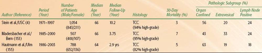
Radical cystectomy is an appropriate standard treatment for patients with high-grade, invasive bladder cancer. The clinical outcomes are presented in Table 64.3. These results should provide a benchmark for outcomes to which other therapies can be compared.
Both laparoscopic and robot-assisted cystectomy have been shown to be feasible and safe, but with a relatively shorter follow-up.177,178–179 Despite an increased materials cost for a robotic-assisted cystectomy, it has been demonstrated to be cost-effective in a subgroup of patients undergoing ileal conduit when the impact of complications are considered in a single institution series.179,180
Morbidity and Mortality of Radical Cystectomy and Lymphadenectomy
The early clinical results and outcomes with regard to the morbidity and mortality of radical cystectomy were disappointing. Lack of universal acceptance of this procedure was attributed to the considerable complication rate and the need for improvements in urinary diversion. Prior to 1970, perioperative complication rates of radical cystectomy were approximately 35%, with a mortality rate of nearly 20%. However, with contemporary medical, surgical, and anaesthetic techniques, along with better patient selection, the mortality and morbidity from radical cystectomy have dramatically decreased (Table 64.3). Importantly, in high-volume centers, the administration of preoperative therapy (radiation or chemotherapy) and the form of urinary diversion performed (continent or incontinent) did not obviously increase the mortality rate of patients undergoing radical cystectomy.150 The issue of the need to select patients for surgery complicates the comparison of bladder-sparing techniques, because frequently those not suitable for surgery will be the ones who appear in the radiotherapy-based series.
The early complication rate following radical cystectomy should not be underestimated in this elderly group of patients. The median age of patients undergoing cystectomy in a University of Southern California series was 66 years.4 Of the 1,054 patients treated, 28% developed an early complication within the first 3 months of surgery. Early complications included all events related to the cystectomy, perioperative care, and urinary diversion. The administration of preoperative therapy (radiation or chemotherapy) and the form of urinary diversion did not significantly increase the early complication rate in these cystectomy patients. Most early complications following radical cystectomy are unrelated to the urinary diversion (85% diversion unrelated) and can be managed conservatively without the need for reoperation in approximately 90% of cases.181 The most common early diversion-unrelated complication is dehydration, while the most common early diversion-related complication following radical cystectomy is prolonged urinary leakage. Overall, the most surgical complications after cystectomy are associated with urinary diversion in relation to the intestinal segments.152,182
Neoadjuvant chemotherapy is discussed in a later section, but it does not seem to increase the perioperative morbidity or mortality.7,183 Preoperative radiation therapy is discussed below.
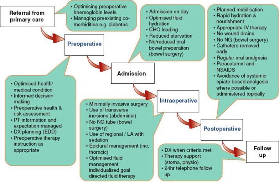
Radical cystectomy may appropriately be performed in carefully selected elderly patients,184 however, this emphasis in the surgical literature on “careful selection” highlights as much the deficiencies as the efficacy of cystectomy as it emphasizes that the published results are not applicable to the entire population. It is emphasized that physiologic age may be more important than chronologic age when determining appropriate candidacy for radical cystectomy. Proper patient selection, strict attention to perioperative details, along with a dedicated and meticulous team-oriented surgical approach are critical components to minimize the morbidity and mortality of surgery and to ensure the best clinical outcomes in all patients following radical cystectomy.185–187 A recommended approach from the authors’ center previously pioneered by the Danish is adoption of an enhanced recovery pathway protocol (Fig. 64.2).188
FIGURE 64.1. Pathways to development of bladder cancer. (Data from Pollard C, Smith SC, Theodorescu D. Molecular genesis of non-muscle-invasive urothelial carcinoma (NMIUC). Expert Rev Mol Med 2010;12:e10.)

FIGURE 64.2. Schematic representation of the enhanced recovery program in patients undergoing a radical cystectomy.
Pathologic Stage and Subgroups
The pathologic stage of the primary bladder tumor and the presence of regional lymph node metastases are the most important survival determinants in patients undergoing cystectomy for bladder cancer.4,153,155 These pathologic determinants may also be categorized into pathologic subgroups that provide risk stratification and direct the need for adjuvant therapy in the appropriately selected individual. Pathologic subgroups are defined as organ-confined, lymph node–negative tumors (P0, Pa, Pis, P1, P2a, P2b), nonorgan confined (extravesical) lymph node–negative tumors (P3, P4), and lymph node positive disease (N+); representing 56%, 20%, and 24% of patients, respectively.155The 5- and 10-year recurrence-free survival for the entire group of 1,054 patients in the University of Southern California series was 68% and 66%, respectively (Table 64.3). Most deaths occurred within the first 3 years following radical cystectomy and were attributed to cancer recurrence. However, with longer follow-up (>3 years), most deaths in this elderly group of patients were primarily related to comorbid diseases unrelated to bladder cancer. This underscores the effective and durable outcomes of radical cystectomy.
TABLE 64.4 SELECTED DATA FROM THE UNIVERSITY OF SOUTHERN CALIFORNIA BLADDER CANCER STUDY OF RADICAL CYSTECTOMY
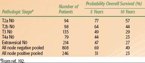
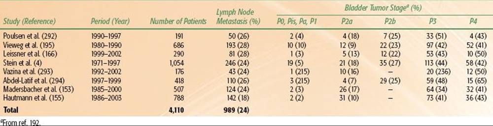
TABLE 64.5 INCIDENCE OF LYMPH NODE METASTASIS FOLLOWING RADICAL CYSTECTOMY, CORRELATION TO PRIMARY TUMOR
Organ-Confined, Lymph Node–Negative Tumors
In the University of Southern California series, 56% of patients demonstrated pathologically organ-confined, lymph node–negative tumor.4 The outcomes in this pathologic subgroup were excellent (Table 64.4), with a 5- and 10-year recurrence-free survival of 85% and 82%, respectively. No significant survival differences were observed when comparing superficially noninvasive (Pis, Pa), lamina propria invasive (P1), and muscle-invasive (P2a, P2b) tumors as long as the tumor was confined to the bladder without evidence of lymph node involvement. Similar outcomes for patients with pathologic nonmuscle-invasive bladder tumors following cystectomy have been previously reported.4,155,189,190 Collectively, these data support the treatment of patients with surgery when a tumor is confined to the bladder, without evidence of extravesical extension or lymph node metastasis. Treatment delays in patients with invasive bladder cancer should be avoided. Evidence suggests that prolonged delays may lead to more advanced pathologic stage and decreased survival in patients with muscle-invasive bladder cancer.160 Furthermore, caution should be taken in delaying definitive therapy in patients with high-risk, nonmuscle-invasive bladder tumors or those with nonmuscle-invasive tumors that have not appropriately responded to conservative forms of therapy.149
Extravesical, Lymph Node–Negative Tumors
Nonorgan confined (extravesical), lymph node–negative tumors were found in 20% of University of Southern California series patients undergoing cystectomy (Table 64.4). No obvious survival differences between extravesical P3 and P4 (node-negative) tumors were observed. The 5- and 10-year recurrence-free survival for this pathologic subgroup was 58% and 55%, respectively. Similar outcomes were reported by Madersbacher et al.153 and Dhar et al.,171who demonstrated a 56% 5-year recurrence-free survival for the same pathologic subgroup. Patients with locally advanced tumors have higher recurrence rates and decreased survival compared with those with organ-confined, lymph node–negative tumors.191 The 1997 AJCC TNM staging system192 used for these studies stratifies extravesical tumor involvement (previously defined as pT3b) into microscopic (pT3a) and gross (pT3b) extravesical tumor extension. No significant difference was observed in the recurrence-free and overall survival in patients when evaluating for pT3a and pT3b extravesical extension.191 The incidence of lymph node involvement was similar (approximately 45%); however, the presence of lymph node involvement was associated with a higher risk of recurrence and worse survival compared with node-negative patients.
Lymph Node–Positive Disease
Perhaps one-fourth of patients in large cystectomy series will have positive lymph nodes (Tables 64.3 and 64.5). Although patients with lymph node tumor involvement are a high-risk group of patients, nearly one-third of these patients were alive at 5 years in the series by Stein et al.4 It is possible that the surgical approach employing an extended lymph node dissection provides some survival advantage in selected individuals with node-positive disease.171The role of adjuvant therapy in these patients is difficult to assess due to the absence of adequately powered trial data (see on the Role of Systemic Therapy section). In an analysis of lymph node–positive patients carried out by the University of Southern California group, the administration of adjuvant chemotherapy was a significant and independent predictor for recurrence and overall survival in this group,193 however, case-mix effects will be prominent in this type of retrospective analysis as patients need to survive long enough and be fit enough to even start chemotherapy to figure in the analysis. Inevitably this will bias results in favor of adjuvant chemotherapy by excluding those who died early or who never became fit enough postoperatively to receive chemotherapy. The authors own experience with attempting to recruit to adjuvant chemotherapy trials suggests that only the most fit patients are able to recover quickly enough postoperatively to undergo systemic chemotherapy, so these data are highly likely to be biased by underlying patient fitness. A further interesting feature of the BC2001 trial,194 discussed in detail below, was that although no attempt was made to include pelvic nodes in the treatment field, the rate of nodal relapse was low at around 6% with radiotherapy alone, falling to 4% with chemoradiation. One possible interpretation of these data is that successful treatment of the primary, with either surgery or radiotherapy, may affect the subsequent behavior of low-volume nodal disease.
The prognosis in patients with lymph node–positive disease can be stratified by the number of lymph nodes involved (tumor burden), the stage of the primary tumor, and the presence of lymph node capsule perforation.4,173Patients with fewer than five positive lymph nodes among those with lymph node–positive organ-confined bladder tumors had a significantly improved recurrence-free survival rate.4,166,195
The number of lymph nodes involved with tumor and the extent of the lymph node dissection are both important variables for patients undergoing cystectomy for bladder cancer. Stein et al.193 examined 246 patients with lymph node tumor involvement following radical cystectomy to evaluate other prognostic factors in this high-risk group. They used lymph node density to account for the extent of the lymph node dissection (number of lymph nodes removed) and the tumor burden (number of positive lymph nodes) following cystectomy for patients with lymph node–positive disease. Lymph node density as defined in this study was a significant and independent prognostic variable in patients with lymph node metastases. Future staging systems and the application of adjuvant therapy in clinical trials may consider applying these concepts to better stratify this high-risk group of patients.
Recurrence Following Radical Cystectomy
Recurrence following radical cystectomy for bladder cancer correlates well with the pathologic stage and subgroup.4,153 With long-term follow-up (median, >10 years) recurrences in the University of Southern California series were classified as local (pelvic), distant, and urethral. Local recurrences were defined as those occurring within the soft tissue field of exenteration. Distant recurrences were defined as those occurring outside the pelvis, while urethral tumors were classified as a new primary tumor occurring in the retained urethra. Overall, 30% of all patients in the University of Southern California series experienced tumor recurrence.4 The median time to any recurrence was 12 months, with 86% of all patients developing their recurrences within the first 3 years of cystectomy. Of the 311 patients who developed a recurrence, the median time to distant recurrence was 12 months, while the median time to local recurrence was 18 months. Late tumor recurrences, defined as 5 years or more after surgery, do occur and underscore the need for lifelong follow-up.
Pelvic (Local) Recurrence
Pelvic recurrence rates of 6% to 9% are reported in large series,153,155,190 with higher rates in tumors with extravesical spread or node-positive disease at cystectomy.4
TABLE 64.6 RECURRENCE-FREE SURVIVAL AND THE INCIDENCE OF RECURRENCE IN SELECTED STUDIES OF RADICAL CYSTECTOMY

Metastatic (Distant) Recurrence
Recurrences following radical cystectomy are most commonly found at distant sites. This is consistent with the benefits observed in the neoadjuvant chemotherapy trials, where the maximum effect would be expected on low-volume, occult metastases rather than large-volume primary disease (discussed below). Distant recurrence rates of 20% to 35% are reported in large series (Table 64.6). However, overall death rates from bladder cancer are higher than this, so there would appear to be either underreporting of deaths in these series or the cases selected were not representative of wider experience.
Urethral Recurrence
Urethral tumor recurrence in patients with a history of bladder cancer following radical cystectomy represents a second manifestation of the multicentric defect of the primary transitional cell mucosa that led to the original bladder tumor. The term urethral recurrence is therefore somewhat misleading, suggesting a failure of definitive treatment of the bladder cancer. Most urethral tumors probably represent simply another occurrence of the transitional cell carcinoma in the remaining urothelium. Because radical cystectomy with orthotopic diversion has increasingly been performed, the fate of the retained urethra has become an increasingly important oncologic issue.
The advent of orthotopic lower urinary tract reconstruction has provided a more natural voiding pattern in patients following radical cystectomy. Approximately 85% of all patients undergoing cystectomy for TCC of the bladder at the University of Southern California research center receive an orthotopic neobladder substitute. From an oncologic perspective, only those with a positive surgical margin at the proximal urethra (distal to the apex of the prostate in men and just distal to the bladder neck in women) on intraoperative frozen section are absolutely excluded from orthotopic reconstruction. This enthusiasm to preserve the native urethra following radical cystectomy and allow for orthotopic reconstruction has rightfully increased concerns for a urethral recurrence in these patients.
Prior to the orthotopic era in women, urethral tumor recurrence was not an important oncologic issue because the entire urethra was removed at the time of cystectomy. With a better understanding of female pelvic anatomy and the innervation of the urinary sphincter and continence mechanism in women,196 along with the identification of various pathologic risk factors for urethral tumor involvement in these patients, orthotopic diversion has now become a commonly performed form of urinary diversion in women following cystectomy.197 Tumor involving the bladder neck is the most important risk factor for urethral tumor involvement in women.198,199 Although bladder neck involvement is a significant risk factor for urethral tumors, not all women with tumor involving the bladder neck will have urethral tumors. Approximately 50% of female patients with tumor at the bladder neck will have an uninvolved urethra free of tumor. In this situation, the patient may potentially be considered an appropriate candidate for orthotopic diversion. Furthermore, intraoperative frozen-section analysis of the distal surgical margin is an accurate and reliable means to pathologically evaluate the proximal urethra.199
A growing population of male patients reconstructed to the urethra following cystectomy exists today. With longer follow-up, could this expose them to a greater risk for a urethral recurrence? The historical incidence of urethral recurrence in the retained urethra following cystectomy for bladder cancer ranges from 6% to 10%.200,201 Specific clinical and pathologic risk factors that have been identified to provided risk assessment for urethral recurrence include multifocal tumors, CIS, tumor involvement of the prostate (particularly invasion of the prostatic stroma), and the form of urinary diversion (orthotopic or cutaneous) performed.198,200,202–204
Stein et al.205 have evaluated urethral recurrence in a large group of male patients undergoing radical cystectomy and urinary diversion for TCC of the bladder. In this study, the clinical and pathological results of 768 consecutive male patients undergoing radical cystectomy with a median follow-up of 13 years were analyzed. Of these 768 patients, 397 men (51%) underwent an orthotopic diversion (median follow-up 10 years) and 371 men (49%) underwent a cutaneous diversion (median follow-up 19 years). Overall, 45 patients (7%) developed a urethral recurrence. The median time to a urethral recurrence was 2 years (range, 0.2 to 13.6 years). Of these 45 patients, 16 men (5%) had an orthotopic and 29 (9%) had a cutaneous form of urinary diversion. In this cohort of male patients, multiple risk factors were analyzed with regard to urethral recurrence. In a multivariate analysis, two important variables were identified that significantly increased the risk of a urethral tumor recurrence following cystectomy, including any prostate involvement and the form of urinary diversion. The estimated 5-year probability of a urethral recurrence was 5% without prostate involvement, which increased to 12% and 18% with superficial (prostatic urethra and ducts) and invasive (stroma) prostate involvement, respectively. Patients undergoing an orthotopic diversion demonstrated a statistically significantly lower risk of urethral recurrence compared with those undergoing a cutaneous form of urinary diversion.
The follow-up and management of the urethra in male patients treated for high-grade invasive bladder cancer is of importance. The indications and timing of a prophylactic urethrectomy in those undergoing cystectomy and a cutaneous diversion is debatable. It may include urethrectomy at the time of cystectomy based on preoperative clinical parameters, based on the intraoperative frozen-section analysis of the urethral margin, or a delayed urethrectomy based on final pathologic evaluation of the cystectomy specimen. These issues are best detailed with the patient preoperatively ensuring proper informed consent.
Management of the Retained Urethra Following Cystectomy
Intraoperative frozen-section analysis of the proximal urethra by an experienced pathologist is a reliable and accurate means to determine indications for orthotopic diversion in all patients. It is good practice to proceed with an orthotopic neobladder in men and women whose intraoperative frozen section of the proximal urethra is free of tumor. This approach does not appear to increase the risk of a urethral recurrence in these patients.197,206 Male patients with known prostatic tumor involvement should not necessarily be excluded from an orthotopic substitute if the intraoperative biopsy is normal. Similarly, female patients with bladder neck involvement should not necessarily be excluded from an orthotopic neobladder if the intraoperative biopsy is also normal. All patients should be carefully counseled regarding the need for follow-up, the long-term risks of a urethral recurrence, and the possible need for urethrectomy following cystectomy.
Salvage Cystectomy
Salvage cystectomy after prior radiotherapy was discussed above in the section comparing surgical with bladder-sparing approaches.
Summary of Surgical Therapy
Surgery is an important mode of therapy for invasive bladder cancer and undoubtedly can provide durable disease control for many patients with the disease. In addition, an integrated approach with other treatment modalities such as neoadjuvant chemotherapy and radiotherapy is essential if the best results are to be achieved in the entire patient population, especially those who are borderline fit for major surgery. Radical cystectomy probably provides the best local pelvic control of the disease and provides accurate evaluation of the primary bladder tumor along with the regional lymph nodes yielding important prognostic information. This, coupled with the evolution and successful application of orthotopic lower urinary tract reconstruction in both men and women, has provided patients a more physiological and acceptable means to store and eliminate urine. However, most deaths from bladder cancer still occur due to distant metastases, presumably present at the time of original surgery, so further improvements in outcome will depend on the development of both better systemic therapies and also predictive (as opposed to prognostic) markers that will allow better selection of therapies.
TABLE 64.7 INDICATIONS FOR RADIOTHERAPY IN BLADDER CANCER

Radiotherapy
External-Beam Radiotherapy
Whatever the merits or otherwise of radiotherapy as a treatment for bladder cancer, its use in the United States has declined markedly since the 1980s, and the treatment is now used mainly in certain centers such as Boston in carefully selected patients rather than as a mainstream alternative to surgery. As discussed above, patterns vary worldwide with the U.S. pattern of care predominating. Potential indications for radiotherapy are summarized in Table 64.7. Patients with radiological node-positive or metastatic disease should be managed predominantly with either chemotherapy or palliative approaches such as local radiotherapy to bulk disease (see below). Patients with radiological node-negative, muscle-invasive disease may be considered for radical radiotherapy. In North America, radiotherapy is administered as part of a package of care comprising maximal TURBT, chemotherapy, and radiotherapy, the so-called trimodality therapy (Fig. 64.3).207,208
The schedules used are complex but can be summarized as initial maximal TURBT followed by initial radiotherapy combined with synchronous chemotherapy, usually with cisplatinum followed by interim check cystoscopy. Patients experiencing a good response to treatment continue to consolidation chemoradiotherapy and then adjuvant polychemotherapy, usually with a cisplatinum base. Patients remain on long-term cystoscopic surveillance. Noninvasive recurrence can be managed by further TURBT and intravesical therapy. Those with isolated muscle-invasive recurrence or failure to respond (but with no systemic relapse) can undergo salvage cystectomy with or without further chemotherapy.
The optimal radiotherapy schedule has yet to be established. In North America, split schedules often used are 39 or 40 Gy in 1.8- or 2-Gy fractions with an interval cystoscopy; patients with responding disease proceed to a total dose of 64 to 66 Gy. Generally, these schedules achieve long-term survival comparable to surgical series.209,210 A significant risk of cystectomy remains, however, with 22% undergoing immediate cystectomy, 13% delayed cystectomy for local recurrence, and 65% retaining a functioning bladder.211
In some countries, particularly the United Kingdom and Australia, an alternative schema is used (Fig. 64.4). As in the trimodality approach, patients will undergo maximal TURBT for diagnostic and staging purposes. They can then be treated either with primary surgery (as elsewhere) or alternatively with either neoadjuvant chemotherapy or primary radiotherapy. The role of cystectomy was discussed above, and the evidence and use of neoadjuvant therapy is discussed in the section Role of Systemic Therapy. In the United Kingdom, the radiotherapy itself is given as a single radical course, usually to the whole bladder, only with no attempt to treat the nodes (as discussed below). Typical dose schedules would be 64 Gy in 32 fractions or hypofractionated schedules such as 55 Gy in 20 fractions. Following radiotherapy, patients undergo cystoscopic surveillance as in the U.S. model, with salvage cystectomy for isolated local failure.
There are key differences between these two approaches. The first and most obvious is that in most disease sites radical radiotherapy is not given as a split course but as a single treatment as in the United Kingdom. To understand how this approach has arisen, it has to be understood that trimodality therapy is offered as an alternative to cystectomy in countries where the prevailing opinion is that surgery is the treatment of choice. Patients undergoing this treatment are offered what is termed “selective bladder preservation,” with the multiple checkpoints allowing early exit to surgery aimed at providing reassurance to surgeons in particular that the opportunity for cure is not being lost. The median ages (mid-60s) reported in studies using this approach reflect this patient selection147,208,212 and are similar to large surgical series.4,141 The UK approach is completely different in this respect as there is a long tradition of using radical radiotherapy after TURBT, and the much older patient groups compared with surgical series reflect a different decision-making process in which younger, more fit patients are more likely to get surgery and older or less fit patients radiotherapy.10,12,142,143,194 Interestingly, the long-term bladder preservation rates in older series seem similar to the rates for salvage cystectomy postradiotherapy occurring in approximately one-fourth of patients managed with radiotherapy alone at 10 years’ median follow-up,213 with around two-thirds of surviving patients retaining their bladders. The authors own more recent chemoradiotherapy series suggests a higher rate of bladder preservation with the use of synchronous chemotherapy combined with full-dose radical radiotherapy.11
FIGURE 64.3. Trimodality therapy.
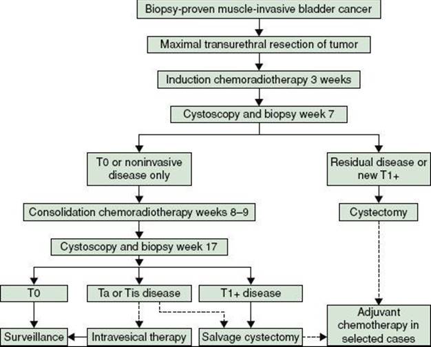
FIGURE 64.4. Integration of neoadjuvant chemotherapy, radiotherapy, and surgery in the United Kingdom.
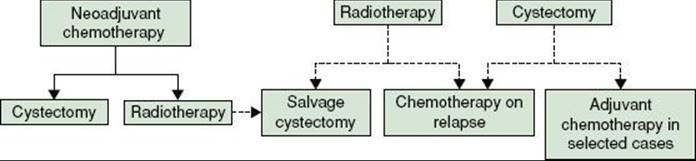
FIGURE 64.5. Acute toxicity of radiotherapy with and without synchronous chemotherapy. Graphs show worst grade of on-treatment toxicity by radiotherapy dose and week. Darkest bars indicate National Cancer Institute common terminology class grade 4; white indicates grade 0. Proportions with a grade 3 or 4 at any time on treatment: 64/178 (36.0%) chemotherapy (CT) versus 50/182 (27.5%). No CT-stratified chi-square test (P = .07).
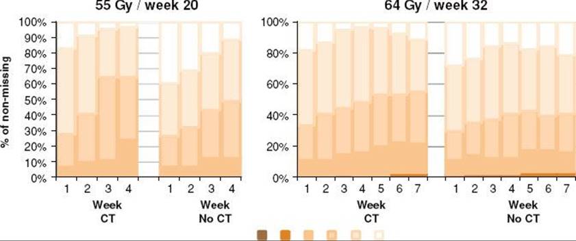
Treatment Results
It is difficult to compare results of older series to more contemporary ones for a number of reasons. First, staging systems have changed over the years, as have imaging techniques. Patients who may have been staged as organ confined in the early or pre-CT era may now be more accurately staged as more advanced with contemporary imaging. In contrast, surgical series will report accurate pathological stage. It is well documented that more accurate staging will bring about a paradoxical improvement in reported results by stage due to upstaging of apparently early disease cases, the so-called Will Rogers phenomenon.214 Treatment techniques in the past were obviously less refined, with little or no ability for conformal beam shaping and hence toxicity would have been higher. Nonetheless, it is clear from large series that tumor control could be attained in significant numbers of patients. For example, a study from Western General Hospital, Edinburgh, conducted between 1971 and 1982, reported treatment results from 963 patients. The reported stage mix was: T1, 20%, T2, 32%, T3, 40%, and T4, 8%, with the administered dose of 55 Gy in 20 fractions—a widely used UK schedule. The overall 5- and 10-year survival rates were 30% and 18%, respectively.215 This study reports tumor control at cystoscopy in 46% of patients, and this seems to be a pretty typical response rate with radiotherapy alone.
Similar control rates were reported in the radiotherapy alone arm of the National Cancer Institute of Canada (NCIC) trial comparing radiotherapy with chemoradiotherapy with cisplatinum212 and also in the radiotherapy only arms at 2 years of the UK trials BCON12 and BC2001194 at 2 years. The latter two trials give an accurate reflection of the results of modern radiotherapy. They ran contemporaneously between 2000 and 2009 and between them recruited more than 800 patients from around 70 UK sites, including all major UK radiotherapy centers. Both trials had similar designs, including near identical entry criteria (essentially T2–4aN0M0, with a small number of T1G3 patients entered the BCON trial), the same control arms (radical radiotherapy to bladder only to either 55 Gy in 20 fractions or 64 Gy in 32 fractions), and the same outcome measures (locoregional disease-free and overall survival). Both trials assessed both acute and late toxicity (Figs. 64.5 and 64.6). In addition, the BC2001 trial included an optional second randomization comparing whole-bladder radiotherapy with a reduced dose to uninvolved bladder of 80% of the isocenter dose. The BC2001 permitted neoadjuvant chemotherapy, which was a stratification factor. Around one-third of the chemoradiotherapy patients also received neoadjuvant chemotherapy.
The trials give a comprehensive insight into UK radiotherapy practice and great detail on outcomes. Median age in both trials was 73 to 74 years, with age range up to 90 years. Overall, around 40% of patients received 55 Gy in 20 fractions and 60% 64 Gy in 32 fractions. In both trials, over 95% of patients received at least 90% of the target dose in both arms, with no reduction associated with the two radio-sensitizing treatments. Although the protocol recommended complete debulking at TURBT, this was only achieved in one-third to one-half of the cases, probably reflecting technical factors at surgery rather than deliberate intent.
The overall 5-year survival rate was 50% for radiotherapy plus carbogen and nicotinamide compared with 39% for radiotherapy alone (48% and 35%, respectively, if T1 tumors are excluded), with the greatest effect seen in T2 tumors. For BC2001, the estimated 5-year survival with radiotherapy alone was very similar at 34% (95% CI, 25% to 43%).
FIGURE 64.6. Late toxicity in the BC2001 trial. Darkest bars indicate Radiation Therapy Oncology Group (RTOG) grade 4; white indicates grade 0. Proportion of patients with grade 3 or 4 RTOG toxicity at any month during follow-up (6 months onward), up to 3 months before a recurrence: RT: 12.9% sRT vs. 17.9% RHDV; odds ratio (95% confidence interval) 1.34 (0.55, 3.24); P = .52; CT: 8.3% CT vs. 15.7% no CT; odds ratio 0.48 (0.21, 1.10); P = .07. Patients with no available assessment were excluded from analysis. RT, radiotherapy; sRT, standard volume radiotherapy; RHDV, reduced high dose volume radiotherapy; CT, chemotherapy with radiotherapy.
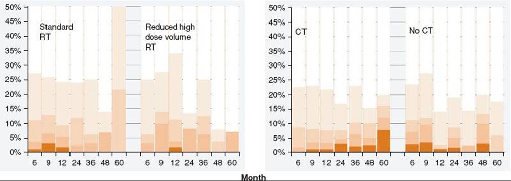
FIGURE 64.7. Invasive locoregional disease-free survival with or without chemotherapy.
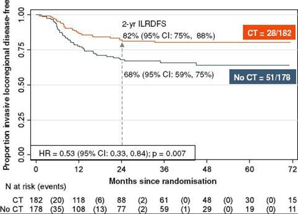
Toxicity
The toxicity of radical radiotherapy varies with the dose and schedule used. Within BC2001 and BCON, there were two schedules: 55 Gy in 20 fractions and 64 Gy in 32 fractions. Acute toxicity is shown in Figure 64.5 for both schedules, with and without chemotherapy with 5-fluorouacil/mitomycin-C.
As can be seen, the majority of patients experience only grade 1 or 2 toxicity with very low rates of grade 4 events. Synchronous chemotherapy increased toxicity but was predominantly grade 1 or 2, with a nonstatistically significant effect on grade 3 or 4 events (27.5 vs. 36%; P = .07; boundary for significance set at 0.01 to reflect multiplicity of testing). The reduced volume randomization failed to demonstrate a reduction in overall toxicity. However, when the volume of bowel irradiated was calculated, this did show a significant reduction; in an exploratory analysis, volume of bowel irradiated did correlate with the risk of grade 3 or 4 toxicity. There was no increase in acute toxicity observed in BC2001 if patients had received prior neoadjuvant chemotherapy.
Treatment completion rates in both trials were very high, with an excess of 95% of patients receiving the prescribed dose. The majority of patients failing to complete did so for tumor-related reasons rather than toxicity. Late toxicity was of great interest in both trials and was analyzed somewhat differently. In BCON, the cumulative rates of grade 3 or 4 events were reported, which gives the worst-case scenario but does not reflect the overall toxicity at any given time point posttreatment. BC2001 reports the toxicity rates at prespecified time points.
There are a number of points of note here. First, late toxicity was the same with both randomizations in BC2001 (i.e., reduced-volume radiotherapy did not impact late toxicity). More significantly, the addition of synchronous chemotherapy also had no effect on reported late side effects. In addition, at any given point, 75% to 80% of patients report no late toxicity at all. This is supported by bladder capacity measurements in BC2001, which show a mean change in bladder volume at 1 and 2 years of <5 mL. Of those reported side effects, fewer than 5% report grade 4 events and fewer than 10% overall grade 3. Very similar findings pertain to the BCON trial.12 In 2009, Efstathiou et al.216 reported late toxicity results in 285 patients who had participated in four Radiation Therapy Oncology Group trimodality therapy trials. Overall 5.7% reported persistent late genitourinary and 1.9% gastrointestinal toxicity of grade 3 or above, consistent with the BCON and BC2001 results. The good toxicity and functional results associated with radiotherapy are borne out by surveys of quality of life and symptoms in cystectomy and radiotherapy patients carried out by Henningsohn et al.13,217–220 Furthermore, radiotherapy and cystectomy patients report different patterns of symptoms, with sexual dysfunction being more prominent in surgical patients and bowel symptoms more prominent in radiotherapy patients. The preservation of sexual function by radiotherapy is particularly striking given that the patients were an average of around 10 years older.
Effect of Synchronous Chemotherapy
There are a large number of phase I or II trials from North America,208,216,221–222,223,224–230,231,232–237 mainland Europe,5,238–240 and the United Kingdom241,242 that have examined various chemotherapy agents in combination with radiotherapy. There are, however, only a few trials reported in which radiotherapy alone is compared with radiotherapy with synchronous chemotherapy. The only randomized study using the trimodality therapy approach was carried out by the NCIC and reported in 1996.212 This study compared radiotherapy to 40 Gy in 20 fractions with the same schedule combined with cisplatinum 100 mg/m2 twice weekly é 3. Patients then underwent interim cystoscopy and either consolidation radiotherapy to 20 Gy in 10 fractions or cystectomy depending on response and fitness for surgery. Synchronous cisplatinum had no effect on the rate of distant metastasis, consistent with a lack of effect in neoadjuvant trials.243 The study, however, was relatively small and could only have detected very large effects. Concurrent cisplatinum had a highly significant effect on pelvic recurrence, with 25 of 48 control patients having a pelvic recurrence, compared with 15 of 51 cisplatinum-treated patients (P = .036; HR 0.50; 90% CI, 0.29 to 0.86). It should be noted that a similar platinum schedule given prior to radiotherapy had no effect whatsoever on recurrences, either local or distant.213,243
There are two randomized trials of radio sensitization using UK schedules: the BC2001 and BCON studies, as already discussed. These two large phase III randomized control trials reported results in 2009 to 2010. These trials used radiosensitization with either concurrent chemotherapy (BC2001) or carbogen and nicotinamide (BCON). BC2001 has shown a significant improvement in locoregional disease-free survival with concurrent 5-fluorouracil and mitomycin-C of 34% (HR 0.66; 95% CI, 0.46 to 0.95) driven by a reduction of 47% in invasive locoregional recurrences (HR 0.53; P = .007), which is very similar to that observed in the NCIC trial with cisplatinum using the trimodality approach.212 Figure 64.7 shows the Kaplan-Meier curves for invasive locoregional disease-free survival. Survival data show a trend toward an improvement in overall survival (HR 0.81; P = .16), although the data are immature.194 The BCON trial12 with synchronous carbogen and nicotinamide narrowly failed to meet its primary end point of an improvement in local relapse-free survival (HR 0.87; 63% vs. 74%; P = .1) but did report an improvement in overall survival at 3 years (46% vs. 59%; HR 0.86; 95% CI, 0.745 to 0.996; P = .04).
Taken together the trial data with chemoradiotherapy suggests good tolerability even in relatively elderly patients with excellent late toxicity profiles in the majority of patients whether treated with the North American trimodality approach216 or the UK single treatment block.194 The similar hazard ratios observed with cisplatinum and 5-fluorouacil/mitomycin-C suggests that a range of chemotherapy approaches can probably be used with the selection based on toxicity. It is noteworthy that comparisons of platinum and 5-fluorouacil-based chemoradiotherapy combinations have been carried out in anal cancer, with the two approaches being similarly effective.244 The high rates of locoregional control seen make radical chemoradiotherapy a viable treatment option for many patients presenting with muscle-invasive bladder cancer.
FIGURE 64.8. Patterns of failure after chemoradiotherapy in BC2001.

Pattern of Failure
As already discussed above, a significant proportion of patients will experience local failure and undergo salvage surgery. The BC2001 study gives a good indication of patterns of failure using the single block approach to radical radiotherapy. A summary of relapse patterns in the chemoradiotherapy arm of the trial is shown in Figure 64.8.
There are a number of features of note here. First, the majority of locoregional failures are in the bladder, and there are more noninvasive than invasive recurrences. This underlines the need for regular surveillance postradiotherapy and the requirement for good integration of radiotherapy and surgical services if patients are to be managed by bladder conservation. In the authors’ experience, the majority of noninvasive recurrences can be successfully managed conservatively without the need for cystectomy. For those with invasive recurrence, cystectomy remains an option if the patients are sufficiently fit. The low rate of nodal relapse in BC2001 is also of interest, as no attempt was made to include pelvic nodes in the field, although lower pelvic nodes would have been included in the treated volume. For improvement in the metastatic relapse rate, better systemic therapies must be found. However, the rate of second cancer is also notable and probably relates to the age of the patients and the historic high rate of tobacco consumption in the bladder cancer population.
Altered Fractionation Schedules
There are old data examining hyperfractionation in comparison to conventional treatment to 64 Gy in 32 fractions, both given as a split course. The data suggest an improvement with the hyperfractionated regimen; however, given the suboptimal nature of the control arm, it is hard to draw firm conclusions from this.245,246 A study from the Royal Marsden Hospital compared 64 Gy in 32 fractions to 60.8 Gy in 32 fractions over 26 days in 228 patients.247Hypofractionated schedules are widely used in the United Kingdom and elsewhere for radical treatment and for palliation (as discussed below). There are no modern trials comparing these schedules to 64 Gy in 32 fractions.
Palliative Radiotherapy
A prospective randomized trial was conducted in 500 patients to compare the outcome in two treatment groups. Group 1 received 35 Gy in 10 fractions and group 2 received 21 Gy in 3 fractions. Five hundred patients were recruited, but data on symptomatic improvement at 3 months were only available on 272 patients. Of these, 68% achieved symptomatic improvement (71% for 35 Gy, 64% for 21 Gy), with no evidence of a difference in efficacy or toxicity between the two arms. On the basis of these results, 21 Gy in 3 fractions is widely used in the United Kingdom as a palliative schedule.248 The data are congruent with other data on hypofractionation for palliation249,250 as well as the growing trend for hypofractionation in other pelvic sites, in particular the prostate.
Preoperative Irradiation
Preoperative radiotherapy has been investigated in the past, but interest in the technique has waned, largely because none of the trials carried out showed any evidence of worthwhile benefit. In addition, the observed chemosensitivity of bladder cancer lead to a wave of neoadjuvant chemotherapy trials carried out in the 1980s and 1990s, effectively ending interest in the technique. Given the subsequent improvements in both radiotherapy and surgical technique, it may be appropriate to revisit the possibility of combining surgery with radiotherapy, particularly given the apparent plateau in progress with systemic therapy since the licensing of gemcitabine more than 10 years ago. Most of the studies in the literature are old, retrospective, nonrandomized comparisons and little can be concluded from them.251
There are few randomized trials in the literature, and those that are available are also old, for the reasons outlined above. A study from the Royal Marsden Hospital randomized patients to preoperative radiotherapy to 40 Gy in 4 weeks followed by cystectomy, against definitive radiotherapy to 60 Gy in 6 weeks. The 5-year survival in patients receiving the combined therapy was 38% versus 29% for those treated with radiotherapy alone, the difference not reaching statistical significance.252 This trial of course did not compare radiotherapy plus surgery versus surgery alone and thus does not address the primary question in this section. However, it is noteworthy that no tumor was found in 31% of the cystectomy specimens, a similar complete response rate to that observed in the intergroup neoadjuvant MVAC study.7 Similar response rates have been reported in other series.253
A second randomized study was carried out by the Memorial Sloan-Kettering Cancer Center in New York comparing radiotherapy to 40 Gy in 4 weeks followed by cystectomy 4 weeks later with a shorter 20 Gy in a 5-day course with immediate cystectomy modeled on the Swedish preoperative rectal cancer trials. Again this study is not informative on the primary issue of the utility of adding radiotherapy to surgery but does demonstrate the feasibility and safety of such an approach.254 A retrospective review from the MD Anderson Cancer Center assessed the use of preoperative irradiation (50 Gy in 5 weeks) in patients with T3b bladder tumors.255 The 5-year local control rate was 91% in the preoperative group (n = 92) compared with 72% for those treated with radical cystectomy alone (n = 43; P = .003).
Parsons and Million256 reviewed the results of retrospective studies and six prospective randomized trials on the use of preoperative irradiation. They concluded that the use of the technique may improve outcomes by up to 15% to 20% at 5 years. In particular, they noted that many preoperative radiotherapy series report pathological complete response rates of around one-third, similar to that seen with neoadjuvant chemotherapy. There are no modern trials of preoperative radiotherapy, and the topic seems to be ripe for revisiting with modern techniques, particularly given the low long-term toxicity associated with chemoradiotherapy schedules, as summarized above.
Postoperative Radiotherapy
As with many areas of bladder cancer practice, there are little in the way of randomized data on the use of adjuvant radiotherapy following surgery. When used, it is mostly based on the grounds of positive surgical margins or tumor spillage at surgery, where a high local recurrence rate can be anticipated. With the use of neoadjuvant chemotherapy worldwide being at low levels, chemo-naive patients at high risk of recurrence can be offered adjuvant chemotherapy, although the evidence base for this approach is also somewhat thin. What few data there are on radiotherapy suggests that limited doses are well tolerated.257 Given the more solid evidence base for neoadjuvant chemotherapy and the failure of adjuvant chemotherapy trials to accrue, the use of postoperative radiotherapy would also seem to be a topic well-worth revisiting with modern treatment techniques. An approach combining neoadjuvant chemotherapy, surgery, and adjuvant radiotherapy for patients at high risk of local recurrence would combine all three of the major bladder cancer treatment modalities in a novel fashion.
Electron Beam
Electron beams are really only of interest in the context of intraoperative radiotherapy. The role of intraoperative radiotherapy for carcinoma of the bladder needs to be assessed in a prospective trial and cannot at present be recommended outside of a suitable study.
Neutron Beam
There are a small number of studies examining the use of neutrons beams for bladder cancer therapy. A single randomized trial failed to show a survival advantage but did show increased morbidity.258,259 There are currently no reasons to recommend neutron therapy for bladder cancer.
Treatment Techniques
Patient Position and Immobilization
• The patient should be planned and treated in the same position; supine with arms on their chest. Knee and ankle immobilization should be used to ensure patient positioning is reproducible.
• The rectum should be empty of flatus and feces. The use of daily microenemas may be considered.
• Patients should be asked to empty the bladder 15 minutes prior to scan.
• While breathing normally, the patient should have a CT scan performed with 3 to 5-mm slice spacing. Patients are scanned from bottom of ischial tuberosities to 3 cm above the dome of the bladder or bottom of L5 (whichever is higher). A flat top CT scanner should be used.
• Neither intravenous nor oral contrast is thought to be of benefit in this instance.
• Reference tattoos should be made at the base of the abdomen and over each hip. The location of the tattoos should be marked on the planning scan by the use of radio-opaque markers to allow cross-referencing of planning scan and setup instructions.
Volume or Field Localization
• The gross tumor volume can be difficult to define and should integrate information from the staging CT or MRI as well as the TURBT. MRI-CT fusion may be helpful, where available.
• The use of fiducial markers or contrast medium such as lipiodol at the time of TURBT has been explored and may help identify tumor for image-guided adaptive radiotherapy.
• There are few data on the optimal radiotherapy volume. A standard approach is to define the planning target volume as the whole bladder, identified by its noninvolved outer bladder wall with a 1.5-cm margin plus extravesical extent of tumor with a 2-cm margin.
• All planning and treatment should be carried out with the bladder empty to minimize the risk of geographic miss and to keep the treated volumes as small as possible. Patients with significant residual volumes post voiding should be considered for planning and treatment with a catheter in situ, although this is likely to increase urinary toxicity.
• There are no data to support the routine irradiation of radiologically negative lymph nodes. The nodal relapse rate in the BC2001 trial, with planning target volume and clinical target volume defined as above, was only 3% in the chemoradiotherapy arm and 6% with radiotherapy only.
Brachytherapy
There is considerable historic literature on the use of brachytherapy, particularly from the Netherlands. Reported results seem to be very good, for example, van der Werf-Messing et al.260 report the outcomes in 328 patients treated with 3 é 3.5 Gy external irradiation followed by a radium implant. Overall 5- and 10-year survival was 56% for T2 tumors and 39% and 13%, respectively, for T3 disease. Similar results have been obtained in more recent series with iridium-192 implants and manual afterloading.261,262 There are also descriptions in the literature of the use of high-dose rate afterloading, although one paper suggests that this may be less effective and more toxic than low-dose rate treatment.263 The striking feature of most of these series is the very low patient numbers and long time spans reported, suggesting that these techniques are only rarely used, even in centers with the relevant expertise.
Hyperthermia
Details on hyperthermia in combination with external-beam radiation therapy or chemotherapy are beyond the scope of this chapter.
Treatment of Patients with Uncommon Bladder Tumors
Squamous cell carcinoma is uncommon in the developed world, and many reported cases may in addition reflect sampling of squamous elements within a transitional cell carcinoma. As these tumors are often excluded from systemic therapy trials, there are few data on outcomes with treatment as most papers are retrospective collections of unconnected observations.264–265,266 It appears that local failure may be more prevalent than distant relapse, but even that observation is based on very few cases. Urachal adenocarcinomas are extremely rare. Surgical resection with a partial cystectomy and en bloc resection of the urachal ligament with umbilicus is the treatment of choice in the setting of localized disease. There is currently no definitive role for neoadjuvant or adjuvant chemotherapy in this tumor. Unfortunately, there are many patients who present with metastatic disease, which currently is not likely to be curable. There is no standard chemotherapy regimen for these patients. Carcinosarcoma is even more rare and appears to have a poor prognosis.267–269 Similar considerations apply to small cell carcinoma, although the consensus seems to be that metastasis is more likely and initial response to chemotherapy is usually good. These patients generally get treated along the same lines as small cell lung carcinoma, but the benefit of prophylactic cranial irradiation is unclear.270–275
ROLE OF SYSTEMIC THERAPY
Up to 50% of patients will develop metastatic disease. Bladder cancer is a chemosensitive disease, with responses reported to a range of agents. Systemic combination chemotherapy can be effective and result in tumor responses and symptom control. Overall response rates may be as high as 70%,286 with a median survival of approximately 12 to 14 months.35 These agents were initially evaluated in the metastatic setting and subsequently in the neoadjuvant and adjuvant settings. However, despite over 30 years of research in the field, there remain many unanswered questions, including the role of adjuvant therapy and optimal second-line chemotherapy.
Neoadjuvant Chemotherapy
There are two principal rationales for neoadjuvant chemotherapy: first, to improve survival in patients with micrometastatic disease,277 and second, to preserve the bladder by shrinking the primary tumor to facilitate radiotherapy as an alternative definitive therapy to surgery.278 The potential disadvantage of neoadjuvant chemotherapy, and a frequent reason cited for its nonuse, is the delay in definitive treatment (cystectomy or chemoradiotherapy), because this may lead to disease progression in a proportion of nonresponding patients who may conceivably become inoperable or unsuitable for radical organ preservation treatment. However, it is equally plausible that by undergoing neoadjuvant chemotherapy these patients can be identified as those with biologically aggressive disease and therefore spared the morbidity of futile radical therapy. It should also be noted that a significant proportion, possibly up to two-thirds, of bladder cancer patients are elderly with multiple comorbidities and poor renal function and, therefore, unsuitable for neoadjuvant chemotherapy. However, in the authors’ experience, a patient who is considered fit for a radical cystectomy is likely to be fit for chemotherapy as well.
There are two particularly key studies in the area of neoadjuvant chemotherapy. The South Western Oncology Group (SWOG) neoadjuvant study comparing surgery alone with 3 cycles of MVAC followed by surgery reported an estimated median survival of 6.2 years versus 3.8 years in favor of patients having neoadjuvant chemotherapy (P = .027).7 Updated results from the UK MRC/EORTC neoadjuvant chemotherapy trial, which used 3 cycles of cisplatinum, methotrexate, and vinblastine (CMV) prior to surgery or radiotherapy, show a statistically significant 16% reduction in the risk of death (HR 0.84; 95% CI, 0.72 to 0.99; P = .037), corresponding to an increase in 10-year survival from 30% to 36% after neoadjuvant chemotherapy.279 In total, 976 patients with high-grade T2 to T4a urothelial bladder cancer accrued over 5.5 years from 106 institutions were randomly assigned to 3 cycles of neoadjuvant CMV chemotherapy (n = 491) or no chemotherapy (n = 485), followed by the institution’s choice of definitive therapy with either radical cystectomy or radiation therapy. Of patients in the chemotherapy and no chemotherapy groups, 42% and 43%, respectively, received radiation therapy alone as definitive therapy. Pathologic complete response with neoadjuvant chemotherapy was 33%. Overall survival at 3 years in the two groups was 55.5% versus 50%, respectively. The recent update, after 8 years of follow-up, showed an increase in 3-year survival from 50% to 56%, an increase in 10-year survival from 30% to 36%, and an increase in median survival time of 7 months (from 37 to 44 months) in CMV-treated patients compared with those treated with local therapy only. The SWOG study showed that of the 82% patients who underwent cystectomy, 38% had no evidence of disease pathologically. Patients who achieved pT0 status had a better prognosis than those who did not, although this difference may be accounted for by better disease biology rather than treatment effect. By definition, the SWOG study did not address organ preservation, because the mandated treatment plan was for surgery following neoadjuvant chemotherapy.
Meta-analyses show a 5% overall survival benefit at 5 years with cisplatin-containing regimens.31 Thus, there have been two large randomized neoadjuvant studies in muscle-invasive bladder cancer, both showing significant survival advantage. Although many disciplines in cancer care would consider these data sufficient to change the standard of care, this does not seem to have taken place for the management of invasive bladder cancer.280 The lack of widespread adoption of neoadjuvant chemotherapy may relate to the selected patients in clinical trials, not reflecting typical muscle-invasive bladder cancer patients who may be older or have impaired renal functions, which may limit the applicability of some chemotherapy regimens. GC,33 standard,7 or accelerated281 MVAC are widely used with definitive radical treatment (either surgery or radiotherapy/synchronous chemoradiotherapy) 4 to 6 weeks later. It is worth noting that these trials have been restricted to patients with well-preserved renal function (typically glomerular filtration rate >60 mL/min), thus excluding a significant proportion of bladder cancer patients who are elderly or have ureteric obstruction and therefore deemed unsuitable for neoadjuvant chemotherapy. This regimen requires in-patient or prolonged hydration and therefore has a significant impact on patient quality of life and health service resources. Clinical trials investigating chemotherapy regimens that may broaden the spectrum of patients receiving cisplatin-based treatment have been conducted in palliative settings, for example, a split-dose regimen of cisplatin in combination with gemcitabine on days 1 and 8 of a 21-day schedule allowed safe treatment of patients with calculated glomerular filtration rate as low as 40 mL/min in the day-case setting.282 This regimen could be tested within a randomized clinical trial in the neoadjuvant setting and may go far in increasing the uptake of neoadjuvant chemotherapy. It is the responsibility of urology and oncology colleagues to work together to provide state-of-art care for our patients with muscle-invasive bladder cancer, which should include neoadjuvant chemotherapy prior to surgery or organ-preservation therapy in fit patients.
Adjuvant Chemotherapy
Adjuvant chemotherapy has the potential advantage of enabling better patient selection based on the findings at surgical and pathological staging. The major disadvantages, although, are the delay in systemic therapy and the inability to assess the response to chemotherapy in the absence of measurable disease. Randomized adjuvant chemotherapy studies (Table 64.8) have been conducted with the methodological flaws of inadequate sample size, early closure, and suboptimal choice of chemotherapy, preventing a clear interpretation of the results. A systematic review and meta-analysis of updated individual patient data from all available randomized controlled trials in the adjuvant setting has been performed. Updated data were collected, validated, and reanalyzed for 491 patients from 6 randomized controlled trials, representing 90% of all patients randomized in cisplatin-based combination chemotherapy trials and 66% of patients from all eligible trials. In view of this, the power of this meta-analysis was limited. The overall hazard ratio for survival of 0.75 (95% CI, 0.60 to 0.96; P = .019) suggests a 25% relative reduction in the risk of death favoring adjuvant chemotherapy. However, the impact of trials that stopped early, of patients not receiving allocated treatments, or not receiving salvage chemotherapy is less clear.283 An Italian multicenter randomized phase III trial enrolled 194 patients (one-third of its target) with muscle-invasive TCC and assigned them to 4 cycles of GC or observation after cystectomy. The trial was stopped early due to poor accrual. After a median follow-up of 32.5 months, relapses were similar in both groups (43% vs. 45%) with no difference in disease-free survival. The 3-year overall survival was 67% for the chemotherapy arm and 48% for the observation arm and the 3-year disease-free survival was 47% and 35%, respectively, suggesting no statistically significant improvement in either survival rate with adjuvant GC in these patients.284 A large phase III trial by EORTC (protocol 30994) evaluating observation versus adjuvant chemotherapy with one of the three chemotherapy regimens (GC, MVAC, or high-dose MVAC) in high-risk bladder cancer (pT3–4 and/or node-positive disease) was also prematurely closed due to poor accrual after enrollment of 278 of a planned 1,344 patients. This appropriately designed and sized study thus failed to conclusively address the issue of adjuvant chemotherapy following cystectomy. A possible reason for the failure of this trial was the tight window postsurgery for commencing chemotherapy. In the authors’ experience, relatively few patients were able to enter the trial sufficiently soon due to slow postoperative recovery. This suggests that adjuvant chemotherapy may be less suitable than neoadjuvant chemotherapy in this patient population. The adjuvant chemotherapy question still requires international collaboration with an appropriately sized pragmatic study for a conclusive answer. In various disease sites neoadjuvant chemotherapy followed by adjuvant chemotherapy is an acceptable norm with level I evidence. With an anticipated rise in uptake of neoadjuvant chemotherapy, patients with bladder cancer should not be precluded from this strategy where neoadjuvant chemotherapy is followed by radical treatment followed by a trial of adjuvant chemotherapy versus no chemotherapy in an appropriately sized phase III trial that meets its recruitment target, thus providing a definite answer for or against this strategy. Previous failures in meeting recruitment targets can be overcome by using better-tolerated regimens in common use now and by extending the recruitment window from 12 to 16 weeks to allow full recovery postsurgery and to allow more dose-intense adjuvant chemotherapy treatment.
TABLE 64.8 RANDOMIZED TRIALS OF ADJUVANT CHEMOTHERAPY
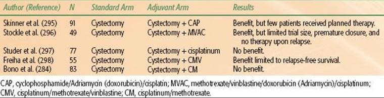
Choice of Chemotherapy Regimen in Metastatic Disease
Currently, systemic combination chemotherapy is the only treatment that may prolong survival in patients with metastatic disease. The chemosensitivity of bladder cancer is demonstrated by objective response rates of 12% to 73% and complete response rates of 0% to 35%.285 Although antitumor activity has been demonstrated with several single agents, the median survival associated with single-agent therapy is short (4 to 6 months). Prior to the development of gemcitabine, a range of trials were done with doublet regimes, which typically showed median survival times of approximately 8 months.285 The development of the four drug combination for MVAC chemotherapy extended this to over 12 months and became the established standard of care.286
The triplet combination CMV is also widely used but has not been compared with MVAC directly. Experimental data suggested that a combination of gemcitabine and cisplatin given using an appropriate schedule (simultaneous or close proximity exposure) can act synergistically. Synergy may be mediated either by inhibition of ribonucleotide reductase by gemcitabine, depleting the deoxynucleotide pool required for DNA replication and thereby inhibiting excision repair of cisplatin-induced DNA crosslinks, or by gemcitabine incorporation into DNA, facilitating cisplatin crosslink formation.287,288 The combination of the two drugs proved active and tolerable33 and was subsequently compared with MVAC in a phase III trial. Patients were randomized to GC (gemcitabine 1,000 mg/m2 days 1, 8, and 15; cisplatin 70 mg/m2 day 2) or standard MVAC every 28 days for a maximum of 6 cycles. Four hundred five patients were randomized (GC, n = 203; MVAC, n = 202). The groups were well balanced with respect to prognostic factors. Overall survival was similar on both arms (HR 1.04; 95% CI, 0.82 to 1.32; P = .75), as were time to progressive disease (HR 1.05; 95% CI, 0.85 to 1.30), time to treatment failure (HR 0.89; 95% CI, 0.72 to 1.10), and response rate (GC, 49%; MVAC, 46%). More GC patients completed 6 cycles of therapy, with fewer dose adjustments. The toxic death rate was 1% on the GC arm and 3% on the MVAC arm. More GC than MVAC patients had grade 3 or 4 anaemia (27% vs. 18%, respectively) and thrombocytopenia (57% vs. 21%, respectively). More MVAC patients, had grade 3 or 4 neutropenia (82% vs. 71%), neutropenic fever (14% vs. 2%), neutropenic sepsis (12% vs. 1%), grade 3 or 4 mucositis (22% vs. 1%), and alopecia (55% vs. 11%). Because of the higher incidence of neutropenic fever and mucositis, more hospital admissions were required for the MVAC arm (49 admissions for a total of 272 days) than for the GC group (9 admissions for a total of 33 days), resulting in considerably greater hospital resource utilization. The quality of life was maintained during treatment on both arms; however, patients on GC fared better regarding weight, performance status, and fatigue. This study thus demonstrated that GC provides a similar survival to MVAC with a better safety profile and tolerability.34,35 Therefore, although this trial was not designed to show equivalence of the two regimens, many interpret the results as showing therapeutic noninferiority and have adopted GC as the new standard in view of the better tolerability. With the upper boundary of the confidence interval of the adjusted hazard ratio for survival close to 1.2, noninferiority can reasonably be assumed. Therefore, GC is a valuable alternative for the growing elderly patient population with metastatic bladder cancer who may derive equal benefit from this regimen as compared with MVAC but with fewer side effects.
The addition of paclitaxel to GC was evaluated in a phase III clinical trial by EORTC (protocol 30987), which enrolled 627 patients with advanced urothelial carcinoma. The regimens were well tolerated. Results showed that the GCP arm resulted in a higher rate of overall response rate (57% vs. 46%) CR (15% vs. 10%) and survival (15.7 vs. 12.8 months) compared with GC arm, but these differences were not statistically significant.289
Combination of docetaxel and cisplatin (DC) has been compared with MVAC in a multicenter phase III clinical trial by the Hellenic Co-operative Oncology Group.290 Patients (n = 220) were randomly assigned to MVAC every 4 weeks versus docetaxel plus cisplatin every 3 weeks. Treatment with MVAC resulted in significantly superior response rate (54.2% vs. 37.4%), median time to progression (9.4 vs. 6.1 months), and median survival (14.2 vs. 9.3 months), suggesting that MVAC was superior to DC. Toxicity of MVAC was considerably lower than that previously reported for MVAC administered without granulocyte colony-stimulating factor. Currently GC, MVAC, and high-dose MVAC with granulocyte colony-stimulating factor support are the acceptable standard of care as first-line chemotherapy in advanced or metastatic bladder cancer setting.
Second-Line Chemotherapy Treatment
The role of salvage chemotherapy after relapse following first-line chemotherapy remains an important subject of recent clinical trials. The enrollment in such trials is challenging in view of patients’ poor performance status and deranged renal functions. Although various phase I or II studies have been reported, to date there is only one completed phase III trial comparing vinflunine with best supportive care (BSC) alone. Patients (n = 370) were randomly assigned in a 2 to 1 ratio to receive vinflunine plus BSC (n = 253) or BSC alone (n = 117). Both arms were well balanced. Grade ≥3 toxicities for the vinflunine arm were neutropenia (50%), febrile neutropenia (6%), anemia (19%), fatigue (19%), and constipation (16%). A median survival advantage of 2.3 months (6.9 months for vinflunine plus BSC vs. 4.6 months for BSC) was achieved but was not statistically significant (P = .287). Cox multivariate analysis adjusting for prognostic factors showed a statistically significant effect of vinflunine on overall survival (P = .036), reducing the death risk by 23%. Objective response rate (8.6% vs. 0%), disease control (41.4% vs. 24.8%), and progression-free survival (3.0 vs. 1.5 months) were all statistically significant, favoring vinflunine. Because vinflunine was well tolerated, it is a reasonable second-line therapy option for patients with bladder cancer who have relapsed following cisplatin-based therapy.291 Future second-line chemotherapy studies should incorporate vinflunine as the standard of care arm when testing any experimental treatment in a randomized trial.
REFERENCES
1. Lotan Y, Kamat AM, Porter MP, et al. Key concerns about the current state of bladder cancer: a position paper from the Bladder Cancer Think Tank, the Bladder Cancer Advocacy Network, and the Society of Urologic Oncology. Cancer 2009;115(18):4096–4103.
2. Bryan RT, Wallace DM. Have we abandoned the “superficial” in bladder cancer? Eur Urol 2009;56(6):1091.
3. Riley GF, Potosky AL, Lubitz JD, et al. Medicare payments from diagnosis to death for elderly cancer patients by stage at diagnosis. Med Care 1995;33(8):828–841.
4. Stein JP, Lieskovsky G, Cote R, et al. Radical cystectomy in the treatment of invasive bladder cancer: long-term results in 1,054 patients. J Clin Oncol 2001;19(3):666–675.
5. Rödel C, Grabenbauer GG, Kuhn R, et al. Combined-modality treatment and selective organ preservation in invasive bladder cancer: long-term results [see comment]. J Clin Oncol 2002;20(14):3061–3071.
6. Hayter CR, Paszat LF, Groome PA, et al. The management and outcome of bladder carcinoma in Ontario, 1982–1994. Cancer 2000;89(1):142–151.
7. Grossman HB, Natale RB, Tangen CM, et al. Neoadjuvant chemotherapy plus cystectomy compared with cystectomy alone for locally advanced bladder cancer [see comment] [erratum appears in N Engl J Med2003;349(19):1880]. N Engl J Med2003;349(9):859–866.
8. Konety BR, Joslyn SA. Factors influencing aggressive therapy for bladder cancer: an analysis of data from the SEER program. J Urol 2003;170(5):1765–1771.
9. Sriplakich S, Jahnson S, Karlsson MG. Epidermal growth factor receptor expression: predictive value for the outcome after cystectomy for bladder cancer? BJU Int 1999;83(4):498–503.
10. Munro NP, Sundaram SK, Weston PM, et al. A 10-year retrospective review of a nonrandomized cohort of 458 patients undergoing radical radiotherapy or cystectomy in Yorkshire, UK. Int J Radiat Oncol Biol Phys2010;77(1):119–124.
11. James ND, Hussain SA, Hall E, et al. Radiotherapy with or without chemotherapy in muscle-invasive bladder cancer. N Engl J Med 2012;366(16):1477–1488.
12. Hoskin PJ, Rojas AM, Bentzen SM, et al. Radiotherapy with concurrent carbogen and nicotinamide in bladder carcinoma. J Clin Oncol 2010;28(33):4912–4918.
13. Henningsohn L, Wijkstrom H, Dickman PW, et al. Distressful symptoms after radical radiotherapy for urinary bladder cancer. Radiother Oncol 2002;62(2):215–225.
14. International Agency for Research on Cancer. GLOBOCAN 2008: Cancer incidence, mortality, and prevalence worldwide. Available at: http://globocan.iarc.fr/.
15. Ploeg M, Aben KK, Kiemeney LA. The present and future burden of urinary bladder cancer in the world. World J Urol 2009;27(3):289–293.
16. Bryan RT, Zeegers MP, Bird D, et al. 20-years of bladder cancer research in the West Midlands: a comparison of two large cohorts. J Urol 2010;183:e196.
17. Zlotta AR, Cohen SM, Dinney C, et al. BCAN think tank session 1: overview of risks for and causes of bladder cancer. Urol Oncol 2010;28(3):329–333.
18. Volanis D, Kadiyska T, Galanis A, et al. Environmental factors and genetic susceptibility promote urinary bladder cancer. Toxicol Lett 2010;193(2):131–137.
19. Salem S, Mitchell RE, El-Alim El-Dorey A, et al. Successful control of schistosomiasis and the changing epidemiology of bladder cancer in Egypt. BJU Int 2011;107(2):206–211.
20. Hein DW. N-acetyltransferase 2 genetic polymorphism: effects of carcinogen and haplotype on urinary bladder cancer risk. Oncogene 2006;25(11):1649–1658.
21. Miller AB. The etiology of bladder cancer from the epidemiological viewpoint. Cancer Res 1977;37(8 Pt 2):2939–2942.
22. Hoffman D, Masuda Y, Wynder EL. Alpha-naphthylamine and beta-naphthylamine in cigarette smoke. Nature 1969;221(5177):255–256.
23. Lopez-Abente G, Gonzalez CA, Errezola M, et al. Tobacco smoke inhalation pattern, tobacco type, and bladder cancer in Spain. Am J Epidemiol 1991;134(8):830–839.
24. Claude J, Kunze E, Frentzel-Beyme R, et al. Life-style and occupational risk factors in cancer of the lower urinary tract. Am J Epidemiol 1986;124(4):578–589.
25. Sorahan T, Lancashire RJ, Sole G. Urothelial cancer and cigarette smoking: findings from a regional case-controlled study. Br J Urol 1994;74(6):753–756.
26. Kaufman DS, Shipley WU, Feldman AS. Bladder cancer. Lancet 2009;374(9685):239–249.
27. Wallace DM, Bryan RT, Dunn JA, et al. Delay and survival in bladder cancer. BJU Int 2002;89(9):868–878.
28. Kiemeney LA, Witjes JA, Verbeek AL, et al. The clinical epidemiology of superficial bladder cancer. Dutch South-East Cooperative Urological Group. Br J Cancer 1993;67(4):806–812.
29. Millan-Rodriguez F, Chechile-Toniolo G, Salvador-Bayarri J, et al. Multivariate analysis of the prognostic factors of primary superficial bladder cancer. J Urol 2000;163(1):73–78.
30. Sylvester RJ, van der Meijden AP, Oosterlinck W, et al. Predicting recurrence and progression in individual patients with stage Ta T1 bladder cancer using EORTC risk tables: a combined analysis of 2596 patients from seven EORTC trials. Eur Urol2006;49(3):466–465.
31. Neoadjuvant chemotherapy in invasive bladder cancer: update of a systematic review and meta-analysis of individual patient data advanced bladder cancer (ABC) meta-analysis collaboration. Eur Urol 2005;48(2):202–205.
32. Sternberg CN, Yagoda A, Scher HI, et al. Preliminary results of M-VAC (methotrexate, vinblastine, doxorubicin and cisplatin) for transitional cell carcinoma of the urothelium. J Urol 1985;133(3):403–407.
33. Moore MJ, Winquist EW, Murray N, et al. Gemcitabine plus cisplatin, an active regimen in advanced urothelial cancer: a phase II trial of the National Cancer Institute of Canada Clinical Trials Group. J Clin Oncol1999;17(9):2876–2881.
34. von der Maase H, Hansen SW, Roberts JT, et al. Gemcitabine and cisplatin versus methotrexate, vinblastine, doxorubicin, and cisplatin in advanced or metastatic bladder cancer: results of a large, randomized, multinational, multicenter, phase III study. J Clin Oncol 2000;18(17):3068–3077.
35. von der Maase H, Sengelov L, Roberts JT, et al. Long-term survival results of a randomized trial comparing gemcitabine plus cisplatin, with methotrexate, vinblastine, doxorubicin, plus cisplatin in patients with bladder cancer. J Clin Oncol2005;23(21):4602–4608.
36. Hussain SA, James ND. The systemic treatment of advanced and metastatic bladder cancer. Lancet Oncol 2003;4(8):489–497.
37. Rehn L. Blasengeschwultse bei Fuchsein-arbeitern. Arch Clin Chir 1895;50:588–600.
38. Lower GMJ. Concepts in causality: chemically-induced human urinary bladder cancer. Cancer 1982;49:1056–1066.
39. Hueper WC, Wiley FH, Wolfe HD. Experimental production of bladder tumours in dogs by administration of beta-naphthylamine. Indust Hyg Toxicol 1938;20:46–84.
40. Case RAM, Hosker ME, McDonald DB, et al. Tumors of the urinary bladder in workmen engaged in the manufacture and use of certain dyestuff intermediates in the British chemical industry. Role of aniline, benzidine, alpha-naphthylamine, and beta-naphthylamine. Br J Indust Med 1954;11:75–104.
41. Clayson D. Working hypothesis for the mode of carcinogenesis of aromatic amines. Br J Cancer 1953;7:460–471.
42. King CM, Phillips B. Enzyme-catalyzed reactions of the carcinogen H-hydroxy-2-fluorenylacetamide with nucleic acid. Science 1968;159:1351–1353.
43. Radomski JL, Brill E. Bladder cancer induction by aromatic amines: role of N-hydroxy metabolites. Science 1970;167:992–993.
44. Risch A, Wallace DM, Bathers S, et al. Slow N-acetylation genotype is a susceptibility factor in occupational and smoking related bladder cancer. Hum Mol Genet 1995;4(2):231–236.
45. Probert JL, Persad RA, Greenwood RP, et al. Epidemiology of transitional cell carcinoma of the bladder: profile of an urban population in the south-west of England. Br J Urol 1998;82(5):660–666.
46. Morrison AS, Ahlbom A, Verhoek WG, et al. Occupation and bladder cancer in Boston, USA, Manchester, UK, and Nagoya, Japan. J Epidemiol Commun Health 1985;39(4):294–300.
47. Sorahan T, Hamilton L, Jackson JR. A further cohort study of workers employed at a factory manufacturing chemicals for the rubber industry, with special reference to the chemicals 2-mercaptobenzothiazole (MBT), aniline, phenyl-beta-naphthylamine and o-toluidine. Occup Environ Med 2000;57(2):106–115.
48. Guha N, Steenland NK, Merletti F, et al. Bladder cancer risk in painters: a meta-analysis. Occup Environ Med 2010;67(8):568–573.
49. Strope SA, Montie JE. The causal role of cigarette smoking in bladder cancer initiation and progression, and the role of urologists in smoking cessation. J Urol 2008;180(1):31–37.
50. Baris D, Karagas MR, Verrill C, et al. A case-control study of smoking and bladder cancer risk: emergent patterns over time. J Natl Cancer Inst 2009;101(22):1553–1561.
51. Freedman ND, Silverman DT, Hollenbeck AR, et al. Association between smoking and risk of bladder cancer among men and women. JAMA 2011;306(7):737–745.
52. Lammers RJ, Witjes WP, Hendricksen K, et al. Smoking status is a risk factor for recurrence after transurethral resection of non-muscle-invasive bladder cancer. Eur Urol 2011;60(4):713–720.
53. Moore LE, Baris DR, Figueroa JD, et al. GSTM1 null and NAT2 slow acetylation genotypes, smoking intensity and bladder cancer risk: results from the New England bladder cancer study and NAT2 meta-analysis. Carcinogenesis 2011;32(2):182–189.
54. Rothman N, Garcia-Closas M, Chatterjee N, et al. A multi-stage genome-wide association study of bladder cancer identifies multiple susceptibility loci. Nat Genet 2010;42(11):978–984.
55. Sanderson S, Salanti G, Higgins J. Joint effects of the N-acetyltransferase 1 and 2 (NAT1 and NAT2) genes and smoking on bladder carcinogenesis: a literature-based systematic HuGE review and evidence synthesis. Am J Epidemiol 2007;166(7):741–751.
56. Koutros S, Silverman DT, Baris D, et al. Hair dye use and risk of bladder cancer in the New England bladder cancer study. Int J Cancer 2011;129(12):2894–2904.
57. Yuan JM, Chan KK, Coetzee GA, et al. Genetic determinants in the metabolism of bladder carcinogens in relation to risk of bladder cancer. Carcinogenesis 2008;29(7):1386–1393.
58. Garcia-Closas M, Ye Y, Rothman N, et al. A genome-wide association study of bladder cancer identifies a new susceptibility locus within SLC14A1, a urea transporter gene on chromosome 18q12.3. Hum Mol Genet2011;20(21):4282–4289.
59. Grotenhuis AJ, Vermeulen SH, Kiemeney LA. Germline genetic markers for urinary bladder cancer risk, prognosis and treatment response. Future Oncol 2010;6(9):1433–1460.
60. Liang FX, Bosland MC, Huang H, et al. Cellular basis of urothelial squamous metaplasia: roles of lineage heterogeneity and cell replacement. J Cell Biol 2005;171(5):835–844.
61. Hodder SL, Mahmoud AA, Sorenson K, et al. Predisposition to urinary tract epithelial metaplasia in Schistosoma haematobium infection. Am J Trop Med Hyg 2000;63(3–4):133–138.
62. Tawfik HN. Carcinoma of the urinary bladder associated with schistosomiasis in Egypt: the possible causal relationship. Princess Takamatsu Symp 1987;18:197–209.
63. Elem B, Patil PS. Pattern of urological malignancy in Zambia. A hospital-based histopathological study. Br J Urol 1991;67(1):37–39.
64. el-Mawla NG, el-Bolkainy MN, Khaled HM. Bladder cancer in Africa: update. Semin Oncol 2001;28(2):174–178.
65. Al Adnani MS, Saleh KM. Schistosomiasis and bladder carcinoma in southern Iraq. J Trop Med Hyg 1983;86(3):93–97.
66. Cooppan RM, Bhoola KD, Mayet FG. Schistosomiasis and bladder carcinoma in Natal. S Afr Med J 1984;66(22):841–843.
67. Shaaban AA, Orkubi SA, Said MT, et al. Squamous cell carcinoma of the urinary bladder. Ann Saudi Med 1997;17(1):115–119.
68. Tricker AR, Mostafa MH, Spiegelhalder B, et al. Urinary excretion of nitrate, nitrite and N-nitroso compounds in Schistosomiasis and bilharzia bladder cancer patients. Carcinogenesis 1989;10(3):547–552.
69. Sheweita SA, Abu El-Maati MR, El-Shahat FG, et al. Changes in the expression of cytochrome P450 2E1 and the activity of carcinogen-metabolizing enzymes in Schistosoma haematobium-infected human bladder tissues. Toxicology 2001;162(1):43–52.
70. Lin JI, Yong HS, Tseng CH, et al. Diffuse cystitis glandularis associated with adenocarcinomatous change. Urology 1980;15(4):411–415.
71. Davis CP, Cohen MS, Gruber MB, et al. Urothelial hyperplasia and neoplasia: a response to chronic urinary tract infections in rats. J Urol 1984;132(5):1025–1031.
72. Bullock PS, Thoni DE, Murphy WM. The significance of colonic mucosa (intestinal metaplasia) involving the urinary tract. Cancer 1987;59(12):2086–2090.
73. Delnay KM, Stonehill WH, Goldman H, et al. Bladder histological changes associated with chronic indwelling urinary catheter. J Urol 1999;161(4):1106–1108.
74. Bell TE, Wendel RG. Cystitis glandularis: benign or malignant? J Urol 1968;100(4):462–465.
75. Andersen JA, Hansen BF. The incidence of cell nests, cystitis cystica and cystitis glandularis in the lower urinary tract revealed by autopsies. J Urol 1972;108(3):421–424.
76. Wiener DP, Koss LG, Sablay B, et al. The prevalence and significance of Brunn’s nests, cystitis cystica and squamous metaplasia in normal bladders. J Urol 1979;122(3):317–321.
77. Young RH, Bostwick DG. Florid cystitis glandularis of intestinal type with mucin extravasation: a mimic of adenocarcinoma. Am J Surg Pathol 1996;20(12):1462–1468.
78. Jacobs LB, Brooks JD, Epstein JI. Differentiation of colonic metaplasia from adenocarcinoma of urinary bladder. Hum Pathol 1997;28(10):1152–1157.
79. Hampson SJ, Falzon M, Cowie AG. Intestinal metaplasia of the bladder: implications for management. Br J Urol 1992;69(3):323–324.
80. Sidransky D, Frost P, Von Eschenbach A, et al. Clonal origin bladder cancer. N Engl J Med 1992;326(11):737–740.
81. Novelli M, Cossu A, Oukrif D, et al. X-inactivation patch size in human female tissue confounds the assessment of tumor clonality. Proc Natl Acad Sci USA 2003;100(6):3311–3314.
82. Chern HD, Becich MJ, Persad RA, et al. Clonal analysis of human recurrent superficial bladder cancer by immunohistochemistry of P53 and retinoblastoma proteins. J Urol 1996;156(5):1846–1849.
83. Tsai YC, Simoneau AR, Spruck CH 3rd, et al. Mosaicism in human epithelium: macroscopic monoclonal patches cover the urothelium. J Urol 1995;153(5):1697–1700.
84. Gaisa NT, Graham TA, McDonald SA, et al. The human urothelium consists of multiple clonal units, each maintained by a stem cell. J Pathol 2011;225(2):163–171.
85. Simon R, Eltze E, Schafer KL, et al. Cytogenetic analysis of multifocal bladder cancer supports a monoclonal origin and intraepithelial spread of tumor cells. Cancer Res 2001;61(1):355–362.
86. Hartmann A, Rosner U, Schlake G, et al. Clonality and genetic divergence in multifocal low-grade superficial urothelial carcinoma as determined by chromosome 9 and p53 deletion analysis. Lab Invest 2000;80(5):709–718.
87. Hafner C, Knuechel R, Zanardo L, et al. Evidence for oligoclonality and tumor spread by intraluminal seeding in multifocal urothelial carcinomas of the upper and lower urinary tract. Oncogene 2001;20(35):4910–4915.
88. Steidl C, Simon R, Burger H, et al. Patterns of chromosomal aberrations in urinary bladder tumours and adjacent urothelium. J Pathol 2002;198(1):115–120.
89. Jones PA, Baylin SB. The fundamental role of epigenetic events in cancer. Nat Rev Genet 2002;3(6):415–428.
90. Lindgren D, Gudjonsson S, Jee KJ, et al. Recurrent and multiple bladder tumors show conserved expression profiles. BMC Cancer 2008;8:183.
91. Majewski T, Lee S, Jeong J, et al. Understanding the development of human bladder cancer by using a whole-organ genomic mapping strategy. Lab Invest 2008;88(7):694–721.
92. Bryan RT, Zeegers MP, James ND, et al. Biomarkers in bladder cancer. BJU Int 2009;105(5):608–613.
93. Crawford JM. The origins of bladder cancer. Lab Invest 2008;88(7):686–693.
94. Knowles MA, Elder PA, Williamson M, et al. Allelotype of human bladder cancer. Cancer Res 1994;54(2):531–538.
95. Hurst CD, Fiegler H, Carr P, et al. High-resolution analysis of genomic copy number alterations in bladder cancer by microarray-based comparative genomic hybridization. Oncogene 2004;23(12):2250–2263.
96. Duggan BJ, Gray SB, McKnight JJ, et al. Oligoclonality in bladder cancer: the implication for molecular therapies. J Urol 2004;171(1):419–425.
97. Bryan RT, Hussain SA, James ND, et al. Molecular pathways in bladder cancer: part 1. BJU Int 2005;95(4):485–490.
98. Bryan RT, Hussain SA, James ND, et al. Molecular pathways in bladder cancer: part 2. BJU Int 2005;95(4):491–496.
99. Hanahan D, Weinberg RA. The hallmarks of cancer. Cell 2000;100(1):57–70.
100. Hanahan D, Weinberg RA. Hallmarks of cancer: the next generation. Cell 2011;144(5):646–674.
101. Reya T, Morrison SJ, Clarke MF, et al. Stem cells, cancer, and cancer stem cells. Nature 2001;414(6859):105–111.
102. Bonnet D, Dick JE. Human acute myeloid leukemia is organized as a hierarchy that originates from a primitive hematopoietic cell. Nat Med 1997;3(7):730–737.
103. Brandt WD, Matsui W, Rosenberg JE, et al. Urothelial carcinoma: stem cells on the edge. Cancer Metastasis Rev 2009;28(3–4):291–304.
104. Bryan RT. Bladder cancer and cancer stem cells: basic science and implications for therapy. Sci World J 2011;11:1187–1194.
105. Gupta PB, Chaffer CL, Weinberg RA. Cancer stem cells: mirage or reality? Nat Med 2009;15(9):1010–1012.
106. Visvader JE, Lindeman GJ. Cancer stem cells in solid tumours: accumulating evidence and unresolved questions. Nat Rev Cancer 2008;8(10):755–768.
107. Birkhahn M, Mitra AP, Cote RJ. Molecular markers for bladder cancer: the road to a multimarker approach. Expert Rev Anticancer Ther 2007;7(12):1717–1727.
108. Castillo-Martin M, Domingo-Domenech J, Karni-Schmidt O, et al. Molecular pathways of urothelial development and bladder tumorigenesis. Urol Oncol 2010;28(4):401–408.
109. Cheng L, Zhang S, MacLennan GT, et al. Bladder cancer: translating molecular genetic insights into clinical practice. Hum Pathol 2011;42(4):455–481.
110. Knowles MA. Molecular pathogenesis of bladder cancer. Int J Clin Oncol 2008;13(4):287–297.
111. Shariat SF, Karam JA, Lerner SP. Molecular markers in bladder cancer. Curr Opin Urol 2008;18(1):1–8.
112. Youssef RF, Mitra AP, Bartsch G Jr, et al. Molecular targets and targeted therapies in bladder cancer management. World J Urol 2009;27(1):9–20.
113. Goebell PJ, Knowles MA. Bladder cancer or bladder cancers? Genetically distinct malignant conditions of the urothelium. Urol Oncol 2010;28(4):409–428.
114. Knowles MA. Molecular genetics of bladder cancer. Br J Urol 1995;75(Suppl 1):57–66.
115. Knowles MA. Molecular genetics of bladder cancer: pathways of development and progression. Cancer Surv 1998;31:49–76.
116. Knowles MA. The genetics of transitional cell carcinoma: progress and potential clinical application. BJU Int 1999;84(4):412–427.
117. Knowles MA. What we could do now: molecular pathology of bladder cancer. Mol Pathol 2001;54(4):215–221.
118. Knowles MA. Molecular subtypes of bladder cancer: Jekyll and Hyde or chalk and cheese? Carcinogenesis 2006;27(3):361–373.
119. Knowles MA. Bladder cancer subtypes defined by genomic alterations. Scand J Urol Nephrol Suppl 2008;218:116–130.
120. Han Y, Chen J, Zhao X, et al. MicroRNA expression signatures of bladder cancer revealed by deep sequencing. PLoS One 2011;6(3):e18286.
121. Bryan RT, Wei W, Shimwell NJ, et al. Assessment of high-throughput high-resolution MALDI-TOF-MS of urinary peptides for the detection of muscle-invasive bladder cancer. Proteomics Clin Appl 2011;5(9–10):493–503.
122. Schiffer E, Vlahou A, Petrolekas A, et al. Prediction of muscle-invasive bladder cancer using urinary proteomics. Clin Cancer Res 2009;15(15):4935–4943.
123. Hawkins RD, Hon GC, Ren B. Next-generation genomics: an integrative approach. Nat Rev Genet 2010;11(7):476–486.
124. Edwards TJ, Dickinson AJ, Natale S, et al. A prospective analysis of the diagnostic yield resulting from the attendance of 4020 patients at a protocol-driven haematuria clinic. BJU Int 2006;97(2):301–305.
125. Khadra MH, Pickard RS, Charlton M, et al. A prospective analysis of 1,930 patients with hematuria to evaluate current diagnostic practice. J Urol 2000;163(2):524–527.
126. Sobin LH, Gospodarowicz MK, Wittekind C, eds. UICC International Union Against Cancer. TNM classification of malignant tumours, 7th ed. Hoboken, NJ: Wiley-Blackwell, 2010.
127. Mostofi FK, Sobin LH, Torloni H. Histological typing of urinary bladder tumours. Geneva: World Health Organization, 1973.
128. Goessl C, Knispel HH, Miller K, et al. Is routine excretory urography necessary at first diagnosis of bladder cancer? J Urol 1997;157(2):480–481.
129. European Association of Urology. Guidelines. 2011. Available at: http://www.uroweb.org/guidelines/online-guidelines/.
130. Miladi M, Peyromaure M, Zerbib M, et al. The value of a second transurethral resection in evaluating patients with bladder tumours. Eur Urol 2003;43(3):241–245.
131. European Organisation for Research and Treatment of Cancer. Bladder cancer calculator. Available at: http://www.eortc.be/tools/bladdercalculator/.
132. Sylvester RJ, Oosterlinck W, van der Meijden AP. A single immediate postoperative instillation of chemotherapy decreases the risk of recurrence in patients with stage Ta T1 bladder cancer: a meta-analysis of published results of randomized clinical trials. J Urol 2004;171(6 Pt 1):2186–2190.
133. Mowatt G, N’Dow J, Vale L, et al. Photodynamic diagnosis of bladder cancer compared with white light cystoscopy: systematic review and meta-analysis. Int J Technol Assess Health Care 2011;27(1):3–10.
134. Vrooman OP, Witjes JA. Urinary markers in bladder cancer. Eur Urol 2008;53(5):909–916.
135. Lamm DL. Carcinoma in situ. Urol Clin North Am 1992;19(3):499–508.
136. American Urology Association. Guidelines on managment of bladder cancer. Available at: http://www.auanet.org/content/guidelines-and-quality-care/clinical-guidelines.cfm?CFID=7269186&CFTOKEN=35fc09c2077430ac-60CBB5BC-5056–9935-CB9E31CB2BE6EDED&jsessionid=8430be70f51e5fd1550d764f6f755119572e.
137. Malmstrom PU, Sylvester RJ, Crawford DE, et al. An individual patient data meta-analysis of the long-term outcome of randomised studies comparing intravesical mitomycin C versus bacillus Calmette-Guerin for non-muscle-invasive bladder cancer. Eur Urol 2009;56(2):247–256.
138. van der Meijden AP, Sylvester RJ, Oosterlinck W, et al. Maintenance bacillus Calmette-Guerin for Ta T1 bladder tumors is not associated with increased toxicity: results from a European Organisation for Research and Treatment of Cancer Genito-Urinary Group phase III trial. Eur Urol 2003;44(4):429–434.
139. Jahnson S, Damm O, Hellsten S, et al. A population-based study of patterns of care for muscle-invasive bladder cancer in Sweden. Scand J Urol Nephrol 2009;43(4):271–276.
140. Huddart RA, Hall E, Lewis R, et al. Life and death of spare (selective bladder preservation against radical excision): reflections on why the spare trial closed. BJU Int 2010;106(6):753–755.
141. Dalbagni G, Genega E, Hashibe M, et al. Cystectomy for bladder cancer: a contemporary series. J Urol 2001;165(4):1111–1116.
142. Kotwal S, Choudhury A, Johnston C, et al. Similar treatment outcomes for radical cystectomy and radical radiotherapy in invasive bladder cancer treated at a United Kingdom specialist treatment center. Int J Radiat Oncol Biol Phys 2008;70(2):456–463.
143. Addla SK, Naidu P, Maddineni SB, et al. Long term survival following radical cystectomy for TCC of the bladder—comparison between primary and radical cystectomy. J Urol 2009;181(4):(abstr 1754).
144. Koiso K, Shipley W, Keuppens F, et al. The status of bladder-preserving therapeutic strategies in the management of patients with muscle-invasive bladder cancer. Int J Urol 1995;2(Suppl 2):49–57.
145. Neoadjuvant cisplatin, methotrexate, and vinblastine chemotherapy for muscle-invasive bladder cancer: a randomised controlled trial. International Collaboration of Trialists. Lancet 1999;354:533–540.
146. Harland SJ, Kynaston H, Grigor K, et al. A randomized trial of radical radiotherapy for the management of pT1G3 NXM0 transitional cell carcinoma of the bladder. J Urol 2007;178(3 Pt 1):807–813.
147. Michaelson MD, Shipley WU, Heney NM, et al. Selective bladder preservation for muscle-invasive transitional cell carcinoma of the urinary bladder. Br J Cancer 2004;90(3):578–581.
148. Prout GR, Marshall VF. The prognosis with untreated bladder tumors. Cancer 1956;9(3):551–558.
149. Stein JP. Indications for early cystectomy. Urology 2003;62(4):591–595.
150. Shariat SF, Karakiewicz PI, Palapattu GS, et al. Outcomes of radical cystectomy for transitional cell carcinoma of the bladder: a contemporary series from the Bladder Cancer Research Consortium. J Urol 2006;176(6 Pt 1):2414–2422.
151. Shariat SF, Palapattu GS, Amiel GE, et al. Characteristics and outcomes of patients with carcinoma in situ only at radical cystectomy. Urology 2006;68(3):538–542.
152. Hautmann RE, Volkmer BG, Schumacher MC, et al. Long-term results of standard procedures in urology: the ileal neobladder. World J Urol 2006;24(3):305–314.
153. Madersbacher S, Hochreiter W, Burkhard F, et al. Radical cystectomy for bladder cancer today—a homogeneous series without neoadjuvant therapy. J Clin Oncol 2003;21(4):690–696.
154. Shelley MD, Barber J, Wilt T, et al. Surgery versus radiotherapy for muscle invasive bladder cancer. Cochrane Database Syst Rev 2002;1:CD002079.
155. Hautmann RE, Gschwend JE, de Petriconi RC, et al. Cystectomy for transitional cell carcinoma of the bladder: results of a surgery only series in the neobladder era. J Urol 2006;176(2):486–492.
156. Hautmann RE, de Petriconi RC, Volkmer BG. 25 years of experience with 1,000 neobladders: long-term complications. J Urol 2011;185(6):2207–2212.
157. Hautmann RE. Words of wisdom. Re: how close are we to knowing whether orthotopic bladder replacement surgery is the new gold standard? Evidence from a systematic review update. Eur Urol 2011;59(2):303–304.
158. Nagele U, Anastasiadis AG, Stenzl A, et al. Radical cystectomy with orthotopic neobladder for invasive bladder cancer: a critical analysis of long-term oncological, functional, and quality of life results. World J Urol2012;30(6):725–732.
159. Herr H, Lee C, Chang S, et al. Standardization of radical cystectomy and pelvic lymph node dissection for bladder cancer: a collaborative group report. J Urol 2004;171(5):1823–1828.
160. Sanchez-Ortiz RF, Huang WC, Mick R, et al. An interval longer than 12 weeks between the diagnosis of muscle invasion and cystectomy is associated with worse outcome in bladder carcinoma. J Urol 2003;169(1):110–115.
161. Lee CT, Madii R, Daignault S, et al. Cystectomy delay more than 3 months from initial bladder cancer diagnosis results in decreased disease specific and overall survival. J Urol 2006;175(4):1262–1267.
162. Hadjizacharia P, Stein JP, Cai J, et al. The impact of positive soft tissue surgical margins following radical cystectomy for high-grade, invasive bladder cancer. World J Urol 2009;27(1):33–38.
163. Birkmeyer JD, Stukel TA, Siewers AE, et al. Surgeon volume and operative mortality in the United States. N Engl J Med 2003;349(22):2117–2127.
164. Finks JF, Osborne NH, Birkmeyer JD. Trends in hospital volume and operative mortality for high-risk surgery. N Engl J Med 2011;364(22):2128–2137.
165. Herr HW, Bochner BH, Dalbagni G, et al. Impact of the number of lymph nodes retrieved on outcome in patients with muscle invasive bladder cancer. J Urol 2002;167(3):1295–1298.
166. Leissner J, Ghoneim MA, bol-Enein H, et al. Extended radical lymphadenectomy in patients with urothelial bladder cancer: results of a prospective multicenter study. J Urol 2004;171(1):139–144.
167. Stein JP. The role of lymphadenectomy in patients undergoing radical cystectomy for bladder cancer. Curr Oncol Rep 2007;9(3):213–221.
168. Stein JP, Quek ML, Skinner DG. Lymphadenectomy for invasive bladder cancer: I. historical perspective and contemporary rationale. BJU Int 2006;97(2):227–231.
169. Stein JP, Quek ML, Skinner DG. Lymphadenectomy for invasive bladder cancer. II. technical aspects and prognostic factors. BJU Int 2006;97(2):232–237.
170. Burkhard FC, Roth B, Zehnder P, et al. Lymphadenectomy for bladder cancer: indications and controversies. Urol Clin North Am 2011;38(4):397–405.
171. Dhar NB, Klein EA, Reuther AM, et al. Outcome after radical cystectomy with limited or extended pelvic lymph node dissection. J Urol 2008;179(3):873–878.
172. Herr HW, Faulkner JR, Grossman HB, et al. Surgical factors influence bladder cancer outcomes: a cooperative group report. J Clin Oncol 2004;22(14):2781–2789.
173. Fleischmann A, Thalmann GN, Markwalder R, et al. Extracapsular extension of pelvic lymph node metastases from urothelial carcinoma of the bladder is an independent prognostic factor. J Clin Oncol 2005;23(10):2358–2365.
174. Studer UE, Collette L. Morbidity from pelvic lymphadenectomy in men undergoing radical prostatectomy. Eur Urol 2006;50(5):887–889.
175. Abdelhady M, Abusamra A, Pautler SE, et al. Clinically significant prostate cancer found incidentally in radical cystoprostatectomy specimens. BJU Int 2007;99(2):326–329.
176. Pettus JA, Al-Ahmadie H, Barocas DA, et al. Risk assessment of prostatic pathology in patients undergoing radical cystoprostatectomy. Eur Urol 2008;53(2):370–375.
177. Challacombe BJ, Bochner BH, Dasgupta P, et al. The role of laparoscopic and robotic cystectomy in the management of muscle-invasive bladder cancer with special emphasis on cancer control and complications. Eur Urol2011;60(4):767–775.
178. Nix J, Smith A, Kurpad R, et al. Prospective randomized controlled trial of robotic versus open radical cystectomy for bladder cancer: perioperative and pathologic results. Eur Urol 2011;57(2):196–201.
179. Smith A, Kurpad R, Lal A, et al. Cost analysis of robotic versus open radical cystectomy for bladder cancer. J Urol 2011;183(2):505–509.
180. Lee R, Chughtai B, Herman M, et al. Cost-analysis comparison of robot-assisted laparoscopic radical cystectomy (RC) vs open RC. BJU Int 2011;108(6 Pt 2):976–983.
181. Stein JP, Dunn MD, Quek ML, et al. The orthotopic T pouch ileal neobladder: experience with 209 patients. J Urol 2004;172(2):584–587.
182. Farnham SB, Cookson MS. Surgical complications of urinary diversion. World J Urol 2004;22(3):157–167.
183. Sherif A, Holmberg L, Rintala E, et al. Neoadjuvant cisplatinum based combination chemotherapy in patients with invasive bladder cancer: a combined analysis of two Nordic studies. Eur Urol 2004;45(3):297–303.
184. Hollenbeck BK, Miller DC, Taub D, et al. Aggressive treatment for bladder cancer is associated with improved overall survival among patients 80 years old or older. Urology 2004;64(2):292–297.
185. Koupparis AJ, Dunn J, Gillatt D, et al. Improvement of an enhanced recovery protocol for radical cystectomy. BJMSU 2011;3(6):237–240.
186. Melnyk M, Casey RG, Black P, et al. Enhanced recovery after surgery (ERAS) protocols: time to change practice? Can Urol Assoc J 2011;5(5):342–348.
187. Pruthi RS, Nielsen M, Smith A, et al. Fast track program in patients undergoing radical cystectomy: results in 362 consecutive patients. J Am Coll Surg 2011;210(1):93–99.
188. Kehlet H. Multimodal approach to control postoperative pathophysiology and rehabilitation. Br J Anaesth 1997;78(5):606–617.
189. Amling CL, Thrasher JB, Frazier HA, et al. Radical cystectomy for stages Ta, Tis and T1 transitional cell carcinoma of the bladder. J Urol 1994;151(1):31–35.
190. Yafi FA, Aprikian AG, Chin JL, et al. Contemporary outcomes of 2287 patients with bladder cancer who were treated with radical cystectomy: a Canadian multicentre experience. BJU Int 2011;108(4):539–545.
191. Quek ML, Stein JP, Clark PE, et al. Microscopic and gross extravesical extension in pathological staging of bladder cancer. J Urol 2004;171(2 Pt 1):640–645.
192. American Joint Committee on Cancer. Urinary bladder. In: AJCC cancer staging manual. Philadelphia: Lippincott-Raven, 1997:241–243.
193. Stein JP, Cai J, Groshen S, et al. Risk factors for patients with pelvic lymph node metastases following radical cystectomy with en bloc pelvic lymphadenectomy: concept of lymph node density. J Urol 2003;170(1):35–41.
194. James N, Hussain SA, Hall E, et al. Results of a 2é2 phase III randomized trial of synchronous chemo-radiotherapy compared to radiotherapy alone and standard versus reduced high dose volume radiotherapy in muscle invasive bladder cancer. J Clin Oncol 2010;28(15S):(abstr 4517).
195. Vieweg J, Gschwend JE, Herr HW, et al. Pelvic lymph node dissection can be curative in patients with node positive bladder cancer. J Urol 1999;161(2):449–454.
196. Colleselli K, Stenzl A, Eder R, et al. The female urethral sphincter: a morphological and topographical study. J Urol 1998;160(1):49–54.
197. Stein JP, Ginsberg DA, Skinner DG. Indications and technique of the orthotopic neobladder in women. Urol Clin North Am 2002;29(3):725–734.
198. Stein JP, Cote RJ, Freeman JA, et al. Indications for lower urinary tract reconstruction in women after cystectomy for bladder cancer: a pathological review of female cystectomy specimens. J Urol 1995;154(4):1329–1333.
199. Stein JP, Esrig D, Freeman JA, et al. Prospective pathologic analysis of female cystectomy specimens: risk factors for orthotopic diversion in women. Urology 1998;51(6):951–955.
200. Freeman JA, Esrig D, Stein JP, et al. Management of the patient with bladder cancer. Urethral recurrence. Urol Clin North Am 1994;21(4):645–651.
201. Freeman JA, Tarter TA, Esrig D, et al. Urethral recurrence in patients with orthotopic ileal neobladders. J Urol 1996;156(5):1615–1619.
202. Hardeman SW, Soloway MS. Urethral recurrence following radical cystectomy. J Urol 1990;144(3):666–669.
203. Levinson AK, Johnson DE, Wishnow KI. Indications for urethrectomy in an era of continent urinary diversion. J Urol 1990;144(1):73–75.
204. Stenzl A, Bartsch G, Rogatsch H. The remnant urothelium after reconstructive bladder surgery. Eur Urol 2002;41(2):124–131.
205. Stein JP, Grossfeld GD, Ginsberg DA, et al. Prognostic markers in bladder cancer: a contemporary review of the literature. J Urol 1998;160(3 Pt 1):645–659.
206. Stein JP, Clark P, Miranda G, et al. Urethral tumor recurrence following cystectomy and urinary diversion: clinical and pathological characteristics in 768 male patients. J Urol 2005;173(4):1163–1168.
207. Zietman AL, Sacco D, Skowronski U, et al. Organ conservation in invasive bladder cancer by transurethral resection, chemotherapy and radiation: results of a urodynamic and quality of life study on long-term survivors. J Urol2003;170(5):1772–1776.
208. Hagan MP, Winter KA, Kaufman DS, et al. RTOG 97–06: initial report of a phase I-II trial of selective bladder conservation using TURBT, twice-daily accelerated irradiation sensitized with cisplatin, and adjuvant MCV combination chemotherapy. Int J Radiat Oncol Biol Phys 2003;57(3):665–672.
209. Kaufman DS, Winter KA, Shipley WU, et al. Phase I–II RTOG study (99–06) of patients with muscle-invasive bladder cancer undergoing transurethral surgery, paclitaxel, cisplatin, and twice-daily radiotherapy followed by selective bladder preservation or radical cystectomy and adjuvant chemotherapy. Urology 2009;73(4):833–837.
210. Mak RH, Zietman AL, Heney NM, et al. Bladder preservation: optimizing radiotherapy and integrated treatment strategies. BJU Int 2008;102(9 Pt B):1345–1353.
211. Kaufman DS, Winter KA, Shipley WU, et al. The initial results in muscle-invading bladder cancer of RTOG 95–06: phase I/II trial of transurethral surgery plus radiation therapy with concurrent cisplatin and 5-fluorouracil followed by selective bladder preservation or cystectomy depending on the initial response. Oncologist 2000;5(6):471–476.
212. Coppin CM, Gospodarowicz MK, James K, et al. Improved local control of invasive bladder cancer by concurrent cisplatin and preoperative or definitive radiation. The National Cancer Institute of Canada Clinical Trials group. J Clin Oncol1996;14(11):2901–2907.
213. Cooke PW, Dunn JA, Latief T, et al. Long-term risk of salvage cystectomy after radiotherapy for muscle-invasive bladder cancer. Eur Urol 2000;38(3):279–286.
214. Christensen D. The Will Rogers phenomenon: roping the effects of a new cancer staging system. J Natl Cancer Inst 2003;95(15):1105–1106.
215. Duncan W, Quilty PM. The results of a series of 963 patients with transitional cell carcinoma of the urinary bladder primarily treated by radical megavoltage X-ray therapy. Radiother Oncol 1986;7(4):299–310.
216. Efstathiou JA, Bae K, Shipley WU, et al. Late pelvic toxicity after bladder-sparing therapy in patients with invasive bladder cancer: RTOG 89-03, 95-06, 97-06, 99-06. J Clin Oncol 2009;27(25):4055–4061.
217. Henningsohn L, Wijkstrom H, Steven K, et al. Relative importance of sources of symptom-induced distress in urinary bladder cancer survivors. Eur Urol 2003;43(6):651–662.
218. Henningsohn L, Wijkstrom H, Pedersen J, et al. Time after surgery, symptoms and well-being in survivors of urinary bladder cancer. BJU Int 2003;91(4):325–330.
219. Henningsohn L, Steven K, Kallestrup EB, et al. Distressful symptoms and well-being after radical cystectomy and orthotopic bladder substitution compared with a matched control population. J Urol 2002;168(1):168–174.
220. Henningsohn L, Wijkstrom H, Dickman PW, et al. Distressful symptoms after radical cystectomy with urinary diversion for urinary bladder cancer: a Swedish population-based study. Eur Urol 2001;40(2):151–162.
221. Shipley WU, Prout GR Jr, Einstein AB, et al. Treatment of invasive bladder cancer by cisplatin and radiation in patients unsuited for surgery. JAMA 1987;258(7):931–935.
222. Shipley WU, Prout GR Jr, Kaufman SD, et al. Invasive bladder carcinoma. The importance of initial transurethral surgery and other significant prognostic factors for improved survival with full-dose irradiation. Cancer1987;60(3 Suppl):514–520.
223. Marks LB, Kaufman SD, Prout GR Jr, et al. Invasive bladder carcinoma: preliminary report of selective bladder conservation by transurethral surgery, upfront MCV (methotrexate, cisplatin, and vinblastine) chemotherapy and pelvic irradiation plus cisplatin. Int J Radiat Oncol Biol Phys 1988;15(4):877–883.
224. Shipley WU, Kaufman SD, Prout GR Jr. The role of radiation therapy and chemotherapy in the treatment of invasive carcinoma of the urinary bladder. Semin Oncol 1988;15(4):390–395.
225. Shipley WU. Cisplatin and external beam radiation in patients with invasive bladder cancer. Int J Radiat Oncol Biol Phys 1989;16(6):1649–1650.
226. Prout GR Jr, Shipley WU, Kaufman DS, et al. Preliminary results in invasive bladder cancer with transurethral resection, neoadjuvant chemotherapy and combined pelvic irradiation plus cisplatin chemotherapy. J Urol1990;144(5):1128–1134.
227. Prout GR Jr, Shipley WU, Kaufman DS, et al. Interval report of a phase I–II study utilizing multiple modalities in the treatment of invasive bladder cancer. A bladder-sparing trial. Urol ClinNorth Am 1991;18(3):547–554.
228. Kaufman DS, Shipley WU, Althausen AF. Radiotherapy and chemotherapy in invasive bladder cancer with potential bladder sparing. Hematol Oncol Clin North Am 1992;6(1):179–194.
229. Shipley WU, Kaufman DS, Griffin P, et al. Radio-chemotherapy for invasive carcinoma of the bladder. Front Radiat Ther Oncol 1992;26:142–152.
230. Kaufman DS, Shipley WU, Griffin PP, et al. Selective bladder preservation by combination treatment of invasive bladder cancer. N Engl J Med 1993;329(19):1377–1382.
231. Zietman AL, Shipley WU, Kaufman DS. The combination of cis-platin based chemotherapy and radiation in the treatment of muscle-invading transitional cell cancer of the bladder. Int J Radiat Oncol Biol Phys 1993;27(1):161–170.
232. Douglas RM, Kaufman DS, Zietman AL, et al. Conservative surgery, patient selection, and chemoradiation as organ-preserving treatment for muscle-invading bladder cancer. Semin Oncol 1996;23(5):614–620.
233. Kachnic LA, Shipley WU, Griffin PP, et al. Combined modality treatment with selective bladder conservation for invasive bladder cancer: long-term tolerance in the female patient. Cancer J Sci Am 1996;2(2):79.
234. Tester W, Caplan R, Heaney J, et al. Neoadjuvant combined modality program with selective organ preservation for invasive bladder cancer: results of Radiation Therapy Oncology Group phase II trial 8802. J Clin Oncol1996;14(1):119–126.
235. Shipley WU, Winter KA, Kaufman DS, et al. Phase III trial of neoadjuvant chemotherapy in patients with invasive bladder cancer treated with selective bladder preservation by combined radiation therapy and chemotherapy: initial results of Radiation Therapy Oncology Group 89–03. J Clin Oncol 1998;16(11):3576–3583.
236. Shipley WU, Kaufman DS, Heney NM, et al. An update of combined modality therapy for patients with muscle invading bladder cancer using selective bladder preservation or cystectomy. J Urol 1999;162(2):445–450.
237. Zietman AL, Shipley WU, Kaufman DS. Organ-conserving approaches to muscle-invasive bladder cancer: future alternatives to radical cystectomy. Ann Med 2000;32(1):34–42.
238. Housset M, Maulard C, Chretien Y, et al. Combined radiation and chemotherapy for invasive transitional- cell carcinoma of the bladder: a prospective study. J Clin Oncol 1993;11:2150–2157.
239. Dunst J, Rödel C, Zietman A, et al. Bladder preservation in muscle-invasive bladder cancer by conservative surgery and radiochemotherapy. Semin Surg Oncol 2001;20(1):24–32.
240. Rödel C, Grabenbauer GG, Kuhn R, et al. Organ preservation in patients with invasive bladder cancer: initial results of an intensified protocol of transurethral surgery and radiation therapy plus concurrent cisplatin and 5-fluorouracil. Int J Radiat Oncol Biol Phys 2002;52(5):1303–1309.
241. Hussain SA, Moffitt DD, Glaholm J, et al. A phase II study of synchronous chemoradiotherapy for locally advanced bladder cancer. Br J Cancer 2001;85:16.
242. Choudhury A, Swindell R, Logue JP, et al. Phase II study of conformal hypofractionated radiotherapy with concurrent gemcitabine in muscle-invasive bladder cancer. J Clin Oncol 2011;29(6):733–738.
243. Wallace DM, Raghavan D, Kelly KA, et al. Neo-adjuvant (pre-emptive) cisplatin therapy in invasive transitional cell carcinoma of the bladder. Br J Urol 1991;67(6):608–615.
244. Ajani J, Winter K, Gunderson L, et al. Fluorouracil, mitomycin, and radiotherapy vs fluorouracil, cisplatin, and radiotherapy for carcinoma of the anal canal: a randomized controlled trial. Int J Radiat Oncol Biol Phys2011;79(2):490–495.
245. Naslund I, Nilsson B, Littbrand B. Hyperfractionated radiotherapy of bladder cancer. A ten-year follow-up of a randomized clinical trial. Acta Oncol 1994;33(4):397–402.
246. Edsmyr F, Andersson L, Esposti PL, et al. Irradiation therapy with multiple small fractions per day in urinary bladder cancer. Radiother Oncol 1985;4(3):197–203.
247. Horwich A, Dearnaley D, Huddart R, et al. A randomised trial of accelerated radiotherapy for localised invasive bladder cancer. Radiother Oncol 2005;75(1):34–43.
248. Duchesne GM, Bolger JJ, Griffiths GO, et al. A randomized trial of hypofractionated schedules of palliative radiotherapy in the management of bladder carcinoma: results of medical research council trial BA09. Int J Radiat Oncol Biol Phys2000;47(2):379–388.
249. Hoskin PJ, Price P, Easton D, et al. A prospective randomised trial of 4 Gy or 8 Gy single doses in the treatment of metastatic bone pain. Radiother Oncol 1992;23(2):74–78.
250. Price P, Hoskin PJ, Easton D, et al. Prospective randomised trial of single and multifraction radiotherapy schedules in the treatment of painful bony metastases. Radiother Oncol 1986;6(4):247–255.
251. Matsui K, Takebayashi S, Watai K, et al. Combination radiotherapy of urinary bladder carcinoma with chemohyperthermia. Int J Hyperthermia 1991;7(1):19–26.
252. Bloom HJ, Hendry WF, Wallace DM, et al. Treatment of T3 bladder cancer: controlled trial of pre-operative radiotherapy and radical cystectomy versus radical radiotherapy. Br J Urol 1982;54(2):136–151.
253. Miller LS. Bladder cancer: superiority of preoperative irradiation and cystectomy in clinical stages B2 and C. Cancer 1977;39(2 Suppl):973–980.
254. Whitmore WF Jr, Batata MA, Hilaris BS, et al. A comparative study of two preoperative radiation regimens with cystectomy for bladder cancer. Cancer 1977;40(3):1077–1086.
255. Cole CJ, Pollack A, Zagars GK, et al. Local control of muscle-invasive bladder cancer: preoperative radiotherapy and cystectomy versus cystectomy alone. Int J Radiat Oncol Biol Phys 1995;32(2):331–340.
256. Parsons JT, Million RR. Planned preoperative irradiation in the management of clinical stage B2-C (T3) bladder carcinoma. Int J Radiat Oncol Biol Phys 1988;14(4):797–810.
257. Mohiuddin M, Kramer S, Newall J, et al. Combined pre- and postoperative adjuvant radiotherapy for bladder cancer: results of RTOG/Jefferson study. Cancer 1981;47(12):2840–2843.
258. Duncan W, Williams JR, Kerr GR, et al. An analysis of the radiation related morbidity observed in a randomized trial of neutron therapy for bladder cancer. Int J Radiat Oncol Biol Phys 1986;12(12):2085–2092.
259. Pointon RS, Read G, Greene D. A randomised comparison of photons and 15 MeV neutrons for the treatment of carcinoma of the bladder. Br J Radiol 1985;58(687):219–224.
260. van der Werf-Messing BH, Menon RS, Hop WC. Carcinoma of the urinary bladder category T2, T3, NX, MO treated by interstitial radium implant. Prog Clin Biol Res 1988;260:511–524.
261. Van Poppel H, Lievens Y, Van Limbergen E, et al. Brachytherapy with iridium-192 for bladder cancer. Eur Urol 2000;37(5):605–608.
262. de Crevoisier R, Ammor A, Court B, et al. Bladder-conserving surgery and interstitial brachytherapy for lymph node negative transitional cell carcinoma of the urinary bladder: results of a 28-year single institution experience. Radiother Oncol2004;72(2):147–157.
263. Pos FJ, Horenblas S, Lebesque J, et al. Low-dose-rate brachytherapy is superior to high-dose-rate brachytherapy for bladder cancer. Int J Radiat Oncol Biol Phys 2004;59(3):696–705.
264. Kassouf W, Spiess PE, Siefker-Radtke A, et al. Outcome and patterns of recurrence of nonbilharzial pure squamous cell carcinoma of the bladder: a contemporary review of the University of Texas MD Anderson Cancer Center experience. Cancer2007;110(4):764–769.
265. Kastritis E, Dimopoulos MA, Antoniou N, et al. The outcome of patients with advanced pure squamous or mixed squamous and transitional urothelial carcinomas following platinum-based chemotherapy. Anticancer Res2006;26(5B):3865–3869.
266. Serretta V, Pomara G, Piazza F, et al. Pure squamous cell carcinoma of the bladder in western countries. Report on 19 consecutive cases. Eur Urol 2000;37(1):85–89.
267. Akoluk A, Barazani Y, Slova D, et al. Carcinosarcoma of the bladder: case report and review of the literature. Can Urol Assoc J 2011;5(4):E69–E73.
268. Hoshi S, Sasaki M, Muto A, et al. Case of carcinosarcoma of urinary bladder obtained a pathologically complete response by neoadjuvant chemoradiotherapy. Int J Urol 2007;14(1):79–81.
269. Lopez-Beltran A, Pacelli A, Rothenberg HJ, et al. Carcinosarcoma and sarcomatoid carcinoma of the bladder: clinicopathological study of 41 cases. J Urol 1998;159(5):1497–1503.
270. Mukesh M, Cook N, Hollingdale AE, et al. Small cell carcinoma of the urinary bladder: a 15-year retrospective review of treatment and survival in the Anglian Cancer Network. BJU Int 2009;103(6):747–752.
271. Kaya AO, Coskun U, Yildiz R, et al. Primary pure small cell carcinoma of the urinary bladder that responded to carboplatin plus etoposide: a case report and review of the literature. J BUON 2009;14(4):703–706.
272. Ismaili N, Heudel PE, Elkarak F, et al. Outcome of recurrent and metastatic small cell carcinoma of the bladder. BMC Urol 2009;9:4.
273. Ismaili N, Ghanem S, Mellas N, et al. Small cell carcinoma of the urinary bladder: a case report and review of the literature. J Cancer Res Ther 2009;5(2):133–136.
274. Ismaili N, Arifi S, Flechon A, et al. Small cell cancer of the bladder: pathology, diagnosis, treatment and prognosis. Bull Cancer 2009;96(6):E30–E44.
275. Bex A, de Vries R, Pos F, et al. Long-term survival after sequential chemoradiation for limited disease small cell carcinoma of the bladder. World J Urol 2009;27(1):101–106.
276. Bajorin DF, Dodd PM, Mazumdar M, et al. Long-term survival in metastatic transitional-cell carcinoma and prognostic factors predicting outcome of therapy. J Clin Oncol 1999;17(10):3173–3181.
277. Whitmore WF Jr. Management of invasive bladder neoplasms. Semin Urol 1983;1(1):34–41.
278. Sternberg CN, Calabro F. Neo-adjuvant chemotherapy in invasive bladder cancer. World J Urol 2001;19(2):94–98.
279. Griffiths G, Hall R, Sylvester R, et al. International phase III trial assessing neoadjuvant cisplatin, methotrexate, and vinblastine chemotherapy for muscle-invasive bladder cancer: long-term results of the BA06 30894 trial. J Clin Oncol 2011;29(16):2171–2177.
280. Bochner BH. Chemotherapy: standardizing the care of invasive bladder cancer. Nat Rev Clin Oncol 2011;8(8):454–455.
281. Sternberg CN, de Mulder PH, Schornagel JH, et al. Randomized phase III trial of high-dose-intensity methotrexate, vinblastine, doxorubicin, and cisplatin (MVAC) chemotherapy and recombinant human granulocyte colony-stimulating factor versus classic MVAC in advanced urothelial tract tumors: European Organization for Research and Treatment of Cancer Protocol no. 30924. J Clin Oncol 2001;19(10):2638–2646.
282. Hussain SA, Geh I, Riley P, et al. A phase I/II study of gemcitabine and cisplatin in metastatic or relapsed TCC of the bladder in an out-patient setting. Proc Am Soc Clin Oncol 2002;21:(abstr 2493).
283. Advanced Bladder Cancer (ABC) Meta-analysis Collaboration. Adjuvant chemotherapy in invasive bladder cancer: a systematic review and meta-analysis of individual patient data Advanced Bladder Cancer (ABC) Meta-analysis Collaboration. Eur Urol2005;48(2):189–199.
284. Bono AV, Benvenuti C, Reali L, et al. Adjuvant chemotherapy in advanced bladder cancer. Italian Uro-Oncologic Cooperative Group. Prog Clin Biol Res 1989;303:533–540.
285. Juffs HG, Moore MJ, Tannock IF. The role of systemic chemotherapy in the management of muscle-invasive bladder cancer. Lancet Oncol 2002;3(12):738–747.
286. Sternberg CN, Yagoda A, Scher HI, et al. M-VAC (methotrexate, vinblastine, doxorubicin and cisplatin) for advanced transitional cell carcinoma of the urothelium. J Urol 1988;139(3):461–469.
287. Bergman AM, Ruiz van Haperen VW, Veerman G, et al. Synergistic interaction between cisplatin and gemcitabine in vitro. Clin Cancer Res 1996;2(3):521–530.
288. Crul M, van Waardenburg RC, Bocxe S, et al. DNA repair mechanisms involved in gemcitabine cytotoxicity and in the interaction between gemcitabine and cisplatin. Biochem Pharmacol 2003;65(2):275–282.
289. Bellmunt J, von der Maase H, Mead GM. Randomized phase III study comparing paclitaxel/cisplatin/gemcitabine (PCG) and gemcitabine/cisplatin (GC) in patients with locally advanced (LA) or metastatic (M) urothelial cancer without prior systemic therapy. American Society of Clinical Oncology Annual Meeting. Chicago; 2007. J Clin Oncol 2007;25(18S):(abstr LBA5030).
290. Bamias A, Aravantinos G, Deliveliotis C, et al. Docetaxel and cisplatin with granulocyte colony-stimulating factor (G-CSF) versus MVAC with G-CSF in advanced urothelial carcinoma: a multicenter, randomized, phase III study from the Hellenic Cooperative Oncology Group. J Clin Oncol 2004;22(2):220–228.
291. Bellmunt J, Theodore C, Demkov T, et al. Phase III trial of vinflunine plus best supportive care compared with best supportive care alone after a platinum-containing regimen in patients with advanced transitional cell carcinoma of the urothelial tract. J Clin Oncol 2009;27(27):4454–4461.
292. Poulsen AL, Horn T, Steven K. Radical cystectomy: extending the limits of pelvic lymph node dissection improves survival for patients with bladder cancer confined to the bladder wall. J Urol 1998;160(6 Pt 1):2015–2019.
293. Vazina A, Dugi D, Shariat SF, et al. Stage specific lymph node metastasis mapping in radical cystectomy specimens. J Urol 2004;171(5):1830–1834.
294. Abdel-Latif M, Abol-Enein H, El-Baz M, et al. Nodal involvement in bladder cancer cases treated with radical cystectomy: incidence and prognosis. J Urol 2004;172(1):85–89.
295. Skinner DG, Daniels JR, Russell CA, et al. The role of adjuvant chemotherapy following cystectomy for invasive bladder cancer: a prospective comparative trial. J Urol 1991;145(3):459–464.
296. Stockle M, Meyenburg W, Wellek S, et al. Advanced bladder cancer (stages pT3b, pT4a, pN1 and pN2): improved survival after radical cystectomy and 3 adjuvant cycles of chemotherapy. Results of a controlled prospective study. J Urol 1992;148(2 Pt 1):302–306.
297. Studer UE, Bacchi M, Biedermann C, et al. Adjuvant cisplatin chemotherapy following cystectomy for bladder cancer: results of a prospective randomized trial. J Urol 1994;152(1):81–84.
298. Freiha F, Reese J, Torti FM. A randomized trial of radical cystectomy versus radical cystectomy plus cisplatin, vinblastine and methotrexate chemotherapy for muscle invasive bladder cancer. J Urol 1996;155(2):495–499.