
Larissa Lee, Ross Berkowitz, and Ursula Matulonis
Ovarian neoplasms encompass a wide array of benign and malignant tumors with diverse histologic cell types, clinical features, and survival outcomes. Primary malignant tumors of the ovary include the epithelial ovarian cancers, germ cell tumors, and sex cord tumors. Low malignant potential (LMP) tumors of the ovary are noninvasive epithelial tumors often confined to the ovary, although extraovarian tumor implants may be detected. Metastases to the ovary occur from other primary malignancies including uterine, gastrointestinal (Krukenberg tumors), and breast cancers. Primary lymphoma, sarcoma, and melanoma of the ovary are rare. Relative to its incidence, epithelial ovarian cancers have substantially high mortality because effective screening tools are lacking; only 25% are detected as stage I at diagnosis, and current therapies for advanced cancer, although improving, have reached a therapeutic plateau. Surgery is the mainstay for diagnosis, staging, and the initial treatment for ovarian cancer. Platinum-based chemotherapy is indicated for patients with high-risk or advanced disease. Novel agents, such as antiangiogenics and poly (ADP-ribose) polymerase (PARP) inhibitors, are under active investigation in the adjuvant and/or recurrent setting. The use of whole-abdomen irradiation (WAI) or intraperitoneal (IP) radioisotopes is primarily historical, and radiation therapy now has a limited role in the management of ovarian cancer. Nonetheless, palliative radiotherapy may be of significant benefit for symptomatic disease relapse or select patients with localized recurrence.
 ANATOMY
In premenopausal women, the ovaries are almond-shaped, gray-pink solid organs that measure approximately 4 é 2.5 é 1 cm, with an average weight of 4 to 5 g. After menopause, the ovaries atrophy and become nonfunctional and smaller in size. When normally positioned, the ovary is attached by the meso-ovarium to the broad ligament that covers the uterus and fallopian tubes. The infundibular pelvic, or suspensory, ligament extends from the surface of the ovary to the lateral pelvic wall, forming the superior and lateral aspect of the broad ligament (Fig. 71.1). The blood supply of the ovary is derived from the ovarian arteries, which arise from the aorta immediately below the level of the renal arteries and course through the retroperitoneum and infundibular pelvic ligaments. The venous return of the ovary empties to the renal vein on the left and directly to the vena cava on the right. The primary lymphatic drainage of the ovary parallels the course of the ovarian veins, with secondary lymphatic flow passing through the inguinal canal and to the iliac nodal system.1 Histologically, the outer cortex of the ovary is covered by a layer of pseudocolumnar or cuboidal epithelium, termed the germinal epithelium of Waldeyer or ovarian surface epithelium (OSE). The inner medulla consists of a superficial tunica albuginea and dense stromal tissue filled with blood vessels and spindled, “muscle-like” connective tissue. Within the medulla, maturing follicles are present throughout the various layers.
The fallopian tubes are positioned horizontally within the superior part of the broad ligament and extend from the superior posterior portion of the uterine fundus to the ovaries. The fallopian tubes are hollow, muscular viscera that are in direct communication with the peritoneal cavity. The ovarian artery anastomoses with the uterine artery to supply the fallopian tube, and venous drainage is through the pampiniform plexus to the ovarian vein and uterine plexus. The mucosa of the fallopian tube contains a rich network of intercommunicating lymphatic sinusoids that anastomose with adjacent organs and drain into the ovarian lymphatics and para-aortic and iliac lymph nodes.2 The fallopian tubes consist of four separate histologic layers: the mucosa, submucosa, muscularis (external longitudinal and inner circular layers), and outer serosal layer, which is continuous with the visceral peritoneum of the uterus. The mucosa is intricately folded, with the number of folds increasing from the interstitial portion to the ampulla. The epithelium is composed mainly of ciliated cells and secretory cells. Cyclic changes are evident in the tubal epithelium, similar to those of the endometrium, in response to estrogen and progesterone.
FIGURE 71.1. Anatomy of the ovary and female reproductive tract. (Asset provided by the Anatomical Chart Company.)

 EPIDEMIOLOGY
Ovarian cancer is the second most common gynecologic malignancy in the United States after endometrial cancer. Approximately 22,280 women in the U.S. received a diagnosis of epithelial ovarian cancer in 2012, and 15,500 died of the disease3,4 (Fig. 71.2). Ovarian cancer represents the fifth leading cause of cancer-related death in U.S. women, following lung, breast, colorectal and pancreatic cancers. The lifetime risk of ovarian cancer is approximately 1 in 72 women with a median age at diagnosis of 63 years; >80% of women are diagnosed after the age of 40 years.5 The incidence of ovarian cancer rises with increasing age and peaks in the eighth decade of life. Differences in race and ethnicity are apparent in the age-adjusted annual incidence per 100,000 women, which in 2005 to 2009 was highest for White women (13.4), followed by Hispanic (11.3), American Indian/Alaska Native (11.2), Black (9.8), and Asian/Pacific Islanders (9.8).5 The incidence of ovarian cancer also varies significantly by geography, with the highest rates in North America and northern Europe, which are three to seven times higher than that observed in Japan.6Mortality rates for ovarian cancer have been decreasing in the United States since 1975, when the 5-year survival rate reported by the Surveillance, Epidemiology, and End Results (SEER) Program was 37%. Between 2001 and 2007, 5-year survival increased to 44%, predominantly driven by survival gains among White women. Black women have disproportionately lower 5-year survival rates, estimated at 34% in the same time period.5
Primary cancer of the fallopian tube was once considered a rare disease, accounting for 0.2% to 0.5% of female gynecologic malignancies. However, the true incidence has likely been underestimated considering a large proportion of extrauterine high-grade serous carcinomas may actually originate in the fimbriated end of the fallopian tube.7,8 Precursor lesions, known as tubal intraepithelial carcinomas (TICs) have been detected in the fallopian tubes of prophylactic salpingo-oophorectomy specimens from high-risk patients. The model of the fallopian tube as the primary site of origin for extrauterine high-grade serous carcinoma is supported by epidemiologic, morphologic, and genetic data, as discussed later.
PATHOGENESIS
The female reproductive tract originates from the Müllerian ducts, which are paired embryologic structures of mesodermal origin that give rise to the fallopian tubes, uterus, cervix, and upper vagina. Ovarian tissue is composed of embryonic yolk sac cells that give rise to the ova or germ cells, stromal cells that produce the steroid hormones, and mesothelium that provides the epithelial covering for the follicle cysts. These cell types give rise to germ cell tumors, sex cord–stromal tumors, and the epithelial tumors of the ovary, respectively. The traditional view of ovarian cancer pathogenesis is that all tumor subtypes arise from the OSE. In the “incessant ovulation hypothesis,” ovarian cancer develops from an aberrant repair process as a result of repeated rupture of the surface epithelium during each ovulatory cycle, thought to produce inflammation and scarring that serves as a nidus for carcinogenesis.9–11 A second proposed mechanism is related to hormonal and reproductive factors such as persistent exposure to gonadotropins and elevated estradiol levels that stimulate malignant transformation.12,13 Strong evidence exists that many borderline tumors and low-grade carcinomas of the ovary arise from cortical inclusion cysts (CICs) within the ovarian parenchyma. These benign cysts are composed of Müllerian epithelium that closely resembles the fallopian tube. CICs are thought to result from invaginations of the OSE into the ovarian stroma through repeated ovulation and aging, acquiring a Müllerian phenotype through metaplasia.8 An alternate explanation is that remnants of Müllerian-derived epithelia from the fallopian tube may adhere to the ovarian surface and become incorporated into CICs in a process known as endosalpingiosis. Nonetheless, as a result of hormone exposure, damage repair processes, and inflammation within the ovary, neoplastic transformation gives rise to a variety of Müllerian-cell type differentiations, including serous carcinomas that resemble the fallopian tube, mucinous tumors as seen in the endocervix, endometrioid tumors from the endometrium, and glycogen-rich clear cell cancers similar to secretory-phase endometrial glands.
High-grade serous ovarian carcinomas are infrequently associated with benign or borderline precursor lesions within the ovary, a contradiction of the OSE-CIC model. Furthermore, high-grade serous carcinomas of the ovary share distinct morphologic and genetic characteristics with high-grade serous carcinomas of other extrauterine sites, including serous fallopian tube carcinoma and primary peritoneal serous carcinoma. In this context, a second model of ovarian carcinogenesis has emerged with the recognition of the distal fallopian tube as the primary site of origin for many high-grade serous carcinomas. With rigorous pathologic processing, occult fallopian tube cancer has been detected in the fimbria of prophylactic salpingo-oophorectomy specimens from high-risk patients.14,15 Furthermore, TICs have been found in a large proportion of high-grade serous carcinomas initially designated as primary ovarian cancers.16 An adenoma-carcinoma sequence has since been described that involves a dysplastic tubal precursor lesion with loss of p53 detectable by immunohistochemical staining (“p53 signature”) with progression to a TIC characterized by increased proliferation, followed by the development of frank invasive serous carcinoma.7,17 The concept of the fallopian tube as the primary site of high-grade serous carcinoma suggests that previously undetected serous TICs may spread to the adjacent ovary or peritoneal cavity early in the disease process of serous carcinogenesis.
FIGURE 71.2. Ovarian cancer incidence and deaths in the United States from 2005 to 2012. (Courtesy of the American Cancer Society.)
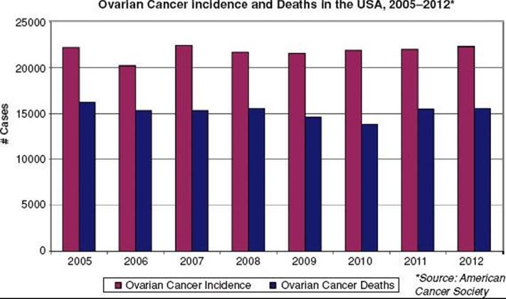
TABLE 71.1 TWO-PATHWAY MODEL OF EPITHELIAL OVARIAN CARCINOGENESIS

In the two-pathway model of ovarian carcinogenesis, type I tumors include all major histologic subtypes (serous, endometrioid, mucinous, clear cell, and transitional) with low nuclear and architectural grade that may be linked to well-defined benign precursor lesions, and type II tumors account for the bulk of high-grade serous carcinomas (Table 71.1). The type I tumors are associated with distinct molecular alterations, such as mutations in KRAS, BRAF, and PTEN, which are rarely found in type II serous tumors.18 Mutually exclusive KRAS and BRAF mutations, both of which activate the oncogenic MAPK signaling pathway, are observed in 65% of serous borderline tumors but are rarely seen in high-grade serous carcinomas.19 KRAS mutations also occur in Müllerian histologic subtypes, including 60% of mucinous, 5% to 16% of clear cell, and 4% to 5% of endometrioid carcinomas.18 Mutations in PTEN,which lead to constitutive activation of the related PI3 kinase signaling pathway, have been detected in 20% of endometrioid carcinomas,20 whereas activating mutations in PIK3CA have been identified in 30% of clear cell cancers.21Somatic mutations in ARID1A, a novel tumor suppressor gene, were recently identified in 46% of ovarian clear cell cancers and 30% of endometrioid cancers. ARID1A mutation and corresponding loss of BAF250a expression, a key component in chromatin remodeling, were also seen in preneoplastic lesions and may represent an early event in the transformation of endometriosis.22 In contrast, type II tumors exhibit a high frequency of p53 mutation and widespread DNA copy number change. Inactivation of the BRCA pathway, a critical component of DNA repair by homologous recombination, is also a hallmark of high-grade serous carcinomas.23 Disruption of the BRCA pathway by germline or somatic mutation, epigenetic silencing, or microRNA regulation has been observed in >50% of serous cancers,24 and functional assays show defective formation of homologous recombination repair foci following DNA damage.25
Angiogenesis plays an important role in normal ovarian function, as heavy vascularization occurs at the beginning of each ovulatory cycle with predictable variation in the serum levels of vascular endothelial growth factor (VEGF). Epithelial ovarian cancers frequently overexpress VEGF, fibroblast growth factor, platelet-derived growth factor (PDGF), and angiopoietin.26 In preclinical models, expression of VEGF provides a survival advantage to transformed cells of the ovary. Other studies have found an association between preoperative serum VEGF level and clinical outcome.27 Targeting the tumor microenvironment in ovarian cancer is an attractive treatment strategy given the inherent genomic instability of high-grade serous cancers.
TABLE 71.2 FACTORS INFLUENCING THE RISK OF OVARIAN CANCER
RISK FACTORS
Epidemiologic studies have identified hormonal, genetic, and environmental factors that may play an important role in ovarian carcinogenesis (Table 71.2). The OSE-CIC model is consistent with well-established epidemiologic data indicating that suppression of ovulation results in substantial reduction of ovarian cancer risk. New insights into the pathogenesis of high-grade serous carcinomas have emerged through the study of high-risk populations with a genetic predisposition for the development of ovarian cancer. Nevertheless, the pathogenic mechanisms are not well understood and likely multifactorial, associated with both patient-related and environmental factors.
Patient-Related Factors
Risk factors associated with a higher frequency of ovulatory events, such as advancing age, low parity, infertility, early menarche, and late menopause, support the proposed mechanism of incessant ovulation.28–31 Although infertility is associated with ovarian cancer, the use of fertility drugs has not been conclusively linked.32–34 Suppression of ovulatory events from pregnancy, breast-feeding, and oral contraceptive use may explain the observed protective benefit.30,31,35 With oral contraceptive use for 4, 8, or 12 years, the risk of ovarian cancer is reduced by 40%, 53%, and 60%, respectively.36 In a collaborative meta-analysis of 45 epidemiologic studies from 21 countries, the use of oral contraceptives was significantly associated with a decreased risk of ovarian cancer in ever users (hazard ratio [HR] 0.73, p <.0001), with larger reductions observed for longer duration of use.37 The authors conclude that 200,000 ovarian cancers and 100,000 deaths have been prevented since the introduction of oral contraceptives 50 years ago.
Other patient-related risk factors such as polycystic ovarian disease and endometriosis may act through hormonal mechanisms attributable to elevated gonadotropins or by chronic inflammation. In a meta-analysis of eight case-control studies, women with polycystic ovarian disease had a 2.5-fold increase in ovarian cancer risk.38 Endometriosis has been shown to be an independent risk factor for ovarian cancer with an estimated rate of malignant transformation of 2.5%.39 Ovarian cancers that arise from endometriosis are most often low-grade tumors with endometrioid or clear cell histology and are associated with a better prognosis.40 Altered hormonal levels such as elevated androgens may also increase risk, whereas progestins have been shown to be protective.41 Large prospective cohort studies have demonstrated an increase in ovarian cancer risk and mortality with the use of exogenous estrogen and hormone replacement therapy.42–44 Surgical procedures, including hysterectomy and tubal ligation, are independently associated with a 34% reduction each in ovarian cancer risk, although the protective mechanism is unknown.29,45–47 Chronic inflammatory conditions such as pelvic inflammatory disease and tuberculous salpingitis were once believed to be causative factors in the development of fallopian tube malignancies, although this theory remains unproven.
Genetic Factors
Heredity tumors account for 10% to 15% of all ovarian cancers, and family history is the strongest risk factor after increasing age.48,49 The risk of developing ovarian cancer in the general population is approximately 1.4%, whereas the lifetime risk for a woman with one first-degree family member with ovarian cancer is 5% and climbs to 7% with two first-degree relatives. If a hereditary syndrome is present, the lifetime risk is on the order of 25% to 50% with an age of diagnosis approximately 10 years younger than women with sporadic disease.50
Three distinct familial ovarian cancer syndromes have been identified by pedigree analysis with autosomal dominant patterns of inheritance.51,52 BRCA1 and BRCA2 mutations are the most common genetic alterations, with up to 90% of all hereditary cases associated with a deleterious mutation of the BRCA1 gene, located on chromosome 17q21, or the BRCA2 gene, located on chromosome 13q22. The lifetime risk of ovarian cancer is approximately 40% in BRCA1 mutation carriers and 18% in BRCA2 mutation carriers.53 BRCA-associated ovarian cancers are most frequently invasive serous adenocarcinomas and less likely borderline or mucinous tumors.54 Mutation carriers are also more likely to present with advanced stage disease and have poorly differentiated tumors.55 Nonetheless, BRCA1 or BRCA2 mutation carriers have a more favorable clinical course with a significantly longer recurrence-free and overall survival compared to noncarriers. The improved outcome appears related to a higher sensitivity to platinum chemotherapy across first and subsequent lines of treatment.56,57,58–59,60
The importance of the fallopian tube in the pathogenesis of extrauterine serous carcinoma was initially recognized in BRCA1 and BRCA2 mutation carriers. In a prospective study of 483 BRCA1 mutation carriers, the incidence of fallopian tube cancer was reported as 120 times that of the general population.61 High-grade serous primary peritoneal carcinomas are also frequently observed in mutation carriers.62 Further attention was focused on the fallopian tube as the primary site of origin following reports of epithelial dysplasia and occult serous carcinomas in prophylactic salpingo-oophorectomy specimens from mutation carriers.63–66 In contrast to the standard pathologic sampling of the ampullary region of the fallopian tube in ovarian cancer cases, more extensive complete sectioning of the tube revealed an abundance of lesions in the fimbria of the distal tube.14,15–16 The detection of early serous carcinomas, or TICs, lent further support to the emerging concept of the fallopian tube as the primary site for extrauterine serous carcinoma. In this context, the National Comprehensive Cancer Network (NCCN) guidelines recommend that any woman with a diagnosis of ovarian, fallopian tube, or primary peritoneal carcinoma be referred to a cancer genetics professional for consideration of BRCA mutation testing.67 BRCA screening in a population of women with non-mucinous ovarian cancer detected germline mutations in 14% of women, including 22% with high-grade serous cancer, reinforcing the importance of offering testing to all patients.68
Hereditary nonpolyposis colorectal syndrome, or Lynch type II cancer syndrome, is responsible for the remaining 10% of hereditary ovarian cancers.69 In this syndrome, germline mutations of DNA mismatch repair genes lead to an increased risk of colorectal, stomach, endometrial, and ovarian cancer owing to underlying microsatellite instability. A total of seven mismatch repair genes have been identified with mutations in MLH1 and MSH2 accounting for 90% of the observed mutations in Lynch syndrome families.70,71 Mutations in MSH6 and PMS2 have been reported in the remaining 10% of families, whereas alterations in the remaining three identified genes are uncommonly observed. Inactivating mutations in specific genes may modify the underlying cancer predisposition. Women with MSH6 mutations are twice as likely to develop endometrial cancer compared to carriers of MSH2 or MLH1 gene mutations, which are the most common alterations underlying colorectal cancer risk.72 Women with MSH2 gene mutations have been shown to have a risk of epithelial ovarian cancer twice that of MLH1 carriers. In contrast to BRCA1 and BRCA2 mutation carriers, women with Lynch syndrome may present with a variety of nonserous epithelial tumor types, including endometrioid and clear cell histologies. Ovarian cancer associated with Lynch syndrome is more often diagnosed at an earlier stage with well to moderately differentiated tumor grade. The lifetime risk of ovarian cancer in women with Lynch syndrome is estimated at 3% to 14% and is most commonly diagnosed in the fifth decade of life.73 In women with a family or personal history suggestive of Lynch syndrome, tumor testing may be performed by microsatellite instability analysis or immunohistochemistical staining for mismatch repair genes. Direct sequencing of the mismatch repair genes is used for detection of germline mutations. Other less common germline mutations have been identified by genomic sequencing and involve the following genes: BARD1, BRIP1, CHEK2, MRE11A, NBN, PALB2, RAD50, RAD51C, and TP53.74
Other genetic disorders linked with nonepithelial ovarian cancers include Peutz-Jeghers syndrome, which is associated with an increased risk of sex cord–stromal tumors, and gonadal dysgenesis, associated with dysgerminomas and gonadoblastomas.
Environmental Factors
Environmental or physical causes of ovarian cancer have been investigated through numerous case-control studies. Women in developing countries have a lower incidence of ovarian cancer than those living in industrialized nations,75 although to date, no specific chemical carcinogens have been identified. Chronic exposure to asbestos-related products, including talc products, have long been implicated in the development of ovarian cancer.76 The Nurses’ Health Study reported a modestly elevated risk of invasive serous cancers with talc use (relative risk [RR] 1.4), although no association was detected with overall ovarian cancer risk.77 The development of mucinous ovarian cancer has been found to be associated with cigarette smoking (RR 2 to 2.2) but not other epithelial subtypes.78,79
Dietary and metabolic factors have also been explored in large population-based studies as possible contributors to ovarian cancer risk. To date, there have been no consistent associations of coffee or alcohol consumption with increased risk. Large epidemiologic studies also showed no relationship between consumption of animal fat and the development of ovarian cancer.80,81 In a recent Swedish population-based cohort study of the effect of body mass index on cancer risk in >35,000 women, a 36% higher risk of cancer was observed in obese women (body mass index ≥30) relative to women with body mass index in the normal range (18.5 to 25); cancer sites most strongly related to obesity were endometrium, ovary (risk for top quartile 2.09; 95% confidence interval [CI], 1.13 to 4.13), and colon.82 Obesity has also been associated with increased ovarian cancer mortality in a large prospective study of adults in the U.S.83 There is no clear relationship between exercise and ovarian cancer risk.
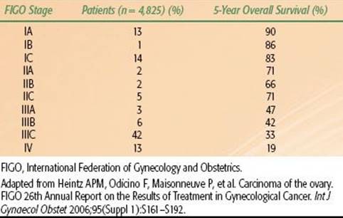
TABLE 71.3 EPITHELIAL OVARIAN CANCER STAGE DISTRIBUTION AND SURVIVAL BY STAGE
SCREENING
In the absence of a reliable screening test, most women with ovarian cancer are diagnosed with advanced-stage disease. In contrast to women with localized disease (stage I/II) who have estimated 5-year survival rates of 70% to 90%, overall survival for women with advanced disease (stage III/IV) is poor, ranging from 20% to 45% (Table 71.3). The low sensitivity and specificity of the available screening modalities and the low prevalence of the disease in the general population have hindered the development of a robust screening test. Recent screening approaches have been associated with a low positive predictive value and unacceptable false-positive rates without impacting disease mortality in the general population or high-risk groups.
Several screening strategies have been investigated, including pelvic exam, the serum tumor marker CA-125, and transvaginal ultrasound (TVUS), either alone or in combination.84 CA-125 values >35 U/mL are observed in 80% of patients with epithelial ovarian cancer, including 90% of women with advanced-stage disease, but only in 50% of early-stage patients.85 CA-125 is nonspecific for ovarian cancer, as it can be elevated in benign gynecologic and nongynecologic conditions as well as nongynecologic cancers. Screening with CA-125 alone has been found to lack specificity in an average-risk population of postmenopausal women. Ultrasound alone is able to detect early-stage disease in an average-risk population,86–88 although the significant false-positive rate remains a concern. In a United Kingdom multimodality screening study, diagnostic surgeries were performed nine times more frequently to detect one cancer in the group screened by TVUS alone compared to multimodality screening (CA-125 and TVUS).89
Several large screening trials currently in progress in the United States, United Kingdom, and Japan have been designed to assess reduction in mortality with early detection of ovarian cancer. The Prostate, Lung, Colorectal, and Ovarian Cancer (PLCO) screening trial in the United States randomized 78,216 women aged 55 to 74 to annual screening with CA-125 and TVUS versus usual medical care. Following baseline screening, 570 surgical procedures, including 325 laparotomies, were performed; 29 tumors were detected, 9 of which were of LMP.90 The positive predictive value for the detection of invasive ovarian cancer was 3.7% for an abnormal CA-125, 1.0% for an abnormal TVUS, and 23.5% for both. Publication of the mortality data with 12-year follow-up showed no difference in the stage of cancer detected by screening (90% stage III or IV) and no reduction in cause-specific or overall mortality for women who underwent screening.91 Furthermore, diagnostic evaluation for women with false-positive screening resulted in a serious complication rate of 15%.
A second trial, the United Kingdom Collaborative Trial of Ovarian Cancer Screening (UKCTOCS), has accrued >200,000 postmenopausal women to evaluate multimodality screening with TVUS and serum CA-125. In the prevalence screen, the sensitivity, specificity, and positive predictive values of multimodality screening for invasive epithelial ovarian and tubal cancers were 89.5%, 99.8%, and 35.1%, respectively.89 The initial results of baseline screening were more promising than the U.S. study, with 43% of invasive cancers detected as stage I or II, which may reflect differences in study design and the diagnostic algorithm. The effect of multimodality screening on mortality is awaiting further follow-up. The multicenter, randomized Japanese study utilizes a similar screening strategy with TVUS and CA-125 and has accrued >80,000 women. Unlike the U.S. and UK trials, there was a nonsignificant trend for the detection of more stage I cancers in the screened population (63% vs. 38%), although mortality rates have not yet been reported.92
The present data do not support routine screening for ovarian cancer in the general population. All of the North American expert groups, including the U.S. Preventive Services Task Force, the American College of Obstetricians and Gynecologists (ACOG), the Society of Gynecologic Oncologists (SGO), and the Canadian Task Force on the Periodic Health Examination, recommend against routine screening in asymptomatic women. The incidence of ovarian cancer is relatively low in the general population; thus, the concerns for false-positive screening and the associated risks of exploratory surgery are significant.93 However, women at high risk for ovarian cancer with BRCA1or BRCA2 mutations or hereditary nonpolyposis colorectal syndrome could potentially benefit from chemoprevention; screening with pelvic examinations, TVUS, and CA-125 on a biannual or annual basis; or prophylactic surgery.94Although the efficacy of screening has been disappointing in the high-risk population,95 the ACOG, SGO, and NCCN guidelines recommend routine screening with pelvic exam, CA-125 and TVUS for women with hereditary ovarian cancer syndromes beginning at the age of 30 to 35, or 5 to 10 years earlier than the age of diagnosis of ovarian cancer in the family. The U.S. National Institutes of Health Consensus Development Panel recommends that prophylactic salpingo-oophorectomy be considered in women with ovarian cancer syndromes at age 35 years or after childbearing is complete, as the risk reduction with salpingo-oophorectomy is 80%.96,97 Removal of the ovaries also reduces the risk of breast cancer in BRCA mutation carriers.98
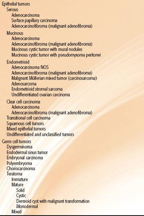
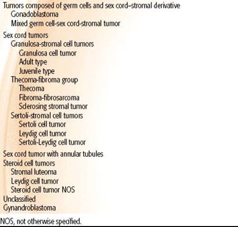
TABLE 71.4 WORLD HEALTH ORGANIZATION CLASSIFICATION OF OVARIAN TUMORS
HISTOLOGIC CLASSIFICATION
The World Health Organization and International Federation of Gynecology and Obstetrics (FIGO) have adopted a unified classification of the common epithelial, germ cell, sex cord, and stromal tumors99 (Table 71.4). Most ovarian malignancies (60% to 65%) are epithelial, with germ cell tumors (20%), sex cord–stromal tumors (5%), and metastases to the ovary (5% to 10%) accounting for the remainder.100 Serous tumors are most common, comprising 50% to 60% of epithelial tumors. Other subtypes include mucinous carcinoma in 10%; endometrioid carcinoma, 8%; clear cell carcinoma, 3% to 5%; transitional, 3% to 5%; and undifferentiated carcinoma, 1%. Bilateral presentation occurs frequently in epithelial tumors, most commonly in serous tumors followed by endometrioid (15%) and mucinous tumors (5% to 10%).
FIGURE 71.3. Serous tumor of low malignant potential of the ovary at low power (A) and high power (B). At low power, note the progressively branching papillary fronds with fibrovascular support and detached papillary clusters; at high power, note the serous epithelium with nuclear hyperchromasia and cytologic atypia. By definition, there is no destructive stromal invasion.

High-grade serous carcinomas are often widely disseminated at diagnosis and account for most deaths from ovarian, tubal, and peritoneal cancers. In contrast, 5-year survival for low-grade serous carcinoma is 85%. Most serous tumors present as large ovarian masses and grossly appear nodular with multiple papillary projections and cysts filled with clear serous fluid. A two-tiered grading system separates high-grade serous carcinomas from low-grade tumors,101,102 which have a similar prolonged natural history to noninvasive borderline tumors, or tumors of LMP.103 As discussed previously, there is increasing evidence that high grade serous carcinomas originate in the fimbria of the fallopian tube and may spread rapidly to the adjacent ovary and peritoneal sites. Low-grade tumors appear to follow a stepwise progression from borderline tumor to invasive carcinoma and involve molecular pathways distinct from their high-grade counterparts.104–106 Although treatment is similar for all serous tumors, low-grade tumors have been shown to be less responsive to chemotherapy.103,107,108
LMP or borderline tumors have nuclear abnormalities and mitotic activity intermediate between benign and malignant tumors of similar cell type but lack stromal invasion (Fig. 71.3). Borderline tumors are a subcategory of ovarian malignancies that account for 10% to 20% of all epithelial neoplasms.109,110 The prognosis, surgical approach, and postoperative treatment recommendations are vastly different as compared with those of their invasive counterparts. The majority of LMPs (75%) present with stage I disease, which directly contrasts with the 75% advanced stages in the invasive epithelial tumors. The 5- and 10-year survival rates for women with LMP tumors are >95%. Significant heterogeneity exists in the biologic behavior of borderline tumors. Women with nonlocalized LMP tumors of the ovary have decreased survival compared to those with localized LMP tumors but are similar to that of women with localized, well-differentiated epithelial ovarian carcinoma.111 Other pathologic features that affect prognosis include the cell type, tumor stage, implant type (invasive vs. noninvasive), the presence of micropapillary architecture, and microinvasion. Extensive sampling of all pathologic specimens is required to firmly establish the diagnosis.
Most endometrioid and clear cell carcinomas arise in foci of endometriosis and follow an adenoma to borderline tumor to carcinoma sequence. Endometrioid carcinoma of the ovary resembles its endometrial counterpart (Fig. 71.4), and synchronous primary cancers are detected in 10% of women with ovarian cancer and 5% of women with endometrial cancer.112 Bilateral ovarian involvement, small multinodular ovaries, and surface and hilar spread suggest metastatic involvement from an endometrial primary, particularly if the endometrial tumor is high-grade, deeply invasive, and associated with lymphovascular invasion. A large, cystic, unilateral tumor of low grade arising in a focus of endometriosis likely represents a primary ovarian tumor. Endometrioid tumors appear to have a better prognosis than serous cancers regardless of tumor stage.113 Multiple synchronous primary tumors may represent a field defect related to endometriosis, which is associated with improved survival.114
Mucinous tumors follow a similar progression from cystadenoma to borderline tumor prior to invasion. All subtypes are more likely to be diagnosed when confined to the ovary. Mucinous cancers may grow to a very large size, often measuring up to 20 cm in diameter and filled with large pockets of thick, necrotic, mucinous debris.115,116 Ovarian mucinous tumors closely resemble mucin-secreting tumors originating from other sites—most commonly the gastrointestinal tract. The pathologic distinction between primary and metastatic mucinous carcinoma may be challenging, despite the use of immunohistochemical analysis. Further clinical evaluation is often required to exclude a clinically occult nonovarian primary source. Secondary involvement of the ovary may also occur in association with pseudomyxoma peritonei arising from a low-grade tumor often of appendiceal origin.117
Clear cell carcinoma is characterized by clear and hobnail cells with a similar histologic appearance to clear cell carcinoma of the kidney, endometrium, and vagina. Ovarian clear cell carcinoma is often seen in association with venous thromboembolism and hypercalcemia. Although more likely to be stage I than serous carcinoma, clear cell histology is associated with a lower response rate to platinum-based chemotherapy, higher recurrence rate, and worse survival.118 The transitional cell tumors, including Brenner tumors, are benign in most cases (98%) and carry a favorable prognosis.
Ovarian germ cell tumors comprise <5% of ovarian malignancies and are classified by the World Health Organization (Table 71.4). Dysgerminomas are the most common of the germ cell tumors and occur bilaterally in 10% to 20% of cases. The other germ cell tumor types are typically unilateral. Endodermal sinus tumors, also known as yolk sac tumors, are characterized by Schiller-Duvall bodies. Embryonal carcinomas are rare and tend to occur in younger populations; they may be seen with nongestational choriocarcinomas as part of mixed germ cell tumors (10% of all germ cell tumors). Immature teratomas are characterized by immature elements from the germ layers. The grade, treatment recommendations, and outcome are directed by the amount of immature neural elements.
Sex cord–stromal tumors are also classified by the World Health Organization (Table 71.3), with granulosa cell tumors being the most common (70%). Histologically, the granulosa cell tumors are composed of granulosa cells that have a pale, grooved, “coffee bean” nuclei or a rosette of cells surrounding eosinophilic material, a Call-Exner body. Thecomas (hormonally active) and fibromas (hormonally inactive) are both clinically benign tumors and are most common in middle-age women.
The most common histopathology of primary fallopian tube malignancies is papillary serous adenocarcinoma. Other less common Müllerian subtypes include endometrioid, clear cell, and malignant mixed Müllerian tumors.119,120Rare reports of squamous cell carcinoma, immature teratoma, glassy cell tumor, and sarcoma have also been described. Benign tumors are found even less frequently than malignant neoplasms. Metastatic involvement of the fallopian tube is reported in up to 12% of women with uterine cancer and 4% with cervical cancer.121,122
FIGURE 71.4. Endometrioid adenocarcinoma of the ovary. A: At low power, note the tumor composed of back-to-back endometrioid glands. B: At high power, note the squamous metaplasia, commonly seen in this epithelial variant. Assignment of tumor grade is based on similar criteria as for carcinomas arising in the endometrium. This tumor would qualify as well differentiated or grade 1.

PATTERNS OF SPREAD
The primary mode of spread for epithelial ovarian and fallopian tube cancers is transperitoneal, as malignant cells exfoliate into the peritoneal cavity. Intraperitoneal spread is favored by intestinal peristalsis and negative hydrostatic pressure below the diaphragm. The exfoliated tumor cells follow the intra-abdominal fluid stream passing along the paracolic gutters toward the diaphragm, predominately on the right side, before implantation on any peritoneal surface.123,124 Metastatic deposits are frequently seen in the posterior cul-de-sac, paracolic gutters, diaphragmatic surfaces, liver capsule, intestinal surfaces, and omentum. Metastases may also be found in the uterus or contralateral ovary from peritoneal spread or flow through the fallopian tubes. Dense tumor “caking” can cause infiltration into the abdominal organs creating a mass effect on the omentum, ureter, bowel, liver, pancreas, spleen, or adrenals, resulting in advanced disease stage at presentation with associated ascites.
Lymphatic drainage constitutes the second most common pattern of spread. The lymphatic capillaries of the ovary converge on the hilus and follow the ovarian blood vessels in the infundibular ligament to drain to the para-aortic nodes at the level of the renal hilum. Lymphatics also drain along the broad ligament to the hypogastric and external iliac nodes in the pelvis. Less frequently, spread can occur to the inguinal nodes via the round ligament.125Approximately 10% to 15% of women with disease that appears confined to the ovary have para-aortic lymph node involvement,126 which becomes increasingly common in advanced-stage disease. Because of the rich lymphatic supply of the fallopian tubes, lymph node involvement is common even in the absence of tubal musculature involvement.127 Pelvic and para-aortic lymph node involvement has been reported in 10% to 30% of women with fallopian tube cancer at diagnosis.128
Transdiaphragmatic spread occurs to the pleural cavity and is the most common finding in stage IV disease. Hematogenous spread is infrequent at the time of presentation, with only 2% to 3% of patients with parenchymal liver or lung disease. Brain metastasis is also rare. However, >50% of recurrences occur both within and outside the peritoneal cavity at the time of treatment failure.
CLINICAL PRESENTATION
As ovarian cancer has insidious growth and is asymptomatic in early-stage disease, most women do not present until symptoms arise from advanced disease progression. Vague gastrointestinal complaints of dyspepsia, nausea, early satiety, bloating, constipation, or obstipation are common presenting symptoms, as are genitourinary symptoms including frequency, urgency, or incontinence. Other ill-defined symptoms include fatigue, back pain, pain with intercourse, and menstrual irregularities. These nonspecific symptoms can be present for several months but may not trigger diagnostic evaluation until after the symptoms fail to clear with other medical therapy.129 Detection of early-stage disease may occur by palpation of an asymptomatic adnexal mass on routine examination, although most adnexal masses require moderate size for palpation. In premenopausal women, most of these masses are benign, as ovarian cancer represents <5% of adnexal neoplasms. An adnexal mass in a postmenopausal woman has a higher likelihood of malignancy, and surgical exploration is often indicated. Physical examination findings such as a fixed pelvic mass, palpable upper abdominal mass, and ascites are highly suggestive of an ovarian malignancy. According to a consensus statement on the symptoms of ovarian cancer published by the Gynecologic Cancer Foundation, SGO, and American Cancer Society, new and persistent symptoms of bloating, pelvic or abdominal pain, difficulty eating, early satiety, or urinary urgency or frequency should prompt women to seek medical evaluation.
A portion of women with fallopian tube carcinoma may present with early clinical signs and symptoms. Two triads have been described as pathognomonic for fallopian tube malignancy: (a) pelvic pain, pelvic mass, and leukorrhea and (b) vaginal bleeding, vaginal discharge, and lower abdominal pain. However, the percentage of patients presenting with either triad of symptoms has been reported as low as 11%.130 Another classic sign—hydrops tubae profluens—is a sudden emptying of accumulated fluid in the distended fallopian tube that causes profuse, watery, serosanguineous vaginal discharge. The discharge is often accompanied by a decrease in pelvic mass size on physical examination. In a meta-analysis of 122 patients with primary fallopian tube cancer, hydrops tubae profluens was a presenting sign in only 9%.121 In other series,124,127 it has been specifically reported that neither a triad nor hydrops tubae profluens was present in any of the patients reviewed. When clinically apparent, the fallopian tube may be dilated and mimic more benign pathologic processes such as hydrosalpinx, pyosalpinx, or hematosalpinx.131 In more advanced disease with tubal wall invasion, extension of a necrotic mass to involve the ovary may give the initial impression of a tubo-ovarian abscess.
Germ cell and stromal cell malignancies present at an earlier stage than epithelial ovarian or fallopian tube cancers, often related to abdominal discomfort or symptoms of excessive estrogen or androgen production. Granulosa cell tumors may lead to precocious puberty in young girls, and Sertoli-Leydig cell tumors may cause virilization.
DIAGNOSTIC WORKUP
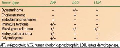
Evaluation of a pelvic mass will be influenced by the patient’s age, clinical presentation, and imaging features. An ovarian mass is more likely to be a malignant neoplasm in the pediatric, perimenopausal, and postmenopausal age groups and benign during the reproductive years. Ultrasound is often the first, noninvasive step for the evaluation of a pelvic mass. Sonographic characteristics suggestive of malignancy include irregular borders; a solid component that is not hyperechoic and often nodular or papillary; Doppler demonstration of flow in the solid component; dense multiple irregular septa (>2 to 3 mm); and the presence of ascites, peritoneal masses, enlarged nodes, or matted bowel. Computed tomography (CT) and magnetic resonance imaging (MRI) may be useful preoperatively for surgical planning to determine the extent of intra- and extra-abdominal disease. Although limited as a screening tool, an elevated CA-125 level may suggest more advanced or greater bulk of disease and high-grade serous histology but in general is a weak predictor of surgical resection.132,133 Human chorionic gonadotropin (β-hCG), α-fetoprotein (AFP), total inhibin, and lactate dehydrogenase levels may aid in the diagnosis of and treatment for nonepithelial germ cell ovarian tumors.
Other conditions may mimic ovarian cancer in their presentation, including colon, gastric, and appendiceal carcinomas as well as metastatic breast cancer and primary lymphoma. Although ideally the primary site of disease is determined in the preoperative setting, the diagnosis often cannot be made accurately until the time of surgery, and frozen section pathologic evaluation may guide surgical approach. Primary malignancies of the fallopian tube have been difficult to diagnose prior to surgical exploration, as the clinical presentation may be similar to that of salpingitis, ovarian abscess, pelvic inflammatory disease, or even ectopic pregnancy. Occult tubal primaries may be detected only by careful pathologic processing.
Referral to a gynecologic oncologist should be considered for any woman with a suspicious pelvic mass, family history of ovarian or fallopian tube cancer, or elevated CA-125.134 Initial evaluation should include a thorough history and full physical and pelvic examination, laboratory studies (complete blood count, chemistries, CA-125, carcinoembryonic antigen, CA 19–9), and imaging (abdominal CT/MRI, directed ultrasound, chest x-ray). Further evaluation with mammography, upper gastrointestinal endoscopy, or colonoscopy may be indicated in some scenarios. Comorbid conditions may prompt additional evaluations, including cardiac risk assessment, pulmonary function testing, and nutritional evaluation.
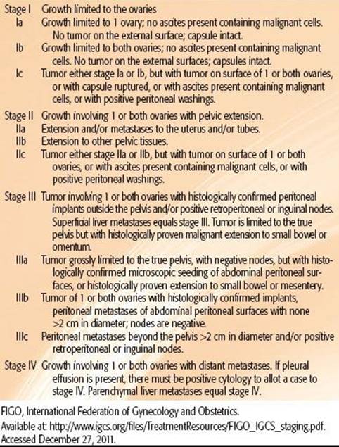
TABLE 71.5 FIGO STAGING FOR OVARIAN CARCINOMA
SURGICAL STAGING
Surgical advances in staging and the therapeutic benefit of a maximal safe tumor resection have improved the progression-free and overall survival of women with ovarian cancer over the past two decades. Traditional staging for ovarian cancer is based on the FIGO staging system shown in Table 71.5.135 A parallel American Joint Committee on Cancer system of TNM staging also exists, with strong correlations between stage and the prognostic value of these substages. Staging of primary fallopian tube cancer was established by FIGO in 1992 based on the ovarian cancer staging system. A proposed modification was later published to permit staging of noninvasive tubal carcinomas and fimbrial carcinomas as well as the inclusion of substaging based on depth of invasion119 (Table 71.6).
Comprehensive surgical staging is the mainstay of diagnosis and initial treatment for ovarian, fallopian tube, and peritoneal cancers.136 In apparent early-stage cancers, surgical evaluation is required for accurate pathologic staging to guide adjuvant treatment recommendations. In patients with advanced disease, surgery represents the initial treatment by providing optimal cytoreduction. Comprehensive surgical staging begins with an exploratory laparotomy via a vertical midline incision followed by collection of peritoneal washings or any ascitic fluid for cytology. The surgeon performs a thorough inspection of all visceral and peritoneal surfaces within the abdomen and pelvis, with particular attention to the intestinal serosal surfaces, mesentery, paracolic gutters, hemidiaphragms, gallbladder, and liver. Systematic biopsies of the bladder serosa, anterior and posterior cul de sac, paracolic gutters, hemidiaphragms, and any suspicious adhesions or implants should be obtained. Total hysterectomy, bilateral salpingo-oophorectomy, infracolic omentectomy, and pelvic and para-aortic lymph nodal sampling may be performed in addition to the intact removal of the adnexal mass when possible. Given the significant risk of contralateral involvement, bilateral lymph node assessment is frequently performed.137,138 In patients with advanced disease, bowel resection, diaphragm stripping, splenectomy, nodal dissection, and radical resection of peritoneal tumor nodules are often necessary to achieve the desired surgical outcome of maximal debulking. Recently, the role of routine appendectomy has been questioned and is not recommended in the absence of visible pathology.139
In select cases, preservation of the contralateral unaffected ovary and the uterus can be achieved in young women wishing to retain fertility who have stage I and/or low-risk tumors (early-stage, low-grade invasive tumors, LMP lesions, or malignant stromal or germ cell tumors).136 In the setting of unilateral salpingo-oophorectomy, comprehensive surgical staging should be performed to exclude occult disease. Exceptions to upfront surgical management include patients with significant medical comorbidities who are poor operative candidates and patients with a complex ovarian cyst where extraovarian disease has not been excluded. Neoadjuvant chemotherapy followed by interval cytoreduction may be considered in patients with bulky stage III or IV disease. Before initiation of neoadjuvant chemotherapy, the pathologic diagnosis should be confirmed by biopsy, fine-needle aspirate, or paracentesis. The NCCN guidelines recommend that patients who are evaluated for neoadjuvant chemotherapy be seen by a fellowship-trained gynecologic oncologist before being deemed a nonsurgical candidate.136
TABLE 71.6 MODIFIED FIGO FALLOPIAN TUBE STAGING

PROGNOSTIC FACTORS
Tumor stage, grade, histology, and optimal cytoreduction by a trained gynecologic oncologist are the most important determinants of survival for ovarian and fallopian tube cancer. Patients with FIGO stage 1A disease who have negative staging laparotomy have 5-year survival rates of 80% to 90%. If extrapelvic disease is detected at the time of surgical staging, the patient is upstaged to stage III disease with corresponding 5-year survival rates of 30% to 50%. Primary or interval cytoreductive surgery should be performed by a gynecologic oncologist based on published evidence of a 6- to 9-month median survival benefit.140–142
The volume of residual disease after primary surgery is also an important determinant of outcome.143–148 Patients who have optimal cytoreduction of tumor have a 22-month improvement in median survival compared to those with suboptimal resection. A meta-analysis of >6,800 women with advanced-stage ovarian cancer treated with adjuvant platinum-based chemotherapy found a 5.5% increase in median survival for every 10% increase in maximal cytoreduction.144 Although the definition of maximal resection varies by study, <1 mm or no visible disease has been associated with improved response to chemotherapy, less platinum resistance, and longer overall survival.143,149,150 Preoperative CA-125 measurement is not independently prognostic, as it likely reflects disease burden. In contrast, the half-life and nadir of CA-125 during induction chemotherapy is associated with improved outcome in ovarian and fallopian tube cancers.120,151–155
Although a strong correlation exists between histologic grade and stage, grade has independent prognostic significance, particularly in early-stage disease. Survival rates for stage I disease with grade 1, 2, or 3 tumors are 96%, 78%, and 62%, respectively.156 The impact of histologic subtype on survival is less robust owing to other more important clinical variables, including stage, grade, and volume of disease. Patients with mucinous and endometrioid subtypes have improved survival rates compared to serous adenocarcinoma, predominately related to the earlier stage at presentation. Clear cell carcinomas appear to be more aggressive than other epithelial malignancies. The 5-year survival rate for stage I clear cell carcinoma is 60% and for other stages is <15%.118
As reflected in the modified FIGO staging system, depth of tubal invasion has been found to correlate with survival in two large retrospective studies of fallopian tube carcinoma.120,157,158 In addition to the depth of tubal invasion, the pathology report should provide information on tumor grade and the presence of lymphovascular invasion.
MANAGEMENT OF EPITHELIAL TUMORS
Treatment for ovarian and fallopian tube cancer depends largely on the stage and grade of disease at presentation. Table 71.7 presents a general schematic for treatment recommendations for women with epithelial ovarian and fallopian tube cancer. Although much less common, primary peritoneal cancers are managed in a similar manner.
Early-Stage Disease
Surgical Therapy
Comprehensive surgical staging should be performed in all women with apparent early-stage disease to confirm that the cancer is confined to the adnexa. Fertility-sparing surgery with unilateral salpingo-oophorectomy may be considered in a small subset of women with stage IA disease if the contralateral ovary is normal in appearance.159 In addition to abdominal exploration and full surgical staging, endometrial biopsy should be performed to sample the endometrium.
Postoperative Management
Early studies by the Gynecologic Oncology Group (GOG) and others have identified a small subgroup of patients with well- to moderately differentiated (grade 1 or 2) stage IA and 1B tumors who have a low risk of relapse and may not require adjuvant therapy.160,161 The NCCN guidelines state that women with early-stage (FIGO 1A or 1B) grade 1 ovarian cancer may be treated with surgical resection and observation with expected 5-year survival rates on the order of 90%.136 If observation is considered for women with early-stage grade 2 disease, full surgical staging should be performed.
TABLE 71.7 SCHEMATIC OF THE PRIMARY TREATMENT FOR EPITHELIAL OVARIAN CANCER

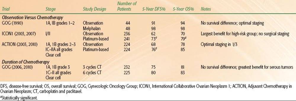
TABLE 71.8 RANDOMIZED STUDIES OF ADJUVANT CHEMOTHERAPY IN EARLY-STAGE OVARIAN CANCER
Adjuvant Chemotherapy
Women with early-stage disease and a less favorable prognosis include patients with grade 3 tumors, clear cell histology, or disease extension beyond the ovarian capsule into the abdominal wall or peritoneum (stage IC or II). Various adjuvant treatment approaches have been investigated to improve clinical outcomes, although the optimal management remains controversial. Few randomized trials have been conducted in this population, and they have been limited by small sample size and lack of power to demonstrate a survival advantage.
Nonetheless, two large multi-institutional trials conducted in Europe randomized women with early-stage ovarian cancer to platinum-based chemotherapy or observation following surgery (Table 71.8). The International Collaborative Ovarian Neoplasm 1 (ICON1) trial enrolled 477 women with FIGO IA-C ovarian cancer inclusive of all tumor grades and histologic subtypes.162 Adjuvant chemotherapy consisted of six cycles of a platinum-based regimen per institutional preference. With a median follow-up of 9.2 years, 10-year recurrence-free survival was 67% for the chemotherapy arm and 57% for observation (HR 0.7, p = .02).163 Overall survival also favored the chemotherapy group (72% vs. 64%, p = 0.06). When stratified by histology and grade, the largest benefit for adjuvant chemotherapy was seen in patients with high-risk disease, defined as FIGO stage 1A grade 3, stages 1B or 1C grades 2 and 3, or any clear cell histology. HRs for recurrence and death for the high-risk subset were 0.52 and 0.48, respectively (both p<.01). The second trial, Adjuvant Chemotherapy in Ovarian Neoplasm (ACTION), assigned 448 women with early-stage ovarian cancer to four to six cycles of platinum-based adjuvant chemotherapy or observation following surgery. Women with FIGO 1A grades 2 and 3 disease, all stages IC-IIA, or any clear cell histology were eligible for the trial. Recurrence-free survival, but not overall survival, was significantly improved in the chemotherapy group (70% vs. 62% at 10 years).164,165 The greatest benefit was seen in the subset of women with nonoptimal surgical staging. In a combined analysis of the ICON1 and ACTION trials, adjuvant chemotherapy was associated with significantly improved overall survival at 5 years (82% vs. 74%) compared to observation.166
The GOG conducted a randomized trial to determine the optimal duration of adjuvant chemotherapy in women with high-risk, surgical stage I disease by comparing three versus six cycles of carboplatin and Taxol chemotherapy.167 Rates of recurrence (25% vs. 20%) and overall survival (81% vs. 83%) were similar at 5 years for three and six cycles of chemotherapy, although six cycles were associated with significantly more toxicity, including neuropathy, granulocytopenia, and anemia. However, subset analysis revealed a significantly lower risk of recurrence for women with serous tumors.168 At 5 years, recurrence-free survival for women with serous tumors was 83% for six cycles of chemotherapy versus 60% for three cycles (HR 0.33, p = .04). Overall survival was also improved at 5 years (86% vs. 73%), although not statistically significant.
Adjuvant Whole-Abdomen Irradiation
WAI was an accepted standard modality in the adjuvant postoperative treatment for completely resected ovarian cancer; however, its use declined significantly after 1975. The practical advantage of WAI was the ability to treat all of the peritoneal surfaces within the abdomen and pelvis, although the delivered dose was limited by the relatively low tolerance of the liver and kidneys. Interest in WAI was established following the publication from Princess Margaret Hospital of a randomized comparison of 147 patients with FIGO 1B, II, or III debulked ovarian cancer who received WAI with a pelvic boost or pelvic radiotherapy followed by chlorambucil.169 WAI significantly improved 10-year survival rates over limited pelvic radiotherapy and chemotherapy (64% vs. 40%, respectively). The greatest magnitude of the survival benefit was observed in patients with <2 cm of residual disease (10-year survival 78% vs. 51%, respectively). No significant benefit was observed in patients with extensive residual tumor.
Subsequent randomized studies did not find a benefit for WAI compared to combined adjuvant treatment modalities. A multi-institutional trial by the National Cancer Institute of Canada (NCIC) randomized 257 high-risk stage I or optimally debulked stage II or III patients to receive melphalan, WAI, or IP 32phosphorus (32P).170 All patients had previously received 22.5 Gy to the pelvis prior to study entry. The WAI arm delivered 22.5 Gy to the abdomen in 2.25-Gy fractions. Actuarial 10-year survival rates failed to demonstrate a statistically significant difference among all groups.171 A second study from the Danish Ovarian Cancer Group (DACOVA) reported similar outcomes for women who received adjuvant WAI versus pelvic radiotherapy with cyclophosphamide or melphalan (4-year survival, 63% vs. 55%, respectively).172
WAI has also been shown to be comparable to single or combination chemotherapy in the adjuvant setting. A prospective study from the MD Anderson Cancer Center randomized 149 women with stage I through III ovarian cancer and <2 cm of residual disease to WAI or melphalan.173 WAI was delivered by a moving strip technique with a pelvic boost. Overall survival at 5 years was 71% for WAI and 72% for the melphalan arm. Severe late gastrointestinal toxicity requiring surgical intervention was reported in 14% of patients who received WAI, attributed to the moving strip technique that delivered excessive pelvic dose. A second prospective trial from the Northwest Oncologic Cooperative Group of Italy randomized 70 women with early-stage high-risk disease to adjuvant WAI or six cycles of cisplatin plus cyclophosphamide. Despite protocol violations in the assigned treatment groups, there was no difference in relapse-free or overall survival when analyzed by treatment received.174 From these studies, WAI appears equivalent to chemotherapy when delivered in patients with optimally debulked disease but is no longer used. Further dose escalation of WAI to 27.5 Gy does not appear to provide additional survival benefit.175
Adjuvant Intraperitoneal 32Phosphorous
IP instillation of 32P was extensively studied as an alternative to external-beam radiotherapy but is no longer in clinical use. In high-risk, early-stage ovarian cancer patients, randomized trials of adjuvant IP 32P versus chemotherapy from the GOG and Norwegian Radium Hospital found no significant differences in disease-free or overall survival in a population with high-risk early-stage disease.161,176,177 Given a higher incidence of bowel toxicity and limitations of delivery, 32P has largely been replaced by platinum and taxane combination chemotherapy as the adjuvant treatment for early-stage and advanced disease.
Advanced-Stage Disease
Surgical Considerations
Maximal cytoreduction is one of the most important prognostic factors for survival in patients with advanced-stage ovarian cancer. Cytoreductive surgery in advanced-stage disease may improve the patient’s disease-related symptoms such as abdominal pain and early satiety and allow for the ability to maintain nutritional status. Optimal debulking may also enhance chemotherapy delivery and response by removal of large hypoxic tumor masses. Bowel resection may be required to remove metastatic implants involving the bowel mesentery or serosa. Extensive upper abdominal surgery, including splenectomy, partial hepatectomy, distal pancreatectomy, and/or diaphragmatic resection, also results in prolonged palliation and improved survival.178,179
Second-look laparotomy (SLL) was introduced to assess the extent of residual disease following cytoreductive surgery and adjuvant chemotherapy. Up to 20% to 50% of patients may have residual disease after adjuvant therapy that was not detected on physical examination or by CA-125 levels or imaging. Although SLL demonstrated the prognostic importance of a pathologic remission, it was not found to have a therapeutic benefit in a GOG trial of 800 patients.180 Therefore, second-look evaluations for the purposes of determining therapy, and not for interval cytoreduction, should be reserved for women enrolled in clinical trials.
Several studies have evaluated the importance of interval cytoreduction following an initial attempt at surgical debulking. In a randomized trial by the European Organisation for Research and Treatment of Cancer (EORTC), 319 women with suboptimally debulked disease (>1 cm residual) were assigned to six cycles of cisplatin/cyclophosphamide chemotherapy or interval cytoreduction after three cycles of chemotherapy, followed by an additional three cycles.181 Progression-free and overall survival were significantly improved with interval cytoreduction (median overall survival 26 vs. 20 months, p = .04) without increased surgical morbidity. However, a subsequent GOG trial of 550 women failed to reproduce these results.182 Following a suboptimal resection in the GOG trial, patients were randomized to three cycles of cisplatin and paclitaxel followed by interval debulking or chemotherapy alone for a maximum of six cycles in both arms. The absence of a survival benefit may be attributable to differences in the chemotherapy used or more aggressive initial surgical management by gynecologic oncologists in the GOG trial.
The role of neoadjuvant chemotherapy has been explored in a randomized EORTC/NCIC trial of 670 women with stage IIIC and IV ovarian, fallopian tube, or primary peritoneal cancer randomized to initial surgical debulking followed by six cycles of cisplatin-based chemotherapy or interval debulking after three cycles, followed by three additional cycles.183 The cohort had extensive bulky disease, with >60% of patients with metastases >10 cm. Although progression-free and overall survival were similar between the two groups, interval surgery achieved optimal cytoreduction more often (81% vs. 42%) and with fewer surgical complications. A major criticism of the trial is that the median survival was significant lower than that of recently reported U.S. trials, which may be related to selection of higher-risk patients. NCCN guidelines state that more data will be necessary prior to recommending neoadjuvant chemotherapy in potentially resectable patients, and upfront debulking surgery remains the treatment of choice in the United States.136
Chemotherapy for Advanced-Stage Disease
Platinum agents are the most active class of compounds in the adjuvant treatment for ovarian cancer. Before 1980, alkylating-based regimens such as cyclophosphamide and doxorubicin were used with clinical response rates of 15% to 20%. GOG 47 demonstrated an improvement in clinical complete response rates (51% vs. 26%) and progression-free survival (13 months vs. 8 months) with the addition of cisplatin to cyclophosphamide and doxorubicin. Cisplatin has since been used extensively in both single-agent and multidrug studies. A meta-analysis of 49 trials from the Advanced Ovarian Cancer Trialists Group found that platinum combination chemotherapy improved survival rates over the same nonplatinum regimen (HR 0.88, 95% CI 0.79 to 0.98) and nonplatinum monotherapy (HR 0.93, 95% CI 0.83 to 1.05).184 The women in the nonplatinum groups routinely received platinum chemotherapy at the time of relapse, likely obscuring the magnitude of the benefit. This meta-analysis also incorporated data from 11 trials that directly compared carboplatin and cisplatin, either as single agents or in combination with other drugs, which suggested no difference in efficacy between the two agents. Carboplatin clearly has fewer treatment-related side effects, including less nephrotoxicity, neurotoxicity, and emetogenic potential, and is considered standard of care. However, carboplatin does have more associated myelosuppression, primarily thrombocytopenia, which is cumulative and may be dose limiting.
In addition to platinum compounds, taxanes have become a cornerstone of the treatment schemas for women with epithelial ovarian cancer. GOG 111 was a randomized study comparing cisplatin and paclitaxel with cisplatin and cyclophosphamide in women with suboptimally debulked, large-volume ovarian cancer. The paclitaxel-containing arm demonstrated improved clinical response rates (73% vs. 60%), progression-free survival (18 months vs. 13 months), and overall survival (38 months vs. 24 months).185 A second GOG study of 614 women with advanced disease and suboptimal resection compared single-agent cisplatin to 24-hour infusion of paclitaxel and to the combination of paclitaxel and cisplatin.186 Cisplatin alone or in combination with paclitaxel resulted in improved clinical response rates and progression-free survival, although overall survival was similar in the three arms. Combination chemotherapy also had lower cumulative toxicity. As several trials have demonstrated the comparable efficacy of cisplatin and carboplatin, combination carboplatin and paclitaxel has become the preferred first-line chemotherapy regimen. Although the standard dosing of intravenous carboplatin and paclitaxel is every 21 days, a phase III trial from Japan demonstrated significant gains in progression-free and overall survival with a dose-dense regimen of weekly paclitaxel in combination with carboplatin given every 3 weeks187 (Table 71.9). Grade 3 or 4 anemia was more common in the dose-dense arm, although other toxicities were similar. Dose-dense paclitaxel and carboplatin is a recommended intravenous regimen by the NCCN.136 Carboplatin and docetaxel is also an acceptable first-line alternative and may be considered for patients at high risk for neuropathy.188 Phase III trials have failed to show a benefit for the addition of a third agent to the carboplatin and paclitaxel regimen. GOG 182-ICON5 was a five-arm randomized trial of 4,312 women that compared the addition of gemcitabine, liposomal doxorubicin, or topotecan to carboplatin and paclitaxel. There were no improvements in progression-free or overall survival with any experimental regimen.189
The role of novel biologics in the first-line treatment for ovarian, fallopian tube, and primary peritoneal cancers is under active investigation. Antiangiogenics have demonstrated activity in the recurrent treatment setting, and the addition of bevacizumab to conventional first-line chemotherapy has recently been tested in two randomized trials. Bevacizumab is a humanized monoclonal antibody directed against the VEGF receptor that may inhibit tumor angiogenesis and improve chemotherapy delivery. GOG 218 reported that the addition of concurrent and maintenance bevacizumab for 15 months significantly improved progression-free survival compared to six cycles of carboplatin and paclitaxel alone (14.1 months vs. 10.3 months).190 The benefit was not seen in the group who received concurrent bevacizumab without maintenance therapy. Rates of hypertension, gastrointestinal perforation, and fistula formation were higher with the use of bevacizumab. With a median follow up of 17.4 months, overall survival was similar between the treatment arms. The ICON7 trial also reported a progression-free but not overall survival benefit with concurrent bevacizumab and carboplatin/paclitaxel chemotherapy.191 The greatest benefit was observed in patients at high risk for progression, defined as FIGO stage IV or >1 cm of residual disease and FIGO stage III (median progression-free survival of 16 months vs. 10.5 months). Concerns about cost and lack of an overall survival benefit have tempered the widespread adoption of bevacizumab to the first-line chemotherapy backbone. The NCCN Ovarian Cancer Panel had a major disagreement about recommending the addition of bevacizumab to upfront therapy with carboplatin/paclitaxel or as maintenance therapy, as reflected in a category 3 recommendation.136 A phase III trial of the oral antiangiogenic nintedanib (BIBF 1120) in combination with carboplatin and paclitaxel is open to accrual for women with newly diagnosed ovarian, fallopian tube, or primary peritoneal cancer. Pazopanib, a potent multitargeted tyrosine kinase inhibitor against VEGF receptor, PDGF receptor, and c-Kit is being tested as maintenance therapy following surgical debulking and first-line chemotherapy.
TABLE 71.9 RANDOMIZED STUDIES OF ADJUVANT CHEMOTHERAPY IN ADVANCED-STAGE OVARIAN CANCER
Intraperitoneal Chemotherapy
Because of the unique peritoneal dissemination of epithelial ovarian and fallopian tube cancer, there has been a significant interest in evaluating IP administration of chemotherapy, which allows for a severalfold increase in drug concentration compared to systemic delivery. However, penetration into tumor tissue may be limited such that therapy is best suited for patients with minimal residual disease after surgical debulking. The National Cancer Institute published a consensus statement in 2006 stating that IP chemotherapy should be offered to every woman with optimally debulked advanced-stage ovarian cancer based on the findings of GOG 172 (Table 71.9), which showed a progression-free and overall survival benefit to IP chemotherapy when compared with intravenous therapy alone (23.6 months vs. 18.3 months and 65.6 months vs. 49.7 months, respectively).192 Only 42% of patients completed all six cycles of IP chemotherapy owing to treatment-related toxicity and catheter-related complications, which included gastrointestinal events, abdominal pain, metabolic abnormalities, neuropathy, catheter infection, and blockage. Despite the higher incidence of toxicity, additional support for IP chemotherapy is derived from a meta-analysis of eight trials comparing IP to intravenous administration of platinum-based chemotherapy. IP delivery resulted in a 22% decrease in the risk of death, which translated into a 12-month median survival benefit.193 NCCN guidelines state that stage II patients may also receive IP chemotherapy, although randomized evidence has not yet been published.136 Despite the clinical advisory, IP chemotherapy has not been consistently adopted for treatment in women with optimally debulked ovarian cancer given the technical demands and increased toxicity. Patients with poor performance status, medical comorbidities, stage IV disease, or advanced age may not tolerate the IP regimen. The feasibility and effectiveness of IP carboplatin will be evaluated in a phase III GOG trial comparing a modified GOG 172 intravenous/IP cisplatin/paclitaxel regimen to IP carboplatin/weekly paclitaxel as well as intravenous carboplatin/weekly paclitaxel (dose-dense regimen). All three arms will receive concurrent and maintenance bevacizumab for 1 year.
CONSOLIDATIVE THERAPY FOR EPITHELIAL OVARIAN CANCER
Consolidative radiotherapy was introduced in hopes of eradicating subclinical residual disease in women who remain at high risk for relapse following surgical cytoreduction and adjuvant chemotherapy. Despite a negative SLL, 30% to 50% of women with confirmed clinical remission ultimately relapse, most commonly in the pelvis or upper abdomen. Consolidative WAI as well as IP radiocolloid have been evaluated in several studies, although randomized data are currently lacking. Radiotherapy is not currently used as consolidative treatment for ovarian or fallopian tube carcinomas, although it may have a role in the salvage or palliative treatment of select cases.
Consolidative Whole-Abdomen Irradiation
The efficacy of WAI has been demonstrated in early-stage patients with minimal residual disease (<2 cm) after cytoreductive surgery. A few early, prospective randomized trials have evaluated the role of consolidative WAI compared to extended chemotherapy in patients with advanced-stage disease (stage III/IV) after initial surgical cytoreduction, adjuvant chemotherapy, and SLL. In all of these trials, disease-free and overall survival rates were not found to be significantly different between WAI and chemotherapy.194–196 A possible limitation of these trials was that women with macroscopic residual disease after SLL were eligible for enrollment.
Two recent trials have evaluated consolidative WAI in patients with complete clinical or pathologic remission following cytoreductive surgery and adjuvant chemotherapy. A randomized study from Austria demonstrated improved disease-free and overall survival for consolidative WAI, with the greatest benefit seen in stage III patients.197 The Swedish-Norwegian Ovarian Cancer Study Group recently reported their long-term results from a randomized trial of stage III patients.198 Following primary cytoreductive surgery and four cycles of cisplatin-based chemotherapy, women with complete remission at second-look surgery were randomized to WAI, six additional cycles of chemotherapy, or observation (if pathologic remission confirmed). In the subgroup with pathologic remission, WAI improved progression-free survival compared to chemotherapy or no further therapy (56%, 36%, and 35%, respectively). Overall survival was not statistically different, although statistical power was limited because only 172 (23%) of 742 enrolled patients were randomized to consolidative treatment.
WAI does not appear to compromise the ability for patients to receive and tolerate salvage chemotherapy following relapse, as recently reported in a phase II study from Princess Margaret Hospital.199 However, the long-term complication rate using conventional radiotherapy techniques is significant. In a multicenter retrospective study from France with a median follow up of 14 years, late symptomatic enteritis occurred in 20% of patients, of which 8% required surgical intervention for bowel obstruction, and death related to bowel complications occurred in 4%.200 At this time, WAI is no longer included in the NCCN guidelines as an option for initial or consolidative treatment in ovarian cancer.
Consolidative Intraperitoneal 32Phosphorous
Randomized data evaluating IP 32P as consolidative treatment following SLL are limited. The Norwegian Radium Hospital randomized 50 patients with stage IA high-grade and IB through III disease and negative SLL to 32P versus observation and found no significant difference between the arms.176 The GOG also conducted a prospective randomized trial of 202 stage III patients with complete clinical remission and microscopically negative disease at SLL.201Compared with patients who received no further therapy, those who received 32P (15 mCi) within 10 days of SLL did not have improved 5-year disease-free survival (36% vs. 42%, respectively) or overall survival (63% vs. 67%, respectively).
Recurrent Ovarian Cancer
Women in clinical remission following initial adjuvant treatment for ovarian cancer are followed with a combination of pelvic examination, abdominal and pelvic CT, and serial CA-125 levels. Detection of early relapse by a rising CA-125 is fairly specific, although the lead time between biochemical and clinical progression may be >6 months. Second-line chemotherapy is not curative; thus, the timing of salvage therapy is controversial. The EORTC conducted a randomized trial of early versus delayed treatment for relapsed ovarian cancer that showed no difference in overall survival between the arms.202 However, the findings have not been widely adopted in the United States owing to criticisms regarding the study design, including lack of stratification by known prognostic factors and nonstandard second-line therapies. An SGO statement encourages physicians to discuss with their patients CA-125 monitoring and the implications regarding treatment and quality of life.
Chemotherapy
The selection of second-line chemotherapy for recurrent ovarian cancer is based on the interval to disease relapse and determination of platinum sensitivity or resistance. Patients who experience an early recurrence within 6 months of the completion of initial chemotherapy, have stable disease, or progress during initial induction chemotherapy are considered platinum-resistant with low likelihood of cure. Women who suffer an early recurrence do have treatment options, including second-line chemotherapy agents (topotecan, weekly paclitaxel, liposomal doxorubicin, gemcitabine, oral etoposide, docetaxel), hormonal therapies, targeted therapeutics, and the opportunity to participate in clinical trials. For women with a late recurrence, retreatment with a platinum-based combination is a reasonable option, and the longer the disease-free interval, the higher the response rate to the agents. With the exception of those women who have a prolonged disease-free interval, the opportunity for a meaningful second remission is low, and one needs to carefully balance quality of life, chemotherapy-related toxicity, cost, and the patient’s goals of care.
Several randomized phase III studies are testing the role of antiangiogenics in the recurrent setting with combination chemotherapy. In the OCEANS trial, the addition of bevacizumab to carboplatin/paclitaxel chemotherapy resulted in improved response rates (79% vs. 57%, p <.001) and progression-free survival (median 12.4 months vs 8.4 months, p <.001) in women with platinum-sensitive recurrent disease.203 Phase II studies of bevacizumab have demonstrated single-agent activity in patients with platinum-resistant recurrent ovarian cancer with response rates of 15% to 20%.204,205 However, one of these studies reported a gastrointestinal perforation rate of 11% in a heavily pretreated population. A number of tyrosine kinase inhibitors have been tested in the recurrent setting as single agents with demonstrated response rates of 3% to 29%.27,206 These agents include cediranib (targets VEGF receptor and c-Kit), sunitinib (targets VEGF receptor, c-Kit, PDGF receptor, RET, and FLT-3), sorafenib (targets VEGF receptor, c-Kit, RAF, and PDGF receptor-b), ENMD2076 (targets VEGF receptor and aurora A), pazopanib (targets VEGF receptor, PDGF receptor, and c-Kit), and cabozantinib (targets VEGF receptor and c-MET). Cabozantinib had a 29% partial response rate in patients with platinum-resistant or refractory ovarian cancer and a 40% response rate in platinum-sensitive disease.207 A randomized phase II study of nintedanib (BIBF 1120) with activity against VEGF receptor, PDGF receptor, and fibroblast growth factor receptor demonstrated a progression-free survival benefit in the maintenance setting, and a phase III study in newly diagnosed patients is under way.
Emerging data is confirming the activity of PARP inhibitors in select patients with recurrent ovarian cancer. This new class of targeted therapy inhibits key enzymes involved in DNA repair and has been shown to be particularly active in treating cancers in BRCA mutation carriers. In the setting of BRCA deficiency, PARP inhibition results in accumulation of double-strand DNA breaks that are lethal to tumor cells through a mechanism known as synthetic lethality. A substantial number of patients with ovarian cancer have been found to have BRCA-deficient tumors related to germline or somatic mutation, epigenetic silencing, or alteration in microRNA levels. Olaparib has shown single-agent response rates ranging from 28% to 41% depending on dose and BRCA mutation status. In a recent trial of 265 women with platinum-sensitive recurrent ovarian cancer, olaparib resulted in a progression-free survival benefit of 8.4 months compared to 4.8 months with placebo (HR 0.35, 95% CI 0.25 to 0.49).208 Further studies of PARP inhibitor agents as single agents and in combination therapy are under way in patients with platinum-sensitive and resistant disease.
Palliative Surgery
Secondary cytoreductive surgery with the intent of prolonging survival may benefit a select subset of patients with a long disease-free interval, good performance status, and single or few sites of recurrence.209 Palliative surgery may also be considered in women who present with symptomatic tumor masses or intestinal obstruction. However, the risk of perioperative mortality and re-obstruction is significant, and the patient’s medical condition, performance status, and anticipated life expectancy should be carefully considered before operative intervention. For patients who are not surgical candidates, percutaneous decompression and intravenous hydration with consideration of palliative chemotherapy and/or hospice referral may be appropriate.
Palliative Radiation Treatment
Radiation therapy may be an effective palliative treatment modality in settings where localized recurrences are causing significant symptoms that are unresponsive to systemic therapy. Symptoms such as bleeding, pain, or obstruction in the pelvis, groin, abdomen, or chest may be palliated with an abbreviated course of radiation treatment. Clinical response rates have been reported in the range of 70% to 100%, with complete clinical response rates of 30% to 70% and a median duration of 5 to 11 months.210–213 Palliative radiotherapy is effective even in patients heavily pretreated with systemic or IP chemotherapy. Pain relief and cessation of bleeding are achieved in >80% of patients, and symptoms from bowel or ureteral obstruction in 65% to 75%.214 Radiation therapy delivered locally to symptomatic sites appears to be of significant and durable benefit and should be considered for palliative purposes in select patients with symptomatic relapses, particularly in those who are refractory to chemotherapy.
The role of salvage radiotherapy in select patients with isolated pelvic recurrences has been suggested by several reports. In a series by Firat and Erickson,215 28 patients with recurrent or persistent disease involving the vagina and/or rectum received palliative radiotherapy delivered by external-beam radiotherapy, brachytherapy, or a combination of both. Bleeding was controlled in all cases, and 79% achieved a complete symptomatic response. Of the 21 patients with no evidence of liver or extra-abdominal disease, 2-year survival was 57% compared to 0% for the 7 patients with liver and extra-abdominal metastases at the time of radiotherapy. In the group of 14 patients with pelvic-confined disease, 5 patients had recurrences in the upper abdomen and 4 patients were long-term survivors >5 years after radiotherapy administration. A second study by Albuquerque et al.216 evaluated the outcomes of 20 women treated with salvage radiotherapy for isolated extraperitoneal recurrence. Most recurrences were in the pelvis, although 3 patients had regional nodal recurrences and 1 patient had an abdominal wall recurrence. The median delivered dose to a tumor-directed volume was 50.4 Gy, and a brachytherapy boost was delivered in select cases. Local recurrence-free survival at 2 years was 89% for patients with optimal debulking prior to radiotherapy compared to 42% for patients with suboptimal debulking or gross residual disease. The corresponding disease-free survival rates at 3 years were 72% and 22%, and overall survival rates at 5 years were 50% and 19%, respectively. Given the exceptional local control rates, minimal toxicity, and long-term survival, select patients with isolated extraperitoneal recurrences may be appropriate candidates for salvage involved-field radiation treatment.
Ovarian cancer metastatic to the brain is a rare occurrence reported in <1% of patients in autopsy cases and in 2% of clinical series.217,218 More recent series have suggested an increased incidence as chemotherapy regimens have become more effective.218 Nonetheless, long-term prognosis is poor, as brain metastases are often a late manifestation of advanced disease, with a median survival time <12 months. In a series of 24 patients with metastatic brain disease treated with whole-brain radiotherapy, stereotactic radiosurgery (SRS), or a combination of whole-brain radiotherapy and SRS, median survival was 8.5 months. Patients with solitary metastatic lesions had significantly improved survival compared to those with multiple metastases of 17 months versus 6 months, respectively.219 Platinum sensitivity was also recently identified as an important prognostic factor in women with metastatic brain involvement from ovarian cancer. In an analysis of 4,277 women treated at six German hospitals, 74 patients (1.7%) had clinical documentation of brain metastases, of which 61 patients received radiotherapy alone or in combination with surgical resection and chemotherapy.220 On multivariate analysis, platinum sensitivity (HR 0.23) and good performance status (HR 0.45) were associated with improved survival, whereas multiple lesions (HR 4.4) and low tumor grade (HR 3.1) were associated with adverse outcome. Although the extent of extracranial disease was not associated with outcome in this study, it is unclear whether the prognostic importance of platinum sensitivity was related to control of intracranial or systemic disease.
RADIATION THERAPY TECHNIQUES
Whole-Abdominal Radiation Therapy
The clinical target volume for WAI includes the entire peritoneum from the diaphragm to the pelvic floor, encompassing both the visceral and parietal surfaces as well as the pelvic and para-aortic nodes. Conventionally, large anterior and posterior fields have been used. The simulation technique requires attention to the excursion of the diaphragm at the superior margin during respiration to ensure appropriate coverage. The field should encompass the pouch of Douglas inferiorly as well as the lateral extent of the peritoneal margins, which may be located outside the pelvic brim in obese patients. Fluoroscopy may be used to assess the range of quiet respiratory motion. Alternatively, image fusion of CT scans obtained at inspiration and expiration or throughout the respiratory cycle (four-dimensional [4D] CT) may be used to design the treatment fields. Extended source to skin distance may be required in some patients to ensure adequate coverage. Organs at risk that are dose limiting include the kidneys, liver, small and large bowel, and bone marrow. Kidney doses are limited to 15 Gy with customized blocking, and whole-liver tolerance is 30 Gy. The standard whole-abdomen dose is 30 Gy delivered in 1.2- to 1.5-Gy fractions. An additional boost may be delivered to the pelvis and para-aortic lymph nodes to 45 to 50 Gy, depending on the clinical requirements. Patients will need to be monitored for acute gastrointestinal and hematologic toxicity as well as nutritional support.
The use of intensity-modulated radiation therapy (IMRT) to deliver WAI has been proposed as a means to reduce the radiation dose to the bone marrow and kidneys to decrease the incidence of myelotoxicity and renal damage. Dosimetric analysis has demonstrated improved planning target volume (PTV) coverage and significant dose reductions to bones with equivalent kidney sparing using dynamic multileaf collimator IMRT when compared with conventional fields.221 The PTV receiving 95% of the prescribed dose improved from 72% to 84%, and the volume of pelvic bones receiving >21 Gy was reduced by a relative 60% from 86% to 35%. Dose inhomogeneity, however, increased slightly, with small regions of underdosing near the kidneys. Similar improvements in PTV dose coverage were reported in a study using IMRT arc therapy.222 It remains to be seen whether the dosimetric advantages gained from WAI-IMRT will translate to a significant and clinically relevant benefit. Furthermore, technical considerations must be considered with great care given the complex anatomy and delineation of the peritoneal cavity boundaries. Patient breathing motion and setup uncertainties will also need to be addressed.
Intraoperative Radiation for Ovarian Cancer
Several studies have suggested that intraoperative radiation therapy (IORT) as part of salvage surgery for locally recurrent gynecologic cancers, including ovarian cancer, may improve locoregional control and overall survival.223,224–227 The largest IORT retrospective study to date that reported results of 22 ovarian cancer patients treated with IORT (median dose, 12 Gy; range, 9 to 14 Gy) suggests that the addition of IORT to cytoreductive surgery may potentially improve locoregional control and achieve palliation in highly selected patients with locally recurrent ovarian cancer.223 Various sites were treated, most commonly the pelvic sidewall. Most patients received additional treatment after IORT, including WAI, pelvic and/or inguinal radiation, and chemotherapy. Locoregional control was achieved in 68% of patients, with a median time to recurrence of 14 months and 5-year disease-free survival of 18% and overall survival of 22%. Overall treatment-related grade 3 toxicities occurred in 41%. Bowel obstruction occurred in seven patients, all of whom received postoperative WAI and two of whom also had a component of locoregional relapse. No long-term neurologic sequelae were reported.
SEQUELAE OF TREATMENT
Acute Toxicity
Acute toxicity from WAI is common but rarely severe. The use of large radiation fields that encompass multiple abdominal organs, including the liver, kidneys, gastrointestinal tract, bladder, spleen, lungs, and pancreas, contributes to the development of predictable side effects. Gastrointestinal side effects are the most common acute and subacute toxicities encountered with WAI given the large volume of bowel within the treatment field. Up to 75% of patients treated with WAI experience mild to moderate diarrhea; severe diarrhea requiring hospitalization and intravenous hydration is seen in 10% of patients. Limiting bowel exposure through the use of shielding or appropriately timed field reductions can minimize or prevent this toxicity. Approximately 60% to 70% may also experience nausea, particularly early during treatment, although emesis occurs infrequently. Premedication with antiemetic therapy 30 to 60 minutes prior to treatment may help to mitigate this side effect. Routine intravenous hydration to prevent dehydration is also recommended. Appetite loss accompanied by weight loss is a frequent concern. It is thus essential to closely monitor and ensure proper nutrition to avoid malnourishment and dehydration that may result in hospitalization and treatment interruption.
Clinically significant liver damage is extremely rare with appropriate shielding.228 Approximately 50% of patients will develop transiently elevated alkaline phosphatase levels; however, symptomatic hepatitis occurs in <1%.229Hematologic toxicity including leukopenia and thrombocytopenia occurs in 10% to 20% of patients, although significant drops in blood counts causing treatment interruption are rare. Splenic damage may occur even at low radiation doses because of the exquisite radiosensitivity of the spleen, resulting in transient reduction in platelet counts.
Urethritis and bladder spasm from pelvic irradiation may occur and should be treated symptomatically. Adequate and careful shielding of the kidneys to limit the delivered dose to <15 Gy is critical to minimize renal damage and failure. Stricture of the ureters or urethra is rare, occurring in <1% of cases and is usually not seen until 3 to 6 months postradiation. Because treatment to the entire peritoneal cavity with adequate margins requires extension of fields above the diaphragm, inclusion of the lung bases bilaterally is required. Chest radiographs may show fibrosis or bibasilar pneumonitis in 5% to 20% of patients but is generally self-limited and rarely symptomatic.
Late Toxicity
Late toxicities were more common with the moving strip technique than with the open-field technique, primarily because of the higher doses and hot spots that were generated. High radiation doses can exceed normal organ tolerances, leading to permanent organ damage and failure. Chronic gastrointestinal damage (i.e., bloating, intermittent diarrhea) occurs in <5% of treated patients. The overall incidence of bowel obstruction has been reported to be 5% to 10% at 5 years in cases in which IP 32P or WAI is used independently. Approximately 50% of these patients who develop bowel obstructions will require surgical intervention (incidence, 3% to 5%); however, recent data suggest that this incidence may be higher and closer to 10% in long-term survivors at 10 years.200 Bowel obstructions occur more frequently with doses >45 Gy and in patients with gapped or abutted split fields. In addition, adhesions in the peritoneal cavity and the combination of additional pelvic radiation to 32P or WAI may double the risk of significant bowel complications up to 20% to 25%.170,176 As with most anatomic sites, escalating doses of radiation come with an increase in rate and degree of toxicity. Major bowel complications from 10 pooled series of 1,098 patients reported an incidence of 1.4% with abdominal dose of 22.5 Gy compared with 14% with 30 Gy.229
MANAGEMENT OF GERM CELL TUMORS
In general, the presentation and management of nonepithelial tumors are similar to those of their epithelial counterparts, as patients usually experience vaginal bleeding, abdominal bloating, or pain and typically require surgical intervention and chemotherapy. On presentation, routine workup is identical to that for other ovarian cancers as outlined previously. Pretreatment AFP and β-hCG levels are of particular importance in diagnosis and treatment. An elevated β-hCG with a normal AFP is strongly suggestive of dysgerminoma. Lactate dehydrogenase and CA-125 levels should also be drawn, as the germ cell tumors may have several tumor markers that can be followed (Table 71.10). Variations in surgical management and adjuvant chemotherapy and radiation do exist among the nonepithelial tumors, and treatments should consider the patient’s desire to maintain fertility while offering the greatest chance for cure.
Most ovarian neoplasms diagnosed in children and adolescents are germ cell tumors, with approximately two-thirds of these tumors being malignant at the time of diagnosis. Germ cell tumors comprise 20% of all ovarian neoplasms and 2% to 5% of all ovarian malignancies. The most common germ cell tumor is the mature cystic teratoma (also the most common ovarian neoplasm); however, fortunately only the minority contain a malignancy or immature elements.
TABLE 71.10 SERUM MARKERS FOR OVARIAN GERM CELL TUMORS

Dysgerminoma
Dysgerminoma is the most common of the malignant germ cell tumors and also has the highest bilaterality rate (20%), with 10% of the ovaries being grossly involved and 10% being microscopically involved. As shown in Table 71.10, dysgerminomas may secrete lactate dehydrogenase and have elevated β−hCG levels. Most women with dysgerminomas receive a diagnosis of early-stage disease, and 80% present before the age of 30 years. In many instances, young women affected by this disease wish to maintain fertility after therapy. The high rate of contralateral disease confers a greater risk with conservative surgical therapy.
Postoperative therapy for patients with dysgerminoma can be separated into those women with stage I disease and those with disease of a more advanced stage. Women with stage IA disease following careful surgical staging can be monitored closely without compromising cure. Approximately 15% to 25% of these women will experience a recurrence, although successful salvage treatment with chemotherapy results in survival rates close to 100%. For women with more advanced stage disease, chemotherapy with three to four cycles of BEP (bleomycin, etoposide, and cisplatin) is recommended.231 In a report from MD Anderson Cancer Center, >95% of the patients with ovarian dysgerminoma were free from relapse at a median of 7 years follow-up.230, Of the 16 women treated with fertility-sparing surgery, 10 patients maintained menstrual function during chemotherapy and 13 patients returned to their prechemotherapy baseline. Five pregnancies were reported, and two of the women had difficulty conceiving.
Dysgerminomas are unique in that they are exquisitely radiosensitive tumors. Radiotherapy may be considered for patients who are not candidates for platinum-based chemotherapy. The appropriate radiation dose for dysgerminoma is 25 Gy in 12 to 14 fractions with a boost of 10 Gy for gross residual disease. However, radiotherapy will affect ovarian function and fertility, which must be taken into consideration when recommending adjuvant radiation therapy in a young patient.
Other Germ Cell Tumors
Nondysgerminomas are almost always unilateral. For apparent early-stage disease, unilateral salpingo-oophorectomy appears to be as effective as more extensive surgery. Patients with stage I grade 1 immature teratomas usually require no further therapy after unilateral salpingo-oophorectomy. All others, including stage I grade 2 and 3 immature teratoma, should be treated with three cycles of BEP chemotherapy. Because of the low number of diagnosed cases, large-scale studies comparing adjuvant therapy are uncommon.
Extrapolation of data regarding the efficacy of chemotherapeutic regimens in treatment for nonseminomatous testicular germ cell tumors has had a great impact on treatment in patients with nondysgerminoma ovarian germ cell tumors. Patients with less well differentiated tumors and all those with endodermal sinus tumor, embryonal carcinoma, choriocarcinoma, or mixed germ cell tumors should receive adjuvant postoperative chemotherapy. Various regimens have been used, including VAC (vincristine, dactinomycin, and cyclophosphamide), PVC (cisplatin, vincristine, and cyclophosphamide), and CVB (cyclophosphamide, vincristine, and bleomycin). The BEP regimen was prospectively evaluated by the GOG in patients with completely resected, surgically staged I, II, and III disease and resulted in a 96% disease-free survival rate.232 NCCN guidelines recommend three to four courses of BEP as the standard treatment for well-staged patients with resected ovarian germ cell tumors. Six cycles of chemotherapy may be considered for patients with gross residual or stage IV disease.
Management of Sex Cord–Stromal Tumors
Sex cord–stromal tumors derive from the intraovarian matrix of mesenchymal and connective tissue elements that supports the germ cells. The most common sex cord–stromal tumors are the granulosa cell tumors, derived from the sex cord cells along with Sertoli cell tumors. The mesenchymal derivatives include fibromas, thecal cell tumors, and Leydig cell tumors. These tumors are responsible for <5% of all ovarian malignancies but account for 90% of all functioning ovarian neoplasms. One-third of the tumors will produce estrogen, progesterone, testosterone, or other androgens. This hormonal expression may lead to presenting signs and symptoms such as precocious puberty, postmenopausal bleeding, hirsutism, or virilization. Sex cord–stromal tumors can develop in women of any age (with the granulosa cell tumors having a bimodal age distribution) with a peak incidence in postmenopausal women around 50 years of age. These tumors typically behave in a benign fashion with LMP. Surgery remains the mainstay of treatment; however, occasionally, postoperative therapy is required, although these tumors are considered relatively insensitive.
Adult granulosa cell tumors account for 95% of all granulosa cell tumors. Most women are diagnosed after 30 years of age, with a median age of 52 years. Abdominal pain, distention, and vaginal bleeding are the most common signs and symptoms. Because of the relative state of estrogen excess produced by these tumors, 25% of women will also have concomitant endometrial pathology, such as hyperplasia or adenocarcinoma.233 These tumors tend to be large with an average diameter of 12 cm. If a granulosa cell tumor is suspected preoperatively, inhibin A and B levels may be elevated and useful in narrowing the differential diagnosis. Ninety percent of patients present with stage I disease, and the tumors are typically unilateral in 90% of cases. Stage is the most important prognostic factor for granulosa cell tumors; other factors include tumor rupture, stage IC disease, poorly differentiated tumor, and tumor size >10 to 15 cm. The 10-year survival for women with stage I disease is approximately 90%, with 15% to 25% of stage I patients ultimately suffering a disease recurrence. The 10-year survival for women with advanced-stage disease is 26% to 49%.
Juvenile granulosa cell tumors are rare, although they account for 90% of the granulosa cell tumors that occur in prepubertal girls and women <30 years of age.234 Similar to the adult form, the juvenile tumors may also secrete estrogen; therefore, the prepubertal girls may present with isosexual precocious puberty. This may be the most dramatic presentation; however, the most common presentation is that of an abdominal mass. As with the adult variant, the juvenile variant is rarely bilateral, with bilaterality occurring in only 5% of cases. More than 90% of cases will be stage I at the time of diagnosis, and other prognostic factors apply as with the adult variant. The 5-year survival rate is 95%, and the prognosis remains poor with advanced-stage or recurrent disease.
Most granulosa cell tumors are unilateral and can be treated with fertility-preserving therapy consisting of unilateral salpingo-oophorectomy and appropriate surgical staging. Given the rarity of pelvic and para-aortic nodal involvement, lymphadenectomy may be omitted. Endometrial sampling should be performed in all women retaining their uterus, as these tumors may be associated with concomitant hyperplasia or adenocarcinoma. In women not wishing to preserve fertility or those who are postmenopausal, complete hysterectomy with bilateral salpingo-oophorectomy is the procedure of choice. Surgery alone is typically curative. However, risk factors for relapse that should be considered in adjuvant treatment for stage 1 disease include tumor rupture, stage IC disease, poorly differentiated tumor, and tumor size >10 to 15 cm. Adjuvant treatment options for high-risk stage I and advanced-stage disease include observation, radiation therapy for localized disease, and BEP chemotherapy. Inhibin levels, if initially elevated, may be a useful tumor marker for surveillance of patients under observation.235
FUTURE DIRECTIONS
Ovarian cancer represents a spectrum of distinct disease processes, ranging from noninvasive borderline tumors to disseminated high-grade serous carcinomas. The cell of origin may be extraovarian in a large proportion of cases, as many high-grade serous carcinomas appear to originate in the distal fallopian tube. Clear cell, endometrioid, and mucinous cancers may arise from metaplastic transformation of the OSE, although an extraovarian origin has been proposed. Recent insights from molecular and genomic studies also support the concept of distinct biologic subtypes of ovarian cancer. Gene expression profiling has revealed greater similarity between ovarian clear cell carcinoma and renal clear cell carcinoma than other ovarian subtypes.236 High-grade serous carcinoma shares genomic and transcriptional features with the basal subtype of breast cancer, characterized by a BRCA-deficient phenotype.23 Driver mutations that activate the PI3 kinase signaling pathway and mutations in the tumor suppressor ARID1A have been identified in endometrioid and clear cell ovarian cancers, which share a strong epidemiologic link with endometriosis.
The recognition of distinct ovarian cancer subtypes has implications for prevention, early detection, clinical trial design, and the identification of new therapeutic targets, particularly for advanced stage and recurrent disease. Results from the large screening trials of postmenopausal women in the United States, United Kingdom, and Japan using CA-125 and TVUS do not at present support routine screening for ovarian and fallopian tube cancer in the general population. Future screening strategies may focus on women at greatest genetic risk by high throughput sequencing and mutation testing. Genome-wide association studies have recently identified new ovarian cancer risk loci.237,238 Novel screening strategies will also be needed to detect precursor lesions within the fallopian tube, particularly for high-grade serous carcinomas. It is not yet known whether prophylactic salpingectomy without removal of the ovaries is an acceptable prevention strategy for premenopausal women with familial ovarian cancer syndromes. Clinical trials will soon incorporate targeted therapies with blood and imaging biomarkers to assess pathway inhibition and accurately measure disease response. Trial end points should also include robust quality-of-life tools to assess the effect of palliative chemotherapy on symptom control as well as evaluation of its toxicity. Although no longer used in the initial management of ovarian cancer, the role of radiation therapy in palliation remains of significant importance for symptomatic recurrence, particularly for women with chemotherapy-refractory disease.
REFERENCES
1. Janovski NA, Paramanandhan TL. Ovarian tumors. Tumors and tumor-like conditions of the ovaries, fallopian tubes and ligaments of the uterus. Major Probl Obstet Gynecol 1973;4:1–245.
2. Kol S, Gal D, Friedman M, et al. Preoperative diagnosis of fallopian tube carcinoma by transvaginal sonography and CA-125. Gynecol Oncol 1990;37(1):129–131.
3. Siegel R, Naishadham D, Jemal A. Cancer statistics, 2012. CA Cancer J Clin 2012;62(1):10–29.
4. American Cancer Society. Cancer facts and figures 2012. 2012;62(1):10–29. Available at: http://www.cancer.org/Research/CancerFactsFigures/CancerFactsFigures/cancer-facts-figures-2012.
5. Howlader N, Noone AM, Krapcho M, et al. SEER cancer statistics review, 1975–2009 (Vintage 2009 Populations). Available at: http://seer.cancer.gov/csr/1975_2009_pops09/.
6. Daly M, Obrams GI. Epidemiology and risk assessment for ovarian cancer. Semin Oncol 1998;25(3):255–264.
7. Levanon K, Crum C, Drapkin R. New insights into the pathogenesis of serous ovarian cancer and its clinical impact. J Clin Oncol 2008;26(32):5284–5293.
8. Karst AM, Drapkin R. Ovarian cancer pathogenesis: a model in evolution. J Oncol 2010;2010:932371.
9. Fathalla MF. Incessant ovulation—a factor in ovarian neoplasia? Lancet 1971;2(7716):163.
10. Casagrande JT, Louie EW, Pike MC, et al. “Incessant ovulation” and ovarian cancer. Lancet 1979;2(8135):170–173.
11. Fleming JS, Beaugie CR, Haviv I, et al. Incessant ovulation, inflammation and epithelial ovarian carcinogenesis: revisiting old hypotheses. Mol Cell Endocrinol 2006;247(1–2):4–21.
12. Choi JH, Wong AS, Huang HF, et al. Gonadotropins and ovarian cancer. Endocr Rev 2007;28(4):440–461.
13. Cramer DW, Welch WR. Determinants of ovarian cancer risk. II. Inferences regarding pathogenesis. J Natl Cancer Inst 1983;71(4):717–721.
14. Medeiros F, Muto MG, Lee Y, et al. The tubal fimbria is a preferred site for early adenocarcinoma in women with familial ovarian cancer syndrome. Am J Surg Pathol 2006;30(2):230–236.
15. Callahan MJ, Crum CP, Medeiros F, et al. Primary fallopian tube malignancies in BRCA-positive women undergoing surgery for ovarian cancer risk reduction. J Clin Oncol 2007;25(25):3985–3990.
16. Kindelberger DW, Lee Y, Miron A, et al. Intraepithelial carcinoma of the fimbria and pelvic serous carcinoma: evidence for a causal relationship. Am J Surg Pathol 2007;31(2):161–169.
17. Lee Y, Miron A, Drapkin R, et al. A candidate precursor to serous carcinoma that originates in the distal fallopian tube. J Pathol 2007;211(1):26–35.
18. Shih Ie M, Kurman RJ. Ovarian tumorigenesis: a proposed model based on morphological and molecular genetic analysis. Am J Pathol 2004;164(5):1511–1518.
19. Singer G, Oldt R III, Cohen Y, et al. Mutations in BRAF and KRAS characterize the development of low-grade ovarian serous carcinoma. J Natl Cancer Inst 2003;95(6):484–486.
20. Obata K, Morland SJ, Watson RH, et al. Frequent PTEN/MMAC mutations in endometrioid but not serous or mucinous epithelial ovarian tumors. Cancer Res 1998;58(10):2095–2097.
21. Kuo KT, Mao TL, Jones S, et al. Frequent activating mutations of PIK3CA in ovarian clear cell carcinoma. Am J Pathol 2009;174(5):1597–1601.
22. Wiegand KC, Shah SP, Al-Agha OM, et al. ARID1A mutations in endometriosis-associated ovarian carcinomas. N Engl J Med 2011;363(16):1532–1543.
23. Bowtell DD. The genesis and evolution of high-grade serous ovarian cancer. Nat Rev 2010;10(11):803–808.
24. Integrated genomic analyses of ovarian carcinoma. Nature 2011;474(7353):609–615.
25. Mukhopadhyay A, Elattar A, Cerbinskaite A, et al. Development of a functional assay for homologous recombination status in primary cultures of epithelial ovarian tumor and correlation with sensitivity to poly(ADP-ribose) polymerase inhibitors. Clin Cancer Res 2010;16(8):2344–2351.
26. Martin L, Schilder R. Novel approaches in advancing the treatment of epithelial ovarian cancer: the role of angiogenesis inhibition. J Clin Oncol 2007;25(20):2894–2901.
27. Liu J, Matulonis UA. Anti-angiogenic agents in ovarian cancer: dawn of a new era? Curr Oncol Rep 2011;13(6):450–458.
28. Negri E, Franceschi S, Tzonou A, et al. Pooled analysis of 3 European case-control studies: I. Reproductive factors and risk of epithelial ovarian cancer. Int J Cancer 1991;49(1):50–56.
29. Whittemore AS, Harris R, Itnyre J. Characteristics relating to ovarian cancer risk: collaborative analysis of 12 US case-control studies. IV. The pathogenesis of epithelial ovarian cancer. Collaborative Ovarian Cancer Group. Am J Epidemiol1992;136(10):1212–1220.
30. Titus-Ernstoff L, Perez K, Cramer DW, et al. Menstrual and reproductive factors in relation to ovarian cancer risk. Br J Cancer 2001;84(5):714–721.
31. Ness RB, Grisso JA, Cottreau C, et al. Factors related to inflammation of the ovarian epithelium and risk of ovarian cancer. Epidemiology 2000;11(2):111–117.
32. Mahdavi A, Pejovic T, Nezhat F. Induction of ovulation and ovarian cancer: a critical review of the literature. Fertil Steril 2006;85(4):819–826.
33. Brinton LA, Lamb EJ, Moghissi KS, et al. Ovarian cancer risk after the use of ovulation-stimulating drugs. Obstet Gynecol 2004;103(6):1194–1203.
34. Kashyap S, Moher D, Fung MF, et al. Assisted reproductive technology and the incidence of ovarian cancer: a meta-analysis. Obstet Gynecol 2004;103(4):785–794.
35. Hinkula M, Pukkala E, Kyyronen P, et al. Incidence of ovarian cancer of grand multiparous women—a population-based study in Finland. Gynecol Oncol 2006;103(1):207–211.
36. Schlesselman JJ. Net effect of oral contraceptive use on the risk of cancer in women in the United States. Obstet Gynecol 1995;85(5 Pt 1):793–801.
37. Beral V, Doll R, Hermon C, et al. Ovarian cancer and oral contraceptives: collaborative reanalysis of data from 45 epidemiological studies including 23,257 women with ovarian cancer and 87,303 controls. Lancet2008;371(9609):303–314.
38. Chittenden BG, Fullerton G, Maheshwari A, et al. Polycystic ovary syndrome and the risk of gynaecological cancer: a systematic review. Reprod Biomed Online 2009;19(3):398–405.
39. Van Gorp T, Amant F, Neven P, et al. Endometriosis and the development of malignant tumours of the pelvis. A review of literature. Best Pract Res Clin Obstet Gynaecol 2004;18(2):349–371.
40. Orezzoli JP, Russell AH, Oliva E, et al. Prognostic implication of endometriosis in clear cell carcinoma of the ovary. Gynecol Oncol 2008;110(3):336–344.
41. Risch HA. Hormonal etiology of epithelial ovarian cancer, with a hypothesis concerning the role of androgens and progesterone. J Natl Cancer Inst 1998;90(23):1774–1786.
42. Lacey JV Jr, Mink PJ, Lubin JH, et al. Menopausal hormone replacement therapy and risk of ovarian cancer. JAMA 2002;288(3):334–341.
43. Rodriguez C, Patel AV, Calle EE, et al. Estrogen replacement therapy and ovarian cancer mortality in a large prospective study of US women. JAMA 2001;285(11):1460–1465.
44. Beral V, Bull D, Green J, et al. Ovarian cancer and hormone replacement therapy in the Million Women Study. Lancet 2007;369(9574):1703–1710.
45. Narod SA, Sun P, Ghadirian P, et al. Tubal ligation and risk of ovarian cancer in carriers of BRCA1 or BRCA2 mutations: a case-control study. Lancet 2001;357(9267):1467–1470.
46. Cibula D, Widschwendter M, Majek O, et al. Tubal ligation and the risk of ovarian cancer: review and meta-analysis. Hum Reprod Update 2011;17(1):55–67.
47. Hankinson SE, Hunter DJ, Colditz GA, et al. Tubal ligation, hysterectomy, and risk of ovarian cancer. A prospective study. JAMA 1993;270(23):2813–2818.
48. Bergfeldt K, Rydh B, Granath F, et al. Risk of ovarian cancer in breast-cancer patients with a family history of breast or ovarian cancer: a population-based cohort study. Lancet 2002;360(9337):891–894.
49. Pal T, Permuth-Wey J, Betts JA, et al. BRCA1 and BRCA2 mutations account for a large proportion of ovarian carcinoma cases. Cancer 2005;104(12):2807–2816.
50. Kerlikowske K, Brown JS, Grady DG. Should women with familial ovarian cancer undergo prophylactic oophorectomy? Obstet Gynecol 1992;80(4):700–707.
51. Lynch HT, Bewtra C, Lynch JF. Familial ovarian carcinoma. Clinical nuances. Am J Med 1986;81(6):1073–1076.
52. Lynch HT, Fitzsimmons ML, Conway TA, et al. Hereditary carcinoma of the ovary and associated cancers: a study of two families. Gynecol Oncol 1990;36(1):48–55.
53. Chen S, Parmigiani G. Meta-analysis of BRCA1 and BRCA2 penetrance. J Clin Oncol 2007;25(11):1329–1333.
54. Lakhani SR, Manek S, Penault-Llorca F, et al. Pathology of ovarian cancers in BRCA1 and BRCA2 carriers. Clin Cancer Res 2004;10(7):2473–2481.
55. Bolton K, Chenevix-Trench G, Goh C, et al. Association between BRCA1 and BRCA2 mutations and survival in women with invasive epithelial ovarian cancer. JAMA 2012;307(4):382–389.
56. Rubin SC, Benjamin I, Behbakht K, et al. Clinical and pathological features of ovarian cancer in women with germ-line mutations of BRCA1. N Engl J Med 1996;335(19):1413–1416.
57. Tan DS, Rothermundt C, Thomas K, et al. “BRCAness” syndrome in ovarian cancer: a case-control study describing the clinical features and outcome of patients with epithelial ovarian cancer associated with BRCA1 and BRCA2 mutations. J Clin Oncol2008;26(34):5530–5536.
58. Chetrit A, Hirsh-Yechezkel G, Ben-David Y, et al. Effect of BRCA1/2 mutations on long-term survival of patients with invasive ovarian cancer: the national Israeli study of ovarian cancer. J Clin Oncol 2008;26(1):20–25.
59. Cass I, Baldwin RL, Varkey T, et al. Improved survival in women with BRCA-associated ovarian carcinoma. Cancer 2003;97(9):2187–2195.
60. Boyd J, Sonoda Y, Federici MG, et al. Clinicopathologic features of BRCA-linked and sporadic ovarian cancer. JAMA 2000;283(17):2260–2265.
61. Brose MS, Rebbeck TR, Calzone KA, et al. Cancer risk estimates for BRCA1 mutation carriers identified in a risk evaluation program. J Natl Cancer Inst 2002;94(18):1365–1372.
62. Piek JM, Torrenga B, Hermsen B, et al. Histopathological characteristics of BRCA1- and BRCA2-associated intraperitoneal cancer: a clinic-based study. Fam Cancer 2003;2(2):73–78.
63. Finch A, Shaw P, Rosen B, et al. Clinical and pathologic findings of prophylactic salpingo-oophorectomies in 159 BRCA1 and BRCA2 carriers. Gynecol Oncol 2006;100(1):58–64.
64. Piek JM, van Diest PJ, Zweemer RP, et al. Dysplastic changes in prophylactically removed fallopian tubes of women predisposed to developing ovarian cancer. J Pathol 2001;195(4):451–456.
65. Carcangiu ML, Radice P, Manoukian S, et al. Atypical epithelial proliferation in fallopian tubes in prophylactic salpingo-oophorectomy specimens from BRCA1 and BRCA2 germline mutation carriers. Int J Gynecol Pathol2004;23(1):35–40.
66. Leeper K, Garcia R, Swisher E, et al. Pathologic findings in prophylactic oophorectomy specimens in high-risk women. Gynecol Oncol 2002;87(1):52–56.
67. National Comprehensive Cancer Network. Genetic/familial high-risk assessment: breast and ovarian. NCCN Clinical Practice Guidelines in Oncology (NCCN Guidelines) 2012. Available at: http://www.nccn.org.
68. Alsop K, Fereday S, Meldrum C, et al. BRCA mutation frequency and patterns of treatment response in BRCA mutation-positive women with ovarian cancer: a report from the Australian Ovarian Cancer Study Group. J Clin Oncol 2012;30(21):2654–2663.
69. Lynch HT, Smyrk T. Hereditary nonpolyposis colorectal cancer (Lynch syndrome). An updated review. Cancer 1996;78(6):1149–1167.
70. Pal T, Permuth-Wey J, Sellers TA. A review of the clinical relevance of mismatch-repair deficiency in ovarian cancer. Cancer 2008;113(4):733–742.
71. Shulman LP. Hereditary breast and ovarian cancer (HBOC): clinical features and counseling for BRCA1 and BRCA2, Lynch syndrome, Cowden syndrome, and Li-Fraumeni syndrome. Obstet Gynecol Clin North Am2010;37(1):109–133.
72. Wijnen J, de Leeuw W, Vasen H, et al. Familial endometrial cancer in female carriers of MSH6 germline mutations. Nat Genet 1999;23(2):142–144.
73. Barrow E, Robinson L, Alduaij W, et al. Cumulative lifetime incidence of extracolonic cancers in Lynch syndrome: a report of 121 families with proven mutations. Clin Genet 2009;75(2):141–149.
74. Walsh T, Casadei S, Lee MK, et al. Mutations in 12 genes for inherited ovarian, fallopian tube, and peritoneal carcinoma identified by massively parallel sequencing. Proc Natl Acad Sci USA 2011;108(44):18032–18037.
75. Jemal A, Bray F, Center MM, et al. Global cancer statistics. CA Cancer J Clin 2011;61(2):69–90.
76. Keal EE. Asbestosis and abdominal neoplasms. Lancet 1960;2(7162):1211–1216.
77. Gertig DM, Hunter DJ, Cramer DW, et al. Prospective study of talc use and ovarian cancer. J Natl Cancer Inst 2000;92(3):249–252.
78. Jordan SJ, Whiteman DC, Purdie DM, et al. Does smoking increase risk of ovarian cancer? A systematic review. Gynecol Oncol 2006;103(3):1122–1129.
79. Tworoger SS, Gertig DM, Gates MA, et al. Caffeine, alcohol, smoking, and the risk of incident epithelial ovarian cancer. Cancer 2008;112(5):1169–1177.
80. Chang ET, Lee VS, Canchola AJ, et al. Diet and risk of ovarian cancer in the California Teachers Study cohort. Am J Epidemiol 2007;165(7):802–813.
81. Schulz M, Nothlings U, Allen N, et al. No association of consumption of animal foods with risk of ovarian cancer. Cancer Epidemiol Biomarkers Prev 2007;16(4):852–855.
82. Lukanova A, Bjor O, Kaaks R, et al. Body mass index and cancer: results from the Northern Sweden Health and Disease Cohort. Int J Cancer 2006;118(2):458–466.
83. Calle EE, Rodriguez C, Walker-Thurmond K, et al. Overweight, obesity, and mortality from cancer in a prospectively studied cohort of U.S. adults. N Engl J Med 2003;348(17):1625–1638.
84. Chu CS, Rubin SC. Screening for ovarian cancer in the general population. Best Pract Res Clin Obstet Gynaecol 2006;20(2):307–320.
85. Bast RC Jr, Klug TL, St John E, et al. A radioimmunoassay using a monoclonal antibody to monitor the course of epithelial ovarian cancer. N Engl J Med 1983;309(15):883–887.
86. Tailor A, Bourne TH, Campbell S, et al. Results from an ultrasound-based familial ovarian cancer screening clinic: a 10-year observational study. Ultrasound Obstet Gynecol 2003;21(4):378–385.
87. Van Nagell JR Jr, DePriest PD, Reedy MB, et al. The efficacy of transvaginal sonographic screening in asymptomatic women at risk for ovarian cancer. Gynecol Oncol 2000;77(3):350–356.
88. Van Nagell JR Jr, DePriest PD, Ueland FR, et al. Ovarian cancer screening with annual transvaginal sonography: findings of 25,000 women screened. Cancer 2007;109(9):1887–1896.
89. Menon U, Gentry-Maharaj A, Hallett R, et al. Sensitivity and specificity of multimodal and ultrasound screening for ovarian cancer, and stage distribution of detected cancers: results of the prevalence screen of the UK Collaborative Trial of Ovarian Cancer Screening (UKCTOCS). Lancet Oncol 2009;10(4):327–340.
90. Buys SS, Partridge E, Greene MH, et al. Ovarian cancer screening in the Prostate, Lung, Colorectal and Ovarian (PLCO) cancer screening trial: findings from the initial screen of a randomized trial. Am J Obstet Gynecol2005;193(5):1630–1639.
91. Buys SS, Partridge E, Black A, et al. Effect of screening on ovarian cancer mortality: the Prostate, Lung, Colorectal and Ovarian (PLCO) Cancer Screening Randomized Controlled Trial. JAMA 2011;305(22):2295–2303.
92. Kobayashi H, Yamada Y, Sado T, et al. A randomized study of screening for ovarian cancer: a multicenter study in Japan. Int J Gynecol Cancer 2008;18(3):414–420.
93. Moyer VA; on behalf of the U.S. Preventive Services Task Force. Screening for ovarian cancer: U.S. preventive services task force reaffirmation recommendation statement. Ann Intern Med 2012;157(12):900–904.
94. Burke W, Daly M, Garber J, et al. Recommendations for follow-up care of individuals with an inherited predisposition to cancer. II. BRCA1 and BRCA2. Cancer Genetics Studies Consortium. JAMA 1997;277(12):997–1003.
95. Bourne TH, Campbell S, Reynolds K, et al. The potential role of serum CA 125 in an ultrasound-based screening program for familial ovarian cancer. Gynecol Oncol 1994;52(3):379–385.
96. National Institutes of Health Consensus Development Conference Statement. Ovarian cancer: screening, treatment, and follow-up. Gynecol Oncol 1994;55(3 Pt 2):S4–S14.
97. Finch A, Beiner M, Lubinski J, et al. Salpingo-oophorectomy and the risk of ovarian, fallopian tube, and peritoneal cancers in women with a BRCA1 or BRCA2 mutation. JAMA 2006;296(2):185–192.
98. Kramer JL, Velazquez IA, Chen BE, et al. Prophylactic oophorectomy reduces breast cancer penetrance during prospective, long-term follow-up of BRCA1 mutation carriers. J Clin Oncol 2005;23(34):8629–8635.
99. Scully R, Young RH, Clement PB. Atlas of tumor pathology: tumors of the ovary, maldeveloped gonads, fallopian tube, and broad ligament. Washington, DC: Armed Forces Institute of Pathology, 1998.
100. Seidman JD, Horkayne-Szakaly I, Haiba M, et al. The histologic type and stage distribution of ovarian carcinomas of surface epithelial origin. Int J Gynecol Pathol 2004;23(1):41–44.
101. Malpica A, Deavers MT, Lu K, et al. Grading ovarian serous carcinoma using a two-tier system. Am J Surg Pathol 2004;28(4):496–504.
102. Silverberg SG. Histopathologic grading of ovarian carcinoma: a review and proposal. Int J Gynecol Pathol 2000;19(1):7–15.
103. Gershenson DM, Sun CC, Lu KH, et al. Clinical behavior of stage II-IV low-grade serous carcinoma of the ovary. Obstet Gynecol 2006;108(2):361–368.
104. Bonome T, Lee JY, Park DC, et al. Expression profiling of serous low malignant potential, low-grade, and high-grade tumors of the ovary. Cancer Res 2005;65(22):10602–10612.
105. Meinhold-Heerlein I, Bauerschlag D, Hilpert F, et al. Molecular and prognostic distinction between serous ovarian carcinomas of varying grade and malignant potential. Oncogene 2005;24(6):1053–1065.
106. Singer G, Stohr R, Cope L, et al. Patterns of p53 mutations separate ovarian serous borderline tumors and low- and high-grade carcinomas and provide support for a new model of ovarian carcinogenesis: a mutational analysis with immunohistochemical correlation. Am J Surg Pathol 2005;29(2):218–224.
107. Gershenson DM, Sun CC, Bodurka D, et al. Recurrent low-grade serous ovarian carcinoma is relatively chemoresistant. Gynecol Oncol 2009;114(1):48–52.
108. Schmeler KM, Sun CC, Bodurka DC, et al. Neoadjuvant chemotherapy for low-grade serous carcinoma of the ovary or peritoneum. Gynecol Oncol 2008;108(3):510–514.
109. Pecorelli S, Odicino, F, Maisonneuve, P, et al. FIGO annual report of the results of treatment in gynaecological cancer. Carcinoma of the ovary. J Epidemiol Biostat 1998;3(75):75–102.
110. Skirnisdottir I, Garmo H, Wilander E, et al. Borderline ovarian tumors in Sweden 1960–2005: trends in incidence and age at diagnosis compared to ovarian cancer. Int J Cancer 2008;123(8):1897–1901.
111. Sherman ME, Mink PJ, Curtis R, et al. Survival among women with borderline ovarian tumors and ovarian carcinoma: a population-based analysis. Cancer 2004;100(5):1045–1052.
112. Soliman PT, Slomovitz BM, Broaddus RR, et al. Synchronous primary cancers of the endometrium and ovary: a single institution review of 84 cases. Gynecol Oncol 2004;94(2):456–462.
113. Storey DJ, Rush R, Stewart M, et al. Endometrioid epithelial ovarian cancer: 20 years of prospectively collected data from a single center. Cancer 2008;112(10):2211–2220.
114. Zaino R, Whitney C, Brady MF, et al. Simultaneously detected endometrial and ovarian carcinomas—a prospective clinicopathologic study of 74 cases: a Gynecologic Oncology Group study. Gynecol Oncol 2001;83(2):355–362.
115. Seidman JD, Ronnett BM, Kurman RJ. Pathology of borderline (low malignant potential) ovarian tumours. Best Pract Res Clin Obstet Gynaecol 2002;16(4):499–512.
116. Yemelyanova AV, Vang R, Judson K, et al. Distinction of primary and metastatic mucinous tumors involving the ovary: analysis of size and laterality data by primary site with reevaluation of an algorithm for tumor classification. Am J Surg Pathol2008;32(1):128–138.
117. Hart WR. Mucinous tumors of the ovary: a review. Int J Gynecol Pathol 2005;24(1):4–25.
118. Sugiyama T, Kamura T, Kigawa J, et al. Clinical characteristics of clear cell carcinoma of the ovary: a distinct histologic type with poor prognosis and resistance to platinum-based chemotherapy. Cancer 2000;88(11):2584–2589.
119. Alvarado-Cabrero I, Young RH, Vamvakas EC, et al. Carcinoma of the fallopian tube: a clinicopathological study of 105 cases with observations on staging and prognostic factors. Gynecol Oncol 1999;72(3):367–379.
120. Baekelandt M, Jorunn Nesbakken A, Kristensen GB, et al. Carcinoma of the fallopian tube. Cancer 2000;89(10):2076–2084.
121. Nordin AJ. Primary carcinoma of the fallopian tube: a 20-year literature review. Obstet Gynecol Surv 1994;49(5):349–361.
122. Anbrokh GB, Anbrokh YM. Morphology of metastatic cancer of the fallopian tube in uterine cervix carcinoma. Neoplasma 1975;22(1):73–79.
123. Pickel H, Lahousen M, Stettner H, et al. The spread of ovarian cancer. Baillieres Clin Obstet Gynaecol 1989;3(1):3–12.
124. Roberts JA, Lifshitz S. Primary adenocarcinoma of the fallopian tube. Gynecol Oncol 1982;13(3):301–308.
125. Vahrson H, Nitz U, Bender HG. Malignancies of the ovaries. In: Vahrson H, ed. Radiation oncology of gynecological cancers. Berlin: Springer, 1997:297–396.
126. Piver MS, Barlow JJ, Lele SB. Incidence of subclinical metastasis in stage I and II ovarian carcinoma. Obstet Gynecol 1978;52(1):100–104.
127. Sedlis A. Carcinoma of the fallopian tube. Surg Clin North Am 1978;58(1):121–129.
128. Maxson WZ, Stehman FB, Ulbright TM, et al. Primary carcinoma of the fallopian tube: evidence for activity of cisplatin combination therapy. Gynecol Oncol 1987;26(3):305–313.
129. Yawn BP, Barrette BA, Wollan PC. Ovarian cancer: the neglected diagnosis. Mayo Clin Proc 2004;79(10):1277–1282.
130. Hanton EM, Malkasian GD Jr, Dahlin DC, et al. Primary carcinoma of the fallopian tube. Am J Obstet Gynecol 1966;94(6):832–839.
131. Anbrokh YM. Macroscopic characteristics of cancer of the fallopian tube. Neoplasma 1970;17(5):557–564.
132. Memarzadeh S, Lee SB, Berek JS, et al. CA125 levels are a weak predictor of optimal cytoreductive surgery in patients with advanced epithelial ovarian cancer. Int J Gynecol Cancer 2003;13(2):120–124.
133. Cooper BC, Sood AK, Davis CS, et al. Preoperative CA 125 levels: an independent prognostic factor for epithelial ovarian cancer. Obstet Gynecol 2002;100(1):59–64.
134. American College of Obstetricians and Gynecologists. ACOG Committee Opinion: number 280, December 2002. The role of the generalist obstetrician-gynecologist in the early detection of ovarian cancer. Gynecol Oncol2002;87(3):237–239.
135. FIGO (International Federation of Gynecology and Obstetrics) annual report on the results of treatment in gynecological cancer. Int J Gynaecol Obstet 2003;83(Suppl 1):ix–xxii, 1–229.
136. National Comprehensive Cancer Network. Ovarian cancer including fallopian tube cancer and primary peritoneal cancer. NCCN Clinical Practice Guidelines in Oncology (NCCN Guidelines) 2013. Available at: http://www.nccn.org.
137. Pereira A, Magrina JF, Rey V, et al. Pelvic and aortic lymph node metastasis in epithelial ovarian cancer. Gynecol Oncol 2007;105(3):604–608.
138. Cass I, Li AJ, Runowicz CD, et al. Pattern of lymph node metastases in clinically unilateral stage I invasive epithelial ovarian carcinomas. Gynecol Oncol 2001;80(1):56–61.
139. Ramirez PT, Slomovitz BM, McQuinn L, et al. Role of appendectomy at the time of primary surgery in patients with early-stage ovarian cancer. Gynecol Oncol 2006;103(3):888–890.
140. Giede KC, Kieser K, Dodge J, et al. Who should operate on patients with ovarian cancer? An evidence-based review. Gynecol Oncol 2005;99(2):447–461.
141. Earle CC, Schrag D, Neville BA, et al. Effect of surgeon specialty on processes of care and outcomes for ovarian cancer patients. J Natl Cancer Inst 2006;98(3):172–180.
142. Du Bois A, Quinn M, Thigpen T, et al. 2004 consensus statements on the management of ovarian cancer: final document of the 3rd International Gynecologic Cancer Intergroup Ovarian Cancer Consensus Conference (GCIG OCCC 2004). Ann Oncol2005;16(Suppl 8):viii7–viii12.
143. Chi DS, Eisenhauer EL, Lang J, et al. What is the optimal goal of primary cytoreductive surgery for bulky stage IIIC epithelial ovarian carcinoma (EOC)? Gynecol Oncol 2006;103(2):559–564.
144. Bristow RE, Tomacruz RS, Armstrong DK, et al. Survival effect of maximal cytoreductive surgery for advanced ovarian carcinoma during the platinum era: a meta-analysis. J Clin Oncol 2002;20(5):1248–1259.
145. Hoskins WJ, McGuire WP, Brady MF, et al. The effect of diameter of largest residual disease on survival after primary cytoreductive surgery in patients with suboptimal residual epithelial ovarian carcinoma. Am J Obstet Gynecol 1994;170(4):974–979; discussion 9–80.
146. Eisenkop SM, Friedman RL, Wang HJ. Complete cytoreductive surgery is feasible and maximizes survival in patients with advanced epithelial ovarian cancer: a prospective study. Gynecol Oncol 1998;69(2):103–108.
147. Winter WE III, Maxwell GL, Tian C, et al. Prognostic factors for stage III epithelial ovarian cancer: a Gynecologic Oncology Group study. J Clin Oncol 2007;25(24):3621–3627.
148. Hoskins WJ, Bundy BN, Thigpen JT, et al. The influence of cytoreductive surgery on recurrence-free interval and survival in small-volume stage III epithelial ovarian cancer: a Gynecologic Oncology Group study. Gynecol Oncol 1992;47(2):159–166.
149. Eisenhauer EL, Abu-Rustum NR, Sonoda Y, et al. The effect of maximal surgical cytoreduction on sensitivity to platinum-taxane chemotherapy and subsequent survival in patients with advanced ovarian cancer. Gynecol Oncol2008;108(2):276–281.
150. Winter WE III, Maxwell GL, Tian C, et al. Tumor residual after surgical cytoreduction in prediction of clinical outcome in stage IV epithelial ovarian cancer: a Gynecologic Oncology Group study. J Clin Oncol 2008;26(1):83–89.
151. Fayers PM, Rustin G, Wood R, et al. The prognostic value of serum CA 125 in patients with advanced ovarian carcinoma: an analysis of 573 patients by the Medical Research Council Working Party on Gynaecological Cancer. Int J Gynecol Cancer1993;3(5):285–292.
152. Riedinger JM, Wafflart J, Ricolleau G, et al. CA 125 half-life and CA 125 nadir during induction chemotherapy are independent predictors of epithelial ovarian cancer outcome: results of a French multicentric study. Ann Oncol2006;17(8):1234–1238.
153. Hefler LA, Rosen AC, Graf AH, et al. The clinical value of serum concentrations of cancer antigen 125 in patients with primary fallopian tube carcinoma: a multicenter study. Cancer 2000;89(7):1555–1560.
154. Zorn KK, Tian C, McGuire WP, et al. The prognostic value of pretreatment CA 125 in patients with advanced ovarian carcinoma: a Gynecologic Oncology Group study. Cancer 2009;115(5):1028–1035.
155. Makar AP, Kristensen GB, Kaern J, et al. Prognostic value of pre- and postoperative serum CA 125 levels in ovarian cancer: new aspects and multivariate analysis. Obstet Gynecol 1992;79(6):1002–1010.
156. Carey MS, Dembo AJ, Simm JE, et al. Testing the validity of a prognostic classification in patients with surgically optimal ovarian carcinoma: a 15-year review. Int J Gynecol Cancer 1993;3(1):24–35.
157. Peters WA III, Andersen WA, Hopkins MP, et al. Prognostic features of carcinoma of the fallopian tube. Obstet Gynecol 1988;71(5):757–762.
158. Schiller HM, Silverberg SG. Staging and prognosis in primary carcinoma of the fallopian tube. Cancer 1971;28(2):389–395.
159. Maltaris T, Boehm D, Dittrich R, et al. Reproduction beyond cancer: a message of hope for young women. Gynecol Oncol 2006;103(3):1109–1121.
160. Vergote I, De Brabanter J, Fyles A, et al. Prognostic importance of degree of differentiation and cyst rupture in stage I invasive epithelial ovarian carcinoma. Lancet 2001;357(9251):176–182.
161. Young RC, Walton LA, Ellenberg SS, et al. Adjuvant therapy in stage I and stage II epithelial ovarian cancer. Results of two prospective randomized trials. N Engl J Med 1990;322(15):1021–1027.
162. Colombo N, Guthrie D, Chiari S, et al. International Collaborative Ovarian Neoplasm trial 1: a randomized trial of adjuvant chemotherapy in women with early-stage ovarian cancer. J Natl Cancer Inst 2003;95(2):125–132.
163. Swart A. Long-term follow-up of women enrolled in a randomized trial of adjuvant chemotherapy for early stage ovarian cancer (ICON1). J Clin Oncol 2007;25(Suppl 18):5509.
164. Trimbos B, Timmers P, Pecorelli S, et al. Surgical staging and treatment of early ovarian cancer: long-term analysis from a randomized trial. J Natl Cancer Inst 2010;102(13):982–987.
165. Trimbos JB, Vergote I, Bolis G, et al. Impact of adjuvant chemotherapy and surgical staging in early-stage ovarian carcinoma: European Organisation for Research and Treatment of Cancer-Adjuvant ChemoTherapy in Ovarian Neoplasm trial. J Natl Cancer Inst 2003;95(2):113–125.
166. Trimbos JB, Parmar M, Vergote I, et al. International Collaborative Ovarian Neoplasm trial 1 and Adjuvant ChemoTherapy in Ovarian Neoplasm trial: two parallel randomized phase III trials of adjuvant chemotherapy in patients with early-stage ovarian carcinoma. J Natl Cancer Inst 2003;95(2):105–112.
167. Bell J, Brady MF, Young RC, et al. Randomized phase III trial of three versus six cycles of adjuvant carboplatin and paclitaxel in early stage epithelial ovarian carcinoma: a Gynecologic Oncology Group study. Gynecol Oncol2006;102(3):432–439.
168. Chan JK, Tian C, Fleming GF, et al. The potential benefit of 6 vs. 3 cycles of chemotherapy in subsets of women with early-stage high-risk epithelial ovarian cancer: an exploratory analysis of a Gynecologic Oncology Group study. Gynecol Oncol2010;116(3):301–306.
169. Dembo AJ, Bush RS, Beale FA, et al. Ovarian carcinoma: improved survival following abdominopelvic irradiation in patients with a completed pelvic operation. Am J Obstet Gynecol 1979;134(7):793–800.
170. Klaassen D, Shelley W, Starreveld A, et al. Early stage ovarian cancer: a randomized clinical trial comparing whole abdominal radiotherapy, melphalan, and intraperitoneal chromic phosphate: a National Cancer Institute of Canada Clinical Trials Group report. J Clin Oncol 1988;6(8):1254–1263.
171. Dent SF, Klaassen D, Pater JL, et al. Second primary malignancies following the treatment of early stage ovarian cancer: update of a study by the National Cancer Institute of Canada—Clinical Trials Group (NCIC-CTG). Ann Oncol 2000;11(1):65–68.
172. Sell A, Bertelsen K, Andersen JE, et al. Randomized study of whole-abdomen irradiation versus pelvic irradiation plus cyclophosphamide in treatment of early ovarian cancer. Gynecol Oncol 1990;37(3):367–373.
173. Smith JP, Rutledge FN, Delclos L. Postoperative treatment of early cancer of the ovary: a random trial between postoperative irradiation and chemotherapy. Natl Cancer Inst Monogr 1975;42:149–153.
174. Chiara S, Conte P, Franzone P, et al. High-risk early-stage ovarian cancer. Randomized clinical trial comparing cisplatin plus cyclophosphamide versus whole abdominal radiotherapy. Am J Clin Oncol 1994;17(1):72–76.
175. Fyles AW, Thomas GM, Pintilie M, et al. A randomized study of two doses of abdominopelvic radiation therapy for patients with optimally debulked stage I, II, and III ovarian cancer. Int J Radiat Oncol Biol Phys1998;41(3):543–549.
176. Vergote IB, Winderen M, De Vos LN, et al. Intraperitoneal radioactive phosphorus therapy in ovarian carcinoma. Analysis of 313 patients treated primarily or at second-look laparotomy. Cancer 1993;71(7):2250–2260.
177. Young RC, Brady MF, Nieberg RK, et al. Adjuvant treatment for early ovarian cancer: a randomized phase III trial of intraperitoneal 32P or intravenous cyclophosphamide and cisplatin—a Gynecologic Oncology Group study. J Clin Oncol2003;21(23):4350–4355.
178. Eisenhauer EL, Abu-Rustum NR, Sonoda Y, et al. The addition of extensive upper abdominal surgery to achieve optimal cytoreduction improves survival in patients with stages IIIC-IV epithelial ovarian cancer. Gynecol Oncol2006;103(3):1083–1090.
179. Chi DS, Eisenhauer EL, Zivanovic O, et al. Improved progression-free and overall survival in advanced ovarian cancer as a result of a change in surgical paradigm. Gynecol Oncol 2009;114(1):26–31.
180. Greer BE, Bundy BN, Ozols RF, et al. Implications of second-look laparotomy in the context of optimally resected stage III ovarian cancer: a non-randomized comparison using an explanatory analysis: a Gynecologic Oncology Group study. Gynecol Oncol2005;99(1):71–79.
181. Van der Burg ME, van Lent M, Buyse M, et al. The effect of debulking surgery after induction chemotherapy on the prognosis in advanced epithelial ovarian cancer. Gynecological Cancer Cooperative Group of the European Organization for Research and Treatment of Cancer. N Engl J Med 1995;332(10):629–634.
182. Rose PG, Nerenstone S, Brady MF, et al. Secondary surgical cytoreduction for advanced ovarian carcinoma. N Engl J Med 2004;351(24):2489–2497.
183. Vergote I, Trope CG, Amant F, et al. Neoadjuvant chemotherapy or primary surgery in stage IIIC or IV ovarian cancer. N Engl J Med 2010;363(10):943–953.
184. Chemotherapy for advanced ovarian cancer. Advanced Ovarian Cancer Trialists Group. Cochrane Database Syst Rev 2000;(2):CD001418.
185. McGuire WP, Hoskins WJ, Brady MF, et al. Cyclophosphamide and cisplatin compared with paclitaxel and cisplatin in patients with stage III and stage IV ovarian cancer. N Engl J Med 1996;334(1):1–6.
186. Muggia FM, Braly PS, Brady MF, et al. Phase III randomized study of cisplatin versus paclitaxel versus cisplatin and paclitaxel in patients with suboptimal stage III or IV ovarian cancer: a Gynecologic Oncology Group study. J Clin Oncol 2000;18(1):106–115.
187. Katsumata N, Yasuda M, Takahashi F, et al. Dose-dense paclitaxel once a week in combination with carboplatin every 3 weeks for advanced ovarian cancer: a phase 3, open-label, randomised controlled trial. Lancet2009;374(9698):1331–1338.
188. Vasey PA, Jayson GC, Gordon A, et al. Phase III randomized trial of docetaxel-carboplatin versus paclitaxel-carboplatin as first-line chemotherapy for ovarian carcinoma. J Natl Cancer Inst 2004;96(22):1682–1691.
189. Bookman MA, Brady MF, McGuire WP, et al. Evaluation of new platinum-based treatment regimens in advanced-stage ovarian cancer: a phase III trial of the Gynecologic Cancer Intergroup. J Clin Oncol 2009;27(9):1419–1425.
190. Burger R, Brady M, Bookman M, et al. Incorporation of bevacizumab in the primary treatment of ovarian cancer. N Engl J Med 2011;365(26):2473–2483.
191. Perren T, Swart AM, Pfisterer J, et al. A phase 3 trial of bevacizumab in ovarian cancer. N Engl J Med 2011;365(26):2484–2496.
192. Armstrong DK, Bundy B, Wenzel L, et al. Intraperitoneal cisplatin and paclitaxel in ovarian cancer. N Engl J Med 2006;354(1):34–43.
193. Jaaback K, Johnson N. Intraperitoneal chemotherapy for the initial management of primary epithelial ovarian cancer. Cochrane Database Syst Rev 2006;(1):CD005340.
194. Bruzzone M, Repetto L, Chiara S, et al. Chemotherapy versus radiotherapy in the management of ovarian cancer patients with pathological complete response or minimal residual disease at second look. Gynecol Oncol1990;38(3):392–395.
195. Lambert HE, Rustin GJ, Gregory WM, et al. A randomized trial comparing single-agent carboplatin with carboplatin followed by radiotherapy for advanced ovarian cancer: a North Thames Ovary Group study. J Clin Oncol1993;11(3):440–448.
196. Lawton F, Luesley D, Blackledge G, et al. A randomized trial comparing whole abdominal radiotherapy with chemotherapy following cisplatinum cytoreduction in epithelial ovarian cancer. West Midlands Ovarian Cancer Group Trial II. Clin Oncol (R Coll Radiol) 1990;2(1):4–9.
197. Pickel H, Lahousen M, Petru E,, et al. Consolidation radiotherapy after carboplatin-based chemotherapy in radically operated advanced ovarian cancer. Gynecol Oncol 1999;72(2):215–219.
198. Sorbe B. Consolidation treatment of advanced (FIGO stage III) ovarian carcinoma in complete surgical remission after induction chemotherapy: a randomized, controlled, clinical trial comparing whole abdominal radiotherapy, chemotherapy, and no further treatment. Int J Gynecol Cancer 2003;13(3):278–286.
199. Dinniwell R, Lock M, Pintilie M, et al. Consolidative abdominopelvic radiotherapy after surgery and carboplatin/paclitaxel chemotherapy for epithelial ovarian cancer. Int J Radiat Oncol Biol Phys 2005;62(1):104–110.
200. Petit T, Velten M, d’Hombres A, et al. Long-term survival of 106 stage III ovarian cancer patients with minimal residual disease after second-look laparotomy and consolidation radiotherapy. Gynecol Oncol 2007;104(1):104–108.
201. Varia MA, Stehman FB, Bundy BN, et al. Intraperitoneal radioactive phosphorus (32P) versus observation after negative second-look laparotomy for stage III ovarian carcinoma: a randomized trial of the Gynecologic Oncology Group. J Clin Oncol2003;21(15):2849–2855.
202. Rustin GJ, van der Burg ME, Griffin CL, et al. Early versus delayed treatment of relapsed ovarian cancer (MRC OV05/EORTC 55955): a randomised trial. Lancet 2010;376(9747):1155–1163.
203. Aghajanian C, Finkler N, Rutherford T, et al. OCEANS: a randomized, double-blinded, placebo-controlled phase III trial of chemotherapy with or without bevacizumab (BEV) in patients with platinum-sensitive recurrent epithelial ovarian (EOC), primary peritoneal (PPC), or fallopian tube cancer (FTC). J Clin Oncol 2011;29(Suppl):LBA5007.
204. Burger RA, Sill MW, Monk BJ, et al. Phase II trial of bevacizumab in persistent or recurrent epithelial ovarian cancer or primary peritoneal cancer: a Gynecologic Oncology Group Study. J Clin Oncol 2007;25(33):5165–5171.
205. Cannistra SA, Matulonis UA, Penson RT, et al. Phase II study of bevacizumab in patients with platinum-resistant ovarian cancer or peritoneal serous cancer. J Clin Oncol 2007;25(33):5180–5186.
206. Horowitz N, Matulonis UA. New biologic agents for the treatment of gynecologic cancers. Hematol Oncol Clin North Am 2012;26(1):133–156.
207. Vergote I, Sella A, Bedell C, et al. Phase II study of XL184 in a cohort of ovarian cancer patients with measurable soft tissue disease. Proceedings of the 22nd EORTC-NCI-AACR Symposium on Molecular Targets and Cancer Therapeutics conference, Berlin, Germany, November 18, 2010.
208. Ledermann J, Harter P, Gourley C, et al. Phase II randomized placebo-controlled study of olaparib (AZD2281) in patients with platinum-sensitive relapsed serous ovarian cancer. J Clin Oncol 2011;29(Suppl):5003.
209. Hauspy J, Covens A. Cytoreductive surgery for recurrent ovarian cancer. Curr Opin Obstet Gynecol 2007;19(1):15–21.
210. Gelblum D, Mychalczak B, Almadrones L, et al. Palliative benefit of external-beam radiation in the management of platinum refractory epithelial ovarian carcinoma. Gynecol Oncol 1998;69(1):36–41.
211. May LF, Belinson JL, Roland TA. Palliative benefit of radiation therapy in advanced ovarian cancer. Gynecol Oncol 1990;37(3):408–411.
212. Tinger A, Waldron T, Peluso N, et al. Effective palliative radiation therapy in advanced and recurrent ovarian carcinoma. Int J Radiat Oncol Biol Phys 2001;51(5):1256–1263.
213. Corn BW, Lanciano RM, Boente M, et al. Recurrent ovarian cancer: effective radiotherapeutic palliation after chemotherapy failure. Cancer 1994;74(11):2979–2983.
214. De Meerleer G, Vandecasteele K, Ost P, et al. Whole abdominopelvic radiotherapy using intensity-modulated arc therapy in the palliative treatment of chemotherapy-resistant ovarian cancer with bulky peritoneal disease: a single-institution experience. Int J Radiat Oncol Biol Phys 2011;79(3):775–781.
215. Firat S, Erickson B. Selective irradiation for the treatment of recurrent ovarian carcinoma involving the vagina or rectum. Gynecol Oncol 2001;80(2):213–220.
216. Albuquerque KV, Singla R, Potkul RK, et al. Impact of tumor volume-directed involved field radiation therapy integrated in the management of recurrent ovarian cancer. Gynecol Oncol 2005;96(3):701–704.
217. Mayer RJ, Berkowitz RS, Griffiths CT. Central nervous system involvement by ovarian carcinoma: a complication of prolonged survivial with metastatic disease. Cancer 1978;41(2):776–783.
218. Cormio G, Maneo A, Parma G, et al. Central nervous system metastases in patients with ovarian carcinoma. A report of 23 cases and a literature review. Ann Oncol 1995;6(6):571–574.
219. Ratner ES, Toy E, O’Malley DM, et al. Brain metastases in epithelial ovarian and primary peritoneal carcinoma. Int J Gynecol Cancer 2009;19(5):856–859.
220. Sehouli J, Pietzner K, Harter P, et al. Prognostic role of platinum sensitivity in patients with brain metastases from ovarian cancer: results of a German multicenter study. Ann Oncol 2011;21(11):2201–2205.
221. Hong L, Alektiar K, Chui C, et al. IMRT of large fields: whole-abdomen irradiation. Int J Radiat Oncol Biol Phys 2002;54(1):278–289.
222. Duthoy W, De Gersem W, Vergote K, et al. Whole abdominopelvic radiotherapy (WAPRT) using intensity-modulated arc therapy (IMAT): first clinical experience. Int J Radiat Oncol Biol Phys 2003;57(4):1019–1032.
223. Yap OW, Kapp DS, Teng NN, et al. Intraoperative radiation therapy in recurrent ovarian cancer. Int J Radiat Oncol Biol Phys 2005;63(4):1114–1121.
224. Del Carmen MG, McIntyre JF, Fuller AF, et al. Intraoperative radiation therapy in the treatment of pelvic gynecologic malignancies: a review of fifteen cases. Gynecol Oncol 2000;79(3):457–462.
225. Garton GR, Gunderson LL, Webb MJ, et al. Intraoperative radiation therapy in gynecologic cancer: update of the experience at a single institution. Int J Radiat Oncol Biol Phys 1997;37(4):839–843.
226. Haddock MG, Petersen IA, Webb MJ, et al. IORT for locally advanced gynecological malignancies. Front Radiat Ther Oncol 1997;31:256–259.
227. Konski AA, Neisler J, Phibbs G, et al. A pilot study investigating intraoperative electron beam irradiation in the treatment of ovarian malignancies. Gynecol Oncol 1990;38(1):121–124.
228. Lund B, Hansen M, Lundvall F, et al. Intestinal obstruction in patients with advanced carcinoma of the ovaries treated with combination chemotherapy. Surg Gynecol Obstet 1989;169(3):213–218.
229. Thomas GM, Dembo AJ. Integrating radiation therapy into the management of ovarian cancer. Cancer 1993;71(4 Suppl):1710–1718.
230. Brewer M, Gershenson DM, Herzog CE, et al. Outcome and reproductive function after chemotherapy for ovarian dysgerminoma. J Clin Oncol 1999;17(9):2670–2675.
231. Williams SD, Blessing JA, Hatch KD, et al. Chemotherapy of advanced dysgerminoma: trials of the Gynecologic Oncology Group. J Clin Oncol 1991;9(11):1950–1955.
232. Williams S, Blessing JA, Liao SY, et al. Adjuvant therapy of ovarian germ cell tumors with cisplatin, etoposide, and bleomycin: a trial of the Gynecologic Oncology Group. J Clin Oncol 1994;12(4):701–706.
233. Miller BE, Barron BA, Wan JY, et al. Prognostic factors in adult granulosa cell tumor of the ovary. Cancer 1997;79(10):1951–1955.
234. Young RH, Dickersin GR, Scully RE. Juvenile granulosa cell tumor of the ovary. A clinicopathological analysis of 125 cases. Am J Surg Pathol 1984;8(8):575–596.
235. Boggess JF, Soules MR, Goff BA, et al. Serum inhibin and disease status in women with ovarian granulosa cell tumors. Gynecol Oncol 1997;64(1):64–69.
236. Zorn KK, Bonome T, Gangi L, et al. Gene expression profiles of serous, endometrioid, and clear cell subtypes of ovarian and endometrial cancer. Clin Cancer Res 2005;11(18):6422–6430.
237. Bolton KL, Tyrer J, Song H, et al. Common variants at 19p13 are associated with susceptibility to ovarian cancer. Nat Genet 2010;42(10):880–884.
238. Song H, Ramus SJ, Tyrer J, et al. A genome-wide association study identifies a new ovarian cancer susceptibility locus on 9p22.2. Nat Genet 2009;41(9):996–1000.