
Richard T. Hoppe
The management of Hodgkin lymphoma (HD) continues to evolve. Since the last edition of this text, molecular imaging has taken on an expanded role as a means for “interim evaluation,” that is, individualization of subsequent therapy based upon the imaging response after just a portion of treatment has been completed. Programs of combined-modality therapy have been established as the standard for early-stage disease, although challenges to the use of radiation in this setting have been raised. New, more precise techniques for radiation therapy delivery have been adopted. For the first time in more than three decades, a new systemic agent has been approved by the U.S. Food and Drug Administration (FDA) for treatment of Hodgkin lymphoma. Large prospective, randomized clinical trials have enabled us to refine treatments, and data regarding late effects continue to influence the development of management approaches.
 ANATOMY
Hodgkin lymphoma almost always begins in lymph nodes. More than 80% of patients with Hodgkin lymphoma present with cervical lymph node involvement, and >50% have mediastinal disease. Isolated extralymphatic involvement in the absence of nodal disease is rare.
EPIDEMIOLOGY AND RISK FACTORS
The reported incidence of Hodgkin lymphoma is slightly less than 3 per 100,000. It accounts for 0.56% of all cancers diagnosed but only 0.23% of all cancer deaths in the United States each year, a death rate that decreased by more than one-third between 1990 and 2006 (0.85 per 100,000 to 0.56 per 100,000).1 There is a slight male predominance (1.2:1). Hodgkin lymphoma is rare in children <10 years of age. The median age of patients at the time of diagnosis is 26 years, and the incidence has a bimodal peak as a function of age.2 The early peak, from ages 25 to 30 years, shows an incidence of approximately 5.5 per 100,000 per year. A second peak, from age 75 to 80 years, shows a similar incidence. However, this peak in older adults may be “contaminated” by cases that were actually anaplastic large-cell or diffuse large-cell lymphoma.
Geographic clusters of patients with HD have been reported, but these are probably only coincidental.3 A relation between HD and previous infection with Epstein-Barr virus (EBV) has been proposed.4 Weiss et al.5 identified components of the EBV genome in the cellular DNA of Reed–Sternberg cells in lymph nodes involved by Hodgkin lymphoma. In addition, Mueller et al.6 identified elevated levels of immunoglobulin G and immunoglobulin A against the EBV capsid antigen and elevated levels of antibody against the EBV nuclear antigen and early antigen D in the serum of patients with Hodgkin lymphoma 3 to 156 months before the diagnosis of Hodgkin lymphoma.
The risk for development of Hodgkin lymphoma is 2.55 times higher among individuals who have a history of infectious mononucleosis than among noninfected control subjects.7 Several series demonstrated an association between EBV infection and mixed-cellularity Hodgkin lymphoma, especially in children in developing countries. An international analysis based on 1,546 patients with Hodgkin lymphoma showed an increased risk for EBV-associated Hodgkin lymphoma in Hispanics (vs. Whites), those with mixed-cellularity histologic subtype (vs. nodular sclerosis), children from economically less developed (vs. more developed) regions, and young adult men (vs. women).8 However, the ultimate relationship between EBV infection and development of Hodgkin lymphoma remains undefined. Studies attempting to link occupational exposures or other etiologic factors with the development of Hodgkin lymphoma have been inconclusive or contradictory.3
NATURAL HISTORY AND CLINICAL PRESENTATION
Patients with Hodgkin lymphoma usually present with painless lymphadenopathy. Some may note systemic symptoms such as unexplained fevers, drenching night sweats, weight loss, generalized pruritus, fatigue, and alcohol-induced pain in tissues involved by Hodgkin lymphoma. Still other patients are diagnosed after detection of a mediastinal mass on a routine chest radiograph.
If contiguity is assumed among the supraclavicular lymph nodes and upper para-aortic nodes/celiac axis/spleen, 90% of patients present with contiguous sites of involvement.9,10 In addition, disease spread after treatment with limited irradiation also occurs in a contiguous fashion in most instances.9 The theory of contiguity of spread and the development of treatment programs including presumptive treatment of uninvolved sites were important conceptual advances in the treatment of Hodgkin lymphoma in the latter half of the twentieth century.
Organ involvement by Hodgkin lymphoma may be secondary to extension from adjacent lymph nodes, such as spread from enlarged mediastinal or bronchopulmonary (pulmonary hilar) nodes directly into the pulmonary parenchyma, or it may be hematogenous, such as nodular disease in the liver or multiple bony sites. Involvement of the bones may cause blastic changes, especially in the vertebrae (creating the classic “ivory vertebra” on plain radiographs), pelvis, sternum, or ribs.
The mechanism of spread of disease to the spleen is unclear. However, the likelihood of disseminated disease, including bone marrow and liver involvement, increases as the extent of disease in the spleen increases.11 Nearly all patients with hepatic or bone marrow involvement by Hodgkin lymphoma have extensive involvement of the spleen.12 Hodgkin lymphoma only rarely involves the gut-associated lymphoid tissues such as Waldeyer ring and Peyer patches. It also only rarely involves the upper aerodigestive tract, central nervous system, and skin.9
The rapidity of Hodgkin lymphoma growth and spread is not predictable. Disease may evolve over a period of several years, demonstrated on serial radiographs or suspected by clinical history, and it is unusual to document progression during evaluation and staging.
There are three “B symptoms” included as part of the staging system for Hodgkin lymphoma (see later discussion). They are fever, drenching night sweats, and significant weight loss. One-third of patients present with one of these symptoms. Fevers may present in the classic waxing-and-waning Pel-Ebstein pattern. Night sweats may be drenching and require a change of bedclothes. The B symptoms may occur even in patients with relatively limited disease (stage II) but are uncommon in stage I disease.
Historically, children have had a particularly good prognosis compared with adults, and therefore different treatment strategies have been developed for children (see Chapter 89).13,14–15 More recently, the results of treatment programs in adults have improved to the same level enjoyed by children. Older adults (>60 years) have a worse prognosis, which often may be secondary to intercurrent illness or the ability to tolerate standard therapies, especially bleomycin, anthracyclines, and extended-field irradiation.16,17
Hodgkin lymphoma may be diagnosed during pregnancy, and many women become pregnant after successful treatment. Special treatment considerations are warranted for the pregnant patient; however, no evidence exists that pregnancy per se has any effect on the natural history of the disease.18,19,20 Although patients infected with human immunodeficiency virus type 1 do not appear to be at increased risk for development of Hodgkin lymphoma, the disease tends to behave differently in infected persons.21,22,23
TABLE 77.1 DIAGNOSTIC AND STAGING PROCEDURES FOR HODGKIN LYMPHOMA

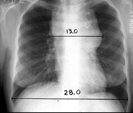
FIGURE 77.1. The mediastinal mass ratio (MMR). This ratio is defined as the maximum single horizontal mediastinal mass measurement divided by the maximum intrathoracic diameter, which is usually near the diaphragm. In this example, MMR = 13.0/28.0 = 0.46.
DIAGNOSTIC WORKUP
Diagnostic and staging procedures commonly used for Hodgkin lymphoma are listed in Table 77.1. Patient age and the presence of intercurrent disease influence the selection of staging studies.24–26
Hematologic evaluation may reveal anemia, leukopenia, lymphopenia, or thrombocytosis. This is often a paraneoplastic effect, but it may be indicative of bone marrow involvement. Anemia, lymphopenia, and hypoalbuminemia are adverse prognostic factors, especially for patients with advanced disease (stage III–IV).27 The serum alkaline phosphatase level may serve as a nonspecific marker of tumor activity or hepatic, bone marrow, or bone disease. The erythrocyte sedimentation rate (ESR) may correlate with response to treatment and subsequent disease activity and is a prognostic factor for patients with limited disease (stage I–II).28 Other useful markers may include the lactate dehydrogenase and β2-microglobulin levels.
Radiographic evaluation should include posteroanterior (PA) and lateral chest radiographs. Mediastinal adenopathy may be quantitated by a measurement of the maximum width of the mediastinal mass divided by the maximum intrathoracic diameter (near the level of the diaphragm) on a standing PA chest radiograph, as shown in Figure 77.1. When this ratio exceeds 1:3, the disease is defined as bulky, and this affects assignment to many clinical trials. Other definitions of bulky mediastinal adenopathy include a mass >10 cm and a ratio of mediastinal mass to the chest diameter at T5-6 exceeding 0.35 (employed in European Organization for the Treatment of Cancer [EORTC] clinical trials). Contrast-enhanced (diagnostic) computed tomographic (CT) scans of the chest, abdomen, and pelvis may reveal adenopathy or organ involvement. If irradiation to the cervical nodes is contemplated, a CT scan of the neck may be indicated in order to identify their precise location for treatment planning. Lymph nodes are usually considered to be enlarged on CT if their short axis measurement exceeds 1 cm.29 Splenomegaly or hepatomegaly alone cannot be interpreted to represent involvement by Hodgkin lymphoma because enlarged spleens often are not involved at the time of splenectomy; however, the presence of focal nodules is usually indicative of involvement.
Positron emission tomography (PET) using 2-fluoro-2-deoxy-D-glucose (FDG), especially with fused CT images (PET-CT scan), has become an important component of initial staging in Hodgkin lymphoma. FDG-PET is more sensitive than CT for detecting disease.30 It may even reveal unsuspected bone or bone marrow disease.31 PET-CT is an essential study for response assessment, is particularly useful for the evaluation of residual masses detected by CT scanning, and may even be a useful prognostic indicator when repeated after just a portion of chemotherapy has been administered.29,32,33
Magnetic resonance imaging may be an alternative to chest or abdominal-pelvic CT scanning for initial staging but has not been used widely.34 Its main value may be in the staging evaluation of women during pregnancy.19
A needle biopsy of the posterior iliac crest bone marrow is appropriate in selected patients. Because the yield is exceedingly low in asymptomatic patients with limited clinical disease, it should be restricted to patients with B symptoms or clinical evidence of subdiaphragmatic disease. The overall incidence of bone marrow involvement in Hodgkin lymphoma is only ~5%.
 STAGING
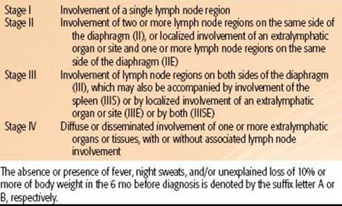
The Ann Arbor staging system for Hodgkin lymphoma, used since 1971, is outlined in Table 77.2.35 The lymphoid regions defined in this system are shown in Figure 77.2. The Ann Arbor system includes designation of a clinical stage, based on the results of the initial biopsy and clinical staging studies, and a pathologic stage, based on the results of any subsequent biopsies, including bone marrow biopsy and those obtained at staging laparotomy. With the exclusion of laparotomy, the generic term stage is now usually employed and reflects the final stage designation after completion of all appropriate staging studies. Deficiencies of the Ann Arbor system include its failure to consider bulk of disease and its lack of a more precise definition of the E-lesion (extralymphatic involvement).36 However, the Cotswolds modification of the Ann Arbor system employs the subscript “x” to designate large mediastinal adenopathy.37
FIGURE 77.2. The lymph node regions as defined in the Ann Arbor staging system. Note that the ipsilateral supraclavicular, cervical, preauricular, and occipital nodes are defined as a single region. The mediastinum and pulmonary hila are defined as separate regions. (From Hoppe RT. The non-Hodgkin lymphomas: pathology, staging, treatment. Curr Probl Cancer 1987;11:363–447; with permission.)

TABLE 77.2 THE ANN ARBOR STAGING CLASSIFICATION FOR HODGKIN’S DISEASE
PATHOLOGIC CLASSIFICATION
The neoplastic cell of classic Hodgkin lymphoma is the Reed–Sternberg cell. It is typically binucleate, with a prominent, centrally located nucleolus in each nucleus, a well-demarcated nuclear membrane, and eosinophilic cytoplasm with a perinuclear halo. However, these cells usually account for <1% of the cells in a lymph node involved by Hodgkin lymphoma. The majority are lymphoid cells, eosinophils, plasma cells, and other normal cells.38
Reed–Sternberg cells probably originate from B-lineage cells at various stages of development, including pre–B-cell and germinal center B-cell origin.39,40 In most instances, the Reed–Sternberg cells stain positively with the lymphocyte activation marker CD30, PAX5, and with variable expression of the antigranulocyte monoclonal antibody CD15. CD20, a marker of mature B cells, may be expressed on a minority of tumor cells with variable intensity in as many as 40% of cases. They stain negatively with CD45, ALK, and J chain.41
There are five histologic subtypes of Hodgkin lymphoma as defined by the World Health Organization modification of the Lukes and Butler system. These include nodular lymphocyte-predominant Hodgkin lymphoma and four subtypes of classic Hodgkin lymphoma: nodular sclerosis, mixed cellularity, lymphocyte-rich, and lymphocyte-depleted.42
Nodular lymphocyte predominant Hodgkin lymphoma (nLPHD) is characterized by an abundance of normal-appearing lymphocytes and a scarcity of abnormal cells. Unlike the other subtypes of Hodgkin lymphoma, the abnormal cells (“L and H cells” or “popcorn cells”) in nLPHD are strongly reactive for CD20, CD45, CD79a, and PAX5 and negative for CD15 and CD30.38,41 nLPHD is often diagnosed in young people. Patients frequently present with early-stage disease, usually in a solitary peripheral nodal site, and systemic symptoms are uncommon (<10%). The natural history is the most favorable of the histologic subtypes. Occasional patients demonstrate a pattern of late relapse but good survival, similar to that observed in the follicular (B-cell) lymphomas. Some investigators suggest that nLPHD would be more appropriately considered a form of non-Hodgkin B-cell lymphoma.43,44–45 As part of its natural history, as many as 14% of patients with nLPHD may transform to an aggressive B-cell lymphoma.46 A reactive process termed progressive transformation of germinal centers may be observed in conjunction with nLPHD.47,48
The other four histologic subtypes of Hodgkin lymphoma are variants of classic Hodgkin lymphoma (cHD). The Reed–Sternberg cells in these cases are CD15+, CD30+, PAX5+, and occasionally CD20+. Nodular sclerosis classical Hodgkin lymphoma (NSHD) is the most common histologic subtype diagnosed in developed countries. Involved nodes often have a thickened capsule and are traversed by broad bands of birefringent collagen that surround nodules of cells consisting of lymphocytes, eosinophils, plasma cells, and tissue histiocytes intermixed with a variable proportion of atypical mononuclear cells and Reed–Sternberg cells. These cells may be in empty (lacunar) spaces, which are artifacts of formalin fixation. The syncytial variant refers to cases in which there are prominent cellular aggregates of atypical cells and histiocytes. The clinical presentation includes common mediastinal involvement, and one-third of patients have B symptoms. The natural history of NSHD is less favorable than that of nLPHD.
Mixed-cellularity classic Hodgkin lymphoma (MCHD) is characterized by a diffuse effacement of lymph nodes by lymphocytes, eosinophils, plasma cells, and relatively abundant atypical mononuclear and Reed–Sternberg cells. Patients with MCHD present more commonly with advanced disease and tend to be slightly older than those with NSHD or nLPHD. The natural history of MCHD is less favorable than that of NSHD.
Lymphocyte-rich classic Hodgkin lymphoma (LRHD) is a relatively recently described entity.49,50 It usually has a nodular growth pattern, but occasionally it has a diffuse one. Previously, many cases of LRHD may have been confused with nLPHD; however, the staining characteristics of the malignant cells in LRHD clearly are consistent with those of classic Hodgkin lymphoma. The clinical characteristics of patients affected by LRHD are similar to those of nLPHD, that is, early stage, absence of B symptoms, and excellent prognosis.
Lymphocyte-depleted classic Hodgkin lymphoma (LDHD) is variable in its microscopic appearance but is generally characterized by a paucity of normal-appearing cells and an abundance of abnormal mononuclear cells, Reed–Sternberg cells, and Reed–Sternberg variants. This subtype may be difficult to differentiate from anaplastic large-cell lymphoma.51 It is an exceedingly uncommon subtype of Hodgkin lymphoma. It tends to occur in older patients and is more likely to be associated with advanced disease and B symptoms. It has the worst prognosis of all histologic subtypes of Hodgkin lymphoma.52
In addition to these major subtypes, interfollicular Hodgkin lymphoma is an uncommon pattern of focal involvement of a lymph node in which there is reactive hyperplasia with a small focus of Hodgkin lymphoma in the interfollicular zone. It is easy to confuse these cases with reactive lymphoid hyperplasia.53
Table 77.3 summarizes the characteristics of patients treated according to the major histologic subtypes of Hodgkin lymphoma at Stanford University from 1989 to 2010. These characteristics are similar to those reported from many other large centers in the United States and western Europe. However, the distribution of histologic subtypes and clinical behavior reported from South America, Asia, Africa, Eastern Europe, and even some parts of the United States indicates a greater proportion of unfavorable histologic subtypes and more aggressive clinical behavior in these developing areas.54,55
TABLE 77.3 MAJOR HISTOLOGIC SUBTYPES CORRELATED WITH CLINICAL CHARACTERISTICS OF 615 ADULT PATIENTS TREATED FOR HODGKIN LYMPHOMA AT STANFORD UNIVERSITY (1989–2011)

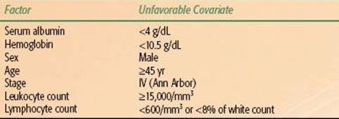
TABLE 77.4 INTERNATIONAL PROGNOSTIC SCORE FOR ADVANCED HODGKIN LYMPHOMA
PROGNOSTIC FACTORS AND THERAPEUTIC IMPLICATIONS
Because most patients with Hodgkin lymphoma are cured, prognostic factors are more important for defining therapy than for predicting outcome. Historically, when treatment programs were less effectie, prognostic factors did have great importance in predicting survival.56
The Ann Arbor stage is likely the most important factor influencing therapy. Data generated at a time when treatment programs were more limited show a marked impact of stage on prognosis. With current management programs, this distinction has been blurred.
The bulk of disease is important, especially in the mediastinum. Bulk may be defined by absolute measurements, ratio of mass to anatomic measurements, surface area on radiographs, or volumetric determinations. In general, the risk of relapse after treatment with single-modality therapy is greater in the presence of bulky mediastinal disease than in nonbulky disease.57 For this reason, combined-modality therapy is the established standard for patients with bulky disease.58,59,60–63
Other measurements of disease severity that may influence treatment selection for Hodgkin lymphoma are the presence of B symptoms, the number of sites of involvement, and the elevation of serum markers such as the ESR. Sophisticated assessments of total tumor burden correlate very well with prognosis.64
The histologic subtype of Hodgkin lymphoma has little impact on therapy, with the exception of identification of cases of nLPHD, for which different treatment algorithms will apply.62 Interobserver agreement among pathologists regarding subclassification is not perfect,65 and after the extent of disease has been determined, histologic subtype of classical Hodgkin lymphoma has little additional impact on prognosis.
Large series report a slightly worse outcome for men than for women.1 However, gender is more important because of its influence on the choice of treatment secondary to potential reproductive complications (see Sequelae of Treatment).
An important international study evaluated a series of prognostic factors among 5,141 patients with advanced Hodgkin lymphoma.27 Seven factors were identified, each of which had an independent and similar impact on prognosis. These included gender, age, Ann Arbor stage, hemoglobin, white cell count, lymphocyte count, and albumin (Table 77.4). This International Prognostic Score is now used for assignment of patients to clinical trials. Patients who have three or more adverse risk factors are often considered to be in an unfavorable prognostic group for advanced Hodgkin lymphoma.
Patients with stage I or II disease often have only one or two adverse factors based on this index. Large clinical trials groups have found other factors, such as number of sites of disease, age, ESR, and presence of B symptoms, to be helpful in stratifying patients according to prognosis. Unfortunately, the criteria for defining “unfavorable” presentations of stage I–II disease vary among clinical trial groups (Table 77.5).
TABLE 77.5 DEFINITION OF “UNFAVORABLE” STAGE I TO II HODGKIN LYMPHOMA IN RECENT CLINICAL TRIALS

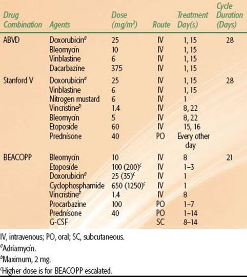
TABLE 77.6 COMMON DRUG COMBINATIONS USED IN THE TREATMENT OF HODGKIN LYMPHOMA
GENERAL MANAGEMENT
Radiation Therapy
Radiation therapy is the most effective therapeutic agent for treating Hodgkin lymphoma and has been used in its management for more than a century.66 Optimal irradiation technique includes careful pretreatment evaluation of disease sites, precise simulation and the use of megavoltage photon beams, fields individually contoured to the patient’s anatomy and tumor configuration, an adequate dose, multifield fractionated treatment, and portal film verification during therapy. Careful attention must be paid to every detail of therapy in order to maximize outcome and minimize risks.67,68
Chemotherapy
The initial successful drug combination for treating Hodgkin lymphoma was nitrogen mustard, vincristine, procarbazine, and prednisone (MOPP), reported by DeVita et al.69 from the National Cancer Institute in 1970. The acute toxicities of treatment at that time were significant and included nausea, vomiting, peripheral neuropathy, constipation, leukopenia, and thrombocytopenia. Late effects of concern included sterility (especially in men) and the risk for secondary myelodysplastic syndrome or leukemia. A number of MOPP-like programs, such as chlorambucil, vinblastine, procarbazine, and prednisone (ChlVPP), achieve comparable results with similar drugs and less toxicity.70
With the introduction of doxorubicin (Adriamycin), completely novel drug combinations were developed. The most successful of these is ABVD, which includes doxorubicin, bleomycin, vinblastine, and dacarbazine.71 ABVD has replaced MOPP as the gold standard of chemotherapy for Hodgkin lymphoma. This is based largely on the results of an intergroup trial that compared MOPP, ABVD, and MOPP/ABVD.72
More recently, in an effort to reduce toxicity, the Stanford V regimen, which almost always includes a component of radiation, was developed as an alternative to ABVD. As another approach, in an effort to enhance efficacy, the German Hodgkin Study Group (GHSG) developed the BEACOPP regimen, which may be administered in a baseline, escalated, or 14-day schedule.73 Table 77.6 summarizes the dosages and scheduling of these drug combinations.70
Most recently, a completely new systemic agent has been approved by the FDA for the treatment of Hodgkin lymphoma. Brentuximab vedotin (BV) is an anti-CD30 monoclonal antibody linked to an antitubulin agent. BV has demonstrated efficacy in CD30+ lymphomas, including Hodgkin lymphoma and anaplastic large-cell lymphoma. It is approved for patients who have had disease recurrence after stem cell transplantation and is being introduced in clinical trials to define its possible use in other settings.74
Combined-Modality Therapy
Combined-modality therapy has become the most common form of general management for patients with Hodgkin lymphoma. Important considerations include the sequence of therapy, the selection of irradiation fields, the decision to irradiate all involved sites, only initially “bulky” sites, or only sites that have not responded completely to chemotherapy, the prescription of dose, and potential overlapping toxicities. Treatment is almost always initiated with chemotherapy. This has the advantages of treating all sites of disease at the outset (especially important in stage III or IV) and reducing bulky disease to facilitate subsequent irradiation (especially in the mediastinum). The irradiation dose used in combined-modality studies in adults ranges from 20 to 36 Gy.
RADIATION THERAPY TECHNIQUES
The principal objective of radiation therapy in Hodgkin lymphoma is to treat involved nodes and regions at high risk for containing disease to a dose associated with a high likelihood of tumor eradication. This requires thoughtful evaluation of all imaging studies, especially diagnostic CT and integrated PET-CT scans, a precise simulation with appropriate immobilization and consideration of organ motion, detailed treatment planning, and effective treatment delivery, including portal imaging and appropriate quality assurance measures.67,68,75,76
The most common techniques for field blocking include multileaf collimation, divergent Cerrobend (Cerro Metal Products, Bellefonte, PA) blocks attached to a Lucite (Lucite International, Southampton, United Kingdom) plate and mounted to the head of the machine, or a combination of both for complex-shaped fields. The use of body molds reduces body movement and rotation and may increase patient comfort.
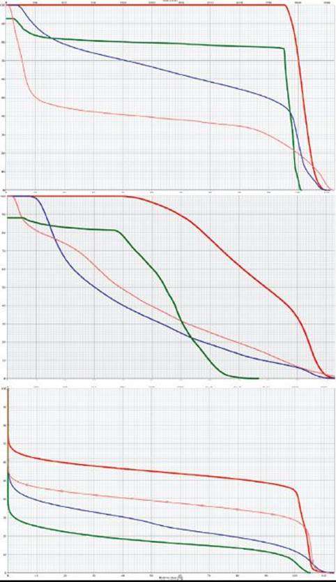
For the majority of clinical scenarios, three-dimensional conformal treatment planning and opposed-field treatment is appropriate, but occasionally intensity-modulated radiation therapy (IMRT) may be indicated.77 IMRT has the inherent advantage of better dose conformality, improved dose–volume histogram (DVH) criteria for the heart, coronary arteries, esophagus, and lungs, and less acute toxicity, but this is at the expense of the low-dose “bath,” which may put larger volumes of normal tissue at risk for the development of secondary cancer, for example, the lungs, breasts (in women), and thyroid gland (Figs. 77.3 and 77.4). This remains a key consideration whenever the use of IMRT is contemplated. IMRT may be most useful in situations of reirradiation, when disease has relapsed in a previously treated site.
Routine three-dimensional (3D) CT simulation is required to optimize treatment field design and analyze DVHs in order to ensure adequate tumor coverage and sparing of organs at risk (OARs). When treatment includes the mediastinum, axillary, and supraclavicular areas, an arms-up position pulls the axillary nodes away from the chest wall and thereby permits more generous lung shielding but may result in an increased dose to the breasts and heart and an enhanced skin reaction in the supraclavicular area. An arms-down or akimbo position permits shielding of the humeral heads and minimizes skin reaction in the tissue folds of the supraclavicular/low neck regions and to the breasts.78 When patients require separate treatment to adjacent regions, the calculation of field separation (gap) is exceedingly important.79 Special additional cord blocking should be used, if possible, when adjacent fields overlie the spinal cord.
FIGURE 77.3. Color-wash dose distributions for three different plans for treating mediastinal Hodgkin lymphoma: axial sections (top) and sagittal sections (bottom) for conventional photon three-dimensional conformal anteroposterior/posteroanterior fields (left), intensity-modulated radiation therapy photon (middle), and anterior proton field (right). Green outline, esophagus; red outline, heart; pink outline, breasts; blue outline, clinical target volume. (Courtesy of Bradford S. Hoppe, MD, MPH.)

In the uncommon situations when radiation therapy alone is used for the treatment of Hodgkin lymphoma (primarily for nLPHD), the National Cancer Center Network (NCCN) guidelines recommend a dose of 30 to 36 Gy to involved and 25 to 30 Gy to uninvolved regions, fractionated at a rate of 7.5 to 10 Gy per week to involved sites.62 Evenly weighted opposed-field treatments, all fields treated daily with fractions of 1.5 to 1.8 Gy, depending on field size and patient tolerance, are the general treatment recommendations. More commonly, radiation therapy is used in the combined-modality setting. Doses vary considerably in different trials, depending on the prognostic category of patients, bulk of disease, and type and duration of chemotherapy. The range of doses considered acceptable according to the NCCN guidelines is 20 to 30 Gy for nonbulky and 30 to 36 Gy for bulky sites of disease, 1.5 to 1.8 Gy per fraction.62
The classic field configurations for the treatment of Hodgkin lymphoma by radiotherapy alone included the mantle and inverted-Y. However, clinical trials have demonstrated an equivalence of “involved-field” treatment (IFRT) with “extended-field” or “subtotal-lymphoid” irradiation in the context of combined-modality therapy programs, and involved-field irradiation has been adopted as the standard for combined-modality therapy.80–82 More recent trials are testing the application of even more restricted fields, referred to as “involved-node radiotherapy” (INRT; discussed later)83 of “involved-region” or “involved-site” irradiation. Because these more limited fields are really portions of the classic treatment fields for Hodgkin lymphoma, an understanding of these classic fields makes the design of limited fields more logical.
FIGURE 77.4. Representative dose–volume histograms for the three plans displayed in Figure 77.3. Anteroposterior/posteroanterior photons (top), intensity-modulated radiation therapy photon (middle), protons (bottom). Red, heart; green, esophagus; blue, lungs; pink, breasts. (Courtesy of Bradford S. Hoppe, MD, MPH.)
PET-CT “Simulation”
In the current treatment paradigm for Hodgkin lymphoma, radiation therapy is nearly always administered in the combined-modality therapy setting. Integrated PET-CT imaging is completed as part of the initial staging for all patients and provides accurate information regarding the initial extent of disease. However, this initial scan is not usually obtained in the treatment position or on a flat couch. After the completion of chemotherapy, the PET-CT scan is usually repeated, and generally one can expect normalization or near normalization of the PET component despite residual disease on CT.84 After the radiation therapy simulation study has been completed (postchemotherapy), the data from the initial staging PET-CT may then be merged with it in order to localize the initial sites of disease. The accuracy of the registration will vary, depending on patient positioning for the two studies, and this needs to be accounted for in ultimate design of the treatment fields.
FIGURE 77.5. A 28-year-old man with massive mediastinal nodular sclerosis Hodgkin lymphoma and right supraclavicular disease, following completion of chemotherapy, with a negative positron emission tomography (PET) scan. White, pretreatment PET+ disease; black, postchemotherapy residual abnormality on computed tomography (CT). A:Design of a modified “involved field” to include the mediastinum, bilateral hila, and supraclavicular areas with an anterior larynx block. Note that inferiorly the field includes the entire length of the original extent of disease plus 2-cm margin. However, laterally the field encompasses only the residual disease on CT plus 2-cm margin. B: Design of an “involved-site” field.
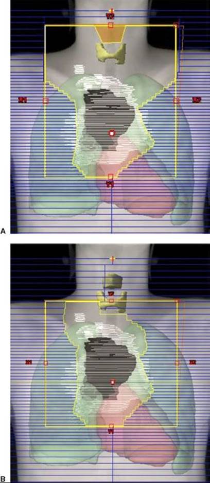
FIGURE 77.6. Representative dose–volume histograms for the fields displayed in Figure 77.5. Small triangles, involved field; small squares, involved site. Yellow, thyroid; pink, heart; blue, lungs. With this configuration of disease the most notable sparing of organs at risk is for the thyroid, with some sparing of the lungs. There is little difference in heart dose. If the disease was more superior in location, sparing of the heart using an involved region would have been more notable.

Supradiaphragmatic Fields
The classic mantle included all of the major lymph node regions above the diaphragm.85 The field extended from the inferior portion of the mandible almost to the level of the insertion of the diaphragm. Individually contoured lung blocks conformed to the patient’s anatomy and tumor localization. In addition to the lung blocks, blocks could be placed over the occipital region and spinal cord posteriorly, the larynx anteriorly, and the humeral heads both anteriorly and posteriorly. The use of these blocks depended on total dose planned and proximity of the adenopathy.
In the classic two-dimensional planned mantle field, the patient was set up supine, with the head fully extended. The superior margin of the field bisected the mandible and passed through the mastoid process. The lateral margins were set to flash the axillae (with humeral head blocks if the arms were at sides or akimbo). The inferior axillary margins were at the level of the inferior tips of the scapulae. The inferior mediastinal border was set at the level of the T10-11 interspace. The lung blocks were designed to provide ~1-cm margin around the mediastinal contours and also encompass the pulmonary hilar lymph nodes. The superiormost point of the lung blocks was no higher than the inferior tip of the head of the clavicle, with the tops of the lung blocks tapered laterally, often parallel to the projection of a posterior rib, in order to expose the high axillary/infraclavicular lymph nodes.
As noted previously, in contemporary management programs of combined-modality therapy, more limited fields are treated. Involved-field treatment includes just portions of the classic radiation treatment fields, and in the original definition implied treatment to the entirety of an involved lymphoid region when any portion of that region was involved. However, the definition of these regions, based on anatomic boundaries, is somewhat arbitrary. Logical considerations mandate modification of these regions in the context of individual patient management. For example, in the common scenario of supraclavicular disease (level IV) without disease any higher in the neck, the submaxillary and submandibular nodes (levels I and II) may be spared. On the other hand, the supraclavicular region is often included when the superior mediastinum is involved because a portion of the superior mediastinum actually superimposes on the lower supraclavicular area in the treatment position. The axillae are not treated unless they are involved. The two-dimensional design of these fields includes a superior border at the top or bottom of the larynx (depending on the extent of supraclavicular disease), lateral borders set at the coracoid processes of the scapulae (to include approximately two-thirds the length of the clavicle), and 0.5- to 1-cm margins beneath the clavicles. With 3D treatment planning, the initially involved lymph nodes (gross tumor volume [GTV]) and adjacent “at-risk” lymph nodes (clinical target volume [CTV]) are outlined on cross-sectional images, and field design is completed to ensure a dose range of 95% to 105% of the prescribed dose to the planning target volume (PTV). The CTV will generally extend 2 to 5 cm proximal and distal to initial PET- or CT-positive disease. The PTV expansion is then ~1 cm (Figs. 77.5A and 77.6).
In the setting of an initial large mediastinal mass, the postchemotherapy treatment fields can usually conform to the width of the residual disease only (unless there was pulmonary parenchymal extension), although the superior and inferior field margins should encompass the initial extent of disease, with margin as noted previously.67 Although special techniques such as deep inspiration breath hold, active breathing control, and respiratory gating are infrequently used in treatment of Hodgkin lymphoma, they do demonstrate improvements in DVH criteria for the lungs and heart that would be especially useful in the setting of treatment for patients with large mediastinal masses.86,87
Organs at Risk
The dose range used for Hodgkin lymphoma (20 to 36 Gy) is below the threshold tolerance for many organs, including the spinal cord. However, intrathoracic structures may be affected by these doses, and careful review of dose–volume histograms is warranted to minimize both acute and late effects. The risk for pneumonitis is related to volume of lung irradiated, total dose, and fraction size. The relationship between mean lung dose or Vx and pneumonitis is complex and may vary for different diseases and depend on patient age, smoking history, presence of intercurrent disease, prior chemotherapy, prior surgery, and so on.88 One study of patients with Hodgkin lymphoma identified a higher risk for Radiation Therapy Oncology Group grade 2 pneumonitis when the mean lung dose exceeded 14 Gy or the V20 exceeded 35%.89 The likelihood of radiation-related pulmonary complications may be increased by the use of bleomycin,90 and the effect that drug combinations with different doses of bleomycin may have on acceptable mean lung dose or V20 values is not known. A guideline followed at Stanford is not to exceed a mean lung dose of 15 Gy after treatment with ABVD or 17 Gy after treatment with Stanford V, treating with 1.5-Gy fractions. When these parameters have been followed, the risk for radiation pneumonitis has been negligible. With respect to late carcinogenesis, because data indicate that lung cancer risk may be increased after doses as low as 5 Gy, it is reasonable to define the lung V5 and try to minimize it if multiple plans are reviewed.91
The criteria for cardiac dose tolerance are not well defined. Again, tolerances will be affected by comorbidities, family history, and prior treatment with cardiotoxic drugs such as doxorubicin. With respect to acute effects (pericarditis), limited data suggest keeping the mean pericardial dose to <26 to 27 Gy and the V30 to <46%.92,93 Data for late cardiac events are less reliable; however, in one study of patients with Hodgkin lymphoma a threshold effect at 30 Gy was suggested for cardiac mortality.94 A more conservative estimate, based on patients irradiated for breast cancer, is that a cardiac V25 of <10% is associated with a very low risk of cardiac mortality.95 This level may be difficult to achieve in patients who present with large mediastinal adenopathy. Treatment techniques such as IMRT may succeed in reducing the cardiac dose but increase the V5 to the lungs or V4 to the breasts, resulting in a higher risk for secondary cancer in those organs (Figs. 77.3 and 77.4).
With respect to the breasts in women, given the excess risk of secondary breast cancer that exists for doses as low as 4 Gy, it is reasonable to track the breast V4 and keep that volume as small as possible, especially for women <30 years of age.96
Subdiaphragmatic Fields
The classic subdiaphragmatic irradiation field for Hodgkin lymphoma was the inverted-Y, which included the retroperitoneal and pelvic lymph nodes and spleen. Sequential treatment to a mantle and inverted-Y field was referred to as total lymphoid irradiation(TLI); if the subdiaphragmatic field did not include the pelvis, the term subtotal lymphoid irradiation was used.
Currently, in the context of combined modality therapy, common radiation therapy fields include the spleen with or without the para-aortic nodes (Fig. 77.7) and unilateral or bilateral pelvic fields (Fig. 77.8). In the two-dimensional design of a para-aortic field, the width of the field generally corresponds to the width of the transverse processes. The spleen may be treated in contiguity with this field. The design of the splenic field requires consideration of respiration and the use of generous (~2 cm) superior and inferior margins or respiratory gating. Three-dimensional planning for this field, especially if the spleen is being irradiated, is essential in order to more accurately localize the spleen and evaluate the DVH for the left kidney (Fig. 77.7). GTV, CTV, and PTV considerations for subdiaphragmatic nodal fields are similar to those for supradiaphragmatic fields, that is, GTV includes prechemotherapy disease on PET or CT, CTV includes 2 to 5 cm proximal and distal, and PTV expansion is 1 cm.
Careful blocking considerations are required when the pelvic region is treated. Because the volume of marrow in the pelvis is substantial, the fields must be shaped carefully to minimize the amount of marrow treated (Fig. 77.8). With two-dimensional planning, the lateral margins are set 1.5 to 2 cm lateral to the widest point of the bony pelvis. Inferiorly, the pelvis field should extend to at least the lesser trochanters, unless there is disease that extends further inferiorly. Gonadal toxicity may also be an issue. In women, the ovaries normally overlie the iliac lymph nodes. To avoid irradiation-induced amenorrhea, an oophoropexy must be performed. This procedure is done by medial or lateral transposition of the ovaries via laparoscopy. The surgeon marks the ovaries with radiopaque sutures or clips and relocates them medially and as low as possible behind the uterine body. A double-thickness (10 half-value layers) midline block is then used; its location is guided by the position of the opacified nodes and transposed ovaries. When the ovaries are at least 2 cm from the edge of this block, the dose is decreased to 8% of that delivered to the iliac nodes.97 Alternatively, one or both of the ovaries can be transposed laterally to a position overlying the iliac wings.
In men, if no special blocking is provided for the testes, the testicular dose may be as high as 10% of the dose delivered to the inguinal-femoral nodes. Use of a double-thickness midline block and a specially constructed testicular shield can reduce this dose to 0.75% to 3.0%, most of which results from internal scatter. The precise dose depends on the position of the testes in relation to the inferior margin of the inguinal-femoral field.
FIGURE 77.7. Example of a para-aortic–spleen field treated with respiratory gating. In this example there was a positron emission tomography+ node at the L1 level, and the inferior portion of the para-aortic field was set at the bottom of L2. The para-aortic nodes are highlighted in light red and the spleen in dark red. Due to the complex shape of this field, it is defined with a combination of multileaf collimators and Cerrobend blocks (shown in light orange) to shield the base of the left lung and the upper half of the left kidney.

FIGURE 77.8. Typical anteroposterior/posteroanterior field for treating unilateral pelvic nodes, in this case for a 34-year-old man with stage IIA lymphocyte-predominant Hodgkin lymphoma. The initial positron emission tomography+ nodes are shown in blue. The field includes margins of at least 2 cm medial and lateral to the nodes and a field width of at least 6 cm.

Involved-Node/Involved-Site Radiotherapy
In conjunction with the evolution of combined-modality therapy programs for all stages of Hodgkin lymphoma, the recognition that late effects may be reduced by using smaller radiation fields, and the introduction of sensitive functional imaging techniques, there has been an effort to continue to reduce radiation field size. The most recent iteration of this concept is the introduction of INRT.83,98,99 As strictly defined by Girinsky et al.83,98 representing the EORTC-GELA Lymphoma Group, INRT requires prechemotherapy diagnostic CT and PET-CT imaging with the patient in the treatment position, postchemotherapy contrast-enhanced CT simulation, and fusion of the prechemotherapy and postchemotherapy images. The fields are designed to treat only the initially involved nodes with modification to avoid OARs. This GTV then becomes the CTV, and a 1-cm expansion of the CTV defines the PTV.
When components of this planning process are missing, for example, the pretreatment PET-CT scan was not done in the treatment position, there is poor registration between scans, or the CT simulation scan was done without intravenous contrast, it may be risky to treat such strictly defined “involved-node” fields. However, field reduction is still possible to what one may term “involved-site” irradiation (Figs. 77.5B and 77.6).
Proton Beam Therapy
The potential dosimetric advantage of treatment with protons, as opposed to photons, is well established, and proton therapy is often employed in the management of prostate cancer and childhood tumors. There is also significant potential advantage of protons over photons (either 3D conformal radiation therapy [3DCRT] or IMRT) in the management of Hodgkin lymphoma. The use of proton beam therapy can be associated with decreased dose to the gut, bone marrow, and other organs but is especially advantageous with respect to mediastinal treatment. Conventional 3DCRT with opposed fields minimizes lung exposure, but portions of the heart, especially the coronary arteries and valves, as well as of the esophagus, cannot be spared. IMRT plans can be quite conformal and spare those structures but at the expense of a low-dose “bath” that includes treatment to the breasts (in women) and lungs, with the potential risk for secondary cancer. With proton therapy, there can be maximal sparing of the esophagus, lungs, and cardiac subunits, thereby minimizing risk to those organs, while at the same time avoiding low-dose exposure to the breasts and lungs, minimizing potential risks and complications of therapy related to those organs (Figs. 77.3 and 77.4).100,101–102,103 Experience is limited, but as more proton centers are established in the United States, it is likely that many patients with mediastinal Hodgkin lymphoma will be referred to these centers.
TABLE 77.7 REPRESENTATIVE RESULTS FROM SELECTED CLINICAL TRIALS FOR EARLY, INTERMEDIATE, AND ADVANCED STAGES OF HODGKIN LYMPHOMA

RESULTS OF THERAPY
Hodgkin lymphoma is responsive to both irradiation and chemotherapy, and a variety of programs may achieve similar survival rates. However, there may be significant differences in freedom from relapse and potential complications of therapy. The results described in the following sections emphasize treatment programs that have been identified as appropriate according to the guidelines of the NCCN.62 The results achieved in recent clinical trials that have defined the current standards are displayed in Table 77.7.
Favorable Prognosis Stage I to IIA Classic Hodgkin Lymphoma
Favorable presentations of stage I to II include patients with stage I to II who do not have systemic symptoms or large mediastinal adenopathy. In some series from Europe and Canada, patients with an elevated ESR (>50), extralymphatic extension (E-lesion), multiple sites of disease (more than two or three), older age (>50 years), or unfavorable histology (mixed cellularity or lymphocyte-depleted) are also excluded and treated according to algorithms for intermediate prognosis (Table 77.7).104
Historically, patients with favorable presentations of stage I to II Hodgkin lymphoma were candidates for treatment with radiation therapy alone, with curative intent and expectations. The treatment volume generally included the mantle and para-aortic fields, as well as the spleen. Results in single-institution and cooperative group trials included 10-year survival rates of 90% and freedom from relapse rates of 80%.57 These results are excellent, but the appearance of late risks of radiation therapy, including secondary neoplasia and cardiovascular disease, resulted in a shift of management to the use of combined-modality therapy.105
The current treatment of choice for these patients is abbreviated chemotherapy plus limited (IF) irradiation. The results of the HD-10 trial of the GHSG suggest that patients with very favorable presentations (no large mediastinal mass, no more than two sites of disease, no E-lesions, and ESR of <50 or <30 if B symptoms are present [see Table 77.7]) may be treated with just two cycles of ABVD, followed by 20-Gy IFRT (8-year survival [OS], 95.1%; freedom from treatment failure [FFTF], 85.9%; progression-free survival [PFS], 86.5%).106 Other patients may be treated very effectively according to the regimen described by Bonadonna et al.80: ABVD times four followed by IFRT (12-year OS, 94%; freedom from progression [FFP], 94%), although the radiation dose may be reduced to 30 Gy. An alternative is to treat with 8 weeks of Stanford V chemotherapy, followed by 30-Gy IFRT (10-year OS, 96%; FFP, 94%; disease-specific survival, 99%).107
Important issues that have been addressed in recent clinical trials include further attenuation of the ABVD regimen by deletion of individual drugs but still incorporating IFRT (GHSG HD13 trial)73 and trials testing the use of ABVD chemotherapy alone. The National Cancer Institute Canada HD6 trial included treatment with ABVD alone, but the comparison arms included subtotal lymphoid irradiation alone for the favorable patients (age, <40 years; lymphocyte predominance or nodular sclerosis histology; ESR, <50; and fewer than four involved regions) and 2 months of ABVD, followed by subtotal lymphoid irradiation for patients with any unfavorable characteristics. Although neither radiation therapy–containing arm would currently be considered appropriate management, the FFP in the two radiation-containing arms of the trial was superior to that of ABVD alone (93% vs. 87%; p = .006).108 In the design of this study, patients randomized to receive ABVD alone underwent repeat CT imaging after two cycles of chemotherapy. Those patients who achieved a complete response or complete response undocumented (35% of patients) received two more cycles of ABVD and no further therapy. Of note, the 5-year FFP in this select group was ~95%.
Based on these data and consensus guidelines of the NCCN and the European Society for Medical Oncology (ESMO),62,63 the most commonly employed treatment for favorable presentations of stage I to IIA Hodgkin lymphoma is combined-modality therapy with chemotherapy plus IFRT. The expected FFP is 90% to 95%. Selected patients may be treated with chemotherapy alone if radiation therapy is contraindicated and the increased risk for relapse is acceptable. Trials in progress (see later discussion) are testing whether interim PET imaging, done after as few as two cycles of ABVD, can help in identifying patients who are suitable for treatment with chemotherapy alone.
The use of radiation therapy alone has a long history in the successful treatment of early-stage Hodgkin lymphoma but has been abandoned for the reasons cited earlier. However, it remains a reasonable option for patients in whom there is a contraindication to chemotherapy.
Fewer than 10% of patients with stage I or II Hodgkin lymphoma present with involvement limited to subdiaphragmatic sites. For patients in whom disease is nonbulky and limited to pelvic lymph nodes with or without extension into the lower para-aortic nodes, the same general treatment principles apply to the management as for those with supradiaphragmatic presentations. In fact, many trials for stage I to II disease included such patients, and the most reasonable approach is to use combined-modality therapy, as outlined previously. In general, the outcome of treatment for these patients is equivalent to that of patients with supradiaphragmatic disease.109,110
Patients with bulky abdominal subdiaphragmatic presentations or those who have involvement of the spleen are generally treated according to guidelines for “unfavorable” stage I or II or else as stage III or IV disease.
Stage I to IIA Nodular Lymphocyte-Predominant Hodgkin Lymphoma
Contrary to the experience with classic Hodgkin lymphoma, it has been noted that patients with limited presentations of nLPHD may achieve long-term disease-free survival after treatment with involved-field or slightly extended field irradiation alone.45,111,112 For example, for a high–cervical-stage IA presentation, treatment may be limited to the ipsilateral neck. For a femoral node presentation, treatment may be limited to the inguinal-femoral region with or without the ipsilateral iliac region. The usual dose is 30 to 36 Gy. There does not appear to be any benefit from the addition of chemotherapy in this setting. For example, in the retrospective review of experience with nLPHD in the GHSG, there was no significant difference in response induction, freedom from treatment failure, or overall survival for involved-field irradiation versus extended-field irradiation versus combined-modality therapy. Based upon these data and data from single-institution studies, the EORTC and GHSG adopted involved field irradiation alone, dose ~30 Gy, as the standard treatment for these patients, which also conforms with the guidelines of the ESMO and NCCN.62,63 Lower doses have been tested, but responses have not been as durable as with conventional doses.113
Stage I to II Classic Hodgkin Lymphoma with Large Mediastinal Adenopathy
Patients with bulky mediastinal Hodgkin lymphoma (mediastinal mass greater than one-third of maximum intrathoracic diameter) are difficult to categorize by the Ann Arbor staging criteria and have a poor outcome when treated with single-modality therapy.58 Early reports confirmed that these patients are best treated with combined-modality therapy.57
When the choice of chemotherapy is made for these patients, the potential overlapping toxicities of doxorubicin and bleomycin with irradiation (cardiac and pulmonary effects) should be considered. The recommended radiation dose in this situation varies from 20 to 36 Gy, but most data cite doses of at least 30 Gy for this cohort.80,114 Doses in the higher part of the range may be considered when the response to chemotherapy is incomplete, PET imaging remains positive after chemotherapy, or the chemotherapy course is abbreviated.
Although ABVD remains the standard chemotherapy for this clinical presentation, both Stanford V and BEACOPP regimens have been tested in clinical trials. The GHSG HD11 trial for patients with intermediate prognosis, which included patients with large mediastinal adenopathy, elevated ESR, the presence of extranodal disease, or more than two sites of involvement, randomized the chemotherapy to BEACOPP baseline versus ABVD and the involved-field radiation therapy dose to 20 versus 30 Gy. The final analysis of that trial indicated that 20 Gy was sufficient only if BEACOPP chemotherapy was used but was inadequate in conjunction with ABVD. However, the toxicity of BEACOPP was greater, resulting in the adoption of ABVDx4 plus 30-Gy IFRT as the standard arm of the next trial of the GHSG, HD14.61 The results for treatment with ABVDx4 plus 30 Gy included 5-year OS of 94%, PFS of 87%, and FFTF of 85%.
The Stanford V regimen has been used in the setting of patents with large mediastinal adenopathy. The 12-week chemotherapy program is followed by irradiation (30 Gy) to all sites >5 cm. In the setting of large mediastinal adenopathy this always included the mediastinum, but the bilateral hilar and supraclavicular areas were also included in the treatment fields.115 The Eastern Cooperative Oncology Group (ECOG) compared treatment with ABVDx6–8 versus Stanford V é 12 weeks, in both cases followed by 36-Gy irradiation to mediastinum and bilateral hilar and supraclavicular areas (E2496). There were no significant differences in 5-year OS (95% and 92%) or FFS (85% and 77%).116
Based on consensus data, the most commonly employed treatment for stage I or II Hodgkin lymphoma in the presence of large mediastinal adenopathy is combined-modality therapy with chemotherapy plus IFRT. The expected FFP is 80% to 90%.
Stage IB or IIB Hodgkin Lymphoma
Approximately 15% to 20% of patients with stage I or II disease have B symptoms. In general, these patients are managed in a fashion analogous to those with stage III to IV disease. However, given the limited anatomic extent of disease in stage I to II, one can make a strong argument to include consolidative involved-field irradiation for these patients, as is recommended by the ESMO and NCCN.62,63
Stage III to IV Disease
Chemotherapy is the mainstay of treatment for patients with stage III to IV Hodgkin lymphoma.117 With respect to the choice of chemotherapy, the landmark study was the prospective, randomized clinical trial conducted by the Cancer and Leukemia Group B. Patients with stage III2A, IIIB, or IV Hodgkin lymphoma were randomly assigned to treatment with MOPP (six to eight cycles), MOPP/ABVD (12 months), or ABVD (six to eight cycles). The results of treatment with MOPP/ABVD and ABVD were equivalent, and both were superior to MOPP alone. Among the 115 patients treated with ABVD chemotherapy, the complete response rate was 82%, the 5-year FFS was 61%, and OS was 73%.72 More recently, the “gold standard” of ABVD has been challenged by the GHSG, which developed the BEACOPP regimen and has demonstrated in a series of trials that results using BEACOPP escalated (often including irradiation) may be superior to those that can be achieved with ABVD. The GHSG HD9 trial compared BEACOPP escalated, BEACOPP baseline, and COPP/ABVD.118 The outcome was best in the BEACOPP escalated arm, with 5-year FFTF of 87% and OS of 91%. However, in an Italian multi-institutional study that compared an initial treatment strategy of ABVD (four to eight cycles) with BEACOPP (four escalated plus four baseline), which took into account the possibility of autologous stem cell transplant as salvage therapy, there was no significant difference in 7-year freedom from second progression or OS (89% vs. 84%), and severe adverse events were more likely in the BEACOPP group.119
The use of combined-modality therapy in stage III to IV disease has a rationale because most patients who relapse after treatment with chemotherapy alone do so in sites of initial disease.120 However, many of the early trials intended to resolve this issue were poorly designed, used chemotherapy programs that are no longer considered to be optimal, or had inadequate accruals. Nevertheless, many trials of systemic therapy for advanced disease have included the selective use of consolidative irradiation.118,119,121
The most definitive trial to address the question regarding effectiveness of consolidative irradiation is the EORTC–Groupe Pierre-et-Marie Curie H34 (20884) trial.122 In this trial, patients were treated with six to eight cycles of nitrogen mustard, vincristine, procarbazine, prednisone, Adriamycin, bleomycin, and vinblastine chemotherapy, and those who achieved a complete response were randomized to no further therapy versus 25-Gy IFRT. No differences in FFTF or OS were identified. A detailed evaluation of causes of death revealed an unusually high risk for secondary myelodysplasia in the group of patients randomized to combined-modality therapy, although a similar risk was not observed in the nonrandomized patients, all of whom received irradiation to a somewhat higher dose! Those patients who achieved only a partial response (by CT criteria) received 30-Gy IFRT. The subsequent FFTF and OS for this group closely paralleled the outcome for patients who had achieved a complete response, suggesting a value to adding IFRT after only a partial response has been achieved.123
The GHSG HD12 trial was another effort to evaluate the impact of consolidative irradiation.73,124 Patients were randomized to either of two different BEACOPP schedules with or without consolidative irradiation, 30 Gy to initial bulk or residual sites of disease. When analyzed “as treated,” there was no significant difference in FFTF (90.4%, irradiated; 87%, nonirradiated) or OS. However, 14% of patients randomized to “no radiotherapy” were actually irradiated after a panel review of response to BEACOPP. This contamination compromises any ability to draw conclusions regarding the role of radiation therapy in advanced disease. The final conclusion of the study was that it did not support the omission of consolidative irradiation for patients with CT evidence of residual disease after chemotherapy.
Although the value of consolidative irradiation after complete response to conventional chemotherapy (ABVD or BEACOPP) has not been proved, there are programs of attenuated chemotherapy in which radiation therapy is an essential component. The Stanford V program includes only 12 weeks of chemotherapy, with very attenuated total doses of some of the drugs (see Table 77.7). Compared with six cycles of ABVD, there is only 50% of the cumulative dose of doxorubicin (Adriamycin) and 25% of the cumulative dose of bleomycin. Radiation therapy (30 to 36 Gy) is routinely added to initially bulky (>5 cm) sites of disease, as well as to macroscopic splenic involvement, and commences 1 to 3 weeks after completion of chemotherapy. The results of this approach have been excellent.115,125 However, the radiation therapy component is essential, because a study that did not employ the same guidelines for radiation therapy resulted in a much worse outcome.126 The ECOG E2496 trial compared management with ABVD versus Stanford V for patients with advanced-stage or locally advanced disease. There was no difference in 5-year FFS (73% for ABVD, 71% for Stanford V) or OS (88% for ABVD, 87% for Stanford V).127 Lack of a difference between ABVD and Stanford V was also the result of the United Kingdom National Cancer Research Institute Lymphoma Group Study ISRCTN 64141244.128
A general conclusion regarding the role of combined-modality therapy compared with chemotherapy alone for patients with stage III to IV disease is that patients who achieve a complete response to a full course of conventional chemotherapy have no proven benefit from the addition of chemotherapy. Nevertheless, irradiation is often added to such programs on a selected basis, especially for bulky disease. In addition, programs of attenuated chemotherapy may realize a benefit from the addition of irradiation, and patients who achieve only a partial response to chemotherapy may have an improved outcome by the addition of irradiation. Ultimately, improved imaging and evaluation of early response to chemotherapy with FDG-PET imaging may help to identify a subset of patients who would truly benefit from consolidative irradiation (see section on Current Clinical Trials).
Pediatric Patients
Most contemporary programs for the management of pediatric Hodgkin lymphoma are based on clinical staging and use chemotherapy alone or combined-modality therapy with low-dose irradiation because higher doses of irradiation are associated with unacceptable risks for growth impairment and late effects.13 To limit growth effects, irradiation doses should not exceed 15 to 25 Gy. Children treated with these programs, all stages combined, are reported to achieve 5-year OS rates of approximately 90% and relapse-free rates of at least 80%.129–133
Older Adult Patients
The treatment of Hodgkin lymphoma in older patients (>60 years) also poses a challenge.17,134–136 They often have less favorable histology and worse performance status. They are more likely to have intercurrent disease that compromises the aggressive management programs used for younger people. Chemotherapy programs may often be modified to minimize cardiac or pulmonary toxicity, and the hematologic reserve in elderly patients more often results in dose reductions or premature discontinuation compared with younger patients.137 Drug combinations that seem to be more tolerable for older adults include ChlVPP,138 procarbazine, Alkeran, and vinblastine,139 and vinblastine, bleomycin, and methotrexate (used primarily for stage I to II).140 With respect to the radiation therapy, patients may need to be treated with slower fractionation programs and observed carefully for signs of weight loss or general decline in performance status. Extended fields are more difficult to tolerate than more limited fields.16
Treatment for Relapse
Treatment for relapse must be individualized. Initial disease characteristics, initial treatment and response duration, relapse sites, and general patient status must be considered in developing an effective secondary treatment program.
In general, patients who were treated initially with irradiation alone for stage I to II disease (now a relatively infrequent occurrence) should receive chemotherapy as the primary salvage treatment.141,142 The efficacy of combination chemotherapy in this setting is similar to that achieved when chemotherapy is used in the primary management of advanced disease (rate of long-term freedom from relapse of 60% or better). The role of irradiation in combination with salvage chemotherapy has not been defined but is quite reasonable to consider if relapse is in a previously unirradiated site.143
More problematic is the management approach to patients who present initially with stage I to II disease and are treated with chemotherapy alone, a group for whom consensus best treatment has not been reached.62 In these patients, relapse may be restricted to initial sites of disease and be quite limited.144 It is possible that in this situation programs using irradiation alone, or at least emphasizing the use of radiation, may be safe and effective, especially given the success in treating some patients with initially advanced disease and limited relapse using this approach.145,146
For patients who present initially with stage III to IV disease and relapse after achieving a complete response to chemotherapy or combined-modality therapy, the standard salvage therapy is high-dose chemotherapy with autologous hematopoietic cell rescue.147 The long-term PFS rate for these patients is expected to be approximately 50%.148 Favorable prognostic factors in this group include a longer duration of response to primary therapy and absence of extranodal disease.149,150 Allotransplantation is not used often for relapsed Hodgkin lymphoma but may be considered in situations of an unsuccessful autotransplant. Reduced-intensity conditioning regimens appear to be safer than myeloablative regimens.151
The Role of Radiation Therapy in Hematopoietic Cell Transplantation
Radiation therapy may be incorporated into high-dose therapy programs such as IFRT, TLI, or total-body irradiation (TBI).147 Fractionated TBI is incorporated into a number of transplantation programs. Its value is debatable, and series that have used regimens with or without fractionated TBI report similar outcomes for both.152 TBI is probably not the most efficacious way to use irradiation in these patients. Recurrent Hodgkin lymphoma is often a locoregional problem rather than a systemic one. In addition, data suggest that irradiation doses in the range used in TBI programs (12 to 15 Gy) are likely to eradicate disease in only about 20% of treated sites.9 It is more logical to limit irradiation to sites of failure or those at high risk for disease, that is, initial sites of disease, especially bulky sites.
Important other issues include the timing of radiation (pretransplant or posttransplant), extent of fields, and dose. The advantages of using radiation therapy as cytoreductive treatment prior to high-dose therapy are that it can effectively reduce the tumor burden before high-dose treatment and the risk of interruption or delay of the locoregional radiation therapy is minimal. The primary disadvantages include potential delay of the high-dose therapy and the potential overlapping toxicities of the locoregional irradiation and high-dose therapy, including mucositis and pneumonitis.153 Cytoreductive radiation treatment may include all sites of relapse, the bulky sites of relapse, sites with an incomplete response, or even more extensive treatment, such as TLI.
Many large published series of high-dose therapy for Hodgkin lymphoma included locoregional irradiation in at least selected patients. In some series, irradiation is given pretransplant, although in the majority of reports it is given after transplant.154 The range of intervals from transplant to irradiation varies from 1 to 4 months. Often, the fields treated include sites of bulky disease (variably defined) at the time of relapse or areas of residual disease after high-dose therapy has been administered. Some included all sites involved at the time of relapse.
The range of radiation doses employed varies substantially in these series, from 18 to 40 Gy. In general, lower doses are employed in situations in which initially nonbulky disease is included in the treatment or if there has been a complete response to high-dose therapy.
The use of locoregional irradiation in high-dose therapy programs has the potential for altering the patterns of failure and perhaps reducing the risk of failure. For example, at Stanford, 49 patients with relapsed stage I to III disease who underwent high-dose therapy for relapse of Hodgkin lymphoma had IFRT as a component of their salvage treatment.155 Their 3-year FFR, OS, and event-free survival (EFS) rates of 100%, 85%, and 85%, respectively, compared with only 67%, 60%, and 54%, respectively, for another group of patients who received high-dose chemotherapy alone. The difference in FFR was statistically significant (p = .04). A similar effect of local irradiation has been reported in cohorts of patients transplanted at several other centers.131,156,157
At Memorial Sloan-Kettering Cancer Center, an intensive program that incorporates pretransplant TLI has been used.158 Patients who had not received previous irradiation were treated with IFRT to 18 Gy and TLI to 18 Gy (both with twice-daily fractionation). Patients who had prior irradiation were treated with IFRT only, if organ tolerance would not be exceeded, to a dose of 18 to 36 Gy in 5 to 10 days (twice-daily fractionation), depending on the prior doses received by the involved sites. The 10-year OS was 56%, and EFS was 56%.159
FOLLOW-UP
Given the effectiveness of primary therapy for Hodgkin lymphoma, the low rate of relapse, the risk for “false-positive” imaging studies, and the expense to the health care enterprise, there is debate regarding the value of routine follow-up for disease detection, beyond addressing patient symptoms.160–162 However, there is no denying that follow-up is important to monitor for complications of therapy and late effects and to ensure health maintenance.
As a rule of thumb, all studies that initially gave abnormal results (e.g., chest radiograph, CT scan, PET scan) should be repeated at the time of completion of therapy to document the completeness of response.26 The subsequent follow-up interval is typically every 2 to 4 months during the first 2 years, every 4 to 6 months during the third and fourth years, and annually thereafter.62
The most important component of follow-up is an interim history and physical examination, which leads to identification of two-thirds of relapses.160 The frequency with which imaging studies should be repeated after the completion of therapy is not well defined. An occasional chest radiograph, especially if chest irradiation was included as a component of therapy, is justifiable. The value of more extensive imaging evaluation in the absence of symptoms or abnormalities is questionable.
The ESR, serum albumin, or other serum marker studies may be followed if these markers were abnormal at presentation. Serum thyroxine (T4) and sensitive thyroid-stimulating hormone (TSH) levels should be obtained at least annually in patients who received irradiation to the neck, to detect subclinical hypothyroidism.
A challenging problem in follow-up evaluation in the past was the interpretation of residual mediastinal abnormality on chest radiograph or chest CT scan. However, this problem has been obviated by the introduction of FDG-PET scanning as a posttreatment assessment tool, with which concern may be limited to those patients who have residual PET activity.32,163,164–165
In almost every case, the first episode of relapse should be documented by biopsy. Inflammatory disease, progressive transformation of germinal centers, or the rebound growth of the thymus in young patients are other reactive processes that can be confused with recurrent Hodgkin lymphoma. All of these processes may be FDG-avid on PET scanning.
SEQUELAE OF TREATMENT
Depending on the fields treated, the acute side effects of radiation therapy may include occipital hair loss, mild skin reaction, sore throat, an altered sense of taste, dysphagia, reflux symptoms, dry cough, nausea, occasional vomiting, diarrhea, and blood count suppression. Most of these sequelae can be managed symptomatically. Complications that may arise in the early phase of the follow-up program are mild radiation pneumonitis, radiation pericarditis, hypothyroidism, herpes zoster, Lhermitte sign, and xerostomia.
Radiation pneumonitis may develop within 6 to 12 weeks after completion of mantle irradiation.166 After classic “mantle” therapy, <5% of patients have symptomatic pneumonitis, manifested by cough, fever, pleuritic chest pain, and an infiltrate on chest radiography that usually conforms to the irradiation fields. Symptomatic management is usually sufficient; however, a small proportion of patients require treatment with corticosteroids, usually beginning with a daily dose of 40 to 60 mg of prednisone (or other corticosteroid equivalent). The initiation of corticosteroid therapy commits one to a course of at least 4 to 6 weeks, with slow, careful tapering to avoid exacerbation of symptoms.
Subclinical hypothyroidism develops in as many as half of patients who receive doses of >30 Gy to the neck.167 It can be detected by an elevation of TSH even with a normal T4 level. Thyroid replacement therapy with L-thyroxine is recommended, with an initial dose of up to 0.1 mg/day. The T4 and TSH values are monitored regularly to make adjustments in the dose. Evidence suggests that thyroid replacement therapy in this setting reduces the risk for development of benign thyroid nodules.168 It is likely that newer techniques of treatment with lower doses and better shielding of the thyroid gland will result in a decrease in this risk.
Herpes zoster can occur during treatment for Hodgkin lymphoma or within the first few years after treatment in 10% to 15% of patients.169 The outbreak is usually limited to one or two contiguous dermatomes. Cutaneous dissemination is uncommon, and visceral involvement is extremely rare. If the cutaneous eruption is identified within 72 hours of its onset, treatment with acyclovir (800 mg five times per day for 7 to 10 days or other antiviral equivalent) can be initiated. This may limit the duration and intensity of infection and decrease the likelihood of cutaneous or visceral dissemination. Currently, zoster vaccine immunization is not recommended for patients who have been treated for lymphoma (www.cdc.gov/vaccines).
Lhermitte sign develops in approximately 10% to 15% of patients after radiation therapy that includes a significant length of the spinal cord and is more likely to occur among patients who have been treated with vinca alkaloids (vincristine and vinblastine). It is marked by paresthesias extending into the arms and legs on neck flexion and may be related to transient demyelinization of the spinal cord. Its onset is usually 1 to 2 months after completion of mantle therapy, and it generally resolves spontaneously after 2 to 6 months. This sequela is not related to the more serious problem of transverse myelitis.
Significant xerostomia may follow irradiation of the Waldeyer lymphoid region or the bilateral submandibular regions, which is rarely necessary, and permanent attention to dental care is required for these patients. Frequent dental prophylaxis and use of fluoride supplements are recommended.
An uncommon but potentially serious complication is overwhelming sepsis after splenectomy or splenic irradiation.170,171 The most serious infections occur with Gram-positive organisms, including Streptococcus pneumoniae,meningococci, and Haemophilusstrains. This risk can be minimized by immunization against these organisms. It is reasonable to immunize patients as soon as a diagnosis of Hodgkin lymphoma has been made. Recommendations for immunization of patients who have been treated previously but not immunized before therapy vary. Recent data suggest that if at least 2 years have passed since treatment, patients can develop adequate antibody titers to H. influenzae type b-conjugate, 4-valent meningococcal polysaccharide vaccine, and 23-valent pneumococcal polysaccharide vaccine.172 Reimmunization is currently recommended every 5 to 7 years.
An important concern of many patients with Hodgkin lymphoma is the possible effect of treatment on reproductive potential. In men, pelvic irradiation may be followed by azoospermia if no special precautions are taken to shield the testes. However, with appropriate testicular shielding, azoospermia is usually only transient, with subsequent recovery of sperm counts to fertile levels.173 Chemotherapy programs such as MOPP, MOPP-like combinations that include alkylating agents and procarbazine, or BEACOPP will cause sterility in most men. However, the ABVD and Stanford V regimens seem to spare male fertility.115,174 Among women, the risk for infertility is influenced by patient age. With respect to the irradiation component, even with a proper oophoropexy and well-planned treatment fields, the scattered dose of irradiation may be sufficient to affect ovarian function and cause menopausal symptoms in women >30 years of age who receive pelvic irradiation.175 Younger women may not have an immediate effect but may enter a premature menopause later in life. Similarly, with respect to chemotherapy that contains alkylating agents, normal menstrual function usually continues in women younger than 25 years but is altered in women older than 30 years.175,176 Again, younger women may later experience an earlier-than-normal onset of menopause. In contrast to MOPP and BEACOPP, the ABVD and Stanford V combinations appear to spare female fertility.115,174
An uncommon but significant treatment complication is the “dropped-head syndrome” secondary to cervical muscle atrophy that occurs in a small proportion of patients treated with high-dose irradiation (>40 Gy) to the neck.177This is a late risk, not usually apparent until >10 years after therapy, and has only been reported with doses of >40 Gy. Management of this problem is challenging, with variable response to physical therapy and use of a “soft collar.” Surgical intervention has been attempted in some patients.178
Long-term follow-up is essential to identify significant adverse late complications of treatment in all patients.105,179,180–182 The most important long-term hazards are secondary malignancies183 and cardiovascular disease.184
Secondary malignancies include leukemia, lymphoma, and solid tumors. In large series, the relative risk for development of a second malignancy after treatment for Hodgkin lymphoma is 2.3 to 2.9, and the absolute excess risk is 44.5 to 47.2 (i.e., 44.5 excess cases per 10,000 patients per year).185,186 It is important to realize that these data are based largely on experiences with radiation therapy and chemotherapy in a different era, that is. larger fields, higher doses, and alkylating agent chemotherapy, and risks are likely much less with contemporary management programs.102,187,188,189
Myelodysplastic syndrome or acute myelogenous leukemia may develop after treatment that includes alkylating agents or procarbazine (e.g., MOPP or BEACOPP) after a latency of 3 to 7 years. The relative risk for this complication was 9.9 to 14.6 and the absolute excess risk 8.8 to 8.9 during the era when alkylating agent chemotherapy was commonly used.185,186 The occurrence of leukemia after treatment with irradiation alone is unusual. It remains controversial whether the risk is greater after combined-modality therapy compared with chemotherapy alone.
Secondary lymphomas are usually of the diffuse large-cell B-cell type. The latent interval is usually >5 years. The relative risk is 5.5 to 14.0, and the absolute excess risk is 5.2 to 9.9.185,186 The development of secondary lymphomas does not seem to be related to any specific component of therapy but may be related to underlying immunosuppression.
Secondary solid tumors are related largely to radiation therapy, although there is an increased risk from chemotherapy alone and an enhanced risk after combined-modality therapy.185,190 Secondary solid tumors usually have a longer latency period (at least 7 to 10 years) than is seen with leukemia. The greatest risks are for lung cancer (absolute excess risk, 9.7 to 14) and female breast cancer (absolute excess risk, 3.1 to 5.1).185,186 Other sites at increased risk reported in different studies include mouth and pharynx, esophagus, stomach, pancreas, liver, colon, bone and soft tissue, melanoma, thyroid, central nervous system, bladder, and female genital organs.
The secondary breast cancer risk has been defined more clearly recently to be greater for younger women. A recent study of >1,000 women younger than 51 years of age at the time of treatment for Hodgkin lymphoma reported a 5.6-fold increased risk for developing invasive breast cancer compared with the general population of women (absolute excess risk, 57 cases per 10,000 persons per year).188 The increased risk was primarily among women who were younger than 40 years at the time they were treated, and the risk was greater for women treated to a full mantle field compared with mediastinal irradiation only. The risk may also be related to radiation dose. Travis et al.96 reported that doses exceeding 4 Gy were associated with an increased risk for developing a secondary cancer. This escalated risk for breast cancer mandates that women should begin mammographic screening as soon as 5 to 7 years after completion of mantle irradiation.62
The lung cancer risk after irradiation exposure is also dose related, with an increased risk for lung cancer following doses as low as 5 Gy.91 In addition, it has been clearly demonstrated that the lung cancer risk is extraordinarily high among irradiated patients who continue to smoke after treatment.191 Because of this inordinately high risk for lung cancer, patients who continue to smoke after treatment should be urged to stop and encouraged to enter into smoking-cessation programs.
Cardiac complications following treatment for Hodgkin lymphoma include pericarditis (which may occur during therapy), valvular dysfunction, conduction abnormalities, coronary artery disease, and ventricular dysfunction.184Radiation pericarditis is a potential risk if the majority of the cardiac silhouette is treated, an uncommon scenario with current management programs. It presents as an acute febrile syndrome associated with chest pain and friction rub, an asymptomatic pericardial effusion diagnosed by chest radiograph or echocardiogram, or constrictive pericarditis or tamponade. Mild manifestations may be managed with conservative medical treatment including analgesics and nonsteroidal anti-inflammatory agents; it usually clears within a few weeks. The syndrome of tamponade or constrictive pericarditis is the most serious. It is seen only rarely in this era and may require surgical intervention.
Long-term cardiovascular sequelae result in increased morbidity and mortality.192,193 Compared with the general population, the risk for cardiac morbidity requiring hospitalization is 2.77 fold for patients treated with both mediastinal irradiation and Adriamycin and 1.82 fold for mediastinal irradiation alone. The relative risk of death from cardiac disease is 3.1 among patients treated for Hodgkin lymphoma.194 Screening studies have shown a significant risk of asymptomatic coronary artery disease, although optimal screening guidelines for patients after mediastinal irradiation have not yet been defined.195 Aggressive management of hypertension, diabetes, and serum lipid abnormalities is recommended for all patients.62,196
Another group of sequelae involves psychosocial problems, fatigue, marital difficulties, and employment issues.197–200 Identification of these problems may promote the development of rehabilitation programs to anticipate and deal with these issues early in the course of treatment.
CURRENT CLINICAL TRIALS
Interim PET Imaging
As noted earlier, the results of PET-CT imaging performed after as few as two cycles of chemotherapy are very predictive of outcome.33 This strategy has been adopted in a number of clinical trials in which treatment is either escalated to more aggressive therapy for patients with positive interim scans or de-escalated for patients with negative studies.
For early-stage lymphoma, several clinical trials groups are testing the deletion of radiation therapy for patients who have negative interim PET scans. In the UK RAPID trial, a PET scan is obtained after ABVDx3, and, if it is negative, patients are randomized to IFRT or no further therapy. In the GHSG HD16 trial, a PET scan is obtained after only two cycles of ABVD, and, if it is negative, patients are randomized to IFRT or no further therapy. In the GHSG HD17 trial (unfavorable stage I or II), patients who have a negative interim PET after BEACOPP escalatedx2 plus ABVDx2 are randomized to IFRT or no further therapy, whereas those with a positive PET are randomized to IFRT or INRT. In the EORTC/GELA/IIT H10 trial, a PET scan was obtained after ABVDx2, and, if it is negative, patients were treated with 1 more month of ABVD (2 more months in H10U) plus INRT (control arm) or two more cycles of ABVD (four more in H10U). Before the study reached its accrual goal, the protocol monitoring committee identified an excess number of events for ABVD alone both in the H10F and H10U, and all subsequent patients went on to receive INRT. The Cancer and Leukemia Group B recently activated studies 50604 and 50801, with virtually identical design as the closed EORTC trials.
In advanced disease, PET is being used to attempt to define the group of patients who may benefit from the addition of irradiation. In the GHSG HD15 trial, patients who have residual masses >2.5 cm at the completion of chemotherapy undergo PET imaging. If the PET scan is positive, they receive IFRT, and if it is negative, they are simply followed.
Systemic Therapy
Because nLPHD marks as a B-cell lymphoma and is CD20+, there has been interest and experience using rituximab, an anti-CD20 monoclonal antibody, in its management. Response rates are high (94% to 97%), with complete response rates of 38% to 41%. This has resulted in the incorporation of rituximab into systemic treatment programs for nLPHD.201,202 In addition, because the Reed–Sternberg cells of cHD sometimes express CD20 and there are benign reactive B cells in the milieu of the lymph nodes affected by cHD, rituximab has been incorporated as a systemic therapy in some trials for cHD.203 Recently, the FDA approved the use of brentuximab vedotin for patients with Hodgkin lymphoma who had relapse after hematopoietic cell transplant. The pivotal trial for this agent reported an overall response rate of 75%, with one-third of patients achieving a complete response.204 This remarkable response rate has led to the rapid incorporation of this agent into trials for advanced or relapsed disease.
SELECTED REFERENCES
A full list of references for this chapter is available online.
9. Kaplan H. Hodgkin disease. Cambridge, MA: Harvard University Press, 1980.
12. Hoppe RT, Cox RS, Rosenberg SA, et al. Prognostic factors in pathologic stage III Hodgkin’s disease. Cancer Treat Rep 1982;66(4):743–749.
13. Hudson M, Korholz D, Donaldson SS. Pediatric Hodgkin lymphoma. In: Hoppe R, Armitage J, Diehl V, eds. Hodgkin lymphoma. Philadelphia: Lippincott Williams & Wilkins, 2007:293–318.
17. Evens AM, Sweetenham JW, Horning SJ. Hodgkin lymphoma in older patients: an uncommon disease in need of study. Oncology. 2008;22(12):1369–1379.
19. Portlock CS, Yahalom J. The management of Hodgkin lymphoma during pregnancy. In: Hoppe R, Armitage J, Diehl V, eds. Hodgkin lymphoma. Philadelphia: Lippincott Williams & Wilkins, 2007:419–426.
22. Tirelli U, Carbone A, Straus DJ. HIV-related Hodgkin disease. In: Mauch P, Armitage J, Diehl V, eds. Hodgkin disease. Philadelphia: Lippincott Williams & Wilkins, 1999:701–712.
24. Gossmann A. Anatomic imaging in Hodgkin Lymphoma. In: Hoppe R, Armitage J, Diehl V, eds. Hodgkin lymphoma. Philadelphia: Lippincott Williams & Wilkins, 2007:133–142.
25. Erturk SM, Ng AK, van den Abbeele AK. Functional imaging in Hodgkin lymphoma. In: Hoppe R, Armitage J, Diehl V, eds. Hodgkin lymphoma. Philadelphia: Lippincott Williams & Wilkins, 2007:143–156.
26. Hodgson D, Gospodarowicz M. Clinical evaluation and staging of Hodgkin lymphoma. In: Hoppe R, Armitage J, Diehl V, eds. Hodgkin lymphoma. Philadelphia: Lippincott Williams & Wilkins, 2007:123–132.
27. Hasenclever D, Diehl V. A prognostic score for advanced Hodgkin’s disease. International Prognostic Factors Project on Advanced Hodgkin’s Disease. N Engl J Med 1998;339(21):1506–1514.
29. Cheson BD, Pfistner B, Juweid ME, et al. Revised response criteria for malignant lymphoma. J Clin Oncol 2007;25(5):579–586.
33. Gallamini A, Hutchings M, Rigacci L, et al. Early interim 2-[18F]fluoro-2-deoxy-D-glucose positron emission tomography is prognostically superior to international prognostic score in advanced-stage Hodgkin’s lymphoma: a report from a joint Italian-Danish study. J Clin Oncol 2007;25(24):3746–3752.
35. Carbone PP, Kaplan HS, Musshoff K, et al. Report of the Committee on Hodgkin’s Disease Staging Classification. Cancer Res 1971;31(11):1860–1861.
37. Lister TA, Crowther D, Sutcliffe SB, et al. Report of a committee convened to discuss the evaluation and staging of patients with Hodgkin’s disease: Cotswolds meeting. J Clin Oncol 1989;7:1630–1636.
42. Swerdlow S, Campo E, Harris N, et al. WHO Classification of tumours of haematopoietic and lymphoid tissues. Lyon, France: IARC, 2008.
44. Nogová L, Reineke T, Brillant C, et al. Lymphocyte-predominant and classical Hodgkin’s lymphoma: a comprehensive analysis from the German Hodgkin Study Group. J Clin Oncol 2008;26(3):434.
45. Chen RC, Chin MS, Ng AK, et al. Early-stage, lymphocyte-predominant Hodgkin’s lymphoma: patient outcomes from a large, single-institution series with long follow-up. J Clin Oncol 2010;28(1):136.
46. Al-Mansour M, Connors JM, Gascoyne RD, et al. Transformation to aggressive lymphoma in nodular lymphocyte-predominant Hodgkin’s lymphoma. J Clin Oncol 2010;28(5):793.
49. Shimabukuro-Vornhagen A, Haverkamp H, Engert A, et al. Lymphocyte-rich classical Hodgkin’s lymphoma: clinical presentation and treatment outcome in 100 patients treated within German Hodgkin’s Study Group trials. J Clin Oncol 2005;23(24):5739.
52. Klimm B, Franklin J, Stein H, et al. Lymphocyte-depleted classical Hodgkin’s lymphoma: a comprehensive analysis from the German Hodgkin study group. J Clin Oncol Oct 10 2011;29(29):3914–3920.
56. Specht L, Hasenclever D. Prognostic factors in Hodgkin lymphoma. In: Hoppe R, Armitage J, Diehl V, eds. Hodgkin lymphoma. Philadelphia: Lippincott Williams & Wilkins, 2007:157–176.
57. Hoppe RT, Coleman CN, Cox RS, et al. The management of stage I–II Hodgkin’s disease with irradiation alone or combined modality therapy: the Stanford experience. Blood 1982;59(3):455–465.
58. Hoppe R, Engert A, Noordijk EM. Treatment of unfavorable prognosis, stage I-II Hodgkin’s disease. In: Mauch P, Armitage JO, Diehl V, et al., eds. Hodgkin’s disease. Philadelphia: Lippincott Williams & Wilkins, 2007.
60. Eghbali H, Raemaekers J, Carde P. The EORTC strategy in the treatment of Hodgkin’s lymphoma. Eur J Haematol Suppl 2005(66):135–140.
61. Eich HT, Diehl V, Görgen H, et al. Intensified chemotherapy and dose-reduced involved-field radiotherapy in patients with early unfavorable Hodgkin’s lymphoma: final analysis of the German Hodgkin Study Group HD11 trial. J Clin Oncol2010;28(27):4199–4206.
62. Hoppe RT, Advani RH, Ai WZ, et al. Hodgkin lymphoma. J Natl Compr Canc Netw 2011;9(9):1020–1058.
63. Eichenaur D, Engert A, Dreyling M. Hodgkin’s lymphoma: ESMO Clinical Practice Guidelines for diagnosis, treatment and follow-up. Ann Oncol 2011;22(Suppl 6):vi55–vi58.
67. Yahalom J, Hoppe RT, Mauch P. Principles and techniques of radiation therapy for Hodgkin lymphoma. In: Hoppe R, Armitage J, Diehl V, eds. Hodgkin lymphoma. Philadelphia: Lippincott Williams & Wilkins, 2007:177–188.
70. Hough R, Hancock BW. Principles of chemotherapy in Hodgkin lymphoma. In: Hoppe R, Armitage J, Diehl V, eds. Hodgkin lymphoma. Philadelphia: Lippincott Williams & Wilkins, 2007:189–204.
72. Canellos GP, Anderson JR, Propert KJ, et al. Chemotherapy of advanced Hodgkin’s disease with MOPP, ABVD, or MOPP alternating with ABVD. N Engl J Med 1992;327(21):1478–1484.
73. Diehl V, Fuchs M. Early, intermediate and advanced Hodgkin’s lymphoma: modern treatment strategies. Ann Oncol 2007;18(Suppl 9):ix71–ix79.
74. Younes A. CD30-targeted antibody therapy. Curr Opin Oncol 2011;23(6):587.
76. Hoppe RT, Hanlon AL, Hanks GE, et al. Progress in the treatment of Hodgkin’s disease in the United States, 1973 versus 1983. The Patterns of Care Study. Cancer 1994;74(12):3198–3203.
77. Girinsky T, Pichenot C, Beaudre A, et al. Is intensity-modulated radiotherapy better than conventional radiation treatment and three-dimensional conformal radiotherapy for mediastinal masses in patients with Hodgkin’s disease, and is there a role for beam orientation optimization and dose constraints assigned to virtual volumes? Int J Radiat Oncol Biol Phys 2006;64:218–226.
80. Bonadonna G, Bonfante V, Viviani S, et al. ABVD plus subtotal nodal versus involved-field radiotherapy in early-stage Hodgkin’s disease: long-term results. J Clin Oncol 2004;22(14):2835–2841.
81. Engert A, Schiller P, Josting A, et al. Involved-field radiotherapy is equally effective and less toxic compared with extended-field radiotherapy after four cycles of chemotherapy in patients with early-stage unfavorable Hodgkin’s lymphoma: results of the HD8 trial of the German Hodgkin’s Lymphoma Study Group. J Clin Oncol 2003;21(19):3601–3608.
82. Ferme C, Eghbali H, Meerwaldt JH, et al. Chemotherapy plus involved-field radiation in early-stage Hodgkin’s disease. N Engl J Med 2007;357(19):1916–1927.
83. Girinsky T, van der Maazen R, Specht L, et al. Involved-node radiotherapy (INRT) in patients with early Hodgkin lymphoma: concepts and guidelines. Radiother Oncol 2006;79:270–277.
85. Kaplan HS. The radical radiotherapy of regionally localized Hodgkin’s disease. Radiology 1962;78:553–561.
86. Claude L, Malet C, Pommier P, et al. Active breathing control for Hodgkin’s disease in childhood and adolescence: feasibility, advantages, and limits. Int J Radiat Oncol Biol Phys 2007;67(5):1470–1475.
89. Koh E-S, Sun A, Tran TH, et al. Clinical dose-volume histogram analysis in predicting radiation pneumonitis in Hodgkin’s lymphoma. Int J Radiat Oncol Biol Phys 2006;66:223–228.
91. Travis LB, Gospodarowicz M, Curtis RE, et al. Lung cancer following chemotherapy and radiotherapy for Hodgkin’s disease. J Natl Cancer Inst 2002;94(3):182–192.
92. Martel MK, Sahijdak WM, Ten Haken RK, et al. Fraction size and dose parameters related to the incidence of pericardial effusions. Int J Radiat Oncol Biol Phys 1998;40(1):155–161.
94. Hancock SL, Tucker MA, Hoppe RT. Factors affecting late mortality from heart disease after treatment of Hodgkin’s disease. JAMA 1993;270(16):1949–1955.
95. Gagliardi G, Constine LS, Moiseenko V, et al. Radiation dose-volume effects in the heart. Int J Radiat Oncol Biol Phys 2010;76(3 Suppl):S77–S85.
96. Travis LB, Hill DA, Dores GM, et al. Breast cancer following radiotherapy and chemotherapy among young women with Hodgkin disease. JAMA 2003;290:465–475.
98. Girinsky T, Ghalibafian M, Bonniaud G, et al. Is FDG-PET scan in patients with early stage Hodgkin lymphoma of any value in the implementation of the involved-node radiotherapy concept and dose painting? Radiother Oncol 2007; 85(2):178–186.
100. Hoppe BS, Flampouri S, Su Z, et al. Consolidative involved-node proton therapy for stage IA-IIIB mediastinal Hodgkin lymphoma: preliminary dosimetric outcomes from a phase ii study. Int J Radiat Oncol Biol Phys2012;83(1):260–267.
103. Hoppe B, Slopsema R, Specht L. Proton therapy for Hodgkin lymphoma. In: Specht L, Yahalom J, eds. Radiotherapy for Hodgkin lymphoma. Berlin: Springer, 2011:197–204.
104. Mauch P. Treatment of favorable prognosis stage I-II Hodgkin lymphoma. In: Hoppe R, Armitage J, Diehl V, eds. Hodgkin lymphoma. Philadelphia: Lippincott Williams & Wilkins, 2007.
105. Hoppe RT. Hodgkin’s disease: complications of therapy and excess mortality. Ann Oncol 1997;8(Suppl 1):115–118.
106. Engert A, Plütschow A, Eich HT, et al. Reduced treatment intensity in patients with early-stage Hodgkin’s lymphoma. N Engl J Med 2010;363(7):640–652.
107. Advani RH, Hoppe R, Baer D, et al. Efficacy of abbreviated Stanford V chemotherapy and involved field radiotherapy in early stage Hodgkin lymphoma: mature results of the G4 trial. Ann Oncol2012.doi:10.1093/annonc/mds542.
108. Meyer RM, Gospodarowicz MK, Connors JM, et al. Randomized comparison of ABVD chemotherapy with a strategy that includes radiation therapy in patients with limited-stage Hodgkin’s lymphoma: National Cancer Institute of Canada Clinical Trials Group and the Eastern Cooperative Oncology Group. J Clin Oncol 2005;23(21):4634–4642.
111. Wirth A, Yuen K, Barton M, et al. Long-term outcome after radiotherapy alone for lymphocyte-predominant Hodgkin lymphoma: a retrospective multicenter study of the Australasian Radiation Oncology Lymphoma Group. Cancer 2005; 104(6):1221–1229.
112. Nogova L, Reineke T, Eich HT, et al. Extended field radiotherapy, combined modality treatment or involved field radiotherapy for patients with stage IA lymphocyte-predominant Hodgkin’s lymphoma: a retrospective analysis from the German Hodgkin Study Group (GHSG). Ann Oncol 2005;16(10):1683–1687.
114. Horning SJ, Williams J, Bartlett NL, et al. Assessment of the stanford V regimen and consolidative radiotherapy for bulky and advanced Hodgkin’s disease: Eastern Cooperative Oncology Group pilot study E1492. J Clin Oncol2000;18:972–980.
115. Horning SJ, Hoppe RT, Breslin S, et al. Stanford V and radiotherapy for locally extensive and advanced Hodgkin’s disease: mature results of a prospective clinical trial. J Clin Oncol 2002;20(3):630–637.
116. Advani R, Hong F, Fisher R. Randomized phase III trial comparing ABVD +radiotherapy and the Stanford V regimen in patients with stage I/II bulky mediastinal Hodgkin lymphoma: a subset analysis of the US Intergroup trial E2496. Blood2010;116(21):4–7.
117. Diehl V, Behringer K, Raemaekers J. Treatment of stage III–IV Hodgkin lymphoma. In: Hoppe R, Armitage J, Diehl V, eds. Hodgkin lymphoma. Philadelphia: Lippincott Williams & Wilkins, 2007:253–270.
121. Johnson PWM, Sydes MR, Hancock BW, et al. Consolidation radiotherapy in patients with advanced Hodgkin’s lymphoma: survival data from the UKLG LY09 randomized controlled trial (ISRCTN97144519). J Clin Oncol2010;28(20):3352.
122. Aleman BM, Raemaekers JM, Tirelli U, et al. Involved-field radiotherapy for advanced Hodgkin’s lymphoma. N Engl J Med 2003;348(24):2396–2406.
123. Aleman BM, Raemaekers JM, Tomisic R, et al. Involved-field radiotherapy for patients in partial remission after chemotherapy for advanced Hodgkin’s lymphoma. Int J Radiat Oncol Biol Phys 2007;67(1):19–30.
124. Borchmann P, Haverkamp H, Diehl V, et al. Eight cycles of escalated-dose BEACOPP compared with four cycles of escalated-dose BEACOPP followed by four cycles of baseline-dose BEACOPP with or without radiotherapy in patients with advanced-stage Hodgkin’s lymphoma: final analysis of the HD12 Trial of the German Hodgkin Study Group. J Clin Oncol 2011;29:4234–4242.
125. Edwards-Bennett SM, Jacks LM, Moskowitz CH, et al. Stanford V program for locally extensive and advanced Hodgkin lymphoma: the Memorial Sloan-Kettering Cancer Center experience. Ann Oncol 2010;21(3):574–581.
126. Chisei T, Bellei M, Luminari S, et al. Long-term follow-up analysis of HD9601 Trial comparing ABVD versus Stanford V versus MOPP/EBV/CAD in patients with newly diagnosed advanced-stage Hodgkin’s lymphoma: a study from the Intergruppo Italiano Linfomi. J Clin Oncol 2011;29(36):4227–4233.
127. Gordon LI, Hong F, Fisher RI, et al. A randomized phase III trial of ABVD vs. Stanford V+/- radiation therapy in locally extensive and advanced stage Hodgkin’s lymphoma: an Intergroup study coordinated by the Eastern Cooperative Oncology Group (E2496) [Abstract]. Blood 2010;116(21):415.
128. Hoskin PJ, Lowry L, Horwich A, et al. Randomized comparison of the Stanford V regimen and ABVD in the treatment of advanced Hodgkin’s Lymphoma: United Kingdom National Cancer Research Institute Lymphoma Group Study ISRCTN 64141244. J Clin Oncol 2009;27(32):5390–5396.
137. Engert A, Ballova V, Haverkamp H, et al. Hodgkin’s lymphoma in elderly patients: a comprehensive retrospective analysis from the German Hodgkin’s Study Group. J Clin Oncol 2005;23(22):5052–5060.
139. Horning SJ, Ang PT, Hoppe RT, et al. The Stanford experience with combined procarbazine, Alkeran and vinblastine (PAVe) and radiotherapy for locally extensive and advanced stage Hodgkin’s disease. Ann Oncol 1992;3:747–754.
141. Canellos G, Josting A. Management of recurrent Hodgkin lymphoma. In: Hoppe R, Armitage J, Diehl V, eds. Hodgkin lymphoma. Philadelphia: Lippincott Williams & Wilkins, 2007.
144. Shahidi M, Kamangari N, Ashley S, et al. Site of relapse after chemotherapy alone for stage I and II Hodgkin’s disease. Radiother Oncol 2006;78(1):1–5.
146. Rueda A, Olmos D, Viciana R, Alba E. Treatment for relapse in stage I/II Hodgkin’s lymphoma after initial single-modality treatment. Clin Lymphoma Myeloma 2006;6(5):389–392.
147. Armitage J, Carella A, Schnitz N. Role of hematopoietic stem-cell transplantation in Hodgkin lymphoma. In: Hoppe R, Armitage J, Diehl V, eds. Hodgkin lymphoma. Philadelphia: Lippincott Williams & Wilkins, 2007:281–292.
148. Lavoie JC, Connors JM, Phillips GL, et al. High-dose chemotherapy and autologous stem cell transplantation for primary refractory or relapsed Hodgkin lymphoma: long-term outcome in the first 100 patients treated in Vancouver. Blood 2005;106:1473–1478.
153. Tsang RW, Gospodarowicz MK, Sutcliffe SB, et al. Thoracic radiation therapy before autologous bone marrow transplantation in relapsed or refractory Hodgkin’s disease. PMH Lymphoma Group, and the Toronto Autologous BMT Group. Eur J Cancer1999;35:73–78.
154. Fermé C, Mounier N, Diviné M, et al. Intensive salvage therapy with high-dose chemotherapy for patients with Advanced hodgkin’s disease in relapse or failure after initial chemotherapy: results of the Groupe d’Études des Lymphomes de l’Adulte H89 Trial. J Clin Oncol 2002;20(2):467–475.
155. Poen JC, Hoppe RT, Horning SJ. High-dose therapy and autologous bone marrow transplantation for relapsed/refractory Hodgkin’s disease: the impact of involved field radiotherapy on patterns of failure and survival. Int J Radiat Oncol Biol Phys1996;36(1):3–12.
157. Kahn S, Flowers C, Xu Z, et al. Does the addition of involved field radiotherapy to high-dose chemotherapy and stem cell transplantation improve outcomes for patients with relapsed/refractory Hodgkin lymphoma? Int J Radiat Oncol Biol Phys2011;81(1):175–180.
159. Yahalom J, Rimner A, Tsang R. Salvage therapy for relapsed adn refractory Hodgkin Lymphoma. In: Specht L, Yahalom J, eds. Radiotherapy for Hodgkin lymphoma. Berlin: Springer, 2011:31–44.
161. Thompson CA, Charlson ME, Schenkein E, et al. Surveillance CT scans are a source of anxiety and fear of recurrence in long-term lymphoma survivors. Ann Oncol 2010;21(11):2262–2266.
163. Advani R, Maeda L, Lavori P, et al. Impact of positive positron emission tomography on prediction of freedom from progression after Stanford V chemotherapy in Hodgkin’s disease. J Clin Oncol 2007;25:3902–3907.
166. Vose JM, Constine LS, Sutcliffe SB. Other complications of the treatment of Hodgkin Lymphoma. In: Hoppe R, Armitage J, Diehl V, eds. Hodgkin lymphoma. Philadelphia: Lippincott Williams & Wilkins, 2007:383–392.
167. Hancock SL, Cox RS, McDougall IR. Thyroid diseases after treatment of Hodgkin’s disease. N Engl J Med 1991;325(9):599–605.
173. Pedrick TJ, Hoppe RT. Recovery of spermatogenesis following pelvic irradiation for Hodgkin’s disease. Int J Radiat Oncol Biol Phys 1986;12:117–121.
174. Viviani S, Santoro A, Ragni G, et al. Gonadal toxicity after combination chemotherapy for Hodgkin’s disease. Comparative results of MOPP vs ABVD. Eur J Cancer Clin Oncol 1985;21:601–605.
175. Horning SJ, Hoppe RT, Hancock SL, et al. Vinblastine, bleomycin, and methotrexate: an effective adjuvant in favorable Hodgkin’s disease. J Clin Oncol 1988;6:1822–1831.
176. Behringer K, Breuer K, Reineke T, et al. Secondary amenorrhea after Hodgkin’s lymphoma is influenced by age at treatment, stage of disease, chemotherapy regimen, and the use of oral contraceptives during therapy: a report from the German Hodgkin’s Lymphoma Study Group. J Clin Oncol 2005;23:7555–7564.
177. van Leeuwen-Segarceanu EM, Dorresteijn LD, Pillen S, et al. Progressive muscle atrophy and weakness after treatment by mantle field radiotherapy in Hodgkin lymphoma survivors. Int J Radiat Oncol Biol Phys2012;82(2):612–618.
179. Ng AK, Mauch P, Hoppe R. Life expectancy in Hodgkin lymphoma. In: Hoppe R, Armitage J, Diehl V, eds. Hodgkin lymphoma. Philadelphia: Lippincott Williams & Wilkins, 2007.
183. van leeuwen FA, Swerdlow AJ, Travis LB. Second cancers after treatment of Hodgkin lymphoma. In: Hoppe R, Armitage J, Diehl V, eds. Hodgkin lymphoma. Philadelphia: Lippincott Williams & Wilkins, 2007:347–370.
184. Hancock S. Cardiovascular late effects after treatment of Hodgkin lymphoma. In: Hoppe R, Armitage J, Diehl V, eds. Hodgkin lymphoma. Philadelphia: Lippincott Williams & Wilkins, 2007:371–382.
185. Swerdlow A, Higgins C, Smith P, et al. Second cancer risk after chemotherapy for Hodgkin’s lymphoma: a collaborative British cohort study. J Clin Oncol 2011;29:4096–4104.
188. De Bruin ML, Sparidans J, van’t Veer MB, et al. Breast cancer risk in female survivors of Hodgkin’s lymphoma: lower risk after smaller radiation volumes. J Clin Oncol 2009;27(26):4239–4246.
191. Travis LB, Gilbert E. Lung cancer after Hodgkin lymphoma: the roles of chemotherapy, radiotherapy and tobacco use. Radiat Res 2005;163:695–696.
194. Hancock SL, Hoppe RT. Long-term complications of treatment and causes of mortality after Hodgkin’s Disease. Semin Radiat Oncol 1996;6:225–242.
195. Heidenreich PA, Schnittger I, Strauss HW, et al. Screening for coronary artery disease after mediastinal irradiation for Hodgkin’s disease. J Clin Oncol 2007;25:43–49.
198. Fobair P, Hoppe RT, Bloom J, et al. Psychosocial problems among survivors of Hodgkin’s disease. J Clin Oncol 1986;4:805–814.
201. Ekstrand BC, Lucas JB, Horwitz SM, et al. Rituximab in lymphocyte-predominant Hodgkin disease: results of a phase 2 trial. Blood 2003;101(11):4285–4289.
204. Katz J, Janik JE, Younes A. Brentuximab Vedotin (SGN-35). Clin Cancer Res 2011;17(20):6428–6436.
205. Noordijk EM, Carde P, Dupouy N, et al. Combined-modality therapy for clinical stage I or II Hodgkin’s lymphoma: long-term results of the European Organisation for Research and Treatment of Cancer H7 randomized controlled trials. J Clin Oncol2006;24(19):3128–3135.