
James E. Hansen, Youn H. Kim, Richard T. Hoppe, and Lynn D. Wilson
Primary cutaneous lymphoma is defined by an accumulation of malignant lymphoid cells in the skin without evidence of extracutaneous disease at the time of diagnosis. The distinction between a primary cutaneous lymphoma and a nodal lymphoma with secondary cutaneous involvement is important and markedly impacts evaluation, staging, prognosis, and therapeutic management. The term primary cutaneous lymphoma encompasses a heterogeneous group of extranodal non-Hodgkin lymphomas, and in 2005 the World Health Organization (WHO) and European Organisation for Research and Treatment of Cancer (EORTC) endorsed a consensus classification system that defines three categories of primary cutaneous lymphomas: cutaneous T-cell and NK-cell lymphomas, cutaneous B-cell lymphomas, and precursor hematologic neoplasms/immature hematologic malignancies (Table 79.1).1 This chapter will review the primary cutaneous lymphomas in the context of this consensus system. A slightly modified classification system was released by the WHO in 2008.2
TABLE 79.1 WHO-EORTC CLASSIFICATION1
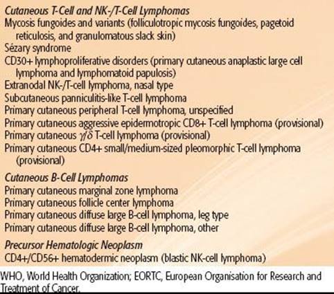
FIGURE 79.1. Pian fungoide, from Jean-Louis-Marc Alibert’s atlas of dermatoses, Descriptions des Maladies de la Peau.

 CUTANEOUS T-CELL AND NK-CELL LYMPHOMAS
Cutaneous T-cell lymphoma (CTCL) is the most common primary cutaneous lymphoma. In the United States, 71% of the 3,884 cases of primary cutaneous lymphoma diagnosed during 2001–2005 were CTCL.3 Similarly, 78% of primary cutaneous lymphoma diagnoses recorded in the Dutch and Austrian Cutaneous Lymphoma Group registry over 1986–20021 and 85% of diagnoses in the Central Cutaneous Lymphoma Registry of the German Society of Dermatology over 1999–2004 were CTCL.4
CTCL subtypes include mycosis fungoides (MF); CD30+ lymphoproliferative disorders; extranodal NK-/T-cell lymphoma, nasal type; subcutaneous panniculitis-like T-cell lymphoma; adult T-cell leukemia/lymphoma; and primary cutaneous peripheral T-cell lymphomas. MF is the most common CTCL, responsible for 54% of CTCL diagnoses in the United States over 2001–2005.3 CD30+ T-cell lymphoproliferative disorders and cutaneous peripheral T-cell lymphomas represent the majority of the remaining cases of CTCL. The remaining CTCL subtypes are extremely rare and represent <1% of primary cutaneous lymphomas.
Mycosis Fungoides
MF is the archetype cutaneous lymphoma. The first case of MF was reported in 1806 by the French dermatologist Jean-Louis-Marc Alibert in his atlas of dermatoses, Descriptions des maladies de la peau (Fig. 79.1). After Alibert released his depiction of MF, approximately 300 similar cases were reported over the next decade, and in modern times approximately 1,500 new cases of MF were diagnosed during 2001–2005.3 MF is a disease of skin-homing CD4+ T-helper cells5 and is most commonly diagnosed in men (male:female ratio of 1.6–2.0:1) with a median age at diagnosis of 55 to 60.1 Significant advances in MF therapy have been made over the past 200 years, but the precise etiology responsible for the development of MF remains elusive.
Cutaneous Disease
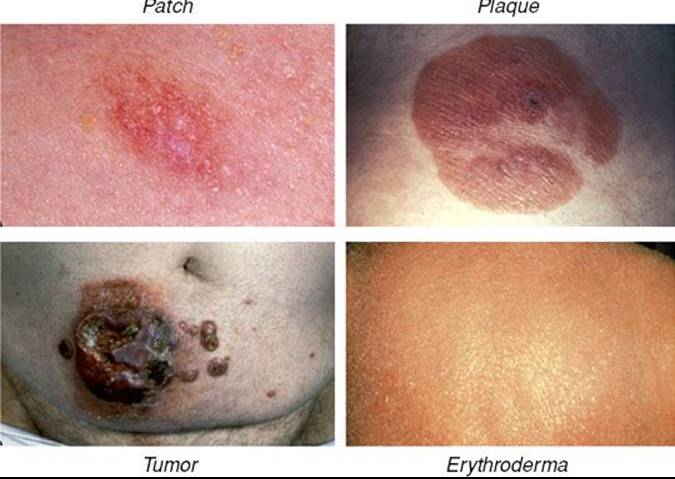
Pruritus, either diffuse or localized to areas of involved skin, is the most common symptom associated with MF. Ulcerated lesions may also cause patients significant pain. Cutaneous lesions in classic (or Alibert-Bazin) MF follow a predictable evolutionary course, and a consensus statement from the International Society for Cutaneous Lymphomas (ISCL), U.S. Cutaneous Lymphoma Consortium (USCLC), and EORTC defines the lesions found in distinct phases of MF.6 In the premycotic phase a small number of red, scaled, macular or patchlike lesions develop in sun-shielded areas of the skin such as the trunk, pelvis, and extremities. These early lesions are unstable and usually regress, followed by development of new lesions. Biopsies of premycotic lesions are rarely diagnostic due to a paucity of malignant lymphocytes in the lesion. With increased deposition of malignant T cells in the skin, the lesions become increasingly durable, and persistent cutaneous patches are characteristic of the patch phase. The ISCL/USCLC/EORTC consensus definition of an MF patch is “any size lesion without induration or significant elevation above the surrounding uninvolved skin: poikiloderma may be present”.6 As the lesions become more densely infiltrated by both malignant and reactive lymphocytes, they evolve into plaques with thickened and raised borders in the plaque phase of classic MF. Plaques are defined by the ISCL/USCLC/EORTC as “any size lesion that is elevated or indurated: crusting or poikiloderma may be present”.6 Plaques may evolve into cutaneous tumors, which the ISCL/USCLC/EORTC defines as “any solid or nodular lesion ≥1 cm in diameter with evidence of deep infiltration in the skin and/or vertical growth”.6 The presence of cutaneous tumors designates the tumor phase of MF. MF may also progress to or present with erythroderma, in which a diffuse erythema involves >80% of the skin surface area.6 Representative images of patches, plaques, tumors, and erythroderma are shown in Figure 79.2.
Leukemic CTCL and the Sézary Syndrome
In rare CTCL cases circulating malignant T cells are identified in the peripheral blood. This phenomenon, sometimes referred to as leukemic CTCL or the Sézary syndrome (SS), most commonly occurs in association with erythroderma but may occur in patients with minimal cutaneous disease.1 The circulating malignant T cells (also called Sézary cells) are atypical T cells with hyperconvoluted nuclei seen at analysis of peripheral buffy coat smear. The circulating cells most commonly possess a CD4+/CD7– or CD4+/CD26– immunophenotype, and flow cytometry allows for quantification of the proportion of circulating malignant T cells. In addition, presence of a dominant circulating malignant clone may be demonstrated by evaluation of the T-cell receptor (TCR) by polymerase chain reaction (PCR) or Southern blotting. The degree of tumor burden in the peripheral blood is denoted as B0 (≤5% atypical or Sézary cells seen on examination of buffy coat smear), B1 (>5% of cells on buffy coat analysis are atypical, but further criteria meeting B2 disease are not met), or B2 (combination of presence of a dominant T-cell clone in the peripheral blood identified by PCR or Southern blot and either ≥1,000 Sézary cells/mm3, an increased amount of CD3+ or CD4+ T cells with CD4:CD8 ratios >10, or increased quantities of abnormal T cells defined as loss of CD7 in >40% of cells or CD26 in >30% of cells).6 If peripheral blood is evaluated for a circulating clone by PCR or Southern blot, B0 or B1 disease may be stratified into B0a or B1a (absence of circulating clone) or B0b or B1b (presence of circulating clone). The mechanism responsible for the development of leukemic disease in MF is unknown and in some cases may simply reflect disease progression. In other cases erythroderma and circulating disease develop simultaneously, which has historically been referred to as SS. The distinction between SS and erythrodermic MF with leukemic involvement (or SS syndrome preceded by MF) has been a point of controversy, and it remains unclear whether the two diseases are distinct or merely variants of one another. At present, SS is specifically defined as the combination of erythroderma with B2 disease.6
FIGURE 79.2. Representative images of cutaneous lesions in mycosis fungoides. (From Smith BD, Wilson LD. Management of mycosis fungoides. Part 1. Diagnosis, staging, and prognosis. Oncology 2003;17:1419–1428, with permission.)
Extracutaneous Disease
In advanced stages of disease MF may progress to involve regional or distant lymph nodes or extracutaneous organ systems (most commonly the lungs, oral cavity, pharynx, or central nervous system), and these sites may cause patients significant pain or functional impairment.7 Of note, in advanced cases of MF the extent of cutaneous or extracutaneous disease may vary significantly in patients of the same clinical stage. Clinical trials that report results by stage alone may therefore be difficult to interpret. The ISCL/USCLC/EORTC consensus statement on clinical end points and response criteria should facilitate improved communication of the degree of disease and response to treatment in future clinical trials.6
Molecular and Cellular Pathophysiology
Examination of early phase MF lesions by hematoxylin and eosin staining commonly reveals epidermotropism, a profound infiltration of lymphocytes into the epidermis. As the lesions progress and become thickened, epidermotropism is gradually lost as the lymphocytes begin to localize more diffusely in the skin. Only a fraction of the skin-homing lymphocytes in MF are malignant, and the malignant T cells may be identified by visualization of their small to medium-sized hyperconvoluted (or cerebriform) nuclei and surrounding lacunae, which give them the impression of being surrounded by a halo. A Pautrier’s microabscess (Langerhans cell surrounded by atypical T cells in the epidermis) is a less common but pathognomonic histologic finding associated with MF.1,5 The malignant cells in MF are CD4+ T-helper cells, which most commonly express a CD3+, CD4+, CD45RO+, CLA+, CCR4+ immunophenotype. Evaluation of the TCR profile of malignant T cells in MF reveals a predominant TCR rearrangement indicative of clonal dominance in a majority of cases.
The specific stimuli and molecular events responsible for activation and development of clonal dominance of a specific T cell are unknown. Genetic profiling studies have identified a number of chromosomal deletions and duplications associated with MF/SS, and microRNA profiling is beginning to reveal distinct molecular profiles associated with different phases of MF. In addition to the molecular aberrations intrinsic to the malignant T cells, the cutaneous microenvironment is also likely to play a key role in the development and maintenance of disease.5,8–11
Diagnosis and Evaluation
Evaluation of a patient with suspected or recently diagnosed MF should include a comprehensive history/physical examination, biopsy of cutaneous lesion(s) and suspicious lymph nodes, and appropriate laboratory and imaging studies. The ISCL has designed a diagnostic algorithm for early-stage MF (not applicable to MF variants) (Table 79.2),12 and specific recommendations for patient evaluation have been released by the ISCL/EORTC.13
History and Physical Examination. The duration of symptoms, evolutionary course of cutaneous disease, and presence or absence of B symptoms should be determined. The percentage of body surface area involved by disease and presence or absence of cutaneous tumors should be documented, and a thorough evaluation of the lymphatic system should be performed. Appropriate images of cutaneous lesions should be recorded so that disease progression or response to therapy may be monitored.
TABLE 79.2 DIAGNOSTIC CRITERIA FOR EARLY STAGE MYCOSIS FUNGOIDESA,12
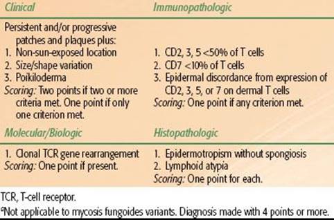
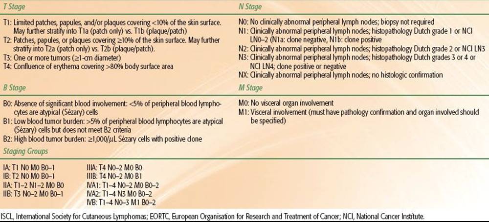
TABLE 79.3 ISCL/EORTC REVISIONS TO MYCOSIS FUNGOIDES STAGING13
Skin Biopsy. Biopsies should be taken from a minimum of two distinct sites of disease. Lesions of the greatest induration will be most likely to yield a diagnosis due to greater numbers of malignant cells. However, scaled lesions are more likely to show epidermotropism. Tissue should be evaluated with hematoxylin and eosin staining, immunostaining for surface marker expression profiles (including CD2, CD3, CD4, CD5, CD7, CD8, CD20, CD30, CD26, CD56, TIA1, granzyme B, βF1), and PCR for clonal TCR rearrangement. Slides should be reviewed by a dermatopathologist.
Excisional biopsies of enlarged or otherwise suspicious lymph nodes (fixed or matted lymph nodes, or lymph nodes ≥1.5 cm or ≥1 cm in the head and neck) should be evaluated as described previously. When multiple suspicious lymph nodes are encountered, the choice of lymph node to be excised should be based on size, fluorodeoxyglucose (FDG) avidity, and location, with priority given to the largest lymph node draining an affected area of skin or the lymph node with the highest standardized uptake value (SUV) on positron emission tomography (PET) scan. If all other factors are equal, priority should be given first to cervical nodes, followed by axillary and then inguinal nodes.13
Laboratory and Imaging Studies. Laboratory studies should include a complete blood count, chemistry panel, liver function tests, and lactate dehydrogenase (LDH). Examination of the peripheral blood for circulating disease by PCR and flow cytometry should also be considered, particularly for patients with erythroderma, nodal, or extracutaneous disease. Bone marrow biopsy should be considered if peripheral blood or extracutaneous organs are found to harbor disease.13 Computed tomography (CT) of the chest, abdomen, and pelvis is recommended in the evaluation of all patients with MF with the exception of patients with patch/plaque disease limited to 10% or less of the body surface area. In these cases a chest x-ray or nodal ultrasound may suffice. PET scanning was shown to be more sensitive than CT in identifying involved lymph nodes, and the role of PET in MF continues to evolve.14
Staging and Prognosis
MF is presently staged using the modified TNMB system proposed by the ISCL/EORTC (Table 79.3).13 Based on a review of 525 patients with MF (staged using the previous 1979 TNMB system), the majority of patients present with early stage disease (30% IA, 25% IB, 11% IIA, 16% IIB, 3% IIIA, 8% IIIB, 6% IVA, and 1% IVB). Only 7% of patients presented with peripheral blood involvement. The correlation of clinical stage to overall and disease-specific survival is presented in Figure 79.3.7 Similar results were obtained in a validation study of the current staging system.15 Of note, stage IA MF is not associated with any increase in mortality risk in comparison to an age- and ethnicity-matched control population, and is associated with only a 16% rate of disease progression at 20 years of follow-up.7
Skin-Directed Therapy
MF is not generally considered to be curable, although extended periods of disease-free survival after treatment have been reported. Efforts to treat MF are primarily focused on preventing progression and ameliorating symptoms. In early-stage disease a number of skin-directed treatments are effective and should be considered as first-line therapy. Topical therapies include corticosteroids (complete response [CR] rates of 63% for T1 and 25% for T2 disease),16nitrogen mustard (CR rates of 76% to 80% for stage IA and 35% to 68% for stage IB),17 carmustine (CR rates of 86% for T1 and 47% for T2 disease),18 and bexarotene gel (CR rate of 21% in early-stage MF).19 Phototherapy with narrow-band ultraviolet B (NB-UVB) (54% CR in stages IA or IB)20 or psoralen + UVA (PUVA) (CR rate of 65%)21 is also frequently used. However, the most effective skin-directed therapy for MF is ionizing radiation.
Ionizing radiation is well suited to the treatment of cutaneous lymphoma. Lymphocytes are highly radiosensitive, and radiation doses may be effectively limited to the epidermis and dermis by appropriate selection of photon or electron energies. Options for irradiation include local superficial irradiation or irradiation of the entirety of the skin via total skin electron beam therapy (TSEBT).
FIGURE 79.3. Overall (A) and disease-specific (B) survival in mycosis fungoides by stage. (Reprinted with permission from Kim YH, Liu HL, Mraz-Gernhard S, et al. Long-term outcome of 525 patients with mycosis fungoides and Sezary syndrome. Arch Dermatol 2003;139:857–866; copyright © 2003 American Medical Association. All rights reserved.)
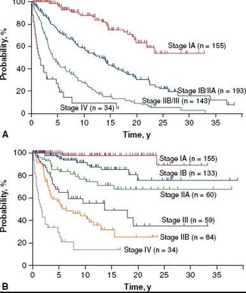
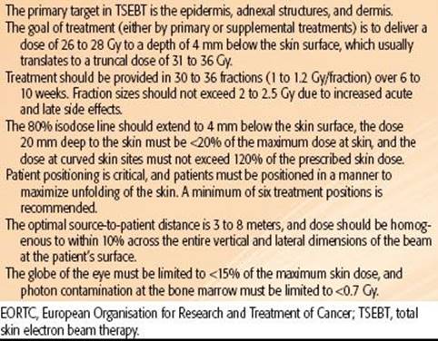
TABLE 79.4 EORTC TECHNICAL RECOMMENDATIONS FOR TSEBT23
Local Superficial Irradiation
A small portion (~5%) of patients with stage IA MF present with “minimal” disease, defined as a solitary lesion or two to three MF lesions clustered sufficiently close to one another that they are amenable to treatment with a single or abutting radiation fields.22 For these patients, treatment with local superficial irradiation may be considered. Treatment fields should be designed to encompass the entirety of the lesion (determined by visual inspection, palpation, and/or appropriate imaging) with a 1- to 2-cm margin, with use of a lead or Cerrobend cutout to conform field borders to the anatomy of the cutaneous lesion. Treatment is most commonly provided with electrons, with energies (usually 6 to 16 MeV) carefully selected to optimize dose penetration. The EORTC recommends that the 80% isodose line is set at the deep border of the dermis,23 which is commonly at a depth of approximately 4.5 mm.
Local superficial radiation is very effective in generating a CR for patients with “minimal” stage IA MF. In a review of 21 patients with “minimal” stage IA MF treated with local superficial radiation (superficial or orthovoltage x-rays or megavoltage electrons) to doses ranging from 20 to 40 Gy in four to five or 10 to 15 fractions, Wilson et al.22 found a CR rate of 97%. Importantly, review of the recurrence rates associated with different total doses of local superficial radiation revealed a 25% local recurrence rate (two of eight fields) with treatment to 20 Gy and an 8% local recurrence rate (two of 25 fields) with treatment to 20 to 40 Gy. Similarly, Cotter et al.24 reported a local recurrence rate of 42% for fields treated to 10 Gy or less, but 0% for fields treated to >30 Gy. More recently, treatment to 8 Gy in 4-Gy fractions has been shown to yield a CR rate of 92%.25 Based on these studies, it is recommended that “minimal” stage IA (i.e., unilesional or up to three closely approximated sites) MF lesions are treated to a dose of 30 to 36 Gy, and treatment as low as 8 Gy in two fractions may be considered as a palliative treatment. Side effects of local superficial radiation are usually limited to mild dermatitis, local alopecia, and pigmentation changes.
Total Skin Electron Beam Therapy
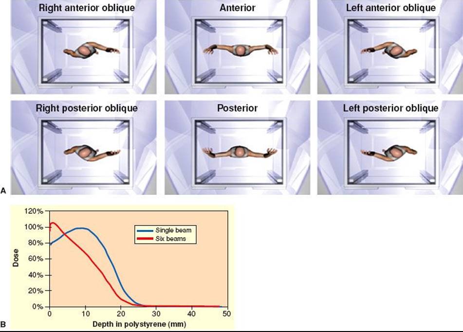
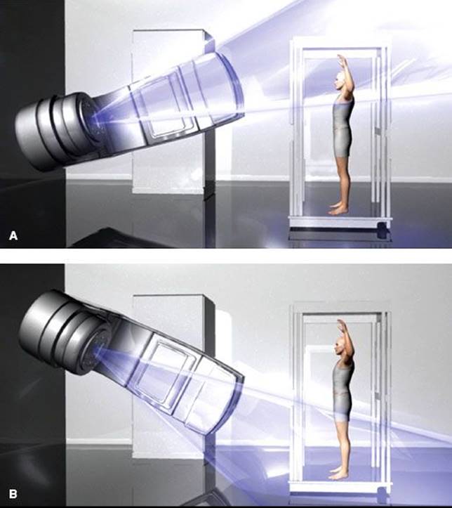
Technique. Total skin electron beam therapy (TSEBT) is technically challenging and should only be attempted in centers with special expertise in its provision, including skilled physics support. EORTC recommendations regarding the technical aspects of TSEBT are presented in Table 79.4. Modern TSEBT is usually accomplished with 6- to 9-MeV electrons generated by a medical linear accelerator directed at a patient standing behind a polycarbonate screen ~3.8 meters from the linear accelerator head. The polycarbonate screen scatters the incident electron beam and contributes to an improved surface dose. Treatment is provided in “cycles,” with one cycle composed of treatment of the patient in six different positions (Fig. 79.4A) over 2 days (three positions each day). Typically, a dose of 2 Gy is provided to the entirety of the skin during one cycle, and two cycles are usually administered per week. Treatment in six positions optimizes dose distribution at the skin surface (Fig. 79.4B). At Yale and Stanford, the anterior, right posterior oblique, and left posterior oblique positions are treated on cycle day 1, and the posterior, right anterior oblique, and left anterior oblique positions on cycle day 2.26,27 When the patient stands in a treatment position, a dual-field technique is used to deliver treatment to a superior and inferior field by angling the gantry 16 to 17.5 degrees above and below horizontal, respectively, the specific angle dependent upon individual machine characteristics (Fig. 79.5). Treatment to the six positions using the dual-field technique, which is in use at Yale and Stanford, delivers maximum dose to a depth of 1 mm, 80% dose to 6 to 7 mm, and 20% dose to 12.5 mm.26,27
FIGURE 79.4. A: The six total skin electron beam therapy (TSEBT) treatment positions as viewed from above. Images not to scale. B: Comparison of depth dose profiles associated with a single treatment position (blue curve) versus six treatment positions (red curve). (A courtesy of Christian Chang, www.christianchang.com, printed with permission from the artist. B from Smith BD, Wilson LD. Management of mycosis fungoides: Part 2. Treatment. Oncology 2003;17:1419–1428, with permission.)
Dose and Fractionation. Sublethal damage repair does not appear to be a major factor in determining the response of MF to ionizing radiation,28 and modern TSEBT is provided with a relatively protracted course of two cycles per week for 9 weeks, which provides a total dose of 36 Gy to the skin surface. The total radiation dose appears to be directly associated with complete response rates, with 18% CR with treatment to <10 Gy, 55% CR with treatment to 10 to 20 Gy, 66% CR with treatment to 20 to 25 Gy, 75% CR with treatment to 25 to 30 Gy, and 94% CR with treatment to 30 to 36 Gy.29 However, lower doses yield impressive rates of overall response (defined as a >50% reduction in cutaneous disease), overall survival, progression-free survival, and relapse-free survival rates. Relapse rates are relatively high even in patients in which a CR is obtained, and the absolute benefit of a CR relative to the increased side effects at higher doses of TSEBT is unclear. The application of reduced-dose TSEBT (10 to 20 Gy) in combination with additional therapies may be a viable alternative to the current standard of 36 Gy,30 but additional studies are necessary before any conclusions may be made in this regard.
Supplemental Treatments. The six treatment positions in TSEBT maximize unfolding of the skin and exposure of the skin surface to the incident electron beam, but areas such as the soles of the feet, perineum, and scalp remain obscured and require supplemental doses to ensure that a minimum of 20 to 28 Gy is administered to a depth of approximately 4 mm. Supplemental treatment to these areas may be accomplished by the use of 120-kV superficial photons with half-value layer (HVL) 4.2-mm Al or low-energy (~6 MeV) electrons with 1-cm bolus to treat the soles of the feet (1 Gy per fraction) and the perineum (1 Gy per fraction). In some setups, the scalp is treated by placing an angled electron reflector above the patient,26 but supplemental boosting is an alternative approach that is incorporated in some centers. Additional areas that may need supplemental dose include thick cutaneous tumors and skin folds secondary to body habitus, and the need for supplemental dose to such areas is based on the judgement of the radiation oncologist.26,27
Side Effects. In a review of perceptions of MF therapy, patients overall considered TSEBT to be a more difficult treatment to endure as compared to other treatments,31 and it is important that patients are advised that symptoms such as pruritus and cutaneous erythema may be exacerbated during therapy. Additional acute side effects that commonly occur include xerosis, dry desquamation, extremity edema, blister/bullae formation over the lower extremities, alopecia (including hair of the scalp, eyebrows, eyelashes, and body), and nail changes (nails may ultimately be lost but usually regrow). Hypohydrosis secondary to damage to sweat glands may occur. Similarly, dryness and irritation of the nasal mucosa may result in nose bleeds. Gynecomastia is a rare occurrence. Late/chronic side effects are minimal and include cataract formation, chronic xerosis, persistent alopecia, dystrophic nails, telangiectasia, and secondary skin cancers including squamous and basal cell carcinomas and melanomas.23,32–35
In an effort to minimize side effects, areas that are most susceptible to TSEBT such as the eyes, lips, hands, fingernails, feet, and testes are blocked during certain cycles of treatment. Shielding of the eye and lens may be accomplished by a combination of internal/external shields, selected based upon the proximity of clinical disease. Usually, if internal eyeshields are used, they are used for only a portion of the therapy (7 to 20 Gy). The lips, hands, and fingernails may be blocked by lead mitts or fingernail shields as clinical circumstances warrant. The feet may be blocked by footboards for a portion of the treatment. A testicular shield may be used during perineal boost treatments.26
FIGURE 79.5. Dual-field technique for total skin electron beam therapy (TSEBT). A: Treatment of superior field. B: Treatment of inferior field. Images not to scale. (Courtesy of Christian Chang, www.christianchang.com, printed with permission from the artist.)
Clinical Efficacy. TSEBT is very effective, particularly for early-stage disease. TSEBT yields a complete response rate of >90% with a 15-year relapse-free survival of 40% in patients with T1 disease, although it is no longer recommended for such limited disease.29 Of note, patients with recurrent disease after a course of TSEBT most commonly have disease restricted to <5% of the skin surface area, and these recurrences are therefore amenable to local salvage therapy with topical therapy or limited superficial radiation. When successful salvage of such limited recurrences is taken into account, relapse-free survival improves to 70% at 15 years.36–38
In T2 disease TSEBT is similarly effective, with a complete response rate of 76% to 90% and a 50% relapse-free survival rate at 5 years and 10% at 10 years. Relapse-free survival is significantly improved by addition of adjuvant PUVA or nitrogen mustard. Specifically, adjuvant PUVA improves 5-year relapse-free survival to 85%, and nitrogen mustard improves 10-year relapse-free survival to 40%.23,38–40
When cutaneous tumors are present (T3 disease), TSEBT is less effective but still yields an impressive complete response rate of 44% to 54% of patients, much greater than any other single modality. Adjuvant treatment should be considered, and retrospective studies suggest that nitrogen mustard may increase the durability of response. Alternatively, a combination of nitrogen mustard and local superficial radiation may be considered for tumors localized to a small percentage of the skin surface.38,40,41 Supplemental boosts should be considered for patients with tumors, and such boost treatment should be provided concomitantly with the initiation of TSEBT or prior to its initiation. The purpose of the boost is to diminish the thickness of the lesion so that electrons from TSEBT can effectively penetrate the entire lesion.
In erythrodermic MF (T4 disease), TSEBT yields a 70% to 100% response rate (for patients with T4N0 disease) and 5-year progression-free survival of 25% to 69%. When disease involves the peripheral circulation or extracutaneous sites, TSEBT is less effective and response and progression-free survival rates decrease to 74% and 36%, respectively. TSEBT appears to be synergistic with extracorporeal photopheresis (ECP), and the combination of TSEBT and ECP is associated with improved disease-specific survival and decreased levels of circulating malignant cells.42–45
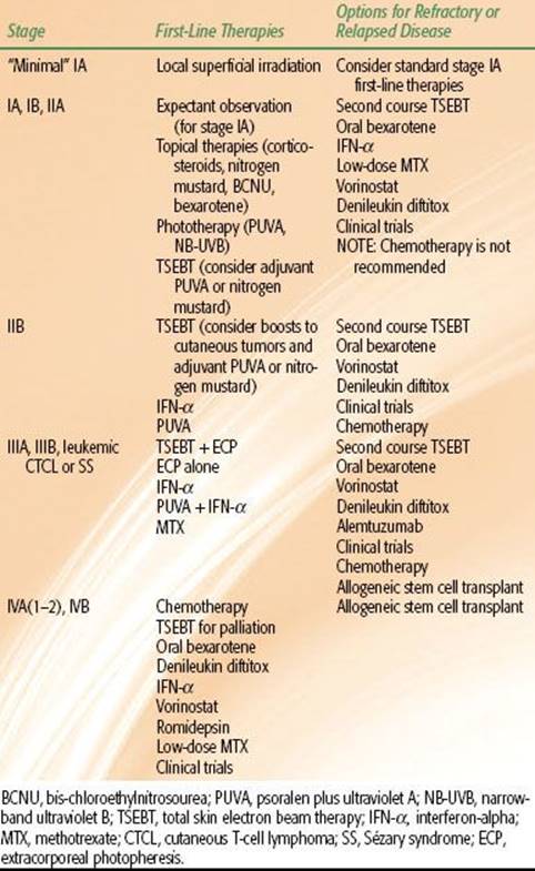
TABLE 79.5 RECOMMENDATIONS FOR TREATMENT OF MF BY STAGE
Palliative Radiotherapy
Symptomatic nodal or visceral disease may be effectively palliated by a brief course of localized radiotherapy, usually to a total dose of 12 to 30 Gy. If this treatment is given in conjunction with a course of TSEBT, a similar dose may be used. In the subset of patients that develop extensive recurrence of cutaneous disease after a previous course of TSEBT, additional courses of TSEBT to lower total doses can be considered and have proven effective in studies at Yale and Stanford.46,47
Systemic Therapy
Skin-directed therapy should be attempted first for patients with early-stage disease, but in patients with advanced or refractory disease systemic therapy may be considered. Options for systemic therapy include oral bexarotene (response rate up to 54% in refractory CTCL at a dose of 300 mg/m2, and bexarotene may be used in combination with other therapies),48,49 denileukin diftitox (a fusion protein consisting of interleukin-2 and a portion of the diphtheria toxin that yields an overall response rate of 30% in refractory CTCL),50 histone deacetylase inhibitors (such as vorinostat and romidepsin, ~30% response rate in refractory CTCL),51,52 alemtuzumab (a monoclonal antibody targeted against the CD52 surface marker with a 38% overall response rate in relapsed or refractory CTCL and 86% response rate in SS),53,54 interferon-α2a (IFN-α) (a biologic modifier with a response rate of 40% to 80% in CTCL as a single agent and with synergistic activity with other MF therapies),55–61 ECP (associated with improvement in peripheral blood and skin involvement, especially in those with T4 disease),62 and cytotoxic chemotherapy (single-agent chemotherapy with agents such as purine or nucleoside analogs, liposomal doxorubicin, and antifolate agents, including methotrexate and pralatrexate, is preferred because no distinct advantages have been identified with the use of multiagent regimens).63–69
Autologous or Allogeneic Stem Cell Transplant
Patients with MF/SS refractory to other therapies may be considered for stem cell transplant. Although autologous stem cell transplants have not proven to be overly effective, greater success has been achieved with allogeneic stem cell transplants. Additional studies are necessary to fully define the role of stem cell transplant in MF. This approach may prove to be a valuable option for patients with refractory disease.70–74
Treatment Recommendations by Stage
Treatment recommendations for MF/SS are presented by stage in Table 79.5.71 The National Comprehensive Cancer Network (NCCN) has also published treatment guidelines for MF/SS.75
Transformed Disease
In some cases, large cell transformation of MF may occur, which is identified on biopsy by >25% large cells in the sample. Large cell transformation may be associated with decreased survival rates and occurs in 8% to 39% of cases. Patients with advanced stage or high levels of β2-microglobulin or LDH are at the greatest risk of large cell transformation. Treatment options include systemic chemotherapy, stem cell transplant, and consolidative or local radiation for localized disease.71
Variants of Mycosis Fungoides
Variant forms of MF that share histologic and clinical features with classic MF but exhibit distinct clinical behavior have been recognized and include folliculotropic MF, Woringer-Kolopp disease, granulomatous slack skin, and hypopigmented MF.
Folliculotropic MF is characterized by a folliculotropic pattern of cutaneous infiltration of malignant T cells. Folliculotropic MF lesions present as patches, plaques, or tumors or may manifest in an acneiform pattern. Lesions primarily develop over the head and neck and are rarely found over the trunk. Localized alopecia secondary to the folliculotropic pattern is common. Folliculotropic MF is associated with a 15-year survival of 41% for early-stage disease. Early-stage disease may be treated with PUVA combined with bexarotene or IFN-α. Local radiation therapy may be advantageous because it can effectively treat to deeper depths of involvement seen in this variant. Advanced-stage disease is minimally responsive to cytotoxic chemotherapy, and alternate options such as irradiation or allogeneic stem cell transplant should be considered.1,76
Woringer-Kolopp disease (pagetoid reticulosis) is characterized by a solitary erythematous and scaling cutaneous patch on an extremity. Biopsy results are similar to classic MF, and the malignant T cells commonly carry a CD3+/CD4+/CD8– or CD3+/CD4–/CD8+ immunophenotype. If CD8+ disease is identified, it is important to consider the alternative diagnosis of CD8+ aggressive peripheral cutaneous T-cell lymphoma, and the distinction between these diagnoses is made by the aggressiveness of disease. Specifically, as compared to CD8+ aggressive peripheral cutaneous T-cell lymphoma, Woringer-Kolopp disease is very slowly progressive and carries an excellent prognosis. Treatment options include skin-directed therapies such as corticosteroids, nitrogen mustard, resection, or local irradiation. Local radiation may be very successful in achieving long-term local control even in locally very advanced disease. In rare cases of diffuse or refractory disease, irradiation is also recommended.1,77
Granulomatous slack skin (GSS) is an extremely rare variant of MF, with <100 cases reported in the literature. It is characterized by cutaneous infiltration of a clonal population of malignant T cells coupled with a granulomatous infiltration. The granulomatous infiltrate ultimately causes destruction of local elastin fibers and impairs the structural integrity and elasticity of the skin, which results in loose or “slack” skin. This loosening of the skin is most commonly observed in the axilla, groin, neck, and breast. Although GSS is itself an indolent disease, it is associated with an increased incidence of secondary lymphomas, most commonly Hodgkin lymphoma or MF. Given the rarity of this disease, it is difficult to make definitive recommendations regarding therapy, but surgical resection or local superficial radiation may be effective.1,78
Hypopigmented MF presents at an earlier age (childhood or adolescence) than classic MF and is characterized by development of hypopigmented patches frequently over the trunk or extremities. In contrast to classic MF, the malignant T cells are often CD8+. Treatment and disease course are otherwise similar to classic MF.79,80
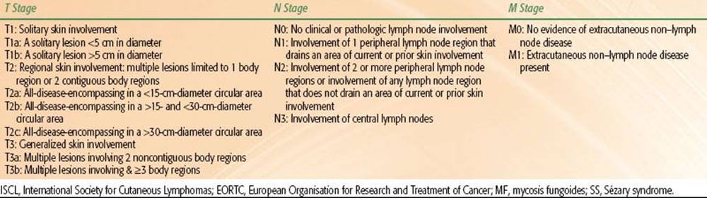
TABLE 79.6 ISCL/EORTC TNM CLASSIFICATION OF CUTANEOUS LYMPHOMA OTHER THAN MF/SS81
Non–Mycosis Fungoides CTCL
Staging
In 2007 the ISCL and EORTC presented a new TNM staging system for non-MF/SS cutaneous lymphomas (Table 79.6).81
CD30+ Lymphoproliferative Disorders
A review of the SEER registry identified 268 cases of primary cutaneous CD30+ lymphoproliferative disorders recorded between 1974 and 2004 (58% male and 42% female patients). The median age of diagnosis was 61, and population-matched 3-year relative survival was 87%, with 5-year disease-specific survival 92%. Localization of disease in the head and neck appears to be a negative prognostic factor.82 The primary cutaneous CD30+ lymphoproliferative disorders recognized by the WHO/EORTC are primary cutaneous anaplastic large cell lymphoma and lymphomatoid papulosis.1 These cannot be differentiated histologically. It is essential to take a careful clinical history to document the distribution of lesions and their clinical course (e.g., history of spontaneous regression). The diagnosis is a clinical-pathologic one. Recently the EORTC, ISCL, and USCLC released consensus recommendations regarding the evaluation and treatment of primary cutaneous CD30+ lymphoproliferative disorders.83
Primary cutaneous anaplastic large cell lymphoma (C-ALCL) is characterized by development of cutaneous plaques, nodules, or tumors that are usually solitary or clustered into a localized area. Multifocal disease is uncommon (occurring in only 20% of patients), and lymph nodes are involved in only 10% of cases. Extracutaneous disease is rarely found. C-ALCL is most commonly diagnosed in males, with a male:female ratio of 2–3:1. When disease is localized to the skin and regional lymph nodes, prognoses are excellent, with anticipated 5-year overall survival and 10-year disease-specific survival rates of 90%. Progression to extracutaneous disease is associated with overall survival, and extensive limb disease (defined as “initial presentation or progression to multiple skin tumors in 1 limb or contiguous body regions”84) is associated with disease-specific survival. Biopsies of C-ALCL lesions reveal a diffuse infiltration of large anaplastic CD4+ T cells, and at least 75% of these anaplastic T cells express CD30. Similar to MF, clonal dominance is frequently detected by PCR evaluation for TCR rearrangement.1
Limited resection or local superficial irradiation is very effective in controlling localized cutaneous disease, and low-dose methotrexate may be considered when widespread cutaneous disease precludes simple resection or irradiation. Local superficial radiation to a dose of 34 to 44 Gy was associated with a 100% CR rate,85 and treatment to 36 to 40 Gy in 2-Gy fractions is recommended. In rare cases in which extracutaneous disease is found, doxorubicin-based chemotherapy should be considered.
Of note, prior to initiating therapy, it is critically important to verify that C-ALCL has been correctly distinguished from the more common systemic ALCL. Molecular studies to evaluate the presence of the (2:5)(p23;q35) chromosomal translocation and expression of anaplastic lymphoma kinase (ALK) or epithelial membrane antigen (EMA) are useful in making this distinction, because the (2:5) translocation and expression of ALK and EMA are found in systemic ALCL but rarely found in C-ALCL.1,86
Lymphomatoid papulosis (LyP) carries an excellent prognosis, with a 5-year overall survival rate of 100%. However, 15% to 20% of patients with LyP will develop MF, C-ALCL, or a Hodgkin lymphoma. LyP is characterized by development of violaceous papular or papulonodular lesions over the trunk or extremities, which typically regress after 3 to 12 weeks, often leaving a residual scar. Biopsy of LyP cutaneous lesions reveals epidermotropic atypical CD3+/CD4+/CD8– lymphocytes, and clonal TCR rearrangement is identified in 60% to 70% of cases. Three distinct histologic subtypes are recognized: LyP A, B, and C. LyP A (characterized by large multinucleated CD30+ cells intermixed with an inflammatory infiltrate) and C (characterized by large CD30+ cells with minimal inflammatory infiltrate) represent 90% of cases. In contrast to types A and C, atypical lymphocytes in LyP B are CD3+/CD4+/CD30–, reminiscent of MF.1
Aggressive therapies such as chemotherapy or radiation should be avoided in LyP. In the rare patient with a large cutaneous burden of disease, skin-directed therapy with PUVA or a topical chemotherapy agent may be considered. In addition, low-dose oral methotrexate may be effective in suppressing the development of new lesions and should be considered if previous LyP lesions caused significant scarring.1
Extranodal NK-/T-Cell Lymphoma, Nasal Type
Extranodal NK-/T-cell lymphoma, nasal type most commonly arises in the nasal cavity and nasopharynx but in select cases may present with cutaneous plaques or tumors over the extremities and trunk and is therefore included in the WHO/EORTC classification of cutaneous lymphomas. Biopsy of a cutaneous lesion reveals a dense lymphoid infiltrate, which may exhibit an epidermotropic component. The malignant cells are most commonly NK in origin with a CD3–/CD2+/CD56+ immunophenotype, but on occasion the malignant cells are derived from cytotoxic T cells. Epstein Barr virus (EBV) is directly associated with disease development, and cutaneous lesions are nearly always EBV+. Consistent with an EBV-mediated pathophysiology, the disease occurs more commonly in geographic distributions in which EBV is endemic (Central America, South America, South Asia). The TCR usually remains in germline configuration when the malignant cells are of NK-cell origin, but clonal TCR rearrangement may be observed in cases in which the malignant cells are derived from cytotoxic T cells. Optimal treatment regimens for cutaneous extranodal NK-/T-cell lymphoma, nasal type have not yet been determined. Treatment with extended field radiotherapy to a median dose of 50 Gy yields a CR rate of 95.4% in early-stage disease, but local or distant recurrences are common and adjuvant therapy with L-asparaginase and other regimens should be considered. Advanced-stage disease is treated primarily with chemotherapy.1,87–89
Subcutaneous Panniculitis-Like T-Cell Lymphoma
Subcutaneous panniculitis-like T-cell lymphoma presents with subcutaneous nodules and/or plaques, which are commonly localized over the legs or trunk. Biopsy reveals a lymphoid infiltrate composed primarily of malignant cytotoxic CD8+ T cells. Prior to the establishment of the WHO/EORTC classification system, α/β and γ/δ T-cell forms of disease were each recognized as subcutaneous panniculitis-like T-cell lymphoma. However, γ/δ T-cell lymphoma is now recognized as a distinct, more aggressive disease and is categorized as a primary cutaneous peripheral T-cell lymphoma. The 5-year survival of the less aggressive α/β disease approaches 80%, and treatment involves combinations of corticosteroids and radiation, and potentially chemotherapy for refractory disease.1 Notably, presence of the hemophagocytic syndrome predicts a worse prognosis.
Adult T-Cell Leukemia/Lymphoma
Infection with the human T-lymphotropic virus 1 (HTLV-1) may result in adult T-cell leukemia/lymphoma in approximately 1% to 5% individuals, and patients may develop associated cutaneous disease characterized by papules, plaques, and tumors. In smoldering disease, cutaneous lesions may be the only sign of pathology. Chemotherapy is the primary therapy for acute adult T-cell leukemia/lymphoma, while smoldering cases may be treated with skin-directed therapies.1
Primary Cutaneous Peripheral T-Cell Lymphoma
Additional CTCLs that do not fit into the previously discussed categories are grouped as “primary cutaneous peripheral T-cell lymphomas,” and provisional subsets of this category described by the WHO/EORTC include primary cutaneous aggressive epidermotropic CD8+ cytotoxic T-cell lymphoma (characterized by cutaneous papules/plaques, nodules, and tumors with ulceration and a propensity for extracutaneous spread of disease), cutaneous γ/δ T-cell lymphoma (most commonly characterized by subcutaneous plaques or tumors in the extremities), primary cutaneous CD4+ small/medium-sized pleomorphic T-cell lymphoma (usually presents as a single cutaneous plaque or tumor on the head and neck or upper trunk and has an indolent clinical behavior), and primary cutaneous peripheral T-cell lymphoma, unspecified (most commonly presents as solitary or generalized nodules). The primary cutaneous peripheral T-cell lymphomas are primarily treated with chemotherapy, with the exception of primary cutaneous CD4+ small/medium-sized pleomorphic T-cell lymphoma, which may be treated with local resection or radiation.1
CUTANEOUS B-CELL LYMPHOMAS
Primary cutaneous B-cell lymphoma (PCBCL) was first recognized as a distinct clinical entity in 1981 and accounts for only 29% of the 3,884 cases of primary cutaneous lymphoma diagnosed in the United States during 2001–2005.3 The WHO/EORTC classification system recognizes three primary categories of CBCL: primary cutaneous marginal zone B-cell lymphoma (PCMZL), primary cutaneous follicle center B-cell lymphoma (PCFCL), and primary cutaneous diffuse large B-cell lymphoma, leg type (PCLBCL-LT).1 Of the CBCL cases diagnosed in the United States in 2001–2005, the most common subtype was primary cutaneous DLBCL (40%, of which only 23% were of leg type), followed closely by cutaneous follicle center lymphoma (30%) and cutaneous marginal zone B-cell lymphoma (25%).3 However, it should be noted that the histopathologic criteria for PLCBCL changed upon adoption of the WHO/EORTC consensus system, and it is likely that most of the PLCBCL cases diagnosed during 2001–2005 would now be reclassified as PCFCL. In a recent review 65% of CBCL cases classified as DLBCL using the older WHO criteria were found to be PCFCL under the current WHO/EORTC classification.90
Primary Cutaneous Marginal Zone B-Cell Lymphoma
PCMZL commonly presents with isolated or multifocal red or violaceous papules, plaques, and/or nodules on the trunk or extremities. The malignant cells are marginal zone B cells with a CD20+/CD79a+/CD5–/CD10– immunophenotype. Biopsies reveal an infiltrate composed of numerous lymphoid cells including marginal zone B cells, lymphoplasmacytoid cells, plasma cells, centroblastlike and immunoblastlike cells, reactive T cells, and reactive germinal centers. The prognosis for patients with PCMZL is excellent, and extracutaneous disease is rarely seen. Five-year overall survival rates of approximately 99% are expected, and cutaneous disease is very responsive to therapy. Cutaneous relapses are common but do not predict decreased survival.1
Primary Cutaneous Follicle Center Lymphoma
In contrast to PCMZL, PCFCL frequently presents with solitary or clustered plaques and tumors on the scalp, forehead, and trunk. Biopsy reveals a nodular or diffuse lymphoid infiltrate containing varying proportions of centrocytes, centroblasts, and reactive T cells growing in patterns ranging from follicular to diffuse. Similar to PCMZL, the malignant cells in PCFCL are of a CD20+/CD79a+ immunophenotype. Clonal rearrangement of immunoglobulin genes is usually observed. Importantly, the chromosomal translocation t(14:18) is infrequently seen in PCFCL, and therefore detection of this translocation should prompt consideration of a nodal or systemic follicular lymphoma. Similar to PCMZL, cutaneous lesions respond well to treatment, but rates of cutaneous relapse are relatively high at 20% overall. Extracutaneous disease is detected in 5% to 10% of patients, and bone marrow biopsy is an important component of evaluation. The 5-year overall survival rate for PCFCL is estimated at 95%.1
Primary Cutaneous Diffuse Large B-Cell Lymphoma, Leg Type
PCLBCL, leg type most commonly presents with violaceous cutaneous tumors over the lower extremities. Despite the nomenclature, anatomic restriction to the legs is not a requirement and in rare cases PCLBCL, leg type may be found in other cutaneous sites than the leg. The disease is characterized by diffuse lymphoid infiltrates with a predominance of centroblasts and immunoblasts, with absence of any significant numbers of centrocytes being a key distinguishing feature from PCFCL. A CD20+, CD79a+, Bcl-6+/–, CD10–, Bcl-2+, MUM-1+, FOXP1+ immunophenotype is usually observed. In contrast to PCMZL and PCFCL, both cutaneous relapses and development of extracutaneous disease are common, and the 5-year overall survival is decreased to 50%. A very small subset of PCLBCL (such as intravascular PCLBCL or anaplastic or plasmablastic PCLBCL) is categorized as “PCLBCL, other.”1
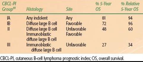
TABLE 79.7 CUTANEOUS B-CELL LYMPHOMA PROGNOSTIC INDEX
Diagnosis and Evaluation
Evaluation of patients with suspected PCBCL should follow the basic protocol recommended by ISCL/USCLC/EORTC previously discussed.13 In addition, a bone marrow biopsy is needed for PCBCL-LT and should be strongly considered in PCFCL (rate of bone marrow involvement may be as high as 11%). In other cases the need for bone marrow biopsy is left to the discretion of the treating physician. A further component to evaluation of PCBCL in European nations includes evaluation for Borrelia burgdorferiinfection.1,91 The Borrelia subspecies B. afzelii that appears to be the causative organism is not found in the United States.92
Staging and Prognosis
PCBCLs should be staged in accordance with the ISCL/EORTC staging system for non-MF/SS cutaneous lymphomas (Table 79.6). A cutaneous B-cell lymphoma prognostic index (CBCL-PI) was developed by correlating outcome data from the Surveillance, Epidemiology, and End Results (SEER) database with PCBCL histology and anatomic location (Table 79.7).93 Four prognostic groups (IA, IB, II, III) were defined by the CBCL-PI based on specific combinations of histology and anatomic location. However, as described earlier, the histologic criteria in use during the years on which the CBCL-PI is based (1973–2001) were changed with the 2005 release of the WHO/EORTC consensus classification system,1 and the CBCL-PI should be used with caution when modern histopathologic classification criteria are used.
In 2011, the International Extranodal Lymphoma Study Group (IELSG) released the cutaneous lymphoma international prognostic index (CLIPI) for indolent CBCL. Independent prognostic factors associated with progression-free survival included serum LDH, morphology (nodule vs. other), and number of distinct cutaneous sites of disease (greater than two). One point is scored for each factor, and patients are grouped into low risk (score 0), intermediate risk (score 1), and high risk (score 2 or 3) with associated 5-year progression-free survival rates of 91%, 64%, and 48%, respectively.94 The clinical utility of this prognostic system in determining risk-adapted therapy remains unclear.
Treatment
No prospective randomized clinical trial data are available to help guide treatment decisions, but the ISCL/EORTC recently performed an extensive survey of the literature pertaining to treatment of the PCBCLs and published consensus recommendations.95 The NCCN has also published treatment guidelines for PCBCLs.96
PCMZL and PCFCL
PCMZL and PCFCL are not expected to be associated with decreased survival relative to control populations (although PCFCL arising in the leg may have a worsened prognosis under the WHO/EORTC classification), and therefore side effects of proposed treatments should be carefully considered. Treatment options include local superficial irradiation, surgical excision, intralesional IFN-α, local and systemic rituximab, antibiotics (where B. burgdorferi is endemic), and chemotherapy.
Local Superficial Irradiation. In the ISCL/EORTC literature review, 132 patients with PCMZL treated with local superficial irradiation were identified, and a 99% complete response rate was observed. Doses ranged from 30 to 45 Gy, and field margins ranged from 1 to 5 cm. Although cutaneous relapse of disease occurred in 46% of patients, extracutaneous progression occurred in only 2% of patients. For PCFCL, 460 patients treated with radiation were reviewed, and the complete response rate was 99% with a 30% relapse rate. Doses ranged from 20 to 54 Gy and margins were 0.5 to 5+ cm. Recently, local superficial irradiation to 4 Gy in two fractions was shown to yield a 72% complete response rate for PCMZL and PCFCL and is recommended as a palliative dose.25,95
Surgery. Excision is associated with a 99% CR rate and 43% cutaneous relapse rate in PCMZL and a 98% CR rate and 40% cutaneous relapse rate in PCFCL.95
IFN-α. Intralesional IFN-a yields a 100% CR rate and 25% local relapse rate for PCMZL and a 100% CR rate and 29% local relapse rate for PCFCL.95
Rituximab. Systemic rituximab (monoclonal antibody targeted against CD20) has proven effective as a treatment for non-Hodgkin lymphomas and has been applied to the treatment of PCBCL. In a review of five patients with PCMZL treated with systemic rituximab, the overall response rate was 60%, and in 10 patients with PCFCL treated with systemic rituximab a 100% overall response rate and 80% complete response rate was observed. Intralesional rituximab may also be effective, with a reported complete response rate of 89% in nine patients with PCMZL, but with a high relapse rate of 62%. Twelve patients with PCFCL treated with intralesional rituximab had a complete response rate of 83% with 40% relapse.97
Chemotherapy. Single-agent treatment with chlorambucil was evaluated in 14 patients with PCMZL, and a complete response rate of 64% with 33% relapse rate was observed. Multiagent chemotherapy with cyclophosphamide, doxorubicin, vincristine, and prednisone (CHOP) was also evaluated in 33 patients with PCMZL, and an 85% complete response rate with 57% relapse rate was observed. In 104 patients with PCFCL treated with CHOP or CHOP-like regimens, an 85% complete response rate with 48% relapse rate was reported.95
ISCL/EORTC Recommendations. For patients who have only one or a few lesions clustered in one region, radiation (30 Gy or more) using a margin of 1 to 1.5 cm is highly effective. Patients presenting with a single small lesion may be treated with resection alone. Scattered lesions that cannot easily be encompassed in one or a few radiation fields may be carefully observed, with treatment reserved for only the most concerning or symptomatic sites. In the setting of diffuse disease, systemic rituximab should be considered. Of note, cutaneous relapses are common, and relapsed disease often responds to retreatment and does not predict for worsened overall survival rate.95
PCLBCL, Leg Type (PCBCL-LT)
PCLBCL-LT is a significantly more aggressive entity in comparison to PCMZL and PCFCL. The primary treatment options that are used in therapy for PCBCL-LT are radiation, rituximab, and multiagent chemotherapy.
Local Superficial Irradiation. The ISCL/EORTC literature review found 101 patients with PCLBCL-LT treated with radiation. In contrast to the more indolent lymphomas in which CR rates were close to 100%, the CR rate for PCLBCL-LT was lower at 88%. The rate of cutaneous relapse rate was high (58%), and even more concerning, 30% of patients were found to have extracutaneous progression of disease.
Rituximab. Systemic rituximab in PCLBCL-LT has been investigated (dose 375 mg/m2 weekly for 4 to 8 weeks) and yielded a CR rate of 38%. Relapse rates are as yet unknown, and additional follow-up is necessary.95
Chemotherapy. Multiagent chemotherapy regimens have been tested in PCLBCL-LT, and in 32 patients treated with CHOP or CHOP-like regimens, an 81% complete response rate has been observed. However, despite aggressive therapy, relapse rates remain high at 54%.95
ISCL/EORTC Recommendations. Based on the aggressive nature of PCLBCL-LT and its propensity to develop extracutaneous disease, recommended treatment in patients able to tolerate multiagent chemotherapy is R-CHOP, with the possible addition of local superficial radiation to distinct cutaneous lesions. In the subset of patients unable to tolerate a multiagent course of chemotherapy, systemic rituximab may be considered. Alternatively, an aggressive course of radiation to all cutaneous sites of disease may be considered, but relapse is expected.95
PRECURSOR HEMATOLOGIC NEOPLASMS
The third category of cutaneous lymphomas in the WHO/EORTC system is precursor hematologic neoplasms/immature hematologic malignancies. At present this category primarily references CD4+/CD56+ hematodermic neoplasm (also referred to as blastic NK-cell lymphoma or blastic plasmacytoid dendritic cell neoplasm), which is characterized by development of red or violaceous cutaneous nodules. Biopsy reveals cutaneous infiltration of abnormal cells with an appearance reminiscent of lymphoblasts and myeloblasts. The origin of the tumor cells in CD4+/CD56+ hematodermic neoplasm is unclear. Immunohistochemistry indicates that the tumor cells lack CD3 and CD8 and express both CD4 and CD56. The expression of CD56 initially led investigators to believe these cells were of NK origin. However, the concomitant expression of CD4 and CD56 is unusual, and further exploration led to the identification of expression of CD123 and TCL1 in these tumor cells, which suggests a possible origin in plasmacytoid dendritic cells. Examination of TCR status in these cells indicates it remains in germline configuration, providing further support for the notion that these cells are not derived from a B- or T-cell lineage. CD4+/CD56+ hematodermic neoplasm is an aggressive malignancy, and it is common for patients to harbor nodal or systemic disease at diagnosis. Due to the propensity for systemic spread of disease, skin-directed therapies are usually unlikely to control disease and chemotherapy is often necessary. Although chemotherapy may induce remission of disease, the duration of remission is typically relatively brief and median survival is only 14 months.1
SUMMARY
The primary cutaneous lymphomas are a diverse group of extranodal non-Hodgkin lymphomas, each with distinct biologic activity. A correct histopathologic diagnosis is therefore critical to the determination of appropriate management. Although significant advances in therapy have been made since the recognition of MF in 1806, numerous questions remain unanswered regarding the pathophysiology and optimal treatment of the cutaneous lymphomas. Recent efforts to develop uniform criteria for classification of disease and response to treatment should facilitate international collaborations. Due to the rarity of these diseases, such international collaborations are critical to the advancement of our understanding of the primary cutaneous lymphomas.
REFERENCES
1. Willemze R, Jaffe ES, Burg G, et al. WHO-EORTC classification for cutaneous lymphomas. Blood 2005;105:3768–3785.
2. WHO classification of tumours of haematopoeitic and lymphoid tissues, 4th ed. Lyon, France: IARC, 2008.
3. Bradford PT, Devesa SS, Anderson WS, et al. Cutaneous lymphoma incidence patterns in the United States: a population-based study of 3884 cases. Blood 2009;113(21):5064–5073.
4. Assaf C, Gellrich S, Steinhoff M, et al. Cutaneous lymphomas in Germany: an analysis of the Central Cutaneous Lymphoma Registry of the German Society of Dermatology (DDG). J Dtsch Dermatol Ges 2007;5(8):662–668.
5. Girardi M, Heald PW, Wilson LD. The pathogenesis of mycosis fungoides. N Engl J Med 2004;350:1978–1988.
6. Olsen EA, Whittaker S, Kim YH, et al. Clinical end points and response criteria in mycosis fungoides and Sezary syndrome: a consensus statement of the International Society for Cutaneous Lymphomas, the United States Cutaneous Lymphoma Consortium, and the Cutaneous Lymphoma Task Force of the European Organisation for Research and Treatment of Cancer. J Clin Oncol 2011;29(18):2598–2607.
7. Kim YH, Liu HL, Mraz-Gernhard S, et al. Long-term outcome of 525 patients with mycosis fungoides and Sezary syndrome. Arch Dermatol 2003;139:857–866.
8. Salgado R, Servitje O, Gallardo F, et al. Oligonucleotide array-CGH identifies genomic subgroups and prognostic markers for tumor stage mycosis fungoides. J Invest Dermatol 2010;130(4):1126–1135.
9. Van Doorn R, van Kester MS, Dijkman R, et al. Oncogenomic analysis of mycosis fungoides reveals major differences with Sezary syndrome. Blood 2009;113(1):127–136.
10. Ballabio E, Mitchell T, van Kester MS, et al. MicroRNA expression in Sezary syndrome: identification, function, and diagnostic potential. Blood 2010;116(7):1105–1113.
11. van Kester MS, Ballabio E, Benner MF, et al. miRNA expression profiling of mycosis fungoides. Mol Oncol 2011;5(3):273–280.
12. Pimpinelli N, Olsen EA, Santucci M, et al. Defining early mycosis fungoides. J Am Acad Dermatol 2005;53(6):1053–1063.
13. Olsen E, Vonderheid E, Pimpinelli N, et al. Revisions to the staging and classification of mycosis fungoides and Sezary syndrome: a proposal of the International Society for Cutaneous Lymphomas (ISCL) and the Cutaneous Lymphoma Task Force of the European Organization of Research and Treatment of Cancer (EORTC). Blood 2007;110(6):1713–1722.
14. Tsai EY, Taur A, Espinosa L, et al. Staging accuracy in mycosis fungoides and Sezary syndrome using integrated positron emission tomography and computed tomography. Arch Dermatol 2006;142(5):577–584.
15. Agar NS, Wedgeworth E, Crichton S, et al. Survival outcomes and prognostic factors in mycosis fungoides/Sezary syndrome: validation of the revised International Society for Cutaneous Lymphomas/European Organisation for Research and Treatment of Cancer staging proposal. J Clin Oncol 2010;28:4730–4739.
16. Zackheim HS, Kashani-Sabet M, Amin S. Topical corticosteroids for mycosis fungoides. Experience in 79 patients. Arch Dermatol 1998;134(8):949–954.
17. Kim YH. Management with topical nitrogen mustard in mycosis fungoides. Dermatol Ther 2003;16:288–298.
18. Zackheim HS. Topical carmustine (BCNU) in the treatment of mycosis fungoides. Dermatol Ther 2003;16(4):299–302.
19. Breneman D, Duvic M, Kuzel T, et al. Phase 1 and 2 trial of bexarotene gel for skin-directed treatment of patients with cutaneous T-cell lymphoma. Arch Dermatol 2002;138(3):325–332.
20. Gathers RC, Scherschun L, Malick F, et al. Narrowband UVB phototherapy for early-stage mycosis fungoides. J Am Acad Dermatol 2002;47(2):191–197.
21. Hermann JJ, Roenigk HH Jr, Hurria A, et al. Treatment of mycosis fungoides with photochemotherapy (PUVA): long-term follow-up. J Am Acad Dermatol 1995;33(2 Pt 1):234–242.
22. Wilson LD, Kacinski BM, Jones GW. Local superficial radiotherapy in the management of minimal stage IA cutaneous T-cell lymphoma (mycosis fungoides). Int J Radiat Oncol Biol Phys 1998;40(1):109–115.
23. Jones GW, Kacinski BM, Wilson LD, et al. Total skin electron radiation in the management of mycosis fungoides: consensus of the European Organization for Research and Treatment of Cancer (EORTC) Cutaneous Lymphoma Project Group. J Am Acad Dermatol 2002;47(3):364–370.
24. Cotter GW, Baglan RJ, Wasserman TH, et al. Palliative radiation treatment of cutaneous mycosis fungoides–a dose response. Int J Radiat Oncol Biol Phys 1983;9(10):1477–1480.
25. Neelis KJ, Schimmel EC, Vermeer MH, et al. Low dose palliative radiotherapy for cutaneous B- and T-cell lymphomas. Int J Radiat Oncol Biol Phys 2009;74(1):154–158.
26. Chen Z, Agostinelli AG, Wilson LD, et al. Matching the dosimetry characteristics of a dual-field Stanford technique to a customized single-field Stanford technique for total skin electron therapy. Int J Radiat Oncol Biol Phys2004;59(3):872–885.
27. Hoppe RT, Fuks Z, Bagshaw MA. Radiation therapy in the management of cutaneous T-cell lymphomas. Cancer Treat Rep 1979;63:625–632.
28. Kim JH, Nisce LZ, D’Anglo GJ. Dose-time fractionation study in patients with mycosis fungoides and lymphoma cutis. Radiology 1976;119(2):439–442.
29. Hoppe RT, Fuks Z, Bagshaw MA. The rationale for curative radiotherapy in mycosis fungoides. Int J Radiat Oncol Biol Phys 1977;2:843–851.
30. Harrison C, Young J, Navi D, et al. Revisiting low dose total skin electron beam therapy in mycosis fungoides. Int J Radiat Oncol Biol Phys 2011;81(4):e651–e657.
31. Yu JB, Khan AM, Jones AW, et al. Patient perspectives regarding the value of total skin electron beam therapy for cutaneous T-cell lymphoma/mycosis fungoides: a pilot study. Am J Clin Oncol 2009;32(2):142–144.
32. Price NM. Electron beam therapy. Its effect on eccrine gland function in mycosis fungoides patients. Arch Dermatol 1979;115:1068–1070.
33. Price NM. Radiation dermatitis following electron beam therapy. An evaluation of patients ten years after total skin irradiation for mycosis fungoides. Arch Dermatol 1978;114:63–66.
34. Licata AG, Wilson LD, Braverman IM, et al. Malignant melanoma and other second cutaneous malignancies in cutaneous T-cell lymphoma. The influence of additional therapy after total skin electron beam radiation. Arch Dermatol 1995;131:432–435.
35. Desai KR, Pezner RD, Lipsett JA, et al. Total skin electron irradiation for mycosis fungoides. Relationship between acute toxicities and measured dose at different anatomic sites. Int J Radiat Oncol Biol Phys 1988;15:641–645.
36. Jones GW, Hoppe RT, Glatstein E, et al. Electron beam treatment for cutaneous T-cell lymphoma. Hematol Oncol Clin North Am 1995;9:1057–1076.
37. Kim YH, Jensen RA, Watanabe GL, et al. Clinical stage IA (limited patch and plaque) mycosis fungoides. A long-term outcome analysis. Arch Dermatol 1996;132:1309–1313.
38. Jones G, Wilson LD, Fox-Goguen L. Total skin electron beam radiotherapy for patients who have mycosis fungoides. Hematol Oncol Clin North Am 2003;17:1421–1434.
39. Quiros PA, Jones GW, Kacinski BM, et al. Total skin electron beam therapy followed by adjuvant psoralen/ultraviolet-A light in the management of patients with T1 and T2 cutaneous T-cell lymphoma (mycosis fungoides). Int J Radiat Oncol Biol Phys1997;38:1027–1035.
40. Chinn DM, Chow S, Kim YH, et al. Total skin electron beam therapy with or without adjuvant topical nitrogen mustard or nitrogen mustard alone as initial treatment of T2 and T3 mycosis fungoides. Int J Radiat Oncol Biol Phys 1999;43:951–958.
41. Wilson LD, Licata AL, Braverman IM, et al. Systemic chemotherapy and extracorporeal photochemotherapy for T3 and T4 cutaneous T-cell lymphoma patients who have achieved a complete response to total skin electron beam therapy. Int J Radiat Oncol Biol Phys 1995;32:987–995.
42. Jones GW, Rosenthal D, Wilson LD. Total skin electron radiation for patients with erythrodermic cutaneous T-cell lymphoma (mycosis fungoides and the Sezary syndrome). Cancer 1999;85:1985–1995.
43. Wilson LD, Jones GW, Kim D, et al. Experience with total skin electron beam therapy in combination with extracorporeal photopheresis in the management of patients with erythrodermic (T4) mycosis fungoides. J Am Acad Dermatol 2000;43:54–60.
44. Introcaso CE, Micaily B, Richardson SK, et al. Total skin electron beam therapy may be associated with improvement of peripheral blood disease in Sezary syndrome. J Am Acad Dermatol 2008;58:592–595.
45. Hansen JE, Wilson LD, Carlson K, et al. Addition of TSEBT to ECP reduces circulating malignant cells in leukemic cutaneous T-cell lymphoma. Int J Radiat Oncol Biol Phys 2009;75(3 Suppl):S480–S481.
46. Wilson LD, Quiros PA, Kolenik SA, et al. Additional courses of total skin electron beam therapy in the treatment of patients with recurrent cutaneous T-cell lymphoma. J Am Acad Dermatol 1996;35:69–73.
47. Becker M, Hoppe RT, Knox SJ. Multiple courses of high-dose total skin electron beam therapy in the management of mycosis fungoides. Int J Radiat Oncol Biol Phys 1995;32:1445–1449.
48. Duvic M, Martin AG, Kim Y, et al. Phase 2 and 3 clinical trial of oral bexarotene (Targretin capsules) for the treatment of refractory or persistent early-stage cutaneous T-cell lymphoma. Arch Dermatol 2001;137(5):581–593.
49. Duvic M, Hymes K, Heald P, et al. Bexarotene is effective and safe for treatment of refractory advanced- stage cutaneous T-cell lymphoma. Multinational phase II-III trial results. J Clin Oncol 2001;19:2456–2471.
50. Olsen E, Duvic M, Frankel A, et al. Pivotal phase III trial of two dose levels of denileukin diftitox for the treatment of cutaneous T-cell lymphoma. J Clin Oncol 2001;19:376–388.
51. Olsen EA, Kim YH, Kuzel TM, et al. Phase IIb multicenter trial of vorinostat in patients with persistent, progressive, or treatment refractory cutaneous T-cell lymphoma. J Clin Oncol 2007;25:3109–3115.
52. Whittaker SJ, Demierre MF, Kim EJ, et al. Final results from a multicenter, international, pivotal study of romidepsin in refractory cutaneous T-cell lymphoma. J Clin Oncol 2010;28(29):4485–4491.
53. Kennedy GA, Seymour JF, Wolf M, et al. Treatment of patients with advanced mycosis fungoides and Sézary syndrome with alemtuzumab. Eur J Haematol 2003;71(4):250–256.
54. Bernengo MG, Martin AG, Kim Y, et al. Low-dose intermittent alemtuzumab in the treatment of Sézary syndrome: clinical and immunologic findings in 14 patients. Haematologica 2007;92(6):784–794.
55. Ross C, Tingsgaard P, Jorgensen H, et al. Interferon treatment of cutaneous T-cell lymphoma. Eur J Haematol 1993;51:63–72.
56. Jumbou O, N’Guyen JM, Tessier MH, et al. Long-term follow-up in 51 patients with mycosis fungoides and Sezary syndrome treated by interferon-alpha. Br J Dermatol 1999;140:427–431.
57. Roenigk HH Jr, Kuzel TM, Skoutelis AP, et al. Photochemotherapy alone or combined with interferon alpha-2a in the treatment of cutaneous T-cell lymphoma. J Invest Dermatol 1990;95:198S–205S.
58. Kuzel TM, Roenigk HH Jr, Samuelson E, et al. Effectiveness of interferon-alpha-2a combined with phototherapy for mycosis fungoides and the Sezary syndrome. J Clin Oncol 1995;13:257–263.
59. Chiarion-Sileni V, Bononi A, Fornasa CV, et al. Phase II trial of interferon-alpha-2a plus psolaren with ultraviolet light A in patients with cutaneous T-cell lymphoma. Cancer 2002;95:569–575.
60. Wollina U, Looks A, Meyer J, et al. Treatment of stage II cutaneous T-cell lymphoma with interferon alfa-2a and extracorporeal photochemotherapy: a prospective controlled trial. J Am Acad Dermatol 2001;44:253–260.
61. Stadler R, Otte HG, Luger T, et al. Prospective randomized multicenter clinical trial on the use of interferon -2a plus acitretin versus interferon-2a plus PUVA in patients with cutaneous T-cell lymphoma stages I and II. Blood1998;92:3578–3581.
62. Lim HW, Edelson RL. Photopheresis for the treatment of cutaneous T-cell lymphoma. Hematol Oncol Clin North Am 1995;9:1117–1126.
63. Koizumi K, Sawada K, Nishio M, et al. Effective high-dose chemotherapy followed by autologous peripheral blood stem cell transplantation in a patient with the aggressive form of cytophagic histiocytic panniculitis. Bone Marrow Transplant 1997;20:171.
64. Foss FM, Ihde DC, Breneman DL, et al. Phase II study of pentostatin and intermittent high-dose recombinant interferon alfa-2a in advanced mycosis fungoides/Sezary syndrome. J Clin Oncol 1992;10:1907.
65. Foss FM, Ihde DC, Linnoila IR, et al. Phase II trial of fludarabine phosphate and interferon alfa-2a in advanced mycosis fungoides/Sezary syndrome. J Clin Oncol 1994;12:2051.
66. Duvic M, Talpur R, Wen S, et al. Phase II evaluation of gemcitabine monotherapy for cutaneous T-cell lymphoma. Clin Lymphoma Myeloma 2006;7:51.
67. Duvic M, Forero-Torres A, Foss F, et al. Oral forodesine is clinically active in refractory cutaneous T-cell lymphoma: results of a phase I/II Study. Blood 2006;108:2467.
68. Wollina U, Dummer R, Brockmeyer NH, et al. Multicenter study of pegylated liposomal doxorubicin in patients with cutaneous T-cell lymphoma. Cancer 2003;98(5):993–1001.
69. Foss FM. Evaluation of the pharmacokinetics, preclinical and clinical efficacy of pralatrexate for the treatment of T-cell lymphoma. Expert Opin Drug Metab Toxicol 2011;7(9):1141–1152.
70. Molina A, Zain J, Arber DA, et al. Durable clinical, cytogenetic, and molecular remissions after allogeneic hematopoietic cell transplantation for refractory Sezary syndrome and mycosis fungoides. J Clin Oncol 2005;23:6163.
71. Prince HM, Whittaker S, Hoppe RT. How I treat mycosis fungoides and Sezary syndrome. Blood 2009;114(20):4337–4353.
72. Wu PA, Kim YH, Lavori PW, et al. A meta-analysis of patients receiving allogeneic or autologous hematopoietic stem cell transplant in mycosis fungoides and Sézary syndrome. Biol Blood Marrow Transplant 2009;15(8):982–990.
73. Jacobsen ED, Kim HT, Ho VT, et al. A large single-center experience with allogeneic stem cell transplantation for peripheral T-cell non-Hodgkin lymphoma and advanced mycosis fungoides/Sezary syndrome. Ann Oncol2011;22(7):1608–1613.
74. Duvic M, Donato M, Dabaja B, et al. Total skin electron beam and non-myeloablative allogeneic hematopoietic stem-cell transplantation in advanced mycosis fungoides and Sezary syndrome. J Clin Oncol 2010;28(14):2365–2372.
75. National Comprehensive Cancer Network Guidelines Version 4.2011, Mycosis fungoides/Sezary syndrome.
76. Gerami P, Rosen S, Kuzel T, et al. Folliculotropic mycosis fungoides: an aggressive variant of cutaneous T-cell lymphoma. Arch Dermatol 2008;144(6):738–746.
77. Lee J, Viakhireva N, Cesca C, et al. Clinicopathologic features and treatment outcomes in Woringer-Kolopp disease. J Am Acad Dermatol 2008;59(4):706–712.
78. Kempf W, Ostheeren-Michaelis S, Paulli M, et al. Granulomatous mycosis fungoides and granulomatous slack skin: a multicenter study of the Cutaneous Lymphoma Histopathology Task Force Group of the European Organization For Research and Treatment of Cancer (EORTC). Arch Dermatol 2008;144(12):1609–1617.
79. Neuhaus IM, Ramos-Caro FA, Hassanein AM. Hypopigmented mycosis fungoides in childhood and adolescence. Pediatr Dermatol 2000;17(5):403–406.
80. El-Shabrawi-Caelen L, Cerroni L, Medeiros LJ, et al. Hypopigmented mycosis fungoides: frequent expression of a CD8+ T-cell phenotype. Am J Surg Pathol 2002;26(4):450–457.
81. Kim YH, Willemze R, Pimpinelli N, et al. TNM classification system for primary cutaneous lymphomas other than mycosis fungoides and Sezary syndrome: a proposal of the International Society for Cutaneous Lymphomas (ISCL) and the Cutaneous Lymphoma Task Force of the European Organization of Research and Treatment of Cancer (EORTC). Blood 2007;110(2):479–484.
82. Yu JB, Blitzblau RC, Decker RH, et al. Analysis of primary CD30+ cutaneous lymphoproliferative disease and survival from the Surveillance, Epidemiology, and End Results database. J Clin Oncol 2008;26(9):1483–1488.
83. Kempf W, Pfaltz K, Vermeer MH, et al. European Organization for Research and Treatment of Cancer (EORTC), International Society of Cutaneous Lymphoma (ISCL) and United States Cutaneous Lymphoma Consortium (USCLC) consensus recommendations for the treatment of primary cutaneous CD30-positive lymphoproliferative disorders: lymphomatoid papulosis and primary cutaneous anaplastic large-cell lymphoma. Blood2011;118(15):4024–4035.
84. Woo DK, Jones CR, Vanoli-Storz MN, et al. Prognostic factors in primary cutaneous anaplastic large cell lymphoma: characterization of clinical subset with worse outcome. Arch Dermatol 2009;145(6):667–674.
85. Yu JB, McNiff JM, Lund MW, et al. Treatment of primary cutaneous CD30+ anaplastic large-cell lymphoma with radiation therapy. Int J Radiat Oncol Biol Phys 2008;70(5):1542–1545.
86. Liu HL, Hoppe RT, Kohler S, et al. CD30+ cutaneous lymphoproliferative disorders. The Stanford experience in lymphomatoid papulosis and primary cutaneous anaplastic large cell lymphoma. J Am Acad Dermatol2003;49:1049–1058.
87. Li YX, Wang H, Jin J, et al. Radiotherapy alone with curative intent in patients with stage I extranodal nasal-type NK/T-cell lymphoma. Int J Radiat Oncol Biol Phys 2012;82(5):1809–1815.
88. Jaccard A, Hermine O. Extranodal/natural killer T-cell lymphoma: advances in the management. Curr Opin Oncol 2011;23(5):429–435.
89. Mraz-Gernhard S, Natkunam Y, Hoppe RT, et al. Natural killer/natural killer-like T-cell lymphoma, CD56+, presenting in the skin: an increasingly recognized entity with an aggressive course. J Clin Oncol 2001;19:2179–2188.
90. Senff NJ, Hoefnagel JJ, Jansen PM, et al. Reclassification of 300 primary cutaneous B-cell lymphomas according to the new WHO-EORTC classification for cutaneous lymphomas: comparison with previous classifications and identification of prognostic markers. J Clin Oncol 2007;25(12):1581–1587.
91. Kutting B, Bonsmann G, Metze D, et al. Borrelia burgdorferi-associated primary cutaneous B cell lymphoma: complete clearing of skin lesions after antibiotic pulse therapy or intralesional injection of interferon alpha-2a. J Am Acad Dermatol 1997;36(2 Pt 2):311–314.
92. Aberer E, Fingerle V, Wutte N, et al. Within European margins. Lancet 2011;377(9760):178.
93. Smith BD, Smith GL, Cooper DL, et al. The cutaneous B-cell lymphoma prognostic index: a novel prognostic index derived from a population-based registry. J Clin Oncol 2005;23(15):3390–3395.
94. Mian M, Marcheselli L, Luminari S, et al. CLIPI: a new prognostic index for indolent cutaneous B cell lymphoma proposed by the International Extranodal Lymphoma Study Group (IELSG 11). Ann Hematol 2011;90(4):401–408.
95. Senff NJ, Noordijk EM, Kim YH, et al. European Organization for Research and Treatment of Cancer and International Society for Cutaneous Lymphoma consensus recommendations for the management of cutaneous B-cell lymphomas. Blood2008;112(5):1600–1609.
96. National Comprehensive Cancer Network Guidelines Version 4.2011, Primary cutaneous B-cell lymphoma.
97. Morales AV, Advani R, Horwitz SM, et al. Indolent primary cutaneous B-cell lymphoma: experience using systemic rituximab. J Am Acad Dermatol 2008;59(6):953–957.