
Elizabeth H. Baldini
 INCIDENCE
Soft tissue sarcomas (STS) comprise a heterogeneous group of rare malignancies that vary extensively by anatomic location, histology, and biologic behavior. They arise from connective tissues and can occur at any anatomic site. Annually, there are almost 11,000 expected new cases of STS in the United States, and this accounts for approximately 0.7% of all new cancer diagnoses.1 The median age at diagnosis for all STS is 65 years, but incidence varies by histologic subtype.2 For example, embryonal rhabdomyosarcoma is common in children; synovial sarcoma occurs mostly in young adults; and pleomorphic high-grade sarcoma, liposarcoma, and leiomyosarcoma are seen mostly in the elderly.
ETIOLOGY AND GENETICS
For the great majority of STS, there is no known etiology. A minority of cases can be attributed to environmental or genetic factors.2,3 Associated environmental factors include radiation exposure; chemical exposures, such as vinyl chloride, dioxin, arsenical pesticides, and phenoxyherbicides; immunosuppression; lymphedema (Stewart-Treves syndrome); and viruses (human immunodeficiency virus, human herpes virus type 8). Certain clinical syndromes are associated with a genetic predisposition for the development of sarcoma. A few examples include Li-Fraumeni syndrome and Werner syndrome, which are associated with the development of STS as well as other malignancies; neurofibromatosis type 1, which is associated with the development of malignant peripheral nerve sheath tumors; and familial adenomatous polyposis (Gardner syndrome), which is associated with the development of abdominal desmoid tumors.
HISTOLOGIC CLASSIFICATION
The World Health Organization divides soft tissue tumors into four categories: benign; intermediate, locally aggressive (e.g., desmoid fibromatosis); intermediate, rarely metastasizing (e.g., plexiform fibrohistiocytic tumor); and malignant.2 Further, there are more than 50 histologic subtypes of STS. The most common subtypes include high-grade pleomorphic sarcoma, liposarcoma, leiomyosarcoma, synovial sarcoma, and malignant peripheral nerve sheath tumor. Together, these account for about 75% of STS cases.2 Histologic diagnosis is determined largely by tumor cell morphology. Immunohistochemical staining helps refine the diagnosis in many cases, and several of the histologic subtypes have characteristic translocations. Some of these characteristic translocations include: Ewing’s tumor, t(11,22); synovial sarcoma, t(X,18); myxoid liposarcoma, t(12,16); and clear cell sarcoma, t(12,22).2 There are several grading systems. The two most widely used are the U.S. National Cancer Institute (NCI) and the French Federation Nationale des Centres de Lutte Contre le Cancer grading systems, both of which employ a three-tiered system of low, intermediate, and high grade.4 Furthermore, there are certain STS subtypes for which grading is not applicable.2
Given the rarity of STS as well as the numerous histologic subtypes, it is not surprising that diagnoses vary even among STS specialists. Several reports have examined concordance rates for histologic diagnosis or grade among pathologists. Agreement rates between nonspecialists and specialists range from 24% to 68%5–9 Even among sarcoma specialists, concordance rates can vary from 60% to 90%.10,11 For this reason, it is very important to submit diagnostic slides for review by an experienced sarcoma pathologist prior to embarking on definitive treatment.
FIGURE 83.1. Comparison of computed tomography (CT) and magnetic resonance imaging (MRI) axial slices for a synovial sarcoma of the left gluteus medius muscle in a 56-year-old man. A: The CT axial slice shows a vaguely defined soft tissue mass in the left gluteus medius muscle. B: The anatomic definition of the soft tissue sarcoma is seen much more clearly on the corresponding T1 postgadolinium axial slice of the MRI.

NATURAL HISTORY
STS can occur anywhere in the body. The most common site of presentation is an extremity, specifically the thigh. The approximate distribution of STS sites at presentation is extremity, 60% (lower extremity, 45%, upper extremity, 15%); trunk, 15% to 20%; retroperitoneum, 10% to 15%; and head and neck, 8%.12–14,15 Interestingly, certain histologic subtypes have predilections for specific sites. For example, angiosarcoma commonly occurs in the head and neck, desmoid tumors frequently occur in the abdomen associated with Gardner syndrome, and epithelioid carcinoma often involves the hand or forearm.16–18
In the classic reports by Simon and Enneking19 and Enneking et al.,20 the local behavior of STS is well described. STS tends to invade longitudinally along musculoaponeurotic planes. These tumors rarely transgress fascial boundaries or invade bone. As the sarcoma grows, it compresses surrounding normal tissue to form a pseudocapsule, which contains a compression zone and a reactive zone. The latter comprises edema, inflammatory cells, and tumor cells. Furthermore, Simon and Enneking19 have shown that microscopic tumor cells perforate through and extend beyond the pseudocapsule.
Unlike most solid tumors, STS rarely spread to lymph nodes. Three series, each with more than 1,000 consecutive patients with STS, cited only 1.8% to 3.7% of lymph node involvement at the time of initial presentation.21–23However, there are a few histologic subtypes for which lymph node involvement is more common. Notable lymph node involvement rates have been demonstrated for epithelioid sarcoma (20% to 35%), clear cell sarcoma (10% to 18%), rhabdomyosarcoma (20% to 25%), and cutaneous angiosarcoma (10% to 15%).18,22–26
The American College of Surgeons Patterns of Care Study for adult STS showed that during the years of that study, 23% of patients had metastatic disease at presentation.15 The single most common site of distant metastasis (34%) was the lung; other metastatic sites included bone (24%), liver (16%), brain (3%), and “other” (24%). Two additional studies that examined patterns of distant recurrence (primarily in high-grade STS) also showed the lung to be the most common site for distant spread, reporting 38% to 52% of first recurrences in the lung.13,27 Furthermore, as was the case for lymph node spread, certain histologic subtypes exhibit distinct patterns of recurrence. For example, myxoid liposarcoma has a predilection for spread to the retroperitoneum as well as other extrapulmonary sites. Among reported recurrences, 48% to 71% occur in the retroperitoneum, 20% in extrapulmonary soft tissue, and 15% to 17% in bone.28,29–30 For retroperitoneal STS, the most common site of recurrence is locally in the retroperitoneum.31–34 These tumors also have a predilection for spread to the liver as well as the lung.32–34 Lastly, myxofibrosarcoma exhibits higher local recurrences rates than other STS, sometimes with multiple local recurrences necessitating amputation; conversely, the rate of distant recurrence to the lung for this histology is lower than for other STS types.35–37
INITIAL EVALUATION
Most STS cases present as a painless mass. When taking a history, one should ask how long the mass has been present, if it has changed in size and at what rate, and if there are any associated local or systemic symptoms. It is also important to ask about potential risk factors for STS, including a history of radiation exposure or a family history of malignancies including STS. On physical examination, one should assess the mass for characteristics such as size, depth, fixation to underlying structures, the presence of overlying skin changes, and potential evidence of neurovascular compromise. On the general examination, one should also note if there are any stigmata of neurofibromatosis, such as neurofibromas or café au lait spots (as there is an association between neurofibromatosis type I and malignant peripheral nerve sheath tumors).3
Imaging workup should include evaluation of the primary site as well as sites of potential metastatic spread. For STS of the extremity, trunk, or head and neck, evaluation of the primary tumor with a magnetic resonance imaging (MRI) scan is generally preferred to computed tomography (CT) scanning.38–39,40 The T1-weighted images of the MRI scan provide excellent definition of the anatomic relation between the tumor and adjacent structures. T2-weighted images demonstrate the tumor and associated edema. As peritumoral edema can contain malignant cells, it is crucial to identify this for both radiation and surgical planning.41 Demas et al.39 compared MRI and CT for STS lesions in the extremity and reported that for 23% of cases, the MRI scans showed tumor involvement in muscles that appeared normal on CT scan. On the other hand, a prospective study showed no significant difference between the two imaging modalities when performed for preoperative evaluation.42 Nonetheless, most practitioners believe that MRI is superior to CT for evaluation of soft tissue tumors of the extremity, trunk, and head and neck. Figure 83.1 depicts a case of synovial sarcoma of the left gluteus medius muscle. The mass is difficult to discern on CT imaging (Fig. 83.1A) but very clearly delineated on MRI (Fig. 83.1B). Figure 83.2 is a second comparative example of CT and MRI and shows a leiomyosarcoma of the proximal anterior thigh. Although, the differences between the imaging modalities are not as pronounced as in Figure 83.1, the MRI again provides superior information compared to the CT scan. The MRI demonstrates probable arterial wall invasion, fascial enhancement of the vastus medialis muscle, and definition of tumor spiculations into the adjacent fat and along the biopsy tract. Chest CT scan is recommended to rule out pulmonary metastases for all cases except low-grade tumors or small (<5 cm) high-grade lesions, and even in these latter cases, the author often obtains a baseline chest CT at her institution. Currently, there is no clearly defined role for positron emission tomography (PET) as part of the diagnostic evaluation. However, PET scanning may have potential utility to help distinguish malignant peripheral nerve sheath tumors from benign neurofibromas in patients with neurofibromatosis.43 Lastly, given that myxoid liposarcoma has a predilection for spread to the retroperitoneum, CT of the abdomen and pelvis is recommended as part of the initial evaluation for patients with this histologic subtype.28,29–30
Incisional biopsy or CT-guided core biopsy are the desired diagnostic approaches, and they are preferred to fine-needle aspiration (FNA).44,45 Each of these procedures typically provides enough tissue for adequate pathologic assessment of both histologic subtype and tumor grade. FNA can confirm malignancy for recurrent disease, but typically does not yield enough tissue to establish an initial diagnosis. The diagnostic biopsy should be performed carefully with the subsequent definitive resection in mind.38,46 Tumor cells can potentially seed a biopsy tract or incision, thereby necessitating removal of tracts and skin incisions at the time of surgical resection. It is important that the biopsy approach does not transgress an uninvolved compartment or joint as this would create a situation where a much more radical resection would need to be performed. Consequences of inappropriately placed biopsies can be significant and include the need to perform more complex operations, including amputation, with the potential for subsequent loss of function, local recurrence, and death.46
FIGURE 83.2. Comparison of computed tomography (CT) and magnetic resonance imaging (MRI) axial slices for a high-grade leiomyosarcoma of the proximal anterior thigh in a 53-year-old man. A: The CT axial slice shows a soft tissue mass in the left anterior upper thigh anterior to the superficial femoral artery and vein. B: The T1 postgadolinium image of the MRI at the same level provides superior resolution compared to the CT. It shows a loss of fat plane between the mass and the superficial femoral artery suggestive of arterial invasion (long arrow), linear fascial enhancement overlying the vastus medialis muscle (short arrow), and tumor spiculations into adjacent subcutaneous fat and to the skin along the biopsy tract.

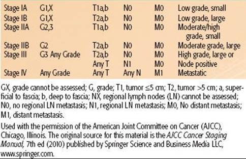
TABLE 83.1 AMERICAN JOINT COMMITTEE ON CANCER TNM STAGE GROUPINGS FOR SOFT TISSUE SARCOMAS
STAGING
The American Joint Committee on Cancer (AJCC) published the seventh edition of the TNM (tumor, node, metastasis) staging manual in 2010.47 The significant factors related to staging of STS include grade (1 to 3), tumor size (≤5 cm vs. >5 cm), location superficial or deep to fascia, and presence of lymph node or distant organ involvement. Stage groupings are shown in Table 83.1. The AJCC staging system for STS does not account for histologic subtype or tumor site, nor does it stratify for tumor size >5 cm. This is unfortunate, given that all of these factors are predictive of survival, but understandable given the complexities of staging systems.
 PROGNOSTIC FACTORS FOR SURVIVAL AND LOCAL RECURRENCE
Many reports have evaluated patient and tumor characteristics to determine prognostic factors for disease-free survival (DFS) or overall survival (OS) and local recurrence (LR). The most powerful predictor for DFS and OS is the AJCC TNM stage of the tumor. Five-year DFS rates for stages I, II, and III STS are 86%, 72%, and 52%, respectively.47 The staging system incorporates several variables, the two most important of which are grade and tumor size. It is also valuable to consider predictive factors individually. Results of multivariate analyses for DFS from several large studies are shown in Table 83.2. Although there are differences among series, several independent prognostic factors are consistently demonstrated. The single most important individual prognostic factor for lower survival rates is high grade.48–53 Five-year DFS rates range from 44% to 67% for high-grade tumors compared to 90% to 100% for low-grade tumors.48–50,53 Other significant predictors for DFS include tumor size, depth, and site. Tumors >5 cm are associated with 5-year DFS rates of about 55% to 70% compared to rates of about 78% to 100% for tumors <5 cm.48,50,53 DFS rates for tumors >10 cm are even lower and range from 33% to 60%.48,50 Five-year DFS rates for tumors that are deep to the fascia range from 58% to 70%, whereas those for tumors superficial to fascia are 81% to 92%.48,50 Furthermore, patients with tumors located in the head and neck or retroperitoneum have lower survival rates than those with tumors located in the extremity or superficial trunk.49,52,54,55 Reports vary regarding the impact of histologic subtype on DFS, but several cite leiomyosarcoma and malignant peripheral nerve sheath tumor as adverse prognostic factors.48,54–56 Older age at presentation, positive resection margins, bone or neurovascular invasion, gender, and race all show mixed results regarding their predictive value for DFS (Table 83.2). As stated earlier, lymph node involvement for STS is rare; but if present, it is an adverse prognostic factor.21–23
Consistently demonstrated significant predictors for LR include positive margins of resection, presentation with locally recurrent disease, older age, and head and neck or retroperitoneal location. Rates of LR for tumors resected with positive margins range from 28% to 56% compared to 0% to 20% for those with negative margins.48,49,57–63 Patients who present with locally recurrent disease are at higher risk for LR (25% to 47%) than those who present with primary disease (11% to 21%).48,58,61 Age has been analyzed with varying cutoff values including >50 years, >64 years, and as a continuous variable. Repeatedly, older age has been associated with higher LR rates.48,49,52,54
TABLE 83.2 SIGNIFICANT ADVERSE PROGNOSTIC FACTORS FOR DISEASE SPECIFIC SURVIVAL OR OVERALL SURVIVAL ON MULTIVARIATE ANALYSES FROM SEVERAL LARGE PATIENT SERIES

MANAGEMENT AND OUTCOME FOR SOFT TISSUE SARCOMAS OF EXTREMITY AND TRUNK
Because of the rarity of STS and the numerous forms in which it can present with respect to tumor histology, site, and size, there are many nuances related to optimal management. Furthermore, delivery of treatment requires a multimodality team that includes experienced pathologists; radiologists; surgeons from the disciplines of surgical oncology, orthopedics, and reconstructive surgery; radiation oncologists; medical oncologists; nurses; physical therapists; and social workers. Treatment goals include complete eradication of tumor with optimal function preservation and minimal treatment-related toxicities. Execution of these goals is complex, and, for this reason, STS is best treated by an experienced team at a specialized sarcoma center. Reports from Sweden, the United Kingdom, and the United States have shown inferior quality of treatment delivery and outcome for STS treated outside of specialized centers.64–66
Surgery
In almost all cases, appropriate surgical resection is a prerequisite for curative treatment of STS. A range of surgical procedures has been employed for the treatment of STS with varying levels of success. These procedures include marginal resection or excisional biopsy, wide resection, and radical resection or amputation. A marginal resection refers to simple removal of the tumor with its pseudocapsule. This is also often described as a “shell-out,” and this is the procedure commonly performed when the diagnosis of STS is not suspected. LR rates after marginal resection range from 42% to 93%.12,20,67–70 This is not surprising as it is known that microscopic tumor cells can extend beyond the pseudocapsule and up to several centimeters beyond palpable gross tumor.19,41 Marginal resection is not an appropriate treatment.
At the other end of the spectrum is radical resection, which involves removal of all of the muscles and neurovascular structures within the compartment where the tumor resides or amputation. Reported LR rates after radical resection are much lower and range from 0% to 18%.12,20,59,67–69 These LR rates are acceptable, but the cost of loss of limb (or loss of an entire compartment) is high. Amputation was a common procedure for STS of the extremities up to the 1970s. The intermediate procedure is a wide resection. Wide resection is also described as conservative surgery (CS), limb-sparing surgery, or function-sparing surgery. It involves en bloc removal of tumor with a rim of normal tissue varying in width from about 1 cm to several centimeters depending on anatomic constraints. This procedure preserves good function (limb salvage) but as a treatment by itself is usually associated with moderately high LR rates, ranging from 25% to 60%.12,20,67–69 Wide resection/CS combined with pre- or postoperative radiation therapy (RT) is the current standard of care for most high-grade STS.
Surgeons should attempt to attain negative margins at the time of definitive resection. As previously stated, the presence of positive margins is consistently associated with increased LR rates even when RT is used.48,49,52,57–63,71Because STS is so rare in comparison to benign soft tissue lesions, the initial procedure performed for a STS is often an unplanned excision (shell-out) with resulting positive margins. It is important to perform a definitive re-excision in these situations, if possible, as the likelihood of finding significant residual disease is on the order of 24% to 63%.8,72,73–75,76–79 As part of the re-resection, incisions, biopsy tracts, drain sites, and any tissues contaminated by the first surgery need to be removed en bloc along with tumor-bed margins. Unfortunately, this often results in a greater scope of surgery and increased functional deficit than if an initial planned excision had been performed by an experienced oncologic surgeon.46 Lastly, although the goal of resection is to attain negative margins, if this would require debilitating surgery, such as resection of a nerve, vessel, or bone, a function-sparing approach with a planned positive margin is sometimes accepted. These decisions are best handled by experienced sarcoma surgeons. Gerrand et al.60 compared LR rates resulting from procedures with planned positive margins (abutting critical structures) to those of procedures with unplanned (unexpected) positive margins. They found a 4% LR rate for the former compared with a 32% to 38% LR rate for the latter.
TABLE 83.3 RANDOMIZED CONTROLLED TRIALS INCLUDING RADIATION THERAPY FOR SOFT TISSUE SARCOMAS OF THE EXTREMITIES AND TRUNK

Conservative Surgery and Radiation Therapy
Three sentinel randomized trials have been performed and have established RT combined with CS as the standard management for most (high-grade) STS of the extremities and trunk (Table 83.3). The first of these trials was conducted by Rosenberg et al.59 at the NCI. Patients with high-grade STS of the extremity were randomized to amputation or to CS and postoperative external beam RT (60 to 70 Gy). Patients in both treatment arms received postoperative doxorubicin, cyclophosphamide, and methotrexate; in the RT arm, chemotherapy was started 3 days prior to RT and continued concurrently with RT. LR rates were 0% (0 of 16) and 15% (4 of 27) for patients treated with amputation and with CS and RT, respectively (P = .06). There was no significant difference in survival rates. This trial was instrumental in setting a new standard for limb-sparing local management of STS. Amputations are now performed sparingly and in <15% of cases.80,81
The next question posed was whether adjuvant RT is necessary. Two more hallmark trials randomized patients between CS alone and CS plus RT.71,82 Both of these trials showed improved local control with the addition of RT. At the NCI, patients with high-grade STS of the extremity were randomized to treatment with CS and postoperative chemotherapy (doxorubicin and cyclophosphamide) with or without concurrent external-beam RT (63 Gy). Patients with low-grade tumors were randomized to CS with or without postoperative external-beam RT (63 Gy). LR rates for the high-grade tumors were 20% (9 of 44) for CS and chemotherapy compared to 0% (0 of 47) for CS, chemotherapy, and RT (P = .003). For the low-grade tumors, LR rates were 33% (8 of 24) for CS alone and 4% (1 of 26) for CS and RT (P = .016). There were no significant differences in survival rates between the two groups. At Memorial Sloan-Kettering Cancer Center (MSKCC), patients with STS of the extremity and trunk were randomized to treatment with CS alone or CS plus adjuvant brachytherapy (BRT).71 BRT catheters were sewn into the tumor bed in a parallel array with 1-cm spacing between catheters and extension of catheters 1.5 to 2.0 cm beyond the tumor bed. Iridium-192 was loaded into the catheters and a dose of 42 to 45 Gy was delivered over 4 to 6 days. LR rates for high-grade tumors were 30% (19 of 63) for CS alone compared with 9% (5 of 56) for CS plus BRT (P = .0025). No difference in LR rates by treatment was seen for low-grade tumors; rates were 26% (6 of 23) and 36% (8 of 22) for patients treated with CS alone and CS plus BRT, respectively. As in the two prior trials, there were no significant differences in survival outcomes. Given that BRT did not improve local control for low-grade tumors in this trial, BRT is not recommended for low-grade STS. Furthermore, for high-grade tumors treated with BRT, LR rates are higher in the setting of positive margins.83 Therefore, BRT as monotherapy is only recommended for high-grade tumors resected with negative margins.84 (In the setting of positive resection margins, a combination of external-beam RT and BRT or external-beam RT alone is preferred.) These two randomized trials have established the role for RT combined with CS for the management of high-grade (and, in select cases, low-grade) STS. In modern series, local control following CS and RT is excellent, with most reported LR rates being <15%.53,58,63,85–91
It is important to note that the current treatment recommendation for most low-grade STS of the extremity and trunk is wide excision alone.92 As long as negative margins are obtained, LR rates are expected to be well under 20%. Relative indications for RT in the setting of low-grade tumors include situations of positive resection margins, locally recurrent disease following initial wide excision, and tumor location that would not be amenable to subsequent salvage surgery.
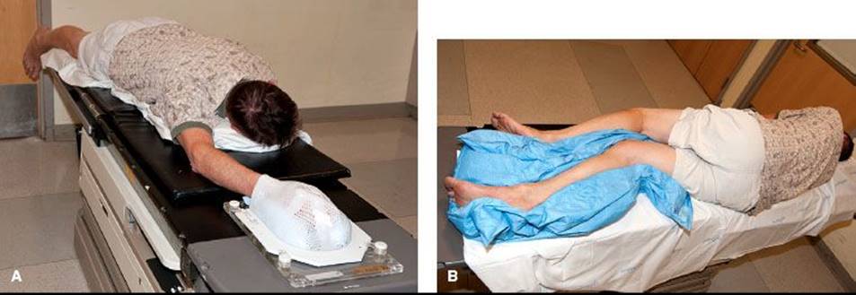
FIGURE 83.3. Examples of patient positions for treatment of upper and lower extremity soft tissue sarcoma (STS). Patients are immobilized in custom casts. A: The “swimmer position” is often an ideal position for treating STS of the hand, forearm, or distal upper arm. B: This is a right decubitus position to treat a STS in the right posterior or anterior distal thigh or lower leg. The right leg is placed directly on the table. The left leg is flexed at the knee, positioned anterior to the right leg, and the legs are separated as much as possible.
Radiation Therapy
Positioning the Patient
The first step of RT planning is to determine the appropriate positioning of the patient. This can be challenging, and sometimes simulation needs to be performed in a few different positions to ascertain the optimal arrangement. A limb should be positioned to allow treatment with as many potential beam angles as possible. In general, this requires that the limb is positioned as far away from the trunk (for upper extremities) or from the opposite limb (for lower extremities) as possible. Suitable positions for upper extremity lesions often include abduction of the arm away from the body with supination or pronation of the arm determined by the location of the tumor. Another good approach for upper extremity lesions is the “swimmer position,” whereby the patient is prone with the arm extended above the head (Fig. 83.3A). This position enables an almost 360-degree approach for all but proximal lesions close to the head. For lower extremity lesions, there are several potential positions. Tumors in the medial compartments are often treated with the majority of the beams in an anterior-posterior/posterior-anterior–like orientation and therefore positioning the patient supine with the legs spread apart often suffices. For proximal thigh lesions, one or both legs are often abducted and flexed in the “frog-leg position” to enable reduction of skin folds in the inguinal region, optimal sparing of the perineum and genitalia, and maximal separation from the contralateral leg. Tumors in the true anterior or posterior compartments are often the most challenging. Sometimes the supine position and oblique fields will be feasible, but, for most of these cases, the author’s approach is to place the patient in a decubitus position (Fig. 83.3B). The author places the patient in the right-sided decubitus position for right-sided tumors (and vice versa), as she finds it is more stable to have the treated leg directly on the table. The untreated leg is flexed at the knee and placed either posterior or anterior to the treated leg, and the legs are separated as much as possible. For men with proximal thigh lesions, the author typically places the genitalia in netting, which is pulled to the contralateral side. Most of time, the genitalia can be positioned such that they will be out of the path of any beams, but the dose delivered from internal scatter cannot be avoided. For this reason, the author counsels all men with treatment fields close to the genitalia to consider sperm banking if they wish to preserve fertility. Similarly, the author recommends fertility consultation for women with treatment fields close to the ovaries. The author also typically calculates and documents doses delivered to the testicles or ovaries in appropriate situations. Patients with an extremity STS that is to be treated with intensity-modulated RT (IMRT) are often positioned in a more neutral supine posture.93 This is possible because of the multiple beam angles used in IMRT planning. Once the position is determined, the patient is immobilized in a reproducible fashion. A custom cast is highly recommended for almost all scenarios.
Target Volumes and Treatment Fields
Preoperative Radiation Therapy
Definitions of appropriate treatment volumes for STS of the extremities and trunk have not been formally tested. Historically, large longitudinal margins of at least 5 cm and sometimes >10 cm have been used with good success. The Radiation Therapy Oncology Group (RTOG) Sarcoma Working Group reported a consensus for appropriate target volumes for preoperative RT and this is described as follows.94 The gross tumor volume (GTV) is defined as the gross tumor delineated by the T1 postgadolinium MRI. Fusion of the diagnostic MRI and planning CT for optimal target definition is strongly encouraged. Clinical target volume (CTV) is defined as the GTV plus 3-cm margins in the longitudinal directions and 1.5-cm margins radially. These margins can be truncated if they extend beyond the compartment or into an intact fascial barrier, bone, or skin. Peritumoral edema on T2 MRI will often be included within the CTV as defined above. If the edema is more extensive and appears suspicious, the CTV can be enlarged to include it at the discretion of the radiation oncologist. Of note, more clarity is needed to determine in which situations peritumoral edema should be included in the CTV. One report showed the presence of sarcoma cells beyond the gross tumor in 10 of 15 patients. The location of the cells varied from 1 to 4 cm beyond the tumor but did not correlate with the location or extent of peritumoral edema on MRI.41 It is reasonable to try to include the edema in the CTV, but if this would require a significant increase in the treatment field beyond the RTOG consensus guidelines, it becomes a judgment call. The planning target volume (PTV) definition is not specified in the consensus statement, but typically it is defined as the CTV plus 5 to 10 mm. Application of the GTV and CTV definitions of the RTOG consensus group results in treatment planning fields very similar to the historical standard of 5-cm margins and 2.5- to 3-cm margins from GTV to the field edge in the longitudinal and radial dimensions, respectively. Figure 83.4 depicts MRI, dosimetry, and treatment field images for a STS of the left posterior thigh that was treated with three-dimensional (3D) conformal preoperative RT.
Several series using treatment volumes similar to those described above have reported excellent local control rates. Kim et al.88 reported a series of 56 patients treated using target volume definitions very similar to the consensus recommendations and described a local control rate of 88.5%. The Canadian Sarcoma Group’s randomized trial of preoperative versus postoperative RT also employed similar preoperative treatment fields (except that the CTV expansion in the longitudinal direction was typically 4 cm rather than 3 cm) and reported a 5-year actuarial local control rate of 93%.86 Lastly, a review of 768 patients treated with preoperative and postoperative RT at Princess Margaret Hospital between 1990 and 2006 demonstrated a local control rate of 92%. Among the 60 recurrences, 82% occurred within the treatment fields.87 As a group, these three publications provide firm support that the current guidelines produce treatment fields that are sufficiently large.
FIGURE 83.4. An unclassified pleomorphic high-grade sarcoma of the posterior left thigh in a 61-year-old man that was treated with preoperative three-dimensional-conformal radiation therapy (50 Gy) using opposed oblique fields. A: T1 postgadolinium image of the magnetic resonance imaging (MRI) scan. B: T2 image of the MRI scan showing peritumoral edema (arrows). C: Axial slice of the graphic plan showing the gross tumor volume (red), clinical target volume (blue), planning target volume (orange), and covering isodose lines. D: Digital reconstructed radiograph showing the treatment field and target volumes.

Postoperative Radiation Therapy
For postoperative treatment, the nomenclature for GTV and CTV is variably described and partly a matter of semantics given that there is actually no “gross tumor.” It can be helpful to draw a GTV in the location where the gross tumor was preoperatively, as sometimes this volume is used for cone-down volumes. The CTV should encompass all the tissues handled during the surgery including the incision and any drain sites. (Postoperative changes seen on MRI help define the operative bed.) An additional longitudinal margin of 2 to 4 cm and a radial margin of 1.5 to 2 cm is generally added to the operative bed to form the CTV. The same principles apply as listed above in terms of using the T1 postgadolinium images for GTV definition, fusing the planning CT and MRI when possible, and editing the CTV to exclude bone, an intact fascial plane, skin, or extension beyond an uncontaminated compartment. The PTV is typically CTV plus 5 to 10 mm. As is the case in the preoperative guidelines, the end result of these GTV and CTV definitions is a distance from the operative bed to the field edge of about 2 to 5 cm in the longitudinal direction, which matches the historical standard. A second (and sometimes third) course field reduction is typically used in the postoperative setting. CTV margins for the reduced field(s) vary and can include about 2 cm on the operative bed or on the initial GTV.95,96 These postoperative treatment field definitions are associated with excellent local control rates and are the current standard of care.86,87,93,97,98 Figure 83.5 shows dosimetry images for a malignant solitary fibrous tumor of the left upper back that was treated with postoperative RT using IMRT.
The excellent local control rates resulting from the use of standard preoperative and postoperative treatment fields establish that these fields are adequately large, but they do not address the question of whether these field sizes could be reduced. As such, the necessity of such large treatment fields has been called into question. Initial development of these large fields predated the CT and MRI era, when imaging modalities to define STS were suboptimal. Moreover, the BRT experience at MSKCC produced excellent local control rates using treatment volumes that extended only 1.5 to 2 cm beyond the tumor bed.71 Lastly, several single-institution series of treatment with surgery alone have been associated with very good local control rates ranging from 0% to 20% (see the Surgery Alone section).99,100–106 Specifically, Baldini et al.102 found no recurrences in 36 patients with resection margins ≥1 cm compared to an actuarial 10-year LR rate of 13% (4 of 38) for those with margins <1 cm. Based on these observations, it is reasonable to query whether treatment field sizes can be reduced. Two ongoing studies ask this question. The first is a trial, in which patients receiving postoperative RT were randomized to standard fields with 5-cm margins or to tailored fields.107 The CTV for the tailored fields was defined as GTV plus 2 cm in all directions. The second study is RTOG-0630, which is a phase II study using image-guided RT and reduced fields.108 The CTV is defined as GTV plus 2-cm longitudinal margins for tumors <8 cm and 3-cm margins for tumors >8 cm. The CTV also includes suspicious edema on MRI T2 images. Results of these trials will be very informative, but until more evidence is available, the author recommends the standard treatment volumes as described above.
FIGURE 83.5. A malignant solitary fibrous tumor of the left upper back in a 45-year-old woman who underwent marginal excision of a 3.5 cm tumor with multiple positive margins. Radical re-excision showed residual microscopic tumor and negative margins. Postoperative radiation therapy was delivered using intensity-modulated radiation therapy (50.4 Gy) for the first course and opposed oblique fields for the second course (12.6 Gy) to a total dose of 63 Gy. A: Axial slice of a planning image shows the clinical target volume (CTV), which includes the operative bed with a margin (blue), planning target volume (PTV) (orange), and covering isodose lines. B: Coronal planning image shows CTV (blue), PTV (orange), and isodose lines.

Brachytherapy
The CTV for BRT treatment as monotherapy should include the operative bed with a margin. The American Brachytherapy Society recommendations state that there is no clear consensus for the appropriate size of the margin.84 The MSKCC randomized trial used margins of 1.5 to 2 cm beyond the tumor bed.71 A more detailed description of BRT modalities, techniques, and indications is beyond the scope of this chapter. The American Brachytherapy Society published detailed recommendations in 2001 and those are a good reference.84
Doses
The standard dose for preoperative external-beam RT is 50 Gy delivered in 2-Gy fractions.49,53,89,109 In the situation of positive margins, a postoperative external-beam RT boost of 16 to 20 Gy (delivered in 1.8- to 2-Gy fractions) is sometimes delivered. The efficacy of this boost dose has not been proven and, as it may be associated with increased toxicity, its use has been called into question.110 Other techniques to deliver an additional boost dose include BRT (both low-dose rate and high-dose rate) and intraoperative electron therapy. Due to variabilities in patient selection for these procedures, it is difficult to determine the relative benefit of the additional dose delivered by these modalities.95,111,112–115 For postoperative external-beam RT, treatment usually commences about 4 to 6 weeks following surgery and once the wound is fully healed. Recommended total doses are 60 to 66 Gy (delivered in 1.8- or 2-Gy fractions) for the case of negative margins and 66 to 68 Gy for positive margins.49,63,97,98,116,117 The first course of treatment is typically treated to a dose of 45 to 50 Gy and the balance of the dose is either given in one reduced field or split about evenly between two reduced fields. The standard dose for low–dose-rate BRT is 45 Gy.71,84 For treatment combining external-beam RT with BRT as a boost, doses are typically 45 to 50 Gy for external beam and 15 to 25 Gy for the BRT component, for a total of approximately 65 Gy.84,111
Principles of Treatment Planning
As for any other malignancy, the basic principles of RT planning are to achieve good coverage of the PTV with maximal sparing of adjacent normal structures. For extremity lesions, the important normal structures are the limb itself, soft tissues, bones, and joints. For proximal thigh lesions, the perineum and genitals are also relevant. For STS of the trunk, adjacent normal structures can include small bowel, kidneys, spinal cord, stomach, liver, and lung. Basic tenets for treating the extremity are to “spare a strip” of the limb circumference (to prevent subsequent lymphedema and pain), to avoid treating the whole thickness of bone to high doses (to diminish risk of fracture), and to avoid treating an entire joint to high doses (to decrease joint stiffness). It is not always possible to preserve fertility (and in any borderline situation, one should offer sperm banking or fertility consultation). Every effort should be made to place the testicles as far out of the field as possible to avoid direct treatment and to minimize the contribution from internal scatter.
Specific dosing guidelines for structures of the extremity are as follows. Spare as much as possible of the limb circumference from receiving any dose, and try to spare a 1-cm thickness at a minimum. With 3D-conformal techniques, part of the limb can usually be excluded entirely from the treatment fields (Fig. 83.4C,D). With IMRT, in order to achieve more dose conformality, the tradeoff is that the volume of tissue that receives a low dose is increased; for some of these cases, the entire circumference of the limb may receive some low dose. The acceptable low dose that can be delivered to the entire limb has not been well established and will most likely vary with factors such as the total dose delivered, total volume of tissue treated, and the location in the limb. However, as a guideline, it is helpful to contour a strip of limb circumference to use as an avoidance structure in order to keep part of the treated limb dose to a minimum. The whole-joint dose should be <40 to 45 Gy. Higher doses can be delivered to part of the joint if needed. Full thickness bone irradiation should be avoided if possible, and the mean and maximum doses to the whole bone should also be kept to a minimum. Dickie et al.118 performed a detailed analysis of dosimetric predictors for bone fracture using a matched pair analysis and found that radiation-related bone fractures were reduced if the following parameters were met: volume of bone receiving ≥40 Gy (V40) <64%, mean dose to bone <37 Gy, and maximum dose to bone <59 Gy. Normal-tissue complication probabilities for organs that may be affected by treatment of a truncal STS are listed in the Quantitative Analysis of Normal Tissue Effects in the Clinic document.119 If RT treatment of a trunk lesion may potentially ablate ipsilateral kidney function, a renal scan should be performed to ensure adequate function of the contralateral kidney. To achieve sufficient coverage of superficial target volumes (in either the preoperative or postoperative setting), tissue-equivalent bolus material is often applied to intact skin as well as to incision sites. In the preoperative setting, if one reviews the case with the surgeon and determines that an appropriately sized paddle of skin will be removed along with a superficial tumor, one can consider omitting bolus.
Three-dimensional-conformal RT (3D-CRT) and IMRT are both acceptable external-beam treatment techniques. The principal dosimetric differences between these two techniques are that, compared with 3D-CRT, IMRT typically achieves comparable full target coverage, improved conformality of the dose distribution around the target volume, reduced volumes of high doses to normal tissues, greater volumes of low doses to normal tissues, and greater total body exposure due to increased monitor units.120,121 Three-dimensional-CRT has a longer track record for STS treatment with well-established outcomes and toxicity end points. Fewer long-term data are available for IMRT, and optimal dose–volume histogram metrics for normal structures, such as limb circumference, are yet to be defined. The data for IMRT that do exist, however, are encouraging. Alektiar et al.93 reported a series of 41 patients with STS of the lower extremity treated with IMRT. With a median follow-up time of 3 years, local control was excellent (94%) and the toxicity profile was comparable to that of 3D-CRT series. Alektiar et al.122 performed a second analysis comparing outcomes for patients with high-grade STS of the extremity treated with either BRT or IMRT at MSKCC. The authors acknowledged the limitations of this retrospective comparison, but reported superior local control for IMRT compared to BRT with 5-year local control rates of 92% and 81%, respectively (P = .04). On the other hand, Hall120 raised a caveat that IMRT may be associated with a higher rate of radiation-associated second cancers compared to 3D-CRT, resulting from the use of more monitor units with a greater total body exposure from leakage, and from a larger volume of normal tissue exposure to low doses of RT, resulting from the greater number of treatment fields. He estimated a potential doubling of second cancers in older patients from a rate of approximately 1.5% to 3% at 10 years. (These rates could be higher for children.) Larger patient numbers and longer follow-up are still needed to confirm the favorable findings reported for IMRT thus far.
There are several other exciting technological advances for treatment, which include IMRT dose painting, image-guided radiation therapy (IGRT), adaptive RT, particle-beam radiotherapy (protons, carbon ions), and stereotactic body RT (SBRT). Dose paintingis an advanced application of IMRT in which differential doses are delivered to different areas of the target volume simultaneously. The idea is to deliver higher doses to areas of the tumor that are believed to be more radioresistant (such as hypoxic regions), areas that may contain a higher burden of disease, or areas where the surgeon may have more difficulty with resection. This technique has been used in the setting of retroperitoneal sarcoma and is appealing.123 Image-guided radiation therapy refers to serial imaging of patient setup prior to treatment so that appropriate positional adjustments can be made beforehand. Because of the increased certainty of treatment accuracy, in many cases the added margins for “setup error” can be reduced, which allows for smaller treatment fields and potential dose escalation without undue normal tissue toxicity. For example, although spinal cord tolerance is 50 Gy, Hansen et al.124 described the ability to treat paraspinal sarcomas to 59.4 Gy using IGRT with CT imaging to better assess patient position prior to treatment. IGRT and reduced treatment fields were also used in the phase II RTOG-0630 trial; patient accrual is complete and the results are awaited.108 Another benefit of IGRT is that if imaging during a course of treatment demonstrates changes in patient anatomy (e.g., due to weight loss) or changes in tumor shape, a second radiation plan can be developed to adapt to these new data. This is referred to as adaptive radiation therapy and might be relevant for sarcomas that respond dramatically to radiation, such as myxoid liposarcoma.125,126
Particle beams such as protons and heavier ions (carbon ions) have more favorable physical and biologic characteristics than photons, which make them appealing for clinical use. Specifically, because of the Bragg peak dose distribution property, treatment plans can be created with steep dose falloff at field borders.127 This allows for ideal sparing of adjacent critical normal structures as well as opportunities for safe dose escalation. Proton-beam treatment for malignancies has been in use at a few centers for several decades. There are published data for treatment of chordomas and chondrosarcomas of the skull base as well as for paraspinal and sacral bone tumors. There have been no randomized studies comparing photons and protons, but there are several single-institution reports for protons that show very good results. Local control rates for skull-base chordomas treated with protons range from 46% to 90% and for skull-base chondrosarcomas range from 75% to 100%.128,129,130–132,133,134–135 Carbon ions have been used for clinical treatment only since the 1990s. Schulz-Ertner et al.136,137 reported a 74% 4-year local control rate for skull-base chordomas treated with carbon ions and a 90% local-control rate for skull-base chondrosarcomas. The group from Chiba, Japan, reported a 73% local-control rate for unresectable bone and STS and a 96% local-control rate for sacral chordomas.138,139 These early results are impressive, but confirmatory long-term follow-up data are needed.
Stereotactic body radiation therapy is a technique that delivers highly focused photon radiation doses to extracranial lesions. Typically the dose schedules are hypofractionated, with large ablative fraction sizes ranging from 6 to 30 Gy per fraction, for a course of 1 to 5 treatments. With respect to sarcoma, SBRT may have a role in the treatment of oligometastatic disease. Excellent local control rates, ranging from 73% to 96%, have been reported for treatment of metastases to the lung as well as other sites for a mix of tumors including sarcoma.140,141,142 There are less data regarding the role of SBRT for definitive treatment of primary sarcomas, but there may be a role in select patients with tumors located in or adjacent to sensitive normal structures. For example, a study of SBRT for 14 patients with primary sarcoma of the spine reported local control for 5 of 7 patients treated with SBRT alone and for 5 of 7 patients treated with SBRT and surgery.143 Figure 83.6 shows an MRI and dosimetry for a case of an unresectable leiomyosarcoma of the inferior vena cava that was treated with definitive SBRT at Brigham and Women’s Hospital.
IMRT with or without dose painting, IGRT, adaptive RT, particle beams, and SBRT all show potential dosimetric and biological advantages compared to conventional 3D-CRT photon treatment. Promising data are available for all of these approaches, but they are limited. There will likely be appropriate places for all of these technologies in the armamentarium for sarcoma treatment, but further careful study is needed to define the optimal role of each technology and to ensure that toxicities are acceptable.
FIGURE 83.6. A 72-year-old man with a 1.8 é 1.8 cm unresectable leiomyosarcoma of the intrahepatic inferior vena cava in close proximity to the liver, esophagus, and heart that was treated with stereotactic body radiation therapy (SBRT) (60 Gy in 5 fractions). A: Coronal T1-weighted magnetic resonance image slice showing the tumor. B: Coronal slice of the SBRT graphic plan showing the internal target volume (blue), planning target volume (orange), and covering isodose lines.

Radiation Therapy Toxicities
Acute Radiation Therapy Toxicities
Acute toxicities following RT treatment of the extremities include skin erythema and possible desquamation in high-dose areas, problems with wound healing, localized alopecia, and fatigue. Moist desquamation can be quite uncomfortable, but this heals typically quickly after completion of RT. Depending on tumor location, treatment of STS of the superficial trunk can be associated with additional toxicities such as nausea, bowel irritability, or esophagitis.
The most significant of these acute sequelae is problems with wound healing. Several retrospective single-institution series have reported wound complication rates following preoperative RT of 25% to 46%.89–91,95,144,145–147,148Rates following postoperative RT are lower and range from 6% to 29%.90,91,144,146 The most definitive data pertaining to wound complications are provided by the landmark randomized trial performed by the Canadian Sarcoma Group, in which the study end point was major wound complications within 120 days of surgery.109 Major wound complications were defined as those requiring a second operation for wound repair or wound management requiring an invasive procedure, readmission, or persistent deep packing for 120 days or longer. This trial randomized 190 patients to preoperative RT (50 Gy ± 16 to 20 Gy postoperative boost) or postoperative RT (66 to 70 Gy). With a median follow-up time of 3.3 years, the study met early stopping rules and closed. Wound complication rates were 35% for patients treated with preoperative RT compared to 17% for those treated with postoperative RT (P = .01). Further, lower extremity site and tumor size >10 cm were independent predictors for wound complications on multivariate analysis. Others have also found large tumor size and lower extremity site to be risk factors for wound complications.89,145,146 Baldini et al.89 reviewed the experience at Brigham and Women’s Hospital and Dana-Farber Cancer Institute for 103 patients with STS of the trunk or extremity treated with preoperative RT. On multivariate analysis, significant independent predictors for wound complications were tumor size >10 cm, tumor proximity to skin surface <3 mm, vascularized flap closure, and diabetes mellitus. In an attempt to decrease wound complications following preoperative RT, Dickie et al.149 conducted a phase II trial for 59 patients with lower-extremity sarcoma. Together with the surgeon, the radiation oncologist contoured the area of the subsequent surgical flaps and designated it as an avoidance structure for IMRT planning. The resulting rate of wound complications was 30.5%, which was not statistically different from the value reported in the randomized trial. Interestingly, for patients who developed wound complications, there was a trend for higher mean and maximum doses to the flaps. It may be that higher doses to the flaps were a necessary planning outcome for patients with tumors in close proximity to the skin surface, which would be consistent with the Baldini report showing increased complications for tumors within 3 mm of the skin surface. Efforts continue to minimize postoperative wound complications.
Chronic Radiation Therapy Toxicities
The most significant chronic toxicities following RT to the extremities include edema, subcutaneous fibrosis, decreased muscle strength, decreased range of motion, pain, and, less commonly, bone fracture and peripheral nerve damage. Published rates of these toxicities vary significantly, as series have different patient inclusion criteria (with respect to tumor site and treatment) and some report only moderate or severe complications, while others report any degree of complication. Nonetheless, reported rates for edema are 10% to 20%, for fibrosis they are 30% to 60%, and for bone fracture they are 0.07% to 7%.97,150–152,153,154–156 Stinson et al.150 reviewed 145 patients treated at the NCI with surgery and RT and reported the following complication rates: fibrosis 57%, moderate to severe decreased range of motion 32%, moderate to severe decreased muscle strength 20%, contracture 20%, ≥ grade 2 edema 19%, pain requiring narcotics 7%, and bone fracture 6%. Furthermore, several studies have correlated higher complication rates with higher doses and larger field sizes. Doses >60 or 63 Gy have been associated with higher rates of fibrosis, edema, bone fracture, pain, decreased muscle strength, and decreased range of motion.97,118,150,151,154,157 Large RT field sizes have also been associated with more edema, fibrosis, and joint stiffness.150,151
Radiation Timing: Preoperative Versus Postoperative
External-beam RT can be delivered either prior to or following definitive resection. Reported local recurrence rates with each approach are similar and range from 3% to 27% for preoperative RT and 8% to 28% for postoperative RT.53,86–88,90,91,158,159,160 The randomized trial of preoperative versus postoperative RT conducted by the Canadian Sarcoma Group showed local control rates of 93% and 92% for the preoperative and postoperative groups, respectively.86 Similarly, there is no clear difference in DFS or OS associated with either approach. The Canadian randomized trial initially showed improved survival for the preoperative RT treatment arm, but updated 7-year results showed no differences in any recurrence or survival outcomes.86,109 One retrospective report that assessed 821 patients using the National Oncology Database showed improved DFS rates for patients treated with preoperative RT.54However, these results should be considered with caution given the retrospective nature of the report and associated potential selection biases.
Although the efficacy of these approaches seems to be similar, the toxicity profiles are clearly different. Preoperative RT is associated with a well-established increased risk of acute wound complications. Several retrospective reports have described wound complication rates on the order of 25% to 46%.89,90,95,145–147 As described in detail above (see the Acute Radiation Therapy Toxicities section), the definitive data for wound complication rates come from the Canadian randomized trial in which the primary end point was wound complications. In that trial, wound complication rates were 35% for patients treated with preoperative RT compared to 17% for those treated with postoperative RT (P = .01).109 Although wound complications adversely affected functional outcome for patients in the early postoperative period (6 weeks after surgery), this toxicity was largely reversible, as evidenced by the fact that function was equivalent between the treatment arms 1 year after resection.
On the other hand, patients treated with postoperative RT have a higher rate of chronic and generally irreversible toxicities, which include subcutaneous fibrosis, joint stiffness, edema, and bone fractures. These late toxicities have been reported in retrospective series but, again, good data come from the Canadian randomized trial.150,151,154,157,161 Davis et al.151 reported that grade 2 or higher late toxicity rates were higher for the postoperative RT group than the preoperative group. Specifically, for the postoperative and preoperative groups, respectively, rates of grade 2+ complications were as follows: subcutaneous fibrosis, 48% versus 31.5%; joint stiffness, 23% versus 18%; and edema, 23% versus 15.5%. These differences were not statistically significant, but the study was not powered to detect these differences. Statistically significant associations were seen between larger field size and rates of fibrosis, joint stiffness, and edema. Additionally, in a retrospective series, Holt et al.154 reported increased rates of bone fracture for patients treated postoperatively (60 to 66 Gy) compared to those treated preoperatively (50 Gy). The rates were 7% and 0.6%, respectively (P = .007).
It is helpful to summarize the relative advantages and disadvantages for preoperative versus postoperative RT. For preoperative RT, the advantages include the ability to treat with smaller RT fields and lower doses, both of which are associated with reduced long-term toxicities. Lower RT doses are also associated with reduced treatment time, lower costs, and a hypothetical possibility of lower second-malignancy risks. Other potential advantages of preoperative RT include the ability to render unresectable or marginally resectable tumors resectable, the potential to prevent tumor seeding of the operative bed or systemic circulation, and an increased efficacy of RT from good oxygenation of tissues due to unperturbed tumor vasculature. Furthermore, in most cases, defining the tumor volume for RT planning is relatively straightforward given that it is in situ. The main disadvantage of preoperative RT is the higher risk of major wound complications and its concomitant increased morbidity and cost. However, most major wound complications are treatable and, therefore, this toxicity is generally considered reversible. Another disadvantage of preoperative RT is that the resected specimen is potentially less informative on pathology review due to the prior treatment.
Postoperative RT has the advantage that the complete tumor specimen is available for pathology review for determination of histology and margin status. Another important advantage is the lower risk of major wound complications. A disadvantage of postoperative RT is the necessity for larger treatment volumes and higher doses, which are associated with higher chronic long-term toxicities such as subcutaneous fibrosis, joint stiffness, edema, pain, and bone fractures. For the most part, these toxicities are irreversible. In theory, the interrupted vasculature related to surgery may create a hypoxic environment, rendering RT less effective (this is a potential explanation for the need for higher doses compared to the preoperative setting). Lastly, determining an RT target definition is often more complex in the postoperative setting, as it requires reconstruction of where the tumor resided as well as definition of the entire operative field.
In conclusion, pre- and postoperative RT are associated with equivalent efficacies but different toxicity profiles, the most salient of which are increased (reversible) wound complications for preoperative RT and increased (irreversible) long-term toxicities of fibrosis, edema, and joint stiffness for postoperative RT. The topic is controversial, but preoperative RT is generally the author’s preferred approach for STS of the extremities or trunk. Even when a patient is at high risk of developing a wound complication, the author often prefers preoperative RT, as most wound problems are highly treatable with eventual recovery of good function.
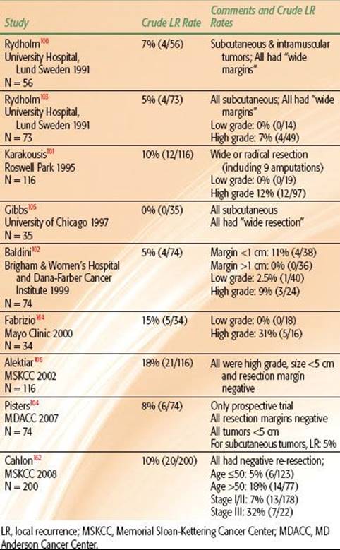
TABLE 83.4 RESULTS OF SELECT SINGLE-INSTITUTION SERIES OF SOFT TISSUE SARCOMAS TREATED WITH SURGERY ALONE
Surgery Alone
As stated previously, conservative surgery and RT is the standard treatment for high-grade STS of the extremity and trunk. However, there are also several reports that demonstrate excellent outcomes following treatment with surgery alone (Table 83.4). It is important to acknowledge that these are all single-institution series, all but one are retrospective, and patients were highly selected for treatment. These factors render the results less generalizable, so caution is recommended when considering this approach. Nonetheless, crude and actuarial LR rates in these select studies range from 0% to 20% and most are 10% or lower.99,100–106,162 What the appropriate selection criteria for this approach are remains unclear, but potential factors can be inferred by close analysis of these reports. Excellent local control appears to be associated with wide resection performed for tumors in a subcutaneous location. Rydholm et al.103described only 4 local recurrences (5%) among 73 subcutaneous tumors treated with wide excision. In addition, Gibbs et al.105 reported no local recurrences among 35 patients with subcutaneous STS treated with wide excision alone. Among these tumors, 47% were high grade and 32% were >5 cm.
Pisters et al.104 described a prospective series of patients with tumors <5 cm who had negative resection margins and were treated with surgery alone. The overall crude LR rate was 8%, and among the subcutaneous tumors, the rate was only 5%. Furthermore, surgical technique and margin status are important. In the series reported by Rydholm et al.,100 a large proportion of the cases were resected without an initial biopsy and, thus, without the potential for tumor seeding of intervening tissues.
Several of the reports also describe a meticulous surgical approach to wide resection with removal of a cuff of normal tissue as well as intact fascia.100,103,105 In the prospective trial of Pisters et al.,104 negative resection margins were a required criterion for treatment by resection alone. The report by Baldini et al.102 quantified surgical resection margins. In that series, 74 patients were treated with surgery alone and the overall 10-year actuarial LR rate was 7%. Interestingly, there were no recurrences seen for 36 patients with resection margins ≥1 cm compared to a 10-year actuarial LR rate of 13% (4 of 38) for those with margins <1 cm.
Conversely, the authors of a retrospective study from the Institut Gustave Roussy also quantified margin status.163 They reported a 10-year actuarial LR rate of 35% for patients treated with surgery alone who had resection margins ≥1 cm. Further, they found the addition of RT for patients with these characteristics was not associated with a significant local control benefit. As the local recurrence rate in that series is high, it is difficult to draw meaningful conclusions except to reinforce the concept that treatment with surgery alone should be done with care.
Cahlon et al.162 assessed 200 patients who were treated with surgery alone at MSKCC following re-resection showing no evidence of disease. Although the overall 5-year actuarial LR rate was only 9%, on multivariate analysis, age >50 and stage III disease were both predictors for higher LR rates. If both of these factors were present, the LR rate was 31%. The LR rates for low-grade tumors treated with surgery alone in these series were all very low and range from 0% to 5%.100,102–105,164 In fact, the standard treatment recommendation for low-grade STS is wide resection alone. With this approach and negative margins, LR rates are typically <20%.92 (Indications for adjuvant RT for low-grade tumors include positive resection margins, locally recurrent disease following initial treatment with surgery alone, or a tumor location that would not be amenable to subsequent salvage surgery.)
In sum, there is most certainly a subset of patients with STS of the extremity and trunk for whom wide excision alone is appropriate treatment. The selection criteria for this strategy remain undefined but will perhaps include, but not be limited to, some of the following: subcutaneous tumors; tumors resected with wide negative margins >1 cm or an intact fascia; low-grade tumors; tumors representing primary presentation of disease (e.g., not locally recurrent); tumor locations amenable to limb-sparing salvage surgery for recurrence; and patient willingness to comply with follow-up. Before surgery alone can become standard of care for select patients with STS, the eligibility criteria need to be elucidated and tested in a prospective multi-institutional trial. Other than for low-grade tumors resected with negative margins, treatment with surgery alone should be employed cautiously.
Neoadjuvant, Concurrent, or Adjuvant Chemotherapy
Locally advanced (stage III) STS of the extremities and trunk has a significant risk of distant recurrence, and for this reason, the addition of systemic therapy to treatment algorithms is appealing. There is an established role for chemotherapy as part of the treatment for rhabdomyosarcoma and Ewing’s sarcoma in children and in adults treated per pediatric protocols, many of which include adults up to age 50.165,166 For other histologies of adult STS, no clear role for chemotherapy has been defined, and this group is the subject of the discussion below. The two most standardly used drugs in the management of STS are doxorubicin and ifosfamide, with gemcitabine-based regimens increasingly used, particularly in patients with leiomyosarcoma.167,168
There are several potential benefits to a neoadjuvant chemotherapy approach. These include the potential to treat micrometastatic disease early in the treatment course; the potential to decrease the scope of the resection if sufficient response is achieved; the ability to ascertain the chemotherapy response or lack thereof for an individual patient, which could guide the use of additional (postoperative) chemotherapy; and enhanced drug delivery to the tumor with corresponding increased efficacy, given that the tumor vasculature has not been disrupted in the neoadjuvant setting. Few reports address the role of neoadjuvant chemotherapy for high-risk STS of the extremities and trunk. A prospective randomized phase II trial conducted by the European Organisation for Research and Treatment of Cancer included 137 patients who were randomized to receive or not receive 3 cycles of doxorubicin and ifosphamide prior to resection with selective use of postoperative RT.169 Unfortunately, the study was closed due to poor accrual and therefore lacks sufficient power for one to draw definitive conclusions. The results showed no statistically significant differences between treatment arms for DFS or OS. Specifically, the 5-year DFS rate for the chemotherapy group was 56% compared with 52% for the observation group; the corresponding 5-year OS rates were 65% and 64%, respectively. Two other retrospective reports on the use of neoadjuvant chemotherapy showed mixed results.170,171
Similarly, informative data are scarce regarding the use of concurrent preoperative chemotherapy and radiotherapy. Several trials have shown that treatment with RT and concurrent doxorubicin, ifosphamide, gemcitabine, or temozolomide is both feasible and safe.172–176 There are also two reports of an interdigitated chemotherapy and RT approach. The first was a pilot trial conducted at the Massachusetts General Hospital, which enrolled 48 patients. Treatment involved an interdigitated approach of chemotherapy (mesna, doxorubicin, ifosphamide, and dacarbazine) and 44 Gy given as a split course.177 Results were very good and associated with improved survival compared with historical controls. The RTOG subsequently enrolled 66 patients in a phase II trial with a very similar treatment approach.178 Efficacy was somewhat comparable to that of the pilot study, but toxicities were greater, with 5% treatment-related deaths and 83% grade 4 toxicities reported.
The most data available pertain to the use of chemotherapy in the adjuvant setting. A meta-analysis reported by the Sarcoma Meta-Analysis Collaboration (SMAC) included 1,568 patients treated in 14 randomized trials using adjuvant doxorubicin-based chemotherapy. It showed that chemotherapy was associated with statistically higher rates of LR-free survival and DFS at 10 years.179 Ten-year LR-free survival rates were 81% and 75%, respectively, for the chemotherapy and observation groups (P = .02); the corresponding values for 10-year DFS were 55% and 45% (P = .001). For 10-year OS, there was no clear benefit, with rates of 54% for the chemotherapy group and 50% for the observation group (P = .12). However, exploratory analysis showed a significant 7% survival benefit for the subset of patients with STS of the extremities. Subsequent to this meta-analysis, several more randomized trials of adjuvant chemotherapy were performed using anthracycline or ifosphamide-based chemotherapy; many of these showed trends for survival benefits with chemotherapy, but none were statistically significant.180–184 (One study from Italy initially reported a significant survival benefit due to chemotherapy at 4 years, but the survival benefit did not hold up with 7.5-year follow-up.180,181) SMAC updated their meta-analysis in 2008 with the inclusion of four additional trials (three adjuvant and one neoadjuvant).169,180,182,183,185 The new analysis included 18 randomized trials with 1,953 patients and reported statistically significant absolute reductions of 4% for LR and 9% for distant recurrence and an absolute improvement of 6% for survival attributable to chemotherapy.185 However, the fact that this update did not include the largest negative trial renders the results less conclusive.184
All of the above studies included a mix of STS histologic subtypes, which is probably not appropriate as we learn more about the varied biologic behaviors of individual STS entities. For example, several trials have shown that synovial sarcoma and round cell liposarcoma are particularly sensitive to chemotherapy.186–189 It may be that a small survival benefit does indeed exist for select subsets of patients (such as those with synovial sarcoma or round cell liposarcoma), but the available studies are underpowered and hindered by the inclusion of a mix of histologic subtypes and tumor sites. Going forward, trials should include centralized pathology review and stratification by histology.
In totality, although there are hints of efficacy for certain subgroups, the available data do not support the routine use of chemotherapy for locally advanced STS. However, for select high-risk patients with high-grade and large tumors (>8 to 10 cm), it is reasonable to address the pros and cons of neoadjuvant, concurrent or interdigitated, or adjuvant chemotherapy on an individual patient basis. As toxicity can be significant, concurrent approaches are best undertaken at experienced centers.
Isolated Limb Perfusion, Isolated Limb Infusion, and Chemotherapy with Regional Hyperthermia
Isolated limb perfusion (ILP) is a complicated technique that has been used in Europe for the treatment of locally advanced STS that would otherwise require amputation. This procedure involves isolating the arterial and venous circulation of the limb by connecting it to an extracorporeal circulation, where it is oxygenated and instilled with systemic agents, most commonly, melphalan and tumor necrosis factor. A tourniquet is applied to the limb to prevent leakage into the systemic circulation and the limb is often treated with hyperthermia as well. The treatment can have significant morbidity, but reported limb salvage success rates are quite high.190–193 Isolated limb infusion (ILI) employs low-flow isolated limb perfusion without oxygenation and has been developed as a simpler alternative to ILP. Available data suggest comparable efficacy and less toxicity for ILI compared to ILP.194,195 Lastly, chemotherapy with regional hyperthermia (delivered via an external electromagnetic field) is another approach for locally advanced disease. A randomized trial of doxorubicin, ifosphamide, and etoposide, with or without regional hyperthermia delivered before and after local therapy, has shown superior DFS and progression-free survival rates for the regional hyperthermia group.196 All of these techniques are complicated to deliver and associated with significant potential toxicities. However, they represent valuable potential alternatives to amputation in such settings as in transit metastases of epithelioid or clear cell sarcoma or extensive local recurrences after prior surgery and RT.
FUTURE DIRECTIONS
In summary, success rates for the treatment of stages I and II STS of the extremities and trunk are currently high, with local control rates of ≥85% and 5-year survival rates of 90% and 81%, respectively.47 For these patients, we should continue to explore ways to reduce treatment-related morbidity related to surgery and RT. These strategies should include development of innovative techniques to reduce postoperative wound complications, efforts to reduce RT field sizes and to deliver more conformal therapy, and definition of patient subsets that can be effectively treated with surgery alone. For situations in which it is difficult to achieve local control, more aggressive local treatment is needed; this could include the use of RT dose escalation using IGRT, IMRT with dose painting, heavy particles, SBRT, or concurrent chemoradiation strategies. Lastly, patients with stage III disease have a high rate of distant relapse and death. For these patients, novel systemic therapies are needed. The discovery of the targeted agent imatinib mesylate for the treatment of gastrointestinal stromal tumors has been associated with great success, and similar discoveries for other histologies are anticipated.197 As the field of STS continues to move forward, it is likely that treatment algorithms for the various histologic subtypes will be developed.
ACKNOWLEDGMENT
I would like to acknowledge the editorial assistance of Susanna Hilfer and Barbara Silver.
SELECTED REFERENCES
A full list of references for this chapter is available online.
2. Fletcher CDM, Unni KK, Mertens F, eds. World Health Orgnization classification of tumours: pathology and genetics of tumours of soft tissue and bone. Lyon: IARC Press, 2002.
15. Lawrence W Jr, Donegan WL, Natarajan N, et al. Adult soft tissue sarcomas. A pattern of care survey of the American College of Surgeons. Ann Surg 1987;205(4):349–359.
19. Simon MA, Enneking WF. The management of soft-tissue sarcomas of the extremities. J Bone Joint Surg Am 1976;58(3):317–327.
20. Enneking WF, Spanier SS, Malawer MM. The effect of the Anatomic setting on the results of surgical procedures for soft parts sarcoma of the thigh. Cancer 1981;47(5):1005–1022.
28. Guadagnolo BA, Zagars GK, Ballo MT, et al. Excellent local control rates and distinctive patterns of failure in myxoid liposarcoma treated with conservation surgery and radiotherapy. Int J Radiat Oncol Biol Phys2008;70(3):760–765.
35. Haglund KE, Raut CP, Nascimento AF, et al. Recurrence patterns and survival for patients with intermediate- and high-grade myxofibrosarcoma. Int J Radiat Oncol Biol Phys 2012;82(1):361–367.
36. Mutter RW, Singer S, Zhang Z, et al. The enigma of myxofibrosarcoma of the extremity. Cancer 2012;118(2):518–527.
37. Sanfilippo R, Miceli R, Grosso F, et al. Myxofibrosarcoma: prognostic factors and survival in a series of patients treated at a single institution. Ann Surg Oncol 2011;18(3):720–725.
38. Toomayan GA, Robertson F, Major NM. Lower extremity compartmental anatomy: clinical relevance to radiologists. Skeletal Radiol 2005;34(6):307–313.
39. Demas BE, Heelan RT, Lane J, et al. Soft-tissue sarcomas of the extremities: comparison of MR and CT in determining the extent of disease. AJR Am J Roentgenol 1988;150(3):615–620.
41. White LM, Wunder JS, Bell RS, et al. Histologic assessment of peritumoral edema in soft tissue sarcoma. Int J Radiat Oncol Biol Phys 2005;61(5):1439–1445.
42. Panicek DM, Gatsonis C, Rosenthal DI, et al. CT and MR imaging in the local staging of primary malignant musculoskeletal neoplasms: report of the Radiology Diagnostic Oncology Group. Radiology 1997;202(1):237–246.
46. Mankin HJ, Mankin CJ, Simon MA. The hazards of the biopsy, revisited. Members of the Musculoskeletal Tumor Society. J Bone Joint Surg Am 1996;78(5):656–663.
47. Edge SB, Byrd DR, Compton CC, et al., eds. AJCC cancer staging manual. 7th ed. New York: Springer, 2010.
48. Pisters PW, Leung DH, Woodruff J, et al. Analysis of prognostic factors in 1,041 patients with localized soft tissue sarcomas of the extremities. J Clin Oncol 1996;14(5):1679–1689.
49. Zagars GK, Ballo MT, Pisters PW, et al. Prognostic factors for patients with localized soft-tissue sarcoma treated with conservation surgery and radiation therapy: an analysis of 1225 patients. Cancer 2003;97(10):2530–2543.
50. Coindre JM, Terrier P, Guillou L, et al. Predictive value of grade for metastasis development in the main histologic types of adult soft tissue sarcomas: a study of 1240 patients from the French Federation of Cancer Centers Sarcoma Group. Cancer2001;91(10):1914–1926.
51. Parsons HM, Habermann EB, Tuttle TM, et al. Conditional survival of extremity soft-tissue sarcoma: results beyond the staging system. Cancer 2011;117(5):1055–1060.
52. LeVay J, O’Sullivan B, Catton C, et al. Outcome and prognostic factors in soft tissue sarcoma in the adult. Int J Radiat Oncol Biol Phys 1993;27(5):1091–1099.
53. Suit HD, Mankin HJ, Wood WC, et al. Treatment of the patient with stage M0 soft tissue sarcoma. J Clin Oncol 1988;6(5):854–862.
54. Sampath S, Schultheiss TE, Hitchcock YJ, et al. Preoperative versus postoperative radiotherapy in soft-tissue sarcoma: multi-institutional analysis of 821 patients. Int J Radiat Oncol Biol Phys 2011;81(2):498–505.
55. Gutierrez JC, Perez EA, Franceschi D, et al. Outcomes for soft-tissue sarcoma in 8249 cases from a large state cancer registry. J Surg Res 2007;141(1):105–114.
56. Canter RJ, Beal S, Borys D, et al. Interaction of histologic subtype and histologic grade in predicting survival for soft-tissue sarcomas. J Am Coll Surg 2010;210(2):191–198.
57. Trovik CS, Bauer HC, Alvegard TA, et al. Surgical margins, local recurrence and metastasis in soft tissue sarcomas: 559 surgically-treated patients from the Scandinavian Sarcoma Group Register. Eur J Cancer 2000;36(6):710–716.
58. Gronchi A, Casali PG, Mariani L, et al. Status of surgical margins and prognosis in adult soft tissue sarcomas of the extremities: a series of patients treated at a single institution. J Clin Oncol 2005;23(1):96–104.
59. Rosenberg SA, Tepper J, Glatstein E, et al. The treatment of soft-tissue sarcomas of the extremities: prospective randomized evaluations of (1) limb-sparing surgery plus radiation therapy compared with amputation and (2) the role of adjuvant chemotherapy. Ann Surg 1982;196(3):305–315.
60. Gerrand CH, Wunder JS, Kandel RA, et al. Classification of positive margins after resection of soft-tissue sarcoma of the limb predicts the risk of local recurrence. J Bone Joint Surg Br 2001;83(8):1149–1155.
61. Singer S, Corson JM, Gonin R, et al. Prognostic factors predictive of survival and local recurrence for extremity soft tissue sarcoma. Ann Surg 1994;219(2):165–173.
62. Stefanovski PD, Bidoli E, De Paoli A, et al. Prognostic factors in soft tissue sarcomas: a study of 395 patients. Eur J Surg Oncol 2002;28(2):153–164.
63. Fein DA, Lee WR, Lanciano RM, et al. Management of extremity soft tissue sarcomas with limb-sparing surgery and postoperative irradiation: do total dose, overall treatment time, and the surgery-radiotherapy interval impact on local control? Int J Radiat Oncol Biol Phys 1995;32(4):969–976.
64. Gustafson P, Dreinhofer KE, Rydholm A. Soft tissue sarcoma should be treated at a tumor center. A comparison of quality of surgery in 375 patients. Acta Orthop Scand 1994;65(1):47–50.
65. Clasby R, Tilling K, Smith MA, et al. Variable management of soft tissue sarcoma: regional audit with implications for specialist care. Br J Surg 1997;84(12):1692–1696.
66. Guadagnolo BA, Xu Y, Zagars GK, et al. A population-based study of the quality of care in the diagnosis of large (≥5 cm) soft tissue sarcomas. Am J Clin Oncol 2011 (in press).
71. Pisters PW, Harrison LB, Leung DH, et al. Long-term results of a prospective randomized trial of adjuvant brachytherapy in soft tissue sarcoma. J Clin Oncol 1996;14(3):859–868.
72. Fiore M, Casali PG, Miceli R, et al. Prognostic effect of re-excision in adult soft tissue sarcoma of the extremity. Ann Surg Oncol 2006;13(1):110–117.
76. Zagars GK, Ballo MT, Pisters PW, et al. Surgical margins and reresection in the management of patients with soft tissue sarcoma using conservative surgery and radiation therapy. Cancer 2003;97(10):2544–2553.
77. Lewis JJ, Leung D, Espat J, et al. Effect of reresection in extremity soft tissue sarcoma. Ann Surg 2000;231(5):655–663.
78. Davis AM, Kandel RA, Wunder JS, et al. The impact of residual disease on local recurrence in patients treated by initial unplanned resection for soft tissue sarcoma of the extremity. J Surg Oncol 1997;66(2):81–87.
79. Giuliano AE, Eilber FR. The rationale for planned reoperation after unplanned total excision of soft-tissue sarcomas. J Clin Oncol 1985;3(10):1344–1348.
80. Williard WC, Hajdu SI, Casper ES, et al. Comparison of amputation with limb-sparing operations for adult soft tissue sarcoma of the extremity. Ann Surg 1992;215(3):269–275.
82. Yang JC, Chang AE, Baker AR, et al. Randomized prospective study of the benefit of adjuvant radiation therapy in the treatment of soft tissue sarcomas of the extremity. J Clin Oncol 1998;16(1):197–203.
83. Alektiar KM, Leung D, Zelefsky MJ, et al. Adjuvant brachytherapy for primary high-grade soft tissue sarcoma of the extremity. Ann Surg Oncol 2002;9(1):48–56.
84. Nag S, Shasha D, Janjan N, et al. The American Brachytherapy Society recommendations for brachytherapy of soft tissue sarcomas. Int J Radiat Oncol Biol Phys 2001;49(4):1033–1043.
85. Lewis JJ, Leung D, Heslin M, et al. Association of local recurrence with subsequent survival in extremity soft tissue sarcoma. J Clin Oncol 1997;15(2):646–652.
86. O’Sullivan B, Davis A, Turcotte R, et al. Five-year results of a randomized phase III trial of pre-operative vs post-operative radiotherapy in extremity soft tissue sarcoma. ASCO Annual Meeting Proceedings 2004. J Clin Oncol2004;22(14 Suppl):(abstr 9007).
87. Dickie CI, Griffin AM, Parent AL, et al. The relationship between local recurrence and radiotherapy treatment volume for soft tissue sarcomas treated with external beam radiotherapy and function preservation surgery. Int J Radiat Oncol Biol Phys2012;82(4):1528–1534.
88. Kim B, Chen YL, Kirsch DG, et al. An effective preoperative three-dimensional radiotherapy target volume for extremity soft tissue sarcoma and the effect of margin width on local control. Int J Radiat Oncol Biol Phys2010;77(3):843–850.
89. Baldini EH, Lapidus MR, Wang Q, et al. Predictors for major wound complications following pre-operative radiotherapy and surgery for soft tissue sarcoma of the extremity and trunk: importance of tumor proximity to skin surface. Ann Surg Oncol 2011 (in press).
90. Cheng EY, Dusenbery KE, Winters MR, et al. Soft tissue sarcomas: preoperative versus postoperative radiotherapy. J Surg Oncol 1996;61(2):90–99.
91. Kuklo TR, Temple HT, Owens BD, et al. Preoperative versus postoperative radiation therapy for soft-tissue sarcomas. Am J Orthop (Belle Mead NJ) 2005;34(2):75–80.
92. Mendenhall WM, Indelicato DJ, Scarborough MT, et al. The management of adult soft tissue sarcomas. Am J Clin Oncol 2009;32(4):436–442.
93. Alektiar KM, Brennan MF, Healey JH, et al. Impact of intensity-modulated radiation therapy on local control in primary soft-tissue sarcoma of the extremity. J Clin Oncol 2008;26(20):3440–3444.
94. Wang D, Bosch W, Roberge D, et al. RTOG sarcoma radiation oncologists reach consensus on gross tumor volume and clinical target volume on computed tomographic images for preoperative radiotherapy of primary soft tissue sarcoma of extremity in Radiation Therapy Oncology Group studies. Int J Radiat Oncol Biol Phys 2011;81(4):e528.
95. Devisetty K, Kobayashi W, Suit HD, et al. Low-dose neoadjuvant external beam radiation therapy for soft tissue sarcoma. Int J Radiat Oncol Biol Phys 2011;80(3):779–786.
96. O’Sullivan B, Wunder J, Pisters PWT. Target description for radiotherapy of soft tissue sarcoma. In: Gregoire V, Scalliet P, Ang KK, eds. Clinical target volumes in conformal and intensity modulated radiation therapy: a clinical guide to cancer treatment.New York: Springer-Verlag, 2004:205–227.
97. Mundt AJ, Awan A, Sibley GS, et al. Conservative surgery and adjuvant radiation therapy in the management of adult soft tissue sarcoma of the extremities: clinical and radiobiological results. Int J Radiat Oncol Biol Phys1995;32(4):977–985.
100. Rydholm A, Gustafson P, Rooser B, et al. Limb-sparing surgery without radiotherapy based on anatomic location of soft tissue sarcoma. J Clin Oncol 1991;9(10):1757–1765.
101. Karakousis CP, Proimakis C, Walsh DL. Primary soft tissue sarcoma of the extremities in adults. Br J Surg 1995;82(9):1208–1212.
102. Baldini EH, Goldberg J, Jenner C, et al. Long-term outcomes after function-sparing surgery without radiotherapy for soft tissue sarcoma of the extremities and trunk. J Clin Oncol 1999;17(10):3252–3259.
103. Rydholm A, Gustafson P, Rooser B, et al. Subcutaneous sarcoma. A population-based study of 129 patients. J Bone Joint Surg Br 1991;73(4):662–667.
104. Pisters PW, Pollock RE, Lewis VO, et al. Long-term results of prospective trial of surgery alone with selective use of radiation for patients with T1 extremity and trunk soft tissue sarcomas. Ann Surg 2007;246(4):675–681; discussion 81–82.
105. Gibbs CP, Peabody TD, Mundt AJ, et al. Oncological outcomes of operative treatment of subcutaneous soft-tissue sarcomas of the extremities. J Bone Joint Surg Am 1997;79(6):888–897.
106. Alektiar KM, Leung D, Zelefsky MJ, et al. Adjuvant radiation for stage II-B soft tissue sarcoma of the extremity. J Clin Oncol 2002;20(6):1643–1650.
108. O’Sullivan B, Davis AM, Turcotte R, et al. Preoperative versus postoperative radiotherapy in soft-tissue sarcoma of the limbs: a randomised trial. Lancet 2002;359(9325):2235–2241.
109. Al Yami A, Griffin AM, Ferguson PC, et al. Positive surgical margins in soft tissue sarcoma treated with preoperative radiation: is a postoperative boost necessary? Int J Radiat Oncol Biol Phys 2010;77(4):1191–1197.
111. Alektiar KM, Velasco J, Zelefsky MJ, et al. Adjuvant radiotherapy for margin-positive high-grade soft tissue sarcoma of the extremity. Int J Radiat Oncol Biol Phys 2000;48(4):1051–1058.
116. Delaney TF, Kepka L, Goldberg SI, et al. Radiation therapy for control of soft-tissue sarcomas resected with positive margins. Int J Radiat Oncol Biol Phys 2007;67(5):1460–1469.
117. Ballo MT, Zagars GK, Cormier JN, et al. Interval between surgery and radiotherapy: effect on local control of soft tissue sarcoma. Int J Radiat Oncol Biol Phys 2004;58(5):1461–1467.
118. Dickie CI, Parent AL, Griffin AM, et al. Bone fractures following external beam radiotherapy and limb-preservation surgery for lower extremity soft tissue sarcoma: relationship to irradiated bone length, volume, tumor location and dose. Int J Radiat Oncol Biol Phys 2009;75(4):1119–1124.
119. Marks LB, Yorke ED, Jackson A, et al. Use of normal tissue complication probability models in the clinic. Int J Radiat Oncol Biol Phys 2010;76(3 Suppl):S10–S19.
120. Hall EJ. Intensity-modulated radiation therapy, protons, and the risk of second cancers. Int J Radiat Oncol Biol Phys 2006;65(1):1–7.
121. Hong L, Alektiar KM, Hunt M, et al. Intensity-modulated radiotherapy for soft tissue sarcoma of the thigh. Int J Radiat Oncol Biol Phys 2004;59(3):752–759.
122. Alektiar KM, Brennan MF, Singer S. Local control comparison of adjuvant brachytherapy to intensity-modulated radiotherapy in primary high-grade sarcoma of the extremity. Cancer 2011;117(14):3229–3234.
123. Koshy M, Landry JC, Lawson JD, et al. Intensity modulated radiation therapy for retroperitoneal sarcoma: a case for dose escalation and organ at risk toxicity reduction. Sarcoma 2003;7(3–4):137–148.
124. Hansen EK, Larson DA, Aubin M, et al. Image-guided radiotherapy using megavoltage cone-beam computed tomography for treatment of paraspinous tumors in the presence of orthopedic hardware. Int J Radiat Oncol Biol Phys 2006;66(2):323–326.
125. Pitson G, Robinson P, Wilke D, et al. Radiation response: an additional unique signature of myxoid liposarcoma. Int J Radiat Oncol Biol Phys 2004;60(2):522–526.
129. Terahara A, Niemierko A, Goitein M, et al. Analysis of the relationship between tumor dose inhomogeneity and local control in patients with skull base chordoma. Int J Radiat Oncol Biol Phys 1999;45(2):351–358.
133. Weber DC, Rutz HP, Pedroni ES, et al. Results of spot-scanning proton radiation therapy for chordoma and chondrosarcoma of the skull base: the Paul Scherrer Institut experience. Int J Radiat Oncol Biol Phys 2005;63(2):401–409.
137. Schulz-Ertner D, Nikoghosyan A, Hof H, et al. Carbon ion radiotherapy of skull base chondrosarcomas. Int J Radiat Oncol Biol Phys 2007;67(1):171–177.
138. Kamada T, Tsujii H, Tsuji H, et al. Efficacy and safety of carbon ion radiotherapy in bone and soft tissue sarcomas. J Clin Oncol 2002;20(22):4466–4471.
141. Milano MT, Katz AW, Schell MC, et al. Descriptive analysis of oligometastatic lesions treated with curative-intent stereotactic body radiotherapy. Int J Radiat Oncol Biol Phys 2008;72(5):1516–1522.
143. Levine AM, Coleman C, Horasek S. Stereotactic radiosurgery for the treatment of primary sarcomas and sarcoma metastases of the spine. Neurosurgery 2009;64(2 Suppl):A54–A59.
144. Pollack A, Zagars GK, Goswitz MS, et al. Preoperative vs. postoperative radiotherapy in the treatment of soft tissue sarcomas: a matter of presentation. Int J Radiat Oncol Biol Phys 1998;42(3):563–572.
148. Curtis KK, Ashman JB, Beauchamp CP, et al. Neoadjuvant chemoradiation compared to neoadjuvant radiation alone and surgery alone for stage II and III soft tissue sarcoma of the extremities. Radiat Oncol 2011;6(1):91.
149. Dickie CI, Griffin A, Parent A, et al. Phase II study of preoperative intensity modulated radiation therapy for lower limb soft tissue sarcoma. Int J Radiat Oncol Biol Phys 2010;78(3 Suppl):S84–S85 (abstr 181).
150. Stinson SF, DeLaney TF, Greenberg J, et al. Acute and long-term effects on limb function of combined modality limb sparing therapy for extremity soft tissue sarcoma. Int J Radiat Oncol Biol Phys 1991;21(6):1493–1499.
151. Davis AM, O’Sullivan B, Turcotte R, et al. Late radiation morbidity following randomization to preoperative versus postoperative radiotherapy in extremity soft tissue sarcoma. Radiother Oncol 2005;75(1):48–53.
152. Rimner A, Brennan MF, Zhang Z, et al. Influence of compartmental involvement on the patterns of morbidity in soft tissue sarcoma of the thigh. Cancer 2009;115(1):149–157.
154. Holt GE, Griffin AM, Pintilie M, et al. Fractures following radiotherapy and limb-salvage surgery for lower extremity soft-tissue sarcomas. A comparison of high-dose and low-dose radiotherapy. J Bone Joint Surg Am2005;87(2):315–319.
155. Helmstedter CS, Goebel M, Zlotecki R, et al. Pathologic fractures after surgery and radiation for soft tissue tumors. Clin Orthop Relat Res 2001(389):165–172.
156. Alektiar KM, Zelefsky MJ, Brennan MF. Morbidity of adjuvant brachytherapy in soft tissue sarcoma of the extremity and superficial trunk. Int J Radiat Oncol Biol Phys 2000;47(5):1273–1279.
158. Zagars GK, Ballo MT, Pisters PW, et al. Preoperative vs. postoperative radiation therapy for soft tissue sarcoma: a retrospective comparative evaluation of disease outcome. Int J Radiat Oncol Biol Phys 2003;56(2):482–488.
160. Al-Absi E, Farrokhyar F, Sharma R, et al. A systematic review and meta-analysis of oncologic outcomes of pre- versus postoperative radiation in localized resectable soft-tissue sarcoma. Ann Surg Oncol 2010;17(5):1367–1374.
162. Cahlon O, Spierer M, Brennan MF, et al. Long-term outcomes in extremity soft tissue sarcoma after a pathologically negative re-resection and without radiotherapy. Cancer 2008;112(12):2774–2779.
164. Fabrizio PL, Stafford SL, Pritchard DJ. Extremity soft-tissue sarcomas selectively treated with surgery alone. Int J Radiat Oncol Biol Phys 2000;48(1):227–232.
177. DeLaney TF, Spiro IJ, Suit HD, et al. Neoadjuvant chemotherapy and radiotherapy for large extremity soft-tissue sarcomas. Int J Radiat Oncol Biol Phys 2003;56(4):1117–1127.
178. Kraybill WG, Harris J, Spiro IJ, et al. Phase II study of neoadjuvant chemotherapy and radiation therapy in the management of high-risk, high-grade, soft tissue sarcomas of the extremities and body wall: Radiation Therapy Oncology Group Trial 9514. J Clin Oncol 2006;24(4):619–625.
179. Adjuvant chemotherapy for localised resectable soft-tissue sarcoma of adults: meta-analysis of individual data. Sarcoma Meta-analysis Collaboration. Lancet 1997;350(9092):1647–1654.
185. Pervaiz N, Colterjohn N, Farrokhyar F, et al. A systematic meta-analysis of randomized controlled trials of adjuvant chemotherapy for localized resectable soft-tissue sarcoma. Cancer 2008;113(3):573–581.
197. Demetri GD, von Mehren M, Blanke CD, et al. Efficacy and safety of imatinib mesylate in advanced gastrointestinal stromal tumors. N Engl J Med 2002;347(7):472–480.