
Monika L. Metzger, Hiroto Inaba, Stephanie Terezakis, and Louis S. Constine
Childhood lymphomas are gratifying to treat because of their curability. Central to this progress has been single and multi-institutional clinical trials that, in turn, have benefited from advances in our understanding of the normal immune system, through diagnostic imaging and pathology. The primary focus of pediatric trials in the past decade has been stratification of patients into risk groups to refine treatment as follows: (a) less morbid therapy in children with a favorable prognosis, and (b) intensification of therapy in children with an unfavorable prognosis. Furthermore, early response to therapy has been recognized as a very important tool in identifying patients with more sensitive disease in whom therapy can be reduced or patients with more aggressive disease that may benefit from therapy escalation.
Commensurate with the increasingly effective use of chemotherapy has been a more restrictive role for radiotherapy. Our recognition of the long-term sequelae of therapy has played a prominent role in the development of effective treatment strategies.
 HODGKIN LYMPHOMA
Historically, irradiation techniques and doses used successfully in adults caused substantial morbidities (primarily musculoskeletal growth inhibition) in children.1,2 Contemporary treatment programs use a risk-adapted approach in which patients receive varying intensities of multiagent chemotherapy and low-dose involved field irradiation.3–6,7–9,10–11,12,13,14,15,16,17,18,19–20,21–24,25–27 It is now clear that the vast majority of children with Hodgkin lymphoma (HL) can be cured, prompting increased attention to devising nonmorbid therapy for these patients. Considering the excellent outcome for the majority of children and adolescents diagnosed with HL, the identification of biologic factors predicting very good or very poor outcome is critical to direct future refinements in therapy.
EPIDEMIOLOGY
Lymphomas are the third most common form of childhood cancer, comprising 15% of cancer diagnoses in individuals younger than age 20 years. Overall, pediatric HL is more common than non-Hodgkin lymphoma (NHL), with an annual incidence rate of 12.8 per 1 million children (≤19 years old).28
Childhood HL has unique epidemiologic presentations that vary geographically:
• The childhood form occurs in patients age 14 years or younger. It is rare in children <4 years of age, usually occurring in children >10 years. The childhood form of HL is associated with increasing family size and decreasing socioeconomic status. Early and intense exposure to an infectious agent has been speculated to increase the risk for the childhood form of HL.29,30
• The young adult form affects patients aged 15 to 34 years and has a roughly equal incidence between older adolescent males and females. In contrast to childhood HL, young adult HL is associated with a higher socioeconomic status, as found in high-income countries. The risk for young adult HL also decreases significantly with increased sibship size and later birth order.31 Delayed exposure to an infectious agent has been proposed as a risk factor for the development of young adult HL because its epidemiologic features are similar to those seen with paralytic poliomyelitis.30 However, Chang et al.29 demonstrated that early exposure to other children at nursery school and day care seems to decrease the risk of young adult HL, most likely by facilitating childhood exposure to common infections and promoting maturation of cellular immunity.
Biology
HL is unique among the lymphomas because the malignant Hodgkin and Reed-Sternberg (HRS) cells, lymphocytic and histiocytic (L&H) cells, and their variants account for <1% of the tumor cell population. Identical immunologic gene rearrangements in HRS and L&H cells support their origin from a single transformed B cell that subsequently undergoes monoclonal expansion.32,33,34 Two distinct immunophenotypes of HL exist. The first immunophenotype, characteristic of L&H cells, consistently expresses CD20 and J chain and does not express CD30 and CD15.34,35 The second immunophenotype, characteristic of HRS cells, consistently expresses CD30, frequently expresses CD15, and does not express J chain. These immunophenotypes differentiate lymphocyte-predominant HL from classical HL, as outlined in the World Health Organization’s (WHO) classification.36,37 Substantial data exist to support a strong association between HL and the Epstein-Barr virus (EBV).38,39 The incidence of EBV-associated HL varies by age, gender, ethnicity, histologic subtype, and regional economic level.39 EBV-positive tumor genomes are more frequent in children <10 years of age and in those who live in low-income countries (see Chapter 77). HL is also associated with congenital (e.g., ataxia telangiectasia) and acquired (e.g., human immunodeficiency virus [HIV]) immunodeficiency states.40 Familial cases of HL suggest a genetic predisposition to the disease or a common environmental exposure. Concordance of HL has been observed in first-degree relatives (particularly of the same gender) and in parent–child pairs.41
Pathologic Classification
The WHO classification categorizes HL into two major groups of HL—classical HL and lymphocyte predominant (LP) HL—according to their biological and clinical features.36,37 Classical HL is further subclassified into nodular sclerosing (NS), mixed cellularity (MC), lymphocyte-rich (LR), and lymphocyte-depleted (LD) histologies based on their unique morphology. The pathologic characteristics are the same as in adults and are described in Chapter 77. The relative distribution of the subtypes differs in younger children compared with adolescents and adults. LP is relatively more common (13%) in children younger than age 10, whereas LD is exceedingly rare. Although NS is the most common subtype in all age groups, it is more frequent in adolescents (77%) and adults (72%) than in younger children (44%). Conversely, MC is more common in younger children (33%) than in adolescents (11%) or adults (17%).42
Clinical Presentation
Most children (80%) present with cervical lymphadenopathy. The lymph nodes are often fixed, firm, rubbery, and painless. Mediastinal involvement is present in 76% of adolescents but only in 33% of children aged 1 to 10 years.43Occasionally patients are diagnosed after the onset of respiratory distress, although isolated mediastinal disease is rare as is isolated infradiaphragmatic HL, both occurring in <5% of patients. One-third of patients have one or more of the so-called B symptoms at diagnosis (unexplained fever >38°C with recurrent episodes during the previous month, drenching night sweats recurrent during the previous month, or weight loss of more than 10% in the 6 months preceding diagnosis).43,44 Cytokine production induced by the HRS cells are responsible for these manifestations as well as a variety of other clinical and pathologic features of HL, including anorexia, pruritus, fibrosis, eosinophilia, thrombocytosis, plasmacytosis, and immunodeficiency.45
Diagnostic Work-Up
The diagnosis of HL is made by lymph node biopsy and is confirmed pathologically by the presence of HRS cells and their mononuclear variants. The diagnosis is facilitated through an excisional lymph node biopsy, which enables evaluation of the malignant HRS cells within the characteristic architectural changes associated with the specific histologic subtypes. The recommended procedures for pretreatment evaluation of the child with HL are similar to those for the adult. Because bone marrow involvement at initial presentation is uncommon and rarely occurs as an isolated site of extranodal disease, bone marrow biopsy can be restricted to patients with B symptoms or stage III or IV disease.
Imaging studies of the thorax include a chest radiograph and a computed tomography (CT) scan, which alters treatment decisions in at least 10% of patients through delineation of radiographically inapparent disease involving subcarinal, hilar, or cardiophrenic angle nodes, and in extranodal sites (pleura, chest wall, or pericardium). Using the ratio of the measurement of the mediastinal mass to the maximum diameter of the intrathoracic cavity on an upright chest radiograph is a standard method to assess mediastinal bulk (mediastinal to thoracic ratio of ≥33%). Infradiaphragmatic disease is best assessed by CT scan or magnetic resonance imaging (MRI). The optimal CT evaluation requires oral and intravenous contrast agents to distinguish lymphadenopathy from other infradiaphragmatic structures. CT evaluation of abdominopelvic disease may be compromised in suboptimally contrasted studies and in children who lack retroperitoneal fat. In these cases, MRI may provide better assessment of disease involvement in the retroperitoneal lymph nodes.46 Nuclear imaging studies such as positron emission tomography (PET) scanning are helpful in staging and monitoring treatment response, particularly in cases with persistent radiographic abnormalities or “rebound” thymic growth after completion of therapy.47 Splenic and hepatic involvement by HL is suggested by the presence of enlarged organs with areas of abnormal density on CT or MRI scans, as well as increased fluorodeoxyglucose (FDG) uptake on PET scan.
Staging Systems
As for adults, children with HL are staged according to the system devised at the Ann Arbor Staging Conference in 1970 and revised at the Cotswolds meeting. Refer to Chapter 77 for complete staging information.
Prognostic Factors
As the treatment of HL has improved, the factors that influence outcome have diminished in importance. However, several factors continue to influence the choice and success of therapy. These factors are interrelated in that disease stage, bulk, and biologic aggressiveness are frequently codependent.48,49 A further complication in the determination of prognostic factors is that relevant variables often depend on staging evaluation and treatment. Similarly, patients with early stage disease have different prognosticators than patients with advanced stage disease.48 Illustrating the complexity of this subject are data from a multi-institutional study that specifically address prognostic factors in 320 children with clinical stage I to IV disease. On univariate analysis stage IV, NS HL, B symptoms, white blood cell count (WBC) of ≥11,500/mm3, hemoglobin ≤11.0 g/dL, bulky mediastinal disease, extranodal disease, and erythrocyte sedimentation rate (ESR) ≥50 mm per hour were significant for inferior disease-free survival (DFS) and overall survival (OS). By multivariate analysis, male gender; stage IIB, IIIB, or IV disease; WBC ≥11,500/mm3; and hemoglobin ≤11.0 g/dL were significant for inferior DFS and OS. Prognosis was associated with the number of adverse factors.50
The following are some generalizations regarding prognostic variables derived from both adult and pediatric data:
• Stage of disease is the most significant prognosticator of treatment outcome. Stage IV disease with multiple organ involvement confers an exceptionally poor prognosis when managed with conventional therapy.50,51
• Bulk of disease is reflected in the disease stage but more specifically is determined by the volume of distinct areas of involvement and the number of disease sites. Large mediastinal adenopathy, defined as a mass of more than one-third of the intrathoracic diameter, is associated with an increased risk of disease recurrence, particularly when managed with radiation therapy alone.43,52–55 However, overall survival remains high because of the effectiveness of salvage chemotherapy.52,53,55 Patients with multiple sites of involvement (usually defined as three or more sites) have an inferior freedom from relapse and survival in some but not all reports.54–57
• Presence of systemic symptoms (B disease), which result from cytokine secretion, reflects biologic aggressiveness and correlates with an increased risk of relapse compared with the absence of such symptoms (A disease).44,50
• Abnormally high levels of certain serum markers have been reported to be prognosticators of a negative outcome.58 Whether these elevated levels are caused by more malignant biology of disease or by increased tumor volume is unclear. In most reports, independent prognostic significance for serum markers is unproven except in select clinical scenarios.48,50
• Histologic subtype may correlate with prognosis. LD histology confers a worse outcome than do the other subtypes, but it is exceedingly rare.55 Patients with LP histology are more commonly early stage59 and have an excellent outcome. Most modern combined modality trials do not show any histology survival difference.21,50 Recently, the Children’s Oncology Group (COG) reported a survival advantage for patients with mixed cellularity HL treated in their low-risk study AHOD0431.60
• Finally, patient age appears to be a determinant of patient outcome.42,61 Children <10 years of age have been observed to fare better than older patients, possibly because they are more likely to have early stage or A disease.42,62
The use of these prognostic factors is reflected in the risk grouping that various pediatric trials employ for assignment of therapy. Such stratification is discussed below in the “Combination Chemotherapy, Combined Modality Therapy, and Risk-Adapted Therapy” section and is reflected in the summary treatment recommendations.
General Management
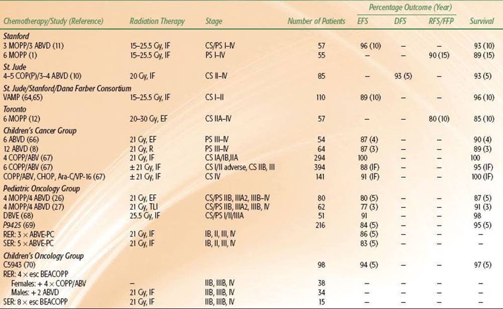
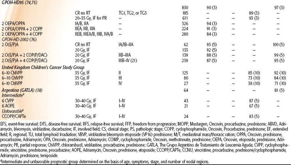
Although HL is one of the few pediatric malignancies that has an adult counterpart with similar natural history and biology, determination of the optimal therapeutic approach for children with this disease is complicated by their longevity and increased risk for adverse treatment-related side effects over time. In particular, radiation therapy doses and fields used in adults can produce significant musculoskeletal growth retardation and cardiovascular and pulmonary effects, as described later in this chapter.1,2,63 Consequently, the various successful approaches to treatment of HL in children (Tables 89.1, 89.2, and 89.3) must be considered in terms of efficacy and morbidity, and this is influenced by the developmental status of the patient.
TABLE 89.1 TREATMENT RESULTS OF COMBINED-MODALITY TRIALS OF PEDIATRIC HODGKIN LYMPHOMA

Radiation Therapy
Radiation therapy (RT) to extended fields to a curative dose range of 30 to 40 Gy produced the first cures in patients with HL, and similar treatment paradigms were used for adults and children. Five-year DFS rates following extended-field radiation therapy in surgically staged children with early stage disease ranged from 60% to 80%.4,5,9,64,65 However, it became apparent that the late effects of RT posed significant risks in terms of morbidity and mortality in pediatric HL survivors.1,52,63,83–88,89,90–92
Combined-modality treatment programs evolved in an effort to reduce therapy-related toxicities; reduced radiation dose was combined with non–cross-resistant chemotherapy in pediatric patients. These regimens reduced the dose-related toxicity of both chemotherapeutic agents and RT and improved DFS in advanced stage patients.1,8,10,11,26,27,93 Advances in radiation technology and diagnostic imaging led to a reduction in RT field size, permitting involved-field radiotherapy (IFRT) treatment approaches that more effectively shield normal tissues. Although radiation alone has been largely abandoned as a treatment strategy in children, risk-adapted treatment strategies still integrate low-dose (15 to 25.5 Gy) IFRT as a critical component of combined-modality regimens and as salvage therapy for patients with refractory or relapsed disease.
TABLE 89.2 TREATMENT RESULTS OF CHEMOTHERAPY ALONE IN PEDIATRIC HODGKIN LYMPHOMA TRIALS
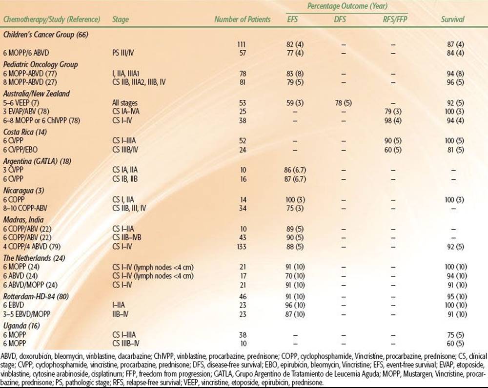
TABLE 89.3 TREATMENT RESULTS IN CHILDREN WITH HODGKIN LYMPHOMA WITH RADIATION THERAPY ALONE

Combination Chemotherapy, Combined Modality Therapy, and Risk-Adapted Therapy
The development of non–cross-resistant chemotherapy combinations provided the first effective therapy for advanced HL and formed the cornerstone for contemporary risk-adapted therapy. The specific chemotherapeutic combinations have changed as their morbidities have become better understood, but most treatment regimens include MOPP (mechlorethamine, vincristine, procarbazine, and prednisone), ABVD (doxorubicin, bleomycin, vinblastine, and dacarbazine), or therapies derived from one of the combinations (Table 89.2).
Despite the excellent disease control with ABVD,6,85,93,94 bleomycin and doxorubicin cause pulmonary and cardiovascular damage, respectively. To reduce the treatment-related toxicities associated with 6 cycles of either MOPP or ABVD, strategies alternating the two regimens, and using fewer cycles of each, have been developed. The combined ABVD and MOPP regimens used for children have produced excellent disease control with apparent diminished toxicity.10,11,27
Contemporary therapy for pediatric HL using chemotherapy alone or combined-modality therapy produces long-term DFS in 85% to 100% of patients with localized disease and 70% to 90% of patients with advanced disease.3,6,7–8,10,11,13,14,15,16, 17,18,20,22–24,25–27,67,71,73,74,77,78,83,94,95 Single-modality chemotherapy treatment usually involves more cycles of chemotherapy, whereas combined-modality therapies prescribe low-dose, involved-field radiation in lieu of several chemotherapy cycles. Although early treatment results appear comparable with results of more mature trials of combined-modality therapy, the long-term efficacy and treatment toxicities associated with chemotherapy alone have not been reported. Earlier randomized pediatric trials prospectively comparing outcomes in patients treated with chemotherapy alone with those treated with combined-modality therapy have not definitively established the superiority of one treatment approach over the other.26,93 However, other trials suggest a better outcome in patients with intermediate or advanced disease treated with combined-modality therapy.26,74 Children’s Cancer Group (CCG) investigators closed enrollment into a randomized controlled trial comparing outcomes in patients treated with contemporary risk-adapted combined-modality therapy with COPP/ABV (cyclophosphamide, vincristine, prednisone, procarbazine/doxorubicin, bleomycin, vinblastine) hybrid chemotherapy and low-dose, involved-field radiation to those treated with COPP/ABV chemotherapy alone based on a significantly higher 3-year event-free survival (EFS) in patients randomized to also receive radiotherapy.67 However, early follow-up did not demonstrate a significant difference in OS among the groups due to the successful retrieval of relapsed patients following salvage therapy. Similarly, the German HL-DAL-90 protocol treated intermediate- and high-risk patients with 2 cycles of OPPA (vincristine, prednisone, procarbazine, doxorubicin) or OEPA (vincristine, etoposide, prednisone, doxorubicin) (females and males, respectively), followed by 2 to 4 cycles of COPP. All patients received 20 to 35 Gy IFRT. Five-year EFS in the intermediate- and high-risk groups was 93% and 86%, respectively, which was comparable to that seen in the low-risk group. OS (5-year) for all three risk groups was ≥94%.17,20,74,75 In the subsequent HL-95 trial, which employed the same chemotherapy but omitted IFRT in complete responders, the EFS for intermediate- and high-risk patients combined who did not receive radiotherapy dropped to 79% compared with 91% for those who did get radiotherapy.74
Currently, consensus regarding the optimal therapy for children and adolescents with HL has not been established. Treatment with chemotherapy alone is preferred by many investigators desiring to avoid long-term sequelae of radiation, including musculoskeletal growth impairment, cardiovascular dysfunction, and solid tumor carcinogenesis. However, single modality chemotherapy protocols typically use higher cumulative doses of chemotherapeutic agents with dose-related toxicity, especially alkylating agents. Moreover, low-dose involved-field radiation therapy has substantially reduced many of the undesirable radiation-associated sequelae. Therefore, current investigations aim to selectively combine chemotherapy and radiation in an effort to improve the therapeutic ratio.
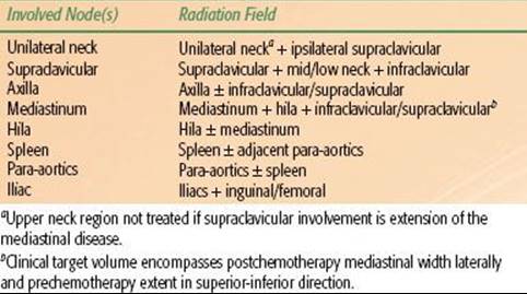
Ongoing risk-adapted trials aim to identify patients who require radiation to optimize DFS and patients who can be cured by limited chemotherapy alone (Table 89.4).
TABLE 89.4 INVOLVED FIELD RADIATION GUIDELINES
Favorable Risk
In the 1990s, pediatric HL investigators evaluated risk-adapted therapies using fewer cycles of combination chemotherapy and lower radiation doses and treatment volumes in clinically staged patients with favorable disease presentations. Favorable disease presentations were characterized by localized nodal involvement in the absence of B symptoms and bulky mediastinal lymphadenopathy. The number of involved nodal regions, the presence of extranodal extension, hilar lymphadenopathy, and peripheral nodal bulk comprised the other risk factors considered in some of these studies. Most treatment regimens prescribed 2 to 4 cycles of novel chemotherapy combinations that limited or omitted alkylating agent and anthracycline chemotherapy and bleomycin. The results of these trials demonstrated that excellent treatment outcomes could be maintained in patients with favorable presentations of localized HL using minimal therapy.13,17,19,20,25,65,67,68,71,74,96 Ongoing trials are testing the feasibility of omitting chemotherapy in patients with excellent response to chemotherapy.
Intermediate and Unfavorable Risks
In most clinical trials, intermediate risk consists of stage I, II, and IIIA patients with unfavorable disease features such as more than three nodal sites, presence of bulky mediastinal lymphadenopathy (mediastinal ratio ≥33%, peripheral nodal mass ≥6 to 10 cm), or extranodal extension. High-risk HL patients are those with stage IIIB and any with stage IV. Patients with stage IIB disease are sometimes considered intermediate and sometimes high risk according to the study group. Two treatment approaches are used for intermediate and high-risk patients, both of which include chemotherapy regimens derived from the original MOPP and ABVD combinations. MOPP has largely been replaced by COPP because cyclophosphamide is less myelosuppressive and leukemogenic than mechlorethamine.97
In the conventional treatment approach, combination chemotherapy is administered on a twice monthly schedule for 6 to 8 months.8,10,11,17,19,20,26,27,67,72,74,93 More recent trials are evaluating the second treatment approach, which has been used successfully in adults with HL. These protocols prescribe dose-intensive, multiagent chemotherapy in an abbreviated schedule over a period of 3 to 5 months. Prototypes of the dose-intensive therapy include the MOPP/ABV hybrid, Stanford V, or BEACOPP (bleomycin, etoposide, doxorubicin, cyclophosphamide, vincristine, procarbazine, prednisone) regimens.67,70,98,99,100 These treatments alternate myelosuppressive and nonmyelosuppressive chemotherapy combinations in a weekly schedule with the aim of reducing the development of resistant disease. Growth factor support is required to facilitate bone marrow recovery and maintain the compacted treatment schedule. Low-dose, involved-field radiation therapy generally is used to consolidate remission after completion of chemotherapy. In ongoing pediatric trials, the response to chemotherapy influences the ultimate amount of therapy. For example, in the Stanford/St. Jude/Dana Farber protocols, rapid early responders to chemotherapy, defined by complete response after 2 cycles of chemotherapy, are being targeted for reduced treatment (less or no radiation therapy, depending on the risk group). In the last COG trial for high-risk patients, rapidity of response was evaluated after 4 cycles of BEACOPP. Rapid responders received consolidation therapy according to gender in order to avoid or minimize sex-specific long-term toxicities of therapy. Females received 4 cycles of COPP/ABV without IFRT to avoid secondary breast cancers, while males received 2 cycles of ABVD with IFRT. Slow responders received 4 more cycles of BEACOPP followed by IFRT. OS was 97%, with significant acute hematologic toxicity and two patients developing secondary leukemia.70 The current European trial, based on the long-standing German experience, is also testing a gender-based approach in which a procarbazine-free OEPA/COPDAC (cyclophosphamide, vincristine, prednisone, dacarbazine) is given to boys and OEPA/COPP to girls, in order to reduce the alkylator exposure in boys and maintain fertility.76
The last COG intermediate risk HL study treated patients with stage IA or IIA bulky, IB, IIB, IIIA, or IVA disease with 2 cycles of ABVE-PC (doxorubicin, bleomycin, vincristine, etoposide, prednisone, cyclophosphamide) prior to response evaluation. Those with a rapid early response to therapy (>60% reduction in the tumor dimension) and those who achieve complete recovery after an additional 2 cycles of the same chemotherapy were randomized to 21 Gy IFRT or no additional treatment. Patients with slow early response after 2 cycles of chemotherapy were randomized to either standard therapy (an additional 2 cycles of ABVE-PC + 21 Gy IFRT) or intensified therapy (2 cycles of ABVE-PC, 2 cycles of DECA [dexamethasone, etoposide, cisplatin, cytarabine] + 21 Gy IFRT). Early results show an improved outcome for rapid early responders of 87% compared with 78% for slow early responders (P = .0001), while radiotherapy did not improve outcome in the rapid early responders (P = .07).101
Sequence of Therapy
The most effective sequence of therapy in the setting of combined chemotherapy and irradiation is not unequivocally established. However, chemotherapy is usually the first modality. This allows assessment of drug response, maximization of the amount of drug treatment, and shrinkage of disease and more limited fields of irradiation. Rarely, focal irradiation prior to chemotherapy is necessary due to significant symptomatic compromise such as airway obstruction.
Refractory or Relapsed Disease
Treatment failures in pediatric HL patients typically develop within the first 3 years, although late relapses have been reported, particularly in patients with lymphocyte-predominant HL. The most common site of relapse following risk-adapted therapies remains the primary site of disease.102 Because of the excellent outcome for the majority of children and adolescents with HL, investigations regarding salvage strategy have been limited. Prognosis following relapse is dependent on the primary therapy given and the timing of relapse. Standard multiagent chemotherapy and radiation therapy may salvage 40% to 50% of patients who relapse 1 or more years after primary therapy, but treatment complications such as second malignancies may reduce long-term survival. More aggressive salvage chemotherapy without stem cell transplant has been shown to provide satisfactory outcome for patients with late relapses.103 Patients with primary refractory disease (i.e., those unresponsive to initial therapy or who relapse within 1 year of primary therapy) have a very poor prognosis. Intensive cytoreductive chemotherapy followed by myeloablation and autologous hematopoietic stem cell transplantation is reported to salvage 30% to 60% of these patients, but relapses after 5 years have been observed, and long-term treatment complications may predispose to early mortality.104,105–107
Historically, because of higher treatment-related mortality, allogeneic transplantation did not provide a survival advantage compared with autologous stem cell transplantation. However, allogeneic bone marrow transplantation has been shown to be a useful treatment in healthy patients who have failed or cannot have an autologous transplant.108
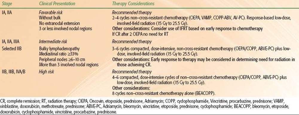
TABLE 89.5 GUIDELINES FOR TREATMENT SELECTION IN PEDIATRIC HODGKIN LYMPHOMA
Techniques of Radiation Therapy
As discussed above, most children with HL will be treated with combined chemotherapy and low-dose IFRT using a risk-adapted approach. The use of radiation therapy alone has been largely abandoned due to concerns for late effects, including cardiac complications and secondary malignancies. The Patterns of Care studies demonstrated the relation between radiation technique and outcome.57 The results of patients treated with RT alone were improved in institutions that treated a large number of HL patients. The HD-DAL 90 trial (German-Austrian pediatric multicenter trial) showed that upfront centralized review of patients entered into the study altered the treatment approach for a large number of children.109 Because technique is discussed in Chapter 77, only select points applicable to children will be discussed here.
Using risk-adapted strategies, most children with HL are treated with combined chemotherapy and low-dose IFRT. The definition of involved fields depends on the anatomy of the region in terms of lymph node distribution and patterns of disease extension into regional areas. Involved fields typically should include not just the identifiably abnormal lymph nodes but also the entire lymph node region containing the involved nodes (Table 89.5). Historically, consideration has been given to radiating the treatment area bilaterally (e.g., both sides of the neck) in young children in an effort to avoid growth asymmetry. However, current combined modality treatment strategies use low radiation doses of 15 to 25 Gy and, therefore, unilateral radiation fields are appropriate if the disease is confined to one side of the neck. However, field definitions often are protocol specific.
Strong consideration must be given to excluding normal tissues in an effort to reduce the risk of acute and late effects, including secondary malignancies and cardiac complications. Every effort should be made to exclude breast tissue, and potentially the thyroid gland, in patients with isolated mediastinal disease with no evidence of axillary or neck involvement. Clearly, careful planning and judgment are necessary. With the use of CT-based treatment planning, normal structures can be identified and doses minimized.89,110,111–115 The treatment of involved supradiaphragmatic fields or a mantle field can pose a clinical challenge to treat precisely the lymph node regions that are involved while still protecting critical adjacent normal tissues. These fields can be simulated with the arms up over the head or down with hands on the hips. The former pulls the axillary lymph nodes away from the lungs, allowing greater lung shielding. However, the axillary lymph nodes then move into the vicinity of the humeral heads, which should be blocked in growing children. Thus, the position chosen involves weighing concerns regarding lymph nodes, lung, and humeral heads. Attempts should be made to exclude or position breast tissue under the lung–axillary blocking.
In risk-adapted strategies using combined modality therapy, radiation dose is dependent on the choice of systemic therapy that is variously defined and often protocol specific. In general, doses of 15 to 25 Gy are used, with modifications based on patient age, treatment response, the presence of bulk or residual (postchemotherapy) disease, and normal tissue concerns. In some situations, a boost of 5 Gy is appropriate. Although IFRT remains the standard when patients are treated with combined-modality therapy, response-adapted RT, which is limited to areas of initial bulk disease (generally defined as ≥5 or 6 cm at the time of disease presentation) or postchemotherapy residual disease (generally defined as ≥2 cm, or residual PET avidity), is under investigation.
An approach studied in Europe by the European Organisation for Research and Treatment Center–Groupe d’Etudes des Lymphomes de l’Adulte is the use of involved node RT (INRT), which restricts RT to the initially involved lymph node.116 This is based on the premise that chemotherapy can effectively eliminate microscopic disease that may exist within adjacent but clinically uninvolved nodes. This concept is supported by the observation that among patients treated with chemotherapy alone, initially involved lymph nodes are the most common site of recurrence.117 INRT uses all available clinical information including pre- and postchemotherapy imaging with CT and FDG-PET scan to define the treatment field according to the prechemotherapy extent of disease.118,119 However, uninvolved lymph node regions are not necessarily included within the clinical target volume (CTV). For example, in contrast to conventional IFRT, uninvolved hila are not included in the CTV for mediastinal presentations, and the length of the treated volume is not routinely extended beyond 1 cm for the planning target volume (Fig. 89.1). However, the margins for INRT are defined differently depending on the protocol and cooperative group.112,120,121 Regardless, successive reduction in treatment volume may reduce the toxicity of therapy.
FIGURE 89.1. A: Digitally reconstructed radiographs demonstrating typical radiation therapy (RT) fields for historic mantle RT, contemporary involved-field RT (IFRT), and involved node RT (INRT) for a female patient with stage I disease involving the upper mediastinum. The postchemotherapy volume of initially involved mediastinal nodes is shown in green. Hila are shown in blue and violet. B: Reduction in dose to breast and lung for the same patient. For each volume the prescribed dose to the CTV is the same. The resulting mean dose to breast and lung tissue with mantle RT is a set value equal to 1. The proportional reduction in normal tissue dose occurs as a result of the reduction in treated volume with IFRT and INRT. (From Hodgson DC, Hudson MM, Constine LS. Pediatric Hodgkin lymphoma: maximizing efficacy and minimizing toxicity. Semin Radiat Oncol 2007;17:230–242; copyright 2007, with permission from Elsevier.)

Steep radiation dose gradients, using modern radiation techniques such as three-dimensional conformal radiation (3D-CRT) and intensity-modulated radiotherapy (IMRT), are achieved to spare normal structures, particularly for patients who are at risk for treatment complications. For example, 3D-CRT of an anterior mediastinal mass could avoid radiation to normal structures, such as the spine, breast, heart, or lung tissue located behind the mass. IMRT utilizes multiple beams to create concavities and avoid dose to surrounding tissues.110,111–114,122 Although conformality is improved, the radiation dose is spread over a larger volume of tissue, and additional monitoring units are required to deliver treatment compared to conventional methods. As a result, integral dose is increased, which has limited the use of IMRT in the pediatrics population, particularly because low doses are required for treatment of HL. IMRT remains controversial in the treatment of children as a result of the concerns that increased integral dose, and increased radiation scatter within the smaller body habitus of a child could lead to an increase in the development of secondary malignancies.123,124 However, IMRT may still provide dosimetric advantages in particular cases, such as patients who have received prior radiation or in patients with bulky mediastinal disease. Proton therapy for pediatric HL is also an area under current active investigation. In particular, the steep dose gradient offered by protons may improve the dose distribution in specific clinical scenarios, such as in patients with superior mediastinal masses in whom the dose to breast and lung may be reduced.125,126
Ultimately, each clinical scenario must be considered independently to determine the optimum RT technique, taking into account the characteristics of the tumor, patient’s age, normal structures at risk, and comorbidities.
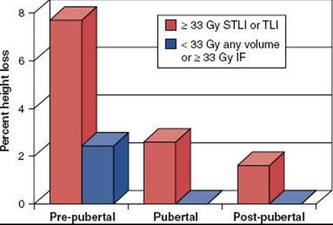
FIGURE 89.2. Relative height impairment for different age and treatment groups in pediatric Hodgkin lymphoma. (From Willman K, Cox K, Donaldson S. Radiation induced height impairment in pediatric Hodgkin’s disease. Int J Radiat Oncol Biol Phys 1994;28:85–92; copyright 1997, with permission from Elsevier.)
Complications of Radiation Therapy
Acute Effects
The acute side effects of radiation therapy are related to the involved site treated. Due to the lower doses used in pediatrics, the most common toxicities include temporary loss or change in taste, xerostomia, esophagitis, low posterior scalp epilation, skin erythema, and occasionally dyspepsia, nausea, and vomiting. Acute effects of para-aortic irradiation include early onset nausea and vomiting, which usually abates after the second or third treatment without antiemetic therapy. Patients who receive larger fields may be subject to bone marrow suppression, the severity of which may be influenced by the systemic regimen preceding radiation.
Long-Term Effects
Musculoskeletal
Height reduction is a potential long-term consequence of irradiation, most frequently with doses >20 Gy and most severe in prepubertal children. Growth arrest or delay is modest with the low doses currently used in modern pediatric HL treatment strategies (Fig. 89.2).2,127,128 Interclavicular shortening and hypoplasia of the neck muscles may occur in children irradiated before puberty, which is particularly dependent on dose delivered; the severity of these complications is more pronounced in children <5 years of age at the time of irradiation. Slipped capital femoral epiphysis occurs in ≤50% of young children whose femoral heads have been irradiated. Avascular necrosis of the femoral or humeral heads is rare if appropriate shielding is provided. Radiation doses of 20 to 40 Gy to the mandible may result in dental abnormalities, such as stunted tooth development, incomplete calcification, premature apical closure or eruption, and root tapering with apical constriction if the tooth germ tissue is in the irradiated field.129
Cardiovascular
Radiation-associated pericardial and myocardial disease are related to dose (including fraction size) and volume and are complicated by the use of anthracyclines. Cardiac sequelae, including pericarditis and effusion, valvular thickening, biventricular dysfunction, and coronary artery disease, all are observed with irradiation to the heart.1,63,68,85,130 Increase in mortality risk, due to premature coronary artery disease and acute myocardial infarction, has been demonstrated in patients who received mediastinal radiation in doses >30 Gy before 20 years of age.83 Conversely, modern treatment approaches based on tailored low-dose radiation and less cardiotoxic chemotherapy, sometimes including cardioprotectants, will optimistically reduce such effects. Radiation doses of <30 Gy and techniques that use adequate cardiac shielding and avoid anterior weighting of the treatment fields appear to reduce the risk of cardiac complications, particularly pericarditis and myocarditis.63,85 Although the proximal coronary arteries are not shielded by a central cardiac block, the 45-fold excess mortality risk from acute myocardial infarction associated with higher radiation doses has diminished substantially with current approaches.131 Also, dose to the heart can now be calculated using advanced radiation techniques and dose can be constrained using conformal techniques in order to keep the risk of long-term development of cardiac injury to a minimum. Unfortunately, the impact of low-dose RT to the heart may not become apparent until larger cohorts of survivors treated with modern combined modality regimens have longer follow-up.
Pulmonary
With doses of 25 Gy,132 the incidence of pulmonary complications, including pneumonitis or fibrosis, is low. Use of bleomycin can increase the risk of acute pulmonary pneumonitis and late pulmonary fibrosis in patients receiving pulmonary RT. In a report from the CCG, 9% of children treated with ABVD and 21 Gy of mantle irradiation exhibited clinically significant pulmonary damage.8 These results were confirmed recently in a study evaluating pediatric patients with any chest radiation who developed radiation pneumonitis. The incidence was found to be low and most often related to therapies including bleomycin.133
Thyroid
Thyroid dysfunction may result from neck or upper mediastinal irradiation and most often is manifested by an elevated serum concentration of thyroid-stimulating hormone (TSH). The incidence of hypothyroidism varies, but it appears to be directly related to irradiation dose. Constine et al.134 noted an increased serum TSH in 4 of 24 (17%) children who received mantle irradiation of ≤26 Gy and in 74 of 95 (78%) who received >26 Gy. Approximately one-fourth of these children had a concomitant low thyroxine level. In this series, 36% of children experienced spontaneous improvement in thyroid function.
In a Childhood Cancer Survivor Study, thyroid abnormalities were self-reported by 34% of patients surveyed. Hypothyroidism was the most common thyroid disturbance, with a relative risk of 17.1 (P <.0001) compared with sibling controls. Risk factors for hypothyroidism include increased dose of radiation, older age at diagnosis of HL, female sex, and white race.135 The estimated actuarial risk of hypothyroidism for survivors treated with ≥45 Gy was 50% at 20 years from diagnosis. Other thyroid abnormalities identified in excess from sibling controls included hyperthyroidism (eightfold excess risk), thyroid nodules (27-fold excess risk), and thyroid cancer (18-fold excess risk).136
Pelvic Late Effects
Gonadal injuries, including infertility and impaired secretion of sex hormones, are potential complications of pelvic irradiation.137 In women, irradiation of the pelvis can affect the ovaries, resulting in premature menopause or fertility impairment.137 In a report of female HL survivors by the Late Effects Study Group, 42% of women treated with alkylating agent chemotherapy and subdiaphragmatic radiation had experienced menopause by age 31 years, compared with 5% of control subjects.138 In childhood cancer survivors who continued to have spontaneous menses more than 5 years after their cancer diagnosis, the cumulative incidence of nonsurgical menopause was 8% by age 40 years, representing a 13-fold higher risk compared with a sibling control group. Risk factors for premature menopause include attained age, exposure to increasing doses of ovarian radiation, increasing alkylating agent score (based on number of agents and cumulative dose), and diagnosis of HL.136 Normal pregnancies, without increased risk of fetal wastage, spontaneous abortion, or birth defects, have been reported after pelvic irradiation.137 In an effort to preserve ovarian function, it is recommended that the ovaries are transposed via oophoropexy to a shielded area laterally or inferomedially near the uterine cervix prior to the initiation of radiation.
In boys irradiated to the pelvis, oligospermia is common, but it is reversible (usually by 18 to 24 months) in most cases if the radiation dose scattered to the shielded testes is small.2,139,140 Permanent oligospermia may occur, however, after full-dose pelvic irradiation.140 Several investigations have demonstrated that fertility is compromised in boys treated with gonadotoxic combinations like COPP, even when cycles of alkylating agent chemotherapy are limited.141,142 Anthracycline-based regimens such as ABVD (or similar hybrid) are associated with recovery of spermatogenesis after a temporary period of azoospermia.143,144 In the modern treatment era, risk-adapted strategies seek to reduce or eliminate gonadotoxic treatments, resulting in excellent prospects to preserve fertility in boys.141
Second Malignant Neoplasm
The 15-year actuarial risk of second malignant neoplasms ranges from 8% to 15%.83,131,145–146,147,148–155 Patients are at risk for developing secondary malignancies including leukemias (e.g., acute myeloblastic leukemia) and solid tumors, most commonly breast, thyroid, bone, and soft tissues. The risk of leukemia, which exhibits a peak frequency in the first 5 to 10 years after treatment, is associated primarily with the use of alkylating agents.145,147 In contrast to secondary hematopoietic malignancies, the risk of developing a second solid tumor increases with time after therapy, with a latency usually exceeding 10 years from diagnosis.
Several studies reveal an increased ratio of observed to expected risk of second tumors for girls compared with boys, with median follow-up of 10 to 16 years.81,145,156 A portion of this risk results from breast cancer, which is the most common solid second malignant neoplasm following the treatment of children. The increased risk of breast cancer is a significant concern for women treated for HL at a younger age. This increase in risk of breast cancer is related to the patient’s age at the time of RT and has also been shown to decrease with a decrease in radiation dose.157,158 Travis et al.158 reported a large case-control study that included 105 women who developed breast cancer within a cohort of more than 3,800 female survivors diagnosed with HL at age ≤30. This cohort also included patients who had received either very low-dose RT (<4 Gy) or no radiation to the breast area where breast cancer developed. For all patients who received RT alone (>4 Gy), the relative risk of breast cancer was 3.2 and increased to 8.0 in the highest radiation dose group. This study demonstrated a dose–response relation between RT and the risk of breast cancer development.
Multiple studies have now demonstrated that lower RT dose and smaller treatment fields should translate into a reduction in late effects when used judiciously.89,91,159,160 Historical extended field treatments included a substantial amount of breast tissue in the field design, which were then exposed to full-dose RT. Interestingly, breast tissue exposure was most significant when the axillae was irradiated in a typical mantle field as opposed to the mediastinal and hilar region. The practice of prophylactically irradiating the axillae is no longer performed, and, thus, the volume of breast tissue exposed to RT using involved field or involved node radiotherapy approaches is substantially less.89Using CT-based planning, the breast tissue can also be contoured as an avoidance structure and thereby shielded appropriately when designing RT fields in the region, a practice that was not used 30 years ago.
Current recommendations for the screening of late effects of therapy after childhood cancer can be found in the long-term follow-up guidelines developed by the COG, which are constantly updated (http://www.survivorshipguidelines.org).
Future Investigations
Analyzing patterns of disease recurrence should enhance our understanding of directions for therapeutic intensification. Most patients will relapse in areas of initial disease involvement, even in this era of combined-modality therapy.161,162 Preventing these recurrences will require ingenuity if additional toxicity is to be avoided. Some generalizations regarding ongoing efforts in clinical trials for pediatric HL are as follows:
• Identification of prognostic biologic markers that correlate with tumor response for a more accurate risk stratification.
• Patients with early stage disease have an excellent prognosis, and, therefore, therapeutic aims focus on reducing the intensity of therapy.
• Patients with disease of an intermediate stage or prognosis are studied appropriately with questions that are intended to increase efficacy without increasing toxicity. Usually this entails modification of existing chemoradiation programs. The rapidity of response to therapy may offer a means to risk adapt subsequent treatment and is under investigation.
• Patients with advanced-stage disease require more effective treatment regimens. This may be attained by increasing the dose intensity or the rate of drug delivery; however, more attractive approaches under discussion are the incorporation of some of the new agents found to be effective in heavily pretreated patients (like brentuximab vedotin) into existing regimens. The role of radiation therapy in such trials will continue to be important.
• Identification of genetic host predisposition to treatment toxicity to better guide risk-adapted therapy.
Targeted therapy for HL focuses either on monoclonal antibodies directed against receptors and antigens expressed on the surface of HRS or on small molecules that target the well-defined signaling pathways that promote HRS survival and are triggered by these receptors.163 Brentuximab vedotin, an anti-CD30 antibody conjugated to a synthetic antimicrotubule agent, monomethyl auristatin E, recently approved by the U.S. Food and Drug Administration for the treatment of relapsed or refractory HL, has shown excellent response in refractory HL.164 Previous trials with naked anti-CD30 antibodies were disappointing.165,166 Histone deacetylase (HDAC) inhibitors are small molecules that affect gene transcription by inhibiting posttranscriptional histone modification. Although there are a variety of enzymes that play a role in this process, the most studied drugs are vorinostat167 and panobinostat,168 both inhibit class I and II (pan-HDAC inhibitors). Everolimus, an inhibitor of mammalian target of rapamycin, a frequently activated pathway in HRS, has produced encouraging clinical responses in relapsed HL.169 Other compounds under investigation directly or indirectly inhibit NF-κB, a nuclear transcription factor that regulates the expression of a variety of genes that play a crucial role in viral replication, tumorigenesis, apoptosis, various autoimmune diseases, and inflammation.170
Identifying the most promising agents and moving them into frontline therapy in order to spare further toxicity escalation while improving outcome will be the goal of upcoming trials. Adoptive immunotherapy with EBV-specific cytotoxic T-lymphocytes and other tumor-specific T cells are other attractive strategies under investigation for the therapy of relapsed or refractory HL.171–173
NON-HODGKIN LYMPHOMA
Childhood NHLs are a heterogeneous group of malignancies with variable histopathology, site of origin, and clinical manifestations. Childhood NHLs are diffuse, high grade, and poorly differentiated; extranodal involvement is common, and dissemination occurs early and often.174 This is in striking contrast to adult NHL, in which low- and intermediate-grade nodal disease predominate.174,175 These differences in pathology and clinical behavior observed between children and adults explain the markedly different presentations, staging practices, and treatment strategies for these age groups.
Epidemiology
In the United States, NHL is diagnosed in approximately 800 children and adolescents younger than 20 years of age, each year. According to the Surveillance, Epidemiology, and End Results program of the National Cancer Institute, NHL accounts for about 8% of all cases of childhood cancer.28 The incidence increases with age, being rare in the child <3 years old; incidence is higher in males than in females (ratio of 3 to 1), and higher in whites than in African Americans (ratio of 1.5 to 1). In younger children, NHL is more frequent than HL, whereas the reverse is true for adolescents.
Although NHL is related to several genetic and environmental factors, its cause and pathogenesis remain unclear.174,176 A small proportion of cases are seen in association with inherited immunodeficiencies (e.g., Wiskott-Aldrich syndrome, X-linked lymphoproliferative disease, ataxia-telangiectasia),177,178 or acquired immunodeficiencies (e.g., HIV infection or immunosuppressive therapy in patients receiving solid organ or bone marrow transplants).179,180Evidence of EBV infection has been demonstrated in the majority of endemic (mainly in equatorial Africa) Burkitt tumors and in about 15% of sporadic cases (outside of Africa), suggesting a significant role for this virus in lymphomagenesis through unknown mechanisms.181,182–183 EBV infection has also been associated with the development of posttransplant lymphoproliferative disease (PTLD), a B-cell lymphoproliferative disorder seen in patients after solid organ or hematopoietic stem cell transplantation.184,185
Pathologic Classification
The classification of NHL by the WHO was updated in 2008 (Table 89.6). Pediatric NHL cases are divided into four major histopathologic subtypes based on morphology and immunophenotype in the order of frequency: (a) Burkitt lymphoma (BL), (b) lymphoblastic lymphoma (LL), (c) diffuse large B-cell lymphoma (DLBCL), and (d) anaplastic large cell lymphoma (ALCL).28,37,176 Identification of large numbers of monoclonal antibodies directed against surface antigens has allowed subclassification of the NHLs according to immunophenotype (see Chapter 78).186 Thorough and efficient evaluation of histology and immunophenotype is a key to the determination of diagnosis, prognosis, and appropriate course of treatment because pediatric NHLs are usually aggressive. Cytogenetic analysis and identification of molecular markers provide a more precise means of characterization of these tumors and will likely contribute to the understanding of pathogenesis as well as the development of novel therapeutic approaches.187–189
TABLE 89.6 BIOLOGIC CHARACTERISTICS OF THE FOUR MAJOR SUBTYPES OF CHILDHOOD NON-HODGKIN LYMPHOMA AS DEFINED BY HISTOLOGY AND IMMUNOPHENOTYPE

TABLE 89.7 CLINICAL CHARACTERISTICS OF CHILDHOOD NON-HODGKIN LYMPHOMA

Clinical Presentation
The signs and symptoms in children with NHL correlate with the histologic subtype, disease site(s), and extent of involvement (Table 89.7). Clinical presentation is different between endemic and sporadic BL. Abdomen is the most common site of disease in sporadic BL, which presents with abdominal pain, distension, mass, nausea, vomiting, and gastrointestinal bleeding, followed by head and neck region involvement. Bone marrow involvement is seen in about 20% of sporadic disease and can present as BL.190 However, in endemic BL, jaw involvement is the most common, and bone marrow involvement is rare.191
The majority (50% to 70%) of children with precursor T-cell LL present with rapidly enlarging neck and mediastinal lymphadenopathy.176,188 Mediastinal masses can cause respiratory symptoms (e.g., cough, wheezing, shortness of breath, and orthopnea) by compressing airways or cause neck, face, and upper extremity swelling due to obstruction of the superior vena cava (SVC). Hemodynamic compromise due to pericardial effusions may also occur. However, children and adolescents with precursor B-cell LL tend to have limited disease in sites including skin, bone, and peripheral lymph nodes.192,193
DLBCL usually presents with nodal disease, especially in the abdomen, although bone (single or multiple sites) is also a relatively common site.194,195 Primary mediastinal large B-cell lymphoma can be locally invasive and may present with SVC syndrome.196
The most frequent sites of involvement in systemic ALCL are peripheral nodes, mediastinal lymph nodes, and extranodal sites such as skin, soft tissue, and bone.197,198 Spontaneous regression or waxing and waning of disease has been observed.
Diagnostic Evaluation
Tissue diagnosis and investigation to determine the clinical extent of disease should be completed expeditiously. It is important that appropriate therapy be initiated promptly because of the extremely rapid growth rate of pediatric NHL.174 Although histology and immunophenotype remain the primary means of establishing the definitive diagnosis, karyotype and molecular studies will be vital for implementation of an optimal treatment plan. Thus, it is critical that an adequate amount of tissue be obtained at biopsy by performing either open biopsy or core-needle biopsy. If the patient’s condition does not permit an open biopsy (e.g., a large mediastinal mass is causing airway obstruction or compression of SVC), then use of a less invasive means of sampling tumor cells with as minimal anesthesia as possible, such as percutaneous fine-needle aspiration or examination of pleural fluid, ascites, peripheral blood, or bone marrow (Table 89.8), is warranted.199,200 The use of irradiation or steroids before biopsy for respiratory distress may result in rapid shrinkage of the mediastinal mass but may jeopardize the ability to establish a tissue diagnosis.201,202
Bilateral bone marrow biopsies are superior to a single aspirate or biopsy for identifying bone marrow involvement, which is often patchy in distribution in NHL.176 Omitting this procedure may result in understaging. FDG-PET scan combined with CT is now widely available and preferred to gallium scans as part of the assessment of the extent of disease at diagnosis, speed of response to therapy, and posttherapy remission status.203,204 Data in adults with HL and NHL and children with HL suggest that PET scanning to assess the rapidity of response may be prognostic.205,206 However, further confirmation is needed to determine whether PET scanning has predictive value for detecting recurrence of pediatric NHL.
Laparotomy is not performed routinely for staging purposes because combination chemotherapy is part of the primary treatment. Still, laparotomy may be necessary in selected cases for diagnostic purposes, but aggressive debulking procedures are not advised. MRI may be helpful in detecting disease at specific sites, particularly the nervous system, but it is not routinely used.
TABLE 89.8 INVESTIGATIONS REQUIRED FOR ACCURATE STAGING AND DIAGNOSIS OF CHILDHOOD LYMPHOMAS
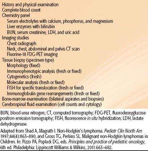
TABLE 89.9 STAGING AND GROUPING SYSTEM FOR CHILDHOOD NON-HODGKIN LYMPHOMA

Staging Systems
The Ann Arbor staging system (commonly used in adult NHL) has proved to be of limited use in pediatric NHL because of the unique clinical presentation and course of the disease in children. These special features of childhood NHL include the preponderance of extranodal presentations, the noncontiguous pattern of disease spread, and a tendency to evolve into leukemia and to involve the central nervous system (CNS). To address the uniqueness of childhood NHL, staging systems specific for pediatric NHL have been developed. The St. Jude Children’s Research Hospital staging system is used most widely (Table 89.9).175
The distinction between lymphoma and leukemia lies in the percentage of malignant cells in a bone marrow aspirate. Patients with more than 25% blasts in bone marrow are considered to have acute leukemia, and those with between 5% and 25% marrow involvement are considered to have stage IV NHL. However, any identifiable tumor cell in the cerebrospinal fluid constitutes CNS disease and therefore stage IV disease.
Of interest, several of the clinical cooperative groups have adopted the designation of limited disease (including stages I and II) versus advanced disease (stages III and IV) for the purpose of determining appropriate treatment strategies. European investigators have developed a clinical grouping system of the B-cell lymphomas (i.e., BL and DLBCL) that is based on extent of disease, modified St. Jude staging criteria, and relative risk of relapse.207Treatment intensity is based on the designation of group A, B, or C disease, which correlates to low-, intermediate-, or high-risk disease, respectively (Table 89.9). This strategy has also proven very effective in recent treatment trials.208–210
Prognostic Factors
The most important factor in determining prognosis in childhood NHL is stage, which takes into account other known prognostic variables such as tumor burden, site, and extent of involvement211 as well as response to therapy.210The fact that most cases of pediatric NHL are of the diffuse, high-grade, and aggressive subtypes may have obscured the prognostic value of histology; few reports support its role as a prognostic factor.212
More sensitive techniques such as flow cytometry to detect lymphoma-specific immunophenotype and RT polymerase chain reaction to evaluate chromosomal translocations or immunoglobulin rearrangements have shown that bone marrow and blood involvement are much more frequent than would be predicted by morphologic examination.213,214,215–216 Higher detected amounts of minimal disseminated disease at diagnosis are associated with worse prognosis in all the major subtypes of pediatric NHL.
With genetic analyses of mature B-cell NHL in children and adolescents, it was shown that deletions of 13q and gains of 7q in addition to c-myc translocations have a significant, negative effect on prognosis.217,218
In pediatric CD30-positive ALCL, there almost always is an anaplastic lymphoma kinase (ALK) rearrangement with translocation t(2;5)(p23;q35), which is associated with a favorable prognosis. This is in contrast with adult cases of ALCL, in which ALK rearrangements are less common and are associated with poor prognosis. In addition, ALK autoantibodies were detected frequently in pediatric patients. High antibody titers correlated with significantly lower amounts of circulating tumor cells and cumulative incidence of relapses, suggesting that a robust pre-existing immune response to an oncoantigen from an oncogenic chromosomal translocation inhibits lymphoma dissemination and relapse.219
Treatment and Results
The overall survival of children with NHL was poor before the advent of multiagent chemotherapy in the mid-1970s.211 Even in cases in which the disease apparently was localized, cure rates as low as 20% were reported. Failure, in most cases, was caused by early systemic dissemination of disease. In modern regimens, chemotherapy is the primary therapeutic modality for all histologies and stages of childhood NHL. The dramatic improvement in the overall rate of survival of those with childhood NHL can be attributed not only to the development of highly effective, multiagent chemotherapy regimens but also to the systematic evaluation of these diseases and their treatment by pediatric cooperative group clinical trials. Cure rates of 85% to 95% in patients with limited disease and 70% to 90% in those with extensive involvement have been reported (Tables 89.10 and 89.11).
Surgery is indicated only for diagnostic purposes or in the case of an abdominal emergency, such as intussusception or acute appendicitis due to abdominal mass effect at presentation. Patients with extensive NHL may present with a number of life-threatening complications that require urgent intervention, including respiratory distress from airway obstruction, SVC syndrome, spinal cord compression, intestinal obstruction, hydronephrosis, renal insufficiency, or tumor lysis syndrome. The priority in these situations is to maximize supportive care and initiate specific chemotherapy as soon as a diagnosis has been established.
TABLE 89.10 TREATMENT OUTCOMES FOR LIMITED STAGE NON-HODGKIN LYMPHOMA

Systemic Chemotherapy
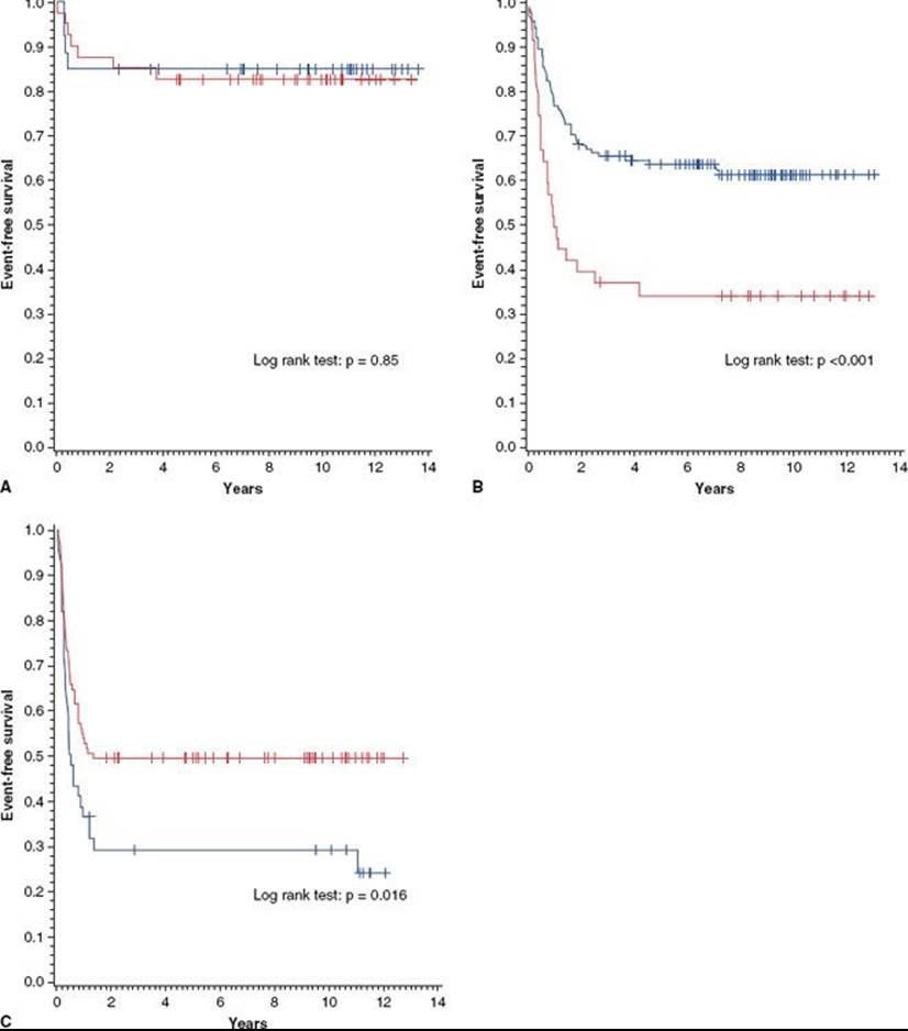
The importance of tailoring therapy to address the inherent biologic differences of the various histologic subtypes became evident early in the evolution of NHL therapy (Tables 89.10 and 89.11). The CCG performed an important randomized clinical trial to compare the cyclophosphamide-based four-drug regimen COMP (cyclophosphamide, vincristine, methotrexate [MTX], and prednisone) and the 10-drug LSA2L2 regimen (cyclophosphamide, vincristine, prednisone, daunomycin, MTX, cytarabine, thioguanine, asparaginase, carmustine [BCNU], and hydroxyurea).223,244,245 COMP was a lymphoma-targeted regimen and LSA2L2 was designed for acute lymphoblastic leukemia (ALL) therapy. For localized NHL, no difference was seen between the two treatment regimens, regardless of histology (Fig. 89.3A). However, outcomes of patients with stage 3 or 4 disease were significantly related to treatment arm and histology. The LSA2L2 regimen (ALL approach) was superior to COMP for lymphoblastic lymphoma (Fig. 89.3B; 5-year EFS 64% vs. 35%), whereas the COMP regimen was superior to LSA2L2 for BL (Fig. 89.3C; 5-year EFS 50% vs. 29%).
Based on these results, further refinements were performed for BL and DLBCL. The St. Jude Total B regimen incorporated sequential high-dose (HD) MTX and escalating continuous infusion of cytarabine.190 This regimen resulted in an excellent outcome for patients with stage III disease; however, those with stage IV disease had only a 20% long-term EFS rate. The Pediatric Oncology Group (POG), POG-8617 protocol, derived from the Total B regimen, which incorporated early administration of high-dose pulse cytarabine, resulted in improved outcomes for patients with stage IV disease.246 During this same time period, a study from the National Cancer Institute (NCI-77–04) showed an equally improved result for patients with advanced stage disease using a cyclophosphamide, doxorubicin, vincristine, and prednisone (CHOP) and HD-MTX regimen.247 Further improvement in treatment outcome has been reported by Patte et al.248 and Reiter et al.249 These regimens incorporated the above mentioned agents (COMP, HD-MTX, and high-dose cytarabine) and etoposide (VP-16) and/or ifosfamide. Patte et al.210 have reported excellent results for children with mature B-cell lymphomas using the Lymphoma Malignancy B (LMB) LMB-89 regimen, with approximately 87% of patients with stage IV disease or B-ALL experiencing long-term EFS. For patients with CNS disease, a group with a poorer prognosis, they reported a 79% long-term EFS rate. Burkitt lymphoma/leukemia is a rapidly growing tumor and is often associated with tumor lysis syndrome (including hyperuricemia). Careful attention to the control of metabolic abnormalities during induction greatly reduced early mortality associated with initial treatment. The international LMB-96 study (CCG, Societe Francaise d’Oncologie Pediatrique [SFOP], and United Kingdom Children’s Cancer Study Group [UKCCG]) randomized patients with group B and C disease to receive standard LMB doses versus stepwise-reduced doses. Efficacy data show that the 4-year EFS of group B patients was >90% in all arms of the study.210 Thus, in Group B patients, nonescalation of cyclophosphamide in the second course of COPADM3 (cyclophosphamide, Oncovin [vincristine], prednisone, Adriamycin [doxorubicin], HD-MTX 3 g/m2) and deletion of the maintenance sequence did not compromise the excellent outcome. In contrast, dose reduction for group C patients was associated with a poorer outcome: 4-year EFS was 90% for the full-dose arm versus only 80% for the dose-reduced arm.208 Finally, group C patients with CNS involvement had an inferior outcome, with 3-year EFS rates of 71% (vs. 87% for group C patients without CNS involvement). Patients with mediastinal large B-cell lymphoma were also associated with a poorer treatment outcome. In the LMB-96 study, the 4-year EFS for these patients was 72%, a finding consistent with those of other studies. It appears, therefore, that these patients require either novel or more intensive therapy.210
TABLE 89.11 TREATMENT OUTCOMES FOR ADVANCED STAGE NON-HODGKIN LYMPHOMA BASED ON HISTOLOGIC SUBTYPE
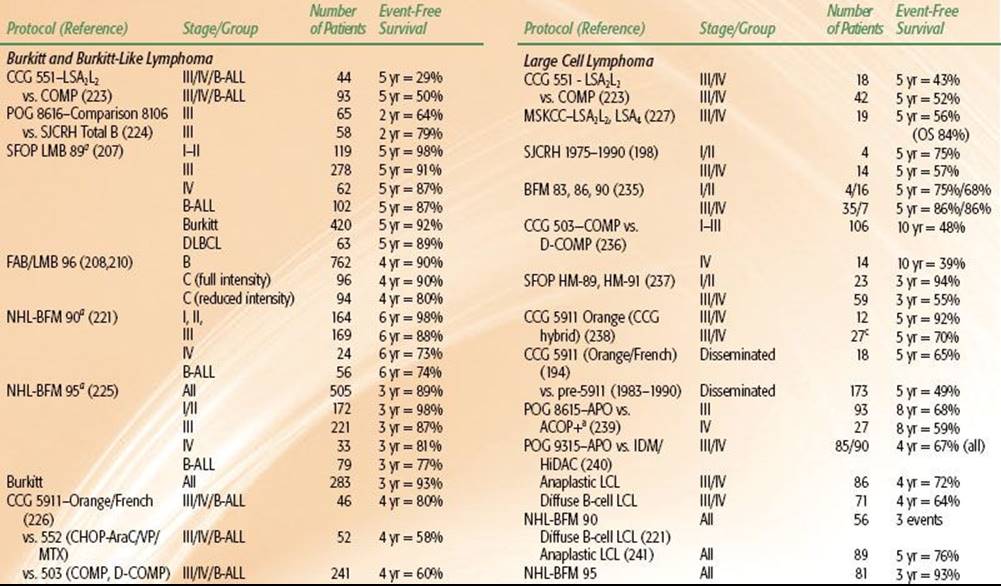

FIGURE 89.3. A: Event-free survival of 68 non-Hodgkin lymphoma patients with localized disease by treatment group. Solid line, COMP (41 patients); dotted line, LSA2L2 (27 patients). B: Event-free survival of patients with disseminated lymphoblastic lymphoma by treatment group. Solid line, COMP (40 patients); dotted line, LSA2L2 (124 patients). C: Event-free survival of patients with disseminated undifferentiated lymphoma by treatment group. Solid line, COMP (93 patients); dotted line, LSA2L2 (44 patients). COMP, cyclophosphamide, vincristine, methotrexate, and prednisone; LSA, cyclophosphamide, vincristine, prednisone, daunomycin, methotrexate, cytarabine, thioguanine, asparaginase, carmustine [BCNU], and hydroxyurea. (From Anderson JR, Jenkin RDT, Wilson JF, et al. Long-term follow-up of patients treated with COMP or LSA2L2 therapy for childhood non-Hodgkin’s lymphoma: a report of CCG-551 from the Children’s Cancer Group. J Clin Oncol 1993;11:1024–1032; reprinted with permission. © 1993, American Society of Clinical Oncology.)
The majority of successful regimens to treat advanced-stage LL are derived from regimens for ALL. They are multiagent regimens comprising induction, consolidation, and maintenance phases delivered over 18 to 30 months. The early St. Jude study, Total X-High Risk, demonstrated the survival efficacy of the addition of cytarabine and teniposide to an otherwise standard antimetabolite-based ALL regimen.250 Epipodophyllotoxins have also been used in other protocols232,234 but have been omitted from recent regimens because of the risk of secondary leukemia. Controversy still exists about the role of HD-MTX in the management of advanced-stage LL. Excellent results were achieved with the LMT81 regimen of the SFOP when multiple courses of HD-MTX (3 g/m2 per course) were added to an LSA2L2 backbone.251 The BFM (Berlin-Frankfurt-Munster) -90 regimen included 4 courses of HD-MTX (5 g/m2 per dose) as a consolidation phase and produced one of the best outcomes to date (5-year EFS for stage III 90% and stage IV 95%).229 The POG-9404 regimen, which examined the role of HD-MTX in a backbone of intensive doxorubicin and weekly l-asparaginase, was terminated early because of an inferior outcome for children with T-ALL in the arm without HD-MTX.233 Although the POG study was not powered to examine the effect of HD-MTX in advanced-stage precursor T-LL, the 5-year EFS rate of patients not receiving HD-MTX (87.8%) was higher than that of those receiving it (81.7%) (P = .38). Asparaginase is thought to be an important component of effective LL therapy. The POG-8704 study demonstrated a survival advantage for patients who received an additional 20 weekly doses of l-asparaginase after consolidation therapy.232 Anthracyclines are also thought to be important in the treatment of precursor T-cell ALL and LL231; and the use of anthracyclines in reinduction (delayed intensification) phases, a strategy used in the BFM regimens, COG-A5971, and St. Jude Total studies, may also improve outcome.229,252
Advanced systemic ALCL has been treated with short-pulse chemotherapy, proven effective in B-cell lymphomas, or with modifications of high-risk ALL protocols.240,241,242 In the BFM B-NHL studies, children with stage II unresected and stage III ALCL received 6 courses of chemotherapy, and patients with stage IV or multifocal bone disease received 6 intensified courses, which included moderate or HD-MTX, dexamethasone, ifosfamide, cyclophosphamide, VP-16, cytarabine, doxorubicin, and intrathecal chemotherapy. The EFS rate was about 75% for patients with stages II, III, and IV disease.241 A follow-up study using this backbone of chemotherapy demonstrated that intrathecal chemotherapy can be eliminated if HD-MTX is given.242 However, adding vinblastine during induction and as maintenance for a total treatment duration of 1 year in the ALCL-99 study significantly delayed the occurrence of relapses but did not reduce the risk of failure.243 Importantly, the failure rate for incompletely resected stage I disease was similar to that for stage II and stage III or IV disease in this study.253 The POG study used the APO (doxorubicin, prednisone, vincristine) regimen for children with large cell lymphoma, which included systemic ALCL.240 Randomized study with or without the addition of consolidation cycles of intermediate-dose MTX and HD-cytarabine did not show a benefit of such regimens, and the EFS rate was approximately 70% for patients with stages III and IV ALCL.
Role of Radiation Therapy
With the development of effective multiagent chemotherapy regimens, radiation therapy for local control of primary disease (exclusive of bone) or for CNS prophylaxis has been virtually eliminated. Radiation is reserved for emergency treatment of mediastinal disease or symptomatic neurologic compromise such as spinal cord compression, palliation of pain, consolidation before bone marrow transplantation in patients with recurrent disease, and treatment of overt symptomatic CNS lymphoma at diagnosis or relapse.
Primary Site and Involved-Field Irradiation (Exclusive of Primary Bone Lesions)
To reduce the potential acute and late effects associated with radiation therapy, protocols have reduced treatment-field size and radiation dose to approximately 20 Gy.222,254 Results of these limited trials showed that lower doses and smaller treatment fields can induce local control and survival rates similar to those achieved with extended fields in early-stage NHL. In a randomized trial, POG observed no difference in results between induction therapy with CHOP alone and CHOP plus 27 Gy IFRT.255 Most investigators have abandoned the use of IFRT in localized early-stage pediatric NHL.
The role of RT in advanced-stage disease is limited. In the one published randomized trial that studied the use of chemotherapy with or without irradiation, the 2-year EFS rate in both treatment arms was only 38%, a value so low that the potential contribution of RT may have been masked by ineffective systemic control.175 Nonetheless, because of concern for the potential of RT to compromise the delivery of chemotherapy and cause late effects in the pediatric population, IFRT has been eliminated from the design of advanced-stage pediatric NHL trials. Recent results of chemotherapy-alone trials tend to support this practice.256
RT can be considered for treatment of localized residual disease after induction chemotherapy or at the time of relapse. The RT dose prescribed is dependent on histologic subtype and involved site of disease, but frequently a dose range of 30 to 40 Gy is used (e.g., 30 Gy is often used for small-cell lymphocyte/LL, but higher doses may be used for large-cell subtypes). The data for the dose–response relation by histology in adult NHL are reviewed in detail in Chapter 78. For palliation, RT at total doses as low as 4 Gy in 2-Gy fractions can result in rapid relief from symptoms associated with such conditions as SVC syndrome, acute respiratory distress, spinal cord compression, and orbital proptosis.257 Local radiotherapy delivered at a dose range of 20 to 30 Gy may be more appropriate for palliation of cranial nerve deficits.258
Primary Non-Hodgkin Lymphoma of Bone
Primary NHL of bone (PBL) is a heterogeneous type of NHL that comprises approximately 2% of NHL in children and adolescents. It has a high rate of systemic spread, even in patients with clinically apparent early-stage presentation. Localized RT to the involved bone was included as part of standard management in early chemotherapy trials. Total doses used for PBL (45 to 55 Gy) have historically been higher than those used for nodal disease. Studies attempting to reduce the risk of late morbidity by eliminating irradiation have been successful because of the increased incidence of second primary bone tumors observed after combined-modality therapy and the potential for growth delay or arrest in young children.259
Prophylaxis and Overt Central Nervous System Disease
CNS involvement was diagnosed in 141 of 2,381 (5.9%) patients and was associated with an advanced stage of NHL in BFM studies.260 The percentage of patients with CNS involvement was as follows: Burkitt lymphoma/leukemia (8.8%), precursor B–lymphoblastic lymphoma (5.4%), anaplastic large-cell lymphoma (3.3%), T-cell–lymphoblastic lymphoma (3.2%), diffuse large B-cell lymphoma (2.6%), and primary mediastinal large B-cell lymphoma (0%).
In the only published randomized trial of CNS presymptomatic therapy in pediatric NHL, 1 of 18 (6%) children randomly assigned to cranial irradiation and intrathecal MTX experienced an isolated CNS relapse, whereas 4 of 16 (25%) of those who did not receive any specific form of CNS prophylactic therapy experienced CNS relapse.175
Concern for increased neurotoxicity with the use of cranial irradiation in children prompted investigators to test the efficacy of intrathecal chemotherapy alone for CNS prophylaxis. Prophylactic cranial irradiation was not used in BFM-95 for patients with LL, but the outcome of these patients was not inferior to that of patients on BFM-90 or BFM-86 therapies.230 St. Jude NHL-13 included intensive intrathecal (IT) chemotherapy for CNS therapy and excluded prophylactic cranial radiation.261
Indications for cranial irradiation are currently limited to patients with overt symptomatic CNS lymphoma at diagnosis, particularly when unresponsive to initiation of chemotherapy or dexamethasone or when there is CNS relapse.262 Due to the relative efficacy of intrathecal MTX and cranial irradiation compared with that of chemotherapy alone, CNS-directed RT is often integrated at the time of CNS relapse. Importantly, St. Jude TOTXV252 and the Dutch Childhood Oncology Group ALL-9263 studies showed that cranial irradiation can be safely eliminated from ALL treatment if effective risk-directed chemotherapy and IT therapy are given. The 5-year cumulative risk of isolated CNS relapse in patients in these studies was 2.7% and 2.6%, respectively, which is within the range (1.5% to 4.5%) of risk of CNS relapse observed in clinical ALL trials using prophylactic cranial irradiation. Careful follow-up of patients after CNS therapy to assess disease status and treatment-related complications is crucial, regardless of whether cranial irradiation is used. Current recommendations for the screening of late effects of therapy after childhood cancer can be found in the long-term follow-up guidelines developed by the COG, which are constantly updated.
Testicular Lymphoma
Testicular involvement at diagnosis is uncommon (about 5% of children with disseminated BL and Burkitt-like NHL).264 Testicular disease does not seem to confer a poor prognosis, and it is curable with intensive combination chemotherapy alone. Local treatment (i.e., surgery or radiation) is avoidable; therefore, gonadal function can be preserved.
Posttransplant Lymphoproliferative Disorder
PTLD encompasses a spectrum of abnormal B-cell lymphoid proliferation that occurs following transplant (mostly within 2 years) in the setting of compromised T-cell immunity, antirejection immunosuppressive therapy and is almost always associated with EBV infection.184,265
As knowledge of the risk factors for PTLD increases, attention has turned to prevention of disease. Limiting the amount of immunosuppression by adjusting the type, combination, and doses of drugs after transplant is important. Although their efficacy is not proven, antiviral agents such as acyclovir and ganciclovir, along with high doses of intravenous immunoglobulin, are commonly used as prophylaxis of PTLD.184,266 EBV-associated PTLD is usually preceded by an increase in the number of latently infected B cells. It is generally accepted practice to perform serial peripheral quantitative blood EBV-PCR monitoring, with a planned decrease or change in immunosuppressive drugs in the event of rising EBV load.184 Historically, withdrawal of immune suppression has been the mainstay of therapy, but the reported success varies greatly, from 20% to 80%. Patients with localized or polymorphic disease are more likely to respond than those with disseminated or monomorphic PTLD.185 However, even if patients are responsive to reduction of immunosuppression, there is an increased risk of rejection, which may threaten the viability of the allograft.267 The use of rituximab, a humanized anti-CD20 monoclonal antibody, was associated with better survival in those with PTLD.268 However, the significant effect of rituximab causing B-cell lymphocytopenia and hypogammaglobulinemia must be monitored. Chemotherapy is considered for patients for whom reduction of immunosuppression and rituximab have failed.184,185 To avoid toxicity, regimens with lower doses of chemotherapy (e.g., cyclophosphamide, prednisone) than those used to treat B-cell NHL in children have been used. Other strategies include cytokine therapy (e.g., alfa-interferon) and cellular immunotherapy (e.g., EBV-specific T cells).
Future Investigations
In children with localized disease (stages I and II), the excellent survival rates achieved with reduced therapy support the practice of minimizing treatment to reduce the incidence and severity of adverse late effects. For the small number of children who do suffer a relapse, secondary treatment with salvage chemotherapy regimens is highly successful. In children with advanced stage NHL (stages III and IV), future trials are aimed at developing more effective therapeutic regimens that include new types of active chemical agents, monoclonal antibodies, and molecularly targeting agents. Nelarabine, a pro-drug of the deoxyguanosine analog ara-G, has been evaluated in pediatric T-cell malignancies.269 The use of monoclonal antibodies directed to B-cell antigens, particularly CD20, in addition to CHOP therapy, improves the initial response rates and durations of remission for adults with B-cell lymphoma.270Similar approaches are being explored in the pediatric B-cell lymphomas. Similarly, antibodies to the CD30 antigen on ALCL cells, as well as small-molecule inhibitors, such as ALK inhibitors, are in early-phase trials.271 New diagnostic strategies such as PET scan with FDG or scans with newer radioisotopes may contribute to improved outcomes through more accurate staging and response monitoring, thus allowing early intervention when primary treatments fail. Detection of minimal disseminated or minimal residual disease, which has proven to be an effective end point of therapeutic efficacy in children with leukemia, is being evaluated in childhood NHL. The challenges lie in using new technologies to investigate the cytogenetic and molecular subtypes of pediatric NHL, understand the key events in lymphomagenesis, identify critical genes that can be used for targeted therapy, develop techniques to assess minimal disseminated or residual disease, and develop therapeutic strategies that are more effective but less toxic.
SELECTED REFERENCES
A full list of references for this chapter is available online.
1. Donaldson SS, Hancock SL, Hoppe RT. The Janeway lecture. Hodgkin’s disease—finding the balance between cure and late effects. Cancer J Sci Am 1999;5(6):325–333.
3. Baez F, Ocampo E, Conter V, et al. Treatment of childhood Hodgkin’s disease with COPP or COPP-ABV (hybrid) without radiotherapy in Nicaragua. Ann Oncol 1997;8(3):247–250.
4. Barrett A, Crennan E, Barnes J, et al. Treatment of clinical stage I Hodgkin’s disease by local radiation therapy alone. A United Kingdom Childrens Cancer Study Group study. Cancer 1990;66(4):670–674.
5. Bayle-Weisgerber C, Lemercier N, Teillet F, et al. Hodgkin’s disease in children. Results of therapy in a mixed group of 178 clinical and pathologically staged patients over 13 years. Cancer 1984;54(2):215–222.
6. Behrendt H, Brinkhuis M, Van Leeuwen EF. Treatment of childhood Hodgkin’s disease with ABVD without radiotherapy. Med Pediatr Oncol 1996;26(4):244–248.
10. Hudson MM, Poquette CA, Lee J, et al. Increased mortality after successful treatment for Hodgkin’s disease. J Clin Oncol 1998;16(11):3592–3600.
11. Hunger SP, Link MP, Donaldson SS. ABVD/MOPP and low-dose involved-field radiotherapy in pediatric Hodgkin’s disease: the Stanford experience. J Clin Oncol 1994;12(10):2160–2166.
13. Landman-Parker J, Pacquement H, Leblanc T, et al. Localized childhood Hodgkin’s disease: response-adapted chemotherapy with etoposide, bleomycin, vinblastine, and prednisone before low-dose radiation therapy-results of the French Society of Pediatric Oncology Study MDH90. J Clin Oncol 2000;18(7):1500–1507.
15. Oberlin O, Boilletot A, Leverger G. Clinical staging, primary chemotherapy and involved field radiotherapy in childhood Hodgkin’s disease. Eur Paediatr Haematol Oncol 1985;2:65.
17. Ruhl U, Albrecht M, Dieckmann K, et al. Response-adapted radiotherapy in the treatment of pediatric Hodgkin’s disease: an interim report at 5 years of the German GPOH-HD 95 trial. Int J Radiat Oncol Biol Phys2001;51(5):1209–1218.
19. Schellong G. The balance between cure and late effects in childhood Hodgkin’s lymphoma: the experience of the German-Austrian Study-Group since 1978. German-Austrian Pediatric Hodgkin’s Disease Study Group. Ann Oncol 1996;7(Suppl 4):67–72.
20. Schellong G, Dorffel W, Claviez A, et al. Salvage therapy of progressive and recurrent Hodgkin’s disease: results from a multicenter study of the pediatric DAL/GPOH-HD study group. J Clin Oncol 2005;23(25):6181–6189.
25. Vecchi V, Pileri S, Burnelli R, et al. Treatment of pediatric Hodgkin disease tailored to stage, mediastinal mass, and age. An Italian (AIEOP) multicenter study on 215 patients. Cancer 1993;72(6):2049–2057.
26. Weiner MA, Leventhal BG, Marcus R, et al. Intensive chemotherapy and low-dose radiotherapy for the treatment of advanced-stage Hodgkin’s disease in pediatric patients: a Pediatric Oncology Group study. J Clin Oncol1991;9(9):1591–1598.
27. Weiner MA, Leventhal B, Brecher ML, et al. Randomized study of intensive MOPP-ABVD with or without low-dose total-nodal radiation therapy in the treatment of stages IIB, IIIA2, IIIB, and IV Hodgkin’s disease in pediatric patients: a Pediatric Oncology Group study. J Clin Oncol 1997;15(8):2769–2779.
33. Kuppers R, Rajewsky K. The origin of Hodgkin and Reed/Sternberg cells in Hodgkin’s disease. Annu Rev Immunol 1998;16:471–493.
50. Smith RS, Chen Q, Hudson MM, et al. Prognostic factors for children with Hodgkin’s disease treated with combined-modality therapy. J Clin Oncol 2003;21(10):2026–2033.
59. Bodis S, Kraus MD, Pinkus G, et al. Clinical presentation and outcome in lymphocyte-predominant Hodgkin’s disease. J Clin Oncol 1997;15(9):3060–3066.
65. Donaldson SS, Link MP, Weinstein HJ, et al. Final results of a prospective clinical trial with VAMP and low-dose involved-field radiation for children with low-risk Hodgkin’s disease. J Clin Oncol 2007;25(3):332–337.
66. Hutchinson RJ, Fryer CJ, Davis PC, et al. MOPP or radiation in addition to ABVD in the treatment of pathologically staged advanced Hodgkin’s disease in children: results of the Children’s Cancer Group phase III trial. J Clin Oncol 1998;16(3):897–906.
67. Nachman JB, Sposto R, Herzog P, et al. Randomized comparison of low-dose involved-field radiotherapy and no radiotherapy for children with Hodgkin’s disease who achieve a complete response to chemotherapy. J Clin Oncol 2002;20(18):3765–3771.
68. Tebbi CK, Mendenhall N, London WB, et al. Treatment of stage I, IIA, IIIA(1) pediatric Hodgkin disease with doxorubicin, bleomycin, vincristine and etoposide (DBVE) and radiation: a Pediatric Oncology Group (POG) study. Pediatr Blood Cancer2006;46(2):198–202.
69. Schwartz CL, Constine LS, Villaluna D, et al. A risk-adapted, response-based approach using ABVE-PC for children and adolescents with intermediate- and high-risk Hodgkin lymphoma: the results of P9425. Blood2009;114(10):2051–2059.
70. Kelly KM, Sposto R, Hutchinson R, et al. BEACOPP chemotherapy is a highly effective regimen in children and adolescents with high-risk Hodgkin lymphoma: a report from the Children’s Oncology Group. Blood2011;117(9):2596–2603.
71. Oberlin O, Leverger G, Pacquement H, et al. Low-dose radiation therapy and reduced chemotherapy in childhood Hodgkin’s disease: the experience of the French Society of Pediatric Oncology. J Clin Oncol 1992;10(10):1602–1608.
74. Dorffel W, Luders H, Ruhl U, et al. Preliminary results of the multicenter trial GPOH-HD 95 for the treatment of Hodgkin’s disease in children and adolescents: analysis and outlook. Klin Padiatr 2003;215(3):139–145.
75. Ruhl U, Albrecht M. The German multinational GPOH-HD 95 trial: treatment results and analysis of failures in pediatric Hodgkin’s disease using combination chemotherapy with and without radiation. Int J Radiat Oncol Biol Phys 2004;60(1):S31.
76. Mauz-Korholz C, Hasenclever D, Dorffel W, et al. Procarbazine-free OEPA-COPDAC chemotherapy in boys and standard OPPA-COPP in girls have comparable effectiveness in pediatric Hodgkin’s lymphoma: the GPOH-HD-2002 study. J Clin Oncol2010;28(23):3680–3686.
77. Kung FH, Schwartz CL, Ferree CR, et al. POG 8625: a randomized trial comparing chemotherapy with chemoradiotherapy for children and adolescents with stages I, IIA, IIIA1 Hodgkin disease: a report from the Children’s Oncology Group. J Pediatr Hematol Oncol 2006;28(6):362–368.
89. Hodgson DC, Hudson MM, Constine LS. Pediatric Hodgkin lymphoma: maximizing efficacy and minimizing toxicity. Semin Radiat Oncol 2007;17(3):230–242.
97. Schellong G, Potter R, Bramswig J, et al. High cure rates and reduced long-term toxicity in pediatric Hodgkin’s disease: the German-Austrian multicenter trial DAL-HD-90. The German-Austrian Pediatric Hodgkin’s Disease Study Group. J Clin Oncol1999;17(12):3736–3744.
98. Diehl V, Franklin J, Hasenclever D, et al. BEACOPP, a new dose-escalated and accelerated regimen, is at least as effective as COPP/ABVD in patients with advanced-stage Hodgkin’s lymphoma: interim report from a trial of the German Hodgkin’s Lymphoma Study Group. J Clin Oncol 1998;16(12):3810–3821.
100. Horning SJ, Hoppe RT, Breslin S, et al. Stanford V and radiotherapy for locally extensive and advanced Hodgkin’s disease: mature results of a prospective clinical trial. J Clin Oncol 2002;20(3):630–637.
103. Schellong G, Dorffel W, Claviez A, et al. Salvage therapy of progressive and recurrent Hodgkin’s disease: results from a multicenter study of the pediatric DAL/GPOH-HD study group. J Clin Oncol 2005;23(25):6181–6189.
104. Claviez A, Sureda A, Schmitz N. Haematopoietic SCT for children and adolescents with relapsed and refractory Hodgkin’s lymphoma. Bone Marrow Transplant 2008;42(Suppl 2):S16–S24.
108. Claviez A, Canals C, Dierickx D, et al. Allogeneic hematopoietic stem cell transplantation in children and adolescents with recurrent and refractory Hodgkin’s lymphoma: an analysis of the European Group for Blood and Marrow Transplantation. Blood2009;114(10):2060–2067.
110. Cella L, Liuzzi R, Magliulo M, et al. Radiotherapy of large target volumes in Hodgkin’s lymphoma: normal tissue sparing capability of forward IMRT versus conventional techniques. Radiat Oncol 2010;5:33.
116. Girinsky T, Van Der MR, Specht L, et al. Involved-node radiotherapy (INRT) in patients with early Hodgkin lymphoma: concepts and guidelines. Radiother Oncol 2006;79(3):270–277.
119. Girinsky T, Specht L, Ghalibafian M, et al. The conundrum of Hodgkin lymphoma nodes: to be or not to be included in the involved node radiation fields. The EORTC-GELA lymphoma group guidelines. Radiother Oncol2008;88(2):202–210.
127. Donaldson SS. Effects of irradiation on skeletal growth and development. In: Green DM, D’Angio GJ, eds. Late effects of treatment for childhood cancer. New York: Wiley-Liss, 1992.
130. King V, Constine LS, Clark D, et al. Symptomatic coronary artery disease after mantle irradiation for Hodgkin’s disease. Int J Radiat Oncol Biol Phys 1996;36(4):881–889.
136. Sklar CA, Mertens AC, Mitby P, et al. Premature menopause in survivors of childhood cancer: a report from the childhood cancer survivor study. J Natl Cancer Inst 2006;98(13):890–896.
147. Bhatia S, Yasui Y, Robison LL, et al. High risk of subsequent neoplasms continues with extended follow-up of childhood Hodgkin’s disease: report from the Late Effects Study Group. J Clin Oncol 2003;21(23):4386–4394.
164. Younes A, Bartlett NL, Leonard JP, et al. Brentuximab vedotin (SGN-35) for relapsed CD30-positive lymphomas. N Engl J Med 2010;363(19):1812–1821.
174. Gross T, Perkins S. Malignant non-Hodgkin lymphomas in children. In: Pizzo PA, Poplack DG, eds. Principles and practice of pediatric oncology, 6th ed. Philadelphia: Lippincott Williams & Wilkins, 2011:663–682.
175. Murphy SB. Classification, staging and end results of treatment of childhood non-Hodgkin’s lymphomas: dissimilarities from lymphomas in adults. Semin Oncol 1980;7(3):332–339.
181. Burkitt D. Determining the climatic limitations of a children’s cancer common in Africa. Br Med J 1962;2(5311):1019–1023.
184. Gross TG, Savoldo B, Punnett A. Posttransplant lymphoproliferative diseases. Pediatr Clin North Am 2010;57(2):481–503.
186. Jaffe ES. The role of immunophenotypic markers in the classification of non-Hodgkin’s lymphomas. Semin Oncol 1990;17(1):11–19.
191. Magrath IT. African Burkitt’s lymphoma. History, biology, clinical features, and treatment. Am J Pediatr Hematol Oncol 1991;13(2):222–246.
196. Seidemann K, Tiemann M, Lauterbach I, et al. Primary mediastinal large B-cell lymphoma with sclerosis in pediatric and adolescent patients: treatment and results from three therapeutic studies of the Berlin-Frankfurt-Munster Group. J Clin Oncol2003;21(9):1782–1789.
208. Cairo MS, Gerrard M, Sposto R, et al. Results of a randomized international study of high-risk central nervous system B non-Hodgkin lymphoma and B acute lymphoblastic leukemia in children and adolescents. Blood2007;109(7):2736–2743.
209. Gerrard M, Cairo MS, Weston C, et al. Excellent survival following two courses of COPAD chemotherapy in children and adolescents with resected localized B-cell non-Hodgkin’s lymphoma: results of the FAB/LMB 96 international study. Br J Haematol2008;141(6):840–847.
210. Patte C, Auperin A, Gerrard M, et al. Results of the randomized international FAB/LMB96 trial for intermediate risk B-cell non-Hodgkin lymphoma in children and adolescents: it is possible to reduce treatment for the early responding patients. Blood2007;109(7):2773–2780.
213. Coustan-Smith E, Sandlund JT, Perkins SL, et al. Minimal disseminated disease in childhood T-cell lymphoblastic lymphoma: a report from the Children’s Oncology Group. J Clin Oncol 2009;27(21):3533–3539.
215. Mussolin L, Pillon M, d’Amore ES, et al. Minimal disseminated disease in high-risk Burkitt’s lymphoma identifies patients with different prognosis. J Clin Oncol 2011;29(13):1779–1784.
216. Shiramizu B, Goldman S, Kusao I, et al. Minimal disease assessment in the treatment of children and adolescents with intermediate-risk (stage III/IV) B-cell non-Hodgkin lymphoma: a Children’s Oncology Group report. Br J Haematol 2011;153(6):758–763.
218. Poirel HA, Cairo MS, Heerema NA, et al. Specific cytogenetic abnormalities are associated with a significantly inferior outcome in children and adolescents with mature B-cell non-Hodgkin’s lymphoma: results of the FAB/LMB 96 international study. Leukemia 2009;23(2):323–331.
220. Link MP, Shuster JJ, Donaldson SS, et al. Treatment of children and young adults with early-stage non-Hodgkin’s lymphoma. N Engl J Med 1997;337(18):1259–1266.
221. Reiter A, Schrappe M, Tiemann M, et al. Improved treatment results in childhood B-cell neoplasms with tailored intensification of therapy: a report of the Berlin-Frankfurt-Munster Group trial NHL-BFM 90. Blood1999;94(10):3294–3306.
225. Woessmann W, Seidemann K, Mann G, et al. The impact of the methotrexate administration schedule and dose in the treatment of children and adolescents with B-cell neoplasms: a report of the BFM Group study NHL-BFM95. Blood 2005;105(3):948–958.
227. Mora J, Filippa DA, Qin J, Wollner N. Lymphoblastic lymphoma of childhood and the LSA2-L2 protocol: the 30-year experience at Memorial Sloan-Kettering Cancer Center. Cancer 2003;98(6):1283–1291.
228. Reiter A, Schrappe M, Parwaresch R, et al. Non-Hodgkin’s lymphomas of childhood and adolescence: results of a treatment stratified for biologic subtypes and stage—a report of the Berlin-Frankfurt-Munster Group. J Clin Oncol 1995;13(2):359–372.
229. Reiter A, Schrappe M, Ludwig WD, et al. Intensive ALL-type therapy without local radiotherapy provides a 90% event-free survival for children with T-cell lymphoblastic lymphoma: a BFM group report. Blood2000;95(2):416–421.
230. Burkhardt B, Woessmann W, Zimmermann M, et al. Impact of cranial radiotherapy on central nervous system prophylaxis in children and adolescents with central nervous system-negative stage III or IV lymphoblastic lymphoma. J Clin Oncol2006;24(3):491–499.
232. Amylon MD, Shuster J, Pullen J, et al. Intensive high-dose asparaginase consolidation improves survival for pediatric patients with T cell acute lymphoblastic leukemia and advanced stage lymphoblastic lymphoma: a Pediatric Oncology Group study. Leukemia 1999;13(3):335–342.
233. Asselin BL, Devidas M, Wang C, et al. Effectiveness of high-dose methotrexate in T-cell lymphoblastic leukemia and advanced-stage lymphoblastic lymphoma: a randomized study by the Children’s Oncology Group (POG 9404). Blood 2011;118(4):874–883.
234. Abromowitch M, Sposto R, Perkins S, et al. Shortened intensified multi-agent chemotherapy and non-cross resistant maintenance therapy for advanced lymphoblastic lymphoma in children and adolescents: report from the Children’s Oncology Group. Br J Haematol 2008;143(2):261–267.
235. Reiter A, Schrappe M, Tiemann M, et al. Successful treatment strategy for Ki-1 anaplastic large-cell lymphoma of childhood: a prospective analysis of 62 patients enrolled in three consecutive Berlin-Frankfurt-Munster Group studies. J Clin Oncol1994;12(5):899–908.
237. Brugieres L, Deley MC, Pacquement H, et al. CD30(+) anaplastic large-cell lymphoma in children: analysis of 82 patients enrolled in two consecutive studies of the French Society of Pediatric Oncology. Blood1998;92(10):3591–3598.
238. Cairo MS, Krailo MD, Morse M, et al. Long-term follow-up of short intensive multiagent chemotherapy without high-dose methotrexate (‘Orange’) in children with advanced non-lymphoblastic non-Hodgkin’s lymphoma: a children’s cancer group report. Leukemia 2002;16(4):594–600.
239. Laver JH, Mahmoud H, Pick TE, et al. Results of a randomized phase III trial in children and adolescents with advanced stage diffuse large cell non-Hodgkin’s lymphoma: a Pediatric Oncology Group study. Leuk Lymphoma2002;43:105–109.
240. Laver JH, Kraveka JM, Hutchison RE, et al. Advanced-stage large-cell lymphoma in children and adolescents: results of a randomized trial incorporating intermediate-dose methotrexate and high-dose cytarabine in the maintenance phase of the APO regimen: a Pediatric Oncology Group phase III trial. J Clin Oncol 2005;23(3):541–547.
241. Seidemann K, Tiemann M, Schrappe M, et al. Short-pulse B-non-Hodgkin lymphoma-type chemotherapy is efficacious treatment for pediatric anaplastic large cell lymphoma: a report of the Berlin-Frankfurt-Munster Group Trial NHL-BFM 90. Blood2001;97(12):3699–3706.
242. Brugieres L, Le Deley MC, Rosolen A, et al. Impact of the methotrexate administration dose on the need for intrathecal treatment in children and adolescents with anaplastic large-cell lymphoma: results of a randomized trial of the EICNHL Group. J Clin Oncol 2009;27(6):897–903.
243. Le Deley MC, Rosolen A, Williams DM, et al. Vinblastine in children and adolescents with high-risk anaplastic large-cell lymphoma: results of the randomized ALCL99-vinblastine trial. J Clin Oncol 2010;28(25):3987–3993.
253. Attarbaschi A, Mann G, Rosolen A, et al. Limited stage I disease is not necessarily indicative of an excellent prognosis in childhood anaplastic large cell lymphoma. Blood 2011;117(21):5616–5619.
255. Link MP, Donaldson SS, Berard CW, et al. Results of treatment of childhood localized non-Hodgkin’s lymphoma with combination chemotherapy with or without radiotherapy. N Engl J Med 1990;322(17):1169–1174.
260. Salzburg J, Burkhardt B, Zimmermann M, et al. Prevalence, clinical pattern, and outcome of CNS involvement in childhood and adolescent non-Hodgkin’s lymphoma differ by non-Hodgkin’s lymphoma subtype: a Berlin-Frankfurt-Munster Group report. J Clin Oncol 2007;25(25):3915–3922.
261. Sandlund JT, Pui CH, Zhou Y, et al. Effective treatment of advanced-stage childhood lymphoblastic lymphoma without prophylactic cranial irradiation: results of St. Jude NHL13 study. Leukemia 2009;23(6):1127–1130.
262. Barredo JC, Devidas M, Lauer SJ, et al. Isolated CNS relapse of acute lymphoblastic leukemia treated with intensive systemic chemotherapy and delayed CNS radiation: a Pediatric Oncology Group study. J Clin Oncol2006;24(19):3142–3149.
263. Veerman AJ, Kamps WA, Van Den BH, et al. Dexamethasone-based therapy for childhood acute lymphoblastic leukaemia: results of the prospective Dutch Childhood Oncology Group (DCOG) protocol ALL-9 (1997–2004). Lancet Oncol2009;10(10):957–966.
266. Gross TG, Steinbuch M, DeFor T, et al. B cell lymphoproliferative disorders following hematopoietic stem cell transplantation: risk factors, treatment and outcome. Bone Marrow Transplant 1999;23(3):251–258.
271. Merkel O, Hamacher F, Sifft E, et al. Novel therapeutic options in anaplastic large cell lymphoma: molecular targets and immunological tools. Mol Cancer Ther 2011;10(7):1127–1136.