
Gary Deng and Barrie R. Cassileth
 BASIC PRINCIPLES OF SUPPORTIVE CARE AND QUALITY OF LIFE
“Quality of life” entered the medical lexicon for the first time in 1976 with the groundbreaking publication by Priestman and Baum.1 Attention was paid to quality of life increasingly throughout the 1980s, spurred in part by efforts to differentiate among numerous chemotherapeutic agents similar in their ability to treat malignancies. In 1989, the Institute of Medicine issued a Quality of Life and Technology Assessment document, supporting the importance of quality of life and its appropriate measurement with validated, patient-reported instruments.2 Today, it is widely recognized that existing cancer treatments, in addition to affecting the disease itself, can negatively impact the patient’s physical, psychosocial, cognitive, and other aspects of well-being, which, in the aggregate, we call quality of life.3
When assessing quality of life, the importance of patient-reported outcome is emphasized, as patients can best describe their symptoms and the consequent impact on their lives. The U.S. Food and Drug Administration published a useful document, “Guidance for Industry Patient-Reported Outcome Measures: Use in Medical Product Development to Support Labeling Claims” in December 2009.4
As the current voluminous literature suggests (there were 37,963 MEDLINE hits for “cancer quality of life” as of this writing), our understanding of what impairs cancer patients’ well-being has expanded, and new ways to identify and manage these problems have emerged. As it became widely recognized that cancer patients have multiple concurrent symptoms from comorbidities as well as from cancer and its treatment, a new focus on symptom clusters, rather than on individual symptoms, has been stressed.5,6 This important concept embodies a relation among concurrent symptoms based on a common etiology or mechanism or by producing outcomes different from those that would be produced by a single symptom alone. It also includes the idea of “symptom burden,” the associated level of patient or survivor distress.
A joint report of the National Cancer Institutes of the United Kingdom, Canada, and the United States on supportive care emphasized the importance of assessing and treating multiple symptoms simultaneously. It indicates that some symptoms are more likely to cluster than others and thus may share a common cause (e.g., pain, fatigue, and depression).7 Research on this topic began relatively recently and much more is required, but some data are beginning to emerge. Examples include an analysis of 25 symptoms from 922 patients with advanced cancer that revealed seven clusters: (a) fatigue or anorexia cachexia, (b) neuropsychological, (c) upper gastrointestinal (GI), (d) nausea and vomiting, (e) aerodigestive, (f) debility, and (g) pain. Many symptoms are associated with site of radiotherapy (RT). For example, emesis is most likely with radiation to the chest and upper abdomen, while diarrhea and other GI symptoms tend to occur with RT to the lower digestive tract.
It is widely agreed that recognition of symptom clusters should lead to better understanding of symptom pathophysiology, to targeted therapies, and improved quality of life. Using this approach may also reduce polypharmacy, lessen drug side effects, and produce pharmacoeconomic benefits.8 A cancer anorexia-cachexia syndrome is described, consisting of a combination of anorexia, tissue wasting, malnutrition, weight loss, and loss of compensatory increase in feeding, the result of complex interaction between cancer growth and host response.9 The statistical techniques used to identify symptom clusters remain an area of research. The potential clinical importance of symptom clusters are being actively investigated.10,11
 CONSTITUTIONAL SYMPTOMS: FATIGUE AND RELATED MOOD DYSFUNCTION
Fatigue remains a major problem for cancer patients, even after treatment for underlying anemia and other contributing medical conditions.12 RT-produced fatigue typically is short-lived and far less severe than chemotherapy-generated fatigue. This symptom is associated with depression and anxiety. It is also related to the areas of the body that are treated with RT. Although most surveys are careful to request information about cancer-related fatigue, it may not be possible for all patients to distinguish among various potential etiologies, including comorbidities or life problems.
It is necessary to bear in mind the complex, reciprocal relationship between physical dysfunction or distress, individual capacity to cope effectively, anxiety or depression, and sleep disturbance and fatigue. Moreover, the pathogenesis of fatigue, not yet well understood, is thought to play an important role. In most studies, fatigue returns to prediagnosis levels not long after completion of RT or chemotherapy. Prediagnosis levels are not necessarily minimal or no fatigue; rather, they reflect personality and coping characteristics as well as other life factors.
Prevalence and Severity of Fatigue Associated with Radiotherapy
In a prospective study of 28 men receiving radical external-beam RT for prostate cancer, the prevalence of moderate to severe fatigue increased from 7% at baseline to 32% at RT completion. Fatigue significantly interfered with walking ability, normal work, daily chores, and enjoyment of life, but only at the end of RT. Improvement occurred after completion of treatment, but at 6.5 weeks of follow-up remained higher than at baseline. Neither age, Gleason score, prostate-specific antigen, T-stage, hormone therapy duration, nor RT dose and fractions were significantly associated with fatigue scores.13
Similar results are seen in studies of breast cancer patients. In 38 women alive with no evidence of disease 2.5 years after adjuvant RT for localized breast cancer, there was no significant difference between chronic fatigue levels at 2.5 years after RT and pretreatment values. Neither age nor hormonal therapy was associated with fatigue levels, but cancer-related distress correlated closely with fatigue scores.
Personality patterns tend to be stable over time and typically predictive of how patients will react to cancer diagnosis and treatment. Patients with pretreatment elevated fatigue, anxiety, or depression are at risk for chronic fatigue. RT did not contribute to posttreatment fatigue in this patient sample. Field sizes (whole-breast vs. partial breast) and age in breast RT were positively associated with maximum radiation-induced fatigue.14
Compared with women who received adjuvant RT, women receiving adjuvant chemotherapy were more than twice as likely to develop fatigue during the course of therapy.15 In a typical study, during and for 3 months after primary RT for breast cancer, fatigue increased from 33% to 93%, and gradual improvement occurred during the following 3 months.16 Among 115 Taiwanese nasopharyngeal carcinoma patients, significantly higher symptom distress was seen for patients undergoing RT compared with those who completed RT 1 to 3 years previously.17
In patients with advanced cancer, fatigue levels initially worsened with RT, stabilized at week 8, and returned to baseline by week 27.18 Patients with brain metastases who received whole-brain RT (69% of 104 patients) experienced severe fatigue and many problems with cognition, whereas only 34% of those receiving only radiosurgery reported side effects. Only 5% of radiosurgery patients reported fatigue.19
Treatment
Management of cancer-related fatigue (CRF) is challenging and will be maximally beneficial only when a multidisciplinary approach is applied. The National Comprehensive Cancer Network (NCCN) developed practice guidelines for the management of CRF. The most recent version, 1.2012, defines CRF as a distressing, persistent, subjective sense of physical, emotional, and/or cognitive tiredness or exhaustion related to cancer or cancer treatment that is not proportional to recent activity and that interferes with usual function.
When evaluating CRF, the nature of cancer, treatment history, comorbidities, concurrent medications, pain, emotional distress, anemia, sleep disturbance, nutritional imbalance, and decreased functional status should all be assessed as potential causes or contributing factors. The management approach progresses from education, to behavior changes, to nonpharmacologic, to pharmacologic interventions.
Education of patient and family members should form the foundation of CRF management. Patient should be counseled on self-monitoring of fatigue levels, energy conservation techniques, and the use of distraction. Simple behavioral changes in daily life, such as setting priorities, pacing daily activities, delegating as much as possible, scheduling activities at times of peak energy, and structuring a daily routine to promote quality of sleep can go a long way to reduce fatigue.
When specific interventions are warranted, nonpharmacologic interventions should be tried first. Initiation of an exercise program, referral to physical therapy, occupational therapy, or rehabilitation medicine may help enhance activity levels. Psychosocial interventions, such as cognitive behavioral therapy, educational therapy, and supportive expressive therapy, can be implemented to address depression, anxiety, and adjustment disorders. Nutrition deficits and imbalance often occur and may be overlooked in these patients. Weight, caloric intake, fluid intake, electrolyte abnormalities, and micronutrients deficiency from an imbalanced diet should be identified and nutritional counseling provided. One of the most common causes of fatigue is inadequate amount and poor quality of sleep. Control of stimulus, optimizing sleep environment, and promotion of sleep hygiene are all important. Massage therapy to reduce tension and stress is often helpful.
When nonpharmacologic interventions do not produce desired results, carefully selected pharmacologic interventions should be considered. Comorbidities and concurrent medications are taken into account. Psychostimulants, such as methylphenidate or modafinil, remain investigational and should be reserved for patients with severe symptoms and prescribed only after treatment and disease-specific morbidities have been characterized or excluded. Adequate treatment of pain, emotional distress, and anemia with pharmacologic agents should be achieved. Optimize treatment for sleep dysfunction, nutritional imbalance, and other comorbidities should also take place.
SALIVARY GLAND INJURY: XEROSTOMIA
Xerostomia, the subjective experience of dry mouth, is among the most common complaints experienced by cancer patients treated with RT to the head and neck area. It is caused by salivary gland dysfunction as a result of damage in the field of radiation. Histologically, irradiated salivary glands demonstrate acinar atrophy and chronic inflammation. Inflammatory changes and fibrosis are observed in periductal and intralobular areas, whereas the ductal system remains relatively intact.20,21
Salivary dysfunction develops immediately and predictably. A 50% to 60% decrease in salivary flow occurs during the first week. As RT continues and the total radiation dose increases, salivary function decreases accordingly in a dose-dependent fashion. After initial deterioration, a recovery phase may be seen, with patients reporting reduced xerostomia even though salivary flow remains depressed. This may result from adaptation to the sensation of xerostomia and compensatory response from surviving functional glandular tissues. However, salivary function usually continues to decline for 6 to 8 months after therapy, and many patients show no recovery even at 12 months.22,23In some patients, xerostomia may be permanent.
In addition to oral discomfort, radiation-induced salivary gland injury contributes to systemic problems, including loss of appetite, chronic esophagitis, gastroesophageal reflux, and sleep disruption due to the need for frequent mouth moistening and subsequent polyuria.24 The lubricating, buffering, and antimicrobial effects of saliva maintain the integrity of oral tissue (dental and mucosal). Saliva also assists in speech, taste perception, mastication, bolus formation, and swallowing.25 Decreased salivation can lead to dental caries, periodontal diseases, a shift of oral flora, poor tolerability to dental prosthesis and inflammation, and atrophy and ulceration of mucosa. As a result, radiation-induced xerostomia has a debilitating impact on health and overall quality of life in head and neck cancer patients and survivors.26
Prevention
The extent of radiation-induced salivary dysfunction is influenced by radiation field, radiation dose, and initial volume and function of the salivary gland. Several approaches have been developed to prevent or minimize injury to salivary glands. They include salivary gland transplantation, intensity-modulated RT, and amifostine therapy.27
In several earlier studies, surgical transfer of submandibular glands into the submental space prior to radiation therapy resulted in prevention of xerostomia.28–30,31 A 2-year follow-up showed that 83% to 92% of patients reported no or minimal xerostomia.31,32 Advances in three-dimensional conformal radiation therapy and intensity-modulated RT technology make it possible to conduct gland-sparing RT. Several studies showed that both subjective and objective measures of salivatory function are preserved. Limiting the mean dose of the parotid glands to ≤26 Gy decreases the risk of long-term xerostomia. Local treatment failure rates are not affected by intensity-modulated RT.33–37,38
Intravenous (IV) amifostine, a thiol-containing radio protectant, administered at 200 mg/m2 daily 15 to 30 minutes before irradiation, reduced acute and chronic xerostomia in an open label phase III study. Antitumor treatment efficacy was preserved; however, mucositis was not reduced. Nausea, vomiting, hypotension, and allergic reactions were the most common side effects.39 Subcutaneous administration of amifostine has been explored for reduced side effects.40–42 A multicentered phase III randomized trial failed to show that subcutaneous amifostine is superior to IV amifostine in terms of patient compliance or efficacy.43 Other agents are less promising. Pilocarpine during radiation therapy was compared to salivary gland transfer in prevention of xerostomia and found to be inferior.44,45 Cevimeline, a muscarinic agonist, was evaluated in randomized controlled trial with conflicting results.46,47
Treatment
Current treatment of RT-induced xerostomia includes dietary and oral hygiene, saliva substitution, or stimulation of salivation by moistening agents or medications.27,48,49 Cold, tepid, soft food, and beverages are preferred. Hard, spicy foods should be avoided. In patients without residual salivary function, saliva substitutes are used to relieve xerostomia. Water is commonly used and preferred by patients. Other types of mouthwash such as saline, bicarbonate, glycerol, or commercial formulations are available. Artificial saliva has been designed to mimic natural saliva. It may contain carboxymethylcellulose, porcine and bovine mucin, or xanthan gum. In patients with residual salivary function, increased flow of natural saliva can be achieved by stimulation with chewing gum, sucking ointment, sugarless candies, menthol, acid, vitamin C, or lozenges developed to provide antimicrobial enzymes.
Several sialogogues, defined as systemic salivary gland stimulants, have been tested with mixed results. They are typically muscarinic agonists such as pilocarpine, bethanechol, carbachol, or cevimeline. Other classes of agents include neostigmine, physostigmine, nicotinic acid, potassium iodide, bromhexine (a mucolytic), and anethole trithione.21 Current data support the use of pilocarpine. Further studies are needed to determine the long-term efficacy and safety of cevimeline and bethanechol.27
The most extensively studied pharmacologic treatment for xerostomia is pilocarpine. Oral administration at 5 to 10 mg, 3 times daily, is the standard regimen. Several randomized, double-blind, placebo-controlled trials have shown clinical efficacy and safety of pilocarpine in treating radiation-induced xerostomia.50–52 In a multicenter study, 54% of the 207 study subjects reported reduction in the overall severity of xerostomia. Only 25% of those receiving placebo reported improvement. Speaking ability improved in 33% of patients receiving pilocarpine versus 18% of those receiving placebo. Saliva production also improved, but this did not correlate with subjective symptom relief.50 In another multicenter trial that involved 162 patients, both subjective symptom and objective measurement of saliva flow improved significantly in those receiving pilocarpine versus placebo. Best results were obtained with continuous treatment for 8 to 12 weeks.51
Some patients require pilocarpine treatment for 2 months or longer to achieve maximum effect. Sweating, the most common side effect, is experienced by 37% to 65% of patients. In one study, 6% and 29% of patients in the 5- and 10-mg groups, respectively, dropped out because of the adverse effects.50 Because of the cholinergic activity of pilocarpine, it is not recommended for patients with cardiovascular disease, and it is contraindicated in patients with narrow-angle glaucoma and uncontrolled asthma.53
Acupuncture has been shown to stimulate saliva production.54–56 It even shows some benefit in pilocarpine-resistant xerostomia.57 Patients with more severe symptoms appear to benefit more from acupuncture treatment.58Acupuncture given concurrently with RT was shown to significantly reduce xerostomia and improve quality of life.59 Yet an acupuncture-like transcutaneous electrical nerve stimulation given concomitantly with RT failed to do the same.60 Acupuncture appears to modulate the function of the autonomic nervous system, which may stimulate salivary gland function and induce salivary flow.61–64 Functional magnetic resonance imaging changes in the brain were associated with acupuncture treatment.65 In summary, acupuncture appears to be a low-risk intervention that offers a potential future treatment for RT-induced xerostomia.27,66
TABLE 97.1 RECOMMENDATIONS FOR PREVENTION AND TREATMENT OF ORAL AND GASTROINTESTINAL MUCOSITIS

MUCOSAL INJURY: ORAL MUCOSITIS, NAUSEA AND VOMITING, DIARRHEA, AND OTHER GASTROINTESTINAL TOXICITY
Radiation therapy causes mucosal injury. When such injury occurs in the oral cavity, as commonly seen in patients irradiated at the head and neck area, it is called stomatitis or oral mucositis. When the injury occurs in nonoral alimentary tract mucosa, it presents as esophagitis, gastritis, enteritis, colitis, or proctitis. These injuries manifest as pain, dysphagia, odynophagia, nausea, vomiting, and diarrhea, typically described as GI toxicity. Mucosal injuries by radiation appear to share the same underlying molecular pathogenesis, regardless of anatomic location.67–69 Some favor the terminology of alimentary mucositis to describe the hierarchy and constellation of toxicity to the oral and GI mucosa.70 Depending on the site of irradiation, dosage, and fractionation, patients’ risks of mucositis vary. More than 50% of patients receiving radiation to the head and neck, abdomen, or the pelvis will experience moderate-to-severe mucositis. Accelerated fractionation increases the risk. Stem cell transplant recipients who received total-body irradiation have more severe and prolonged symptoms.71 Graft-versus-host disease further exacerbates mucosal injury.
Recent research indicates that the pathogenesis of mucositis is not simply the result of nonspecific epithelial cell death. Rather it may involve a more complex pan–tissue process.72–74 The complexity of the pathogenesis of mucositis reflects the dynamic interactions of all of the cell and tissue types that comprise the epithelium and submucosa. Genetic predisposition, circadian variables, epithelial type and characteristics, and local microbial environment all play a role in determining the risk of mucosal injury.75,76,77 New therapies are being developed based on these new findings.76,78
A practice guideline developed in 2004 by the Multinational Association of Supportive Care in Cancer and International Society of Oral Oncology was updated in 2007.73,79 Recommendations related to RT are summarized in Table 97.1.
Oral Mucositis Prevention
Midline mucosa-sparing blocks were shown to protect the aerodigestive tract and significantly reduce acute toxicity during RT for head and neck cancer without compromising tumor control.80 Another technique is three-dimensional treatment planning with conformational dose delivery. It reduces the volume of mucosa exposed to irradiation.81 Topical benzydamine, a drug with anti-inflammatory, analgesic, and antimicrobial effects, reduces the frequency and severity of oral ulcers and pain in several randomized controlled trial. It inhibits the production of proinflammatory cytokines, including tumor necrosis factor-α.82,83 Chlorhexidine failed to prevent radiation-induced oral mucositis (Fig. 97.1).84–86
Basic oral care is the foundation of care for oral mucositis. There is a lack of evidence supporting one protocol over another. Therefore, feasibility, adherence, performance, and outcomes are more important than the use of specific agents. Three randomized and three nonrandomized trials showed that implementation of a systematic protocol improved outcome.87–92 Protocols consisting of brushing, flossing, bland rinses, and moisturizers should be implemented for all patients. An interdisciplinary approach to oral care (nurse, physician, dentist, dental hygienist, dietician, pharmacist, and others as relevant) is preferred. Dental examinations and treatment are important prior to the start of cancer therapy, especially for those with head and neck cancer, and should continue throughout active treatment and follow-up.79
Studies testing amifostine for oral mucositis have been disappointing. Although it appeared useful in the prevention of xerostomia, inconsistent results have been reported for its use for oral symptoms.93
New classes of agents are being investigated. Recombinant human keratinocyte growth factor-1 (rhuKGF-1, palifermin) was shown to reduce mucositis in patients with hematologic malignancies receiving high-dose chemotherapy and total-body irradiation with autologous stem cell transplantation.94 Palifermin reduces incidence of severe oral mucositis in head and neck cancer patients receiving definitive chemoradiotherapy and delays the onset of severe symptoms when compared to placebo. Yet the differences are not significant after multiplicity adjustment.95 Its use in non–stem cell transplantation settings is not recommended based on current data.
On the other hand, a local granulocyte macrophage colony-stimulating factor mouthwash should not be used in efforts to prevent oral mucositis in the transplant setting. Other growth factors and cytokines are in early stage of development, including epidermal growth factor, transforming growth factor-β, glucagon-like peptide-2, lactoferrin, anti-inflammatory amino acid decapeptide, recombinant human interleukin-11, and insulin-like growth factor-1.96Natural product and dietary supplements such as glutamine, PV701 (milk-derived protein extract), several vitamins (A, B12, E), folate, aloe vera (a plant extract), probiotics, and Curcumin, an extract from turmeric, were shown to hold promise in reducing radiation-induced mucositis. Most of the studies are not of sufficient quality to support a recommendation.
Oral Mucositis Treatment
Pain management is an important component of the management of oral mucositis. Most studies were done in the setting of chemotherapy-induced mucositis, instead of radiation-induced oral mucositis. Systemic and topical analgesics are used, as are coating agents. The use of opioids, nonopioids, and adjuvant medications is covered in more detail in Chapter 96. These agents can be given via oral, transmucosal, transdermal, or IV routes. Use of topical agents is widespread in practice, with practices and institutions using their own favorite formulation. Typically, these are compounded mixtures with nicknames such as “magic mouth wash.” Common ingredients include viscous lidocaine, milk of magnesia, chlorhexidine, and diphenhydramine. Despite their popularity, there is no significant evidence supporting their effectiveness or tolerability.86,97–102 A recent systematic review recommends against use of antibiotic lozenges or sucralfate for the prevention of radiation therapy–induced oral mucositis. Guidelines could not be generated because of conflicting data or insufficient evidence on topical anesthetics or analgesics (morphine, fentanyl).103
FIGURE 97.1. (A, B) Radiotherapy-induced oral mucositis.

Gastrointestinal Mucositis Prevention
Several medications have been shown to significantly reduce the frequency and severity of radiation-induced GI mucositis, which usually presents as diarrhea and pain. Depending on the location, the GI mucositis may be termed esophagitis, enteritis, colitis, proctosigmoiditis, or proctitis. External-beam irradiation to the pelvis as part of treatment for prostate, rectal, or cervical cancer produces lower GI injury in the majority of patients.
In a randomized, controlled trial of pelvic irradiation, sulfasalazine, 1 g orally, twice daily, reduces GI toxicity from 93% to 80% and diarrhea from 86% to 55%, when compared with placebo. Grade 4 diarrhea was reduced from 16% of the patients to none.104 Amifostine is an antioxidant that appears to protect normal cells from radiation injury preferentially to cancer cells.105 Amifostine was shown in several studies to prevent proctitis in patients receiving standard-dose RT for rectal cancer.93 The frequency, onset, and duration of acute rectal toxicity was reduced.106–108 When used in patients receiving combined chemoradiation for non–small cell lung cancer, amifostine significantly reduced the need for morphine to control pain from severe esophagitis,109 but its efficacy was mixed in other settings.106,110,111 IV amifostine is not without side effects. Other routes of administration that might reduce side effects are under study.
Other agents that did not show significant benefit include glutamine,110 oral sucralfate,112,113 rectal administration of sucralfate,114 and other anti-inflammatories commonly used in ulcerative colitis, such as 5-aminosalicylates,115mesalazine,116 and olsalazine.117 They should not be used to prevent radiation GI toxicity.
Gastrointestinal Mucositis Treatment
Nausea and Vomiting
In addition to measures discussed here that aim to treat the underlying pathology of radiation-induced mucosal injury, symptomatic treatment should also be provided. Radiation-induced nausea and emesis tend to be undertreated. Factors that influence radiation-induced emesis include single and total dose rate; fractionation; field-size and irradiated volume; site of irradiation and organs included in the radiation field; patient positioning; radiation technique, energy, and beam quality; previous or simultaneous influencing therapy; and general health status of the patient.118 Evidence-based practice guidelines developed by national organizations differ in specific recommendations and in when recommendations apply, reflecting the limited amount of high-level evidence available to date. The updated guidelines from the Multinational Association of Supportive Care in Cancer,119 American Society of Clinical Oncology,120 and NCCN121 are summarized in Table 97.2.
Diarrhea
Symptomatic management of radiation-induced diarrhea is similar to that of chemotherapy-induced diarrhea but may not require hospitalization.122,123 Diarrhea usually occurs during the third week of fractionated abdomen or pelvic RT. Guidelines were developed by an expert panel and updated in 2004.124
For mild to moderate diarrhea, the initial management should include dietary modifications.125 Patients should eat small, frequent, protein-rich meals. Adequate fluid intake (35 mL/kg/day) is necessary. Liquids should be taken primarily between meals. Soluble fibers such as oats, pectin, guar, and psyllium help retain stool consistency. Spices, alcohol, caffeine, high-osmolar beverages, and high-lactose food should be avoided.125
Loperamide remains the mainstay of pharmacologic treatment. It should be started at 4 mg followed by 2 mg every 4 hours or after every unformed stool (maximum, 16 mg per day). Unlike in chemotherapy, where loperamide may be discontinued after initial response, standard doses of loperamide should be continued for the duration of RT. This is because the long duration of fractionated radiation may cause repeated injury to the intestinal mucosa. The dose is increased to 2 mg every 2 hours if the diarrhea persists for more than 24 hours.
If diarrhea has not resolved after another 24 hours on the higher dose of loperamide, the drug should be continued and a second-line agent, such as tincture of opium (paregoric), an antimotility agent, can be added. Diphenoxylate and atropine can also be used, although they do not have as favorable a side effect profile as loperamide. The patient may require outpatient evaluation and IV fluid. Antibiotics and complete stool and blood workup are usually not necessary in the absence of signs of dehydration or infection. Octreotide and glutamine have been studied and found of no benefit.126,127 Probiotic supplementation showed beneficial effect in the prevention and treatment of radiation-induced diarrhea in animal studies, but high-quality clinical studies are lacking. If diarrhea is severe, persistent, or complicated, hospitalization may be considered.124
TABLE 97.2 GUIDELINES FOR PREVENTION AND TREATMENT OF RADIATION-INDUCED NAUSEA AND VOMITING

SKIN INJURY: ACUTE DERMATITIS AND CHRONIC SKIN CHANGES
Radiation-induced skin injury can lead to acute dermatitis or chronic skin changes.128,129 These changes can occur at both the entrance and exit site of the irradiation beam. Severity is determined by the dose, fractionation, beam, volume, and surface area. Patient-specific factors also play a role, such as poor nutrition status, pre-existing vascular condition or connective tissue disease, excessive skin folds, or genetics.130 The pathophysiology is a combination of direct radiation injury and a subsequent inflammatory response. Free radicals from ionizing radiation cause alteration of DNA, proteins, lipids, and carbohydrates. Epithelial basal cells, vascular endothelial cells, and Langerhans cells are damaged. A cascade of proinflammatory cytokines, thrombotic factors, growth factors, and other molecules is activated.131
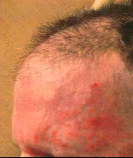
Acute skin changes may become visible after 10 to 14 hours. Grade 1 changes include mild generalized erythema and dry desquamation, pruritus, scaling, dyspigmentation, and hair loss. After 4 or 5 weeks of radiotherapy and radiation doses to the skin of 40 Gy or greater, grade 2 dermatitis may develop, with tender or edematous erythema, moist desquamation in skin folds, and considerable pain. They tend to peak 1 to 2 week after the last treatment and start healing 3 to 5 weeks after radiation. Complete healing may take 1 to 3 months. Occasionally, dermatitis may progress to grade 3, characterized by confluent moist desquamation, or even grade 4, with ulcers, hemorrhage, and necrosis.128,132 Chronic changes may develop months or years after the initial exposure. Postinflammatory hypo- or hyperpigmentation, textural changes (xerosis and hyperkeratosis), loss of hair follicles and sebaceous glands, atrophy, telangiectasia, or subcutaneous fibrosis are among the manifestations. Fibrosis can result in tissue retraction, pain, and limitation of movement. Scalp appears more tolerant to radiation injury than the skin of the face, neck, trunk, and extremities. Affected skin can be predisposed to ulcers and skin breakdown.133,134 In some patients, radiation recall dermatitis may occur. This happens when a patient who has completed RT encounters a drug and develops skin reaction similar to acute radiation dermatitis. The drugs are usually cytotoxic agents. It is probably due to local cutaneous immunologic responses to the challenging agent (Fig. 97.2).135
Mild acute dermatitis is treated symptomatically. Washing with water, gentle cleansing with a mild agent, wearing loose, nonbinding clothing, and avoidance of irritants, antiperspirants, and ultraviolet exposure all help. When erythema and dry desquamation occurs, creams or ointments (petrolatum-based, castor oil, balsam of Peru, trypsin, trolamine) can be used. Topical sucralfate or hyaluronic acid was shown to be efficacious in some controlled studies.136,137 In a phase III study in breast cancer patients receiving postoperative RT, an extract from the calendula plant significantly reduced the occurrence of moderate to severe acute dermatitis from 63% to 41% when compared with trolamine, a nonsteroidal agent.138 Other topical agents containing aloe vera, D-panthenol, almond, or chamomile can also be tried. The use of these agents is supported only by uncontrolled studies or anecdotal evidence.139,140
The value of topical antioxidants has not been established, and topical steroids are controversial, with research producing conflicting results. There are concerns of infection and skin atrophy, known side effects of topical steroids. At best, steroids may ameliorate the symptoms, but they do not prevent the dermatitis.141 Topical ascorbic acid lotion (vitamin C) did not show discernible benefit for the prevention of radiation dermatitis.142
When acute dermatitis becomes severe, usual wound care should be applied to the erosions and ulcerations. Key measures are keeping the site clean and moist, pain management, protection from contamination, debridement, and infection control.134,143 During radiation treatment, hydrogel dressings, hydrocolloid dressing, burn pads, or foam dressings can be applied. If the wound is infected, ionic silver powder, topical antibiotics, cadexomer iodine, or maltodextrin powder can be added. Referral to wound care specialists should be made.139 In recent years, more specific agents have been investigated, such as topical granulocyte-macrophage colony-stimulating factor, tacrolimus, pimecrolimus, and platelet-derived growth factor.128
Chronic skin changes from radiation injury are harder to treat. Chronic fibrosis is associated with high incidence of skin breakdown and infection. A team approach should be adopted that includes wound care, physical therapy, deep massage, and pain management to address cosmetic and quality of life issues. Pentoxifylline (Trental) appears to have an antifibrotic effect. Oral pentoxifylline (800 mg per day) and vitamin E (1,000 IU per day) for 6 months significantly reduce radiation-induced fibrosis.144 Prophylactic use of pentoxifylline significantly reduces late skin changes, fibrosis, and soft tissue necrosis in a randomized controlled study, possibly through its protective effect against vascular pathology.145 Intramuscular liposomal copper or zinc superoxide dismutase, subcutaneous interferon-γ, or hyperbaric oxygen therapy has also been used.
FIGURE 97.2. Acute dermatitis after whole-brain radiation.
GENITOURINARY TRACT INJURY
Urinary Symptoms
Irradiation to the pelvic region as part of treatment for cancer of the prostate, uterus, ovary, cervix rectum, or urinary bladder can cause urinary problems due to injury to mucosa, vasculature, and smooth muscles.146,147 Acute reactions occur within 3 to 6 months of treatment. Chronic changes occur later. Acute reactions present as dysuria, frequency, and urgency as a result of radiation cystitis. They are usually not as severe as some of the cystitis caused by chemotherapy. Strictures or fistula can develop during the years following RT.148,149
If infection is ruled out, symptomatic relief with phenazopyridine (Pyridium) is usually the first-line treatment for acute symptoms. It is given at 200 mg orally, 3 times a day. Phenazopyridine accumulates in the urine essentially unchanged and acts as a topical analgesic within the bladder. Patients should be warned that phenazopyridine turns the urine into a bright orange color and can stain clothing. If the symptoms are not adequately relieved, antispasmodics can be added. Oxybutynin (Ditropan) or flavoxate (Urispas) help relax the smooth muscles and reduce urinary urgency and frequency.150 Tolterodine (Detrol) is a cholinergic antagonist that is also effective for overactive bladder. It causes less dry mouth but its response rate is lower.151 Trospium (Sanctura) was documented to improve symptoms in radiation-induced cystitis and is significantly better tolerated than immediate-release oxybutynin.152
Intravesical infusion of hyaluronic acid or chondroitin sulfate, injection of botulinum toxin A into bladder wall, and hyperbaric oxygen therapy have shown benefit.153–156 IV WF10 (tetrachlorodecaoxide), an immunomodulator, was reported to be beneficial.157,158 In patients with severe pain, aggressive pain control with opioids may be needed.
Symptomatic management of chronic changes is similar to that of acute reactions. Dilatation or placement of a permanent catheter may be required for significant obstruction. Patients not responding to less-aggressive treatment may be candidates for reconstructive surgery to repair the stricture, sphincter failure, or fistula.
Female Sexuality
High-dose radiation to the pelvis causes varying degrees of sexual dysfunction related to injury to the ovaries and vagina.159,160 Ovarian failure as a result of pelvis irradiation leads to postmenopausal changes. Acute injury occurs during the course of RT and the following few months. It usually presents as vaginal and vulval mucositis, pain, and ulceration. Chronic changes are less frequent than acute changes, which can develop more than 3 months after the completion of treatment. Chronic changes include fibrosis, loss of elasticity and sensation, susceptibility to trauma and infection, postcoital bleeding, and dyspareunia.161–163
Maintenance of local hygiene, aggressive treatment of infection, and regular dilatation of the vaginal canal help reduce the acute reaction. Hormone replacement therapy and application of lubricants for mucosal dryness can be used to treat acute injury. To prevent chronic changes, uses of vaginal dilators, lubricants, and supplemental estrogen were shown to be helpful. When fibrosis is established, treatment may require more drastic measures, such as hyperbaric oxygen therapy or surgical reconstruction. Although these are common options in clinical practice, the level of evidence supporting their use varies.159
Most studies of use of topical estrogen showed benefit.164,165 Radiation causes damage to the epithelium, which may persist for another 3 to 6 months after therapy. Topical estrogen promotes epithelial regeneration. Benzydamine is an anti-inflammatory that also has analgesic, local anesthetic, and antimicrobial effects. It can be applied topically to achieve a higher local tissue concentration. It reduced both subjective symptoms and objective observation of vaginal mucositis.166,167 There have been several uncontrolled studies of hyperbaric oxygen therapy in the treatment of established necrotic wound resulting from perineal and vaginal radiation; the strength of evidence is modest.168,169 In women with severe radiation damage, such as perineal defect or obliteration of vagina, reconstructive surgery may be considered. All reported studies are retrospective.159
Male Sexuality
When planning RT for prostate cancer, its effect on male sexual function must be considered and discussed with the patient. Although the rate of erectile dysfunction (ED) is lower in patients receiving RT versus radical prostatectomy, sexual dysfunctions remain one of the most important posttreatment quality of life issues.170 A survey showed that 68% of men aged 45 to 70 years were willing to trade off a 10% or greater advantage in 5-year survival to maintain sexual potency.171 Onset of ED is gradual, usually beginning about 6 months posttreatment and continuing to deteriorate for 4 years.172
RT does not appear to reduce testosterone production or cause pelvic nerve injury.173 In addition to psychological reasons, vascular changes after RT appear to be the predominant cause of postradiation male sexual dysfunction. As such, smoking and hypertension are risk factors.174,175 With external-beam RT, the rates of ED vary from 7% to 72%, a wide range attributed to the study populations. Brachytherapy is associated with 2% to 89% of ED.176Diminished sexual desire, decreased orgasmic pleasure, and a reduced ejaculation volume are other problems reported by patients.177
Treatment of post-RT sexual dysfunction should take a multidisciplinary approach, including psychosocial evaluation and counseling, pharmacologic intervention, and exploration of the use of mechanical devices.176,178 The mainstay of pharmacologic treatment is phosphodiesterase inhibitors in patients with arteriogenic ED. A randomized double-blinded, placebo-controlled crossover trail suggests a positive response to sildenafil (Viagra). Yet the overall response rate is low. Only 21% of patients improved during sildenafil treatment but not during placebo treatment.179 Tadalafil (Cialis) is found beneficial in these patient populations too.180,181
Intracavernosal injection of prostaglandins or phentolamine papaverine is also effective. For patients who are refractory to pharmacologic intervention, implantation of a penile prosthesis can be considered. Minimal intraoperative and postoperative complications and an excellent patient satisfaction rate were reported.182 Vacuum devices are another option.
NUTRITION
Nutritional support is very important for patients undergoing RT. Patients may be malnourished when they come for initial treatment. Cancer creates a catabolic state. Anorexia, early satiety, nausea and vomiting, and involvement of the alimentary tract by cancer all contribute to impaired nutrition intake, digestion, and absorption. Once RT is started, a patient’s nutritional status can deteriorate, especially in those with GI toxicity. A weight loss of more than 20% of total body weight is associated with poorer outcome. Nutritional support measures include prescription of appetite enhancers, provision of a high-quality diet, ensuring adequate enteral intake via tube feeding, and hyperalimentation with parenteral nutrition.183 A multidisciplinary approach should be taken with the involvement of GI physicians, nutritionists, and nursing staff.
A systematic review showed that only progestins (megestrol [Megace]) and corticosteroids (methylprednisolone, prednisolone, and dexamethasone) are supported by evidence for cancer-related anorexia. A number of other drugs have been tested. They include metoclopramide, cyproheptadine, pentoxifylline, melatonin, erythropoietin, eicosapentaenoic acid (fish oil), androgenic steroids (nandrolone or fluoxymesterone), ghrelin, interferon, and cannabinoid (dronabinol [Marinol]). Data are mixed. No strong recommendation can be made at this point regarding these agents.184,185
Cancer patients tend to have a higher protein turnover. Adequate protein intake is critical in patients undergoing cancer treatment. Daily intake of 1.5 to 2.0 g of protein per kilogram of ideal body weight generally maintains a positive nitrogen balance. Caloric intake help maintain the weight. Between 30% and 90% of total calories can come from carbohydrates. Fat provides energy, serves as a vehicle for other nutrients, and performs other important biological functions. It usually makes up around 30% of the content in enteral formulas. Vitamins, minerals, and other micronutrients should be included. Sufficient water intake is required to offset the 2.5-L daily fluid loss. Dietary counseling improves outcomes in selected populations.186 Body weight and serum markers (albumin, transferrin, and prealbumin) can be monitored to assess whether the nutritional support is adequate.187–189
In patients with moderate to severe radiation-induced oral mucositis or esophagitis, oral nutritional support can be challenging. Tube feeding bypasses the injured tissues and provides direct access to the absorption surface. Nasogastric or nasojejunal tubes enable short-term access. For longer term access (>30 days), gastrostomy, gastrojejunostomy, and jejunostomy tubes can be placed endoscopically, radiologically, or surgically. Percutaneous endoscopic gastrostomy is increasingly the method of choice, often placed prophylactically when severe upper GI toxicity is anticipated.190–193 Gastrostomy tube placement is not without risk. Some series suggest a 17% morbidity rate. In 3% of patients, serious complications such as peritonitis, sepsis, perforation, and dislodgement were reported.194,195 Metastasis to percutaneous endoscopic gastrostomy site has also been reported, likely due to direct implantation of cancer cells. For patients with reflux esophagitis, gastroparesis, aspiration pneumonia, or limited stomach volume, a jejunostomy tube may be placed instead. The feeding tube should be cared for properly to prevent displacement or malfunctioning such as clogging.
When adequate nutrition intake can be achieved, enteral intake is preferred over parenteral support because it uses and helps maintain the existing alimentary functions. It is less expensive, safer, and associated with fewer side effects. However, in patients without a functioning GI tract because of obstruction, poor GI motility, intractable vomiting, severe diarrhea, short bowel syndrome, or severe pancreatitis, total parenteral nutrition may be appropriate. Implementation of total parenteral nutrition requires special expertise and is done in a concerted manner between the cancer-treating team and the nutrition support team.196,197
ACKNOWLEDGMENTS
The authors thank Dr. Joseph Huryn, Chief, Dental Service, and Dr. Liang Deng, Dermatology Service, at Memorial Sloan-Kettering Cancer Center for providing the photographs for this chapter, and Jyothirmai Gubili for expert editorial assistance.
SELECTED REFERENCES
A full list of references for this chapter is available online.
4. McLeod LD, et al. Interpreting patient-reported outcome results: US FDA guidance and emerging methods. Expert Rev Pharmacoecon Outcomes Res 2011;11(2):163–169.
7. Hagen NA, et al. The Birmingham International Workshop on Supportive, Palliative, and End-of-Life Care Research. Cancer 2006;107(4):874–881.
11. Kirkova J, et al. Cancer symptom clusters: clinical and research methodology. J Palliat Med 2011;14(10):1149–1166.
18. Brown P, et al. Will improvement in quality of life (QOL) impact fatigue in patients receiving radiation therapy for advanced cancer? Am J Clin Oncol 2006;29(1):52–58.
27. Jensen SB, et al. A systematic review of salivary gland hypofunction and xerostomia induced by cancer therapies: management strategies and economic impact. Support Care Cancer 2010;18(8):1061–1079.
31. Zhang Y, et al. Prevention of radiation-induced xerostomia by submandibular gland transfer. Head Neck 2012;34(7):937–942.
38. Little M, et al. Reducing xerostomia after chemo-IMRT for head-and-neck cancer: beyond sparing the parotid glands. Int J Radiat Oncol Biol Phys 2012;83(3):1007–1014.
43. Bardet E, et al. Subcutaneous compared with intravenous administration of amifostine in patients with head and neck cancer receiving radiotherapy: final results of the GORTEC 2000–02 phase III randomized trial. J Clin Oncol2011;29(2):127–133.
44. Jha N, et al. Phase III randomized study: oral pilocarpine versus submandibular salivary gland transfer protocol for the management of radiation-induced xerostomia. Head Neck 2009;31(2):234–243.
45. Rieger JM, et al. Functional outcomes related to the prevention of radiation-induced xerostomia: oral pilocarpine versus submandibular salivary gland transfer. Head Neck 2012;34(2):168–174.
49. Kahn ST, Johnstone PA. Management of xerostomia related to radiotherapy for head and neck cancer. Oncology (Williston Park) 2005;19(14):1827–1832.
58. Pfister DG, et al. Acupuncture for pain and dysfunction after neck dissection: results of a randomized controlled trial. J Clin Oncol 2010;28(15):2565–2570.
59. Meng Z, et al. Randomized controlled trial of acupuncture for prevention of radiation-induced xerostomia among patients with nasopharyngeal carcinoma. Cancer 2012;118(13):3337–3344.
60. Wong RK, et al. Phase II randomized trial of acupuncture-like transcutaneous electrical nerve stimulation to prevent radiation-induced xerostomia in head and neck cancer patients. J Soc Integr Oncol 2010;8(2):35–42.
65. Deng G, et al. Functional magnetic resonance imaging (fMRI) changes and saliva production associated with acupuncture at LI-2 acupuncture point: a randomized controlled study. BMC Complement Altern Med 2008;8:37.
70. Peterson DE, et al. Alimentary tract mucositis in cancer patients: impact of terminology and assessment on research and clinical practice. Support Care Cancer 2006;14(6):499–504.
73. Sonis ST, et al. Perspectives on cancer therapy-induced mucosal injury: pathogenesis, measurement, epidemiology, and consequences for patients. Cancer 2004;100(9 Suppl):1995–2025.
74. Yeoh AS, et al. Nuclear factor kappaB (NFkappaB) and cyclooxygenase-2 (Cox-2) expression in the irradiated colorectum is associated with subsequent histopathological changes. Int J Radiat Oncol Biol Phys2005;63(5):1295–1303.
75. Anthony L, et al. New thoughts on the pathobiology of regimen-related mucosal injury. Support Care Cancer 2006;14(6):516–518.
77. Peterson DE, Lalla RV. Oral mucositis: the new paradigms. Curr Opin Oncol 2010;22(4):318–322.
79. Keefe DM, et al. Updated clinical practice guidelines for the prevention and treatment of mucositis. Cancer 2007;109(5):820–831.
93. Bensadoun RJ, et al. Amifostine in the management of radiation-induced and chemo-induced mucositis. Support Care Cancer 2006;14(6):566–572.
94. Hensley ML, et al. American Society of Clinical Oncology 2008 clinical practice guideline update: use of chemotherapy and radiation therapy protectants. J Clin Oncol 2009;27(1):127–145.
95. Le QT, et al. Palifermin reduces severe mucositis in definitive chemoradiotherapy of locally advanced head and neck cancer: a randomized, placebo-controlled study. J Clin Oncol 2011;29(20):2808–2814.
96. von Bultzingslowen I, et al. Growth factors and cytokines in the prevention and treatment of oral and gastrointestinal mucositis. Support Care Cancer 2006;14(6):519–527.
115. Baughan CA, et al. A randomized trial to assess the efficacy of 5-aminosalicylic acid for the prevention of radiation enteritis. Clin Oncol (R Coll Radiol) 1993;5(1):19–24.
116. Resbeut M, et al. A randomized double blind placebo controlled multicenter study of mesalazine for the prevention of acute radiation enteritis. Radiother Oncol 1997;44(1):59–63.
117. Martenson JA Jr, et al. Olsalazine is contraindicated during pelvic radiation therapy: results of a double-blind, randomized clinical trial. Int J Radiat Oncol Biol Phys 1996;35(2):299–303.
118. Feyer PC, Stewart AL, Titlbach OJ. Aetiology and prevention of emesis induced by radiotherapy. Support Care Cancer 1998;6(3):253–260.
119. Feyer PC, et al. Radiotherapy-induced nausea and vomiting (RINV): MASCC/ESMO guideline for antiemetics in radiotherapy: update 2009. Support Care Cancer 2011;19(Suppl 1):S5–S14.
120. Basch E, et al. Antiemetics: American Society of Clinical Oncology clinical practice guideline update. J Clin Oncol 2011;29(31):4189–4198.
121. Urba S. Radiation-induced nausea and vomiting. J Natl Compr Canc Netw 2007;5(1):60–65.
122. Gwede CK. Overview of radiation- and chemoradiation-induced diarrhea. Semin Oncol Nurs 2003;19(4 Suppl 3):6–10.
123. O’Brien BE, Kaklamani VG, Benson AB 3rd. The assessment and management of cancer treatment-related diarrhea. Clin Colorectal Cancer 2005;4(6):375–383.
124. Benson AB 3rd, et al. Recommended guidelines for the treatment of cancer treatment-induced diarrhea. J Clin Oncol 2004;22(14):2918–2926.
125. Stern J, Ippoliti C. Management of acute cancer treatment-induced diarrhea. Semin Oncol Nurs 2003;19(4 Suppl 3):11–16.
126. Martenson JA, et al. Phase III, double-blind study of depot octreotide versus placebo in the prevention of acute diarrhea in patients receiving pelvic radiation therapy: results of North Central Cancer Treatment Group N00CA. J Clin Oncol2008;26(32):5248–5253.
128. Hymes SR, Strom EA, Fife C. Radiation dermatitis: clinical presentation, pathophysiology, and treatment 2006. J Am Acad Dermatol 2006;54(1):28–46.
153. Shao Y, Lu GL, Shen ZJ. Comparison of intravesical hyaluronic acid instillation and hyperbaric oxygen in the treatment of radiation-induced hemorrhagic cystitis. BJU Int 2012;109(5):691–694.
154. Hazewinkel MH, et al. Prophylactic vesical instillations with 0.2% chondroitin sulfate may reduce symptoms of acute radiation cystitis in patients undergoing radiotherapy for gynecological malignancies. Int Urogynecol J2011;22(6):725–730.
155. Smit SG, Heyns CF. Management of radiation cystitis. Nat Rev Urol 2010;7(4):206–214.
156. Chuang YC, et al. Bladder botulinum toxin A injection can benefit patients with radiation and chemical cystitis. BJU Int 2008;102(6):704–706.
159. Denton AS, Maher EJ. Interventions for the physical aspects of sexual dysfunction in women following pelvic radiotherapy. Cochrane Database Syst Rev 2003;1:CD003750.
176. Incrocci L, Slob AK, Levendag PC. Sexual (dys)function after radiotherapy for prostate cancer: a review. Int J Radiat Oncol Biol Phys 2002;52(3):681–693.
179. Watkins Bruner D, et al. Randomized, double-blinded, placebo-controlled crossover trial of treating erectile dysfunction with sildenafil after radiotherapy and short-term androgen deprivation therapy: results of RTOG 0215. J Sex Med 2011;8(4):1228–1238.
180. Ricardi U, et al. Efficacy and safety of tadalafil 20 mg on demand vs. tadalafil 5 mg once-a-day in the treatment of post-radiotherapy erectile dysfunction in prostate cancer men: a randomized phase II trial. J Sex Med2010;7(8):2851–2859.
184. Davis MP, et al. Appetite and cancer-associated anorexia: a review. J Clin Oncol 2004;22(8):1510–1517.