Sunil K. Srivastava,
Cynthia A. Toth
|
overview |
|
Surgical therapy for neovascular age-related macular degeneration (AMD) continues to evolve with refinements in technique and integration with developments in pharmacologic therapy. Currently available surgical treatments for neovascular AMD may provide useful options for salvage of vision when medical treatments are not enough. Some of the techniques discussed such as subretinal hemorrhage displacement, and macular translocation with 360-degree retinectomy may be effective in restoring vision in patients with lesions not generally treatable with medical therapy. Other therapies such as RPE transplantation are in early stages of promising development. The indications and techniques and evolution of the surgical therapies for AMD are discussed in this chapter. |
The treatment of neovascular age-related macular degeneration (AMD) has included laser photocoagulation, photodynamic therapy, radiation, use of pharmacologic agents, and in some cases surgical intervention. Surgical therapies have been utilized primarily for neovascular AMD and also in few cases of geographic atrophy. The surgeries for AMD presented here: submacular surgery, hemorrhage displacement, limited or full macular translocation, and transplantation of retinal pigment epithelium (RPE) will continue to evolve, affected by the rapid evolution of novel pharmacologic therapies.
|
Major Complications |
|
Submacular Surgery Retinal detachment Proliferative vitreoretinopathy Vitreous hemorrhage Cataract formation Persistence or recurrence of CNV Displacement of Subretinal Hemorrhage Retinal detachment Proliferative vitreoretinopathy Vitreous hemorrhage Cataract formation Recurrence of subretinal hemorrhage Persistence or recurrence of CNV Limited Macular Translocation Retinal detachment Proliferative vitreoretinopathy Recurrence of CNV after laser or other therapy Cataract formation Macular fold Macular hole Subretinal hemorrhage Scleral perforation |
|
Major Complications |
|
Full Macular Translocation Retinal detachment Proliferative vitreoretinopathy Recurrence of CNV or subretinal hemorrhage Cystoid macular edema Hypotony Glaucoma Epiretinal membrane Keratopathy Note that many of the more common, but not all, complications reported with each surgery are included on this list. |
SUBMACULAR SURGERY
The goal of submacular surgery is to remove the choroidal neovascularization (CNV) and hemorrhage if present and to re-establish the normal anatomy between retina and RPE. The poor visual outcomes associated with the Macular Photocoagulation Study and the encouraging results in early submacular surgery case series generated interest in these procedures.[1-3]
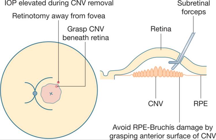
The surgeon considers lesion size and location when determining a site for CNV removal through a posterior retinotomy. During vitrectomy the posterior hyaloid is separated. A retinotomy is created and balanced salt solution is injected beneath the retina or a pick may be passed over the CNV to separate retinal connections. The surface of the membrane is grasped with forceps and it is removed from the subretinal space (Fig. 149.1). If the membrane is not free, attachments are cut with a blade or scissors. Most importantly, intraocular pressure is elevated prior to and during removal of the CNV, then lowered once hemostasis is assured. For subretinal hemorrhage (clot) removal, subretinal tissue plasminogen activator (tPA) 12.5-50 ?g is sometimes injected for thrombolysis 30-45 min prior to hemorrhage removal. Blood is then aspirated or pulled from the subretinal space. A fluid to air or gas exchange is performed and the patient is positioned upright or face down for the first postoperative day.[2-7]
|
|
|
|
FIGURE 149.1 Submacular surgery technique for removal of choroidal neovascularization. The focal retinotomy is located away from the fovea with attention to limiting subretinal instrument contact with retinal pigment epithelium and choroid. |
The Submacular Surgery Trials (SSTs), randomized prospectively clinical trials, compared submacular surgery to observation for two groups of AMD patients, those with large or poorly demarcated, new subfoveal CNV (group N)[6] and those with predominantly hemorrhagic lesions (group B).[7] For group N, at 2 years the surgery and observation groups had no significant difference in results. A successful outcome (defined as <1 line best corrected vision loss) occurred in 41% and 44%, respectively. In both groups at 24 months there was a 2 line median vision loss and median acuity dropped from 20/100 to 20/400. Analysis of vision-related quality of life scores revealed higher scores in the SST surgery group; however, based on the visual acuity findings, submacular surgery could not be recommended for patients with AMD and new subfoveal lesions. Given these results, submacular surgery is currently not indicated for the treatment of CNV associated with AMD.
|
Key Features |
|
Submacular surgery trials (SST) showed no visual benefit with surgery when compared to observation for patients with new subfoveal CNV secondary to AMD. For predominantly hemorrhagic lesions secondary to AMD the SST found a reduced risk of severe vision loss with surgery when compared to observation, but a high risk of rhegmatogenous retinal detachment in the surgery group. |
In SST group B (hemorrhagic lesions) the surgical technique was similar to that described above, with both blood and CNV removed whenever possible.[7] The surgeon chose whether to use tPA (used in 38% of Group B cases). At 24 months, the majority, 56% of surgery eyes and 59% of observation eyes, experienced loss of ?2 lines acuity, and 18% of eyes in each group improved at least 2 lines. In contrast to earlier case series which had shown a trend toward better visual acuity when subretinal blood was removed within 1 week,[5] the SST did not show improvement in acuity in the surgery group compared to observation; however, there was no cutoff with regards to the length of time that the hemorrhage was present, nor was there documentation of good visual acuity before the hemorrhage. Compared to the observation group, surgery eyes had higher rates of retinal detachments (16% vs 2%) and of cataract surgery (44% vs 6%), but a lower rate of severe vision loss (greater than 6 lines) (21% vs 36%).[7] It is difficult to recommend submacular surgery for submacular hemorrhage in AMD given the high rates of complications and the pressure of other treatment options.
The observation of poor vision after intervention in both of these AMD surgery groups in the SST is thought likely due to damage to the retina and underlying RPE by CNV, hemorrhage, surgical manipulation and postoperative absence of RPE in the surgical bed.[8,9] The development of alternate therapies for neovascular AMD has further diminished interest in submacular surgery alone.
DISPLACEMENT OF SUBMACULAR HEMORRHAGE
The prognosis for visual acuity is poor for submacular hemorrhage associated with neovascular AMD,[10-12] especially with larger and thicker hemorrhages which cause photoreceptor damage from clot contraction, direct iron toxicity, and the blockade of metabolic products exchange.[13,14] In light of the potential for damage to retina and RPE during clot extraction, even with tPA, Heriot (presentation at American Academy of Ophthalmology Vitreoretinal Update, San Francisco, CA, 1997) proposed subretinal hemorrhage displacement. He described injection of tPA into the vitreous cavity, followed by injection of a gas bubble with the goal of clot lysis and then displacement with positioning. Although subretinal hemorrhage was successfully displaced using this method, with some improvement in visual acuity,[15,16] it was unclear whether intravitreal tPA reached the subretinal space, since others displaced blood with intravitreal injections of gas alone and prone positioning.[17]
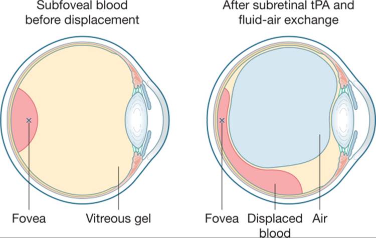
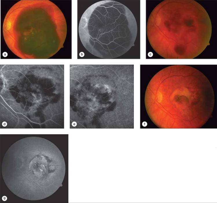
Alternately, Haupert et al described successful hemorrhage displacement after pars plana vitrectomy with subretinal tPA injection followed by fluid to air or gas exchange and prone postoperative positioning in 11 patients.[18] Two series followed with a similar technique that added postoperative supine positioning for the first hour and head down[19] or upright positioning for blood displacement (Fig. 149.2).[20]Patients in all three series had either partial or complete displacement of the blood from the subfoveal space with early vision improvement in the majority of eyes. With follow-up averaging from 3 to 17 months, hemorrhage recurred in 4-28% of eyes. Unlike surgery for removal of subretinal hemorrhage, there was only one retinal detachment in the 57 eyes in these three series.[18-20] Final visual acuity decreased compared to best postoperative acuity though some patients received photodynamic therapy or pegaptanib sodium injection.[19,20] These studies are limited by their small numbers and lack of control group (Fig. 149.3).
|
|
|
|
FIGURE 149.2 Illustration of hemorrhage displacement after injection of subretinal tPA, fluid-air exchange and upright positioning. Hemorrhage displacement with upright positioning based on research and diagrams presented by Marcin Stopa, MD, Duke University Eye Center Resident's Day, June 24, 2005. |
|
|
|
|
FIGURE 149.3 Subretinal hemorrhage displacement. (a) Preoperative color fundus photograph and (b) late fluorescein angiogram of an 8-day-old subretinal hemorrhage. Preoperative distance visual acuity was 20/400 in this eye with no previous treatment. (c) Color fundus photograph and (d) early and (e) late fluorescein angiograph 1 week after hemorrhage displacement by vitrectomy, subretinal tPA injection and fluid-air exchange. At this exam the patient received photodynamic therapy and subsequently this eye was treated with two injections of intravitreal bevacizumab. Color fundus photograph (f) and late fluorescein angiogram (g) 9 months later when visual acuity was 20/400. |
In surgical therapy of subretinal hemorrhage, the earlier submacular surgical techniques led to less invasive approaches with the addition of adjuvants such as photodynamic or anti-VEGF therapy after hemorrhage displacement. Submacular surgery in randomized trials showed no significant improve ment compared to observation. Though the rate of severe vision loss was lower with surgical removal of hemorrhage than with observation, the rate of retinal detachment was high. Given the poor natural history of submacular hemorrhage, and the low rate of complications after intravitreal or subretinal injection of tPA and air or gas displacement, this therapy is a reasonable consideration, though timing and coordination with use of adjuvant anti-VEGF or other therapy awaits results of further study. A meta-analysis of submacular surgery procedures provides a useful background for this decision making, though techniques such as hemorrhage displacement are considered together with hemorrhage removal in the analysis.[21]
MACULAR TRANSLOCATION
In contrast to submacular surgery or hemorrhage displacement, macular translocation surgery seeks to relocate the macula to a healthier site of RPE and choriocapillaris to prevent further degeneration and possibly recover vision. Since the initial report of Machemer,[22] the technique of macular translocation has evolved into two techniques: limited macular translocation (LMT) or full macular translocation with 360° retinectomy (MT360, also known as retinal rotation).
LIMITED MACULAR TRANSLOCATION
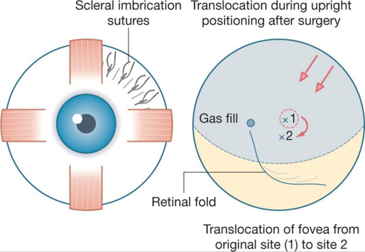
LMT relocates the retina by pinning detached retina against the eye wall using a partial air or gas fill to create a downward shift in the detached fovea and inferior retina to a site of healthier RPE and choriocapillaris; the shift is accentuated by also folding the choroid and sclera in the superotemporal quadrant in the bed of the retinal detachment.[23,24] Horizontal mattress sutures preplaced in the superior temporal sclera are tied to infold the sclera only after completion of vitrectomy and detachment of temporal retina through injection of subretinal fluid. Then a partial fluid to air exchange is performed. Translocation occurs after surgery when the patient is positioned upright. The bubble holds the retina against the imbrication and subretinal fluid accumulates inferiorly shifting the retina. After reattachment, an inferior retinal fold is seen early in the postoperative period (Fig. 149.4).
|
|
|
|
FIGURE 149.4 Illustration of limited macular translocation technique with partial retinal detachment and displacement of subretinal fluid and of the fovea with partial fluid-gas exchange along with scleral infolding. |
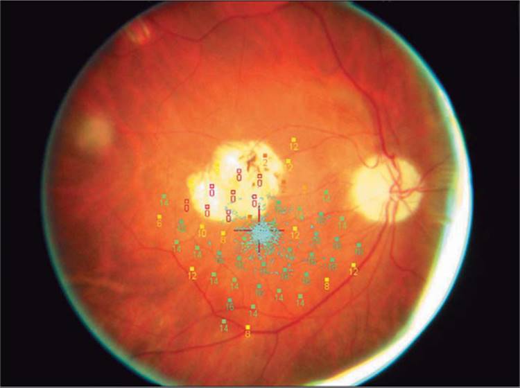
LMT changes the CNV location from subfoveal to juxtafoveal or extrafoveal, depending on distance of translocation, with laser photocoagulation typically applied to the CNV early after surgery (Fig. 149.5).[25]Foveal displacement with LMT averages 1200-1600 ?m depending on the technique and series. In the largest series reported (102 patients), effective translocation off the CNV was seen in 60% of patients and mean visual acuity was unchanged with 40% of eyes gaining ?2 Snellen lines, 29% of eyes unchanged and 31% losing ?2 Snellen lines.[25] CNV recurred in 35% of eyes after effective macular translocation and 10 eyes developed retinal detachments. A retrospective comparison of LMT versus photodynamic therapy found comparable postoperative visual acuities (both 20/200) but vision loss greater with photodynamic therapy.[26] The improvement in new therapies for subfoveal, small CNV may reduce efforts to relocate CNV to an extrafoveal location for treatment. If small subfoveal scars persist after alternate therapy and diminish acuity, LMT might then be considered for relocation of the fovea.
|
|
|
|
FIGURE 149.5 Microperimetry field with good fixation (pale blue dots) 2 years after limited macular translocation surgery and laser treatment of the choroidal neovascular membrane (which became juxtafoveal after the retina was shifted by the limited macular translocation surgery). |
FULL MACULAR TRANSLOCATION WITH 360° RETINECTOMY
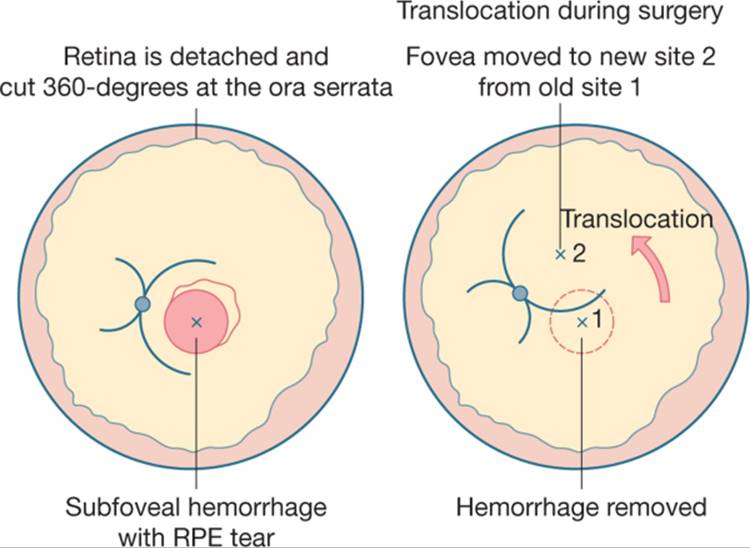
Machemer initially proposed MT360 surgery in the era of submacular surgery development. Though recognized as complex surgery, MT360 allows ? 3000 ?m foveal movement, enough to translocate off large hemorrhage, large CNV, or fibrosis. The retinal rotation produces torsional diplopia, treated by Eckardt et al with extraocular muscle surgery to counter-rotate the globe.[27] MT360 surgery currently includes a thorough vitrectomy followed by injection of subretinal fluid to totally detach the retina. The peripheral retina is cut at the ora serrata, subretinal membrane and/or blood is removed, and the retina is translocated, usually superiorly, off the CNV bed and pinned in the new location with perfluorocarbon liquid (Fig. 149.6). Retinectomy margins are photocoagulated and the perfluorocarbon is exchanged for silicone oil which is removed within months. Extraocular muscle surgery is performed at the time of translocation or in a separate surgery.
|
|
|
|
FIGURE 149.6 Illustration summarizing macular translocation with 360° retinectomy surgery. The fovea is shifted onto a new RPE bed during surgery and is held in place with silicone oil during the early postoperative period. |
Translocation case series have included subfoveal CNV, RPE tears, disciform scars, hemorrhages, previous photocoagulation, and photodynamic therapy. With median follow-up ranging from 10 to 21 months, seven of nine studies showed stabilization or improvement of vision after macular translocation surgery[29] and distance acuity improved in 52-66% of patients in three series.[27,28,30,31] At 1 year after MT360, prospective studies have also shown improvement in reading speed, color vision, contrast, near acuity,[32] critical print size,[33] and in vision-related quality of life.[34]
Retinal detachment rates after macular translocation were highest in early case series (up to 43%).[29] Other complications include cystoid macular edema, CNV recurrence, hypotony, epiretinal membranes, and keratopathy.[29] In the meta-analysis of surgery for AMD, estimated rates of complications were highest for macular translocation surgery.[21] Despite these complication rates, in a prospective 4 year study of 64 patients, Toth found that acuity of 20/80 or better was maintained in 46-51% of eyes from 1 through 4 years after MT360 with RD rate below 8% in this series.[31]
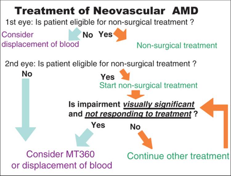
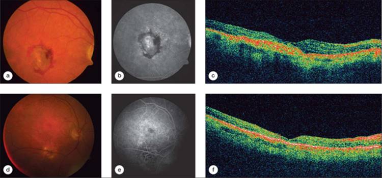
In light of the risks of complications, and the potential for vision gain, the decision whether to pursue MT360 is complex. Nonsurgical therapies now display improved results with low rates of complications.[35-37] Thus in cases where patients fail primary therapies or when there is no effective treatment MT360 is a reasonable salvage therapy, but one would not likely consider surgical intervention until medical therapies have first been utilized (Fig. 149.7), particularly since MT360 has resulted in good vision recovery in patients after photodynamic therapy[38] and after anti-VEGF therapy (Fig. 149.8).
|
|
|
|
FIGURE 149.7 Decision-making algorithm for treating AMD with vision loss. |
|
|
|
|
FIGURE 149.8 Macular translocation in an eye that has already received juxtafoveal laser photocoagulation, photodynamic therapy and pegaptanib injection. (a) Preoperative color fundus photograph, (b) late angiogram and (c) OCT scan of right eye show a fibrotic subfoveal lesion with almost no leakage. The visual acuity at distance was 20/200, and at near was J16 with +4 add. The patient had received four treatments with photodynamic therapy in the 20/800 fellow eye. (d) Color fundus photograph, (e) late fluorescein angiographic frame, and (f) OCT after macular translocation surgery in the right eye. Visual acuity recovered to 20/50 distance and 20/40 near with the patient resuming normal reading tasks. |
TRANSPLANTATION OF RETINAL PIGMENT EPITHELIUM
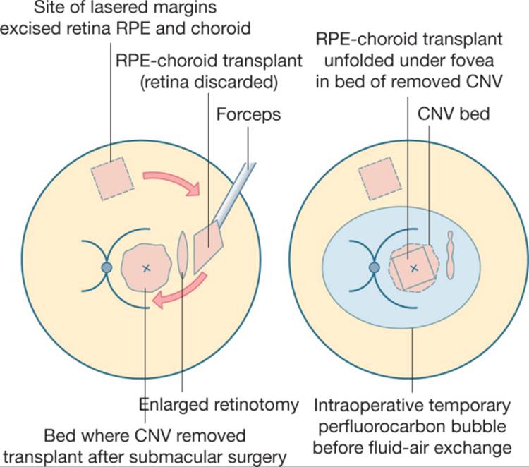
Work on transplantation of healthy RPE into the subfoveal space of eyes with AMD has followed from reports of poor acuity after submacular surgery and of improved acuity after macular translocation onto a healthier RPE bed. The pigment cell transplantation techniques are combined with conventional submacular surgery in eyes with neovascular AMD. In one technique, RPE cells are first harvested from irrigation of the nasal subretinal space and are directly transplanted onto the site of CNV removal.[39] Alternately, an adjacent RPE patch or a choroidal-RPE patch excised from the superior periphery is placed beneath the fovea (Fig. 149.9).[40-44]These techniques are still early in their evolution and results have been limited with some notable recovery of acuity despite complications such as graft failure, fibrosis, and retinal detachment. Refinements in the surgery and improvements in the generation/harvesting of RPE grafts will hopefully make this a viable option for patients.[39-44]
|
|
|
|
FIGURE 149.9 Illustration of RPE-choroid transplant surgery. Choroidal neovascularization is removed as in Figure 149.1. The RPE-choroid transplant is harvested from the mid-periphery, avoiding large choroidal vessels with retina removed from the surface. The transplant is placed into the subretinal space through an enlarged retinotomy and is unrolled to flatten beneath the retina and an intravitreal perfluorocarbon bubble. |
|
Key Features |
|
In light of the complexity of surgery, macular translocation with 360° peripheral retinectomy could be considered for the second eye with vision loss from hemorrhagic or neovascular AMD when primary therapies fail or are ineffective. |
The techniques described have been centered on management of neovascular AMD and not geographic atrophy. Although submacular surgery provides no advantage to eyes with geographic atrophy, macular translocation surgery or RPE transplantation techniques could be useful in cases of loss of RPE, if the retina and choriocapillaris could maintain function or recover after the surgery. In small series, patients with geographic atrophy have recovered acuity after macular translocation, though later some eyes have developed recurrent geographic atrophy in a pattern echoing the preoperative lesion.[45,46] It may become possible to combine surgery - whether translocation or transplantation - with adjuvant therapies to prevent recurrent atrophy in the future. Until then, response to these therapies provides new insight into the progression of atrophic disease and should be cautiously pursued.
As treatment for neovascular AMD shifts from laser photocoagulation to intravitreal delivery of antineovascular drugs, long-term sustained-delivery devices will likely be developed. Adaptation of current devices which allow sustained delivery of corticosteroids[47-49] or new devices will provide sustained intravitreal delivery of pharmacologic agents for AMD and reduce the risks associated with multiple intravitreal injections.
The first line of therapy for neovascular AMD primarily involves medical and laser therapy. Surgical techniques such as hemorrhage displacement, macular translocation, and RPE cell transplantation play a role in the treatment paradigm (Fig. 149.9) of neovascular AMD. Developments in surgical technique, adjuvants (such as growth factors), and drug therapy will have a significant impact on the evolution of future surgical treatments of AMD.
REFERENCES
1. Macular Photocoagulation Study Group : Laser photocoagulation of subfoveal recurrent noevascular lesions in age-related macular degeneration: results of a randomized clinical trial. Arch Ophthalmol 1991; 109:1232-1241.
2. Thomas MA, Dickinson JD, Melberg NS, et al: Visual results after surgical removal of subfoveal choroidal neovascular membranes. Ophthalmology 1994; 101:1384-1396.
3. Thomas MA, Grand MG, Williams DF, et al: Surgical management of subfoveal choroidal neovascularization. Ophthalmology 1992; 99:952-968.
4. De Juan E, Machemer R: Vitreous surgery for hemorrhagic and -brous complications of age-related macular degeneration. Am J Ophthalmol 1988; 105:25-29.
5. Lewis H: Intraoperative -brinolysis of submacular hemorrhage with tissue plasminogen activator and surgical drainage. Am J Ophthalmol 1994; 5:559-568.
6. Submacular Surgery Trials Research Group : Surgery for subfoveal choroidal neovascularization in age-related macular degeneration: ophthalmic -ndings: SST report no. 11 Ophthalmology 2004; 11:1967-1980.
7. Submacular Surgery Trials Research Group : Surgery for hemorrhagic choroidal neovascular lesions of age-related macular degeneration: ophthalmic -ndings: SST report no. 13. Ophthalmology 2004; 11:1993-2006.
8. Grossniklaus HE, Hutchinson AK, Capone A, et al: Clinicopathologic features of surgically excised choroidal neovascular membranes. Ophthalmology 1994; 101:1099-1111.
9. Nasir MA, Sugino I, Zarbin MA: Decreased choriocapillaris perfusion following surgical excision of choroidal neovascular rmembranes in age-related macular degeneration. Br J Ophthalmol 1997; 81:481-489.
10. Scupola A, Coscas G, Sourbrane G, Balestrazzi E: Natural history of macular subretinal hemorrhage in age-related macular degeneration. Ophthalmologica 1999; 213:97-102.
11. Avery RL, Fekrat S, Hawkins BS, Bressler NM: Natural history of subfoveal subretinal hemorrhage in age-related macular degeneration. Retina 1996; 16:183-189.
12. Bennett SR, Folk JC, Blodi CF, Klugman M: Factors prognostic of visual outcome in patients with subretinal hemorrhage. Am J Ophthalmol 1990; 109:33-37.
13. Glatt H, Machemer R: Experimental subretinal hemorrhage in rabbits. Am J Ophthalmol 1982; 94:762-773.
14. Toth CA, Morse LS, Hjelmeland LM, Lander MBD: Fibrin directs early retinal damage after experimental subretinal hemorrhage. Arch Ophthalmol 1991; 109:723-729.
15. Schulze SD, Hesse L: Tissue plasminogen activator plus gas injection in patients with subretinal hemorrhage caused by age-related macular degeneration: predictive variables for visual outcome. Graefes Arch Clin Exp Ophthalmol 2002; 240:717-720.
16. Hassan AS, Johnson MW, Schneiderman TE, et al: Management of submacular hemorrhage with intravitreous tissue plasminogen activator injection and pneumatic displacement. Ophthalmology 1999; 106:1900-1906.
17. Daneshvar H, Kertes PJ, Leonard BC, Peyman G: Management of submacular hemorrhage with intravitreal sulfur hexafluoride: a pilot study. Can J Ophthalmol 1999; 34:385-388.
18. Haupert CL, McCuen II BW, Jaffe GJ, et al: Pars plana vitrectomy, subretinal injection of tissue plasminogen activator and fluid-gas exchange for displacement of thick submacular hemorrhage in age-related macular degeneration. Am J Ophtalmol 2001; 131:208-215.
19. Olivier S, Chow DR, Packo KH, et al: Subretinal recombinant tissue plasminogen activator injection and pneumatic displacement of thick submacular hemorrhage in age-related macular degeneration. Ophthalmology 2004; 6:1201-1208.Erratum. Ophthalmology 2004; 9: 1640.
20. Singh RP, Patel C, Sears JE: Management of subretinal macular haemorrhage by direct administration of tissue plasminogen activator. Br J Ophthalmol 2006; 4:429-431.
21. Falkner CI, Leitich H, Frommlet F, et al: The end of submacular surgery for age-related macular degeneration? A meta-analysis. Graefes Arch Clin Exp Ophthalmol 2007; 245:490-501.
22. Machemer R, Steinhorst UH: Retinal separation, retinotomy and macular relocation. II. A surgical approach for age-related macular degeneration. Graefes Arch Clin Exp Ophthalmol 1993; 231:635-641.
23. De Juan E, Lowenstein A, Bressler NM, et al: Translocation of the retina for management of subfoveal chroidal neovascularization. II: a preliminary report in humans. Am J Ophthalmol 1998; 125:635-646.
24. Lewis H, Kaiser P, Lewis S, Estafanous M: Macular translocation for subfoveal choroidal neovascularization in age-related macular degeneration: a prospective study. Am J Ophthalmol 1999; 128:135-146.
25. Fujii GY, De Juan E, Pieramici DJ, et al: Inferior limited macular translocation for subfoveal chroidal neovascularization secondary to age-related macular degeneration: 1 year visual outcome and recurrence report. Am J Ophthalmol 2002; 134:69-74.
26. Pawlak D, Glacet-Bernard A, Papp M, et al: Limited macular translocation compared with photodynamic therapy in the management of subfoveal choroidal neovascularization in age-related macular degeneration. Am J Ophthalmol 2004; 137:880-887.
27. Eckardt C, Eckardt U, Conrad HG: Macular rotation with and without counterrotation of the globe in patients with age-related macular degeneration. Graefes Arch Clin Exp Ophthalmol 1999; 237:313-325.
28. Toth CA, Freedman SF: Macular translocation with 360° peripheral retinectomy: impact of technique and surgical experience on visual outcomes. Retina 2001; 21:293-303.
29. Cahill M, Toth CA: Macular translocation surgery. In: Stephen JR, ed. Retina, 4th edn. Philadelphia, PA: Mosby; 2004.
30. Pertile G, Claes C: Macular translocation with 360° retinotomy for management of age-related macular degeneration with subfoveal choroidal neovascularization. Am J Ophthalmol 2002; 134:560-565.
31. Mruthyunjaya P, Stinnett SS, Toth CA: Change in visual function after macular translocation surgery with 360° peripheral retinectomy for neovascular age-related macular degeneration. Ophthalmology 2004; 111:1715-1724.
32. Toth CA, Lapolice DJ, Banks AD, Stinnett SS: Improvement in near visual function after macular translocation surgery with 360° peripheral retinectomy. Graefes Arch Clin Exp Ophthalmol 2004; 242:541-548.
33. Fujikado T, Asonuma S, Ohji M, et al: Reading ability after macular translocation surgery with 360° retinotomy. Am J Ophthalmol 2002; 134:849-856.
34. Cahill MT, Stinnett SS, Banks AD, et al: Quality of life after macular translocation with 360 degree peripheral retinectomy for age related macular degeneration. Ophthalmology 2005; 112:144-151.
35. Blinder KJ, Bradley S, Bressler NM, et al: Effect of lesion size, visual acuity, and lesion composition on visual acuity change with and without vertepor-n therapy for choroidal neovascularization secondary to age-related macular degeneration: TAP and VIP report no. 1. Am J Ophthalmol 2003; 136:407-418.
36. Gragoudas ES, Adamis AP, Cunningham ET, et al: Pegaptanib for neovascular age-related macular degeneration. N Engl J Med 2004; 351:2805-2816.
37. Heier JS, Antoszyk AN, Pavan PR, et al: Ranibizumab for treatment of neovascular age-related macular degeneration: a phase I/II multicenter, controlled, multidose study. Ophthalmology 2006; 113:633-642.e1-4.
38. Park CH, Toth CA: Macular translocation surgery with 360° peripheral retinectomy following ocular photodynamic therapy of choroidal neovascularization. Am J Ophthalmol 2003; 136:830-835.
39. Binder S, Krebs I, Hilgers RD, et al: Outcome of transplantation of autologous retinal pigment epithelium in age-related macular degeneration: a prospective trial. Invest Ophthalmol Vis Sci 2004; 45:4151-4160.
40. Stanga PE, Kychenthal A, Fitzke FW, et al: Retinal pigment epithelium translocation after choroidal neovascular membrane removal in age-related macular degeneration. Ophthalmology 2002; 109:1492-1498.
41. Van Meurs JC, ter Averst E, Hofland LJ, et al: Autologous peripheral retinal pigment epithelium translocation in patients with subfoveal neovascular membranes. Br J Ophthalmol 2004; 88:110-113.
42. Van Meurs JC, Van Den Biesen PR: Autologous retinal pigment epithelium and choroid translocation in patients with exudative age-related macular degeneration: short-term follow-up. Am J Ophthalmol 2003; 136:688-695.
43. Stanga PE, Kychenthal A, Fitzke FW, et al: Retinal pigment epithelium translocation after choroidal neovascular membrane removal in age-related macular degeneration. Ophthalmology 2002; 109:1492-1498.
44. MacLaren RE, Bird AC, Sathia PJ, Awylard GW: Long-term results of submacular surgery combined with macular translocation of the retinal pigment epithelium in neovascular age-related macular degeneration. Ophthalmology 2005; 112:2081-2087.
45. Eckardt C, Eckardt U: Macular translocation in nonexudative age-related macular degeneration. Retina 2002; 22:786-794.
46. Cahill MT, Mruthyunjaya P, Bowes Rickman C, Toth CA: Recurrence of retinal pigment epithelial changes after macular translocation with 360 degrees peripheral retinectomy for geographic atrophy. Arch Ophthalmol 2005; 123:935-938.
47. Jaffe GJ, Martin DF, Callanan D, et al: Fluocinolone acetonide implant (Retisert) for noninfectious posterior uveitis: thirty-four week results of a multicenter randomized clinical study. Ophthalmology 2006; 113:1020-1027.
48. Tan DT, Chee SP, Lim L, et al: Randomized clinical trial of Surodex steroid drug delivery system for cataract surgery: anterior versus posterior placement of two Surodex in the eye. Ophthalmology 2001; 108:2172-2181.
49. Srivastava S, Mruthyunjaya P, Wiser J, et al: Intravitreal sustained-release fluocinolone acetonide device to treat severe experimental uveitis, Orlando, Florida: Association for Research in Vision and Ophthalmology; 2005:3536.