David Dueker,
Ibrahim Al Jadaan
HISTORY
GENERAL CONSIDERATIONS
A simple linear progression from complaint to history to examination will rarely suffice in the evaluation of a patient with glaucoma. Frequently, particularly when the diagnosis is primary open-angle glaucoma, the patient's presenting complaint has little or no connection with the as-yet-undiscovered glaucoma. Instead, the patient presents needing stronger bifocals or concerned about a floater or simply for 'a routine check-up'. Abnormalities suggestive of glaucoma are then uncovered during the examination, and this discovery leads to a more directed history. Thus, the findings lead back to further history taking, which may, in turn, suggest additional areas for examination. In this common scenario - the initial discovery of an asymptomatic glaucoma - the focus of the clinical visit will rapidly shift away from the now 'minor' presenting complaint to a much more serious issue. It is important to remember that this turn of events is often shocking and unwelcome to the patient. Although the physician rightly feels that by detecting glaucoma she or he renders a crucial service, the patient, who perhaps came prepared only for the idea of new glasses, must suddenly adjust to the possibility of having a chronic, potentially blinding disease. It is important to communicate clearly during this difficult time for the patient - to convey support and understanding and to lay out a clear plan of action. In addition, although the question of glaucoma rightly becomes the principal focus in these circumstances, the patient's own presenting complaint must not be neglected. Listening well and responding to the patient's stated problems will help develop the trust and commitment necessary for successful long-term treatment of glaucoma.
In less developed regions of the world, patients may not have their glaucoma discovered during examinations for other problems or in the course of routine check-ups. Instead, asymptomatic glaucoma may be allowed to follow a relentless course toward severe visual loss before medical attention is sought. Indeed, a patient may even ignore glaucoma-induced blindness in one eye as long as the other remains functional; not realizing that the same fate is slowly unfolding in the better eye. In extreme instances, the patient may not seek help until the remaining good eye is noticeably impaired. With glaucoma, unfortunately, symptomatic visual impairment is a usually a late development, occurring only when optic nerve damage is quite advanced. Even when glaucoma has already delivered a harsh lesson of its damaging potential, careful education is still needed to help the patient understand the nature of the disease and rationale for treatment.
Often, the time scheduled for a routine office visit is simply inadequate to fully evaluate a newly discovered glaucoma. As an alternative, one may explain the initial findings to the patient and schedule a follow-up visit for further testing to confirm the diagnosis and initiate any needed treatment. This is certainly preferable to making a diagnosis with incomplete information and starting treatment without thorough instruction and support. This option must be tempered, of course, by consideration of the urgency to begin treatment and by the type of glaucoma involved.
MEDICAL HISTORY AND GENERAL HEALTH
The general health of a patient is relevant to a broad range of ocular diseases, and the glaucomas, as a group, have multiple interactions with systemic disease. Many elements of the medical history obtained in the course of a routine eye examination relate directly or indirectly to glaucoma and its management.[1] For example, diabetes mellitus is a risk factor for several different forms of glaucoma through mechanisms that are separate and distinct from one another, e.g., primary open-angle glaucoma, neovascular glaucoma, ghost cell glaucoma, and pseudophakic pupillary-block glaucoma.
Sickle cell anemia trait is another example of a systemic disorder with multiple links to glaucoma. Retinal ischemia caused by the disease may lead to anterior segment neovascularization and neovascular glaucoma. Glaucoma secondary to traumatic hyphema is also more likely to occur in a patient with a sickling tendency. Furthermore, when glaucoma does occur, the eye is much less able to tolerate even moderate elevations of pressure because of the tendency to vascular occlusions. Finally, the treatment options are limited by the fact that some of the standard medications for acute glaucoma - acetazolamide and mannitol - are contraindicated because they can cause acidosis and hemoconcentration.
A variety of cardiovascular diseases - hypertension, atherosclerosis, cardiac failure, hypercoagulable states, and hypercholesterolemia - may diminish blood supply to the optic nerve, increasing susceptibility to glaucomatous damage. Treatment of cardiac disease, in the form of systemic ?-adrenergic blocking agents, may mask glaucoma by reducing intraocular pressure and may also reduce the response to therapy for glaucoma with topical ?-blockers. With further regard to topical ?-blockers, such drugs are hazardous in patients with bronchial asthma and certain other medical conditions, so careful attention to general medical status is mandatory. Treatment of systemic hypertension with calcium channel blockers may help to protect the optic nerve from damage in normal-tension glaucoma and may even prove to be a useful intraocular pressure-lowering agent for many glaucomas.[2]
Several forms of arthritis are associated with uveitis, which may lead to the development of a secondary glaucoma through multiple mechanisms. Ocular inflammation is common with ankylosing spondylitis and pauciarticular juvenile rheumatoid arthritis; it is less so with Reiter's syndrome and adult rheumatoid arthritis. Sarcoidosis may cause a granulomatous iritis, leading to synechial formation, seclusion of the pupil, and secondary angle-closure glaucoma. Systemic corticosteroid therapy of arthritis and other inflammatory conditions may cause elevated intraocular pressure or aggravate a preexisting glaucoma.
Clearly, multiple pathways may link systemic illness with various glaucomas. For this reason, a thorough medical history is an essential part of the glaucoma evaluation. It is also wise to communicate directly with the patient's primary physician and to update the medical status - e.g., surgery, drug changes - in a routine and recurring fashion.
|
Key Features |
|
Proper evaluation of a patient with known or suspected glaucoma may be summarized by one requirement; it must be comprehensive. A thorough history and a careful physical examination are essential given the many forms this disease group may assume. Family History Knowledge regarding the genetic basis for many of the glaucomas continues to expand. Social History Helps to reveal a patient's ability to understand and successfully manage the challenge of a chronic disease. Medical History May reveal an associated systemic disease and/or warn of potential drug reactions or interactions. Complete Eye Examination Virtually every part of a full ocular examination has the potential to uncover findings relevant to glaucoma. Supplemental Testing Gonioscopy and visual field testing, as well as some method for documenting the physical appearance of the optic nervehead - all are important to establish both current status as well as a baseline for future comparison. |
OCULAR HISTORY
The ocular history in primary open-angle glaucoma may be largely noncontributory. Myopia is more common in these patients than in the general population (whereas hyperopia is decidedly more common in primary angle-closure glaucoma). Also, since glaucoma tends to develop in the older age groups, presbyopia is frequently present. To rule out other mechanisms for glaucoma, one should inquire specifically regarding prior trauma (including surgery), inflammation (including possible steroid use), or infection. These historical questions are often triggered, or reemphasized, during the examination. For example, a patient may deny any recollection of serious trauma, yet the examination reveals a recessed chamber angle. Further careful questioning may uncover a childhood injury that the patient failed to recall during the initial inquiry.
In recent years, refractive surgical procedures have become highly refined and effective, and increasing numbers of patients are choosing this method of refractive correction. Often these patients are young and, in later years, may not think to mention these procedures in a routine ophthalmic history of 'prior eye surgery'. This could be unfortunate, since the corneal thinning produced by these procedures can cause marked underestimates of intraocular pressure. Since an increase in intraocular pressure is one important trigger for increased concern and attention to the possibility of glaucoma, it is possible that some cases may be missed due to falsely low pressure readings after refractive surgery. Careful questioning directed specifically at the issue of prior refractive surgery should help avoid this problem.
Many forms of secondary glaucoma have distinctive ocular histories. Some of these will be volunteered in the course of routine questioning regarding ocular health. A sudden, profound, and lasting visual decrease after central retinal vein occlusion, a preceding event for neovascular glaucoma, will usually be noted and reported by the patient. Hazing of vision with vigorous exercise may be a symptom of pigmentary glaucoma (from sudden release of pigment into the aqueous humor). Because vision clears to normal levels within hours, patients may not mention this spontaneously but will respond if asked specifically. The pain and blurred vision characteristic of angle-closure glaucoma are often distinctive points in an ocular history. However, some patients with angle closure are so impressed by their headache during an attack that this becomes their predominant memory. Further careful, directed questions, in this case triggered by finding a narrow angle on clinical examination, may develop a more complete description of the symptoms. The evaluation process should be continued in this recursive fashion, from history to examination, back to history, and so on, until the best possible agreement between history and examination is developed.
FAMILY HISTORY
The family history is important for several reasons. First of all, positive family history is an important risk factor for primary open-angle glaucoma. It is important to determine, if known, which family members have glaucoma. The risk for primary open angle glaucoma is greater for first-degree relatives of glaucoma patients than for the general population. Educating the patient and the patient's family about their risk for glaucoma is an important public health concern. It is also helpful to note whether the family member uses or used medications only or whether surgery was required. Although such information will be influenced by the practice patterns of the relatives' physicians, it may be a 'soft' indication of the severity of the glaucoma that may be inherited. Frequently, patient education will assist in the early detection of glaucoma in other at-risk family members, thus allowing glaucoma to be diagnosed before the occurrence of extensive damage to the eye.
In addition, several of the childhood glaucomas have well-described hereditary patterns. This can be helpful in diagnosis and genetic counseling. Finally, a positive family history often has important psychosocial overtones.
Patients who have had a close personal interaction with glaucoma in a family member, particularly in a complicated case, will be more than usually anxious when confronted with the disease in themselves. Intense personal experience like this can have a positive effect by encouraging careful adherence to treatment. However, it may also hamper care if 'nothing seemed to work' for the patient's relative; the patient may feel discouraged and unmotivated from the beginning. Clearly, it is important to develop an understanding of an individual patient's perception of glaucoma, to dispel unreasonable fears, and to work towards a positive commitment to therapy.
SUMMARY
History taking is an excellent opportunity to establish a trusting relationship with the patient. The patient is allowed to discuss what he or she perceives as important in the occurrence of the eye problem. The patient's understanding of glaucoma may also be ascertained. Listening is an excellent route for establishing trust and commitment between patient and physician. Thus, a foundation for a long-term relationship between the patient and the physician will be created.
EXAMINATION
EXTERNAL
In primary open-angle glaucoma, the external appearance of the eyes and adnexa is generally unremarkable. However, in other circumstances, the periocular structures may provide clues, obvious or subtle, suggesting a coexisting glaucoma. A hemangioma of the upper lid, e.g., is frequently associated with open-angle glaucoma of the ipsilateral eye in patients with Sturge-Weber syndrome. A less obvious vascular abnormality, dilatation of the episcleral veins, may signal glaucoma caused by an arteriovenous malformation raising the episcleral venous pressure. In addition to noting such specific glaucoma-related findings, the external examination should include a search for signs of previous trauma, proptosis, restriction of movement, photophobia and tearing, and hyperemia.
The pupillary responses are generally normal in primary open-angle glaucoma, but there may be an afferent defect in the event of severe nerve damage. Other glaucomas may show more distinctive pupillary signs. Examples include a fixed midposition pupil in acute angle closure, a miotic pupil in acute iritis, an irregular pupil bound down by posterior synechiae in a case of previous inflammation, displaced or multiple pupils, or both, in the iridocorneal endothelial (ICE) syndrome, and color change in Fuchs' heterochromic iridocyclitis. Therefore, in addition to direct and consensual responsiveness, the pupils should be evaluated for size, number, shape, and equality (preferably in both light and dark). Iris color should also be noted, particularly if pigmentation is heterogenous.
SLIT-LAMP BIOMICROSCOPY
The slit-lamp biomicroscope is extremely valuable for the clinical examination of the eye; and varied findings associated with the glaucomas provide an excellent demonstration of its versatility.[3] Subtle findings in the anterior segment require both the magnified stereo view of the slit lamp and the great variability in lighting that is possible with this instrument. In addition, through the use of auxiliary optical devices, the slit-lamp biomicroscope becomes one of the most capable means for examining the anterior chamber angle and the optic nerve head.
The conjunctiva usually appears normal in primary open-angle glaucoma, as well as in many other types of glaucoma. Mention has already been made of dilated episcleral veins, which is a specific association with glaucoma. A single dilated anterior ciliary artery, a 'sentinel vessel', is an important sign of an anteriorly located tumor, which sometimes presents with glaucoma. More generalized hyperemia is a nonspecific sign of inflammation or irritation and may be associated with several types of secondary glaucoma.
Although initially normal, the conjunctiva may be altered later in the course of glaucoma by an allergic or inflammatory reaction to drug treatment or through formation of drug deposits (adrenochrome) after long-term epinephrine instillation. Histologic signs of long-term toxicity from topical drug therapy of glaucoma have been described, but these changes do not appear to have specific biomicroscopic correlates. The conjunctiva is, of course, an important tissue in the context of filtering surgery. Slit-lamp biomicroscopy is used preoperatively to select an optimal 'filtering bed' (mobile and free from scarring); postoperatively, conjunctival hypovascularity, translucency, and surface microcysts are useful signs of bleb function.
Biomicroscopic examination of the cornea should include an overall assessment of clarity, size, and vascularity, as well as attention to each tissue layer. There are specific findings in the cornea that are helpful in the diagnosis of several of the secondary glaucomas. Some of these signs are subtle and must be specifically looked for.
Not surprisingly, the cornea is generally normal for age in primary open-angle glaucoma, unless the intraocular pressure is high enough to cause corneal edema. However, corneal edema is rare at the usually moderate pressure levels of primary open-angle glaucoma. Rapid development of extremely high pressures (<50 mmHg) as may occur, e.g., with acute angle-closure glaucoma or neovascular glaucoma, is more likely to cause a marked corneal edema. Lower pressures may cause corneal edema when the corneal endothelium is compromised, as with inflammatory precipitates or in the ICE syndrome.
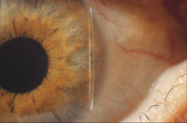
Corneal stromal scars are useful markers of prior trauma or infection-inflammation. An increasingly common cause of stromal scarring is refractive surgery; an important finding since it may signal inaccurate tonomety readings. Careful inspection of the stroma for signs of old interstitial keratitis - midstromal scarring and ghost vessels - is valuable because these otherwise quiet eyes may acquire glaucoma as a late complication. When the cornea is of normal thickness, it is a useful gauge of anterior chamber depth: normal chamber depth is approximately four to five times the corneal thickness centrally. When chamber depth is varying (e.g., progressive shallowing after filtration surgery), and one wishes to chart its progress, a sequential record of depth by corneal thickness can be extremely helpful. A visual estimate of corneal thickness can be used to gauge the distance between the anterior surface of the peripheral iris and the posterior surface of the peripheral cornea in the periphery of the anterior chamber. When this space is less than one-quarter the thickness of the cornea, it is highly suggestive of a dangerously narrow angle (Fig. 194.1).[4]
|
|
|
|
FIGURE 194.1 Slit-lamp photograph of the van Herick test for peripheral chamber depth. |
Abnormalities of Descemet's membrane are not frequent in the glaucomas. In infantile glaucoma, sudden stretching of the globe by pressure may cause ruptures in this membrane. This creates an overlying edema that clears as the endothelium heals over the rupture, leaving curved, usually horizontal, ridges (Haab's striae) on the posterior surface. The region of Descemet's membrane is also the site of major pathologic changes in posterior polymorphous dystrophy, which is associated with glaucoma in 15% of cases.
Several forms of glaucoma are associated with characteristic alterations in or on the endothelial surface of the cornea. Material suspended in the aqueous humor tends to deposit on the endothelium. In pigment dispersion and pigmentary glaucoma, e.g., a narrow spindle of deposited pigment builds up on the central cornea posteriorly - Krukenberg's spindle. In glaucoma associated with intraocular inflammation, cells deposit on the endothelium in sometimes typical formations. These keratic precipitates (KP) may be small round mounds, large juicy clumps ('mutton fat KP' of granulomatous disease), or delicate and stellate (typical of heterochromic iridocyclitis). With time, these deposits tend to become pigmented. Occasionally, pseudoexfoliation material will deposit on the inferior corneal endothelial surface where it may be confused with inflammatory KP. It must be emphasized that inflammation sufficient to produce glaucoma may not be associated with marked signs of anterior chamber inflammation and numerous KP. In fact, KP associated with glaucoma may be restricted to the chamber angle, as discussed in the section on gonioscopy.
In the ICE syndrome, the primary abnormality is apparently in the corneal endothelial cells, with secondary glaucoma and iris distortion and atrophy resulting from posterior migration of these abnormal cells. Since the iris and chamber angle changes may be minimal in one subcategory of this disorder (Chandler's syndrome), careful slit-lamp examination of the corneal endothelium may prove crucial to the diagnosis. In the ICE syndrome, the corneal endothelial cells are large and irregular, and in specular illumination, the endothelial surface has a beaten-silver appearance. A specular microscope is a useful adjunct in suspected cases.
The normal anterior chamber is four to five corneal thicknesses deep centrally with its posterior extent defined by the lens and a relatively flat iris plane, its anterior extent determined by the endothelial surface of the cornea. The normal aqueous humor is optically clear. The depth of the peripheral chamber must be assessed separately because a deep central chamber does not ensure depth in the periphery. The surface contour of the iris is normally flat or slightly bowed forward. Moderate forward bowing may be associated with an increased relative pupillary block or lens enlargement, or both. Marked forward bowing of the iris is seen with iris bombé. Backward bowing - a peripheral concavity of the iris - is a characteristic finding in pigmentary glaucoma.
The iris itself is examined for abnormal surface deposits (e.g., pigment in pigmentary dispersion; pigment or exfoliation material in exfoliation syndrome), pigmented nodules (the ICE syndrome), adhesions to the lens or peripheral cornea (secondary to inflammation), and abnormal surface vessels (rubeosis). The iris may be atrophied in sectors (status after acute angle closure) or patches (ICE syndrome) or diffusely (heterochromic iridocyclitis). Iris transillumination may reveal characteristic defects in the pigment epithelial layer: transillumination along the pupillary margin in exfoliation syndrome or transillumination in a spoke-wheel pattern for pigmentary glaucoma. Previous blunt trauma may leave small wedge-shaped defects in the pupillary margin ('sphincter rupture') and/or peripheral dialysis where the iris base is torn from its insertion, usually in no more than one or two clock hours of the circumference.
The lens - through abnormalities in its fixation, size, location, and integrity - is the prime cause for several glaucomas. If the lens zonules are weak, either through metabolic abnormality or after trauma, the lens may move forward into the pupil, causing pupillary block, a shallow anterior chamber, and angle-closure glaucoma. A lens with loose zonules will show a slight tremulousness - particularly after a refixation movement or blink - when observed at the biomicroscope with a slit of light focused across the anterior lens surface. With greater degrees of dislocation, the lens will decenter, and an edge may become visible along one section of the pupillary margin.
The lens surface often provides useful clues to pathologic processes. Synechiae may occur on the lens during inflammation, and later the iris breaks free spontaneously or because of cycloplegic therapy. This leaves an incomplete circle of pigment on the anterior lens surface near the pupil as a marker of prior inflammation. In exfoliation syndrome, the lens surface is coated with variable amounts of fine, ashen-gray, irregular flakes that are pathognomonic. The typical distribution of this material suggests that the moving pupil scrapes off material wherever it contacts the lens. This results in an undisturbed central round zone lightly coated with material, outside of which there is a fairly even circle of lens surface relatively free of exfoliation material, and finally, in the far periphery, there is once again material in an irregular wreath. These features are best appreciated with a dilated pupil.
As the lens enlarges during cataract formation, relative pupillary block is increased, and the danger of angle closure rises. Indeed, if a lens becomes sufficiently large, it may force the angle closed by direct pressure (phakomorphic glaucoma). In addition, with full or nearly full maturation of a cataract, the capsule may lose its integrity and begin to leak lens material into the aqueous, causing a phacolytic glaucoma. The slit-lamp examination will reveal an aqueous flare, often with fragments of suspended lens material and varying degrees of cellular response.
GONIOSCOPY
Gonioscopy, which is the visual inspection of the anterior chamber angle, is a valuable and, in fact, essential tool for the evaluation of virtually all glaucomas. The initial evaluation of any newly diagnosed case can be complete only if it includes gonioscopic examination. In addition, proper long-term management of glaucoma requires gonioscopy at appropriate intervals because the condition of the angle is not static throughout life. It is influenced by pupil size, ciliary tone, lens size, and other changeable factors. In absolutely stable, easily controlled cases, gonioscopy should be repeated every 2-3 years. However, any time a patient shows an erratic course (e.g., marked fluctuation in pressure, lack of response to previously effective medications), reexamination of the chamber angle is indicated.
Unfortunately, angle examination is not just a simple matter of redirecting the slit-lamp biomicroscope. Light leaving the chamber angle strikes the cornea-air interface at such an oblique angle that it is reflected internally: no image can form outside the eye. To overcome this problem, various lenses have been designed that allow imaging of the angle by first neutralizing the cornea-air interface and then providing a less oblique angle of incidence at the surface where light from the angle enters the air. Numerous types of optical devices have been designed for gonioscopy: all of them can be categorized as either direct or indirect, and there are several useful instruments in each group.[5]
It is not advisable to select one form of gonioscopy and use it to the exclusion of all others. Each has advantages in specific circumstances, as discussed further on. Grant and Schuman[6] advocate a careful laboratory dissection of the anterior chamber angle of an enucleated eye as a means to enhance one's understanding of gonioscopic findings. This is an instructive exercise for the beginning gonioscopist, and repeating this exercise after gaining some clinical experience usually provides understanding not achieved at the first exposure. Direct gonioscopy with the Koeppe lens provides the most natural clinical correlate to this laboratory experience and affords an excellent introduction to the technique. However, regardless of which gonioscopic technique one initially uses, this laboratory dissection is highly recommended.
The image of the angle may be viewed directly or indirectly; both types of gonioscopy have their advantages. In the direct form of gonioscopy, a contact lens with a large spherical front surface is used. This allows the image to leave the eye, by eliminating the cornea-air interface, and then to leave the lens, by allowing the light to strike the lens-air interface almost perpendicularly. A typical direct gonioscopic set-up would include the contact lens designed by Koeppe used in combination with a hand-held binocular microscope and a high-intensity hand-held illuminating source (a Barkan light). Direct ophthalmoscopy has the distinct advantage that all features of the angle are in their normal spatial relationship to the observer. A broad panoramic view of the contours of the angle is obtained with great flexibility in viewing angle and lighting. For direct surgical intervention in the angle, the direct view is the only practical approach. Several specialized surgical lenses have been designed to allow simultaneous instrumentation and viewing of the angle.
Goldmann designed a lens for gonioscopy that allows the image to leave the eye by providing a cornea-lens interface and then redirects the image with a small mirror to the slit-lamp microscope for viewing. Gonioscopy with a mirrored lens is known as indirect gonioscopy. Because of the mirror, the observer sees a reversed image of the angle. With experience, this is usually not a problem, but the initiate to gonioscopy will find this more challenging than the direct view afforded by the Koeppe lens. Because the mirrored lens can be used at the slit lamp, all the advantages of the slit lamp become advantages of the technique, i.e., both the patient and the physician are already familiar with the position for examination, therefore, no special rearrangement of the examining room is necessary. The slit lamp affords stable and well-focused illumination coupled with high-magnification optics. Slit lamp-based gonioscopy also has the advantage of being directly compatible with slit lamp-based lasers.
One extremely useful variation of indirect gonioscopy is a four-mirror lens with a small central region of contact. Because the curve of the lens closely matches the anterior surface of the cornea, no special gonioscopic fluids are used, and the normal tear film provides an optical coupling of the lens to the cornea. This makes the examination convenient and causes less blurring of vision than when gonioscopy is performed using viscous contact fluids. But the special utility of these lenses relates to their small (~5 mm), central, and relatively flat area of contact with the cornea. Because the area of contact is small and central, gentle posterior pressure on the lens will indent the central cornea, displacing aqueous fluid and deepening the periphery of the anterior chamber. This particular maneuver can be helpful in assessing the degree of synechial closure of an extremely narrow angle. Frequently, an angle will appear so narrow that usual gonioscopic techniques do not make clear whether the angle is actually closed by synechiae or is merely extremely narrow, or even touching, but without synechiae formation. By gentle indention of the central cornea with the four-mirror lens, the chamber can be transiently deepened in the periphery, and this differentiation can be made.[7] If the need for iridotomy is questionable in a narrow-angle situation, clear demonstration of early synechiae formation by indentation gonioscopy will resolve the question in favor of iridotomy.
The most widely recognized use for gonioscopy is in the differentiation between narrow-angle glaucoma and open-angle glaucoma. All patients with elevated pressures should have gonioscopy to make this important differentiation. Different systems for grading the narrowness of the angle have been developed in an attempt to help the clinician define the appearance of the angle in concrete terms and make some reasonable prediction of the threat of angle closure. A widely used grading system was proposed by Shaffer.[8] In this system, a wide-open angle (which represents an angle of ~30-40° between iris and peripheral cornea and which allows a view of all angle structures) would be graded 3-4. An angle that is somewhat narrow (open ~20°) is grade 2: the trabecular meshwork can be seen, but views of scleral spur may be intermittent. A grade 2 angle has only a low, but not zero, risk of closure. However, in a grade 1 angle - the angle between iris and peripheral cornea is ~10° - the risk for closure is substantial. In the grade 1 angle, the scleral spur cannot be seen because of the forward bowing of the peripheral iris; only Schwalbe's line and portions of the trabecular meshwork are visible. A grade 0 angle is considered closed and allows no view of any of the angle structures.
Spaeth[9] suggested a somewhat more complex grading system that includes a capability for recording not only the angle between iris and cornea but also an estimate of the height of iris insertion and of the general shape of the iris contour. Such variations serve to emphasize the difficulty of reducing the variability of angle appearances to a single numerical value. Although it may be useful to sort out gonioscopic appearances initially on the basis of four or five angle grades, the duty of the gonioscopist does not stop there. Features such as the peripheral iris contour, the distribution of pigmentation, and consistency of appearance from one region to the next, all demand careful attention. Thus, rather than rely on numerical grades, the gonioscopist should consider using descriptive language to document angle appearance, e.g., 'open to the scleral spur for 360° with mild pigment on the trabecular meshwork'.
It is really not possible to produce a simple and short list of gonioscopic findings that will separate normality from pathology. There are some discrete and obvious findings that will be evident if only looked for. These would include foreign bodies in the angle, large peripheral synechiae, and proliferation of new vessels. Many other findings are more subtle and will be evident only to the observer who has an extensive grounding in the appearance and variations of the normal angle. For this reason, gonioscopy should be performed frequently, not only in patients for whom it is absolutely essential but also in a fair number of normal individuals in order to build an internal sense of the clinical appearance of the chamber angle. For example, the chamber angle of normal infants differs from the adult angle. If the examiner has not observed a number of normal infant angles (which can be done, e.g., during strabismus surgery), it will be difficult to appreciate the unique abnormality of a congenital glaucoma angle. Only by being comfortable and proficient with the technique of gonioscopy will the practitioner achieve full benefit of its ability to contribute to the care of glaucoma.
OPTIC NERVE HEAD
Glaucoma reduces visual function through damage to the optic nerve as it enters the eye. Examination of the nerve is helpful in making the initial diagnosis, in setting a goal for treatment, and in monitoring the success of therapy. Clinically visible damage to the nerve head may take the form of relatively discrete focal damage or a more generalized, diffuse loss of axons. The two modes of damage may occur together.
The normal nerve head consists of some 1.2 million nerve axons and their supporting tissues, including glia, blood vessels, and connective tissue.[10] The axons enter the eye in multiple bundles traveling through openings in the lamina cribrosa and, then, within the eye, spreading out to all regions of the retina. This divergence of axons from the optic nerve head to the retina usually leaves a small depression or 'cup' in the center of the optic nerve head. Between this cup and the edge of the optic disk is a band of orange-pink neural tissue - the neural rim.
The major vessels of the retina travel within the optic nerve into the eye, whereas within the eye they travel on or near the surface of the nerve head and nerve fiber layer. In this way, the vessels mark the surface topography of the nerve head. Because the nerves are packed together at the nerve before spreading out over the retina, the neural rim is usually elevated somewhat above the level of the retina, and gentle curves in the vessel paths, as they cross the neural rim, reflect this.
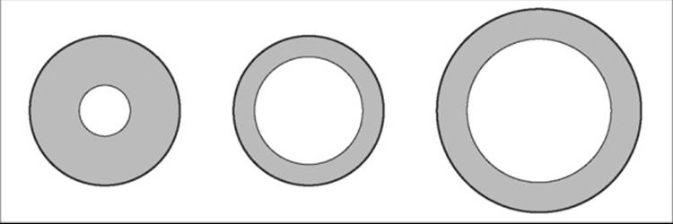
The normal optic disk is slightly oval, with the greatest dimension in the vertical. The normal optic cup is nearly circular, although it may assume a slightly oval shape as well. The size of the cup rather directly reflects the ratio between the size of the disk and the number of nerve fibers. In normals, the number of nerve fibers is not highly variable, whereas disk area may vary four to five times. Therefore, most of the variability in size of the normal cup reflects the size of the optic disk; larger disks have large cups, small disks, small cups. Clearly the size of the cup by itself, or the ratio of cup size to disk size, can have little meaning without considering the absolute size of the disk (Fig. 194.2).[11,12]
|
|
|
|
FIGURE 194.2 Cup:disk ratio is meaningful only when the overall disk size is considered. In this figure, a 0.3 cup is seen at far left in a disk of normal size. The center figure shows a 0.7 cup in a disk of the same size; this is far more suggestive of a pathologic condition than is the 0.7 cup on the right, in which the disk is one-third larger in diameter. In fact, the neural rim area (shaded) of the right nerve is the same as the neural rim area of the nerve on the left. |
The question of cup size has importance in glaucoma because the cup increases in size as nerve fibers are lost to the disease. Generalized diffuse loss of axons produces a uniform enlargement of the cup; focal loss of axons produces localized enlargement of the cup. The whole proposition may also be stated in terms of the neural rim, which is perhaps more meaningful because it is the neural rim that contains the functional tissue. From that perspective, generalized loss produces a rather uniform narrowing of the neural rim, whereas focal loss produces focal narrowing of the rim.
A large circular cup may be a sign of glaucoma (populations of glaucoma patients generally have larger optic nerve cups and smaller neural rim areas than do normal controls), but it may also be a normal variant. If the disk is small and the cup is large, the index of suspicion is higher. Extremely large cups are, of course, also more suspect. However, on a single look, cup size by itself is not highly specific for glaucoma.
A single look at circular cups can be more specific if there is inequality between the two eyes of a patient. If the eyes are otherwise similar, the cups should be similar - within 0.1 in the cup:disk ratio. A difference greater than 0.2 in the cup:disk ratio is highly unusual in the normal population and is likely to represent acquired damage.
The problem of identifying pathologic damage in a circular cup is far simpler if a change occurs over time. Although a slow, age-related loss of axons probably occurs normally, such progression is not detectable by usual clinical means. Therefore, clinical detection of any change is highly suggestive of an uncontrolled pathologic process.
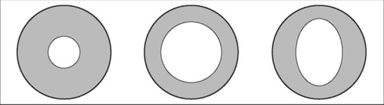
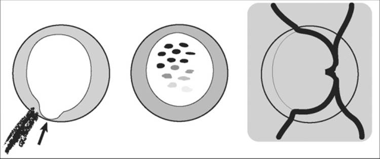
Frequently, glaucoma may selectively damage discrete regions of the nerve head. The two most likely foci for early selective damage are the superior and inferior poles of the nerve, with the inferior pole at somewhat greater risk than the superior. Early loss of neural tissue at the poles leads to a shape change in the optic cup from circular to oval resulting from vertical elongation of the cup. A slightly oval cup may be found in normal individuals, but a marked difference in vertical versus horizontal dimension is suggestive of glaucoma, even on a single examination (Fig. 194.3).[13] An observed change from a circular cup to a vertical elongation of the cup is, of course, a firm sign of an active pathologic process.
|
|
|
|
FIGURE 194.3 Enlargement of the cup may proceed in uniform fashion, going from 0.3 on the left to 0.6 in the center, or the vertical enlargement of the cup may exceed the horizontal, as seen on the right. As an isolated observation, a cup that is vertically oval (right) would raise a somewhat greater suspicion of glaucoma than would the central round cup depicted. |
Selective loss at one or both poles may lead to a focal narrowing of the neural rim, sometimes referred to as a polar notch.[14] The cup in this region is locally extended toward the scleral rim and may ultimately reach it.
As cupping progresses, the vertical cup elongation seen with moderate degrees of damage may begin to revert to a circular shape as the horizontal dimension of the cup expands because of increasing loss of temporal and then nasal axons. Usually, a small group of nasal axons survives in the late stages of damage, subserving a small island of vision in the temporal field. Another visible change that occurs as the cup widens and deepens is that the openings in the lamina cribrosa may be exposed to view.
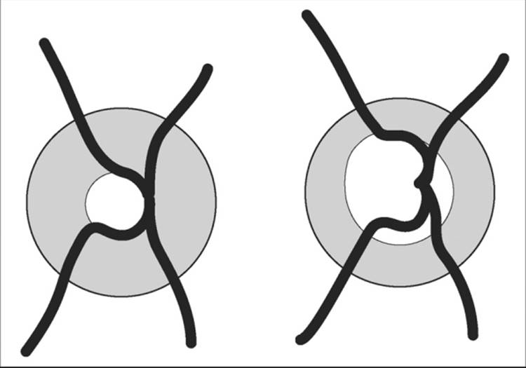
Throughout this process, the surface vessels (with a few notable exceptions discussed further on) shift with the diminishing nerve substance and provide a helpful guide to the altered contours of a damaged nerve. Tracing the path of individual vessels provides useful clues to overall nerve head contours, whereas comparison of the vascular paths on sequential photographs can be a sensitive indicator of change (Fig. 194.4). The vessels are particularly useful as markers when the clinical view of the nerve is gradually obscured, as with an advancing cataract.
|
|
|
|
FIGURE 194.4 The vessels follow the contours of the disk surface providing useful topographic information. Vessel pathways on a normal disk (left) show a gentle rise over the neural rim before flattening out to follow the surface of the retina. In a nerve damaged by glaucoma (right), the vessels show a deviation at their base toward the nasal side (all figures, right eye). The superotemporal vessel reveals flattening of the neural rim and a sudden deviation in course as it falls into a region of cup extension. The inferotemporal vessel previously followed the inferior rim of the cup (left). As the cup enlarged, this circumlinear vessel was left behind, no longer in contact with neural tissue. |
Occasionally, a vessel will not shift as the nerve beneath it atrophies, and this separation between nerve and a previously adjacent vessel often marks pathologic change. A vessel that follows the rim of the optic cup has been called a circumlinear vessel. If the cup expands and the vessel remains in place, a space develops between the vessel and the cup - 'baring of a circumlinear vessel'.[15] This sign is sometimes seen in normal eyes, so it is not specific for glaucoma. However, documented acquired baring, like other observable increases in cupping, is a specific sign of an active pathologic process.
The small vessels of the upper and lower poles of the nerve occasionally bleed, giving rise to a splinter hemorrhage on the disk. This is a rare finding in normal populations; it is most commonly associated with glaucoma but may also be seen with posterior vitreous detachment, ischemic optic neuropathy, or a bleeding diathesis. These small superficial hemorrhages occur in both primary open-angle glaucoma and normal-tension glaucoma, more frequently in the latter. They are frequently associated with focal thinning of the nerve and may presage later field defects, but not always. These hemorrhages are an important sign, since they indicate on a single look an active pathologic process, with glaucoma high on the list of causes (Fig. 194.5).[16]
|
|
|
|
FIGURE 194.5 As cupping advances, focal changes frequently occur in the two areas of highest risk: the inferior and superior poles. On the left, a nerve shows a focal area of total nerve loss - a notch in the inferior neural rim (arrow). Nearby, there is a small splinter hemorrhage, another indicator of localized nerve damage which may be present transiently, and may recur. The center figure depicts nerve damage which is more prominent superiorly. In this instance, the area of more severe damage is marked by increased visibility of the pores in the lamina cribosa. When cataract obscures detail and reduces tissue contrast (right), the vascular pathways often remain immensely helpful as still visible markers of the basic dimensions of the cup. |
Peripapillary atrophy may be found in association with glaucomatous nerve damage. Mild atrophy, showing only an irregular pigment pattern, is frequently a normal finding and not particularly associated with glaucoma. More severe atrophy, when nearly complete loss of pigment and choroidal capillaries reveals large deep choroidal vessels, is more suggestive of glaucoma. The area of such severe atrophy has been found to be spatially correlated with the extent of neural loss, and the atrophic area increases in size with increasing nerve damage. Not all glaucomatous optic nerve loss has associated peripapillary atrophy; but, when present, this finding should trigger increased vigilance.[17] Loss of axons, which presents at the nerve head as changes in the cup and neural rim, can also be detected over the retina. The nerve fiber layer of the retina may be seen as a uniform pattern of closely packed fine bundles radiating from the disk. Glaucomatous damage produces defects in these parallel bundles that appear as radiating dark, flat stripes.[18] The red-free light on the slit lamp or ophthalmoscope can increase the visibility of these defects, and specialized photographic techniques provide even more detail. This finding may be helpful in screening for glaucoma and in managing the disease. Unfortunately, the nerve fiber layer is more difficult to visualize in older age groups and in the presence of cataracts.
METHODS
The nerve head examination is clearly a procedure that must be performed repeatedly and with great care. Many of the signs of active damage are subtle - e.g., small hemorrhages, focal thinning of the nerve rim, vessel shifts. The direct ophthalmoscope provides excellent magnification and good illumination (with the newer light sources), but the view is monocular. This makes assessment of subtle contour changes more difficult, but not impossible. In most instances, the inner and outer borders of the neural rim can be identified with the direct ophthalmoscope, and this can allow numeric grading of the difference between the cup (defined by the inner edge of the neural rim) and the disk (the outer edge of the neural rim). Traditionally, this has been recorded as a cup-to-disk ratio: ranging from zero (no cupping) to 1 (fully cupped nervehead). But considering that the disk is known to vary in size, and that the shape of the disk and cup shape may be asymmetric, the meaning of this simple ratio may vary among individuals. Spaeth has introduced a more detailed, refined grading scheme keyed on the degree and extent of greatest thinning of the neural rim, and including compensation for variable disk size: the disk damage likelihood scale (DDLS).[19] Both approaches to numeric grading of the nervehead can be made with either the direct ophthalmoscope or with a slit lamp stereo view to record the status of the nerve at a given examination.
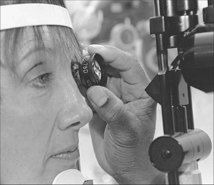
For the examiner who wishes a stereo view, several methods are available. The disk can be imaged in stereo, at high magnification, and in normal orientation using either a fundus contact lens, the central portion of an indirect goniolens, or a Hruby lens with the slit-lamp biomicroscope. The contact lens has the advantage of providing some stability of the lids and globe. An alternative is to use a high-diopter indirect lens (78-90 D) with the slit lamp (Fig. 194.6). These also provide a well-magnified stereo view, but the image is inverted. Once adjusted to the image inversion, this is an excellent method for disk examination. Aspherical lenses of lower dioptic power (60 D, 78 D) used to measure optic disk diameter have been shown to produce values that correlate well with measurements obtained with the Heidelberg retina tomograph, a confocal scanning laser ophthalmoscope.[20] Thus, it is possible to obtain an estimate of neural rim size without using expensive technology. The standard head-mounted binocular indirect ophthalmoscope does not provide sufficient magnification for routine, careful evaluation of the nerve head. The high magnification methods for examining the optic nervehead are also ideal for the vessels and macula, and these regions should be examined as well.
|
|
|
|
FIGURE 194.6 Slit-lamp examination of the optic nerve using a 78-D lens. |
RECORDS
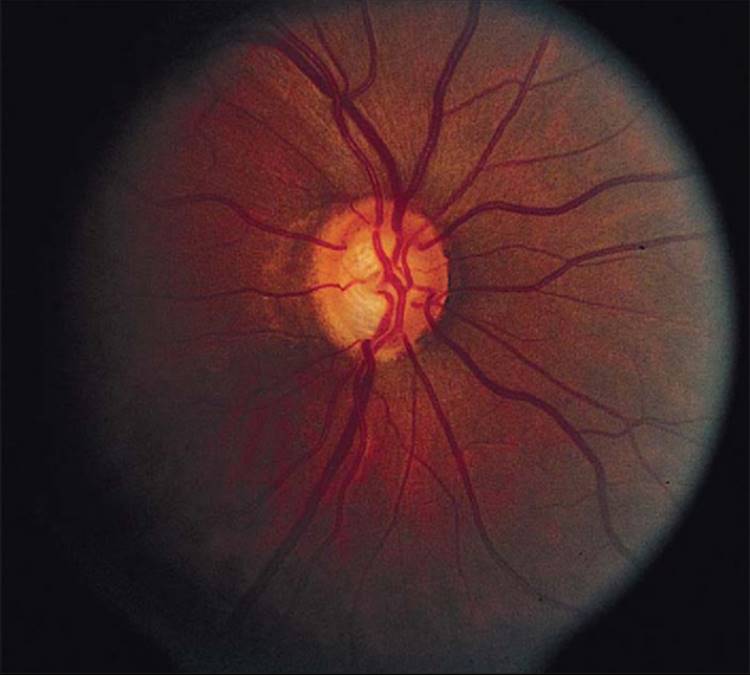
Generally, the nerve head changes caused by glaucoma occur gradually over several years. It is useful, therefore, to have accurate records of nerve head status at several points in time. An ideal method for recording nerve head appearance is high-magnification photography (Fig. 194.7), preferably in stereo. Frequently, subtle but definite changes can be seen in photographs even when no suspicion was raised on the clinical examination. Certainly this reflects the ease with which fine details can be studied and compared in photographs. It is also helpful to make drawings of the nerve head at frequent intervals. These cannot replace the precision of photographs, but they do force the examiner to focus her or his observations and will usually capture the major features of the nerve head. In the absence of photographic capability, of course, careful drawings are mandatory. The clinician should not rely on cup:disk ratios to follow the optic nerve.
|
|
|
|
FIGURE 194.7 Glaucomatous cupping with marked thinning of the neural rim superiorly and temporally and with cupping extending to the rim inferiorly. The severe loss of tissue inferiorly is also reflected in a loss of visible nerve fiber layer in the peripapillary region: this is found in a wedge-shaped area radiating from the edge of the disk from ~6 to 7 o'clock position. In this area, the fine striations of the nerve fiber layer are missing, throwing the vascular borders into stark profile. Contrast this with the same region superiorly where the striations of the nerve fiber layer are still evident, slightly obscuring the vascular margins. |
SUMMARY
When performing the clinical evaluation of a glaucoma patient, the clinician must remember that glaucoma is an eye disease with high-risk management. Glaucoma claims and law suits occur infrequently, but, when glaucoma claims occur, they result in an indemnity payment more than 50% of the time, and these payments are higher than the average ophthalmology indemnity payment.[21] The clinical evaluation of the glaucoma patient should include a comprehensive history and ocular examination as covered in this chapter. Other key elements of the evaluation, specifically perimetry, are covered in specific chapters in this section. Every element of the evaluation must be carefully documented in the patient's record for future comparison and to ensure the quality of care for outside review. The findings of the evaluation, as well as prognosis and treatment options, should be reviewed with the patient.
REFERENCES
1. Berson FG: Clinical examination: history. In: Albert DA, Jakobiec FJ, ed. Principles and practice of ophthalmology, Philadelphia: WB Saunders; 1993:1292-1294.
2. Epstein DL: The patient's history; symptoms of glaucoma. In: Epstein DL, Allingham RR, Schuman JS, ed. Chandler and Grant's glaucoma, 4th edn.. Baltimore: Williams & Wilkins; 1997:25-32.
3. Epstein DL: Examination of the eye. In: Epstein DL, Allingham RR, Schuman JS, ed. Chandler and Grant's glaucoma, 4th edn.. Baltimore: Williams & Wilkins; 1997:33-40.
4. van Herick W: Estimation of width of anterior chamber angle. Am J Ophthalmol 1969; 68:626.
5. Palmberg P: Gonioscopy. In: Ritch R, Sheilds MB, Krupin T, ed. The glaucomas, St Louis: CV Mosby; 1989.
6. Grant WM, Schuman JS: The angle of the anterior chamber. In: Epstein DL, Allingham RR, Schuman JS, ed. Chandler and Grant's glaucoma, 4th edn.. Baltimore: Williams & Wilkins; 1997:51-83.
7. Forbes M: Gonioscopy with corneal indentation: a method for distinguishing between appositional closure and synechial closure. Arch Ophthalmol 1966; 76:488.
8. In: Kolker AE, Hetherington J Jr, ed. Becker and Shaffer's diagnosis and therapy of the glaucomas, 5th edn.. St Louis: CV Mosby; 1976.
9. Spaeth GL: The normal development of the human anterior chamber angle: a new system of descriptive grading. Trans Ophthalmol Soc UK 1971; 91:709.
10. Airaksinen PJ, Tuulonen A, Werner EB: Clinical evaluation of the optic disk and the retinal nerve fiber layers. In: Ritch R, Sheilds MB, Krupin T, ed. The glaucomas, St Louis: CV Mosby; 1989:467-511.
11. Jonas JB, Gusek GC, Guggenmoes-Holzmann I, Naumann GOH: Size of the optic nerve scleral canal and comparison with intravital determination of optic disk dimensions. Graefes Arch Clin Exp Ophthalmol 1988; 226:213.
12. Caprioli J, Miller JM: Optic disk rim area is related to disk size in normal subjects. Arch Ophthalmol 1985; 105:1683.
13. Kirsch RE, Anderson DR: Clinical recognition of glaucomatous cupping. Am J Ophthalmol 1973; 75:442.
14. Read RM, Spaeth GL: The practical appraisal of the optic disk in glaucoma: the natural history of cup progression and some specific disk-field correlations. Trans Am Acad Opthalmol Otol 1974; 78:255.
15. Osher RH, Herschler J: The significance of baring of the circumlinear vessel: a prospective study. Arch Ophthalmol 1981; 99:817.
16. Drance SM, Begg IS: Sector hemorrhage: a probable acute ischemic disk change in chronic simple glaucoma. Can J Ophthalmol 1970; 5:137.
17. Jonas JD: Clinical implications of peripapillary atrophy in glaucoma. Curr Opinion Ophthalmol 2005; 16:84.
18. Hoyt WF, Frisen L, Newman NM: Fundoscopy of nerve fiber layer defects in glaucoma. Invest Ophthalmol 1973; 12:814.
19. Spaeth GL: The disc damage likelihood scale: reproducibility of a new method of estimating the amount of optic nerve damage caused by glaucoma. Trans Am Ophthalmol Soc 2002; 100:181.
20. Lim SC, O'Brien C, Bolton NM: A simple clinical method to measure the optic disk size in glaucoma. J Glaucoma 1996; 5:241.
21. Craven RE: Risk management issues in glaucoma: diagnosis and treatment. Surv Ophthalmol 1996; 40:459.