Joshua D. Stein,
Peter J. McDonnell,
Paul P. Lee
The number of patients undergoing penetrating keratoplasty has grown from 36 000 in 1987 to over 45 000 in 1998.[1] The 1- and even 3-year graft survival rates are now routinely greater than 90% for many ocular indications. Penetrating keratoplasty is one of the most successful of all transplant procedures.[2] As a result, it has been increasingly used at earlier stages in a wide range of ocular disorders for visual rehabilitation. Since the early 1980s, the leading indications for keratoplasty have been pseudophakic and aphakic bullous keratopathy (18% and 11%, respectively) and corneal regrafts (15%).[3,4] Other indications for penetrating keratoplasty are listed in Table 215.1. As more grafts are performed, however, the number of patients with problems associated with keratoplasty will continue to increase.
Anatomic complications associated with keratoplasty include glaucoma, graft melt, endophthalmitis, increased susceptibility to trauma, choroidal hemorrhage, macular edema, graft failure, retinal detachment, and worsening of other ocular disorders.[5,6] An additional visual complication is irregular astigmatism, which may limit the resulting vision even with otherwise anatomic success of the graft. Nevertheless, perhaps the most vexing problems are graft failure and the occurrence of increased intraocular pressure (IOP) after keratoplasty. The leading cause of enucleation (46%) after corneal transplantation is secondary glaucoma.[5] Actual clinical glaucoma after keratoplasty ranges from nearly nonexistent for grafts for keratoconus[7] to as high as 53% for pseudophakic or aphakic bullous keratopathy,[8] and even higher for more unusual conditions, such as viral or infectious keratitis (see Table 215.1). Over all, ?30% of corneal transplants are complicated by the subsequent development or worsening of glaucoma.[5]As the numbers of total grafts and grafts for more severely affected eyes, as well as those for graft failure (i.e., regrafts), increases, the management of postkeratoplasty glaucoma will become more important. Eyes with advanced glaucoma have a significantly elevated risk of graft failure,[10-12] and the surgical treatment of such glaucoma carries a high risk of subsequent graft failure.
TABLE 215.1 -- Indications for Penetrating Keratoplasty and Associated Chronic Glaucoma Rates (Intraocular Pressure)
|
Indication[3,4] |
Mean Follow-Up Period |
Rate (%) |
References |
|
Pseudophakic bullous keratopathy |
1-4 years |
18-53 |
7-9, 32, 142-145 |
|
Corneal regraft |
0.5-2.5 years |
45-50 |
9, 37, 146, 147 |
|
Aphakic bullous keratopathy |
0.5-3 years |
20-70 |
7-9,32, 144, 145, 37, 148 |
|
Trauma |
0.5-2.5 years |
9-55 |
37, 38, 69, 149, 150 |
|
Fuchs' dystrophy |
1 year (minimum) |
0-37 |
7, 9 |
|
Ulcerative disease |
1 year (minimum) |
50[*] |
38 |
|
Keratoconus |
1 year (minimum) |
0-12 |
7, 9, 38 |
|
Scarring and aniridia |
3 years |
56 |
151 |
|
Acanthamoeba keratitis |
4 years |
36 |
152 |
|
Peters' anomaly |
6.4 years |
60 |
153 |
|
Congenital hereditary endothelial dystrophy |
3 years |
0-3 |
154-156 |
|
Iridocorneal endothelial syndrome |
157-159 |
||
|
Viral keratitis |
1-2.5 years |
20-75 |
9, 37, 38 |
|
* |
N = 2. |
GLAUCOMA ASSESSMENT IN PATIENTS UNDERGOING KERATOPLASTY
It was only in the 1960s, with the development of instruments that allowed routine monitoring of IOP immediately after keratoplasty, that the problem of keratoplasty-associated glaucoma became widely recognized.[13-15] Postoperative glaucoma, astigmatism, and irregular corneal surfaces secondary to corneal edema, scarring, and epithelial irregularities before and after keratoplasty often make reliable Goldmann applanation tonometry impossible.[14] When the graft is clear, usually well after the operation, Goldmann or SchiØtz tonometry[16] may be useful. Even in this situation, however, the thickness of the cornea must be kept in mind; in eyes with thin corneas, IOP can be underestimated by as much as 5 mmHg, whereas in those with thick corneas, IOP can be overestimated by as much as nearly 7 mmHg.[17]
|
Key Features: Glaucoma Assessment in Patients Undergoing Keratoplasty |
||||||||||||
|
The MacKay-Marg tonometer, Tonopen, and pneumotonometer permit reliable assessment of IOP both preoperatively and postoperatively. Although a study comparing Goldmann applanation tonometry, Tonopen, and the ocular blood flow tonometry in 69 postkeratoplasty eyes found no significant difference in IOP measurements among these devices,[18] issues about graft thickness and compliance remain important in understanding the choice of measurement devices.
The definition of postkeratoplasty- or keratoplasty-associated glaucoma currently focuses on IOP,[13] frequently without reference to other parameters used to assess patients with glaucoma. This has largely been because of the difficulty in assessing the visual field and neuroretinal structures in eyes with corneal disease sufficient to require keratoplasty. Furthermore, postoperative astigmatism and refractive changes often preclude reliable postoperative assessment and comparison of the visual field, and analysis of the optic nerve and nerve fiber layer may not be possible until after surgery, because of the corneal opacification necessitating keratoplasty.
Although no study has explicitly investigated the feasibility of performing visual field testing and neuroretinal examinations in patients who have undergone keratoplasty, a few studies have suggested that these assessments may be possible in some or many patients.[19,20] In one such study, eight of 17 patients (47%) undergoing Molteno tube implantation had reliable automated visual fields preoperatively and postoperatively and 14 (82%) had a view sufficient to monitor the optic nerve head.[19] In another study, 10 eyes undergoing laser trabeculoplasty with clear grafts 2 years after keratoplasty were reliably monitored with visual field testing.[20] In routine care today, one should attempt to obtain visual fields on all patients with vision greater than hands motion (using Goldmann field testing until the 20/400 level, when it is preferable to switch to automated fields, where appropriate).
Frequency doubling perimetry has shown some promise as an alternative to conventional visual field testing. In a study of patients who underwent keratoplasty, frequency doubling perimetry could detect functional vision loss from glaucoma; its performance was not significantly affected by changes in corneal topography.[21] In addition, when the clarity of the graft permits, some of the newer imaging devices - such as optical coherence tomography, confocal scanning laser ophthalmoscopy, and scanning laser polarimetry - may be useful in determining whether patients who have undergone keratoplasty have structural damage to the optic nerve or nerve fiber layer.
Future efforts to study glaucoma associated with penetrating keratoplasty may include evaluation of other parameters in addition to IOP. This point is particularly important because the American Academy of Ophthalmology, in its Preferred Practice Pattern for primary open-angle glaucoma and angle-closure glaucoma, defines glaucoma as a group of optic nerve diseases, without diagnostic requirements of elevated IOP or visual field defect.[22,23] Thus, published studies cited in this chapter need to be evaluated with these limitations in mind.
Gonioscopy is another important component of the evaluation of patients with glaucoma to assess the status and structure of the filtration angle. Corneal pathology frequently precludes preoperative assessment, even after topical glycerin has been used to reduce corneal edema. Postoperatively, the size of the graft and corneal astigmatism may pose difficulties in attempting gonioscopy. However, evaluation of the angle is critical in helping determine the mechanism of glaucoma after keratoplasty. Most reports of studies in this area do not specify whether gonioscopy was performed routinely in the postoperative setting.
FACTORS IN KERATOPLASTY-ASSOCIATED GLAUCOMA
|
Key Features: Factors in Keratoplasty-Associated Glaucoma |
|||||||||
|
After ?50 years, much has been learned about the behavior of IOP after keratoplasty, including some of the risk factors for increased IOP and some possible mechanisms for postkeratoplasty glaucoma. Unfortunately, although a few large studies have investigated the relative risk factors in postkeratoplasty glaucoma or chronic elevated IOP, no prospective study designed to separate possible confounding factors has been performed.[24,25] In a retrospective study by Kenyon and colleagues involving 229 consecutive keratoplasty patients, 27% of whom had preoperative glaucoma, 34% of eyes developed sustained IOP elevations after surgery.[24] Sixty-two percent of patients with preoperative glaucoma required use of an increased number of medications or had uncontrolled glaucoma at their last visit (mean follow-up of 1.6 years), whereas only 23% of those without preoperative glaucoma developed chronic elevated IOP. In another retrospective study, 22% of 228 consecutive patients undergoing keratoplasty developed glaucoma; among those with glaucoma, 73% achieved IOP control by using ocular hypotensive medications, and the remaining patients required glaucoma surgery.[26] In a series of 500 consecutive patients undergoing keratoplasty, the 10-year cumulative risk for glaucoma was 21%.[27] The largest study to date analyzed data on 747 patients after keratoplasty and found that 11% of eyes developed glaucoma; of these, 52% responded to medical therapy, 29% required filtering surgery, and 19% required cyclodestructive procedures.[28] In that series, 49% of the grafts remained clear, although only 19% of the total sample had a best-corrected visual acuity of 20/60 or better (mean follow-up, 13.4 months).[29]
In the study by Kenyon and colleagues, risk factors associated with a sustained postkeratoplasty elevation of IOP were preoperative glaucoma, aphakia, intraocular lens (IOL) removal, and an operative diagnosis other than keratoconus. No association was found between IOP rise and regrafting, pseudophakia, concomitant vitrectomy, peripheral anterior synechiae (PAS) lysis, iridoplasty, secondary IOL placement, or concomitant cataract extraction and IOL implantation.[24] Another study also observed a higher risk for glaucoma among aphakic patients undergoing keratoplasty than among phakic or pseudophakic patients undergoing keratoplasty.[28] Two studies demonstrated the importance of the underlying ocular condition necessitating keratoplasty.[25,26] In one of these studies, postkeratoplasty glaucoma was found to be associated with bullous keratopathy, herpes virus, and trauma (relative risks of 2.2, 1.9, and 1.1, respectively).[26] In the other study, combined cataract-IOL surgery and PAS formation after surgery were each significant risk factors.[25] Thus, our understanding of postkeratoplasty glaucoma derived from studies with retrospective associations and the resultant rationale for treatment are incomplete and subject to change.
An excellent illustration of this evolution in thought comes from the initial description of the pressure response to keratoplasty. Among the first important points made was the bimodal nature (early and late) of IOP increases after keratoplasty and the predictive role of early pressure rises (within the first week) in glaucoma that develops months to years later.[30,31] In one study of early IOP elevation in 71 consecutive keratoplasties, 53 eyes (75%) had IOP higher than 21 mmHg during the first postoperative week.[30] In another study, 37 of 81 eyes (46%) undergoing keratoplasty combined with cataract extraction or for aphakic bullous keratopathy had an IOP greater than 35 mmHg during the first week.[31] In this study, those eyes with an early elevation of pressure were more likely (76%) than those without such a spike (61%) to have persistently elevated pressures at 6 months.[31]
Overshadowed in the flurry of excitement about these initial descriptions of IOP elevations after keratoplasty was the fact that these differences, and thus the predictive ability of early pressure rises, were not statistically significant. A later study by another group of investigators showed that only 36% of eyes (33 of 91) that developed late glaucoma at a mean follow-up of 3 years had had IOP spikes in the first postoperative week, although 75% of eyes with early spikes would subsequently have late pressure problems.[32] Later studies suggested that changes in operative techniques and postoperative care could reduce the incidence of both early and late IOP spikes.[13,32] One study found that only 7% of 209 eyes had postoperative IOPs greater than 21 mmHg, with none higher than 30 mmHg. The highest IOPs were recorded at 3 months after surgery; by 6 months, the patients' IOPs were similar to their preoperative levels.[33] Another study, conducted prospectively, detected an IOP of 30 mmHg or greater in the first week in only 12% of 155 eyes of all types undergoing keratoplasty.[34] It is evident that even the description of postkeratoplasty glaucoma depends on additional factors, such as the preoperative condition of the eye, the operative technique used, and the postoperative care.
Unaddressed in this context is the role of race in keratoplasty-associated glaucoma. Blacks are more likely than whites to develop glaucoma, at an earlier age and often of a more severe nature. How this observation relates to glaucoma after penetrating keratoplasty has not been explored.
SPECTRUM OF DISEASE
A helpful approach to organizing our understanding of postkeratoplasty glaucoma may be to consider what is known about these glaucomas. The following statements are generally recognized as true:
|
Key Features: Treatment of Postkeratoplasty Glaucoma |
||||||||||||||||||
|
|
1. |
Postkeratoplasty glaucoma occurs much more frequently (at twice or more the rate) in patients with preexisting glaucoma.[32,24,35,36] |
|
|
2. |
The underlying diagnosis precipitating keratoplasty plays a large role in the likelihood of developing glaucoma within 5 years after surgery. Inflammatory, infectious, traumatized, aphakic, and, possibly, pseudophakic eyes are at increased risk for postkeratoplasty glaucoma, as are, to a lesser extent, eyes undergoing corneal regrafts.[9,32,24,26,35,36] Regarding pseudophakic eyes, distinctions should be drawn between eyes with older-style anterior chamber or iris lenses and those with modern posterior capsule or anterior chamber lenses, especially those undergoing phacoemulsification removal. The higher risk is clear in the older-type lenses, whereas there may be no appreciable increase in risk with the modern surgeries and lenses.[24] |
|
|
3. |
Corticosteroids have a role in preventing and inducing postkeratoplasty glaucoma in a significant portion of patients.[8,9,32,37,38,39,40] |
|
|
4. |
Synechial angle closure occurs in many eyes with keratoplasty-associated glaucoma.[38,19,25,41-43] |
|
|
5. |
The part played by conformative changes within the anterior chamber changes induced by penetrating keratoplasty is uncertain.[44-52] |
When evaluated in light of the relative risks from a purely glaucoma perspective, the relative incidence of late-onset glaucoma after penetrating keratoplasty for different conditions (see Table 215.1) becomes more understandable. Certain conditions, such as keratoconus, generally occur in young phakic patients without prior surgery. In these eyes, initial transplants have a low incidence of late glaucoma. In contrast, penetrating keratoplasty in a patient with aphakic bullous keratopathy after intracapsular cataract extraction, with vitreous in the anterior chamber, represents major surgery on an eye that has had previous surgery; such surgery generally occurs in older patients (whose age, alone, is already a glaucoma risk factor) who have had surgical trauma damaging or obstructing their outflow pathways and may have had intervening corticosteroid use or bouts of inflammation. To the degree outflow pathways and any 'reserve' are already compromised, the additional trauma of more surgery in the anterior segment may place the outflow pathways into a steeper decline.
Mechanisms implicated in the development of keratoplasty-associated glaucoma can thus be understood by reference to principles present for other forms of glaucoma. Keratoplasty-associated glaucoma may represent a spectrum of glaucomas caused by one or more specific causes. As such, this offers hope for prevention and improved treatment for glaucoma in eyes after penetrating keratoplasty.
PROPOSED MECHANISMS
Although the list of possible mechanisms is lengthy (Table 215.2), certain mechanisms are more common than others. The four major preoperative factors affecting the risk of keratoplasty-associated glaucoma are the presence or absence of preoperative glaucoma, the phakic status of the eye, the size of the graft compared with the host, and the ocular diagnosis precipitating keratoplasty (see Tables 215.1 and 215.3). Two potential factors intraoperatively are the size of the grafts used and, possibly, the use of viscoelastics. The two major postoperative risk factors are the use of corticosteroids and progressive synechial angle closure (Table 215.4).
TABLE 215.2 -- Mechanisms of Increased Intraocular Pressure After Keratoplasty
|
Open-Angle Glaucoma |
||||||||||||||||||
|
Early Onset |
||||||||||||||||||
|
||||||||||||||||||
|
Intermediate Onset |
||||||||||||||||||
|
||||||||||||||||||
|
Late Onset |
||||||||||||||||||
|
||||||||||||||||||
|
Angle-Closure Glaucoma |
||||||||||||||||||
|
Early Onset |
||||||||||||||||||
|
||||||||||||||||||
|
Late Onset |
||||||||||||||||||
|
TABLE 215.3 -- Factors Associated with Elevated Intraocular Pressure
|
Aphakic (%) |
Pseudophakic |
Phakic |
|
|
Phakic Status |
|||
|
Early rise[10] |
89 |
Unknown |
18 |
|
Late rise[17-20] |
35-53 |
16-39 |
4-20 |
|
Preoperative Glaucoma |
|||
|
Early rise[16,19,23] |
|||
|
With |
48-82 |
||
|
Without |
17-41 |
||
|
Late rise[3,16,19,23,25,88] |
|||
|
With |
76-100 |
38-40 |
|
|
Without |
10-56 |
25-50 |
|
|
Graft Size (All Without |
|||
|
Preoperative Glaucoma) |
|||
|
Early rise[24,35,37] |
|||
|
Same size |
29-56 |
6-25 |
|
|
Oversize |
8-45 |
0-4 |
|
|
Late rise[24,35] |
|||
|
Same size |
31-47 |
24 |
|
|
Oversize |
10-42 |
16 |
TABLE 215.4 -- Postoperative Factors in Elevated Intraocular Pressure
|
Study, by Postoperative Factor |
% |
Patient Population |
|
Corticosteroid Response |
||
|
Krontz and Wood[41] |
60 |
Acute nonangle closure glaucoma after combined surgery |
|
Goldberg et al[37] |
23 |
Mixed |
|
Thoft et al[38] |
17 |
Aphakic bullous keratopathy/pseudophakic bullous keratopathy |
|
Foulks[32] |
7 |
Mixed |
|
Kirkness and Moshegov[7] |
5 |
Mixed |
|
Peripheral Anterior |
||
|
Synechiae Formation |
||
|
Thoft et al[38] |
100 |
Aphakic bullous keratopathy/pseudophakic bullous keratopathy (not clear whether done in all) |
|
Chu et al[70] |
50 |
Pseudophakic bullous keratopathy-iris-suture posterior chamber intraocular lens |
|
Kenyon et al[69] |
86 |
Trauma |
|
Lass and Paven-Langston[85] |
100 |
Mixed |
|
McDonnell et al[19] |
76 |
Mixed |
|
Waring[68] |
46 |
Mixed |
|
Foulks[32] |
14 |
Mixed |
|
Cohen et al[67] |
40 |
Unknown |
PREOPERATIVE FACTORS
As seen in Table 215.1, the rates of chronic glaucoma after keratoplasty differ significantly on the basis of the ocular diagnosis or indication for keratoplasty. From a low of 0-12% for keratoconus to a high of 75% after infectious (viral) keratitis, the rates vary tremendously. As noted earlier, such variation is not surprising, given the underlying patient populations within each of the ocular diseases.
Although the three factors noted in Table 215.3 have been observed in several studies over the past 30 years, no multivariate analysis has assessed possible interactions among variables. For example, aphakic eyes may be more likely to have preoperative glaucoma than phakic eyes. Although this factor can be controlled to some degree by looking at the incidence of preoperative glaucoma among a subset of patients (e.g., with aphakia or pseudophakia, as in Table 215.3),[24] this method precludes generalizing to other patient groups. The importance of having such studies can be seen in one large consecutive series of penetrating keratoplasties, in that when eyes with a preoperative diagnosis of glaucoma were excluded from the analysis, aphakia was no longer associated with a sustained IOP elevation after surgery.[24] Similarly, nearly all of the work in oversize grafting has been in eyes without preexisting glaucoma (see Table 215.3). Although this approach allows for the most uncluttered analysis, it omits critical data in the group shown to be at increased risk - aphakic patients with preoperative glaucoma - without any firm assurance that the data are readily generalizable to other groups. It would thus be anticipated that additional future studies would have sufficient data and numbers to allow us to address these questions in a statistically more significant manner. Indeed, several large studies with multivariate analyses have been published regarding graft survival and failure;[12,53-57] it would be illuminating to evaluate those surgical datasets with the end-point of glaucoma as well.
PHAKIC STATUS
Data from several studies are sufficient to determine the part played by the eye's phakic status in keratoplasties performed by the same surgeon.[9,14,24,32,36,28] However, none of these studies provide sufficient information to allow us to subcategorize by the presence of preoperative glaucoma or the use of oversize grafting. Thus, with the recognition of possible confounding by the other two factors, it is clear that both in the immediate postoperative period and at follow-up to 3 years, aphakic eyes have the highest rate of elevated IOP. Pseudophakic eyes appear to have an intermediate position (again, most likely due to the presence of older style implants and surgical techniques in the older series), and naturally phakic eyes have the lowest rate. As such, we should expect, and so inform our patients, that the risk of elevated IOP among all aphakic patients is at least 40%. A recent retrospective study confirmed that compared with phakic and (modern) pseudophakic patients, patients with aphakic eyes (odds ratio, 6.6) are at significantly increased risk of elevated IOP after keratoplasty (odds ratio, 6.6).[28]
The mechanisms underlying this well-known predisposition are unclear. A partial explanation is the higher rate of preoperative glaucoma among aphakic eyes compared with phakic eyes, given the importance of preoperative glaucoma.[24,34,35] However, this is in dispute, because Foulks[32] found that aphakia was an independent risk factor for postoperative IOP elevation, whereas Simmons and co-workers[24] did not. Studies by Olson,[44] Olson and Kaufman,[45] and Zimmerman and colleagues[46] suggest that the size of the graft may be an important factor because of anterior segment structural changes induced by keratoplasty. Zimmerman and colleagues were able to show that the depth of suture placement had no effect on outflow facility in phakic human eye bank eyes undergoing penetrating keratoplasty. However, unlike through-and-through sutures, nonpenetrating sutures produced a 37% decrease in outflow facility in aphakic eye bank eyes.[46] It was hypothesized that the lens provides the trabecular meshwork with some form of support that is lost when the lens is removed. This loss of trabecular support, in conjunction with distortive forces induced by the keratoplasty, may increase aqueous outflow resistance. A clinical trial to test this hypothesis was discontinued because of persistent suture tract leaks among eyes with through-and-through sutures.[47] A subsequent study showed that the same effect as through-and-through sutures could safely be achieved by using a 0.5-mm oversize graft.[48]
How these factors interact is unknown at this time. Nevertheless, it does appear that phakic eyes have a reduced likelihood of developing keratoplasty-associated IOP elevations.
PREOPERATIVE GLAUCOMA
The role of preoperative glaucoma in predisposing to elevated IOP after keratoplasty has been examined in detail only among aphakic and pseudophakic eyes (see Table 215.3). Although one report states that preoperative glaucoma is significantly associated with postoperative IOP elevation among phakic eyes as well as aphakic eyes,[32] insufficient data are published to allow us to determine the amount of difference. Part of the reason why most studies have concentrated on aphakic and pseudophakic eyes may be the high percentage of keratoplasties performed on aphakic and pseudophakic eyes as well as the relatively low incidence of glaucoma among diseases in phakic eyes with a need for keratoplasty. Similarly, phakic eyes, by definition, have had at least one fewer operation and may have had no prior operations. Nevertheless, it is clear that aphakic eyes with clinically controlled glaucoma preoperatively have at least a 75% risk of losing IOP control postoperatively (see Table 215.3). It is interesting that the rate of IOP derangement in pseudophakic eyes does not appear to be influenced by the preoperative glaucoma status of the eye (see Table 215.3).
The explanations for this phenomenon are even more hypothetical than for phakic eyes. Again, it can be argued that aphakic eyes undergoing penetrating keratoplasty are even more disordered than pseudophakic eyes and that keratoplasty thus represents a greater insult to the eye. It could be that pseudophakic eyes provide more posterior support to the trabecular meshwork than aphakic eyes, so that the postulated trabecular meshwork distortion is less significant in pseudophakic eyes.
Performing penetrating keratoplasty on eyes without adequate pressure control preoperatively or without performing a combined procedure to lower the pressure is generally not recommended. One can expect an even greater frequency and severity of IOP problems in these eyes after surgery. Given the increased risks of suprachoroidal hemorrhage in eyes with uncontrolled IOP,[58] caution is thus required regardless of the timing of the glaucoma surgery.
INTRAOPERATIVE FACTORS
The ability of the surgeon to influence the likelihood and severity of glaucoma has not been widely studied. Unlike the status of cardiac surgery, for example, no analysis in the ophthalmic literature has controlled for who performed the keratoplasty. Given the potential importance of factors such as PAS formation after surgery or wound distortion, the technical skill of performing the surgery may be a critical factor.
At the same time, there are at least two areas that have been investigated. The choice of viscoelastics at the time of keratoplasty may have a role in minimizing the formation of PAS during surgery, and the size of corneal grafts may influence the likelihood of glaucoma.
Choice of Viscoelastics
Because early IOP spikes may be of concern in eyes with advanced glaucoma undergoing keratoplasty, being aware of the IOP course after surgery may be helpful in preventing damage during this time. In a randomized study of 62 patients, those treated with both hyaluronate sodium (Healon) and hyaluronate sodium/chondroitin sulfate (Viscoat) were found to have pressure elevations of 75% above baseline at 10-24 h after surgery.[59] Thus, patients at risk of additional damage at pressure ranges in the high 20s to low 30s might need to receive additional pressure-lowering agents after surgery (if feasible). In another study, a comparison of Healon and Healon GV found that GV was superior to standard Healon in maintaining space in the open anterior chamber.[60] To the extent that this decreases the intraoperative formation of synechiae, it may help avert some progression after surgery. However, the effect of using GV on pressure after surgery was not described. Although no study has assessed the effect of viscoadaptive agents like Healon 5 on IOP after keratoplasty, investigations comparing Healon 5 with other viscoelastics during routine phacoemulsification have found no association between the use of these agents and postoperative IOP rise.[61,62] Unlike with cataract surgery, in which viscoelastics are routinely removed from the eye at the close of the procedure, viscoelastics are often left in the eye after penetrating keratoplasty.
Use of Oversize Grafts
Studies on the role of oversize grafts have been confined almost exclusively to eyes without preoperative glaucoma (see Table 215.3) and mainly in the immediate postoperative period.[49-52,53-57,63] Because many of these studies investigated small numbers of eyes, the failure of some studies to find a statistically significant difference may not mean that the concept supporting oversize grafting is invalid. Similarly, the failure of the studies investigating the chronic elevation of IOP to find a statistically significant difference[49,50] should be evaluated with this in mind, because both suggest a trend toward a protective effect of oversize grafting. Nevertheless, the concept of oversize grafting clearly has not been proved definitively.
The use of oversize grafting - originally suggested by Franceschetti in 1949 and then tested theoretically by Olson,[44] Olson and Kaufman,[45] and Zimmerman and colleagues[46] - has become standard practice. As Olson[52,64] has pointed out, the actual size of the button removed can vary depending on which surface (endothelial or epithelial) is trephined or cut and what the pressure of the eye is (when removed from the host or from a whole-eye donor) at that moment. Therefore, the degree that grafts are oversized (or not) may vary. As such, any conclusion that can be drawn must be tentative. A retrospective series of 3992 keratoplasties found that small trephination size is associated with an increased risk for graft failure.[42] However, another study, which used multivariable analysis to assess graft survival, found that oversize grafting with a disparity larger than 0.25 mm is associated with poorer graft survival.[56]
POSTOPERATIVE FACTORS
The two major postoperative influences cited in altering the course after keratoplasty are the presence of synechial closure of the angle and the dual role played by corticosteroids (see Table 215.4). These represent two mechanisms by which the preoperative factors noted earlier may cause elevated IOP after keratoplasty.
Peripheral Anterior Synechiae
Progressive angle closure carries a poor prognosis for pressure control and graft survival.[9,32,42,65] Table 215.4 shows some of the reported rates of PAS formation as a percentage of eyes that have IOP elevation. Other studies have shown that the presence of PAS may indicate a need to undergo later surgery; in one series, five of eight eyes with PAS subsequently needed surgical intervention for glaucoma at a follow-up of 3 months to 27 years.[66]
The importance of progressive synechial closure has led many surgeons to recommend lysis of synechiae at the time of grafting. Furthermore, intraoperative iridoplasty and synechialysis, perhaps as part of an anterior chamber reconstruction, have successfully reduced the incidence of continued progressive synechial closure after keratoplasty.[67,68] In a review of 39 cases with trauma, Kenyon and associates[69] reported that 86% of eyes with persistent synechiae even after attempted intraoperative anterior chamber reconstruction and lysis of synechiae had glaucoma postoperatively. Thus, the need to be aware of formation of synechiae and to potentially attempt to prevent it is well recognized.
Progressive angle closure offers an attractive mechanism to explain at least part of the population for keratoplasty-associated glaucoma, particularly among those without preoperative glaucoma. Some studies have demonstrated its presence in all eyes with elevated IOP after keratoplasty.[38] None of the reports supporting the role of oversize grafts, aphakia, or the preoperative presence of glaucoma have involved routine gonioscopy of angle structures. However, one major study that did conduct routine gonioscopy found that progressive synechial closure could account for only 14% of eyes with elevated IOP.[32]
CORTICOSTEROIDS
Before researchers had elucidated the role of immune and inflammatory factors in graft rejection and inflammatory glaucomas, the use of corticosteroids in the postoperative period had not been emphasized. The use of potent corticosteroids at frequent intervals was reported to reduce the perceived rate of acute or early IOP elevation.[38] However, with the recognition of the need for corticosteroids to reduce inflammation and decrease allograft rejection came the awareness that a certain proportion of IOP elevations represented corticosteroid responsiveness (see Table 215.4).
The reported rate of corticosteroid-responsive glaucoma ranges from 5% to 60% (see Table 215.4). In an innovative prospective study in which 25 patients with glaucoma after keratoplasty were given topical cyclosporine instead of topical corticosteroids, Perry and coworkers[40] noted that 84% of the patients had a reduction in IOP (mean of 9 mmHg) and 13 patients could discontinue use of one or more glaucoma medications. The importance of this factor is highlighted by its 'occasional' occurrence and the fact that the first therapeutic step should be to reduce the dose of corticosteroids[39] or to use less potent steroids. Insufficient information exists to draw any conclusions about the interrelationships between the role of corticosteroids and the preoperative factors noted earlier.
UNANSWERED QUESTIONS
Many questions remain about whether additional mechanisms are at work in keratoplasty-associated glaucoma. In a large, comprehensive study, 91 of 502 eyes (18%) developed chronic postoperative glaucoma[32]; however, only 32 of these 91 eyes had had glaucoma previously. The proportion of cases involving progressive angle closure, acute angle closure, corticosteroid-induced glaucoma, or allograft rejection was not reported. However, because these known causes added up to 28 cases, at one extreme only four of 502 eyes (1%) could have developed open-angle glaucoma over several years. Although this may be unlikely, it does suggest that previously described mechanisms for other forms of glaucoma could indeed explain a large proportion of eyes with new-onset keratoplasty-associated glaucoma. As with other areas discussed in this chapter, large multivariable analyses are required to answer questions about exactly what the relative risk factors are for elevated IOP and glaucoma after keratoplasty and how best to modify them.
TREATMENT MODALITIES
Glaucoma associated with penetrating keratoplasty is often difficult to control,[70,71,72] largely because of the underlying nature of the ocular conditions necessitating the transplantation and the postoperative complications associated with keratoplasty. For example, aphakic glaucoma, even in the absence of a corneal graft and the presence of an open angle, is often difficult to control. Similarly, angle closure postoperatively reduces the number and effectiveness of treatment options. Thus, a critical consideration is the prevention of those mechanisms that may contribute to the development of postoperative glaucoma.
Despite our best efforts at prevention, some patients with grafts will develop glaucoma. The available options today are much broader and potentially more effective than those of just 25 years ago. They include medical treatment, laser trabeculoplasty, laser iris manipulation, filtering surgery, implant surgery, laser ablation of the ciliary structures, and cryoablation.
A related issue is the effect of penetrating keratoplasty on eyes with preexisting glaucoma and functioning filtering operations. As noted earlier, keratoplasty in eyes with preoperative glaucoma results in reduced glaucoma control in a significant number of patients. Here, too, however, little is known about an important effect of keratoplasty, namely, the effect of keratoplasty on filtering blebs.
The control of glaucoma, however, is only one consideration. The effect of glaucoma treatment on rejection episodes, graft clarity, and graft survival is as important to patients, because the grafts are usually performed in an attempt to increase visual function. Studies have clearly shown that glaucoma is a significant, independent risk factor for graft failure,[11,73] but whether the effect is due to the glaucoma, the treatment given, or both is unknown. The available data on the effects of various surgical approaches come from studies with relatively modest numbers of cases.[32,19,20,74-81] More important, no investigations have addressed the effect of topical and systemic glaucoma medications on graft rejection, graft clarity, and graft survival, even though timolol and pilocarpine are potentially toxic to the epithelium,[82,83] and recent research suggests that topical carbonic anhydrase inhibitors may contribute to corneal decompensation.[84]
MEDICAL THERAPY
Unlike eyes with chronic open-angle glaucoma that have never undergone surgery, eyes with penetrating keratoplasty-associated glaucoma have had at least one major operation, with significant alterations in anterior structures. Treatment is directed at the underlying cause, if the cause can be discerned. In any case, during the acute phase, medical therapy is the initial treatment. Unlike the situation with primary open-angle glaucoma, the risks of glaucoma surgery are compounded by the risks to the graft of additional intraocular surgery, such that surgical therapy is clearly reserved, at this time, for those whose glaucoma is uncontrolled on maximal tolerated medical therapy.
?-Blockers
The reduction of aqueous production by b-blockers is helpful in patients who can tolerate their use.[85,86] Even in aphakic eyes with partial synechial angle closure, timolol was found to reduce IOP to less than 22 mmHg in 70% (9 of 13) of eyes, from a mean baseline of 39.7 mmHg.[85] Another study comparing timolol gel with oral carbonic anhydrase inhibitors for viscoelastic-induced ocular hypertension after keratoplasty found that timolol gel offered better IOP control with fewer systemic side effects.[87] Further data on the effectiveness of ?-blocker therapy in keratoplasty-associated glaucoma have not been published, but widespread experience suggests that it is effective.
Epinephrine
The use of adrenergic agents is limited by several considerations. First, because many eyes are aphakic, the risk of inducing cystoid macular edema is present.[88] Second, the additive effect to topical ?-blockers is minimal, particularly with nonselective agents, such as timolol or levobunolol.[89,90] Third, the presence of synechial closure would limit one proposed mechanism of action, that of increasing conventional outflow. Fourth, induced alterations in the ocular-blood barrier, particularly in the presence of inflammation, may affect graft survival or clarity. Thus, the use of epinephrine compounds should be limited to specific patients and clinical situations, such as phakic patients who have open angles and who are not concurrently using nonselective ?-blockers.
Parasympathomimetic Agents
Both direct- and indirect-acting agents are useful in the treatment of keratoplasty-associated glaucoma.[13,91] However, because they act by increasing conventional outflow through the trabecular meshwork, their effectiveness is markedly reduced in the presence of significant synechial angle closure.
?-Agonists
Apraclonidine (Iopidine), an ?2-agonist, and brimonidine (Alphagan) have been approved by the Food and Drug Administration for chronic use in patients with glaucoma as well as during the perioperative period for laser trabeculoplasty and posterior capsulotomy. Experience suggests that a-agonists can be useful adjuncts, particularly in an acute setting, for keratoplasty-associated glaucoma. Both appear to confer an additional pressure-lowering effect when used in combination with ?-blockers; thus, they can be a useful addition to our medical therapy approach, particularly in the early postoperative period.
Carbonic Anhydrase Inhibitors
Before the development of topical ?-blockers, oral carbonic anhydrase inhibitors were considered the most effective form of medical treatment for keratoplasty-associated glaucoma. They remain effective in most patients, albeit at the risk of well-known systemic side effects. Experience suggests that topical carbonic anhydrase inhibitors (dorzolamide and brinzolamide) also reduce IOP after keratoplasty, with fewer reported systemic side effects. However, a recent series of nine eyes, four of which had undergone keratoplasty, had irreversible corneal decompensation 3-20 weeks after initiation of therapy with topical carbonic anhydrase inhibitors.[84] Additional studies with larger samples are warranted to better assess the effect of topical agents on corneal clarity in eyes with grafts.
Prostaglandin Inhibitors
The development of prostaglandin analogs and prostamides has provided a fifth class of medications for use in patients with glaucoma. By increasing uveoscleral outflow, these drugs decrease IOP through a mechanism additive to most other medications. Somewhat surprisingly, they are also additive in some patients using pilocarpine and other sympathomimetics. Because of their high degree of effectiveness, once a day dosing, and minimal systemic side effect profiles, they are increasingly the first line agent in many patients with glaucoma and the presence of a keratoplasty.
Corticosteroids
As noted above, corticosteroids are needed to reduce postoperative inflammation and to prevent and treat graft rejection. Experience has shown that these agents can be helpful in reducing IOP postoperatively, presumably by reducing inflammation-associated reductions in aqueous outflow.[38] However, they can also cause corticosteroid-induced glaucoma when used more chronically. Thus, patients with increased IOP postoperatively must be assessed for the possible need to either decrease or increase corticosteroids after other potential causes of elevated IOP have been ruled out. If the cause of elevated IOP is in question - as, for example, in the presence of mild inflammation 2 weeks postoperatively - one possible approach is to give potent corticosteroids frequently for 48 h. If the IOP is elevated because of inflammation, IOP reduction should be observed; however, if it is not, then corticosteroids could be tapered to the minimum amount and strength possible to ensure graft survival.
LASER THERAPY
The use of argon and yttrium-aluminum garnet (YAG) lasers or argon lasers to perform a peripheral iridotomy or anterior hyaloid face disruption is dictated by the clinical condition. In those patients who develop pupillary block or malignant glaucoma (in pseudophakic or aphakic eyes) respectively. (The use of neodymium:YAG (Nd:YAG) lasers for cycloablation is discussed later.)
Pupillary Block and Malignant Glaucoma
A patent peripheral iridectomy is often created at the time of penetrating keratoplasty. In aphakic eyes, or those with anterior chamber IOLs, it is mandatory. In those situations in which a complete iridectomy has not been performed or a patent iridectomy has subsequently been closed by inflammation or scarring, a laser iridotomy can be performed to prevent acute and chronic angle-closure glaucoma. Indeed, 6% of cases of elevated IOP in one series were due to acute angle closure.[32]
Malignant (ciliary block, aqueous diversion) glaucoma can be treated with the YAG laser in aphakic and pseudophakic eyes.[92] The aim is to disrupt the anterior vitreous hyaloid face.[92] In the keratoplasty setting, differentiation from pupillary block can be particularly difficult, because postoperative inflammation and the effects of intraoperative iris manipulation can result in loculated aqueous pockets posteriorly. The presence of a patent iridectomy is necessary to rule out a pupillary block mechanism before treatment for malignant glaucoma can be initiated.
Laser Iridoplasty/Gonioplasty
The finding of progressive synechial angle closure after penetrating keratoplasty is an ominous sign because it represents potentially worsening glaucoma over time. Although a few eyes with progressive synechiae may not develop IOP elevation, those that do are extremely difficult to manage.[32] Although no case of laser gonioplasty after keratoplasty has been reported, its successful use in other settings with chronic or secondary angle-closure, with or without trabeculectomy,[93,94] suggests that this may be a potentially useful approach, particularly given the initial results of intraoperative synechialysis (discussed later).
Laser Trabeculoplasty
In selected eyes, laser trabeculoplasty after penetrating keratoplasty may be helpful. Van Meter and colleagues[20] performed laser trabeculoplasty in 10 eyes an average of 25 months after penetrating keratoplasty; the IOP was reduced by more than 5 mmHg in eight eyes (mean of 9.1 mmHg), with a mean follow-up of 23 months. Both eyes without PAS and six of eight eyes with PAS were successfully treated. Eyes without prekeratoplasty glaucoma had a mean pressure reduction of 46%. Four eyes with preoperative glaucoma had a mean decrease of 15%; however, whether these eyes had prior laser trabeculoplasty was unstated.[20]
In another study, Gross and colleagues[95] performed laser trabeculoplasty in 20 aphakic and pseudophakic eyes after grafting. At 9 months, only 45% had an IOP less than 22 mmHg and only 10% met both vision and pressure criteria for success.[95] Thus, laser trabeculoplasty is less successful in aphakic and pseudophakic eyes and in eyes with large amounts of PAS, which limit the amount of treatable angle. In other eyes, particularly those without glaucoma before keratoplasty, laser trabeculoplasty should be considered.
SURGICAL APPROACHES
Available studies on the success and effects of surgical intervention for keratoplasty-associated glaucoma suffer several major shortcomings. First, many of the earlier studies do not present life-table or survival analyses; only some of the newer studies use this necessary statistical technique. Second, the criteria for success are generally stated in terms of IOP and lack of disastrous complications alone, without regard to the optic nerve or visual field used in observing other patients with glaucoma. Again, however, this reflects the relative difficulty in obtaining such parameters in keratoplasty eyes. Relatively few older studies, although almost all studies published since 1990, analyze graft survival as an important end-point, but most do contain enough information to discern that glaucoma surgery poses a significant risk to continuing graft survival. Third, the numbers are generally small, because any one given center is unlikely to have adequate numbers of patients with keratoplasty-associated glaucoma to develop meaningful numbers. With these limitations in mind, it is clear that keratoplasty-associated glaucoma poses significant challenges for its surgical management.
The need for surgical intervention in eyes with keratoplasty-associated glaucoma has been estimated in the past at 0-57%.[32,37,26,28,95-98] Among phakic eyes receiving grafts for inactive interstitial keratitis, the lowest rate of 12% at an average follow-up of 3.7 years was observed.[66] Among a mixed population of patients, Foulks[32] reported a rate of 24% at 3 years. Not surprisingly, among a mixed population of eyes undergoing repeat grafting, Goldberg and colleagues[37] reported a higher rate, of 35%, at a follow-up of only 7-30 months. Among seven eyes in patients who had congenital glaucoma (having had previous glaucoma operations) and who received grafts when they were adults, four eyes required additional glaucoma surgery after the keratoplasty and the other three required medical therapy.[96] Finally, in a study of nine adults who underwent penetrating keratoplasty for corneal edema secondary to congenital glaucoma, during a mean follow-up of 28 months, none required glaucoma surgery.[98]
Because chronic use alpha-agonists and prostaglandin analogues were unavailable before the mid-1990s, reported rates of surgery today are most likely lower than those from earlier studies with equivalent durations of follow-up. However, the use of additional medications is likely to only delay the need for subsequent surgery. One suspects that as longer follow-up occurs with patients, the rates of surgery will once again rise to historic levels.
Because penetrating keratoplasty is often performed on seriously compromised eyes and the procedure, itself, substantially alters ocular structures, glaucoma surgery is often difficult and less successful. In many eyes, the conjunctiva is scarred. In many cases, the eyes are aphakic or pseudophakic. As a result, many eyes are poor candidates for filtration surgery, and implant surgery or cilioablative procedures may be required. Indeed, in many eyes, filtration surgery is technically impossible, owing to the absence of conjunctival tissue at or even near the limbus or its replacement by dense scar tissue.
Filtration Surgery
The use of trabeculectomy has been investigated in a limited number of studies.[32,75,95,97,99-104] As can be seen in Table 215.5, the reported success rate in controlling pressure ranges from 27% to 91%, although the success rate of both controlling pressure and maintaining vision or graft clarity is much lower (27-82%). Since the use of potent antimetabolites, such as mitomycin C, during trabeculectomy has become the standard of care for complex cases in recent years, the rates of success reported for IOP control have improved substantially, compared with some earlier studies. A study by Ishioka comparing the effectiveness of trabeculectomy with and without mitomycin C confirmed the importance of using antimetabolites, with IOPs less than 21 mmHg achieved in 19 of the 26 eyes (73%) that had had mitomycin C but only two of the eight eyes that had not (mean follow-up, 22 months).[104] Moreover, a significantly higher proportion of those in the mitomycin C group than in the nonmitomycin C group maintained graft clarity (69% vs 37%).
TABLE 215.5 -- Trabeculectomy[*]
|
Study |
Criteria |
Mean Follow-Up |
Patient Population |
Success (%) |
|
Foulks[32] |
IOP |
3 years |
Mixed |
80 |
|
Gilvarry et al[75] |
IOP |
3 years |
Mixed |
51 |
|
Gross et al[95] |
IOP and VA |
9 months |
Aphakic/pseudophakic |
27 |
|
Insler[99] |
IOP |
16 months |
Mixed |
43 |
|
Figueiredo et al[100] |
IOP |
16 months |
Aphakic/pseudophakic |
67 |
|
Kirkness et al[101] (before penetrating keratoplasty) |
IOP and VA |
5 years |
Mixed |
27 |
|
Kirkness et al[101] (with penetrating keratoplasty) |
IOP and VA |
5 years |
Mixed |
50 |
|
Kushwaha and Pual[97] |
IOP |
1 years |
Mixed (immediate operation within 2 wk of penetrating keratoplasty) |
53 |
|
Chowers[102] |
IOP and clear graft |
14 months |
Pseudophakic |
91 |
|
WuDunn[103] |
IOP |
2 years |
Mixed |
50 |
|
Ishioka[104] (with MMC) |
IOP |
22 months |
Mixed |
73 |
|
Ishioka[104] (without MMC) |
IOP |
22 months |
Mixed |
25 |
|
* |
Numbers in superscript are reference numbers. IOP, intraocular pressure; MMC, mitomycin C; VA, visual acuity. |
The success of trabeculectomy is influenced by the same factors that alter its success in the nonkeratoplasty setting. Gilvarry and colleagues[75] reported, for example, that the success rate was four times greater in eyes that had only one graft performed as opposed to those with two or more. Similarly, aphakic eyes tend to fare much worse than phakic eyes. In addition, eyes with angles closed by PAS fare worse than eyes without.[75] Thus, the less anterior segment architecture has been disturbed, the more successful intervention becomes.
The timing of the operation should be based on a patient's clinical condition. Although some have advocated early trabeculectomy in these patients on the basis of early IOP rises,[97] the effectiveness does not appear to be increased by doing so. As an alternative, Insler and associates[99] reported that three of seven eyes had an IOP less than 21 mmHg when combined keratoplasty and trabeculectomy were performed, at a mean follow-up of 16 months. In a retrospective study, Kirkness and coworkers[101] reported that combined surgery offered greater 5-year IOP control and graft survival than performing trabeculectomy first, then keratoplasty. However, some caution is warranted because there was a mean interval of 34 months between trabeculectomy and keratoplasty in the filter-first group, with survival dated from the time of keratoplasty. (Perhaps the use of 'older' blebs accounts for the difference in postoperative IOP control.) The higher mean pressure in the filter first group at 1 year after keratoplasty (20 mmHg) versus the combined group (14 mmHg) also could have adversely affected graft survival. Two recent retrospective studies reported results of combined penetrating keratoplasty and trabeculectomy with use of mitomycin C. In one small study, 91% of eyes demonstrated controlled IOPs and 82% maintained clear grafts (mean follow-up, 14 months).[102] In the other study, involving 24 eyes followed postoperatively for at least 2 years, only 50% had adequate IOP control and 60% had clear grafts.[103]
The use of antimetabolites during filtering surgery is not without consequences. For example, use of mitomycin C is associated with an elevated risk of thin cystic blebs which are prone to bleb leaks and endophthalmitis. A small case series described two patients who developed endophthalmitis after undergoing trabeculectomy with mitomycin C for glaucoma following penetrating keratoplasty.[105] When using antimetabolites, it is essential to check carefully for bleb leaks at postoperative visits and to manage leaks aggressively.
Performing filtration surgery, as noted earlier, also appears to pose a risk to the graft, a risk that is greater than that of cataract surgery. (In one small series with large confidence intervals, 5-year survival was 83% after cataract surgery versus 62% after trabeculectomy.)[106] Among eyes in which the surgery succeeds, the rate of graft failure at 3 years ranges from 11% to 29%.[11,32,75] How many failures could be expected in any case is uncertain; this phenomenon could potentially represent the natural history of grafts in these eyes. However, in eyes with failed filtration surgery, the rate of graft failure at 3 years has been reported to be as high as 59%.[75] Whether such failures are due to the severity of the glaucoma or the need for further intervention, to the results of the operation itself, or to a combination of factors is not clear. Nevertheless, this observation does suggest not only the risks posed to the graft by filtration surgery but also the gravity of unsuccessful filtration surgery in eyes that have undergone keratoplasty. Furthermore, as the routine use of antimetabolites has been shown to improve the success of filtration surgery in general, this may ultimately also result in an improved success rate for keratoplasty associated glaucoma surgery.
Cyclodialysis
The success rate for pressure control by performing cyclodialysis ranges from 22% to 35%.[95,97] Among aphakic and pseudophakic eyes, 35% achieved a pressure of 21 mmHg or less at 9 months,[95]whereas 22% of a mixed population were successful at an unknown length of follow-up in another study.[97] When combined with visual criteria, however, the rate of success fell to 15% after 9 months.[95] As such, cyclodialysis should be used only when other options are unavailable or are declined by the patient.
Synechialysis
With the recent repopularization of surgical lysis of PAS,[107,108] the application of this technique has proven useful in the treatment of progressive angle closure after keratoplasty. Performing iridoplasty and synechialysis at the time of keratoplasty reduces the number of patients in whom progressive closure occurs after surgery.[67,69] As mentioned earlier, many authors have noted the importance of performing intraoperative synechialysis when possible and preventing postoperative anterior synechiae or PAS.
The effect of performing synechialysis on the risk for subsequent graft failure is unclear. In one reported case in which graft failure was mentioned, synechialysis preceded graft failure.[74] However, eyes requiring synechialysis tend to be more compromised and thus already at greater risk of subsequent loss of graft clarity or IOP control.
Drainage Devices
Because of the refractory nature of many forms of secondary glaucoma, several drainage devices designed to be implanted in the eye have been developed to overcome the eye's healing responses to filtration and other surgery. These have become widely used for keratoplasty-associated glaucoma, in an attempt to salvage useful vision in eyes before performing ciliodestructive procedures, because so many eyes with keratoplasty-associated glaucoma do not have sufficient conjunctival tissue to undergo trabeculectomy.
With greater experience with drainage devices, it is now clear that several factors should be kept in mind concerning the choice of device and the surgery to be performed. First, across all forms of refractory glaucoma, the ability to achieve longer-term IOP control is dependent largely on the drainage area of the implant device. The larger the surface area of the device, the better the IOP control.[109] Second, the prior performance of some form of ciliary ablation reduces inflow of aqueous such that smaller area devices, such as the single-plate Molteno implant, may be sufficient in such eyes. The evidence for this can be seen in the clinical history of eyes undergoing successful insertion of the single-plate Molteno or Ahmed implants.[19,110] Third, the tube tip should be kept well away from the graft to minimize any contact between the tube tip and graft endothelium. Fourth, as with all other forms of glaucoma surgery, pressure control may erode with time, such that the longer the follow-up, the lower the rate of IOP control is likely to be. As such, studies should be interpreted with this limitation in mind. Fifth, issues regarding the timing and order of surgery (before keratoplasty, concomitant with keratoplasty, or after keratoplasty) have not been adequately addressed, owing to small numbers in the few published studies. Sixth, and perhaps most importantly, there is as significant a risk to the graft's clarity and survival with drainage tube surgery as there is with trabeculectomy or cyclodialysis.
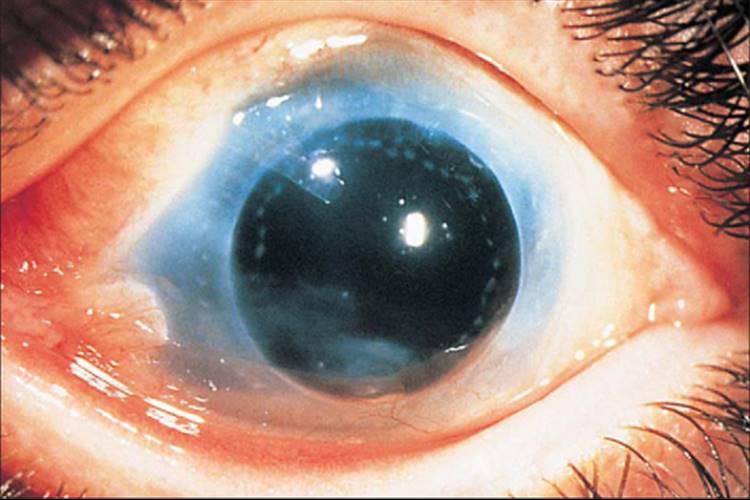
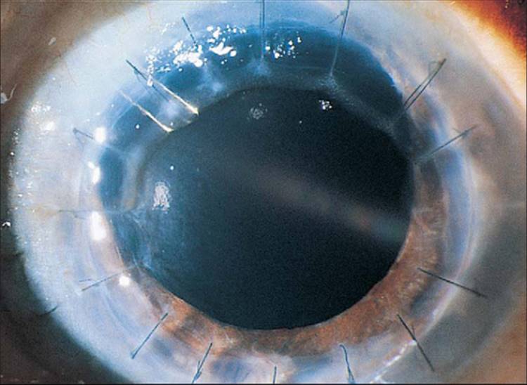
Early reports of use of Molteno plate implantation have noted it to be a promising technique (Figs 215.1 and 215.2). In 17 eyes, 16 aphakic or pseudoaphakic and 13 having had prior glaucoma operations, 12 eyes achieved an IOP of less than 21 mmHg after one implant; two of three eyes (of the unsuccessful five eyes) undergoing a second implant also achieved IOP control, with a mean follow-up of 13 months and ranging to 28 months. This finding of an overall IOP success rate (including reoperations) of 82% offered encouragement for additional investigation for patients with keratoplasty-associated glaucoma.[19]
|
|
|
|
FIGURE 215.1 Low-power photograph of a Molteno tube in an eye with penetrating keratoplasty. |
|
|
|
|
FIGURE 215.2 Higher-power view of eye with a Molteno tube in position. |
Extended follow-up from the same center, however, showed an IOP success rate with just a single-plate Molteno implant (without including success after reoperations) of 53% at 1 year, 40% at 2 years, 33% at 3 years, and 27% at 4 years.[110] Whereas many of these eyes achieved pressure control after additional surgery, the results with single-plate Molteno implants were clearly less than desired. In contrast, subsequent reports from other centers have revealed success rates of 53-96% with double-plate Molteno implants at mean follow-ups of just under 2 years to ?3 years.[111-114] Thus, as in other forms of glaucoma, larger surface areas are essential to obtaining adequate pressure control unless prior cyclodestruction has been performed.
The high proportion of eyes undergoing initial surgery that experienced graft rejection - seven of 17, of which five progressed to failure - raised concerns about the ultimate benefit of the surgery. Thus, although 12 of 17 eyes had the same or better vision after placement of a Molteno plate, five experienced decreased vision.[19] Of the 12 eyes with successful pressure control, three had a decrease in vision due to graft failure. Additional studies have since reported rates of graft failure due to both rejection and nonimmunologic causes to be as high as 53% in the first year, 80% in the second, and 87% at the end of 3 years, in a population with single-plate Molteno implants and most eyes having inadequate pressure control and thus requiring additional operations.[110] However, most studies report lower rates in eyes with better pressure control and without the need for additional operations that might put the graft at risk, but still with rates of graft failure of 29-52% with mean follow-ups between 2 and 3 years.[111-115]Indeed, one study demonstrated a 96% rate of IOP control with either a Molteno double-plate or Schocket implant (insufficient data to separate out by types) at a mean follow-up of 22 months, albeit with a 42% graft failure rate.[112]
Schocket tube implantation has also been used to try to preserve visual function while controlling IOP.[79,80,95,111,112,116,117] Its potential usefulness was suggested in the original report of Schocket tube implantations, in which glaucoma in four of five eyes, including two aphakic eyes and one with keratoplasty, was successfully controlled despite the presence of 360° of synechial angle closure.[116]Subsequent groups have noted a success rate of 50-100% in keratoplasty-associated glaucoma.[78,79,95,111,112,116] Kirkness and colleagues[79] reported a 4-year survival of 68%, including maintaining vision. Six eyes had a single-stage procedure, and 14 had a two-stage procedure; results were better with the two-stage procedure.[79]
In the past few years, two additional drainage devices have become commonly used, the Baerveldt implant and the Ahmed implant. The Baerveldt implant is available in various different sizes, from 250 mm2to 350 mm2, whereas the Ahmed implant has a surface area of 185 mm2 but has a valve-like device to regulate IOP to minimize the occurrence of hypotony after tube insertion. Publications suggest that both are useful devices for the care of keratoplasty-associated glaucoma requiring surgery. The Baerveldt implant was noted to have a 69% IOP success rate (although with a 46% graft failure rate) at a mean follow-up of ?18 months, whereas the Ahmed implant was reported to have an IOP and vision success rate of 75% at 1 year and 52% at 20 months.[118,119] Subsequent publications have demonstrated, as with the Molteno implant, better rates of IOP control and lower, but still significant, rates of graft failure (see Table 215.6 and references).[120]
TABLE 215.6 -- Graft Survival Rates with Different Surgical Modalities[*]
|
Type of Surgery |
Mean Follow-Up Period (years) |
Survival Rate (%) |
|
Trabeculectomy[32,11,75,99] |
2-3 |
71-89 |
|
Trabeculectomy[101,106] |
5 |
45-62 |
|
Trabeculectomy with MMC[102,103] |
1.17-2 |
60-82 |
|
Baerveldt[118,160-162] |
1.2-1.5 |
11-83 |
|
Ahmed[119,120] |
1-3 |
50-75 |
|
Tube shunt (various)[121-123,163] |
1-3 |
48-55 |
|
Molteno, double[111-113] |
2-3 |
48-71 |
|
Shocket[79] |
4 |
68 |
|
Tube (pars plana)[29,121] |
1-2 |
41-83 |
|
Cryoablation[32,19,76,95] |
0.75-3 |
20-59 |
|
Transvitreal endolaser[132] |
1 |
58 |
|
Nd:YAG laser[127] |
2.25 |
56 |
|
Cyclodialysis[95] |
0.75 |
15 |
|
* |
Numbers in superscript are reference numbers. MMC, mitomycin C; Nd:YAG, neodymium:yttrium-aluminum garnet. |
An important procedural consideration when inserting an intraocular glaucoma drainage device in keratoplasty patients is whether to place the implant in the anterior chamber or to perform a pars plana vitrectomy and insertion, as noted above. In a study by Arroyave and colleagues involving 72 eyes that received primarily Baerveldt 350 drainage implants, IOPs of 5-21 mmHg were successfully achieved in 89% of eyes with tube placement in the anterior chamber, compared with 100% of those with placement in the pars plana. Graft clarity at 1 year postoperatively was significantly better in the pars plana group (83% vs 48%). The investigators observed no significant difference in graft survival between eyes whose tubes were inserted at the same time as the keratoplasty and those with a subsequent insertion.[121]In another study, Sidoti reported the results of a retrospective study of 34 consecutive patients receiving various types of implants before keratoplasty, concurrent with keratoplasty, and after previous keratoplasty. The success rate for IOP control was 62%, and that for corneal graft clarity was 41% (mean follow-up, 1 year).[29] Of interest, 44% of patients in this study developed at least one posterior segment complication. In another relatively small study - in which patients with uncontrolled glaucoma and failed penetrating keratoplasty underwent combined pars plana vitrectomy with a temporary keratoprosthesis, penetrating keratoplasty, and pars plana seton insertion - six of 18 eyes had a 2-line or better improvement in visual acuity. Similar to the previous cohort, this sample experienced considerable complications; for example, two eyes had retinal detachments and two eyes became phthisical.[122] Over all, these studies demonstrate that the risk of posterior segment complications, including vitreous hemorrhage, retinal detachment, and macular hole formation, must be weighed against the desirability of keeping the tube tip away from the corneal graft. In deciding whether to perform a pars plana tube insertion, other important considerations include the access to a posterior segment surgeon capable of performing a complete vitrectomy, including the vitreous base; without a complete vitrectomy, the procedure can fail as a result of vitreous occlusion of the tube, limiting or preventing outflow.
Few studies (other than the Sidoti et al study) have examined the timing of drainage device insertion relative to the performing of the penetrating keratoplasty (i.e., before, concurrent with, or after keratoplasty). One study reported that combined penetrating keratoplasty and drainage device insertion successfully controlled IOP in 82% of 55 eyes. During that study's 3-year follow-up, graft clarity was maintained in 55% of eyes. The investigators reported complication rates of 42%, with 18% of the sample experiencing serious complications.[123]
Ciliodestructive Procedures
Before the application of cyclocryotherapy, end-stage glaucoma in eyes with a keratoplasty was often treated by enucleation or functional denervation with retrobulbar alcohol. Cyclocryotherapy is now rarely used, since newer laser forms of ciliodestruction offer safer and more predictable results.
Cryotherapy
The success rate for IOP control ranges from 38% to 100% at follow-ups ranging to 3 years (Table 215.7).[9,32,76,77,95,97,124] Again, the rate of success is lower in aphakic and pseudophakic eyes.[97] Although highly successful from an IOP viewpoint, cryotherapy carries significant morbidity. In one series, 14% of eyes receiving cryotherapy lost more than two Snellen lines of visual acuity within 9 months.[97]
TABLE 215.7 -- Cryotherapy[*]
|
Study |
Criteria |
Mean Follow-Up |
Patient Population |
Success (%) |
|
Binder et al[77] |
IOP |
19.4 months |
Mixed |
100 |
|
Foulks[32] |
IOP |
3 years |
Mixed |
88 |
|
Tragakis et al[124] |
IOP |
20 months |
Mixed |
69 |
|
West et al[76] |
IOP |
10 months |
Mixed |
86 |
|
Gross et al[95] |
IOP and VA |
9 months |
Aphakic/pseudophakic |
41 |
|
IOP only |
77 |
|||
|
Kirkness and Moshegov[9] |
IOP |
Mixed |
62 |
|
|
Kushwaha and Pual[97] |
IOP |
1 years |
Early glaucoma/mixed |
61 |
|
* |
Numbers in superscript are reference numbers. IOP, intraocular pressure; VA, visual acuity. |
Vitreous hemorrhage, retinal detachment, choroidal detachment, cystoid macular edema, uveitis, phthisis, and graft failure are among the reported side effects.[19,76,77] The rate of phthisis is from 0% to 14% at follow-up, ranging to a mean of 19.4 months.[76,77,95,125] As importantly, the rate of graft failure is quite high, from 7% to 80% in series with a follow-up of at least 10 months.[19,32,76,77] In part, failure may be due to injuring the corneal endothelium. In many cases, these eyes are already severely compromised and graft failure might not be unexpected; for example, eight of 36 (22%) eyes in one study had corneal clouding even before cryotherapy.[77] Three other studies report rates of graft failure of 41-80% at follow-ups ranging from 10 months to 3 years.[19,32,76] Thus, cryotherapy carries significant risk to the graft as well as the eye.
Laser Cycloablation
Refinement of transscleral Nd:YAG and more recently transscleral diode cyclophotocoagulation has raised the hope of offering a more controlled and less morbid approach for IOP control. Success rates of 50-100% have been reported at median follow-ups of 6-27 months for YAG cyclophotocoagulation.[80,81,95,126,127] Not surprisingly, aphakic and pseudophakic eyes appear to fare somewhat worse.[95] In a large series, 30 of 39 eyes (77%) with keratoplasty-associated glaucoma achieved IOP control by the final follow-up visit (mean, 27 months).[127] Studies have reported success rates for transscleral diode cyclophotocoagulation ranging from 56% to 79% (Table 215.8)[128-130] (follow-up ranged from a mean of 11 months to a median of 30.5 months). In these studies, a considerable number of eyes required multiple treatments to achieve IOP control. In a series of 52 patients receiving treatment with transscleral diode cyclophotocoagulation, 63% had controlled IOP and 56% maintained graft clarity during the 5-year follow-up.[130]
TABLE 215.8 -- Transcleral Diode Cyclophotcoagulation[*]
|
Study |
Criteria |
Mean Follow-Up |
Patient Population |
Success (%) |
|
Ocakoglu et al[128] |
IOP |
11.4 months |
Mixed |
56[?] |
|
Shah et al[129] |
IOP |
30.5 months |
Mixed |
79 |
|
Beiran et al[130] |
IOP |
5 years |
Mixed |
63 |
|
IOP, intraocular pressure. |
|
* |
Numbers in superscript are reference numbers. |
|
? |
Cumulative. |
Although laser cycloablation has been reported to be less morbid than cryotherapy in some series,[131] reported rates of graft clouding and failure have ranged from 16% to 44% during average follow-ups of 2.5-5 years.[129,130] Furthermore, visual loss of greater than two lines of Snellen acuity has also been reported among eyes treated successfully.[95] Thus, laser cyclophotocoagulation appears to share the same success and complications as the other currently used treatment modalities in the management of keratoplasty-associated glaucoma. Other ways to perform laser cycloablation include the use of a transvitreal route to deliver laser energy (with rates of graft failure and hypotony at 12 months of 31% and 8%, respectively)[132] and, more recently, the use of endoscopic cyclophotocoagulation (ECP).[133]Whether ECP will have the same success rate as diode laser with a lower rate of complications awaits additional clinical experience.
Unfortunately, no prospective study to date has directly compared the success and complication rates among postkeratoplasty trabeculectomies with use of mitomycin C, glaucoma drainage device insertions, and cilioablation. A small retrospective study, however, did compare these three modalities, and the authors found no difference among them in IOP control nor in graft survival but did note nonsignificantly increased rates of graft failure, glaucoma failure, and vision loss among patients undergoing transscleral cyclophotocoagulation.[134] At present, due to the relatively high reported rates of graft failure associated with tube shunt insertion, compared with trabeculectomy, in postkeratoplasty eyes, performing a trabeculectomy is favored as the initial procedure to control the IOP. Future studies, including a randomized, controlled trial comparing outcomes (both pressure control and graft clarity) of these two surgical modalities would be helpful in determining the best way to manage these patients.
Lamellar Keratoplasty and Glaucoma
In recent years, deep lamellar endothelial keratoplasty (DLEK) and Descemet stripping with endothelial keratoplasty (DSEK) have become popular alternatives to conventional penetrating keratoplasty for certain corneal conditions. Each of these two procedures involves replacing the posterior cornea, including the endothelium, with graft tissue while keeping the anterior corneal stroma, Bowman layer, and epithelium. These procedures offer potentially quicker healing, less astigmatism, and better visual acuity, compared with penetrating keratoplasty. Although the effects of these procedures on IOP and the risk for glaucoma remain unknown, a randomized, controlled trial of 26 consecutive patients found significantly higher IOP at 12 months in patients who had undergone conventional penetrating keratoplasty than in those who had been assigned to DLEK.[135] In another prospective cohort, comprising 100 consecutive patients who underwent DLEK and then were followed for 6 months, only one person experienced a sustained IOP elevation requiring glaucoma surgery.[136] In the future, if larger series with longer follow-up periods support the findings from initial reports, IOP control may prove another benefit of performing DLEK over using conventional penetrating keratoplasty.
GRAFT FAILURE
|
Key Features: Graft Failure in Keratoplasty-Associated Glaucoma |
|||||||||
|
It is relatively clear that IOP control can now be readily achieved in most patients with keratoplasty-associated glaucoma. Whether one uses trabeculectomy, cyclodestruction, or a drainage device, IOP success rates of above 70% at 1- or 2-year follow-up, or even longer, are attainable. Also clear, however, is that graft failure, due to both rejection and nonimmunologic causes, is also a common result (Table 215.6). Even in the eyes with the best prognosis for keratoplasty, the presence of glaucoma significantly reduces the likelihood of graft survival (71% vs 89% at 3 years).[11] A study examining 156 patients requiring repeat penetrating keratoplasties for graft failure found that 60% of patients had a concomitant glaucoma diagnosis. Moreover, compared with those who did not have glaucoma, the patients with preoperative glaucoma had a significantly higher rate of subsequent graft rejection (50% vs 32%) and a shorter time to graft failure (18 vs 32 months), regardless of glaucoma therapy.[137] Using multivariate analysis, another study found that a history of glaucoma or ocular hypertension doubled the risk of graft failure, whereas that of having anterior synechiae of the iris raised the risk by 30%[12]; indeed, having a history of glaucoma or ocular hypertension was second only to preoperative endothelial dysfunction (type of disease) in determining the risk of graft failure. In a third study, which used survival analyses, prior glaucoma or uveitis had the largest relative risk of nonimmune failure (relative risk, 3.3).[57] Once the grafts in eyes with glaucoma fail, the subsequent regrafting is further compromised by the greater failure rate in regrafts as opposed to primary grafts, even without the presence of glaucoma.[56,57]
Besides a preoperative history of glaucoma, multivariable analyses have identified the following risk factors for graft failure: the presence of posterior synechiae, the presence of corneal neovascularization, older age, and intraoperative anterior vitrectomy.[138] By using survival analysis, a study of 3992 consecutive primary penetrating keratoplasty case-patients identified these risk factors for graft failure: the use of topical glaucoma medications, the presence of deep stromal vascularization, the presence of posterior synechiae, diabetes mellitus, patient race, and small trephination size.[42]
The etiology of this increased risk of having glaucoma on graft failure is unknown, although many have proposed various mechanisms. Some studies indicate that use of preoperative glaucoma medications may be an important factor.[139,42] The effect of elevated pressure or retarded aqueous circulation on endothelial function in the graft may be another mechanism. That the effect of additional intraocular surgery or manipulations is uniformly negative on the graft can easily be seen in the higher rates of graft failure after surgical intervention than the baseline rates of failure reported. When additional surgical intervention is needed, physical trauma to the endothelium may result in loss of endothelial cells, as has been reported for trabeculectomy with and without antimetabolites.[140] A study showing no accelerated postoperative loss of endothelial cells with Molteno implantation was limited by not having obtained preoperative cell counts.[141] Although many have postulated an immune mechanism for glaucoma-associated failure, some studies indicate that glaucoma tends to increase rates of graft failure through nonimmune failure as opposed to progressive rejection.[57,139]
CONCLUSION
In eyes with preexisting glaucoma, either medically uncontrolled or controllable only with the use of multiple medications, surgical management to lower IOP in advance (preferably) or at the time of keratoplasty is recommended. Such an approach may blunt the early IOP rise and possibly avoid the need for later glaucoma surgery, jeopardizing a clear graft. The decision about which procedure to perform in conjunction with, before, or after keratoplasty requires careful consideration of a patient's status and the options available to the surgeon.
Glaucoma associated with penetrating keratoplasty is a common and growing problem. Although much has been learned in the past 50 years, much more still needs to be discovered. With current level of knowledge, one can draw certain tentative conclusions. First, the incidence of keratoplasty-associated glaucoma varies widely depending on the cause of the corneal disease. Second, eyes with preexisting glaucoma have a high rate of loss of IOP control after keratoplasty. Third, although the underlying mechanisms are unclear, several discrete interventions can be undertaken intraoperatively and postoperatively to try to control IOP. Finally, current methods of treatment leave much to be desired. In particular, eyes requiring surgery face not only the risk of unsuccessful surgery but also the risk of graft decompensation, even when pressure control is achieved.
REFERENCES
1. National Institutes of Health and National Institute of Allergy and Infectious Diseases, Status of NIH-Sponsored Basic and Clinical Research on Transplantation. Available: www3.niaid.nih.gov/about/organization/dait/PDF/feb2000_transplant.pdf Apr 24, 2006
2. Council on Scientific Affairs: Report of the organ transplant council: corneal transplantation. JAMA 1988; 259:719.
3. Robin JB, Gindi JJ, Koh K, et al: An update of the indications for penetrating keratoplasty. Arch Ophthalmol 1986; 104:87.
4. Mohamadi P, McDonnell JM, Irvine JA, et al: Changing indications for penetrating keratoplasty, 1984-88. Am J Ophthalmol 1989; 107:551.
5. Lang GK, Green WR: Clinicopathologic studies of keratoplasty eyes obtained surgically. Cornea 1985; 4:229.
6. Stark WJ, Bruner WE, Maumenee AE: Surgery of the cornea. In: Rice TA, Michels RG, Stark WJ, ed. Ophthalmic surgery, St. Louis: CV Mosby; 1984:129-136.
7. Polack FM: Glaucoma in keratoplasty. Cornea 1988; 7:67.
8. Schanzlin DJ, Robin JB, Gomez DS, et al: Results of penetrating keratoplasty for aphakic and pseudophakic bullous keratopathy. Am J Ophthalmol 1984; 98:302.
9. Kirkness CM, Moshegov C: Post keratoplasty glaucoma. Eye 1988; 2(Suppl):919.
10. Wilson SE, Kaufman HE: Graft failure after penetrating keratoplasty. Surv Ophthalmol 1990; 34:325.
11. Reinhard RT, Kallman C, Cepin A, et al: The influence of glaucoma history on graft survival after penetrating keratoplasty. Graefes Arch Clin Exp Ophthalmol 1997; 235:553.
12. Yamagami S, Suzuki Y, Tsuru T: Risk factors for graft failure in penetrating keratoplasty. Acta Ophthalmol Scand 1996; 74:584.
13. Olson RJ: Glaucoma associated with penetrating keratoplasty. In: Ritch R, Shields MB, Krupin T, ed. The glaucomas, St. Louis: CV Mosby; 1989:1337-1347.
14. Irvine AR, Kaufman HE: Intraocular pressure following penetrating keratoplasty. Am J Ophthalmol 1969; 68:835.
15. Wind CA, Kaufman HE: Validity of MacKay Marg tonometry following penetrating keratoplasty in man. Am J Ophthalmol 1971; 72:117.
16. Buxton JN, Riechers RJ, Aaron SD: Corneal grafts and their effect upon the applanation Schiotz disparity. Arch Ophthalmol 1971; 86:28.
17. Whitacre MM, Stein RA, Hassanein K: The effect of corneal thickness on applanation tonometry. Am J Ophthalmol 1993; 115:592.
18. Rao VJ, Gnanaraj L, Mitchell KW, Figueiredo FC: Clinical comparison of ocular blood flow tonometer, Tonopen, and Goldmann applanation tonometer for measuring intraocular pressure in postkeratoplasty eyes. Cornea 2001; 20:834-838.
19. McDonnell PJ, Robin JB, Schanzlin DJ, et al: Molteno implant for control of glaucoma in eyes after penetrating keratoplasty. Ophthalmology 1988; 95:364.
20. Van Meter WS, Allen RC, Waring GO, Stulting RD: Laser trabeculoplasty for glaucoma in aphakic and pseudophakic eyes after penetrating keratoplasty. Arch Ophthalmol 1988; 106:185.
21. Nguyen NX, Horn FK, Seitz B, et al: Frequency-doubling perimetry in patients following penetrating keratoplasty. Cornea 2004; 23:433-438.
22. American Academy of Ophthalmology: Preferred practice pattern: primary open angle glaucoma, San Francisco, AAO, 2005.
23. American Academy of Ophthalmology: Preferred practice pattern: angle closure glaucoma, San Francisco, AAO, 2005.
24. Simmons RB, Stern RA, Teekhasaenee C, Kenyon KR: Elevated intraocular pressure following penetrating keratoplasty. Trans Am Ophthalmol Soc 1989; 87:79.
25. Kirkness CM, Ficker LA: Risk factors for the development of postkeratoplasty glaucoma. Cornea 1992; 11:427.
26. Franca ET, Arcieri ES, Arcieri RS, Rocha FJ: A study of glaucoma after penetrating keratoplasty. Cornea 2002; 21:284-288.
27. Ing JJ, Ing HH, Nelson LR, et al: Ten-year postoperative results of penetrating keratoplasty. Ophthalmology 1998; 105:1855-1865.
28. Sihota R, Sharma N, Panda A, et al: Post-penetrating keratoplasty glaucoma: risk factors, management and visual outcome. Aust N Z J Ophthalmol 1998; 26:305-309.
29. Sidoti PA, Mosny AY, Ritterband DC, Seedor JA: Pars plana tube insertion of glaucoma drainage implants and penetrating keratoplasty in patients with coexisting glaucoma and corneal disease. Ophthalmology 2001; 108:1050-1058.
30. Olson RJ, Kaufman HE: Intraocular pressure and corneal thickness after penetrating keratoplasty. Am J Ophthalmol 1978; 86:97.
31. Olson RJ, Kaufman HE: Prognostic factors of intraocular pressure after aphakic keratoplasty. Am J Ophthalmol 1978; 86:510.
32. Foulks GN: Glaucoma associated with penetrating keratoplasty. Ophthalmology 1987; 94:871.
33. Nguyen NX, Langenbucher A, Seitz B, et al: Impact of increased intraocular pressure on long-term corneal endothelial cell density after penetrating keratoplasty. Ophthalmologica 2002; 216:40-44.
34. Chien AM, Schmidt CM, Cohen EJ, et al: Glaucoma in the immediate postoperative period after penetrating keratoplasty. Am J Ophthalmol 1993; 115:711.
35. Sekhar GC, Vyas P, Nagarajan R, et al: Post penetrating keratoplasty glaucoma. Indian J Ophthalmol 1993; 41:181.
36. Karesh JW, Nirankari VS: Factors associated with glaucoma after penetrating keratoplasty. Am J Ophthalmol 1983; 96:160.
37. Goldberg DB, Schanzlin DJ, Brown SI: Incidence of increased intraocular pressure after keratoplasty. Am J Ophthalmol 1981; 92:372.
38. Thoft RA, Gordon JM, Dohlman CH: Glaucoma following keratoplasty. Trans Am Acad Ophthalmol Otol 1974; 78:OP 352.
39. Heidemann DG, Sugar A, Meyer RF, Musch DC: Oversized donor grafts in penetrating keratoplasty. Arch Ophthalmol 1985; 103:1807.
40. Perry HD, Donnenfeld ED, Kanellopoulos AJ, Grossman GA: Topical cyclosporin A in the management of postkeratoplasty glaucoma. Cornea 1997; 16:284.
41. Krontz DP, Wood TO: Corneal decompensation following acute angle closure glaucoma. Ophthalmic Surg 1988; 19:334.
42. Price MO, Thompson RW Jr, Price FW Jr: Risk factors for various causes of failure in initial corneal grafts. Archives of Ophthalmology 2003; 121:1087-1092.
43. Vanathi M, Sharma N, Sinha R, et al: Indications and outcome of repeat penetrating keratoplasty in India. BMC Ophthalmology 2005; 5:26.
44. Olson RJ: Aphakic keratoplasty. Arch Ophthalmol 1978; 96:2274.
45. Olson RJ, Kaufman HE: A mathematical description of causative factors and prevention of elevated intraocular pressure after keratoplasty. Invest Ophthalmol Vis Sci 1977; 16:1085.
46. Zimmerman TJ, Krupin T, Brodzki W, Waltman SR: The effect of suture depth on outflow facility in penetrating keratoplasty. Arch Ophthalmol 1978; 96:505.
47. Zimmerman TJ, Waltman SR, Sachs U, Kaufman HE: Intraocular pressure after aphakic penetrating keratoplasty: Through and through suturing. Ophthalmic Surg 1979; 10:49.
48. Bourne WM, Davison JA, O'Fallon WM: The effects of oversize donor buttons on postoperative intraocular pressure and corneal curvature in aphakic penetrating keratoplasty. Ophthalmology 1982; 89:242.
49. Zimmerman T, Olson R, Waltman S, Kaufman H: Transplant size and elevated intraocular pressure. Arch Ophthalmol 1978; 96:2231.
50. Perl T, Charlton KH, Binder PS: Disparate diameter grafting. Ophthalmology 1981; 88:774.
51. Foulks GN, Perry HD, Dohlman CH: Oversize corneal donor grafts in penetrating keratoplasty. Ophthalmology 1979; 86:490.
52. Olson RJ: Discussion. Ophthalmology 1981; 88:780.
53. Vail A, Gore SM, Bradley BA, et al: Clinical and surgical factors influencing corneal graft survival, visual acuity, and astigmatism. Ophthalmology 1996; 103:41.
54. Price FW, Whitson WE, Marks RG: Graft survival in four common groups of patients undergoing penetrating keratoplasty. Ophthalmology 1991; 98:322.
55. The Collaborative Corneal Transplantation Studies Research Group: The collaborative corneal transplantation studies (CCTS). Arch Ophthalmol 1992; 110:1392.
56. Price FW, Whitson WE, Collins KS, Marks RG: Five year corneal graft survival: A large, single center patient cohort. Arch Ophthalmol 1993; 111:799.
57. Boisjoly HM, Tourigny R, Bazin R, et al: Risk factors of corneal graft failure. Ophthalmology 1993; 100:1728.
58. Speaker MG, Guerriero PH, Met JA, et al: A case control study of risk factors for intraoperative suprachoroidal expulsive hemorrhage. Ophthalmology 1991; 98:202.
59. Burke S, Sugar J, Farber MD: Comparison of the effects of two viscoelastic agents, Healon and Viscoat, on postoperative intraocular pressure after penetrating keratoplasty. Ophthalmic Surg 1990; 21:821.
60. Volker Dieben HJ, Regensburg H, Kruit PJ: A double blind, randomized study of Healon GV compared with Healon in penetrating keratoplasty. Cornea 1994; 13:414.
61. Yang H, Zheng D, Huang J, et al: The clinical study of Healon 5 in phacoemulsification and IOL implantation. Yan Ke Xue Bao (Eye Science) 2001; 17:186-190.
62. Vajpayee RB, Verma K, Sinha R, Titiyal JS, Pandey RM, Sharma N: Comparative evaluation of efficacy and safety of ophthalmic viscosurgical devices in phacoemulsification. BMC Ophthalmology 2005; 5:17.
63. Vajpayee RB, Dada T, Ray M, et al: Oversized corneal grafts for corneal opacities with iridocorneal adhesions. Ophthalmology 2001; 108:2026-2028.
64. Olson RJ: Variation in corneal graft size related to trephine technique. Arch Ophthalmol 1979; 97:1323.
65. Polack FM: Keratoplasty in aphakic eyes with corneal edema. Ophthalmic Surg 1980; 11:701.
66. Rabb MF, Fine M: Penetrating keratoplasty in interstitial keratitis. Am J Ophthalmol 1969; 67:907.
67. Cohen EJ, Kenyon KR, Dohlman CH: Iridoplasty for prevention of post keratoplasty angle closure and glaucoma. Ophthalmic Surg 1982; 13:994.
68. Waring GO: Management of pseudophakic corneal edema with reconstruction of the anterior ocular segment. Arch Ophthalmol 1987; 105:709.
69. Kenyon KR, Starck T, Hersh PS: Penetrating keratoplasty and anterior segment reconstruction for severe ocular trauma. Ophthalmology 1992; 99:396.
70. Chu MW, Font RL, Koch DD: Visual results and complications following posterior iris fixated posterior chamber lenses at penetrating keratoplasty. Ophthalmic Surg 1992; 23:608.
71. Wood TO, West C, Kaufman HE: Control of intraocular pressure in penetrating keratoplasty. Am J Ophthalmol 1972; 74:724.
72. Olson RJ, Zimmerman TJ, Kaufman HE: Elevated intraocular pressure after aphakic keratoplasty: Iatrogenic disease and prevention. Ann Ophthalmol 1978; 10:931.
73. Paton D: The prognosis of penetrating keratoplasty. Ophthalmic Surg 1976; 7:36.
74. Lemp MA, Pfister RR, Dohlman CH: The effect of intraocular surgery on clear corneal grafts. Am J Ophthalmol 1970; 70:719.
75. Gilvarry AME, Kirkness CM, Steele AD, et al: The management of post keratoplasty glaucoma by trabeculectomy. Eye 1989; 3:713.
76. West CE, Wood TO, Kaufman HE: Cyclocryotherapy for glaucoma pre or postpenetrating keratoplasty. Am J Ophthalmol 1973; 76:485.
77. Binder PS, Abel R, Kaufman HE: Cyclocryotherapy for glaucoma after penetrating keratoplasty. Am J Ophthalmol 1975; 79:489.
78. Kirkness CM: Penetrating keratoplasty, glaucoma and silicone drainage tubing. Dev Ophthalmol 1987; 14:161.
79. Kirkness CM, Ling Y, Rice NSC: The use of silicone drainage tubing to control post keratoplasty glaucoma. Eye 1988; 2:583.
80. Cohen EJ, Schwartz LW, Luskind RD, et al: Neodymium:YAG laser transscleral cyclophotocoagulation for glaucoma after penetrating keratoplasty. Ophthalmic Surg 1989; 20:713.
81. Levy NS, Bonney RC: Transscleral YAG cyclophotocoagulation of the ciliary body for persistently high intraocular pressure following penetrating keratoplasty. Cornea 1989; 8:178.
82. Wilson RP, Spaeth GL, Poryzees EM: The place of timolol in the practice of ophthalmology. Ophthalmology 1980; 87:451.
83. Johnson DH, Kenyon KR, Epstein DL, Van Buskirk EM: Corneal changes during pilocarpine gel therapy. Am J Ophthalmol 1986; 101:13.
84. Konowal A, Morrison JC, Brown SV, et al: Irreversible corneal decompensation in patients treated with topical dorzolamide. Am J Ophthalmol 1999; 127:403-406.
85. Lass JH, Paven Langston D: Timolol therapy in secondary angle closure glaucoma post penetrating keratoplasty. Ophthalmology 1979; 86:51.
86. Olson RJ, Kaufman HE, Zimmerman TJ: Effects of timolol and Daranide on elevated intraocular pressure after aphakic keratoplasty. Ann Ophthalmol 1979; 11:1833.
87. Kanellopoulos AJ, Perry HD, Donnenfeld ED: Comparison of topical timolol gel to oral acetazolamide in the prophylaxis of viscoelastic-induced ocular hypertension after penetrating keratoplasty. Cornea 1997; 16:12-15.
88. Thomas JV, Gragoudas ES, Blair NP, Lapus JV: Correlation of epinephrine use and macular edema in aphakic glaucomatous eyes. Arch Ophthalmol 1978; 96:625.
89. Thomas JV, Epstein DL: Timolol and epinephrine in primary open angle glaucoma. Arch Ophthalmol 1981; 99:91.
90. Parrow KA, Hong YJ, Shin DH, et al: Is it worthwhile to add dipivefrin HCl 0.1% to topical ?1, ?2 blocker therapy?. Ophthalmology 1989; 98:1338.
91. Shields MB: Textbook of Glaucoma, 2nd edn.. Baltimore: Williams & Wilkins; 1987:374-382.
92. Epstein DL, Steinert RF, Puliafito CA: Neodymium YAG laser therapy to the anterior hyaloid in aphakic malignant (ciliovitreal block) glaucoma. Am J Ophthalmol 1984; 98:137.
93. Kandarakis A, Zimmerman T: Non pupillary block angle closure glaucoma and their treatment by laser. Ann Ophthalmol 1984; 16:914.
94. Fu YA, Liaw ZC: Argon laser gonioplasty with trabeculoplasty for chronic angle closure glaucoma. Ann Ophthalmol 1987; 19:419.
95. Gross RL, Feldman RM, Spaeth GL, et al: Surgical therapy of chronic glaucoma in aphakia and pseudoaphakia. Ophthalmology 1988; 95:1195.
96. Huang SCM, Soong HK, Brenz RM, et al: Problems associated with penetrating keratoplasty for corneal edema in congenital glaucoma. Ophthalmic Surg 1989; 20:399.
97. Kushwaha DC, Pual AK: Incidence and management of glaucoma in postoperative cases of penetrating keratoplasty. Indian J Ophthalmol 1981; 29:167.
98. Ramchandani M, Mohammed S, Mirza S, McDonnell PJ: Penetrating keratoplasty in adults with congenital glaucomas. Eye 2004; 18:703-708.
99. Insler MS, Cooper HD, Kastl PR, Caldwell DR: Penetrating keratoplasty with trabeculectomy. Am J Ophthalmol 1985; 100:593.
100. Figueiredo RS, Araujo SV, Cohen EJ, et al: Management of coexisting corneal disease and glaucoma by combined penetrating keratoplasty and trabeculectomy with mitomycin C. Ophthalmic Surg Lasers 1996; 27:903.
101. Kirkness CM, Steele AD, Ficker LA, Rice NS: Coexistent corneal disease and glaucoma managed by either drainage surgery and subsequent keratoplasty or combined drainage surgery and penetrating keratoplasty. Br J Ophthalmol 1992; 76:146.
102. Chowers I: Ticho U. Mitomycin-C in combined or two-stage procedure trabeculectomy followed by penetrating keratoplasty. J Glaucoma 1999; 8:184-187.
103. WuDunn D, Alfonso E, Palmberg PF: Combined penetrating keratoplasty and trabeculectomy with mitomycin C. Ophthalmology 1999; 106:396-400.
104. Ishioka M, Shimazaki J, Yamagami J, et al: Trabeculectomy with mitomycin C for post-keratoplasty glaucoma. Br J Ophthalmol 2000; 84:714-717.
105. Akova YA: Bulut S. Dabil H. Duman S. Late bleb-related endophthalmitis after trabeculectomy with mitomycin C. Ophthalmic Surg Lasers 1999; 30:146-151.
106. Ficker LA, Kirkness CM, Steele AD, et al: Intraocular surgery following penetrating keratoplasty: The risks and advantages. Eye 1990; 4(pt 5):693.
107. Campbell DG, Vela MA: Modern goniosynechialysis for the treatment of synechial angle closure glaucoma. Ophthalmology 1984; 91:1052.
108. Shingleton BJ, Chan MA, Bellows AR, Thomas JV: Surgical goniosynechialysis for angle closure glaucoma. Ophthalmology 1990; 97:551.
109. Heuer DK, Lloyd MA, Abrams DA, et al: Which is better? One or two?. Ophthalmology 1992; 99:1512.
110. Choi J, Lee PP, McDonnell PJ, et al: Longer term follow up of single plate Molteno implants in eyes with penetrating keratoplasty. Invest Ophthalmol Vis Sci (Suppl) 1993; 34:367.
111. Beebe WE, Starita RJ, Fellman RL, et al: The use of Molteno implant and anterior chamber tube shunt to encircling band for the treatment of glaucoma in penetrating keratoplasty patients. Ophthalmology 1990; 97:1414.
112. Sherwood MB, Smith MR, Driege WT, et al: Drainage tube implants in the treatment of glaucoma following penetrating keratoplasty. Ophthalmic Surg 1993; 24:185.
113. Price FW, Wellemeyer M: Long term results of Molteno implants. Ophthalmic Surg 1995; 26:130.
114. Smith MR, Sherwood MB, McGorray SP: Comparison of the double plate Molteno drainage implant with the Schocket procedure. Arch Ophthalmol 1992; 110:1246.
115. Rapuano CJ, Schmidt CM, Cohen EJ, et al: Results of alloplastic tube shunt procedures before, during, or after penetrating keratoplasty. Cornea 1995; 14:26.
116. Schocket SS, Nirankari VS, Lakhanpal V, et al: Anterior chamber tube shunt to anencircling band in the treatment of neovascular glaucoma and other refractory glaucomas. Ophthalmology 1985; 92:553.
117. Omi CA, de Almeida GV, Cohen R, et al: Modified Schocket implant for refractory glaucoma. Ophthalmology 1991; 98:211.
118. Hodkin MJ, Goldblatt WS, Burgoyne CF, et al: Early clinical experience with the Baerveldt implant in complicated glaucomas. Am J Ophthalmol 1995; 120:32.
119. Coleman AL, Mondino BJ, Wilson MR, Casey R: Clinical experience with the Ahmed glaucoma valve implant in eyes with prior or concurrent penetrating keratoplasties. Am J Ophthalmol 1997; 123:54.
120. Al-Torbak AA: Outcome of combined Ahmed glaucoma valve implant and penetrating keratoplasty in refractory congenital glaucoma with corneal opacity. Cornea 2004; 23:554-559.
121. Arroyave CP, Scott IU, Fantes FE, et al: Corneal graft survival and intraocular pressure control after penetrating keratoplasty and glaucoma drainage device implantation. Ophthalmology 2001; 108:1978-1985.
122. Johnston RH, Nguyen R, Jongsareejit A, et al: Clinical study of combined penetrating keratoplasty, pars plana vitrectomy with temporary keratoprosthesis, and pars plana seton implant. Retina 1999; 19:116-121.
123. Kwon YH, Taylor JM, Hong S, et al: Long-term results of eyes with penetrating keratoplasty and glaucoma drainage tube implant. Ophthalmology 2001; 108:272-278.
124. Tragakis MP, Brown SI: The significance of new anterior synechiae after corneal transplantation. Am J Ophthalmol 1972; 74:523.
125. Wilson SE, Kaufman HE: Graft failure after penetrating keratoplasty. Surg Ophthalmol 1990; 34:325.
126. Wheatcrogt S, Singh A, Caset Y, McAllister J: Treatment of glaucoma following penetrating keratoplasty with transscleral YAG cyclophotocoagulation. Int Ophthalmol 1992; 16:397.
127. Threlkeld AB, Shields MB: Noncontact transscleral Nd:YAG cyclophotocoagulation for glaucoma after penetrating keratoplasty. Am J Ophthalmol 1995; 120:569.
128. Ocakoglu O, Arslan OS, Kayiran A: Diode laser transscleral cyclophotocoagulation for the treatment of refractory glaucoma after penetrating keratoplasty. Curr Eye Res 2005; 30:569-574.
129. Shah P, Lee GA, Kirwan JK, et al: Cyclodiode photocoagulation for refractory glaucoma after penetrating keratoplasty. Ophthalmology 2001; 108:1986-1991.
130. Beiran I, Rootman DS, Trope GE, Buys YM: Long-term results of transscleral Nd:YAG cyclophotocoagulation for refractory glaucoma postpenetrating keratoplasty. J Glaucoma 2000; 9:268-272.
131. Schuman JS, Puliafito CA, Allingham RR, et al: Contact transscleral continuous wave neodymium YAG laser cyclophotocoagulation. Ophthalmology 1990; 97:571.
132. Haller JA: Transvitreal endocyclophotocoagulation. Trans Am Ophthalmol Soc 1996; 94:589.
133. Hollander DA, Lin SC: Delayed therapeutic success with endoscopic cyclophoto-coagulation in treating refractory post-penetrating keratoplasty glaucoma. Br J Ophthalmol 2003; 87:792-793.
134. Ayyala RS, Pieroth L, Vinals AF, et al: Comparison of mitomycin C trabeculectomy, glaucoma drainage device implantation, and laser neodymium:YAG cyclophotocoagulation in the management of intractable glaucoma after penetrating keratoplasty. Ophthalmology 1998; 105:1550-1556.
135. Shimazaki J, Shimmura S, Ishioka M, Tsubota K: Randomized clinical trial of deep lamellar keratoplasty vs penetrating keratoplasty. Am J Ophthalmol 2002; 134:159-165.
136. Terry MA, Ousley PJ: Deep lamellar endothelial keratoplasty: early complications and their management. Cornea 2006; 25:37-43.
137. Aldave AJ, Rudd JC, Cohen EJ, et al: The role of glaucoma therapy in the need for repeat penetrating keratoplasty. Cornea 2000; 19:772-776.
138. Weisbrod DJ, Sit M, Naor J, Slomovic AR: Outcomes of repeat penetrating keratoplasty and risk factors for graft failure. Cornea 2003; 22:429-434.
139. Price FW, Whitson WE, Johns S, Gonzales JS: Risk factors for corneal graft failure. J Refract Surg 1996; 12:134.
140. Pastor SA, Williams R, Hetherington J, et al: Corneal endothelial cell loss following trabeculectomy with mitomycin C. J Glauc 1993; 2:112-113.
141. McDermott ML, Swendris RP, Shin DH, et al: Corneal endothelial cell counts after Molteno implantation. Am J Ophthalmol 1993; 115:93.
142. Waring GO, Welch SN, Cavagh MD, et al: Results of penetrating keratoplasty in 123 eyes with pseudophakic or aphakic corneal edema. Ophthalmology 1983; 90:25.
143. Jensen OM, Haamann P, Schmidt P: Penetrating keratoplasty and transscleral fixation of posterior chamber lens. Acta Ophthalmol Scand 1995; 73:551.
144. Kandrakis AS, Doulas KG, Amariotakis AG: Penetrating keratoplasty and transsclerally suture fixated intraocular lenses. J Refract Surg 1996; 12:S304.
145. Lois N, Cohen EJ, Rapuano CJ, Laibson PR: Long term graft survival in patients with flexible open loop anterior chamber intraocular lenses. Cornea 1997; 16:387.
146. Robinson CH: Indications, complications and programs for repeat penetrating keratoplasty. Ophthalmic Surg 1979; 10:27.
147. Feldman ST, Frucht Perry J, Brown SI: Corneal transplantation in microphthalmic eyes. Am J Ophthalmol 1987; 104:164.
148. Heidemann DG, Dunn SP: Transsclerally sutured intraocular lenses in penetrating keratoplasty. Am J Ophthalmol 1992; 113:619.
149. Doren GS, Cohen EJ, Brady SE, et al: Penetrating keratoplasty after ocular trauma. Am J Ophthalmol 1990; 110:408.
150. Kenyon KR, Kenyon BM, Starck T, Hersh PS: Penetrating keratoplasty and anterior segment reconstruction for severe ocular trauma. Ger J Ophthalmol 1994; 3:90.
151. Kremer I, Rajpal RK, Rapuano CJ, et al: Results of penetrating keratoplasty in aniridia. Am J Ophthalmol 1993; 115:317.
152. Ficker LA, Kirkness C, Wright P: Prognosis for keratoplasty in Acanthoamoeba keratitis. Ophthalmology 1993; 100:105.
153. Gollamudi SR, Traboulsi EI, Chamon W, et al: Visual outcome after surgery for Peter's anomaly. Ophthalmic Genet 1994; 15:31.
154. al Rajhi AA, Wagoner MD: Penetrating keratoplasty in congenital hereditary endothelial dystrophy. Ophthalmology 1997; 104:956.
155. Sajjadi H, Javadi MA, Hemmati R, et al: Results of penetrating keratoplasty in CHED. Cornea 1995; 14:18.
156. Mullaney PB, Risco JM, Teichmann K, Millar L: Congenital hereditary endothelial dystrophy associated with glaucoma. Ophthalmology 1995; 102:186.
157. Crawford GJ, Stulting RD, Cavanagh HD, Waring GO: Penetrating keratoplasty in the management of iridocorneal endothelial syndrome. Cornea 1929; 8:34.
158. Chang PC, Soong HK, Couto MF, et al: Prognosis for penetrating keratoplasty in iridocorneal endothelial syndrome. Refract Corneal Surg 1993; 9:129.
159. Alvim PT, Cohen EJ, Rapuano CJ, et al: Penetrating keratoplasty in iridocorneal endothelial syndrome. Cornea 2001; 20:134-140.
160. Seigner SW, Netland PA, Urban RC, et al: Clinical experience with the Baerveldt glaucoma drainage implant. Ophthalmology 1995; 102:1298.
161. Lloyd MA, Baerveldt G, Heuer DK, et al: Initial clinical experience with the Baerveldt implant in complicated glaucomas. Ophthalmology 1994; 101:640.
162. Lloyd MA, Baerveldt G, Fellenbaum PS, et al: Intermediate term results of a randomized clinical trial of the 350 versus the 500 mm2 Baerveldt implant. Ophthalmology 1994; 101:1456.
163. Alvarenga LS, Mannis MJ, Brandt JD, et al: The long-term results of keratoplasty in eyes with a glaucoma drainage device. Am J Ophthalmol 2004; 138:200-205.