
SHUCHI B. PATEL
1 Compared with plasma, aqueous humor has an increased concentration of which one of these components?
A) Protein
B) Ascorbate
C) Glucose
D) Carbon dioxide
2 Which vessel(s) provides the predominant blood supply to the surface nerve fiber layer of the optic nerve head?
A) Short posterior ciliary artery
B) Peripapillary choroidal vessels
C) Pial vessels
D) Central retinal artery
3 Which one of the following statements is false concerning the condition depicted in Figure 9-1?
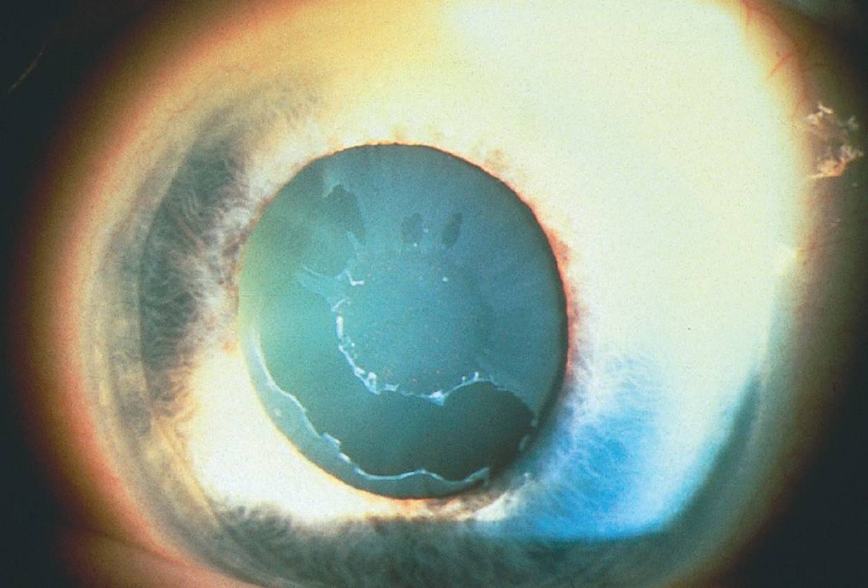
FIGURE 9-1
A) It has a worse prognosis than primary open-angle glaucoma (POAG).
B) It may be monocular or binocular.
C) Lens extraction alleviates the condition.
D) The intraocular pressure (IOP) is often higher than in POAG.
4 Which one of the following is not a risk factor for POAG?
A) Topical corticosteroid response
B) African American heritage
C) Positive family history
D) Diabetes mellitus
5 Which drug used during general anesthesia is associated with an increase in IOP?
A) Halothane
B) Ketamine
C) Valium
D) Phenobarbital
6 Patients with homocystinuria are at increased risk for which of the following?
A) Lens subluxation
B) Angle-closure glaucoma
C) Intravascular thrombosis with general anesthesia
D) All of the above
7 Applanation tonometry gives falsely low readings under all of the following conditions except:
A) high myopia
B) decreased central corneal thickness
C) excessive fluorescein
D) greater than 3 D of with-the-rule astigmatism
8 The reliability of visual field testing becomes suspect when pupil diameter decreases below:
A) 4 mm
B) 3 mm
C) 2 mm
D) 1 mm
9 Which test object has four times the area and the same light intensity as the Goldmann II4e target?
A) III4e
B) II2e
C) II4c
D) V2a
10 A patient is tested on the Humphrey automated perimeter. The machine projects a light at his blind spot, and the patient presses the button. What does this patient’s response represent?
A) False-positive response
B) Fixation loss
C) Short-term fluctuation
D) False-negative response
11 The visual field in Figure 9-2 is caused by a retinal lesion. Which one of the following retinal lesions corresponds to this field? (Note: the fovea is marked by “X”.)
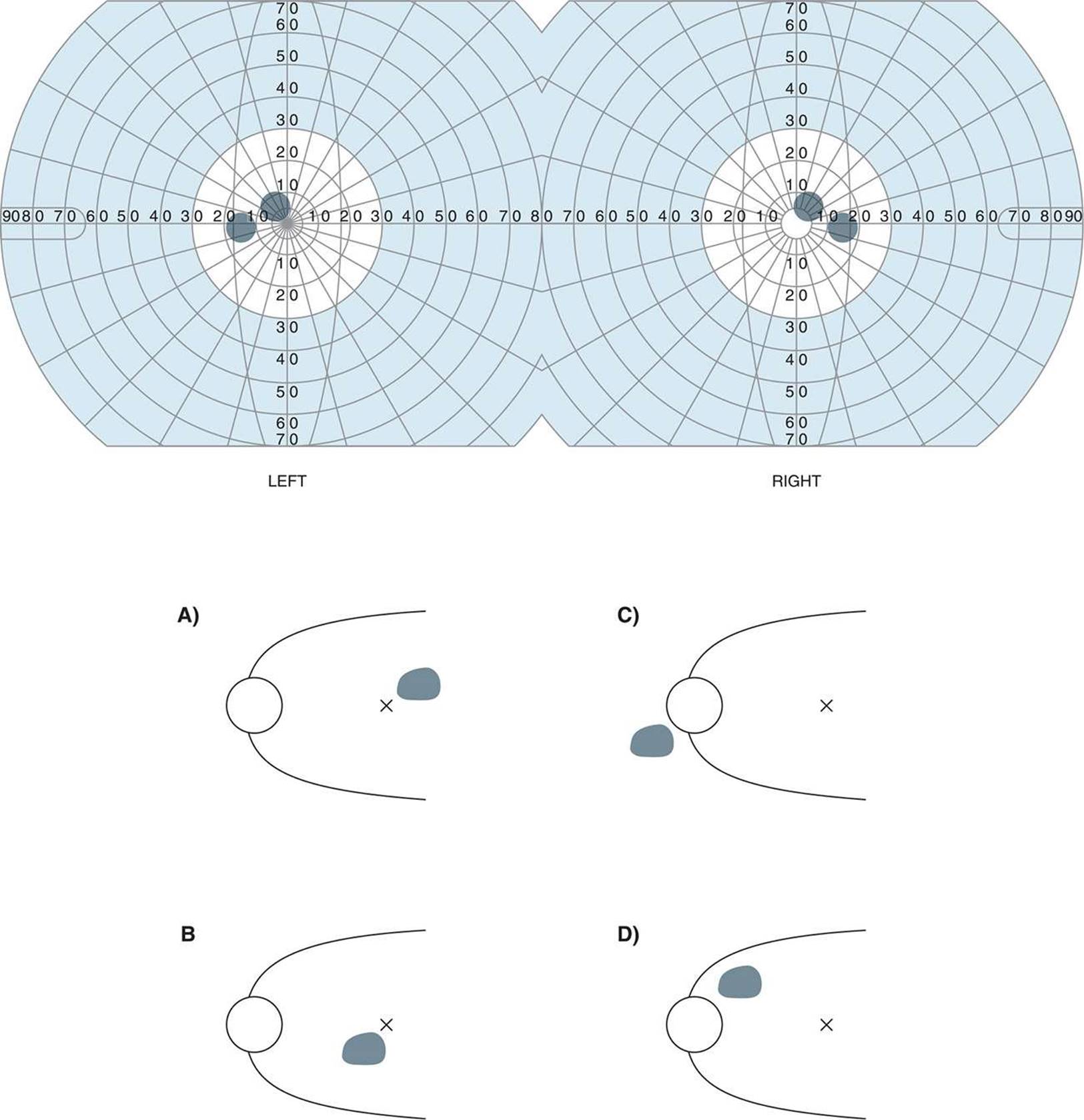
FIGURE 9-2
A)
B)
C)
D)
12 Which one of the following signs is most indicative of glaucomatous optic neuropathy?
A) Figure 9-3A
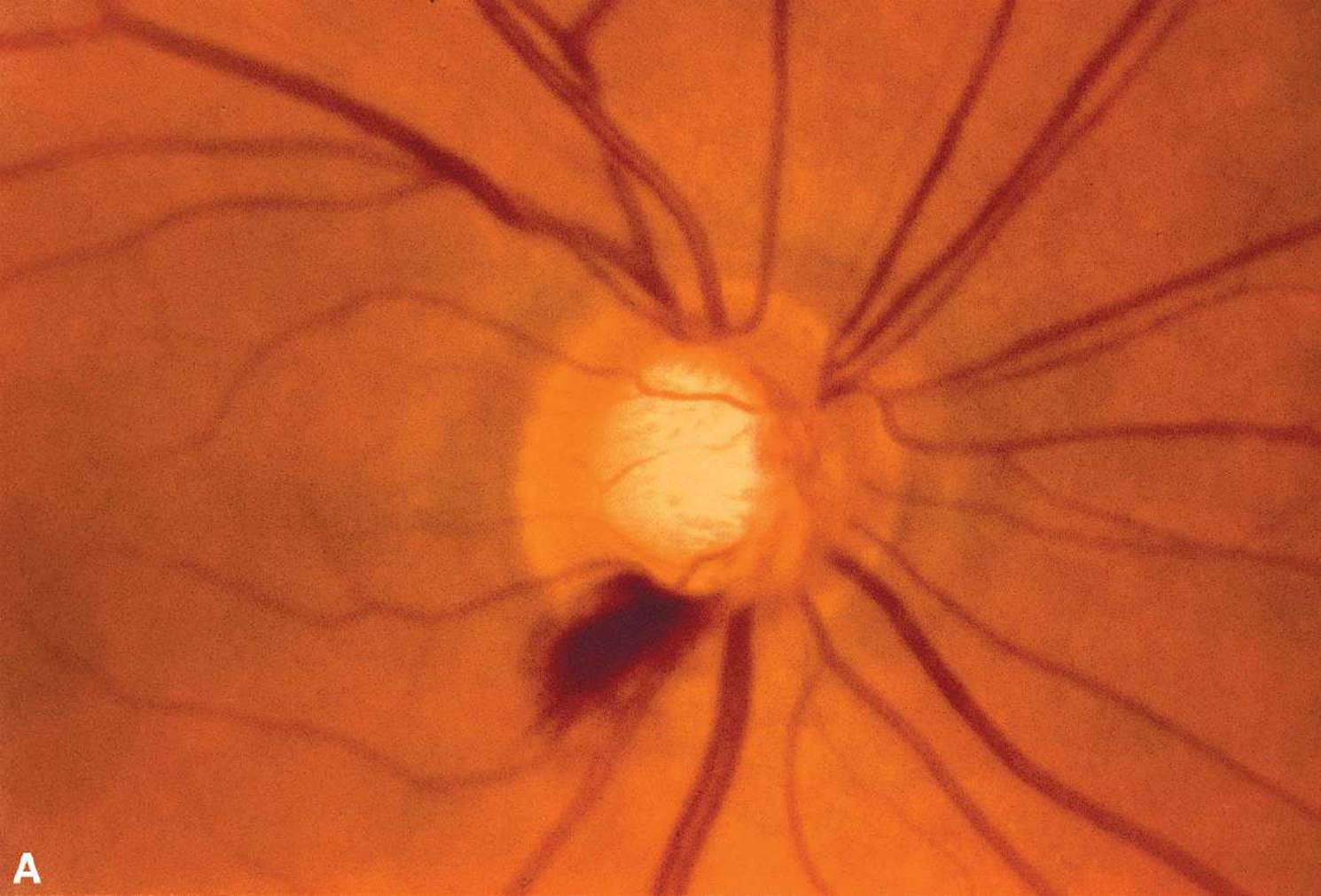
B) Figures 9-3B and C
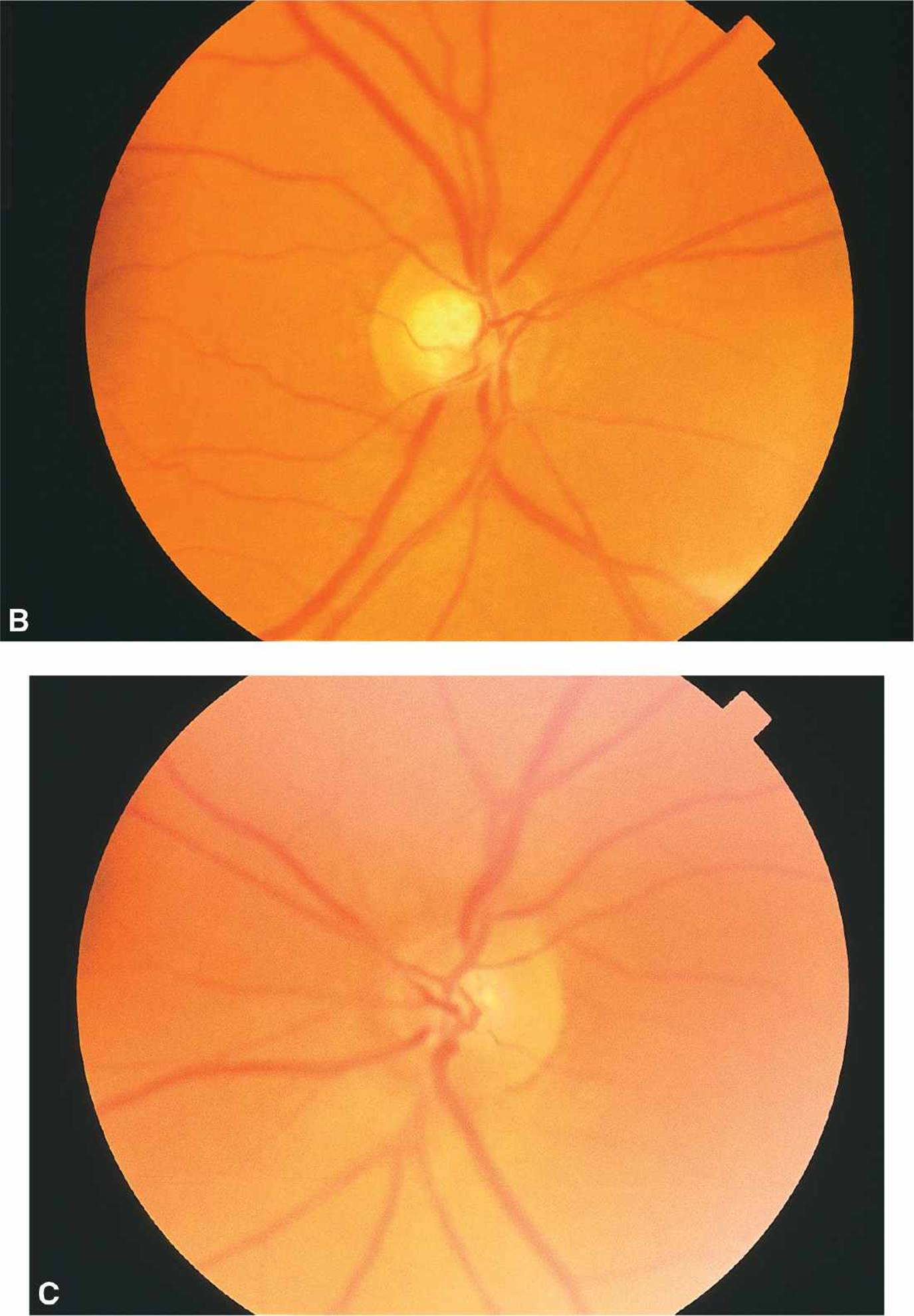
C) Figure 9-3D
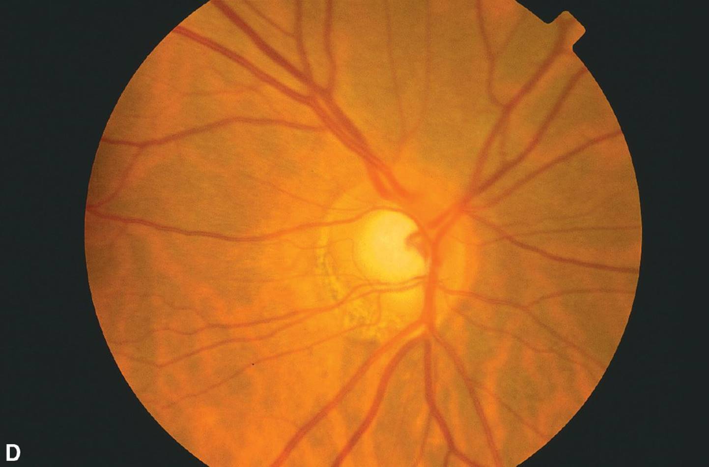
D) Figure 9-3E

FIGURE 9-3
13 Based on histologic studies, what percentage of optic nerve axons may be lost before visual field changes are detected by Goldmann perimetry?
A) 10%
B) 15%
C) 25%
D) 50%
14 All of the following are well-established early signs of glaucomatous damage except:
A) vertical elongation of the cup
B) peripapillary atrophy
C) splinter hemorrhage on disc
D) nerve fiber layer loss
15 All of the following conditions are associated with increased pigmentation of the trabecular meshwork on gonioscopy except:
A) pseudoexfoliation (PXF) syndrome
B) pigment dispersion syndrome
C) prior trauma
D) all of the above
16 Iris transillumination defects are present in all of the following conditions except:
A) oculocutaneous albinism
B) PXF
C) plateau iris syndrome
D) pigment dispersion syndrome
17 All of the following contact lenses for gonioscopy are examples of indirect goniolenses except:
A) Figure 9-4A = Shields, p11b (Koeppe lens)
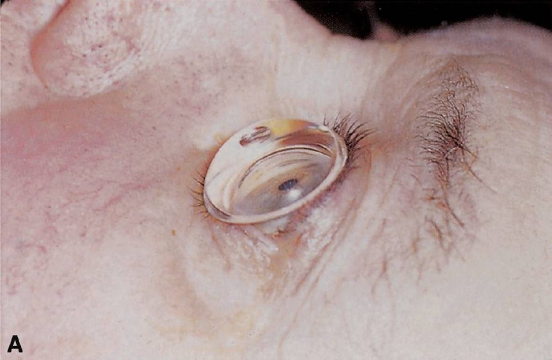
B) Figure 9-4B = Shields, p9c (Posner lens)

C) Figure 9-4C = Shields, p9e (Sussman lens)

D) Figure 9-4D = Shields, p9b (Goldmann lens)

FIGURE 9-4 From Shields MB. Color Atlas of Glaucoma. Williams & Wilkins, 1998.
18 The technique least helpful in evaluating the appearance of a glaucomatous optic nerve is:
A) direct ophthalmoscopy
B) slit-lamp examination using a contact lens
C) indirect ophthalmoscopy using a 20-D lens
D) slit-lamp examination using a 90-D lens
19 Which of the following is least likely to be found in a patient with primary congenital glaucoma?
A) IOP of 23 mm Hg
B) Cup-to-disc ratio of 0.4
C) Corneal diameter of 10.0 mm
D) Open angle with high iris insertion on gonioscopy
20 With respect to uveitic glaucoma, all of the following are true except:
A) Prostaglandins (PGs) such as latanoprost should be used with caution.
B) Argon laser trabeculoplasty (ALT) may be a helpful adjunct if medications are ineffective.
C) Miotics are usually avoided.
D) Treating the intraocular inflammation is as important as lowering IOP.
21 Which one of the following has not been suggested to be a possible pathophysiologic mechanism for optic neuropathy in patients suspected of having normal-tension glaucoma (low-tension glaucoma)?
A) Nocturnal systemic hypotension
B) Vasospasm
C) Shock (hypotensive) optic neuropathy
D) Systemic hypercholesterolemia
22 Which one of the following conditions does not have the same pathogenesis of glaucoma as the others?
A) Sturge–Weber syndrome
B) Thyroid eye disease
C) Aniridia
D) Carotid-cavernous sinus fistula
23 Which surgical procedure would be initially used to manage primary congenital glaucoma with a markedly cloudy cornea?
A) Trabeculectomy with mitomycin C
B) Cyclophotocoagulation
C) Goniotomy
D) Trabeculotomy
24 Which one of the following is the most common cause of glaucoma in eyes being treated for the condition depicted in Figure 9-5?
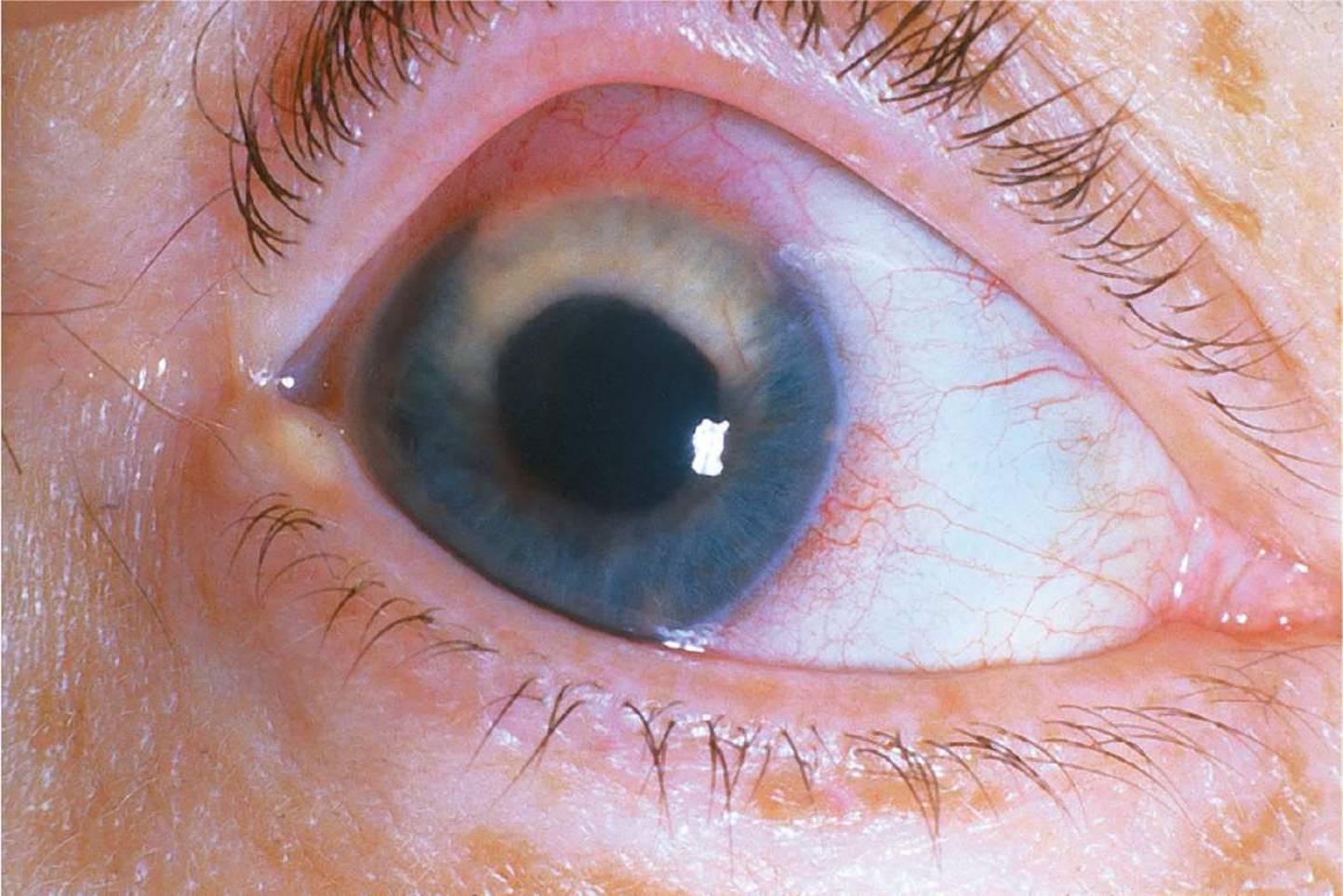
FIGURE 9-5
A) Tumor cells invading the angle
B) Neovascularization
C) Acute-angle closure
D) Uveitis
25 Which one of the following chemical burns is most likely to be associated with an acute elevation of IOP?
A) Chlorine bleach
B) Sulfuric acid
C) Hydrogen peroxide
D) Sodium hydroxide
26 With respect to corticosteroid glaucoma, all of the following are true except:
A) In most cases, after discontinuing the steroid, the IOP returns to normal over a few days to several weeks.
B) The rise in IOP may be delayed for years after starting the steroid.
C) Most cases are caused by long-term oral administration of steroids.
D) Patients with POAG are more susceptible to steroid-induced IOP elevations.
27 Glaucomatous optic neuropathy is associated with damage to which types of retinal cells?
A) Amacrine cells
B) Ganglion cells
C) Bipolar cells
D) Photoreceptors
28 What is the best initial therapy for malignant glaucoma?
A) Pilocarpine 2%
B) Laser iridotomy
C) Mydriatic–cycloplegic therapy
D) Lens removal
29 The condition shown in Figure 9-6 may be associated with which one of the following choices?

FIGURE 9-6
A) Visual loss
B) Papilledema
C) Autosomal recessive inheritance
D) Bilaterality in 25%
30 The most important treatment for a patient with diabetic neovascular glaucoma and good vision is:
A) atropine and topical steroids
B) aqueous suppressants
C) adequate blood glucose and blood pressure control
D) panretinal photocoagulation—consider adding anti-VEGF injections
31 Which one of the following types of glaucoma is least likely to respond to medical therapy alone?
A) Phacolytic glaucoma
B) Pigmentary glaucoma
C) Lens particle glaucoma
D) PXF
32 The eye condition least likely to be associated with aqueous misdirection syndrome is:
A) angle-closure glaucoma
B) uveitis
C) myopia
D) nanophthalmos
33 Topical β-adrenergic antagonists are known to be associated with all of the following side effects except:
A) increased plasma high-density lipoprotein cholesterol levels
B) bronchospasm and airway obstruction
C) weakened myocardial contractility
D) exercise intolerance
34 A patient who has recently undergone panretinal photocoagulation after a central retinal vein occlusion presents with corneal edema, neovascularization of the iris, and an IOP of 58 mm Hg. All of the following medications are appropriate to use except:
A) atropine
B) dorzolamide
C) apraclonidine
D) pilocarpine
35 Ocular side effects of pilocarpine include all of the following except:
A) hyperopia
B) retinal detachment (RD)
C) exacerbation of pupillary block
D) lacrimation
36 Which one of the following is not considered a possible side effect of the hyperosmotic agents?
A) Congestive heart failure
B) Subdural hemorrhage
C) Worsening of obstructive pulmonary disease
D) Mental confusion
37 What is the most likely cause of allergic conjunctivitis in a glaucoma patient? List in order of probability.
1. Latanoprost
2. Dorzolamide
3. Brimonidine
A) 1 > 2 > 3
B) 3 > 2 > 1
C) 3 > 1 > 2
D) 2 > 3 > 1
38 Which statement about carbonic anhydrase inhibitors is false?
A) Aqueous production in the eye is not significantly reduced until more than 90% of the carbonic anhydrase activity is inhibited.
B) Carbonic anhydrase inhibitors cause reduced excretion of urinary citrate or magnesium, therefore predisposing to formation of kidney stones.
C) Carbonic anhydrase inhibitors may cause idiosyncratic and transient acute myopia.
D) Metabolic acidosis is greater with oral acetazolamide than with IV injection of acetazolamide.
39 What is the most common cause of bleb failure?
A) Bleb encapsulation (Tenon cyst formation)
B) Episcleral fibrosis
C) Late bleb leak
D) Closure of the internal sclerostomy
40 Which one of the following is the most important medication to discontinue as far before glaucoma surgery as possible?
A) Pilocarpine
B) Echothiophate
C) Dipivefrin
D) Timolol
41 Medical management of bleb leaks involves all of the following except:
A) bandage soft contact lens
B) autologous blood injection
C) aqueous suppressants
D) 5-fluorouracil (5-FU)
42 Use of 5-FU following filtration surgery has been associated with all of the following except:
A) conjunctival wound leaks
B) suprachoroidal hemorrhage
C) hypotony maculopathy
D) RD
43 Apraclonidine (Iopidine), an α2-adrenergic agonist, has all of the following side effects except:
A) systemic hypotension
B) superior lid retraction
C) dry mouth
D) blanching of conjunctival vessels
44 Dorzolamide (Trusopt) lowers IOP by:
A) increasing uveoscleral outflow
B) decreasing aqueous production
C) increasing conventional (trabecular meshwork) outflow
D) decreasing episcleral venous pressure
45 What combination of medications is most effective in lowering IOP?
A) Timolol and a carbonic anhydrase inhibitor
B) Echothiophate and pilocarpine
C) Pilocarpine and dipivefrin
D) Timolol and dipivefrin
46 PG analogs lower IOP predominantly by which one of the following mechanisms?
A) Increased uveoscleral outflow
B) Enhanced aqueous outflow by stimulation of ciliary muscle contraction
C) Reduced vitreous volume
D) Reduced aqueous production
47 Which one of the following statements about SLT is true?
A) SLT achieves its effect by creating physical openings in the trabecular meshwork through which aqueous humor can pass from the anterior chamber into the canal of Schlemm
B) The chance of posttreatment IOP rise is not influenced by the number of laser burns applied in each treatment session
C) Blanching of the trabecular meshwork is the endpoint seen with each burn
D) Repeating SLT in eyes that were previously treated with ALT is acceptable
48 Laser trabeculoplasty is most likely to be helpful in an eye with which one of the following types of uncontrolled glaucoma?
A) Pigmentary glaucoma
B) Angle-recession glaucoma
C) Iridocorneal endothelial syndrome
D) Inflammatory glaucoma
49 Complications of ALT include all of the following except:
A) iritis with posterior synechiae
B) postoperative IOP spike
C) synechial angle closure
D) cataract formation
50 Which are the most appropriate laser settings for ALT?
A) Spot size: 50 µm, duration: 0.1 seconds, energy: 700 mW
B) Spot size: 50 µm, duration: 0.5 seconds, energy: 500 mW
C) Spot size: 500 µm, duration: 0.1 seconds, energy: 200 mW
D) Spot size: 500 µm, duration: 0.5 seconds, energy: 800 mW
51 The advantages of selective laser trabeculoplasty (SLT) over ALT include all of the following except:
A) SLT is more effective at lowering IOP.
B) SLT uses a potentially repeatable laser.
C) SLT selectively targets pigmented TM cells.
D) general structure of TM intact post-SLT.
52 In which condition would a laser peripheral iridectomy not be indicated?
A) Iris bombé
B) Neovascular glaucoma
C) Acute angle-closure glaucoma
D) Prophylaxis in an eye with narrow angles
53 Compared with the argon laser, the Nd:YAG laser is associated with which one of the following with respect to iridotomies?
A) Late closure of the iridotomy
B) Fewer total applications
C) Less frequent bleeding with application
D) More extensive histologic damage to the treatment site
54 In contrast to trabeculectomy without mitomycin, the use of mitomycin intraoperatively during trabeculectomy may be associated with:
A) lower surgical success rate
B) higher long-term risk of endophthalmitis
C) higher risk of RD
D) more inflammation and a more vascular-appearing bleb
55 Which one of the following procedures has the highest incidence of hypotony?
A) Full-thickness sclerectomy
B) Trabeculectomy with mitomycin C
C) Seton
D) Trabeculectomy with 5-FU
56 The adjunctive use of antifibrotic agents in trabeculectomy is indicated in all of the following situations except:
A) previously failed filtering surgery
B) young myopic patients
C) aphakic/pseudophakic patients
D) neovascular glaucoma
57 The following are true about the drugs used to modulate wound healing post glaucoma surgery, except:
A) BAPN (β-aminoproprionitrile), an inhibitor of lysyl oxidase, blocks collagen cross-linking.
B) 5-FU inhibits fibroblast proliferation by acting selectively on the S phase of the cell cycle.
C) Mitomycin C is an alkylating agent that decreases DNA synthesis by causing DNA cross-linking.
D) Colchicine acts by inhibiting fibroblast migration and proliferation.
58 Which of the following is a theoretical advantage of nonpenetrating glaucoma surgery (nonpenetrating deep sclerectomy/viscocanalostomy)?
A) Lower incidence of postoperative complications
B) Technically easier
C) Better IOP reduction when compared to standard trabeculectomy
D) None of the above
QUESTIONS 59–61 A 34-year-old lawyer is struck in the eye by a golf ball launched by an ophthalmologist. The ophthalmologist rushes the lawyer to his office and examines his eye. A 20% hyphema is present in the anterior chamber. No rupture of the globe is present.
59 The traumatized eye is at risk for developing all of the following types of glaucoma except:
A) angle-closure glaucoma
B) open-angle glaucoma
C) angle-recession glaucoma
D) phacolytic glaucoma
60 Because the IOP is elevated in the traumatized eye, proper management of the hyphema could include all of the following except:
A) corticosteroids
B) β-blockers
C) aminocaproic acid
D) miotic agents
61 The hyphema clears within a week; however, the eye remains hypotonus for several months while retaining good vision. Suddenly, while on vacation in a remote region of the country, the lawyer experiences extreme pain and blurred vision in the previously traumatized eye. Hours later, he is examined in an emergency room of a local rural hospital; his eye has an IOP of 62 mm Hg by Schiotz tonometry. Treatment with timolol drops and acetazolamide tablets is instituted, and the lawyer rushes home to the care of his ophthalmologist. What is the most likely cause for this sudden elevation in IOP?
A) Angle-recession glaucoma
B) Ghost cell glaucoma
C) Recurrent hyphema
D) Spontaneous closure of a cyclodialysis cleft
QUESTIONS 62–64 A mother brings in her 7-month-old son for evaluation of excessive tearing from both of his eyes. On examination, the patient is noted to be photophobic and have bilateral corneal enlargement and corneal clouding.
62 What is the best course of action to take?
A) Send the patient home and instruct the mother on how to perform nasolacrimal sac massage.
B) Perform corneal scrapings and treat as a corneal ulcer.
C) Examine the patient under general anesthesia.
D) Perform B-scan ultrasonography.
63 This patient may experience visual loss for all of the following reasons except:
A) anisometropic amblyopia
B) myopic astigmatism
C) corneal scarring
D) optic nerve damage
64 Initial treatment options for this patient include all of the following except:
A) goniotomy
B) trabeculotomy
C) medical therapy
D) trabeculectomy
65 Indications for surgical intervention after traumatic hyphema include all of the following except:
A) corneal blood staining
B) prolonged presence of a large clot after 15 days
C) rebleeding
D) IOP greater than 45 mm Hg despite maximum-tolerated topical and systemic medications
QUESTIONS 66 and 67 A 65-year-old phakic hyperopic woman undergoes trabeculectomy for uncontrolled POAG. On the first postoperative day, her IOP is 10 mm Hg, a diffuse bleb is present, and the anterior chamber is deep. However, on the second postoperative day, the chamber is shallow with peripheral iridocorneal apposition and an IOP of 22 mm Hg.
66 Appropriate management includes all of the following except:
A) topical cycloplegic medications
B) peripheral laser iridotomy
C) pars plana vitrectomy
D) topical corticosteroids
67 The patient is seen on postoperative day 3 and is noted to have a completely flat anterior chamber, no bleb, and an IOP of 45 mm Hg. At this point, appropriate medical management includes each of the following except:
A) topical cycloplegic medications
B) systemic and topical aqueous suppressants
C) miotic medications
D) topical corticosteroids
68 A 72-year-old phakic African American man undergoes glaucoma-filtration surgery for POAG. Adjunctive mitomycin C is used intraoperatively. Postoperatively, the IOP remains at 2 to 3 mm Hg, and he develops choroidal effusions. Surgical intervention should be considered for all of the following except:
A) impending failure of the bleb
B) continued hypotony beyond 4 weeks
C) kissing choroidals
D) flat anterior chamber with corneal decompensation
69 All of the following may be associated with the abnormality depicted in Figure 9-7 except:
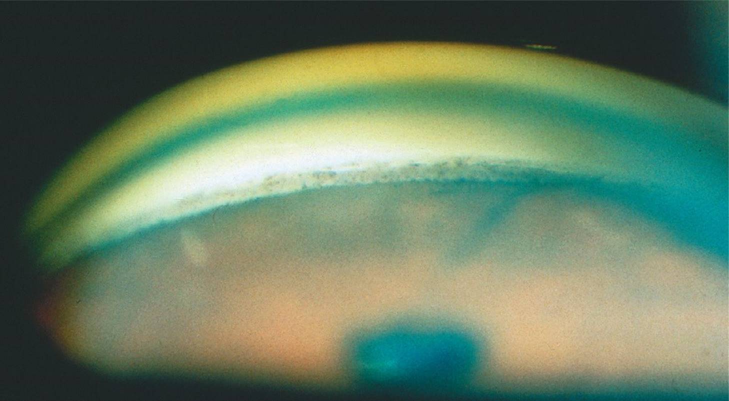
FIGURE 9-7
A) inflammation
B) PXF
C) neoplasm
D) Fuchs heterochromic iridocyclitis
QUESTIONS 70 and 71 A 60-year-old man from India presents with a painful red eye. He has had poor vision in this eye for years, but he feels it has worsened over the past 2 weeks. He denies any ocular trauma. On examination, he is noted to have epithelial edema, heavy flare and cell (large cells), and a dense, brunescent cataract. The IOP is 56 mm Hg.
70 What is the most likely diagnosis?
A) Phacomorphic glaucoma
B) Phacolytic glaucoma
C) Phacoanaphylactic glaucoma
D) Uveitic glaucoma
71 What is the definitive management of this condition?
A) Trabeculectomy
B) Anterior chamber washout
C) Extracapsular cataract extraction
D) Posterior pole vitrectomy
72 According to the Laser Glaucoma Trial, all of the following are true except:
A) ALT is at least as effective at reducing IOP as one glaucoma medical agent, timolol.
B) The majority of patients treated with laser first did not require any additional medical therapy to maintain IOP control.
C) Laser-treated eyes had a slightly lower IOP than medication-treated eyes.
D) ALT is a safe alternate and may be offered as initial therapy to POAG patients.
QUESTIONS 73 and 74 A 67-year-old Asian woman calls complaining of 3 hours of acute right eye pain, blurred vision, and redness of her eye. She has a cloudy cornea with epithelial edema, shallow anterior chamber, 4-mm nonreactive pupil, and IOPs of 62 mm Hg OD and 17 mm Hg OS. Gonioscopy of the left eye reveals a narrow potentially occludable angle with virtually no angle structures visible. She has never had any previous similar episodes.
73 Other findings expected on examination include:
A) keratic precipitates
B) glaukomflecken
C) optic nerve pallor and cupping
D) optic nerve hyperemia and swelling
74 Which one of the following treatments should be attempted first?
A) Peripheral iridectomy
B) Laser trabeculoplasty
C) Trabeculectomy
D) Seton implant
75 Primary angle-closure glaucoma occurs most commonly in patients with shallow anterior chambers. Among the following, which does not contribute to a shallow anterior chamber?
A) Mature lens
B) Hyperopia
C) Ocular hypertension
D) Iris bombé
76 Secondary angle-closure glaucoma may be associated with each of the following conditions except:
A) intraocular tumor
B) scleral buckle for RD
C) nanophthalmos
D) Schwartz syndrome
QUESTIONS 77 and 78 A 72-year-old African American man had a cataract extraction with posterior chamber IOL in the right eye 7 years ago, and 5 years ago he developed a RD, which was repaired with a scleral buckle. He developed intractable glaucoma in that eye with an open angle on gonioscopy. Despite having undergone two sessions of laser trabeculoplasty and now being on maximum-tolerated medical therapy with a visual acuity of 20/200, the IOP remains at 28 mm Hg. Slit-lamp examination reveals 360° of scarred and nonmobile conjunctiva, and there is almost total cupping of the nerve.
77 Which one of the following procedures would be most indicated at this point in this patient’s management?
A) Transscleral cyclophotocoagulation
B) Trabeculectomy with antimetabolite
C) Laser trabeculoplasty
D) Laser iridotomy
78 If a drainage implant were placed in this patient’s eye, which type of implant would be indicated?
A) Double-plate Molteno implant
B) Krupin implant
C) Anterior chamber tube shunt—Shockett procedure
D) Ahmed implant
79 A 65-year-old man undergoes extracapsular cataract surgery. Postoperatively, he has a peaked pupil, hypotony, and incarceration of iris into the wound with leakage. Two weeks later, a grayish white membrane with a scalloped, thickened leading edge was noted on the posterior corneal surface. Treatment of involved iris tissue with argon laser turns this membrane white. Appropriate treatment for this condition includes which one of the following?
A) X-irradiation to involved tissues
B) β-Irradiation to involved tissues
C) Photocoagulation of involved iris tissues with cryotherapy to remaining membranes on corneal tissues
D) Excision of involved iris tissues with cryotherapy to remaining membranes on corneal tissues
80 A diabetic patient has had a complicated RD repaired with silicone oil instillation. Which one of the following is true concerning the peripheral iridectomy?
A) It prevents pupillary block that can occur from neovascularization of the iris.
B) It allows for a much-needed alternate pathway for light entry into the eye.
C) It prevents pupillary block glaucoma that can occur with silicone oil.
D) The iridectomy should have been performed superiorly.
81 Which of the following statements is/are true with respect to the Collaborative Initial Glaucoma Treatment Study (CIGTS)?
A) The study was designed to address the question of medical therapy versus early filtration surgery on the long-term progression of glaucoma.
B) Patients in the early surgery group were more likely to lose visual acuity and visual field during the first few years of follow-up study.
C) After 4 years of follow-up, both groups (medical versus early surgery group) were similar in visual acuity and visual field.
D) All of the above are true.
82 A 75-year-old white man underwent intracapsular cataract extraction 25 years ago. Approximately 15 years ago, he underwent secondary anterior chamber lens placement. Two years ago, he had a penetrating keratoplasty on this eye. Over the past year, he has had recurrent uveitis in this eye, and on referral evaluation in your office, his IOP is 25 mm Hg. Gonioscopy reveals an open angle and a small hyphema. The most appropriate management of his eye would be:
A) aminocaproic acid
B) topical corticosteroids
C) panretinal photocoagulation
D) surgical removal of his anterior chamber lens
83 A young man is seen in your office over a 5-year period with several episodes of unilateral elevation of IOP to the 40 to 50 mm Hg range. During these episodes, fine keratic precipitates and faint flare are noted. A mild ciliary flush is noted. No iris changes are noted. Each episode seems to respond well to topical corticosteroids and topical and systemic aqueous suppression. What is the most likely cause of his episodic glaucoma?
A) Fuchs heterochromic iridocyclitis
B) Juvenile rheumatoid arthritis
C) Posner–Schlossman syndrome
D) Sarcoidosis
84 Features associated with the disease shown in Figure 9-8 include all of the following except:
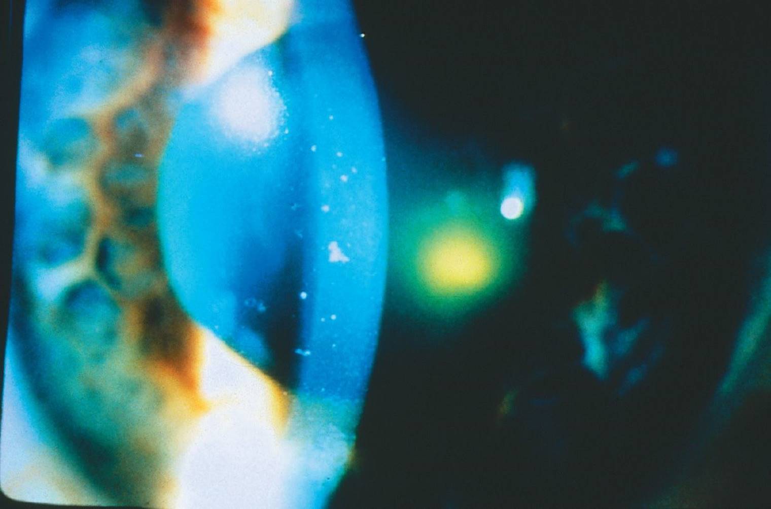
FIGURE 9-8
A) 20% to 60% incidence of glaucoma
B) poor pupillary dilation
C) weak zonules
D) peripheral iris transillumination defects
85 All of the following are true with respect to PG analogs except:
A) Unoprostone isopropyl (Rescula) reduces IOP by increasing uveoscleral outflow.
B) Latanoprost (Xalatan) is a prodrug that becomes biologically active after being hydrolyzed by corneal esterase.
C) Bimatoprost (Lumigan) is a prostamide analog.
D) Conjunctival hyperemia is a reported side effect of the PG analogs.
86 A 46-year-old woman with a long history of insulin-dependent diabetes mellitus presented with a nonclearing vitreous hemorrhage in her left eye. She underwent a vitrectomy to clear the hemorrhage. One week after surgery, she presented with left eye pain, an IOP of 58 mm Hg, and the slit-lamp appearance shown in Figure 9-9. The most likely diagnosis is:
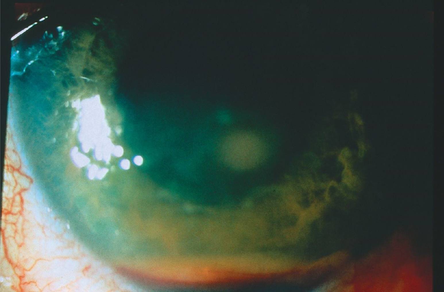
FIGURE 9-9
A) hemolytic glaucoma
B) phacolytic glaucoma
C) ghost cell glaucoma
D) hyphema
QUESTIONS 87 and 88 A 64-year-old pseudophakic woman presented with severe pain in her left eye 6 days after a standard trabeculectomy. Her visual acuity was hand motions, and IOP was 33 mm Hg. The anterior chamber of the left eye was shallow.
87 The diagnosis of this patient’s condition could include each of the following except:
A) malignant glaucoma
B) excessive filtration
C) delayed suprachoroidal hemorrhage
D) incomplete iridectomy with obstruction of sclerostomy
88 This patient is later found to have large choroidal detachments (Fig. 9-10) with central touch. Which one of the following risk factors has not been shown to be associated with this condition?
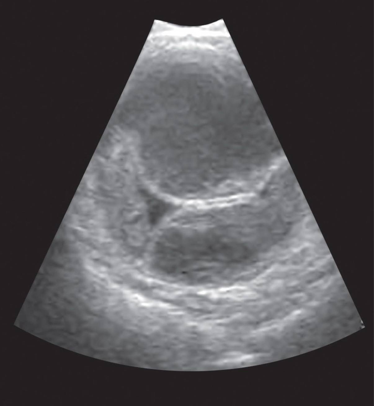
FIGURE 9-10 From Jehle D, Bouvet S, Braden B, et al. Emergency Ultrasound of the Eye and Orbit. Buffalo, NY: Grover Cleveland Press, 2011:58. Figures 7–10, with permission.
A) Preoperative elevated IOP
B) Aphakia
C) Hyperopia
D) Previous vitrectomy
89 An elderly patient underwent an uncomplicated trabeculectomy of the right eye. The next day, the IOP was 1 mm Hg and the patient had the slit-lamp appearance shown in Figure 9-11. The bleb is flat. The retina and choroid appear normal. What is the most likely cause of these findings?
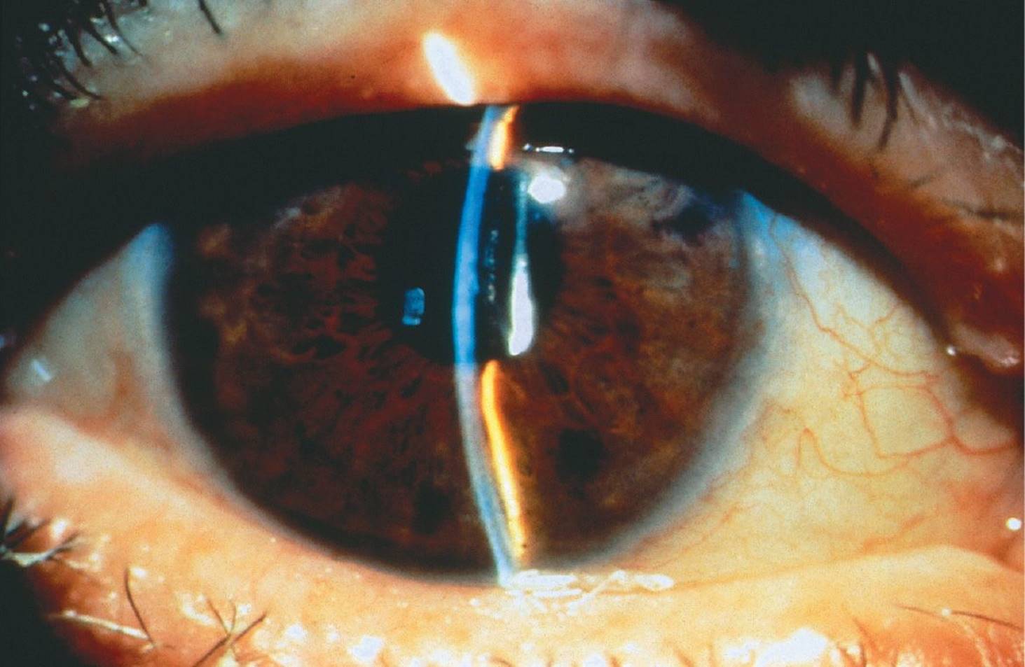
FIGURE 9-11
A) Malignant glaucoma
B) Choroidal detachments
C) Bleb leak
D) Excessive filtration
90 Ocular tumors can cause glaucoma through a variety of mechanisms. Malignant melanomas of the anterior uveal tract might cause glaucoma through each of the following mechanisms except:
A) direct extension of the tumor in the trabecular meshwork
B) obstruction of the trabecular meshwork by macrophages laden with melanin
C) seeding of tumor cells into the outflow channels
D) increase in episcleral venous pressure
91 Reported side effects of the PG analog drops include all of the following except:
A) conjunctival hyperemia
B) cystoid macular edema (CME)
C) conjunctival melanosis
D) increased iris pigmentation
92 Two months ago, a 68-year-old man suffered a central retinal vein occlusion in his left eye. He comes in with a red, painful eye and florid rubeosis (Fig. 9-12). What is the most appropriate treatment for this patient?

FIGURE 9-12
A) Panretinal photocoagulation
B) Seton implantation
C) Diode cyclophotocoagulation
D) Laser iridotomy
93 According to the Ocular Hypertension Treatment Study (OHTS), all of the following are risk factors for glaucoma except:
A) African American race
B) thick corneas
C) increasing age
D) optic nerve anatomy
QUESTIONS 94–97 Visual field defects (Figs. 9-13 to 9-18)
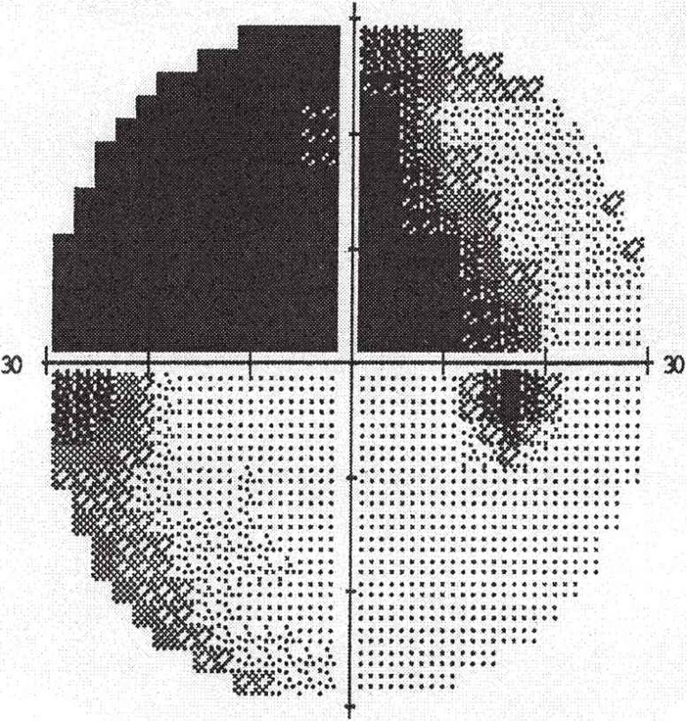
FIGURE 9-13
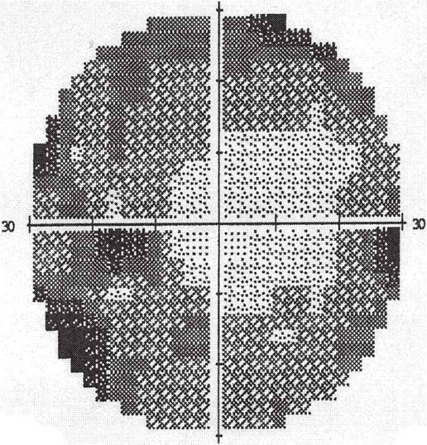
FIGURE 9-14

FIGURE 9-15
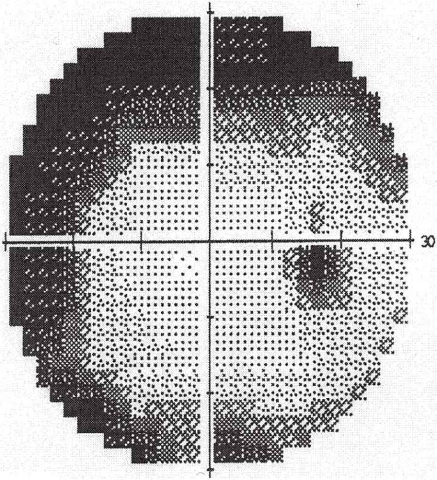
FIGURE 9-16
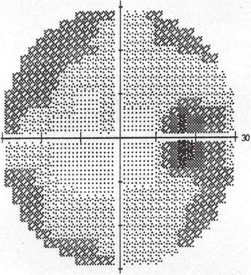
FIGURE 9-17

FIGURE 9-18
94 A patient with which field corresponds to the greatest optic nerve damage?
A) Figure 9-13
B) Figure 9-14
C) Figure 9-16
D) Figure 9-18
95 The visual field in Figure 9-15 was seen in a patient with a healthy-appearing optic nerve and retina. What might account for the findings?
A) A patient pushing the button too frequently
B) Poor head positioning
C) Field test with both eyes open
D) Failure to press the button at the beginning of the test
96 Which field best corresponds to the optic nerve appearance in Figure 9-19?
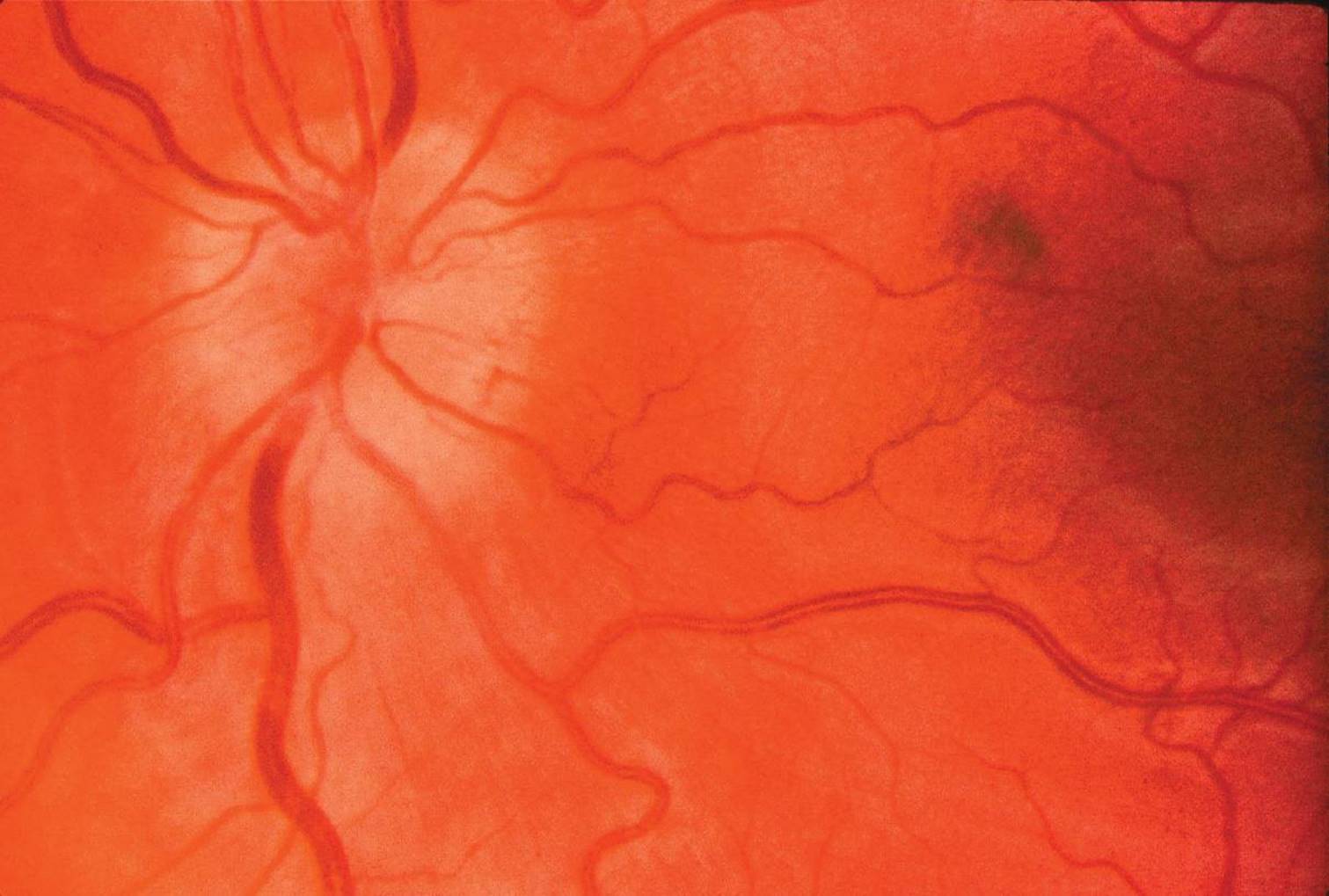
FIGURE 9-19
A) Figure 9-17
B) Figure 9-13
C) Figure 9-14
D) Figure 9-18
97 Figure 9-18 was found upon visual field testing in a 72-year-old man with intact neuroretinal rims on optic nerve evaluation. Which etiology is most likely?
A) POAG
B) Myopia with peripapillary atrophy
C) CNS vascular event
D) Low-tension glaucoma
98 Glaucoma implant surgery (aqueous shunt devices) would be indicated in the following circumstances except:
A) previously failed filtration surgery with antimetabolites
B) uveitic glaucoma
C) congenital glaucoma patient with poor visual potential
D) glaucoma patient with previous vitrectomy with scleral buckle
99 All of the following are potential complications of tube shunt procedures except:
A) corneal neovascularization
B) conjunctival melt
C) hypotony
D) diplopia
100 The following measures can be taken to limit postoperative hypotony with the device in Figure 9-20 except:
A) two-stage procedure
B) collagen plugs
C) pressure-sensitive valve
D) ligature occlusion of tube

FIGURE 9-20
1 B) Ascorbate
Compared with plasma, aqueous is slightly hypertonic and acidic. Aqueous has a marked excess of ascorbate (15 times greater than that of arterial plasma) and a marked deficit of protein (0.2% in aqueous as compared to 7% in plasma).
2 D) Central retinal artery
The four divisions of the optic nerve head correlate roughly with a four-part blood supply (Fig. 9-21). The surface fiber layer is supplied mainly by branches of the central retinal artery. The prelaminar region is supplied by capillaries of the short posterior ciliary arteries. The lamina cribrosa region is also supplied by vessels that come directly from the short posterior ciliary arteries to form a dense plexus in the lamina. The retrolaminar region is supplied by both the ciliary and retinal circulations, with the former coming from recurrent pial vessels. The central retinal artery provides centripetal branches from the pial region.

FIGURE 9-21 LC = lamina cribrosa, S = sclera, C = choroid, R = retina, PCA = posterior ciliary artery, NFL = nerve fiber layer, PL = prelamina, RL = retrolamina, CRA = central retinal artery, ON = optic nerve.
3 C) Lens extraction alleviates the condition.
PXF is an age-related disease involving an accumulation of abnormal fibrillar extracellular material within ocular and systemic tissues. Glaucoma occurs more commonly in eyes with PXF than in those without it. In fact, PXF has been recognized as the most common identifiable cause of glaucoma. The glaucoma is a secondary open-angle one and can be unilateral or bilateral. IOPs can be quite elevated and are often higher than in POAG. Compared to POAG, glaucoma associated with PXF tends to respond less well to medical therapy, to be more difficult to treat, to require surgical intervention more often, and to have a worse overall prognosis. Unfortunately, lens extraction is not curative, although the pressure may be lowered by a few millimeters of Hg following simple cataract extraction.
4 A) Topical corticosteroid response
POAG is a multifactorial disease that may occur on the basis of inherited risk factors. Risk factors for POAG include family history of glaucoma (five to six times greater risk), African American heritage (five to eight times higher incidence than in whites), and age over 50 (the risk of glaucoma increases with each decade of life to nearly a 15% incidence in the population over 80 years of age). It is seen with greater frequency in patients with diabetes mellitus, high myopia, RD, and central retinal vein occlusion. Patients with POAG and relatives of POAG patients have a higher incidence of elevated IOP in response to topical or systemic corticosteroids, but this is not a risk factor for POAG; rather, it is a risk factor for steroid-induced secondary open-angle glaucoma.
5 B) Ketamine
In most cases, patients have a decrease in IOP with general anesthesia. In particular, halothane and the inhalational anesthetics can decrease IOP. Ketamine and trichloroethylene, however, can cause IOP to increase. Tranquilizers and barbiturates may cause a slight decrease in IOP.
6 D) All of the above
Homocystinuria is a rare autosomal recessive condition. Affected patients are generally tall, with osteoporosis, scoliosis, and chest deformities. About 50% of the time, there is associated mental retardation. These patients are at increased risk of thrombotic vascular occlusions, and this should be taken into consideration if general anesthesia is planned. Lens dislocation occurs in 90% of patients and is generally inferior and bilateral. Homocystinuria may lead to angle-closure glaucoma if the lens dislocates into the anterior chamber.
7 C) Excessive fluorescein
If the fluorescein rings are too narrow, the IOP is underestimated. If they are too thick, the IOP is overestimated. Use of excessive fluorescein causes thick mires and an overestimation of IOP. The thickness of the cornea affects IOP readings. If the cornea is thin, the IOP is underestimated. In high myopia, there is decreased scleral rigidity, which may lead to an underestimation of IOP. If corneal astigmatism is greater than 3 D, IOP is underestimated for with-the-rule astigmatism and overestimated for against-the-rule astigmatism.
8 B) 3 mm
A pupillary diameter of less than 3 mm can cause general depression of the field. It is best to test the field with a pupil larger than 3 mm. Patients taking pilocarpine may need to refrain from taking the medication for 24 hours before the test or be dilated at the time of their examination.
9 A) III4e
On the Goldmann perimeter, the test objects can be varied in both size and intensity by using different filters. The size of the test object is represented by the roman numeral (I–V). Each increment of the roman numeral doubles the diameter (and quadruples the area) of the test object. Light intensity can be altered using different neutral density filters. Filters 1 to 4 are in increments of 5 dB each. Filters a to e are in increments of 1 dB each. The III4e test object will have twice the diameter and four times the area of the II4e test object.
10 B) Fixation loss
The Humphrey automated perimeter has a number of ways to test the reliability of the test taker. A fixation loss occurs when the patient responds as if seeing a light when a target is displayed in his blind spot. A false-negative response occurs when the patient fails to respond to a suprathreshold stimulus at a location that would be expected to be seen. This response may indicate a patient who is falling asleep or losing interest. Intermittently, the perimeter will pause and the motorized light will change position, but no stimulus will be presented. If the patient presses the button, a false-positive response is recorded. A nervous or trigger-happy patient may have a high false-positive rate. Short-term fluctuation describes the change in sensitivity when the same point is retested.
11 B)
The center of the visual field corresponds to the fovea. The visual field shows a lesion located superior to the fovea of the left eye between the fovea and the blind spot. Superior defects correspond to lesions inferior to the fovea in the retina.
12 C) Figure 9-3D
Figure 9-3D shows narrowing (notching) of the rim. For the early detection of glaucomatous optic nerve damage, the most important variable appears to be focal narrowing or notching of the neuroretinal rim. Other important variables are optic cup size in relation to optic disc size, disc asymmetry, and presence of disc hemorrhages. However, disc hemorrhages can occur in other conditions (e.g., anterior ischemic optic neuropathy [AION]) and disc asymmetry may be a normal finding if the difference is slight (<0.2 difference).
13 D) 50%
It is known that pathologic changes of the optic nerve precede visual field changes. In fact, up to 50% of optic nerve axons can be lost before any change is detected on the Goldmann visual field. This observation has stimulated interest in measuring the retinal nerve fiber layer thickness via imaging modalities. Different technologies have been developed and are being refined, most notably scanning laser polarimetry (GDx) and optical coherence tomography (OCT). These technologies will likely be useful to find early changes of glaucoma in addition to a careful ophthalmoscopic exam and clinical assessment.
14 B) Peripapillary atrophy
Focal enlargement of the cup appears as localized notching of the rim. The cup can become vertically oval if narrowing of the rim occurs at either the superior or inferior pole of the disc. Splinter hemorrhages usually clear over several weeks but are often followed by localized notching of the rim. Glaucomatous optic atrophy is associated with loss of axons in the nerve fiber layer, which can be best evaluated with red-free illumination. Peripapillary atrophy is not considered to be an exclusive sign of early glaucomatous damage since other conditions such as ocular histoplasmosis or myopia can result in peripapillary atrophy.
15 D) All of the above
In a young normal eye, it is unusual to see any trabecular pigment band. This is because insufficient pigment has filtered through the trabecular meshwork to form a visible pigmented line. If pigmentation is apparent, it is usually most prominent in the inferior angle. The two most common conditions in which the pigment band is very prominent are pigment dispersion syndrome/pigmentary glaucoma and PXF glaucoma. Lesser amounts of trabecular pigmentation can be seen in iritis, diabetes, or following intraocular surgery, trauma, or laser.
16 C) Plateau iris syndrome
In oculocutaneous albinism, there is diffuse transillumination of the iris. Patients with exfoliation syndrome have peripupillary transillumination defects. Peripheral discrete or confluent iris transillumination defects may be seen in pigment dispersion syndrome. Plateau iris syndrome does not customarily produce transillumination defects.
17 A) Figure 9-4A = Shields, p11b (Koeppe lens)
In direct gonioscopy, the angle is visualized directly through the contact lens. In indirect gonioscopy, the light rays are reflected by a mirror in the contact lens. The Zeiss four-mirror lens is an example of an indirect gonioprism, where all four mirrors are inclined at 64°. The Sussman lens is a handheld Zeiss-type gonioprism. The Goldmann mirror is inclined at 62° for gonioscopy. The Koeppe lens is the prototype diagnostic direct goniolens.
18 C) Indirect ophthalmoscopy using a 20-D lens
The small image obtained using indirect ophthalmoscopy does not allow for adequate evaluation of the optic nerve details. Careful examination with the direct ophthalmoscope can provide important information about the pallor of the optic cup; however, the most effective methods include stereoscopic examination using the slit lamp in combination with a posterior-pole contact lens, a 90-D lens, or a Hruby lens.
19 C) Corneal diameter of 10.0 mm
Although congenital glaucoma has an incidence of 1 in 12,500 births, it accounts for about 5% of students in schools for the visually handicapped. It is bilateral in two-thirds of patients; two-thirds of these patients are male; and about 10% of congenital glaucoma is familial (autosomal recessive). Symptoms include tearing, photophobia, and blepharospasm. Signs include:
1. IOP over 21 mm Hg
2. Optic nerve cup-to-disk ratio greater than 0:3 (present in only 2.6% of normal newborns)
3. Horizontal corneal diameter greater than 12.5 mm with or without corneal edema or breaks in Descemet membrane (Fig. 9-22)

FIGURE 9-22
4. Open angle with anterior iris insertion (either flat insertion into the trabecular meshwork or, less commonly, concave insertion with the plane of the iris posterior to scleral spur and anterior iris stroma sweeping upward and inserting into the meshwork)
5. No iris abnormality other than that previously described (i.e., no hypoplasia or corectopia)
Important diagnoses to exclude include neural crest dysgenesis (aniridia, Peter anomaly, Axenfeld–Rieger syndrome), phakomatoses, metabolic abnormalities (Lowe syndrome, homocystinuria, mucopolysaccharidoses), inflammatory conditions (congenital rubella, herpes simplex iridocyclitis), neoplasms (retinoblastoma, juvenile xanthogranuloma), congenital diseases (X-linked megalocornea, Down syndrome, Patau syndrome, Zellweger syndrome, Rubinstein–Taybi syndrome, persistent hyperplastic primary vitreous [PHPV], retinopathy of prematurity), trauma, and steroid use.
20 B) Argon laser trabeculoplasty (ALT) may be a helpful adjunct if medications are ineffective.
Ocular inflammation can lead to glaucoma via a variety of mechanisms, including (1) obstruction of the trabecular meshwork by inflammatory debris, (2) increased viscosity of aqueous humor, (3) neovascularization, (4) uveal effusion, (5) papillary block, and (6) formation of peripheral anterior synechiae. Treatment depends on the underlying condition, but in most cases, inflammation is suppressed by some combination of topical, periocular, or oral corticosteroids. Occasionally, other immunosuppressive agents are needed. Elevated IOP is managed by topical and oral glaucoma medications as needed. Miotics are generally avoided because they lead to increased pain and congestion and promote the formation of posterior synechiae. PG agents are used with caution because they may exacerbate uveitis. ALT is not very helpful in eyes with active inflammation.
21 D) Systemic hypercholesterolemia
Normal-tension glaucoma is not one disease but rather a collection of disease processes characterized by characteristic glaucomatous visual field loss without elevated IOPs. Nocturnal systemic hypotension caused by excessively large late evening or bedtime doses of antihypertensive agents has recently been suggested to cause low-tension glaucoma on the basis of compromised optic nerve blood supply. Vasospasm and shock may also compromise optic nerve blood supply. Systemic hypercholesterolemia has not been implicated in the pathophysiology of low-tension glaucoma.
22 C) Aniridia
The common pathogenesis of glaucoma in the Sturge–Weber syndrome, thyroid eye disease, and carotid-cavernous sinus fistula is an increase in episcleral venous pressure through elevated venous outflow pressure or orbital congestion. The glaucoma associated with aniridia is caused by congenital agenesis of the angle structures.
23 D) Trabeculotomy
Management of congenital glaucoma involves surgery, either goniotomy (70% success) or trabeculotomy (70% to 80% success). A goniotomy requires that the cornea be clear enough to view the meshwork. In the scenario presented, a trabeculotomy ab externo is the procedure of choice because of the cloudy cornea. In general, for primary congenital glaucoma, if repeated attempts at goniotomy and/or trabeculotomy fail, filtering surgery with mitomycin C or seton placement is considered. Topical medication, such as β-blockers, miotics, or carbonic anhydrase inhibitors can temporize and potentially clear the cornea enough to allow goniotomy to be performed. Cyclophotocoagulation and other cyclodestructive procedures usually are reserved for eyes that have undergone other unsuccessful surgeries.
24 B) Neovascularization
Malignant melanomas can be associated with normal, elevated, or depressed IOP. Elevated IOP occurs more frequently with melanomas of the iris/ciliary body than with choroidal melanomas. Glaucoma may occur by a variety of mechanisms, including (1) obstruction of the trabecular meshwork by melanin-containing macrophages (melanomalytic glaucoma), (2) direct extension of tumor into the trabecular meshwork, (3) angle closure from anterior displacement of the lens–iris diaphragm or peripheral anterior synechiae, (4) inflammation, and (5) neovascularization of the angle. Neovascularization of the angle appears to be the most common cause of elevated IOP, especially among eyes treated with radiation.
25 D) Sodium hydroxide
Glaucoma is most often associated with alkali burns but can also be seen after severe acid burns. IOP rises initially because of scleral shrinkage and release of PGs. Later, IOP rises because of inflammation, posterior synechiae causing pupillary block, or acute lens swelling. Finally, IOP may continue to be high due to direct injury to the trabecular meshwork. Filtering surgery may be needed but can be difficult due to conjunctival scarring. A cyclodestructive procedure or seton valve may be required.
26 C) Most cases are caused by long-term oral administration of steroids.
Most cases of corticosteroid glaucoma are caused by drops or ointments instilled in the eye. Steroid creams, lotions, and ointments applied to the face or eyelids may reach the eye in sufficient quantity to raise IOP, as may systemically administered corticosteroids. There have been case reports of increased IOP following the use of steroid inhalers for asthma or steroid nasal spray for allergic rhinitis. The IOP elevation may occur within a week of initiating treatment or may be delayed for years. The first step in managing steroid glaucoma is to discontinue the drug. In most cases, the IOP will return to normal within a few days to several weeks. The residual glaucoma can be treated with glaucoma medicines, laser, or surgery. Corticosteroids are thought to raise IOP by lowering outflow facility perhaps due to an accumulation of glycosaminoglycans in the trabecular meshwork.
27 B) Ganglion cells
The optic nerve carries axons from ganglion cells into the inner retina. The nerve fiber layer is comprised of these ganglion cell axons.
28 C) Mydriatic–cycloplegic therapy
Malignant glaucoma, or aqueous misdirection, occurs when aqueous humor is secreted into and sequestered in the vitreous cavity, pushing the lens, hyaloid face, and iris forward, thus collapsing the anterior chamber and blocking the trabecular meshwork. The IOP often rises significantly, and the anterior chamber is shallow both centrally and peripherally. The common clinical features include (1) shallow or flat anterior chamber, (2) increased or normal IOP, (3) poor response to miotics, (4) favorable response to cycloplegics–mydriatics. Mydriatic–cycloplegics help by pulling the lens–iris diaphragm posteriorly.
29 A) Visual loss
Optic nerve drusen occurs in 2% of optic nerve specimens on histopathology and is bilateral in 50%. Pathologic studies have revealed intra-axonal calcification of mitochondria. The nerve head may be elevated, but no axonal swelling or edema exists. Optic nerve drusen may be inherited in an autosomal dominant fashion and occur more frequently in whites. Visual loss can occur and is thought to result from axonal compression, subretinal hemorrhage, or choroidal neovascular membrane formation. Systemic conditions associated with optic nerve drusen include Paget disease, Ehlers–Danlos syndrome, sickle cell disease, and pseudoxanthoma elasticum.
30 D) Panretinal photocoagulation—consider adding anti-VEGF injections
Initial treatment of a patient with an acute episode of neovascular glaucoma includes a topical β-blocker, an α-agonist, and/or carbonic anhydrase inhibitor as well as atropine and topical steroids. Carbonic anhydrase inhibitors may be less effective in the presence of marked corneal edema. Miotics, PGs, and epinephrine should be avoided. In most cases, it is important to proceed rapidly with panretinal photocoagulation to prevent total angle closure. Following panretinal photocoagulation, new vessels may regress within a few days to a few weeks. If IOP remains elevated despite panretinal photocoagulation and medicines, filtering surgery, seton valve placement, or cyclodestruction may be needed.
31 A) Phacolytic glaucoma
Phacolytic glaucoma results when mature or hypermature cataracts leak soluble lens protein through microscopic defects in the lens capsule. This heavy-molecular-weight protein directly obstructs the outflow of aqueous humor. Macrophages engorged with this material may also obstruct the outflow channels. Although it is desirable to first bring the IOP under medical control with hyperosmotics, carbonic anhydrase inhibitors, and topical β-blockers, definitive therapy requires removal of the lens. Lens particle glaucoma may occur after trauma to the lens capsule or after cataract surgery with retained cortical material. The lens material may resorb spontaneously. Medical management is used to control IOP; however, surgical removal of the lens material may be necessary if medical management fails.
32 C) Myopia
Patients at particular risk for aqueous misdirection are those with crowded anterior segments (i.e., angle closure, nanophthalmos). Postoperative inflammation may cause swelling of the ciliary body and ciliary processes leading to aqueous misdirection. Myopia is not associated with an increased risk of aqueous misdirection.
33 A) Increased plasma high-density lipoprotein cholesterol levels
Blockade of β1-adrenergic receptors slows the pulse rate and weakens myocardial contractility, which can affect the time of exhaustion with heavy exercise. Blockade of β2-adrenergic receptors produces contraction of bronchial smooth muscle. Topical timolol has been shown to decrease plasma high-density lipoprotein cholesterol levels. Carteolol is a topical nonselective β-adrenergic antagonist that has intrinsic sympathomimetic activity and has been shown to have less adverse effect on plasma lipids. Betaxolol is relatively β1-selective and may have less of an effect on the respiratory muscles.
34 D) Pilocarpine
Aqueous suppressants have the best chance of controlling the IOP. Because the trabecular outflow has been occluded by neovascularization, miotic medications will be ineffective and may, in fact, reduce the uveoscleral outflow, further elevating the IOP. Miotics are also less effective in eyes with severely increased IOP.
35 A) Hyperopia
Ocular side effects of pilocarpine include conjunctival vascular congestion, miosis, induced myopia, cataract formation, and temporal or periorbital headaches. Rarely, pilocarpine can be associated with RD. Pilocarpine does not cause hyperopia.
36 C) Worsening of obstructive pulmonary disease
Hyperosmotic agents may aggravate congestive heart failure by an increase in extracellular volume. Backache, headache, mental confusion, and subdural, even subarachnoid, hemorrhages have also been reported.
37 B) 3 > 2 > 1
PG analogs such as latanoprost (Xalatan) more commonly produce conjunctival hyperemia than true allergic conjunctivitis. From 1% to 5% of patients taking dorzolamide (Trusopt) may have allergic symptoms. Up to 20% of patients taking brimonidine (Alphagan) may show such symptoms.
38 D) Metabolic acidosis is greater with oral acetazolamide than with IV injection of acetazolamide.
Metabolic acidosis is greater with IV injection of acetazolamide. Acute myopia is the only ocular reaction commonly associated with carbonic anhydrase inhibitors.
39 B) Episcleral fibrosis
The most frequent cause of failure after filtration surgery is bleb scarring due to episcleral fibrosis. This excessive healing response is largely due to the proliferation of fibroblasts and the production of collagen and glycosaminoglycans. The antimetabolite 5-FU has been used to modulate wound healing after filtration surgery. 5-FU inhibits fibroblast proliferation to prevent episcleral fibrosis. Mitomycin C, an antitumor antibiotic isolated from Streptomyces caespitosus, also suppresses cellular proliferation. Both antimetabolites have been used to modify wound healing in glaucoma surgery.
40 B) Echothiophate
Echothiophate is a strong, relatively irreversible cholinesterase inhibitor. This drug causes a disruption of the blood–aqueous barrier, which may cause increased inflammation after intraocular surgery in eyes pretreated with echothiophate. Indirect agents also block other cholinesterases, including plasma pseudocholinesterase, which deactivates succinylcholine. Patients may be paralyzed for extended periods of time after anesthesia with succinylcholine and need to be warned about this. For these reasons, it is usually advisable to discontinue the drug several weeks before surgery. Many surgeons also like to discontinue dipivefrin several days before surgery to lessen conjunctival and episcleral injection.
41 D) 5-Fluorouracil (5-FU)
Bandage, soft contact lenses, autologous blood injections, and aqueous suppressants have all been used in the management of bleb leaks. Bleb leaks are more common in eyes with thin cystic blebs and after treatment with antimetabolites.
42 D) RD
In addition to inhibiting fibroblast proliferation, 5-FU also inhibits the growth of epithelial cells of the conjunctiva and cornea. It is associated with several undesirable complications, including conjunctival wound leaks, corneal epithelial defects, thin-walled ischemic blebs, hypotony, and suprachoroidal hemorrhage. It has not been associated with increased risk of RD.
43 A) Systemic hypotension
Apraclonidine hydrochloride is a para-amino derivative of clonidine hydrochloride, an α2-adrenergic agonist that is used clinically as a potent systemic antihypertensive agent. Several studies have shown the lack of effect of apraclonidine on blood pressure and pulse. However, a transient dry mouth or dry nose is commonly reported. Ocular side effects include eyelid retraction, mydriasis, and conjunctival blanching.
44 B) Decreasing aqueous production
Dorzolamide is a topical carbonic anhydrase inhibitor that works in an analogous fashion to systemic carbonic anhydrase inhibitors (acetazolamide, methazolamide) by inhibiting carbonic anhydrase on the ciliary epithelium and decreasing aqueous production. Dorzolamide’s main side effects are burning or blurring on instillation and a bitter or metallic taste in the mouth. It appears to produce far fewer systemic side effects than do the oral agents.
45 A) Timolol and a carbonic anhydrase inhibitor
The combined effect of timolol and a miotic or timolol and a carbonic anhydrase inhibitor is significantly greater than the effect of any of the medications alone. The combination of timolol and an epinephrine compound, however, has less additional IOP-lowering effect. Because epinephrine stimulates and β-blockers inhibit β-adrenergic receptors, one drug may interfere with the action of the other. When an epinephrine compound and a miotic are given in combination therapy, the reduction in IOP is usually not to the same degree as timolol and a carbonic anhydrase inhibitor in combination. When other miotics are administered in combination with pilocarpine, they not only fail to increase the IOP-lowering effect but also may interfere with the action of pilocarpine.
46 A) Increased uveoscleral outflow
The PG analogs, of which latanoprost is a member, appear to lower IOP by increasing uveoscleral outflow. Aqueous outflow follows both conventional and nonconventional pathways. The conventional pathway, which accounts for 85% to 90% of aqueous outflow, consists of the trabecular meshwork, Schlemm canal, and episcleral/conjunctival veins. The nonconventional pathway’s principal route is via the uveal tract and sclera. This accounts for about 10% to 15% of total outflow. Whereas conventional outflow is dependent on the baseline level of IOP, the nonconventional pathway is not.
47 D) Repeating SLT in eyes that were previously treated with ALT is acceptable.
The technique of creating laser holes through the trabecular meshwork is known as laser trabeculopuncture (trabeculotomy). This technique is the earliest attempt to treat glaucoma using laser technology, but it has not been successful in people or in animal models. The risk of IOP rise increases with increasing numbers of laser burns. For this reason, some clinicians treat 180° of the angle with 50 laser burns in each session. Laser burns should be placed in the anterior trabecular meshwork.
48 A) Pigmentary glaucoma
Laser trabeculoplasty effectively lowers IOP in patients with POAG, pigmentary glaucoma, or PXF. It is ineffective and may actually worsen the IOP in eyes with inflammatory glaucoma, recessed angles, or membranes in the angle, and in young patients with developmental defects.
49 D) Cataract formation
ALT has been associated with the complications of iritis, postoperative IOP spike, and anterior synechiae formation but not with cataract formation.
50 A) Spot size: 50 µm, duration: 0.1 seconds, energy: 700 mW
A large spot size and long duration provide more of a coagulative effect than might be employed for shrinking iris tissue for an iridoplasty. The smaller spot size and shorter duration provide greater energy for a given area of tissue treated. This is more appropriate for the ALT applied to a very small, thin structure with disruptive, rather than coagulative, effect.
51 A) SLT is more effective at lowering IOP.
SLT works by irradiating and targeting only the melanin-containing cells in the trabecular meshwork, without causing thermal damage to adjacent nonpigmented trabecular meshwork cells and underlying trabecular beams. When treated with SLT, a primarily biologic response is induced in the trabecular meshwork. This response involves the release of cytokines, which trigger macrophage recruitment and other changes leading to IOP reduction. The laser beam bypasses surrounding tissue, leaving it undamaged by light. This is why, unlike ALT, SLT is repeatable several times. ALT patients can receive two treatments in a lifetime, whereas SLT patients can receive two treatments a year. Even though SLT is a promising new technology, further studies need to be done to prove that SLT is in fact better than ALT at decreasing IOP pressure; at best, SLT is currently equivalent to ALT at reducing IOP.
52 B) Neovascular glaucoma
In acute-angle closure, the iris occludes the trabecular meshwork due to pupillary block. This may be relieved, in some cases, by a peripheral iridectomy. Prophylactic iridectomies in eyes with narrow angles may prevent a subsequent attack of angle closure. In iris bombé, synechiae between the iris and the lens block the normal flow of aqueous through the pupil into the anterior chamber. Because of this obstruction, fluid accumulates in the posterior chamber, causing the iris to bow forward and obstruct the angle. A laser peripheral iridotomy creates an alternate pathway for aqueous humor to flow from the posterior to the anterior chamber. Neovascular glaucoma is caused by the growth of new vessels into the angle. This growth requires panretinal photocoagulation to cause regression of the vessels, not a peripheral iridectomy.
53 B) Fewer total applications
The continuous-wave argon laser was the unit most commonly used for creating iridotomies in the early days of laser surgery; however, the pulsed Nd:YAG laser is probably the more commonly used today. Iridotomies created with an argon laser have more extensive early edema and tissue destruction at the margins of treatment histologically as compared with those created with the Nd:YAG laser. Argon laser has the disadvantage of more iritis, pupillary distortion, and late closure of the iridotomy. Clinically, the Nd:YAG laser has the disadvantage of frequent bleeding. In general, Nd:YAG laser iridotomies require fewer total applications with a marked reduction in total energy as compared with argon laser iridotomies. In some cases, it may be advantageous to use both lasers: the argon for its coagulative effects and the Nd:YAG for its disruptive properties.
54 B) Higher long-term risk of endophthalmitis
Mitomycin C is a potent antineoplastic agent that intercalates with DNA and prevents its replication. It is toxic to fibroblasts and vascular endothelial cells and hence gives rise to diffuse, thin, avascular blebs. Recent reports suggest that these thin blebs may carry a higher risk of endophthalmitis than is associated with filtering surgery without the use of mitomycin C. Studies comparing mitomycin with intraoperative or postoperative 5-FU suggest roughly equal outcomes with regard to successful filtration surgery; however, well-controlled, long-term trials still need to be done.
55 A) Full-thickness sclerectomy
The incidence of hypotony is highest with full-thickness procedures such as a posterior lip sclerectomy. Because of this, these procedures are performed less commonly today. Partial thickness procedures, including trabeculectomies with antimetabolites, have lower rates of hypotony. Setons are intermediate in incidence depending on the type of implant and whether a ligature or other device is used to occlude the drainage tube.
56 B) Young myopic patients
Although originally advocated for use in high-risk eyes such as those with aphakia/pseudophakia, neovascular glaucoma, or a history of previously failed surgeries, antifibrotic agents are now routinely used by many surgeons. Antifibrotic agents should be used with caution in young myopic patients due to the risk of hypotony.
57 D) Colchicine acts by inhibiting fibroblast proliferation and migration.
Colchicine affects collagen cross-linking and thereby decreases scar formation.
58 A) Lower incidence of postoperative complications
Nonpenetrating glaucoma surgery includes deep sclerectomy with collagen implant and deep sclerectomy with injection of viscoelastic into Schlemm canal (viscocanalostomy). The surgery involves creating a superficial scleral flap and a deeper scleral dissection underneath to leave behind a thin layer of sclera and Descemet membrane. Preliminary data comparing nonpenetrating procedures to standard trabeculectomy shows better IOP reduction after standard trabeculectomy but a lower incidence of postoperative complications such as hypotony after nonpenetrating procedures. However, the nonpenetrating surgeries are technically more difficult.
59 D) Phacolytic glaucoma
Blunt trauma may produce angle recession glaucoma, a form of secondary open-angle glaucoma. The contusion and hyphema can eventually cause the formation of peripheral anterior synechiae, with the development of chronic angle-closure glaucoma. Phacolytic glaucoma develops from the obstruction of the trabecular meshwork by macrophages laden with lens material leaking from a mature lens.
60 D) Miotic agents
Miotic agents should be avoided in the treatment of hyphema because they can cause breakdown of the blood–aqueous barrier, increase inflammation, and worsen the discomfort of ciliary spasm associated with the traumatic injury.
61 D) Spontaneous closure of a cyclodialysis cleft
Cyclodialysis clefts occur after traumatic injuries. Chronic hypotony usually results. These clefts close spontaneously weeks to months later, usually resulting in a sudden increase in the IOP. Usually, the trabecular outflow system will begin functioning more normally a short period of time after the pressure spike has occurred.
62 C) Examine the patient under general anesthesia.
This is classic for bilateral infantile glaucoma. If possible, the diagnosis may be made in the office through IOP measurement, cycloplegic refraction (induced myopia), and optic nerve examination. In most cases, however, the patient needs to be taken to the operating room to confirm the diagnosis and to initiate surgical correction while under the same anesthesia.
63 A) Anisometropic amblyopia
Most cases of primary congenital glaucoma are bilateral. The signs include:
An enlarged eye (buphthalmos)
Megalocornea
Corneal edema
Haab striae
Corneal scarring and decompensation
Immature angle and TM
Elevated IOP
Cupped optic nerve
Myopia and astigmatism (secondary to enlargement of the globe and K irregularity)—myopic astigmatism
64 D) Trabeculectomy
Congenital glaucoma is a surgical disease. The basic abnormality is a localized dysgenesis of the superficial angle structures, and both trabeculotomy and goniotomy may alleviate this problem. IOP-lowering medications are useful in the management of pediatric glaucoma: they may clear corneal edema, which in turn facilitates surgery; the lower IOP may lessen optic nerve damage until surgery can be performed; and the IOP-lowering medications may be used in the postoperative course if additional lowering of IOP is required. Trabeculectomy may be an option, but given that these young patients tend to heal exuberantly, its success may be limited. Judicious use of antimetabolites, however, may improve the success of trabeculectomy in the pediatric population.
65 C) Rebleeding
Rebleeding is not an indication to operate unless elevated pressure and corneal blood staining are present.
66 C) Pars plana vitrectomy
There are multiple causes for shallowing of the anterior chamber after glaucoma filtration surgery. In this case, the IOP is not grossly elevated and the anterior chamber is not completely flat. Therefore, aqueous misdirection syndrome is unlikely. Pars plana vitrectomy has a role in the management of aqueous misdirection syndrome, but it is not the initial choice of therapy. Pupillary block is a likely cause and may respond to cycloplegia or to laser iridotomy.
67 C) Miotic medications
The most likely diagnosis in this clinical setting is aqueous misdirection (ciliary block glaucoma, malignant glaucoma). Patients at greatest risk include those with shallow anterior chambers: older patients, women, and hyperopes. Maximum cycloplegia and aqueous suppression may help to break the cycle of aqueous being misdirected into the vitreous cavity. Reformation of the anterior chamber with viscoelastic material may also help rotate the ciliary body posteriorly to break the misdirection. Half of eyes with aqueous misdirection may be successfully managed medically. Pseudophakic eyes can, on occasion, be treated successfully with Nd:YAG laser to disrupt the posterior capsule and/or anterior hyaloid face. The rest will require surgical intervention (pars plana vitrectomy with or without lensectomy). A wound leak would present with a shallow chamber and flat bleb; however, the IOP would be low.
68 B) Continued hypotony beyond 4 weeks
Hypotony and choroidal effusions can occur after glaucoma filtration surgery, especially if antimetabolites are used. The therapeutic window of mitomycin C is especially small, meaning that the optimal therapeutic dose is not far from the toxic dose. Overfiltration can occur if excessive doses of mitomycin C are used intraoperatively. Drainage of choroidal effusions should be considered with apparent failure of the bleb. Drainage of choroidal effusions should also be considered with worsening cataract or corneal decompensation.
69 D) Fuchs heterochromic iridocyclitis
An intensely pigmented meshwork may be caused by PXF, pigment dispersion syndrome, inflammation (uveitis), malignant melanoma, trauma, surgery, and hyphema. The meshwork may also be more pigmented in individuals with darkly pigmented irides and may become more pigmented with age. Patients with Fuchs heterochromic iridocyclitis typically have abnormal vessels present in the angle without peripheral anterior syndrome (PAS), but they do not have a hyperpigmented meshwork.
70 B) Phacolytic glaucoma
71 C) Extracapsular cataract extraction
Phacolytic glaucoma occurs when lens proteins leak through an intact lens capsule, inducing a heavy macrophage response. The trabecular meshwork is obstructed with high-molecular-weight lens proteins and bloated macrophages. It usually occurs in older patients with a mature, hypermature, or even Morgagnian cataract. The lens needs to be removed to cure this problem. Phacomorphic glaucoma occurs in patients with large crystalline lenses, causing pupillary block and secondary angle closure. Phacoanaphylactic glaucoma occurs after a traumatic injury ruptures the lens capsule, allowing a granulomatous inflammation and secondary glaucoma.
72 B) The majority of patients treated with laser first did not require any additional medical therapy to maintain IOP control.
The Glaucoma Laser Trial study has demonstrated that ALT is a reasonable alternative to medication (timolol) in the initial treatment of POAG. However, 2 years into the study, 56% of laser-treated eyes needed supplemental medical therapy to control IOP. Laser-treated eyes had a lower mean IOP.
73 D) Optic nerve hyperemia and swelling
In acute angle-closure glaucoma, hydropic degeneration and impaired axoplasmic flow cause swelling and hyperemia of the optic nerve. Glaukomflecken and optic nerve pallor and cupping would indicate previous episodes of angle-closure glaucoma, which the patient denies. Cell and flare may be seen with prolonged attacks, but keratic precipitates are rarely seen.
74 A) Peripheral iridectomy
Large lenses in hyperopic eyes can cause pupillary block and subsequent angle-closure glaucoma. In these situations, a peripheral iridectomy may relieve the pupillary block and relieve the angle-closure attack. If the cornea does not clear sufficiently for a laser peripheral iridectomy, a surgical iridectomy may be necessary. Trabeculoplasty has no role in angle-closure episodes. Trabeculectomy and seton implants may be needed in the future, but they are not the initial therapy of an angle-closure attack.
75 C) Ocular hypertension
Elevated IOP does not necessarily result in shallowing of the anterior chamber. The other conditions may result in shallowing of the anterior chamber or narrowing of the angle.
76 D) Schwartz syndrome
Schwartz syndrome is the name given to open-angle glaucoma after rhegmatogenous RD. The elevated IOP results from obstruction of outflow by inflammation, pigment released from the retinal pigment epithelium (RPE), glycosaminoglycans released by the photoreceptors, or photoreceptor outer segments. It usually resolves after repair of the RD. Scleral buckles may interfere with venous drainage of the uveal tract, leading to swelling and anterior rotation of the ciliary body with resultant angle closure. In nanophthalmos, uveal effusions resulting from obstruction of venous blood flow through abnormally thick sclera cause rotation of the ciliary body anteriorly with resultant angle closure. Intraocular tumors may push the angle closed from posteriorly with the development of chronic angle closure.
77 A) Transscleral cyclophotocoagulation
Transscleral cyclophotocoagulation is useful in many types of refractory glaucoma, such as glaucoma in aphakia or pseudophakia, neovascular glaucoma, glaucoma associated with inflammation, and glaucoma in eyes with multiple failed filtering procedures. Observations in animal and human eyes suggest that the most likely mechanism of IOP-lowering is reduced aqueous production through destruction of ciliary epithelium. Performing a trabeculectomy in an eye with 360° of scarred or nonmobile conjunctiva would be very difficult and is likely to fail. Although repeating trabeculoplasty is helpful in some eyes that have had a good response to initial treatment, most studies have shown a much lower success rate with repeat trabeculoplasty, and it is unlikely that the pressure-lowering response would be adequate for this patient. This patient has an open angle configuration, so an iridotomy would have no further IOP-lowering effect.
78 C) Anterior chamber tube shunt—Shockett procedure
In eyes with coexistent vitreoretinal disease and glaucoma that have previously undergone scleral buckling surgery, placement of an implant device is difficult because of the lack of adequate scleral surface area for securing the seton. In those cases, a silicone tube may be used to shunt fluid from the anterior or posterior chamber to the fibrous capsule surrounding the episcleral encircling element. This procedure allows for drainage of aqueous to a preformed reservoir of large surface area.
79 D) Excision of involved iris tissues with cryotherapy to remaining membranes on corneal tissues
Epithelial downgrowth has been reported most commonly as a complication of cataract surgery, but it can occur after penetrating keratoplasty, glaucoma surgery, penetrating trauma, and partial removal of epithelial cysts of the anterior segment. It is more likely when surgery is associated with hemorrhage, inflammation, vitreous loss, or incarcerated tissue. It usually results in intractable secondary angle-closure glaucoma unless successfully treated. The treatment is difficult and usually unrewarding. All the techniques attempt to close the fistula and destroy the epithelium inside the eye. The extent of iris involvement can be outlined using the argon laser. Irradiation and photocoagulation have been abandoned as ineffective. Excision of involved iris tissue with cryotherapy to remaining corneal membranes is currently the best technique, although good vision is maintained in only a few cases. Prevention is much more effective than the treatment of established disease.
80 C) It prevents pupillary block glaucoma that can occur with silicone oil.
A large inferior peripheral iridectomy (Fig. 9-23) is mandatory when silicone oil is placed into an eye. The oil is lighter than water, and to prevent pupillary block glaucoma, an inferior peripheral iridectomy is performed.

FIGURE 9-23
81 D) All of the above are true.
A standard medical treatment for newly diagnosed glaucoma is to use drops and/or laser treatment to lower the pressure inside the eye. Recent studies have questioned this approach, suggesting that the risk of vision loss from glaucoma could be reduced by instead having immediate filtration surgery. The CIGTS was designed to address this question by comparing the effects of medical treatment to early filtration surgery in newly diagnosed open-angle glaucoma.
The CIGTS found that increased IOP was significantly decreased in both the medically and surgically treated groups, with the surgery group having a larger decrease. However, in the surgery group, the need for subsequent cataract surgery was significantly higher, and patients were more likely to lose visual acuity and visual field during the first few years of follow-up study. After 4 years, patients in both groups were similar in visual acuity and visual field. Few patients developed serious vision loss from glaucoma after either treatment.
The CIGTS investigators concluded that the study results provided no reason to change current treatment approaches to glaucoma. The CIGTS researchers also compared the impact of these two treatments on the patients’ health-related quality of life. Their findings provided no reason to change current treatment approaches to glaucoma.
82 D) Surgical removal of his anterior chamber lens
Uveitis–glaucoma–hyphema syndrome is rarely seen today, but it was a common complication of older, rigid haptic anterior chamber lenses that suffered from poor design, poor finishing characteristics, or excessive mobility, allowing them to chafe the iris surface. Pseudophakic bullous keratopathy was also more common with these types of lenses. Modern flexible haptic anterior lenses are better tolerated and less apt to cause these complications. Therefore, because this patient’s anterior chamber lens is causing problems, it should be removed or replaced.
83 C) Posner–Schlossman syndrome
The clinical picture described resembles Posner–Schlossman syndrome. Fuchs heterochromic iridocyclitis can present with a similar picture but with iris hypochromia and gray-white nodules on the anterior iris.
84 D) Peripheral iris transillumination defects
PXF syndrome is bilateral in 50% of patients and manifests itself in older-age patients. It is recognized by the presence of dandruff-like particles on the pupillary border, anterior lens capsule, zonules, and other areas in the anterior segment. (Fig. 9-8 shows PXF particles on the posterior corneal surface as well as PXF on the anterior lens capsule). The material is distributed widely, including the conjunctiva, orbital tissues, skin, and viscera, supporting the concept that PXF is a systemic disease. Associated eye findings include a Krukenberg spindle, decreased corneal endothelial cell density, a heavily pigmented trabecular meshwork, narrow angle, poor pupillary dilation (iris muscle degeneration and/ or lack of iris stroma elasticity due to accumulation of PXF), nuclear sclerotic cataract, zonular weakness leading to forward subluxation or dislocation of the lens, and peripupillary (not peripheral) transillumination defects which are characteristic of pigment dispersion syndrome. An accumulation of pigment may also be seen along Schwalbe line (Sampaolesi line). About 20% to 60% of patients can have an associated open-angle glaucoma.
85 A) Unoprostone isopropyl (Rescula) reduces IOP by increasing uveoscleral outflow.
The PG analogs are a relatively new class of anti-glaucoma agents. Unlike latanoprost and travoprost, which lower IOP by increasing uveoscleral outflow, bimatoprost decreases IOP by increasing uveoscleral and trabecular outflow. Unoprostone appears to lower IOP by increasing trabecular outflow alone. Latanoprost and travoprost are prodrugs that penetrate the K and become biologically active after being hydrolyzed by corneal esterase. Neither bimatoprost nor unoprostone appears to be a prodrug. An ocular side effect unique to this class of drugs is the darkening of the iris and periocular skin. Other side effects include hypertrichosis of the eyelashes, conjunctival hyperemia, exacerbation of herpes keratitis, CME, and uveitis.
86 C) Ghost cell glaucoma
The candy stripe is a tip-off to ghost cell glaucoma. Two conditions are necessary for the development of this unique form of glaucoma: vitreous hemorrhage (red blood cells degenerate and become rigid in the vitreous) and a break in the anterior hyaloid face (to allow the cells to enter the anterior chamber). The crenated khaki-colored ghost cells layer out in the anterior chamber and can be distinguished from fresher red cells, creating the effect of a candy stripe. Ghost cells cannot escape easily from the meshwork and hence produce intertrabecular obstruction and raise the IOP.
In hemolytic glaucoma, hemoglobin-laden macrophages block the meshwork, whereas in phacolytic glaucoma, macrophages are engorged with lens protein. Hyphema can also produce elevated IOP, especially in the setting of sickle cell hemoglobinopathies, which include sickle cell trait.
87 B) Excessive filtration
A pressure rise during the early postoperative period associated with a flat or shallow anterior chamber can be seen with malignant (ciliary block) glaucoma, an incomplete iridectomy with obstruction of the sclerostomy, or a delayed suprachoroidal hemorrhage. Excessive filtration is usually manifested by hypotony and a flat anterior chamber. Other causes of postoperative hypotony with a flat anterior chamber include choroidal detachments and a conjunctival defect.
88 C) Hyperopia
Delayed suprachoroidal hemorrhages after filtering surgery typically present during the first few postoperative days with severe pain, occasional nausea, and a marked reduction in vision. The IOP is usually elevated, the anterior chamber is shallow or flat, and large choroidal detachments are present. Risk factors associated with this condition include aphakia, pseudophakia, myopia, previous vitrectomy, and preoperative IOP greater than 30 mm Hg.
89 C) Bleb leak
A low, often unrecordable IOP is common during the early postoperative period and is typically associated with a shallow anterior chamber. If there is an obvious hole in the conjunctiva, one would see brisk Seidel positivity and a flat bleb. Spontaneous closure of the defect often is possible with a pressure patch; however, if this technique is not effective, a Simmons scleral compression shell may help close the leak. Other treatment options for bleb leak include defect suturing, trichloroacetic acid, soft contact lens, and autologous blood injection.
Fluid commonly collects in the suprachoroidal space in hypotonous eyes, leading to serous choroidal detachments. The fluid in the detachments is high in protein (67% of plasma concentration). Most serous choroidal detachments resolve spontaneously along with the normal rise in IOP during the first few postoperative days or weeks. In other cases, there may be no apparent conjunctival defect, but filtration may simply be excessive as a result of a large fistula or filtering bleb. Malignant glaucoma is associated with an elevated IOP and a flat anterior chamber.
90 D) Increase in episcleral venous pressure
Episcleral venous pressure may be elevated by conditions that obstruct the superior vena cava, thyroid eye disease, or arteriovenous fistulas. It is not caused by intraocular tumors. In addition to direct extension or seeding of outflow pathways, melanomas may elevate IOP by pigment dispersion, inflammation, hemorrhage, angle closure, or neovascularization of the angle. Melanomalytic glaucoma results from blockage of the trabecular meshwork by macrophages that have engulfed material released from the tumor.
91 C) Conjunctival melanosis
The PG analogs represent a new class of glaucoma drugs aimed at decreasing IOP by increasing uveoscleral outflow of aqueous humour. The most frequent side effects include increased iris hyperpigmentation, iris cyst formation, eyelash hypertrichosis, conjunctival hyperemia, and CME. Systemic side effects are rare.
92 A) Panretinal photocoagulation
The patient pictured has developed neovascular glaucoma after a central retinal vein occlusion. The definitive treatment is to destroy areas of ischemic retina in the eye by panretinal photocoagulation or cryotherapy. Seton implantation would be fraught with complications in this hot, inflamed eye. Diode cyclophotocoagulation is not the first line of therapy for this condition.
93 B) Thick corneas
The primary goal of the OHTS was to determine whether reducing elevated IOP delayed or prevented the onset of glaucoma and subsequent vision loss in people at risk of developing the disease. The study showed that reducing IOP with eye drops was effective at delaying (and possibly preventing) the onset of POAG. The investigators also reported several factors predictive of those who would develop POAG, mainly age, race, IOP, optic nerve anatomy, and central corneal thickness. By considering these factors, the clinician may identify those at risk for developing glaucoma and who are more likely to benefit from early medical treatment. Thin corneas are a risk factor for the development of glaucoma. IOP obtained by applanation tonometry is underestimated in patients with thin corneas, that is, the true IOP is actually greater than that measured.
Figure 9-13 demonstrates a superior arcuate/altitudinal defect. Figure 9-14 shows generalized depression as might be seen with the development of a cataract. Figure 9-15 is a field with scattered, isolated defects, which may be due to retinal lesions or patient artifact. Figure 9-16 shows peripheral constriction. The patient in Figure 9-17 has an enlarged blind spot. Figure 9-18 has an inferior quadrantanopia respecting the horizontal and vertical midlines.
94 A) Figure 9-13
This patient has almost complete loss of his superior visual field. Loss of central fixation is also indicative of more advanced glaucomatous damage. These patients would benefit from reducing their IOP as low as possible.
95 D) Failure to press the button at the beginning of the test
A number of artifacts can result, depending on patients’ understanding of and compliance with the testing procedure. Isolated depressed quadrantic defects result if the patient misses the early portion of the test in which the machine attempts to determine the threshold for each quadrant. A trigger-happy patient would have a field with high false-positive errors and a high mean deviation. If both eyes are open for a field, no blind spot will be plotted.
96 A) Figure 9-17
In acute papilledema, visual acuity is usually normal unless macular edema is present. Color vision and pupillary responses are also normal. The visual field generally shows only an enlargement of the blind spot. The earliest loss of visual field in chronic papilledema is typically in the inferior nasal quadrant.
97 C) CNS vascular event
Retinal and optic nerve lesions produce field defects that do not generally respect the vertical midline. In most patients, the presence or absence of glaucomatous field defects can usually be predicted from the appearance of the optic nerve head. The presence of a field defect that respects the vertical midline should always arouse suspicion of a neurologic lesion (e.g., cerebrovascular accident, tumor), especially when the disc and field changes do not correspond. Some studies have shown that patients with low-tension glaucoma have scotomas with steeper slopes, greater depth, and closer proximity to fixation than POAG patients with higher IOP.
98 C) Congenital glaucoma patient with poor visual potential
Aqueous shunt devices are reserved for those glaucoma cases in which standard filtration surgery would fail or has already failed. Therefore, glaucoma implant surgery is indicated in the following situations:
Failed trabeculectomy
Active uveitis
Neovascular glaucoma
Inadequate conjunctiva
Impending need for PK
A tube shunt can be placed in the presence of a scleral buckle. In post vitrectomy cases, the tube can be placed through the pars plana. It may not be wise to perform incisional surgery in an eye with poor visual potential given the risks and, at times, complicated postoperative course of tube shunt surgery.
99 A) Corneal neovascularization
The following are complications of glaucoma implant procedure: hypotony, shallow chamber, migration/expulsion of tube, conjunctival melts, corneal edema, diplopia, and elevated IOP.
100 C) Pressure-sensitive valve
The Baerveldt implant, as shown in Figure 9-20, does not have a pressure-sensitive valve, so hypotony in the early postoperative period has to be managed by other means. Hypotony is common when drainage implants are installed in a one-stage procedure without complete tube occlusion. The absence of any resistance to aqueous outflow invariably results in reduction of IOP to below physiologic levels. A two-stage procedure has been recommended to limit early hypotony. During the first operation, the scleral plate is sutured to the globe without connecting the tube into the anterior chamber. This procedure is followed by a second operation 2 to 8 weeks later, during which the tube is inserted into the anterior chamber. An alternative to limit overfiltration after one-stage installation involves the temporary occlusion of the tube lumen with a ligature or with semipermeable, biodegradable collagen lacrimal plugs.