
Richard L. McGough, III, MD
Dann Laudermilch, MD
Kurt R. Weiss, MD
ESSENTIALS OF DIAGNOSIS
Orthopedic infections are common entities. Orthopedic infections can arise de novo, even in healthy hosts. Orthopedic infections are unfortunately a common surgical complication as well. Like all surgical complications, the only way to avoid encountering infection is to either ignore the problem or not to perform surgery in the first place. Otherwise, infections can and will occur. Infections, especially iatrogenic and nosocomial infections, are receiving increasing attention and visibility in the lay press. There is no shortage of popular media describing individual or institutional infectious complications and a rapidly evolving movement by the Centers for Medicare and Medicaid Services (CMS) to not reimburse institutions for the treatment of nosocomial infection. For this reason, infection prevention, recognition, and prompt attention are of paramount importance.
The most essential element of diagnosis is an appropriate index of suspicion. Orthopedic infections are frequently subtle, and without a high level of suspicion, treatment will be delayed. Diagnosis is especially difficult for postoperative wounds for a variety of reasons. The first and most important reason for this difficulty is denial: the quality of our work becomes questioned, and the path of least resistance is to deny that a problem exists. This is especially dangerous in the postoperative situation and in compromised hosts. Prompt treatment may salvage the index procedure, and patients with decreased physiologic reserve may possess the reserve to overcome a developing infection but not an established one. A second difficulty with postoperative wounds is the overlapping qualities of subcutaneous hematomas, delayed wound healing, and frank infection. Many postoperative wounds can be slow to heal without being infected. Likewise, different individuals will demonstrate differing levels of swelling, erythema, and tissue warmth in an uncomplicated postoperative course based simply on body habitus, coagulation status, or skin complexion. Our mandate to “do no harm” becomes most difficult in the complex patient who is most threatened, as unnecessary returns to the operating room for unsubstantiated infectious concerns may subject the patient to further risk. Accurate diagnosis remains difficult, as most signs of infection are subjective.
In the first century AD, Celsus described the quartet of calor (warmth), dolor (pain), rubor (redness), and tumor (swelling) as the essential quartet of infection. Two millennia later, these clinical clues are still the “vital signs” of infection. Beyond this, infection should be suspected in patients who are “going the wrong direction” after treatment. More intensive investigations, as detailed later by general category, are warranted in these individuals.
Any discussion of orthopedic infections varies tremendously based on etiology, because, for example, pediatric osteomyelitis is a very different entity than periprosthetic knee infections. Detailed discussions of specific infections are given in topical areas within this book. For the purposes of this chapter, different types of orthopedic infections will be discussed in general categories.
GENERAL CONSIDERATIONS
As alluded to earlier, orthopedic infections can be broadly divided into two categories. The first is spontaneous infection, where no orthopedic surgical intervention has occurred, and some combination of factors contributes to the infection. This encompasses pediatric and adult osteomyelitis, as well as spontaneous soft-tissue infections. The second broad category is that of postoperative or posttraumatic infections. These occur when the soft-tissue envelope has been breached either deliberately (postoperative infections) or traumatically. In modern trauma care, infection can often be attributed to both, as after surgical stabilization of open fractures.
 Pediatric Osteomyelitis
Spontaneous osteomyelitis is a common entity in children. It may occur at any time from birth to adulthood, but generally possesses a decreasing incidence into adolescence, after which it is possible but becomes much rarer (see Clinical Corollary #1). The pathogenesis of pediatric osteomyelitis is thought to be due to the watershed nature of the metaphyseal vascularity, with a low-flow, centripetal system of vascularity. This allows normally encountered blood pathogens a micro-environment in which they may multiply isolated from most circulating lymphocytes, thereby evading initial immune surveillance. The most common pathogen is Staphylococcus aureus, followed by group A Streptococcus and Haemophilus influenzae. Patients suffering from sickle cell anemia have an unusual propensity for Salmonellainfections.
Upon multiplication, bacteria produce matrix metalloproteinases, which degrade the surrounding cancellous bone and allow abscess formation. The abscess further disrupts an already poor blood supply and shields bacteria from the immune response. This devascularization causes osteo-necrosis and formation of a sequestrum, which is a radiographic hallmark of osteomyelitis. This sequestrum is seen as an area of hyperdense, necrotic bone surrounded by lysis on plain radiographs. The rate of progression depends on how robustly the host can respond to the infection. If the host is able to mount a response and surround the sequestrum, it will be encapsulated by a rim of living, immunocompetent bone called an involucrum. If, on the other hand, the infection spreads with enough vigor that the host bone cannot contain it, bacteria will continue to multiply. The abscess will enlarge and eventually destroy the surrounding cortex. This will cause a purulent response that will produce a very aggressive radiographic appearance, mimicking Ewing sarcoma (see Clinical Corollary #1). The purulent material will raise the surrounding periosteum, create a Codman triangle, and produce a permeative appearance in the subjacent cortex, and may even form a soft-tissue mass.
Clinically, children with acute osteomyelitis typically appear ill. Many, if not most, will have a febrile response greater than 38.5°C. Nearly all will complain of localized pain and swelling and, in lower extremity cases, will present with an inability or unwillingness to ambulate. If the area of infection occurs beneath the adjacent joint capsule, an acute septic joint may occur with substantial pain, resistance to motion, and effusion. Radiographs may demonstrate the features described earlier and, in the case of joint sepsis, will have evidence of effusion. Laboratory analysis generally demonstrates leukocytosis with a left shift to greater than 70% neutrophils. Erythrocyte sedimentation rates (ESRs) will be elevated, as will C-reactive protein (CRP) levels. Laboratory results are especially important in the differential diagnosis of pediatric osteomyelitis because radiographs can easily be confused with pediatric sarcomas, especially Ewing sarcoma.
A. Clinical Corollary #1
Patient 1 is a healthy 15-year-old male student athlete who noted the onset of pain, swelling, and cramping in his left thigh 1 month before presentation. He denied any trauma. These symptoms gradually worsened to the point of his being unable to attend school. He also developed fevers, chills, night sweats, and malaise. Radiographs and a magnetic resonance imaging (MRI) scan were obtained by a community physician, and he was subsequently referred to a tertiary medical center.
X-rays were essentially normal (Figures 7–1 to 7–5), but MRI scan revealed extensive bone marrow infiltration and a large soft-tissue mass involving nearly the entire circumference of the femur (Figures 7–6to 7–10). The differential diagnosis at this point included bone neoplasm versus infection. The patient was sent for a computed tomography (CT) scan–guided biopsy. The CT scan suggested intramedullary abscess formation and showed air within the soft tissues (Figure 7–11). Laboratory evaluation showed a white blood cell count of 14.0 × 10³ cells/mL with 79.4% neutrophils. ESR and CRP were 114 mm/h and 22.31 mg/dL, respectively, consistent with infection. Biopsy revealed acute inflammatory cells and bacteria, further substantiating the diagnosis.
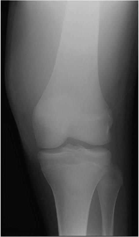
 Figure 7–1. Anteroposterior knee for Patient 1. Beside the suggestion of a soft-tissue mass, there are no obvious destructive changes to the bone.
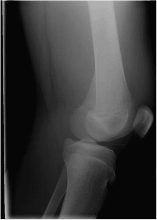
Figure 7–2. Lateral knee for Patient 1.
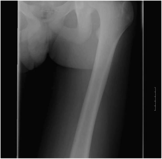
Figure 7–3. Anteroposterior femur for Patient 1.
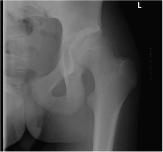
Figure 7–4. Anteroposterior hip for Patient 1.
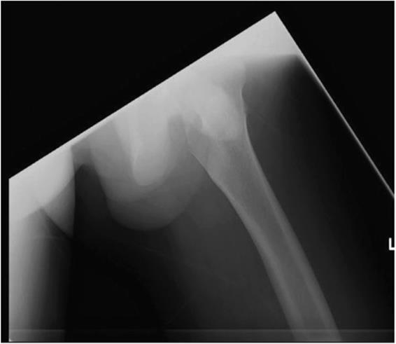
Figure 7–5. Lateral femur for Patient 1.
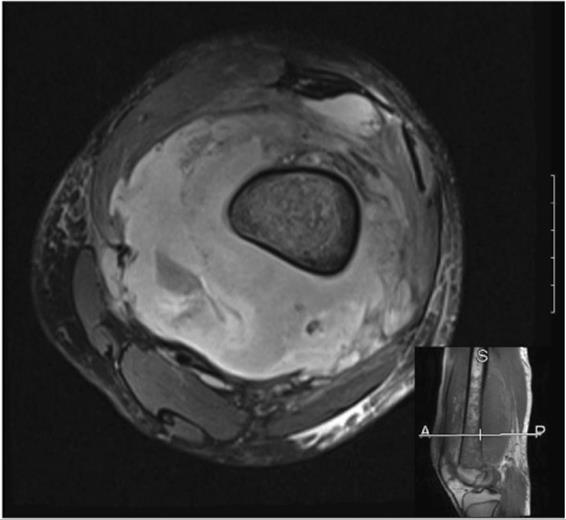
Figure 7–6. Axial T2 MRI image of Patient 1. Note the extensive marrow involvement and nearly circumferential involvement of the femur.
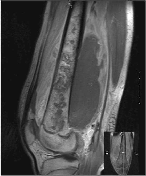
Figure 7–7. Sagittal T1 MRI image of Patient 1.
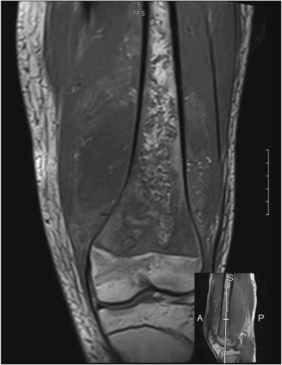
Figure 7–8. Coronal T1 MRI image of Patient 1.

Figure 7–9. Sagittal T2 MRI image of Patient 1. Note the extensive marrow involvement and nearly circumferential involvement of the femur.

Figure 7–10. Coronal fat-saturated MRI image of Patient 1. Note the extensive marrow involvement and nearly circumferential involvement of the femur.
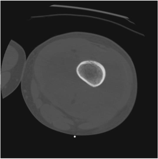
Figure 7–11. This image was obtained at the time of Patient 1’s CT-guided biopsy and shows an axial view of his thigh. There is a suggestion of air in the soft tissues, which supports the diagnosis of infection rather than tumor.
The patient was taken to the operating room where a lateral approach to the femur was made. Copious amounts of purulent material were encountered, and the soft tissues were extensively irrigated and debrided. A 4-cm × 2-cm ovoid hole was made in the lateral cortex of the femur, and flexible reamers were passed into the proximal and distal femur. Antibiotic beads containing 1 g of tobramycin and 3 g of vancomycin per 40 g of polymethylmethacrylate (PMMA) were placed in the intramedullary canal (Figures 7–12 to 7–15). Operative cultures from the purulent material, soft tissue, and bone reamings were all positive for methicillin-sensitive S. aureus, and the patient was placed on intravenous therapy with continuous-infusion oxacillin for 6 weeks under the supervision of a musculoskeletal infectious disease consultant.
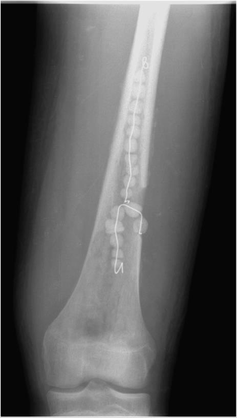
Figure 7–12. Orthogonal view 1 of Patient 1’s femur after debridement, irrigation, intramedullary reaming, and the placement of antibiotic beads. The beads were subsequently removed after 6 weeks of intravenous antibiotic therapy, and Patient 1’s clinical examination improved tremendously. He has returned to limited participation in sports.
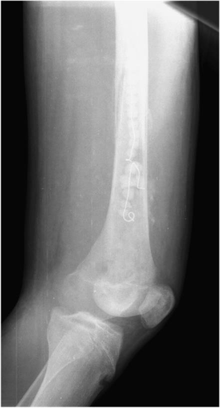
Figure 7–13. Orthogonal view 2 of Patient 1’s femur after debridement, irrigation, intramedullary reaming, and the placement of antibiotic beads. The beads were subsequently removed after 6 weeks of intravenous antibiotic therapy, and Patient 1’s clinical examination improved tremendously. He has returned to limited participation in sports.
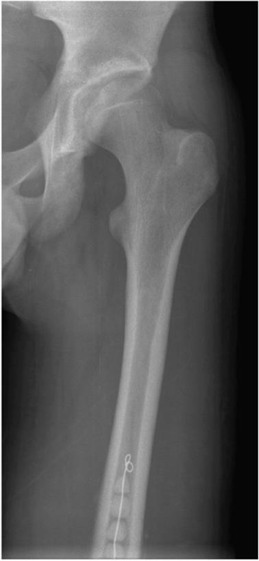
Figure 7–14. Orthogonal view of Patient 1’s femur after debridement, irrigation, intramedullary reaming, and the placement of antibiotic beads. The beads were subsequently removed after 6 weeks of intravenous antibiotic therapy, and Patient 1’s clinical examination improved tremendously. He has returned to limited participation in sports.
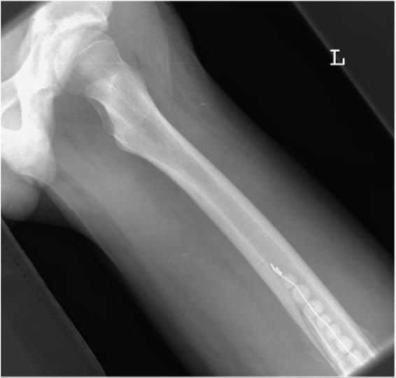
Figure 7–15. Orthogonal view of Patient 1’s femur after debridement, irrigation, intramedullary reaming, and the placement of antibiotic beads. The beads were subsequently removed after 6 weeks of intravenous antibiotic therapy, and Patient 1’s clinical examination improved tremendously. He has returned to limited participation in sports.
Subsequent to the completion of systemic antibiotic therapy, he was taken back to the operating room for removal of the antibiotic beads, irrigation, and repeat intraoperative cultures of the bone and soft tissues. These cultures were all negative. At the most recent follow-up, his knee motion was normal. The laboratory parameters had normalized with an ESR of 3 mm/h and a CRP of less than 0.1 mg/dL.
Adult Osteomyelitis
Adult osteomyelitis is fortunately rare. As the growing child reaches skeletal maturity, the metaphyseal bone vascularity changes, and the pediatric venous sinusoids are eliminated. This produces improved blood flow, decreasing the prevalence of spontaneous osteomyelitis. Most cases of adult osteomyelitis are therefore a result of skin penetration, either deliberate or accidental. A few cases merit special mention.
Aside from accidental or surgical trauma, most cases of adult osteomyelitis occur in individuals who are unable to maintain a normal soft-tissue envelope over subjacent bone. This occurs with disturbing frequency in paraplegics and diabetics, as the normal protective sensation over bony prominences (sacrum, femora, ischial tuberosities, calcanei, and metatarsals) is disrupted. This leads to pressure sores, which, if untreated, will progress to the bone. Once the bone is exposed to air, its vascularity is compromised and osteomyelitis occurs. This form of osteomyelitis is much more chronic than that seen in children, and the symptomatology is quite different. Because many of these individuals are insensate, pain is not a prominent finding. Also, the acute purulent response found in the pediatric case is rarely present, instead manifesting as open, draining sinuses. Infectious indices (ESR/CRP) may demonstrate increased values, but a leukocytosis with left shift is generally absent. Individuals with this condition may persist for years or even decades with osteomyelitis, having no symptoms until developing squamous cell carcinoma (Marjolin ulcer) within the sinus tract.
The situation is quite different in patients who are immunocompromised. Patients who are suffering from human immunodeficiency virus (HIV) or acquired immunodeficiency syndrome (AIDS), who have undergone solid organ or bone marrow transplantation, or who are undergoing cytotoxic chemotherapy may develop acute osteomyelitis. The hallmark of this disease is pain without an obvious source, and this symptom may occur anywhere in the involved bone. This condition may also produce spontaneous joint sepsis, most commonly in the sternoclavicular and sacroiliac joints. Immunocompromised individuals with osteomyelitis will present with pain and fever but will lack leukocytosis, as the dysfunctional immunity producing the osteomyelitis precludes an adequate immune response. Infectious indices are rarely useful, as many of these individuals have other causes of inflammation that will confound these nonspecific test parameters. A very high index of suspicion is required to make this diagnosis, as plain radiographs will likely only demonstrate osteopenia and MRI scanning shows only bony edema on T2-weighted sequences. Generally no abscess forms within the bone. In this instance, prompt diagnosis can be critical because patients do not possess the immuno-logic reserve to fight a fulminant infection.
Collinet-Adler S, Castro CA, Ledonio CG, Bechtold JE, Tsukayama DT: Acinetobacter baumannii is not associated with osteomyelitis in a rat model: a pilot study. Clin Orthop Relat Res 2011;469:274. [PMID: 3008889]
A. Clinical Corollary #2
Patient 2 is a 33-year-old man with a chief complaint of steadily increasing left thigh pain for 1 month. He denied any trauma or constitutional symptoms. He had a negative medical and surgical history but had a social history of heavy drug abuse. He was initially seen at a community hospital where radiographs suggested a permeative lesion in the lateral cortex of the left femur (Figure 7–16). He was subsequently transferred to a tertiary medical center for further management.
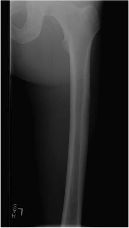
Figure 7–16. Anteroposterior projection of Patient 2’s femur. Note the poorly defined, permeative lesion in the lateral diaphyseal cortex. This was concerning for infection or a bone neoplasm.
At the tertiary medical center, he was found to be afebrile with stable vital signs. Laboratory evaluation disclosed a white blood cell count of 12.4 × 10⁹ cells/L with 70.5% neutrophils and a platelet count of 524 × 103 cells/mL. His ESR and CRP were both elevated at 66 mm/h and 1.96 mg/dL, respectively.
CT scan and MRI scan were obtained. CT scan showed changes in the femur consistent with sequestrum formation, and MRI scan revealed intense soft-tissue edema around the lesion. A presumptive diagnosis of tumor versus infection was made (Figures 7–17 to 7–20).
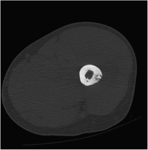
Figure 7–17. Axial CT image suggests the presence of a sequestrum within the lateral cortex of the femur.
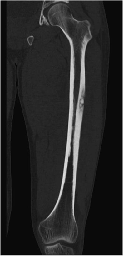
Figure 7–18. Coronal CT image suggests the presence of a sequestrum within the lateral cortex of the femur.
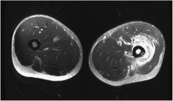
Figure 7–19. Axial MRI image illustrating the dramatic difference between the affected and nonaffected limbs. The image also shows a substantial soft-tissue component to the process.
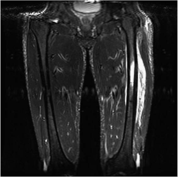
Figure 7–20. Coronal MRI image illustrating the dramatic difference between the affected and nonaffected limbs. The image also shows a substantial soft-tissue component to the process.
The patient was subsequently taken to the operating room where an open biopsy of the left femur and the surrounding tissues was performed. Pathology on frozen section was consistent with acute infection and the diagnosis of osteomyelitis with soft-tissue involvement was made. The muscle around the femur was thoroughly debrided, and the femur itself was likewise debrided with rongeurs, curettes, and a high-speed burr (Figure 7–21). Cultures from the musculature, periosteum, bone, and intermedullary canal were all obtained. After copious irrigation, a strand of antibiotic beads containing 1 g of tobramycin and 3 g of vancomycin per 40 g of PMMA was made and placed next to the femur (Figure 7–22). The wound was closed in layers. Postoperative radiographs showed obliteration of the abnormal bone (Figure 7–23).
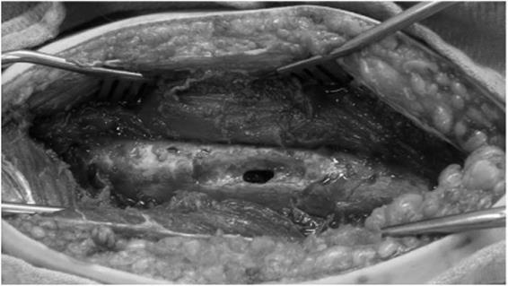
Figure 7–21. Intraoperative view of Patient 2’s femur after biopsy, culture, thorough curettage, and burring of the abnormal bone in the lateral cortex. Bone and soft tissue were sent for pathologic and microbiologic testing. All samples were positive for methicillin-resistant Staphylococcus aureus infection and negative for neoplasm.
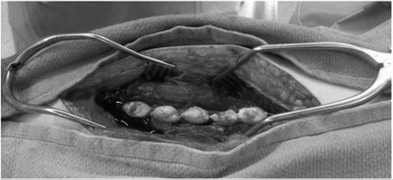
Figure 7–22. A strand of antibiotic-impregnated beads were placed after copious irrigation with antibiotic-containing pulse lavage fluid.
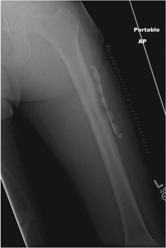
Figure 7–23. Postoperative anteroposterior (AP) radiograph of Patient 2’s femur. Note the removal of the permeative lateral bone (see Figure 7–16 for comparison).
All operative cultures were positive for methicillin-resistant S. aureus (MRSA). The patient was placed on oral antibiotic therapy with linezolid for 6 weeks with plans to remove his antibiotic beads and perform a repeat irrigation and debridement after the completion of antibiotic therapy.
Hamzaoui A, Salem R, Koubaa M, et al: Escherichia coli osteomyelitis of the ischium in an adult. Orthop Traumatol Surg Res 2009;95:636. [PMID: 19944663]
Adult Spontaneous Soft-Tissue Infections
Soft-tissue infections are common. Admissions to medical services for cellulitis, a common skin infection, are frequent in most hospitals. These conditions may occur in the setting of medical comorbidities, such as venous stasis, diabetes, obesity, or immunocompromise. They are generally treated with antibiotics alone, and rarely is surgical intervention necessary.
Deep, abscess-producing skin infections in adults are usually the result of skin penetration. This occurs either due to trauma, iatrogenic skin penetration, or intravenous drug use. These causes are sometimes obvious, as in the case of intravenous drug use, but may be difficult to ascertain, as in septic olecranon bursitis. Bursitis is often presumed to be due to trauma, as the extensor surface of the elbow is frequently traumatized. Surgical treatment of simple abscesses with irrigation and drainage as well as wound packing or vacuum-assisted closure generally yields satisfactory results. Occasionally, intravenous drug abusers can present with a mixed picture of both bone and soft-tissue infection (see Clinical Corollary #2).
Severe, spontaneous, deep soft-tissue infections, those occurring below the fascia, are decidedly rarer, and tend to occur in individuals with some form of immunocompromise. While the often publicized “flesh-eating bacteria” seen in necrotizing fasciitis may infect immunocompetent hosts, those with an immune-compromised comorbidity are at much greater risk. These individuals are generally as follows: patients who are neutropenic secondary to cytotoxic chemotherapy, those suffering from HIV/AIDS, or those with other, frequently autoimmune, diseases that render the normal tissues susceptible to bacteria in the environment. In immunocompromised individuals, the typical signs of infection may be absent, and the patient may simply manifest a high fever (>38.5°C), tenderness, and erythema. Advanced imaging with MRI may demonstrate only edema within the affected area because the patient often does not have adequate immunity to form an abscess. These patients are at great risk, and surgical debridement coupled with broad-spectrum antibiotics may be lifesaving.
Because of the vascular nature of muscle, pyomyositis in immunocompetent individuals is rare. Etiologically, pyomyositis is different than simple abscess formation; in pyomyositis, the bacteria occlude the small vascular inflow into the muscle, producing necrosis. This avascular bed is an ideal culture medium for bacteria, and liquefactive necrosis occurs. Treatment of pyomyositis is more extensive than many soft-tissue infections because all necrotic material must be thoroughly removed before the infection can be controlled. Serial debridement and adjunctive intravenous antibiotics are the mainstays of treatment, as initial debridement often fails to remove all necrotic material.
Necrotizing fasciitis is the most dreaded deep soft-tissue infection. Classically caused by Clostridium perfringens, it can become a rapidly progressive, life-threatening event. Necrotizing fasciitis caused by other organisms may not present with as fulminant a course and may more closely mimic cellulitis or other soft-tissue infections. Necrotizing fasciitis is a clinical diagnosis: although MRI may demonstrate T2 enhancement of the fascia, this finding is extremely nonspecific, and the clinician acting on the clinical findings will yield a much more timely diagnosis. Clinically, the patients are quite ill, with fever, malaise, and localized pain. Signs of systemic sepsis may be present, with mental status changes, tachycardia, or even hypotension. Palpation of the affected area demonstrates a swollen, boggy texture to the skin and soft tissues. The skin may also be hypermobile, similar to after a fasciocutaneous injury. Bullous changes may also occur.
Treatment of necrotizing fasciitis is time dependent. Time should not be wasted obtaining confirmatory diagnostic tests, as these are rarely specific enough to change the clinical diagnosis, and substantial delays may be limb or life threatening. Surgical treatment is extensive and requires debridement of all affected skin and fascia. In fulminant cases, soft-tissue reconstruction is generally required. Debridement to healthy, vascularized skin is mandatory, and debridement of the subjacent muscle may be required. The presence of myonecrosis may make amputation necessary, and the performance of a high amputation such as a hip disarticulation or forequarter amputation may be necessary as a lifesaving measure.
JOINT INFECTIONS
Prosthetic Joint Infections
Prosthetic infection after arthroplasty is a dreaded complication of this procedure, which is performed with increasing frequency in our aging population. With the number of arthroplasties projected to increase dramatically, treating prosthetic infections may become a full-time profession for some orthopedic surgeons. The costs of this treatment are extensive and, when multiplied by our aging population, may become astronomical.
Prosthetic infections are divided into three categories. The first is an acute infection following surgery. In this case, the patient will demonstrate fever, increasing pain, erythema, and poor wound healing. Distinguishing a typical postoperative knee wound from an acutely infected postoperative knee wound is an art, and the gravity of ignoring the infection must be balanced with the morbidity of a return to the operating theater. Fortunately, acute sepsis following arthroplasty is rare (<1%), and most of these situations may be salvaged by aggressive and timely debridement and polyethylene exchange, followed by intravenous antibiotics.
A more difficult problem to distinguish is the subacute prosthetic infection. In this case, a well-functioning, painless arthroplasty becomes acutely painful, warm, and effusive. At times, a suggestive history such as recent dental infection, skin trauma to the ipsilateral limb, or an unrelated invasive procedure may be elicited. If the aforementioned constellation of symptoms has been occurring for only a short period of time (generally <2 or 3 weeks), the joint may be salvaged with aggressive surgical debridement, intravenous antibiotics, and polyethylene exchange. Removal of the bacterial exudate (glycocalyx or biofilm) with Dakin’s solution from the prosthetic surfaces may be helpful.
The most common variety of prosthetic infection is a chronic infection. This occurs when a well-functioning joint of intermediate to long duration (>3 months, but perhaps years after the index procedure) becomes painful, warm, and effusive. Sinus tracts may form in the adjacent skin, leading to chronic drainage. In this situation, the infection has generally been present for weeks to years, and bacterial colonization and biofilm formation have organized on the joint surface. Many surgeons attempt a debridement and polyethylene component exchange if the components are tightly fixed. When the components are loose, or if permanent suppression is planned, the affected joint should generally be removed and replaced with antibiotic-containing bone cement. The patient is then treated with systemic antibiotics for several (usually 6) weeks. If sterilization occurs, as determined by normal CRP and ESR laboratory values and a normal aspirate culture, replantation may then be contemplated.
Traumatic Infections
Traumatic infections are unfortunately common, and their incidence corresponds directly to the energy imparted by the injury. The maxim that “an open fracture is a soft-tissue injury that happens to have a broken bone in it” is true, as devitalized, devascularized tissue becomes rapidly colonized upon exposure to air, soil, or other materials. To minimize the risk of traumatic infection, all open fractures should be treated urgently with thorough debridement and skeletal stabilization. This allows devitalized tissues to declare themselves so that subsequent surgeries facilitate complete removal of all abnormal tissue. Despite the most meticulous care, open fractures may still become infected, and this problem is much more prevalent in certain anatomic locations. Clearly the tibia, a subcutaneous bone throughout much of its circumference, is the most at risk, and modern trauma care often combines soft-tissue reconstruction with bony stabilization to minimize the risk of osteomyelitis.
Septic Arthritis
Nonprosthetic septic arthritis is unusual in adults. Most commonly, a history of penetrating injury will be elicited. Septic arthritis can also be seen in immunocompromised individuals without trauma. Septic arthritis with a past history lacking trauma or immunocompromise is decidedly rare, and a more common crystalline arthropathy, such as gout, is more often the culprit.
Pathologically, a native joint is relatively resistant to infection. While cartilage itself is avascular, the synovium and joint capsule are richly vascularized and provide ample protection from infection. Children can develop septic arthritis from penetration of bacteria from a nearby osteomyelitis due to the configuration of the joint capsule, allowing bacteria to enter the joint space. In adults, osteomyelitis in proximity rarely occurs. This is fortunate, as the sequelae of septic arthritis can be devastating and can lead to rapid joint destruction. Bacterial matrix metalloproteases can rapidly degrade the articular cartilage, leading to severe cartilage damage and end-stage arthrosis.
Clinically, patients present with severe pain, effusion, and resistance to joint motion. Warmth and redness of the surrounding skin may also be present. Unless an osteomyelitis in proximity is also present, both plain radiographs and MRI scanning will reveal only a joint effusion. Arthrocentesis demonstrates an exudative effusion, with a leukocyte count greater than 50 × 109 cells/L. Bacterial cultures are frequently positive, with Staphylococcus species the most frequent causative organisms.
Treatment of septic arthritis is time dependent and demands a high index of suspicion. Awaiting final culture results in a patient with a purulent effusion may cause irreversible joint damage. Debate exists regarding the means of surgical treatment, with open synovectomy being the traditional procedure. Arthroscopic irrigation and debridement may allow more thorough visualization and lavage of all joint surfaces but is more technically demanding. All patients with septic arthritis are treated with antibiotics following surgery, frequently a short intravenous course. Results of this depend on both the cause of the infection and the amount of joint destruction that has occurred.
PATHOGENESIS
General
All clinical infections must be considered as a conflict between the attacking microbes’ ability to cause disease and the host’s immune defenses. Infections are more likely to occur if the organisms are more virulent and if the inoculum is larger. Conversely, an infection is less likely if the host has a greater ability to eradicate the pathogens or if the host has fewer deficits that encourage infection. Additionally, the pathogenesis of infection involving foreign materials and osteomyelitis are discussed.
Organisms
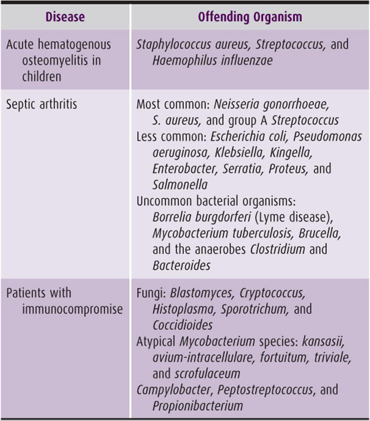
Although the musculoskeletal system may be infected by any infectious agent, the vast majority of infections are bacterial (Table 7–1). S. aureus, Streptococcus, and H. influenzae are the most common causes of acute hematogenous osteomyelitis in children. The most common causes of septic arthritis are Neisseria gonorrhoeae, S. aureus, and group A Streptococcus. Septic arthritis is less often caused by gram-negative organisms, including Escherichia coli, Pseudomonas aeruginosa, Klebsiella, Enterobacter, Serratia, Proteus, and Salmonella. Uncommon bacterial organisms include Borrelia burgdorferi (Lyme disease), Mycobacterium tuberculosis, Brucella, and the anaerobes Clostridium and Bacteroides. Unusual organisms that may preferentially infect immunocompromised patients include fungi (Blastomyces, Cryptococcus, Histoplasma, Sporotrichum, and Coccidioides) and atypical mycobacteria (Mycobacterium kansasii, M. avium-intracellulare, M. fortuitum, M. triviale, and M. scrofulaceum).
Table 7–1. Common offending organisms in orthopedic conditions.
Brook I: Microbiology and management of joint and bone infections due to anaerobic bacteria. J Orthop Sci 2008;13:160. [PMID 18392922]
Peterson MC: Rheumatic manifestations of Campylobacter jejuni and C. fetus infections in adults. Scand J Rheumatol 1994;23:167. [PMID: 8091140]
The increase in the immunocompromised population due to the success of solid organ transplants and advancements in the treatment of HIV/AIDS and autoimmune disorders has increased the spectrum of bacteria that can cause musculoskeletal infection. Likewise, an increase in the number of antibiotic-resistant organisms presents a difficult, constantly evolving challenge in the eradication of infection. MRSA can infect bones, joints, soft tissues, and surgical implants. Other common bacterial species have become more resistant to a variety of drugs, delaying the proper treatment of infection and decreasing the number of antibiotics that can be used effectively. For instance, Acinetobacter baumannii is a pan-resistant bacterial strain that can cause severe lung and soft-tissue infections. Its role in clinically significant osteomyelitis is unknown, although some evidence suggests that it may be of limited importance.
Host Factors
A given host may have several factors that promote or defend against infection. Comorbid medical conditions and compromised immunity play a role in the establishment of infectious disease. The presence (or lack) of implanted devices affects infection risk and treatment strategies. Nutritional status and acute nutritional requirements also play a role.
A. Comorbid Diseases and Host Immunity
Comorbid diseases that are known to contribute to infection risk include diseases such as diabetes mellitus, obesity, peripheral vascular disease, chronic renal and liver disease, cancer, autoimmune diseases, and AIDS. Iatrogenic causes, often as a treatment for other diseases, include the use of cytotoxic chemotherapy, corticosteroids, and inhibitors of inflammatory molecules (such as inhibitors of tumor necrosis factor-alpha). Diabetes mellitus has been shown to increase the risk of infection following surgeries, including total joint surgery, spinal surgeries, and foot and ankle surgery. The role that diabetes plays in infectious disease pathogenesis is related to vascular disease, as well as the effects of high glucose, which can cause granulocyte dysfunction. Obesity, often associated with diabetes, has also been shown in some studies to be an independent risk factor for infection.
B. Nutrition and Infection
Nutritional status must be thought of in terms of host requirements. In an adult with static requirements, poor nutrition may develop due to a variety of medical issues, including advanced age, alcoholism, renal disease, chronic diseases, cancer, malabsorption, and other infirmities. It is also necessary to consider the nutritional requirements of patients who have increased nutritional physiologic demand. These patients at baseline may have been well nourished. However, a significant event (such as trauma) may increase their nutritional requirements and cause a relative nutritional deficiency. Laboratory testing for nutritional deficiency includes levels of transferrin (normal, 70–850 mg/dL), serum albumin (normal, 3.4–5.0 g/dL), total lymphocyte count (normal, 0.8–3.65 × 10³ cells/μL), and prealbumin (normal, 18–38 mg/dL). Values below those shown may indicate malnutrition and consequent immunocompromise.
C. Foreign Material
All biomaterials commonly used for total joint arthroplasty increase the incidence of S. aureus infections. In contrast, biomaterials appear to have no effect on E. coli and Staphylococcus epidermidis infections except when polymethylmethacrylate is used, in which case the incidence rises markedly.
D. Biofilms
Adherence of bacteria to the surface of implants is promoted by a polysaccharide biofilm called glycocalyx that acts as a barrier against host defense mechanisms and antibiotics. In addition, this film makes culture of organisms difficult, even with the use of special techniques. Hence, other approaches must be taken for culture and treatment. Mixing antibiotics such as vancomycin and gentamicin into PMMA cement can theoretically lower the risk of infection from cemented metal joint replacements, presumably by killing surface bacteria before they produce glycocalyx. Before a glycocalyx is produced, the bacteria undergo a process referred to as quorum sensing. Quorum sensing dispersion is a technique under development that might allow clinicians to disrupt a biofilm, therefore making it susceptible to treatment. As stated, the formation of biofilm makes cultures unreliable. Other means, such as the polymerase chain reaction, have been employed to diagnose infection.
Collinet-Adler S, Castro CA, Ledonio CG, Bechtold JE, Tsukayama DT: Acinetobacter baumannii is not associated with osteomyelitis in a rat model: a pilot study. Clin Orthop Relat Res 2011;469:274. [PMID: 3008889]
Lauderdale KJ, Malone CL, Boles BR, Morcuende J, Horswill AR: Biofilm dispersal of community-associated methicillin-resistant Staphylococcus aureus on orthopedic implant material. J Orthop Res2010;28:55. [PMID: 19610092]
PREVENTION
Overview
Prevention of infection starts long before the operative date, continues both during and after surgery, and is influenced by multiple factors and personnel. As discussed previously, medical comorbidities influence the pathogenesis of infection. It follows that the management and prevention of such comorbidities will contribute to infection prevention. Multiple physician and patient factors contribute to medical optimization of comorbidities before surgery. Preoperatively, factors such as nutrition, MRSA colonization, preoperative hygiene, and bathing can contribute to infection risk. Perioperative and intraoperative factors such as proper prepping, operative room sterility, appropriate use of antibiotics, closure material choice, and minimization of intraoperative time can help prevent infection. Finally, optimizing postoperative factors including postoperative antibiotics, management of blood glucose levels, and choices regarding blood transfusions can also positively affect infection rates.
Preoperative Optimization
A. Medical Comorbidities
Some medical comorbidities (and their necessary treatment) represent nonmodifiable risk factors for infection, whereas other present an opportunity for preoperative optimization. Rheumatoid arthritis (RA) provides an excellent example. Individuals with RA have an increased risk of infection, both from the disease and its treatment. Surgeons working in concert with the patient’s rheumatologist can optimize the treatment of RA while decreasing the risk of poor operative outcomes, including infection. This approach to medical comorbidity management can be applied to other diseases as well.
Obesity, for instance, represents a modifiable risk factor that is becoming a more frequently encountered condition in developed countries, especially in the United States. In the total joint literature, obesity has been shown to be associated with an increased risk of infection. An increase of 5 points in body mass index (BMI) has been shown to be associated with a 50% increase in the odds ratio for acquiring a prosthetic joint infection. In the spine literature, obese patients have an odds ratio of 2.2 relative to nonobese patients. The surgeon must be aware of the association between obesity and infection and work with patients to decrease their risk of infection. Unfortunately, weight loss can be extremely difficult, especially in patients with orthopedic conditions. Gastric bypass therapy may be necessary before orthopedic intervention, especially in patients with a BMI greater than 45. This, unfortunately, produces relative malnutrition.
Bosco JA 3rd, Slover JD, Haas JP: Perioperative strategies for decreasing infection: a comprehensive evidence-based approach. J Bone Joint Surg Am 2010;92:232. [PMID: 20048118]
Chen S, Anderson MV, Cheng WK, Wongworawat MD: Diabetes associated with increased surgical site infections in spinal arthrodesis. Clin Orthop Relat Res 2009;467:1670. [PMID: 2690748]
Dowsey MM, Choong PF: Obesity is a major risk factor for prosthetic infection after primary hip arthroplasty. Clin Orthop Relat Res 2008;466:153. [PMID: 2505299]
Dowsey MM, Choong PF: Obese diabetic patients are at substantial risk for deep infection after primary TKA. Clin Orthop Relat Res 2009;467:1577. [PMID: 2674158]
Howe CR, Gardner GC, Kadel NJ: Perioperative medication management for the patient with rheumatoid arthritis. J Am Acad Orthop Surg 2006;14:544. [PMID: 16959892]
Jämsen E, Varonen M, Huhtala H, et al: Incidence of prosthetic joint infections after primary knee arthroplasty. J Arthroplasty 2010;25:87. [PMID: 19056210]
Moucha CS, Clyburn T, Evans RP, Prokuski L: Modifiable risk factors for surgical site infection. J Bone Joint Surg Am 2011;93:398. [PMID: 21325594]
Olsen MA, Nepple JJ, Riew KD, et al: Risk factors for surgical site infection following orthopaedic spinal operations. J Bone Joint Surg Am 2008;90:62. [PMID: 18171958]
B. Nutrition
The overall health of a given host’s immune system influences susceptibility to infection. Indeed, malnutrition can lead directly to immune dysfunction. A recent review highlights the importance of recognizing and correcting patient nutrition. Testing for nutritional status should include albumin (normal, 3.4–5.0 g/dL), total lymphocyte count (normal, 0.8–3.65 × 10³ cells/μL), and transferrin (normal, 70–850 mg/dL) levels. Supplementation to correct deficiency of both macro- and micronutrients should be accomplished preoperatively.
Katona P, Katona-Apte J: The interaction between nutrition and infection. Clin Infect Dis 2008;46:1582. [PMID: 18419494]
C. Staphylococcus aureus Decolonization/Preoperative Bathing
It has been clear for some time that certain organisms possess an increased potential to cause severe infections. S. aureus is one such bacterium. Attempts to thwart infection with S. aureus have led to the development of preoperative decolonization protocols designed to eradicate S. aureus from the skin flora before the patient undergoes surgery. At certain institutions, adult reconstruction surgeons have begun to screen the nares of patients preoperatively. One protocol uses nasal cultures 2–4 weeks before surgery. If positive for S. aureus, patients apply nasal mupirocin twice daily and take chlorhexidine baths for the 5 days immediately prior to surgery. This has been shown to decrease the rate of S. aureus infections, as well as the overall rate of surgical site infections. Similar protocols have been used to prevent infections in other surgical disciplines, prompting the Cochrane group to conclude that nasal mupirocin is effective in preventing infections. Conversely, a separate Cochrane review demonstrated that chlorhexidine baths alone did not change the incidence of infection as compared to washing preoperatively with regular soap and water.
Hacek DM, Robb WJ, Paule SM, Kudrna JC, Stamos VP, Peterson LR: Staphylococcus aureus nasal decolonization in joint replacement surgery reduces infection. Clin Orthop Relat Res 2008;466:1349. [PMID: 2384050]
Rao N, Cannella B, Crossett LS, Yates AJ Jr, McGough R 3rd: A preoperative decolonization protocol for staphylococcus aureus prevents orthopaedic infections. Clin Orthop Relat Res 2008;466:1343. [PMID: 2384036]
Rao N, Cannella BA, Crossett LS, Yates AJ Jr, McGough RL 3rd, Hamilton CW: Preoperative screening/decolonization for Staphylococcus aureus to prevent orthopedic surgical site infection prospective cohort study with 2-year follow-up. J Arthroplasty 2011;26:1501. [PMID: 21507604]
van Rijen M, Bonten M, Wenzel R, Kluytmans J: Mupirocin ointment for preventing Staphylococcus aureus infections in nasal carriers. Cochrane Database Syst Rev 2008;4:CD006216. [PMID: 18843708]
Webster J, Osborne S: Preoperative bathing or showering with skin antiseptics to prevent surgical site infection. Cochrane Database Syst Rev 2007;2:CD004985. [PMID: 16625619]
Perioperative and Intraoperative Factors
A. Prepping
Preparing the patient for surgery requires many steps once the patient has arrived in the operating theatre. The first decision in regard to infection control is whether or not to remove hair. The three options for hair removal are shaving, clipping, and depilatory creams. Alternatively, the hair can be left intact. Traditionally, it has been taught that shaving is inferior, a finding that is supported by a recent Cochrane review. However, no difference was found between removing the hair using the other two approaches and leaving the hair intact. The next step that influences infection control involves the selection of prepping solution. Options include iodophor or chlorhexidine gluconate–based solutions. Furthermore, these can be aqueous or alcohol based. Alcohol-based chlorhexidine gluconate solutions are superior to other preparation solutions when the outcome measure is simply positive cultures from the operative field. However, the extent to which these cultures correlate with an eventual surgical site infection is not clear. Interestingly, in urologic surgery, other preparation solutions have been shown to be superior. This is ostensibly the result either of a statistical phenomenon or a different bacterial milieu in this patient population that responds differently to a given preparation solution. A Cochrane review from 2004 failed to show any significant difference between different preparation solutions.
Edwards PS, Lipp A, Holmes A: Preoperative skin antiseptics for preventing surgical wound infections after clean surgery. Cochrane Database Syst Rev 2004;3:CD003949. [PMID: 15266508]
Ostrander RV, Botte MJ, Brage ME: Efficacy of surgical preparation solutions in foot and ankle surgery. J Bone Joint Surg Am 2005;87:980. [PMID: 15866959]
Saltzman MD, Nuber GW, Gryzlo SM, Marecek GS, Koh JL: Efficacy of surgical preparation solutions in shoulder surgery. J Bone Joint Surg Am 2009;91:1949. [PMID: 19651954]
B. Antibiotics
The use of preoperative antibiotics within 1 hour of surgery is standard, routine, and required for adherence to national Surgical Care Improvement Project (SCIP) protocols. Recommendations for antibiotic timing, duration, selection, and redosing include the following: administration of a first-generation cephalosporin within 1 hour of surgical initiation, use of clindamycin or vancomycin if patient is allergic to cephalosporin, and maintenance of antibiotics for no more than 24 hours total unless infection is suspected or confirmed. A proposed adjunct to systemic antibiotics involves the use of local antibiotics, namely gentamicin. Two separate studies used a rat injury model to show improved bactericidal activity in vivo. Results are yet to be demonstrated in a human, randomized controlled trial.
Cavanaugh DL, Berry J, Yarboro SR, Dahners LE: Better prophylaxis against surgical site infection with local as well as systemic antibiotics. An in vivo study. J Bone Joint Surg Am 2009;91:1907. [PMID: 2714810]
Prokuski L: Prophylactic antibiotics in orthopaedic surgery. J Am Acad Orthop Surg 2008;16:283. [PMID: 21553797]
Yarboro SR, Baum EJ, Dahners LE: Locally administered antibiotics for prophylaxis against surgical wound infection. An in vivo study. J Bone Joint Surg Am 2007;89:929. [PMID: 17473127]
C. Operating Room Sterility
Sterility in the operating theater is the responsibility of anyone who enters the room. This includes the surgical team, the anesthesiology team, the nursing team, the scrub technician team, and the medical equipment team. Lapses in sterile technique can undoubtedly increase bacterial exposure to the patient. However, other factors also contribute, namely the sterility of the air in which the surgery is performed. Ultraviolet (UV) lighting has long been tried as an attempt to reduce the bacterial content of the air. A recent study demonstrated a decreased incidence of surgical site infections with use of UV light in the operating theater. In addition, the sterility of the air and the direction of the air flow are also controllable factors in the operating room. For many years, both vertical and horizontal airflow systems, combined with a variety of filters, have been employed to theoretically lower the risk of surgical site infections. Despite these efforts over many years, there is still no clear consensus on whether such systems decrease, make no difference, or actually increase the risk of airborne contamination.
Brandt C, Hott U, Sohr D, Daschner F, Gastmeier P, Ruden H: Operating room ventilation with laminar airflow shows no protective effect on the surgical site infection rate in orthopedic and abdominal surgery. Ann Surg2008;248:695. [PMID: 18948793]
Owers KL, James E, Bannister GC: Source of bacterial shedding in laminar flow theatres. J Hosp Infect 2004;58:230. [PMID: 15501339]
Ritter MA, Olberding EM, Malinzak RA: Ultraviolet lighting during orthopaedic surgery and the rate of infection. J Bone Joint Surg Am 2007;89:1935. [PMID: 17768189]
Stocks GW, O’Connor DP, Self SD, Marcek GA, Thompson BL: Directed air flow to reduce airborne particulate and bacterial contamination in the surgical field during total hip arthroplasty. J Arthroplasty2011;26:771. [PMID: 20851565]
Stocks GW, Self SD, Thompson B, Adame XA, O’Connor DP: Predicting bacterial populations based on airborne particulates: a study performed in nonlaminar flow operating rooms during joint arthroplasty surgery. Am J Infect Control 2010;38:199. [PMID: 19913327]
D. Closure
The two main options for closure of a surgical wound are sutures and staples. A recent meta-analysis combined the outcomes of six different studies. Despite the methodologic limitations inherent in this meta-analysis, it suggests that closure with staples increases the risk of infection. When closing deep layers with suture, the nature of the filament can be considered. Traditional teaching is that monofilament closure decreases the risk of infection because theoretically, braided sutures provide a greater surface area for microbial contamination than monofilament sutures. This is likely merely anecdotal, however, as no studies have demonstrated superiority of one closure material over another regarding infection. Adherence to good surgical tissue handling techniques including atraumatic closure technique, maintenance of full-thickness fasciocutaneous flaps, and avoidance of excessive tissue undermining or devascularization are likely substantially more important than the choice of closure material.
Smith TO, Sexton D, Mann C, Donell S: Sutures versus staples for skin closure in orthopaedic surgery: meta-analysis. BMJ 2010;340:c1199. [PMID: 20234041]
E. Postoperative Blood Glucose Control
It is well known that diabetes is a risk factor for surgical site infections. However, the consequence of poor perioperative glucose control in terms of surgical site infection is less clear. Recent literature suggests that poor control of postoperative hyperglycemia substantially increases the risk of surgical site infection.
Ata A, Lee J, Bestle SL, Desemone J, Stain SC: Postoperative hyperglycemia and surgical site infection in general surgery patients. Arch Surg 2010;145:858. [PMID: 20855756]
CLINICAL FINDINGS
Despite 2500 years of medical progress, the aforementioned cardinal signs of inflammation described by originally recorded by the Roman encyclopedist Celsus in the first century AD are still our primary means of diagnosing infection. In immunocompetent hosts, many, if not all, of the signs will be present in acute infection. In the postsurgical setting, wound drainage, whether purulent, bloody, or clear, may also indicate that an infectious process is occurring. The postsurgical setting is the most difficult, however, because the signs mentioned earlier are significant for inflammation in general and are not specific for infection. Distinguishing between a routine postoperative wound, a poorly healing postoperative wound, a hematoma, or an infection can be extremely difficult.
In the postoperative situation, infection may be diagnosed by a clinical scenario that does not improve with time. After surgery, pain, swelling, and erythema usually decrease over a period of days to weeks. If, instead of improving, the symptoms of inflammation increase, infection may be present.
Laboratory studies may help to distinguish infection from other diagnoses. The presence of a high white blood cell count, especially if the percentage of neutrophils is greater than 70% of the total, suggests infection. Leukocytosis is not sensitive for localized infection, however, and has greater sensitivity with systemic disease than with a localized problem. An increased ESR is another sensitive, but nonspecific marker. The ESR will increase any time inflammation is present, dramatically reducing its utility in the postoperative situation or in any scenario wherein multiple inflammatory etiologies may be present. An increase in the serum CRP is somewhat more specific for infection, but not specific enough to act on in the absence of other signs or symptoms. The CRP generally increases very rapidly in the presence of infection and then begins to decrease after 48–72 hours. The ESR takes much longer to normalize, often peaking over a period of several days and decreasing much more slowly. Abnormalities in these lab values are corroborative of, but not diagnostic of, a musculoskeletal infection.
DIFFERENTIAL DIAGNOSIS
Infection, the great masquerader, generally is part of an extensive differential diagnosis. In fact, the diagnosis of infection is frequently confounded by many other medical conditions, most of them quite common.
Infection is frequently confused with trauma. In the early stages of a healing contusion, the soft-tissue hematoma mimics an abscess. Adding the complication that hematomas may subsequently become infected only serves to further confuse the clinical scenario. The increasing prevalence of anticoagulation for a number of cardiac or other medical comorbidities renders even the most trivial trauma capable of producing substantial bleeding. Furthermore, metabolizing blood products may yield a febrile response, placing the clinician in a position of possibly operating on a hematoma in an anticoagulated patient or ignoring a large soft-tissue infection in an individual who is substantially medically compromised.
Many musculoskeletal neoplasms may be confused with infections. Most soft-tissue sarcomas produce swelling and may produce pain to palpation. Their heterogeneous imaging characteristics on MRI scan have led numerous surgeons into a “simple irrigation and debridement” that unfortunately enters a sarcoma, thus producing substantial bleeding and possible oncologic compromise. Radiographically, osteosarcoma may look nearly exactly like osteomyelitis. Both produce permeative bony changes with substantial new bone formation. Both are painful and produce swelling. Both occur in the metaphyses of children. Similarly, Ewing sarcoma produces permeative bony changes mimicking those produced by purulence. Because Ewing sarcoma, lymphoma, and osteomyelitis are composed of small blue cells without matrix, all may resemble frank purulence at the time of debridement. The orthopedic oncologist’s admonition to “send every infection to pathology and culture every tumor” was born of this confusion.
Fortunately, needle biopsy may assist in narrowing this differential diagnosis without the potential for causing harm. Unless the patient is septic and in extremis, most suspected infections can be simply aspirated, either with or without radiographic imaging guidance. This approach has multiple advantages:
1. Infections composed of purulent material can be aspirated, and appropriate cultures can be obtained quickly, easily, and before the administration of antibiotics.
2. Needle aspiration is unlikely to cause bleeding complications, even in anticoagulated patients. Hemostasis is almost always achievable using direct pressure, and the patient’s coagulopathy may be corrected before any necessary surgical interventions.
3. In cases where sarcoma enters the differential diagnosis, needle biopsy may not only obtain purulent samples (if infection is present), but will also allow pathologic diagnosis. Although there is current debate regarding whether needle tracks should be excised at the time of sarcoma surgery, excising a needle track is always easier than a surgical incision and does not have the potential complication of an improperly placed incision.
Other inflammatory conditions also mimic infection. Because the physical signs of infection are the signs of inflammation, the two can present nearly identically. Aseptic myonecrosis, as seen in statin-induced myonecrosis, is remarkably similar to infection, with pain, inflammation, and swelling. Diabetic myonecrosis may behave like infection as well. Tumoral calcinosis, as seen in patients with renal insufficiency, produces soft-tissue masses with pain and skin changes, also mimicking abscesses.
COMPLICATIONS
The complications of infection may be truly grave. In situations of systemic sepsis, failure to adequately treat both the local and systemic manifestations of infection may lead to the patient’s demise. Similarly, clostridial fasciitis may become rapidly progressive, causing massive tissue loss, loss of limb, or even death. If the patient’s system fails to clear or suppress either prosthetic infection or osteomyelitis, amputation may be required to control local disease. Even with successful treatment, infection may lead to substantial loss of tissue, function, social status, and income for the affected patient.
One specific complication of chronic infection is the development of squamous cell carcinoma (a “Marjolin ulcer”). This occurs when any chronically (generally of >20 years’ duration) draining infection causes enough irritation to the surrounding skin to cause the development of an invasive carcinoma. Because this becomes a potentially life-threatening situation in the face of limb salvage that has failed to control the baseline infection, amputation is often the treatment of choice.
Bauer T, David T, Rimareix F, Lortat-Jacob A: Marjolin’s ulcer in chronic osteomyelitis: seven cases and a review of the literature [French]. Rev Chir Orthop Reparatrice Appar Mot 2007;93:63. [PMID: 17389826]
TREATMENT
Treatment of orthopedic infections generally involves a multi-modality and often multidisciplinary approach. Although simple skin infections such as cellulitis or folliculitis may respond completely with medical treatments alone, deep orthopedic infections generally require both surgical and medical intervention. The presence of nonimmunocompetent materials, such as dead bone or metal prostheses, frequently mandates a more aggressive approach as the blood flow required for successful antibiotic treatment is obviously not present.
Pediatric Osteomyelitis
Pediatric osteomyelitis is primarily a surgical disease. The slow, sinusoidal blood flow within the metaphyses allows bacteria to multiply in the region (see General Considerations). Once a sequestrum has formed, this necrotic substrate provides a perfect medium for bacterial growth. Infection will progress until an involucrum forms or the bone is entirely destroyed.
Surgery consists of draining the infected bone and removing any sequestrum, if present. If no sequestrum is present, the bone may be opened with a high-speed burr, gouges, drill, or osteotome, and the purulent material drained. If the adjacent joint is also involved, it may be opened, drained, and irrigated. A deep drain is usually placed within the bone and/or joint and left until drainage ceases. Intravenous antibiotics are then instituted and tailored to the culture results, if positive. Four to six weeks of treatment are generally sufficient to produce a cure.
Adult Osteomyelitis
Treatment of adult osteomyelitis is often much more difficult than the pediatric variety. Often the comorbidities surrounding the orthopedic condition are chronic and may only be optimized rather than eliminated. Adult osteomyelitis also requires much more aggressive surgical and medical treatment than the pediatric condition.
Like pediatric osteomyelitis, treating adult osteomyelitis absolutely requires the removal of all necrotic bone. This is frequently much more difficult, as areas of necrosis may be extensive and removal of all dead bone may produce large, segmental bony defects. Similarly, extensive areas of poorly vascularized, fibrotic, or necrotic soft tissue may be present, requiring thorough debridement.
Careful preoperative planning is required. The extent of bony involvement can be estimated preoperatively using CT scanning, as necrotic bone is frequently sclerotic and hyperdense with this modality. If a segmental defect is anticipated or if circumferential debridement would yield a bone at substantial risk for pathologic fracture, stabilization will be necessary. For small defects or situations where a limited number of debridements is anticipated, stabilization using intramedullary nail or plate fixation may be ideal. For larger defects or situations where multiple debridements are necessary, external fixation either using standard or thin wire (Ilizarov) techniques may be ideal. This allows skeletal stabilization with access to the soft tissues. External fixation may ultimately be revised to internal fixation once the infection is eradicated or used as definitive fixation after grafting or bone transport.
Planning for soft-tissue reconstruction is also necessary. Draining sinuses, necrotic tissue, and poorly vascularized areas should be thoroughly and radically debrided. This generally yields soft-tissue defects that require plastic surgical reconstruction, either with rotational or free tissue transfers.
Adult osteomyelitis secondary to pressure phenomena provides a special case (see General Considerations). In this situation, patients are either mentally or physically inhibited from perceiving or responding to pressure. If the pressure sore extends to the bone, osteomyelitis occurs by definition. Bone biopsy is not necessary for diagnosis in this case but may be useful to tailor antibiotic treatment. Surgical treatment, if necessary based on patient wishes, life expectancy, and comorbidities, is often radical with removal of large portions of the sacrum, pelvis, or proximal femur. Plastic reconstruction is generally necessary.
Antibiotic treatment for adult osteomyelitis is often multimodal. Using two or even three agents in combination can facilitate penetration into involved tissues and can help to limit side effects and toxicities. Obtaining a culture from a deep source is very important for speciation, and patients with chronic draining sinuses are often given an antibiotic holiday for several days to weeks to allow deep cultures to grow. Deep tissue cultures are both more sensitive and specific than culture swabs. Culturing sinus tracts or purulent drainage is of no benefit and may cause harm and confusion.
Adult Soft-Tissue Infections
Soft-tissue infections that result from intravenous or subcutaneous drug abuse may be treated by incision and drainage, possibly with open packing and systemic antibiotics. More severe infections, especially in immunocompromised individuals, require much more aggressive treatment. All abscess cavities must be thoroughly opened, and all necrotic tissue removed. Broad-spectrum antibiotics are generally used, and multiple agents may be used in combination, depending on the clinical scenario. Successful treatment demands an immunocompetent host, so any immunomodulating agents should be discontinued. Leukocyte proliferation growth factors may be contemplated in neutropenic hosts.
Treatment of neutropenic myonecrosis is especially difficult. In this scenario, a neutropenic host presents with fever, severe pain, and cellulitis. MRI generally fails to reveal any abscess or other surgical condition, as the host does not possess enough immune function even to create an abscess. Broad-spectrum antibiotics are instituted, as in any neutropenic fever protocol, but will fail to reduce fevers or eliminate erythema. If the patient’s condition continues to deteriorate without another source, the area of erythema should be opened, and the subjacent muscle examined surgically. Frequently, the muscle will be completely necrotic, but without the liquefactive necrosis encountered in the pyomyositis of immunocompetent individuals. Debridement must continue until adequate bleeding and viable muscle are appreciated. The entire necrotic portion of the compartment must be removed. In severe cases, the entire compartment will be necrotic, and all vessels leading into the muscle will have clotted due to bacterial thrombi. This appearance is not dissimilar to that found in advanced compartment syndrome, with patent major vessels and compromised muscular branches. Unlike in compartment syndrome, the entire compartment must be removed as a lifesaving measure.
Prosthetic Infections
As previously mentioned, the treatment of prosthetic joint infection depends on the acuity of the infection from the time of surgery or the inciting event. Early (within 4–6 weeks of surgery or within a few days of an invasive procedure) infections can be treated with surgical irrigation and exchange of the bearing surfaces. After this course of treatment, the surgeon must maintain a high level of vigilance, because this limited approach may not be successful. In chronic infections (months to years after implantation with no inciting event), the surgeon must assume that biofilm has contaminated the metal prosthesis and cannot be salvaged. If the patient is able to tolerate a staged revision and systemic antibiotics, this is the treatment with the greatest chance of success. If the patient cannot tolerate a staged revision due to comorbidities, the options include chronic antibiotic suppression and amputation. As the population ages, joint arthroplasties increase in frequency, and bacterial resistance continues to evolve, prosthetic infections will continue to challenge orthopedic surgeons.
Traumatic Infections
Traumatic infections are an especially difficult problem because they combine the infection risks inherent in any orthopedic surgery as well as the contamination of the traumatic event. The mainstays of modern treatment are prompt, thorough debridement of devitalized tissue and rigid fixation. Not surprisingly, the risk of infection is directly proportional to the degree of soft-tissue damage imparted by the injury. The host’s functional capacity, regenerative capacity, nutrition, and overall health also affect the risk of infection. Although the surgeon has no control over the magnitude of the trauma or the health of the host, the surgeon has complete control over the quality of the debridement and fixation, and therefore must maximize these. The optimal treatment of traumatic infections often involves a multidisciplinary team including orthopedic surgeons, plastic surgeons, and infectious disease specialists. Indeed, the literature suggests that patients treated with a multidisciplinary approach have better outcomes than those who are not.
Cierny G 3rd, DiPasquale D: Treatment of chronic infection. J Am Acad Orthop Surg 2006;14:S105. [PMID: 17003180]
Copley LA: Pediatric musculoskeletal infection: trends and antibiotic recommendations. J Am Acad Orthop Surg 2009;17:618. [PMID: 19794219]
Duzgun AP, Satir HZ, Ozozan O, Saylam B, Kulah B, Coskun F: Effect of hyperbaric oxygen therapy on healing of diabetic foot ulcers. J Foot Ankle Surg 2008;47:515. [PMID: 19239860]
Forsberg JA, Potter BK, Cierny G 3rd, Webb L: Diagnosis and management of chronic infection. J Am Acad Orthop Surg 2011;19(Suppl 1):S8. [PMID: 21304049]
Noel SP, Courtney HS, Bumgardner JD, Haggard WO: Chitosan sponges to locally deliver amikacin and vancomycin: a pilot in vitro evaluation. Clin Orthop Relat Res 2010;468:2074. [PMID: 2895824]
Prokuski L: Treatment of acute infection. J Am Acad Orthop Surg 2006;14(10 Spec No.):S101. [PMID: 17003179]
Stinner DJ, Noel SP, Haggard WO, Watson JT, Wenke JC: Local antibiotic delivery using tailorable chitosan sponges: the future of infection control? J Orthop Trauma 2010;24:592. [PMID: 20736801]
Ziran BH, Rao N, Hall RA: A dedicated team approach enhances outcomes of osteomyelitis treatment. Clin Orthop Relat Res 2003;414:31. [PMID: 12966273]
PROGNOSIS
It is virtually impossible to predict the prognosis of musculoskeletal infections as a whole because the ultimate success of treatment depends on enumerable variables, as we have indicated many times within this chapter. Host factors (age, nutrition, comorbidities, anatomic location), pathogen factors (organism type and virulence), and physician factors (index of suspicion, quality of technique, available resources) all impact tremendously on the patient’s outcome. However, the principles of musculoskeletal infection treatment are the same in every instance. A high index of suspicion, correct interpretation of laboratory and clinical data, thorough surgery when indicated, optimization of host factors, and pathogen-specific antibiotic therapy are the pillars of treatment for musculoskeletal infections. In all instances, adherence to these principles will maximize the patient’s prognosis.