
Susan A. Kecskes
HIGH-YIELD FACTS
• Serum potassium levels reflect only 2% of total body potassium.
• Therapy for hyperkalemia is aimed at halting intake, stabilizing cellular membranes, intracellular translocation, and enhancing elimination.
• Hypokalemia should be corrected orally, if possible. Extreme caution should be exercised during intravenous replacement to avoid hyperkalemia.
POTASSIUM
While only 2% of total body potassium is in the extracellular fluid (ECF), potassium is the main cation in intracellular fluid (ICF). Normal potassium concentration in the ECF is 3.5 to 5.5 mEq/L, compared to approximately 160 mEq/L in the ICF. The sodium–potassium ATPase pump in the cell membrane maintains this large concentration gradient.
Potassium homeostasis is managed through the use of both translocation and excretion. The majority of potassium excretion occurs in the kidney. The kidney can adjust urinary potassium excretion from 5 to 1000 mEq/24 h. Approximately 10% of daily potassium intake is lost through the gastrointestinal tract in stool.
As only 50% of a potassium load is excreted in the first 4 to 6 hours, translocation allows the body to maintain stable ECF potassium. In the first hours after ingestion, potassium is translocated into cells, primarily in the liver and muscle. Potassium uptake is regulated by insulin, epinephrine, aldosterone, and acid–base balance.1 Insulin stimulates the sodium–potassium ATPase pump to promote potassium uptake in the liver and muscle. Catecholamines cause an initial rise in serum potassium as it is released from the liver. Subsequently, serum potassium falls as catecholamines promote movement to ICF. Aldosterone acts through both renal and extrarenal mechanisms to reduce serum potassium. Acid–base changes direct potassium shifts. Acidemia promotes movement of potassium to the ECF, whereas alkalosis favors movement of potassium to the ICF.
 HYPERKALEMI
Hyperkalemia is defined as serum potassium >5.5 mEq/L and can result from increased potassium intake, decreased potassium loss, or from redistribution from the ICF. Increased potassium intake rarely results in an elevation of serum potassium, unless it is iatrogenic or simultaneously associated with decreased excretion. Iatrogenic causes include excessive intravenous administration of potassium, administration of large quantities of cold-stored blood, large doses of the potassium salts of penicillin, or oral intake of potassium-containing salt substitutes. Acute renal failure is the primary cause of decreased excretion. Less commonly, adrenal insufficiency may result in hyperkalemia due to decreased mineralocorticoid activity. Use of potassium-sparing diuretics is also associated with decreased potassium excretion.
Redistribution of potassium from the ICF to the ECF may occur via cell destruction or translocation from intact cells. In patients with trauma, burns, rhabdomyolysis, massive intravascular coagulopathy, or tumor lysis/necrosis, injured cells release stores of intracellular potassium into the circulation. Hematomas in the newborn and gastrointestinal bleeding may result in large volumes of hemolyzing cells and elevated potassium levels. Potassium can be quickly shifted from the ICF to the ECF in response to metabolic acidosis. The ICF is a major part of the body’s buffering system, with extracellular hydrogen ions being exchanged for intracellular potassium ions.
Pseudohyperkalemia is a common occurrence and must be considered in the differential diagnosis of hyperkalemia. It is often associated with hemolysis from the blood draw. Other causes include prolonged tourniquet use, heel squeezing, or use of small-gauge needles. When pseudohyperkalemia is suspected, specimens should be repeated with attention to avoiding such mechanical factors.
Most patients with hyperkalemia are relatively asymptomatic. Neuromuscular symptoms begin with paresthesias and progress to muscle weakness and, ultimately, flaccid paralysis. Cardiac abnormalities are much more likely to produce life-threatening situations. Characteristic changes in the electrocardiogram (ECG) include peaked T waves, prolongation of the PR interval, and progressive widening of the QRS complex. As potassium continues to rise (typically, >8 mEq/L), the classic “sine wave” of hyperkalemia appears (Fig. 82-1). This may rapidly degenerate to asystole or ventricular fibrillation.
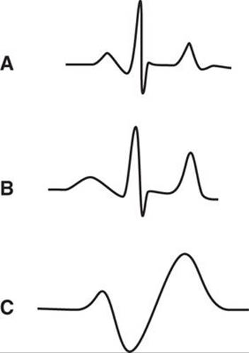
FIGURE 82-1. ECG changes in hyperkalemia. A: Normal ECG. B: ECG with peaked T waves, prolonged PR interval, and widened QRS, seen in moderate hyperkalemia (potassium >7.0 mEq/L). (C) “Sine wave” ECG seen at potassium levels >8 mEq/L.
An ECG should always be obtained when hyperkalemia is suspected, to assess the clinical severity. Serum electrolytes, renal indices (BUN, creatinine, and urinalysis), a complete blood count (CBC), and acid–base status should be measured. Urinary potassium levels may help evaluate the cause of the hyperkalemia. All patients with serum potassium levels >6.5 mEq/L should have continuous ECG monitoring and frequent laboratory follow-up.
Treatment of hyperkalemia depends on the level of serum potassium, along with the clinical symptoms and renal status of the patient. In all cases, intake of potassium and potassium-sparing medication should be halted. In asymptomatic patients with intact renal function and modest (<7 mEq/L) levels of serum potassium, halting intake and follow-up of serum potassium levels may be all that is required (Fig. 82-2). For patients with renal dysfunction, the addition of the potassium-binding agent, sodium polystyrene sulfonate (Kayexalate, 1–2 g/kg po, ng, or pr), or dialysis should be considered to enhance elimination.

FIGURE 82-2. Treatment of Hyperkalemia.
Those patients with serum potassium levels >7 mEq/L or who are symptomatic require aggressive intervention to stabilize the cellular membrane, shift potassium intracellularly, and increase potassium elimination. Membrane stabilization is effected by intravenous administration of calcium. Calcium gluconate, 10%, in a dose of 50 to 100 mg/kg, or calcium chloride, 10%, in a dose of 10 to 25 mg/kg, may be administered over 2 to 5 minutes with continuous ECG monitoring. Onset of action is immediate and the stabilizing effects last 30 to 60 minutes. Potassium may be shifted intracellularly to temporarily reduce serum potassium levels. Administration of sodium bicarbonate (1–2 mEq/kg intravenously over 5–10 minutes) has an onset of action of 5 to 10 minutes and duration of 1 to 2 hours. The dose may be repeated if necessary. Insulin administered in conjunction with glucose effectively shifts potassium to the ICF as well. Dextrose (1 g/kg) may be combined with insulin (0.25 units/kg) and infused over 2 hours. Inhalation of α2-agonists is an attractive alternative for patients with delayed intravenous access.2Nebulized albuterol, in a dose of 2.5 mg for patients <25 kg and 5 mg for patients of 25 kg, and above has been reported to reduce potassium in adult patients with chronic renal failure and is likely to have a similar effect in pediatric patients. It should not be a substitute for appropriate intravenous therapy, but may be used while access is obtained. None of these methods alter total body potassium, so the time they buy should be utilized to enhance elimination of potassium from the body. In the absence of renal failure, loop diuretics and/or thiazides will enhance renal elimination of potassium. Sodium polystyrene sulfonate is a resin that exchanges sodium for potassium at a 1:1 ratio. It is administered through the gastrointestinal tract and may be used in patients with and without renal failure. In patients with renal failure or severely symptomatic cases, dialysis is the definitive therapy. Although hemodialysis is more effective than peritoneal dialysis, the peritoneal route may be more readily available in some locations.
HYPOKALEMIA
Hypokalemia is defined by a serum potassium level <3.5 mEq/L and can result from decreased intake, increased renal excretion, increased extrarenal losses, or a shift of potassium from the ECF to the ICF. A low-potassium diet, eating disorders such as anorexia nervosa, and prolonged administration of intravenous fluids without potassium may all lead to hypokalemia. Increased renal excretion may result from the use of diuretics, osmotic diuresis, hyperaldosteronism, Bartter syndrome, magnesium deficiency, and renal tubular acidosis. Extrarenal losses occur primarily through the gastrointestinal system.1 Vomiting and nasogastric losses may lead to hypokalemia, both from the direct loss of potassium and from secondary hyperaldosteronism associated with hypovolemia. Diarrhea is associated with large potassium losses. Movement of potassium into the cells from the ECF can occur with correction of acidosis, alkalosis, administration of insulin, administration of α2-agonists, or familial hypokalemic periodic paralysis.
Clinical manifestations of hypokalemia are related to its rapidity of onset and degree of severity. Muscle contraction is dependent on membrane polarization and requires a rapid influx of sodium into cells and a comparable efflux of potassium. Hypokalemia impairs this process. The result is alteration of nerve conduction and muscle contraction. Clinical symptoms include muscle weakness, ileus, areflexia, and autonomic instability, often manifested as orthostatic hypotension. Respiratory arrest and rhabdomyolysis can also occur. The ECG can show flattening of the T wave, ST-segment depression, U waves, premature atrial and ventricular contractions, and dysrhythmias, especially in patients who are on digitalis. The kidney has a reduced ability to concentrate urine in hypokalemia, resulting in polyuria. Laboratory data should include serum electrolytes, including magnesium, serum pH, and urine potassium. Urine potassium concentration of <15 mEq/L indicates renal conservation and suggests extrarenal loss. An ECG should be done looking for the alterations just noted.
Since serum potassium levels only measure extracellular potassium concentration, total body concentration may be decreased or normal. Also, potassium must cross the smaller extracellular space to the larger ICF, where the majority of potassium is stored. Both of these factors lead to concern of “overshoot hyperkalemia” during correction. In the patient without life-threatening complications, hypokalemia should be corrected gradually with oral supplementation or, in those patients with a contraindication to oral intake, an increase in the maintenance potassium concentration in the intravenous fluids. Underlying conditions that accompany the hypokalemia, such as alkalosis or hypomagnesemia, should be corrected. Sources of ongoing potassium loss are identified. The loss is then measured and replaced. An effort should be made to determine the cause of the loss and, if possible, treat it. If hypokalemia is associated with digoxin use or life-threatening complications, such as cardiac dysrhythmias, rhabdomyolysis, extreme muscle weakness, or respiratory arrest, intravenous therapy is required. Extreme care should be exercised in the ordering, preparation, and administration of intravenous potassium. Recommendations for dosage of potassium chloride in pediatric patients range from 0.5 to 1 mEq/kg/dose (maximum dose: 40 mEq) to infuse at 0.3 to 0.5 mEq/kg/h (maximum rate: 1 mEq/kg/h).3 Potassium must be diluted prior to intravenous administration. In peripheral lines, the maximum concentration is 80 mEq/L. The maximum recommended central line concentration is 200 mEq/L (usually reserved for severely fluid-restricted patients). Continuous ECG monitoring, along with frequent assessment of serum potassium levels, is essential during intravenous correction of hypokalemia.
REFERENCES
1. Gennari FJ. Current concepts: hypokalemia. N Engl J Med. 1998;339:451.
2. Allon M, Dunlay R, Copkney C. Nebulized albuterol for acute hyperkalemia in patients on hemodialysis. Ann Intern Med. 1989;110:426.
3. Taketomo CK, Hodding JH, Kraus DM, eds. Pediatric Dosage Handbook. 19th ed. Hudson, OH: Lexi-Comp; 2012:1390.
CHAPTER
83
Hypo and Hypercalcemia Abnormalities
Susan A. Kecskes
USE THE FOLLOWING FOR HIGH YIELD FACTS:
• Less then one percent of the body’s calcium is in the circulation.
• Only the ionized form of circulating calcium is physiologically active.
• Parathyroid hormone (PTH), vitamin D, and calcitonin are the hormones that control calcium levels.
• IV calcium can be given as calcium chloride or calcium gluconate but the mg doses are different.
• IV calcium is very irritating to tissues and veins.
CALCIUM
Calcium is one of the most abundant and important minerals in the body. It is essential for skeletal integrity and, in its physiologically active form, is responsible for cellular depolarization, muscle excitation/contraction, neurotransmitter release, hormonal secretion, and the function of both leukocytes and platelets.
Ninety-nine percent of body calcium is stored in bone. Of this, <1% present in the circulation, 40% is bound to proteins such as albumin, 12% is complexed with anions such as phosphate and citrate, and 48% is ionized.1 Serum calcium levels measure ionized, complexed, and protein-bound calcium. Ionized calcium is the physiologically active form. Since approximately half of serum calcium is bound to albumin, the serum calcium level may need to be adjusted for alterations in the albumin level. For every 1 g/dL decrease in serum albumin, “true” serum calcium may be estimated by adding 0.8 mg/dL.2
Corrected total calcium = measured total calcium + 0.8 (4 - serum albumin)
Alternatively, ionized calcium levels are widely available.
Parathyroid hormone (PTH), vitamin D, and calcitonin interact to regulate ionized calcium in a narrow range by controlling intestinal absorption, renal excretion, and skeletal distribution (Fig. 83-1). Ionized calcium levels are sensed by the calcium sensing receptor (CaSR) on the cell surface of parathyroid cells. When ionized calcium falls, PTH is secreted. If levels are elevated, PTH secretion is suppressed. PTH, in turn, promotes bone resorption and increases calcium reabsorption in the kidney. As bone resorption occurs, ionized calcium and phosphorus are released into the circulation. In the kidney, PTH stimulates renal reabsorption of calcium and excretion of phosphorus. It also facilitates conversion of 25-hydroxyvitamin D to the active form, 1,25(OH)2D, which promotes transepithelial transport of calcium and phosphorus in the intestine and the kidney.3
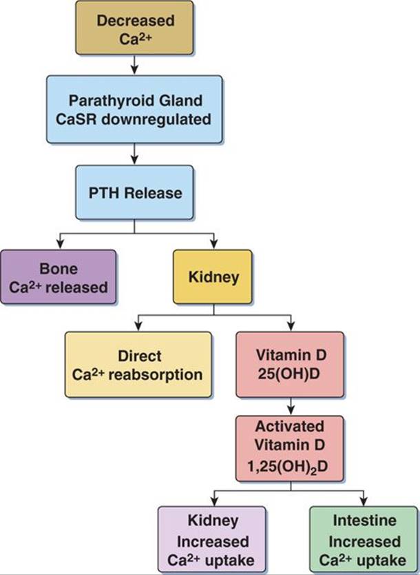
Figure 83-1. Calcium homeostasis: hypocalcemia.
Calcitonin is a hormone released by the thyroid gland which plays a more subdued role in calcium homeostasis. In response to hypercalcemia, calcitonin is released and inhibits osteoclast-mediated bone resorption, and renal and intestinal reabsorption of calcium.4
HYPOCALCEMIA
Hypocalcemia is defined as serum calcium <9 mg/dL. Major etiologies (Table 83-1) are hypoparathyroidism and vitamin D deficiency. Hypoparathyroidism leads to insufficient PTH release to stimulate release of calcium from bone and renal and intestinal preservation of calcium. It can be congenital or acquired. Magnesium is essential for PTH secretion and activation of PTH receptors. Low levels of magnesium may lead to functional hypoparathyroidism and hypocalcemia. Interestingly, high levels of magnesium (as in parenteral administration) may inhibit PTH release and also lead to hypocalcemia.3Vitamin D deficiency or resistance can lead to hypocalcemia, as well as difficulty converting vitamin D to the active form, 1,25(OH)2D. Pseudohypoparathyroidism, characterized by resistance to the effects of PTH, may also lead to hypocalcemia, along with elevated levels of PTH, and phosphorus. Additional etiologies are massive transfusion of citrated blood, phosphate enema toxicity, pancreatitis, and sepsis.
|
TABLE 83-1 |
Causes of Hypocalcemia |
Hypoparathyroidism
• Acquired
• Surgical
• Autoimmune
• Radiation – induced
• Heavy Metal Toxicity
• Iron – thalassemia, hemochromatosis
• Copper – Wilson disease
• Genetic
• DiGeorge (velocardiofacial) syndrome
• Familial hypoparathyroidism
• Familial hypocalcemia with hypercalciuria
• X-linked hypoparathyroidism
Pseudohypoparathyroidism (resistance to PTH)
• Type Ia – Albright syndrome
• Type Ib
• Type II
Magnesium imbalance
• Hypomagnesemia
• Hypermagnesemia
Vitamin D dysfunction
• Deficiency
• Resistance
• Vitamin D dependent rickets, Type II
• Inability to convert to active form
• Chronic renal failure
• Vitamin D dependent rickets, Type I
Loss of circulating calcium
• Hyperphosphatemia
• Phosphate enema toxicity
• Alkalosis
• Citrate toxicity
• Massive transfusion
• Hungry bone syndrome
• Pancreatitis
• Sepsis
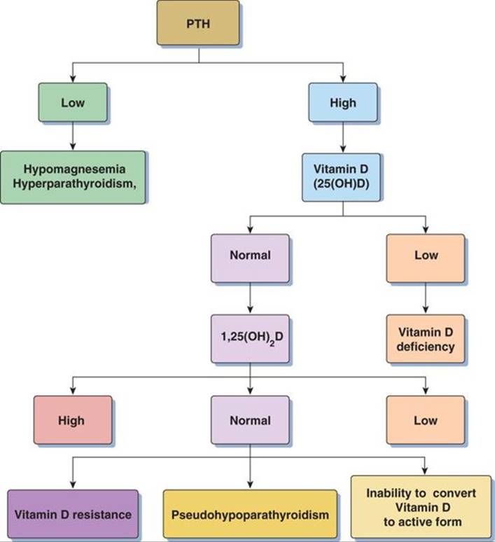
Figure 83-2 Laboratory algorithm for hypocalcemia.
Nonspecific symptoms, including nausea, weakness, paresthesias, and irritability, are typical. Classic physical findings of neuromuscular irritability are Chvostek sign and Trousseau sign. Chvostek sign is positive when there is twitching of the upper lip following tapping of the cheek anterior to the earlobe over the facial nerve. Trousseau sign is positive when there is carpal spasm after inflating a BP cuff on the upper arm above the systolic blood pressure for 3 minutes.3 In more severe cases, tetany, seizures, laryngospasm, and psychiatric manifestations may be seen. The electrocardiogram (ECG) may show prolongation of the QT interval, bradycardia, and/or dysrhythmias. Depression of cardiac contractility and vasomotor dysfunction are seen with low levels of ionized calcium.
Laboratory testing should include ionized and total calcium, magnesium, phosphorus, serum albumin, creatinine, and alkaline phosphatase. Vitamin D and PTH levels may help elucidate the etiology (Fig. 83-2), as may urine calcium and phosphorus levels.
For significant or symptomatic hypocalcemia, intravenous calcium may be administered cautiously with continuous ECG monitoring. Calcium gluconate, 10% (50–100 mg/kg/dose), or calcium chloride, 10% (10–25 mg/kg/dose), may be administered over 2 to 5 minutes.5 Intravenous calcium is very irritating to tissues and veins. It should be diluted prior to administration to a maximum concentration of calcium gluconate, 50 mg/mL or calcium chloride, 20 mg/mL. It is preferably given through a central line or very secure peripheral venous access. It should never be given intramuscularly, subcutaneously, or via an endotracheal route, as tissue necrosis and sloughing will occur. Intravenous calcium predisposes to digitalis toxicity and precipitates when mixed with bicarbonate. Hyperphosphatemic patients are at risk of metastatic calcium deposition with calcium administration and require treatment aimed at lowering phosphorus levels. When hypomagnesemia is present, oral or intravenous correction should be undertaken. Magnesium sulfate may be administered intravenously at a dose of 25 to 50 mg/kg, diluted to a maximum concentration of 200 mg/mL, over 2 to 4 hours.5 Longer-term management may involve oral administration of calcium, vitamin D, and thiazide diuretics.
HYPERCALCEMIA
Hypercalcemia is defined as a serum calcium level >10.5 mg/dL. Although often asymptomatic, complaints may include constipation, anorexia, vomiting, abdominal pain, or pancreatitis. Rarely, lethargy, depression, psychosis, or coma may occur. ECG changes may include QT-segment shortening, bradycardia, heart block, and sinus arrest. Nephrolithiasis can be an important consequence of hypercalcemia.
The conditions in adults that are commonly associated with hypercalcemia (hyperparathyroidism and malignancies of the breast, lung, kidney, and head and neck) are rare in children. In children, hypercalcemia with malignancy is associated with bone metastases or tumor lysis syndrome. Other causes in children include primary or tertiary hyperparathyroidism, hyperthyroidism, vitamin D intoxication, immobilization, thiazide diuretics, milk-alkali syndrome, and sarcoidosis. There are a number of rare, inherited disorders associated with hypercalcemia including idiopathic hypercalcemia of infancy (Lightwood syndrome), Jansen disease, Williams-Beuren syndrome, primary oxalosis, congenital lactase deficiency, and Down syndrome.
Laboratory investigation should include total and/or ionized serum calcium, serum albumin, electrolytes (including magnesium and phosphorus), creatinine, PTH, vitamin D levels, ECG, and urinalysis (Fig. 83-3). Hyperchloremic metabolic acidosis suggests primary hyperparathyroidism.
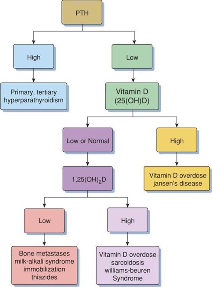
FIGURE 83-3. Laboratory algorithm for hypercalcemia.
In symptomatic patients or those with levels >14 mg/dL, therapy is aimed at expansion of extracellular fluid, calcium excretion, increased bone storage, and definitive treatment of the underlying cause. Volume expansion begins with normal saline and is followed by diuresis with furosemide to promote calcium excretion. Hemodialysis may be required in the setting of renal insufficiency or life-threatening dysrhythmias. Calcitonin, glucocorticoids, mithramycin, and indomethacin have all been used to suppress bone resorption, although the onset of action is >24 hours.
REFERENCES
1. Moore EW. Ionized calcium in normal serum, ulrafiltrates, and whole blood determined by ion-exchange electrodes. J Clin Invest. 1970;49:318.
2. Phillips P, Pain R. Correcting the calcium. Br Med J. 1977;1:1473.
3. Shoback D. Hypoparathyroidism. N Engl J Med. 2008;359:391.
4. Potts JT, Juppner H. Chapter 353. Disorders of the parathyroid gland and calcium homeostasis. In: longo DL, Fauci AS, Kasper DL, et al., eds. Harrison’s Principles of Internal Medicine. 18th ed. New York, NY: McGraw Hill; 2012.
5. Taketomo CK, Hodding JH, Kraus DM, eds Pediatric Dosage Handbook. 19th ed. Hudson, OH: Lexi-Comp; 2012.