
Practical Classification of Pediatric Vasculitis (Curr Rheumatol Rep 2009;11:402)
• 1º vasculitides: Classification based on International Consensus Conference, Vienna
• Large vessel vasculitis: Takayasu arteritis
• Medium vessel vasculitis: Kawasaki dz, cutaneous & systemic polyarteritis nodosa
• Small vessel vasculitis:
• Granulomatous: Wegener granulomatosis & Churg–Strauss syndrome (CSS)
• Nongranulomatous: Henoch–Schonlein purpura, microscopic polyangiitis, isolated cutaneous leukocytoclastic vasculitis, hypocomplementemic urticarial vasculitis
• Other vasculitides: Behcet’ dz, Cogan’s syndrome, unclassified, and 2° vasculitides 2/2 infection, malignancy, drugs or those associated with CTD
Clinical Manifestations
• Focal hypoperfusion: Claudication, HTN, CVA, abdominal pain
• Organ dysfunction: MI, myocarditis, neuropathy, myositis, sensory changes, cutaneous/microvascular effects (palp purpura, GN, pulmonary–renal syndrome)
• Can result in vascular tissue injury w/ vascular leak, aneurysm formation, stenosis, occlusion, rupture & necrosis, Kawasaki disease (mucocutaneous lymph node synd)
• Definition (Pediatr Rev 2008;29:308; Pediatrics 2004;114:1708)
• Acute self-limited vasculitis w/ fever, bilateral nonexudative conjunctivitis, erythema of lips and oral mucosa, extremities swelling, rash, and cervical LAD
• Coronary artery aneurysms or ectasia develop in 15–25% of untreated children
• Epidemiology: Susceptibility assoc w/ ITPKC gene. (Nat Genet 2008;40:35)
• 15 cases per 100,000 <5 yo w/ median age 2 yr in the US; incidence highest in Asians
• 85% pts <5 yo; pts <6 mo or >8 yo less common but ↑ risk coronary art aneurysm
• Diagnostic criteria (AHA/AAP Guidance Reports; updated 2007)
• Confirmed by fever ≥5 d and 4 of the 5 criteria below, and no other explanation
• Extremities Δs: Acute (erythema of palms, soles; edema of hands, feet); subacute (periungual peeling of fingers, toes in wk 2 and 3)
• Polymorphous exanthem
• Bilateral bulbar conjunctival injection (limbic sparing) without exudates
• Mucosal changes: Erythema, lips cracking, strawberry tongue
• Cervical lymphadenopathy (>1.5 cm diameter), usually unilateral
• Exceptions: Pts w/ fever ≥5 d and <4 criteria dx’d w/ Kawasaki if cardiac involvement
• Other clinical manifestations: Pericarditis, noncoronary aneurysms, hydrops of gallbladder, aseptic meningitis, urethritis/meatitis, anterior uveitis
• Modified AHA/AAP algorithm for “incomplete” (atypical) Kawasaki
• Fever at least 5 d and at least 2 clinical criteria for Kawasaki, no other explanation and lab findings consistent with severe systemic inflammation

• Laboratory findings suggestive of KD include the following
• CRP ≥ 3 mg/dL or ESR ≥ 40 mm/hr; WBC ≥ 15,000, anemia (normocytic, normochromic), sterile pyuria (≥10 WBC/HPF), ALT > 50, albumin ≤ 3.0 g/dL, after 7 d of illness Plts > 450,000
• Diagnostic studies: Echo – aneurysms, findings c/w coronary arteritis (perivascular brightness, ectasia, and lack of tapering of coronary arteries), ↓ LV contractility, mild valvular regurg (1° mitral valve) and pericardial effusion
• Treatment (Pediatr Rev 2008;29:308)
• IVIG and Aspirin (ASA): Single dose of 2 g/kg of IGIV over 10–12 hr; best w/I 1st 10 d
• ASA 80–100 mg/kg/d in 4 divided doses during acute phase
• Re-Rx w/ IVIG (2 g/kg) and cont ASA Rx may be indicated for persistent fever (>36 hr) or recurrent fever after initially afebrile ≤48 hr; infliximab has been used
• After fever controlled 4–5 d, ASA dose ↓ to 3–5 mg/kg/d
• ASA d/c’d if no coronary artery abn by 6–8 wk after onset of illness
• Low-dose ASA Rx continued indefinitely in those w/ coronary artery abn
• Even w/ appt Rx, 5% cor artery dilation 1% w/ giant aneurysm; vs. 20–25% w/o Rx
• Cardiac care: Check echo early in acute phase of illness 2 & 6 wk after onset
• F/u by cardiologist in 1st 2 mo assess for arrhythmias, CHF, and valvular regurg
• Development of giant coronary artery aneurysms (≥8 mm in diameter) may need anticoagulant Rx, such as warfarin, to prevent thrombosis
Henoch–Schönlein Purpura
• Definition (Curr Opin Rheumatol 2010;22:598)
• Leukocytoclastic vasculitis of small vessels w/ deposition of IgA1 in vessel walls and renal mesangium; most common vasculitis of childhood
• Epidemiology:10–20 cases per 100,000 children/yr
• Typically presents btw 3–10 yo; 50% cases at or before 5 yo; M:F 2:1
• Pathophysiology (BioDrugs 2001;15:99)
• Assoc w/ a wide variety of microbial pathogens, drugs, environmental agents; significant minority of pts w/ evidence of recent group A strep infection
• Skin findings w/ subepidermal hemorrhages and dermal necrotizing vasculitis
• The vasculitis can also occur in organs, such as the gastrointestinal tract
• ACR criteria for classification of Henoch–Schönlein purpura
• Need ≥2 of the 4 criteria. Presence ≥2 of criteria yields sens 87.1% and spec 87.7%
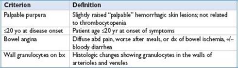
• Clinical manifestations: Usually p/w tetrad of rash, arthralgias, abd pain, & renal dz
• Rash; typically nonblanching, in groups (can persist 3–10 d), most on legs and arms
• Polyarthralgia; >80% pts. Most commonly affects knees and ankles, often w/ edema
• Resolves after a few d and leaves no permanent damage
• Abd pain; >½ pts, often colicky. Dev w/i 8 d of rash. Usually w/ N/V, diarrhea; blood and mucus often w/ stool. Rarely c/b intussusception
• Renal dz: ∼40–50% pts, p/w mild GN w/ proteinuria, microhematuria, and +/− RBC casts. Usually resolves spont but progressive renal dz may develop; those w/ persistent proteinuria likely w/ worsening renal damage
• Renal failure is the most common cause of death in pts who die w/ HSP
• Treatment (Curr Rheumatol Rep 2004;6:195; J Pediatr 2006;149:241)
• Suggested management of HSP (See the table below) (Curr Rheumatol Rep 2009;11:402)
• RCT of 1 mg/kg/d pred ×2 wk then 2 wk taper for severe abd pain ↓ ineffective for purpura, development of nephritis, ↓ing duration of dz or ↓ing recurrence. Usu steroids are not used for joint pain/rash alone
• Rx options for severe renal dz in HSP: High-dose corticosteroids +/− IS (AZA, cyclophosphamide, or cyclosporine); high-dose IVIG; plasma exchange or plasmapheresis; corticosteroids w/ urokinase and warfarin; renal xplant

Steroids, corticosteroids; IS, immunosuppressive; RPGN, rapid progressive glomerulonephritis.
Polyarteritis Nodosa (PAN) (Curr Rheumatol Rep 2009;11:402)
• Definition: Systemic vasculitis involving small and medium muscular arteries
• 1/3 w/ cutaneous PAN w/ limited dz restricted to skin, muscle, joints & periph nerves
• Epidemiology (J Pediatr 2004;145:517): Peak onset ∼9–11 yo (1–16 yo),  >
> slightly
• Etiology: Unknown, infxns (i.e., viral hepatitis, strep and parvo) implicated. Assoc w/ FMF
• Criteria for classification (1990 American College of Rheumatology Criteria)
• Commonly w/ fever, weight loss, malaise, and non-specific abdominal pain
• Classification criteria of ACR not validated for children; 3 of the following
• Unexplained weight loss >4 kg; livedo reticularis; testicular pain or tenderness; myalgias (except shoulder and hip girdle), muscle weakness, leg muscle pain; mononeuropathy or polyneuropathy; new onset DBP >90 mm Hg; ↑ BUN (>40 mg/dL) or Cr (>1.5 mg/dL); hepatitis B virus infection; characteristic arteriographic abn; bx of small or medium-sized artery w/ PMNs
• Diagnostic studies: Labs— ↑ ESR/CRP, ↑WBC, ↑ immunoglob, can also see proteinuria, hematuria, inc BUN/Cr, complement usually nml. +HBsAg (30%), p-ANCA (<20%)
• Angiogram: Aneurysms and focal vessel narrowing
• Bx (sural nerve or skin): Vasculitis with fibrinoid necrosis without granulomas
• Rx: Mainstay Rx is steroids and IS agents (cyclophos, MTX, MMF, anti-TNF, rituximab)
• HBV-related PAN is treated with antiviral therapy
ANCA-related Vasculitis
• Definition (Pediatr Nephrol 2010;25:205): Aberrant interaction btw neutrophils & vascular endothelial cells; includes Wegener granulomatosis, microscopic polyangiitis, renal-limited microscopic polyangiitis, CSS and drug-induced vasculitides (antithyroid Rx, hydralazine, minocycline)
• Epidemiology: Can occur at any age but typically in young and middle-aged adults
• Clinical indications for testing for ANCA (Am J Kidney Dis 1991;18:184)
• Glomerulonephritis, especially rapidly progressive glomerulonephritis
• Pulmonary hemorrhage, especially pulmonary–renal syndrome
• Cutaneous vasculitis, especially with systemic features
• Multiple lung nodules; chronic destructive disease of the upper airways
• Long-standing sinusitis or otitis; subglottic tracheal stenosis
• Mononeuritis multiplex or peripheral neuropathy; retro-orbital mass
• Wegener granulomatosis Classification (2005 EULAR/PRES Criteria, updating 1990 ACR Criteria) (Arthritis Rheum 2009;60:3413)
• Needs 3 of the 6 criteria. Yields sens of 73.6% and a spec of 73.2% in pediatric pts
• Nasal or sinus inflamm (painful/painless ulcers, purulent or bloody nasal discharge)
• Abn CXR or chest CT scan (w/ presence of nodules, fixed infiltrates, or cavities)
• Abn U/A: Microhematuria (>5 RBCs/HPF) or significant proteinuria
• Granulomatous inflamm on bx/necrotizing pauci-immune GN
• Subglottic, tracheal, or endobronchial stenosis
• Anti-PR3 ANCA or cANCA staining
• Churg–Strauss vasculitis Classification (1990 ACR Criteria; no EULAR/PRES Criteria)
• 4 or more of the 6 criteria w/ sens 85% and spec 99.7%
• Asthma: History of wheezing or diffuse high-pitched expiratory rhonchi
• Eosinophilia: >10% on differential WBC count
• Mono- or polyneuropathy: Glove/stocking distribution
• Pulmonary infiltrates that are nonfixed: Migratory or transitory
• Paranasal sinus abn: Hx acute/chronic sinus pain x-ray opacification of sinuses
• Extravascular eosinophils: Demonstrated via bx of artery, arteriole, or venule
Diagnostic Testing
• Antineutrophil cytoplasmic antibody (Kidney Int 2000;57:846)

IF, immunofluorescence; C-ANCA, cytoplasmic staining ANCA; P-ANCA, perinuclear staining ANCA; PR3, proteinase3, MPO, myeloperoxidase.
• Wegener: CXR or CT chest (for nodules, infiltrates, cavities), sinus CT for sinusitis, BUN/Cr, U/A (proteinuria, hematuria, sediment w/ RBC casts, dysmorphic RBCs for renal dz), bx (nec granulomatous inflamm of vessels)
• Microscopic polyangiitis: CXR, U/A (similar to Wegener), bx (necrotizing, pauci-immune inflammation of arterioles, capillaries, and venules)
• Churg-Strauss syndrome: CXR (pulm infiltrates), bx (microgranulomas, fibrinoid necrosis, and thrombosis of small vessels w/ eosinophilic infiltrates)
• Clinical features (Am J Med 2004;117:39)

MPO, myeloperoxidase, PR3, proteinase 3.
• Treatment (Pediatr Nephrol 2010;25:205)
• Wegener granulomatosis or microscopic polyangiitis
• Generalized non-organ–threatening dz = remission w/ MTX 0.3 mg/kg BW qwk orally + prednisone 1 mg/kg qd; MTX & pred for maintenance as well
• Generalized organ-threatening dz – cyclophosphamide (15 mg/kg IV pulse q2wk × 6 mo) w/ oral prednisone 1 mg/kg tapered to 0.2 mg/kg by 6 mo effective, continued till 15 mo then tapered to 0.15 mg/kg × 3 mo more for remission
• Addition of infliximab allows remission 6 wk earlier w/ dec steroid exposure
• Azathioprine and w/ prednisone commonly used for maintenance
• Severe renal disease and immediate life-threatening disease (DAH) = pulses of methylprednisolone and IV cyclophosphamide (3–4 mg/kg daily), consider plasma exchange
• Churg-Strauss syndrome: Initially w/ prednisone 1 mg/kg daily monotherapy
• In severe dz addition of cyclophosphamide used (heart, CNS, GI, kidney involved)