
Definition (Pediatr Rev 2008;29:39)
• Intraluminal bleeding at any site from oropharynx to the anus; definition of upper GI bleed as proximal to the ligament of Treitz and lower GI bleed as distal
• Can p/w bloody vomit (hematemesis), “coffee ground” emesis, hematochezia (BRBPR; LGIB, or rapid UGIB [>20% blood volume]) or melena (tarry black 2/2 digestion, darkness correlates w/ time to pass); r/o other site (nasopharynx)
• Check for false coloring; red food coloring, fruit juice, beets can make vomit or stool red. Pepto-Bismol, iron, grape juice, spinach, and blueberry can make it black
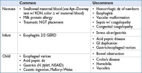
Etiology of Upper GI Bleed (Clin Pediatr (Phila) 2007;46:16)
Differential of UGIB
Etiology of Lower GI Bleed (Pediatr Emerg Care 2002;18:319)
• Anal fissure: Most common cause LGIB <2 yo. Blood coated stool. Painful. Constipation
• Nodular lymphoid hyperplasia: Painless rectal bleeding, usually after viral illness, can be associated with immunodef (IgA def, hypogamma)
• Food allergy: 3 different mechanisms; cow’s milk and soy most likely offenders
• Enterocolitis: Vomiting, bloody diarrhea; +/− malabsorption, FTT
• Colitis: 1st few mos of life, healthy, normal weight w/ blood in stool
• Eosinophilic gastroenteritis: Infiltration w/ eos, peripheral eos, no vasculitis, p/w postprandial N/V, abd pain, watery diarrhea +/− blood, anemia, FTT
• Necrotizing enterocolitis: Risk factors, prematurity, sepsis, LBW, HoTN, asphyxia
• Infectious enterocolitis: Bacterial, viral, or parasitic pathogens. C. diff w/ abx hx
• Hirschsprung disease: 10–30% develop enterocolitis w/ fever, bloody diarrhea
• Meckel diverticulum: 2/2 incomplete obliteration of omphalomesenteric duct
• Painless passage of large amt of blood; otherwise healthy (2/2 heterotopic gastric mucosa in diverticulum causing adjacent ileal mucosa ulceration)
• “Rule of 2s”; 2% pop, 2 in. long, 2 cm diameter, w/i 2 ft of ileocecal junction, w/ types of ectopic tissue (gastric and pancreatic), 2:1 M:F, and 50% w/ sx before 2 yo
• Duplication of bowel same as Meckel but on mesenteric side, not antimesenteric
• Intussusception: Usually <2 yo, colicky abd pain, “sausage-shaped” abd mass w/ late finding of “currant jelly stool.” See ED chapter
• Polyps: Outside of infants these are most common source for painless LGIB
• Painless rectal bleeding
• Juvenile polyps (95% of polyps in children) are 90% hamartomatous (benign) usually singular but multiple (>5 polyps) seen in juvenile polyposis, Peutz–Jeghers (mucocutaneous pigmentation; higher rate GI CA but not 2/2 polyps) and Cowden syndrome
• Adenomatous polyps can be premalignant; found in familial polyposis (AD but 1 in 3 new mutation; sx after 10 yo), Gardner syndrome (AD; soft tissue/bone tumors) and Turcot syndrome
• IBD: Almost all UC and ¼ of Crohn have LGIB, 1 in 4 present before 20 yo
• Angiodysplasia: Vascular ectasia; assoc syndromes (Osler–Weber–Rendu, Turner)
• Hemorrhoids: Rare in childhood, assess for portal HTN; common after adolescence
• Henoch–Schönlein purpura: Typically 4–7 yo (but any age) systemic small vessel vasculitis w/ abd pain and bloody stools, “palpable purpura.” +/− renal and joint
• GI involvement in 45–75% cases and can precede skin findings in 15% of cases
• IgA immune complex mediated
• HUS: Microangiopathic hemolytic anemia, thrombocytopenia, ARF usually preceded (3– 16 d) by bloody diarrhea. (90% D + HUS w/ shiga-like toxin vs. atypical HUS/D – HUS w/o)
• Ischemia: 2/2 midgut volvulus, intussusception, etc.; generally ill appearing
Clinical Manifestations
• Tachycardia most sensitive sign of acute, severe blood loss; orthostasis w/ >20 bpm ↑
• HoTN and decreased capillary refill are ominous late findings (>30% blood vol loss)
• With infants who are breast-feeding, ask mother about presence of breast lesions
• Hemorrhagic disease of newborn; hx – child born athome, no vit K presents DOL 1–5
• Variceal bleed 2/2 extrahepatic portal HTN; no cirrhotic stigmata, + splenomegaly
• Extrahepatic portal HTN 2/2 omphalitis 2/2 neonatal umbilical vein catheter or spontaneous inflammation of umbilical vessels
• Check skin for petechiae or purpura (coagulopathy or HSP), spider angiomata (liver dz), hemangiomas or telangiectasias (Osler–Weber–Rendu)
Diagnostic Studies (Pediatr Rev 2008;29:39)
• Confirm w/ hemoccult for stool guaiac or gastroccult w/ vomitus
• False pos w/ vit C, red meat, veg w/ peroxidase (broccoli, radish, turnips)
• NGT lavage useful if returns blood/coffee grounds = UGIB; can miss duodenal ulcer; if clears w/ lavage, then likely active bleeding has ceased
• Isolated increase in BUN can be a sign of gastric bleeding and absorption
• Plain films: KUB/upright for free air (perforation), pneumatosis intestinalis (NEC), air–fluid levels (obstruction)
• For LGIB w/ sx of colitis check stool cx for Yersinia, Salmonella, Shigella, Campylobacter, Aeromonas, E. coli (including O157:H7), Klebsiella oxytoca, C. diff, perirectal swab for N. gonorrhoeae (if sexually active), & O&P for E. histolytica & T. trichiura (if appt travel hx)
• Upper GI and small bowel follow through best for structural lesions
• Endoscopy: Indicated w/ acute UGIB necessitating transfusion or recurrent bleeding or as first step in diagnosis and evaluation of any GIB; contraindicated in clinically unstable patient
• Retrospective studies show w/ EGD 5 most common dx = duodenal ulcer (20%), gastric ulcer (18%), esophagitis (15%), gastritis (13%), and varices (10%) in children and adolescents (Pediatrics 1979;63:408)
• Rectosigmoidoscopy/colonoscopy: Useful if not active major bleeding
• Most helpful to assess IBD, angiodysplasia, polyps, pseudomembranous colitis
• Barium enema: For Hirschsprung dz, dx and rx of intussusception
• Nuclear medicine (Technetium-99 pertechnetate): Labels ectopic gastric mucosa, best for Meckel diverticulum or intestinal duplication
• Can also do Technetium-99 RBC scan for bleeding (0.05–0.1 mL/min)
• Angiography: Must be >0.5 mL/min for detection
• Can be used for guiding therapeutic approach, i.e., coiling
• Indicated over EGD for hemobilia (bleeding from biliary tract)
• Abdominal U/S +/− Doppler: For specific evaluation (i.e., liver dz or portal HTN)
• In immune compromised consider assessment for CMV, HSV, or Candida esophagitis
Treatment
• Acute management: Volume resuscitation w/ 2 large bore IVs, IV bolus w/ NS or LR, transfusion, if present, correct coagulopathies (FFP, platelets)
• UGIB: Basically 2 sources (1) Mucosal (-itis, ulcers, Mallory–Weiss) (2) Variceal
• Mucosal: Neutralize/decrease acid production (PPIs > H2RA); w/ Mallory–Weiss can coagulate w/ thermal probe or inject dilute epinephrine
• Variceal: Acute bleeding stops spontaneously in 50% w/ rebleed in 40%
• Stop bleeding (band ligation > sclerotherapy by risk profile)
• Decrease portal pressure and splanchnic blood flow: Octreotide > vasopressin
• LGIB: Etiology specific (stool softener for fissure, abx for infectious colitis, rsxn for polyps)