
STEPHEN LUDWIG
BRANDON C. KU
DEFINITION
The complaint of pallor indicates a perceived decrease in rubor in the skin and mucous membranes of a child, which is associated with decreased oxyhemoglobin delivery to the skin or mucous membranes. Potential causes include decreased blood flow, which may be regional (e.g., thrombosis) or systemic (e.g., shock), and normal blood flow with decreased oxygen-carrying capacity (e.g., anemia).
In most cases, the finding of pallor demands that anemia first be considered as this is the most common cause. Exceptions include children who have a constitutional cause of pallor due to their fair complexion and lack of exposure to sunlight. However, most children with pallor should be considered to have low hemoglobin, which should be measured. Ordinarily, a complete blood count with differential count, red blood cell (RBC) indices, and reticulocyte count guide the clinician in differentiating the many causes of anemia and in determining unusual situations in which pallor is not related to anemia. In addition, a peripheral smear with examination of RBC morphology may further guide the clinician in determining the etiology of anemia.
CAUSE AND FREQUENCY
Pallor may be divided into causes involving normal hemoglobin (Table 10-1) and those involving a low hemoglobin level. The etiology of anemia may be separated into causes due to decreased RBC production, increased RBC destruction, or acute blood loss. Causes due to decreased RBC production are generally associated with low reticulocyte count while causes due to increased RBC destruction or acute blood loss are generally associated with increased reticulocyte count. Anemia may also be considered in relation to mechanism, such as trauma, toxin, metabolic tumor, congenital, or mixed etiology (Table 10-2), or in relation to RBC morphology (Table 10-3).
TABLE 10-1. Pallor with anemia.
Decreased erythrocyte or hemoglobin production
Nutritional deficiencies
Iron deficiency
Folic acid and vitamin B12 deficiency or associated metabolic abnormalities
Aplastic or hypoplastic anemias
Diamond-Blackfan anemia
Fanconi anemia
Aplastic anemiaa
Transient erythroblastopenia of childhood
Malignancy: leukemia, lymphoma, neuroblastomaa
Anemia of chronic disease
Abnormal heme and hemoglobin synthesis
Lead poisoninga
Sideroblastic anemias
Thalassemias
Increased Erythrocyte Destruction
Erythrocyte membrane defects: hereditary spherocytosis, elliptocytosis, stomatocytosis, pyknocytosis, paroxysmal nocturnal hemoglobinuria
Erythrocyte enzyme defects
Defects of hexose monophosphate shunt: G6PD deficiency most common
Defects of Embden-Meyerhof pathway: pyruvate kinase deficiency most common
Hemoglobinopathies
Sickle cell syndromea
Unstable hemoglobins
Immune hemolytic anemia
Autoimmune hemolytic anemia (e.g., Evans syndrome)
Isoimmune hemolytic anemia
Infection
Viral: mononucleosis, influenza, coxsackie, measles, varicella, cytomegalovirus
Bacterial: Escherichia coli, Pneumococcus, Streptococcus, thyphoid fever, Mycoplasma
Drugs: antibiotics, methyldopa
Inflammatory and collagen vascular disease
Malignancya
Microangiopathic anemias
Disseminated intravascular coagulationa
Hemolytic uremic syndromea
Thrombotic thrombocytopenic purpura
Cavernous hemangioma
Blood Loss
Severe traumaa
Anatomic lesions
Meckel diverticulum
Peptic ulcer
Idiopathic pulmonary hemosiderosisa
aCan present as life-threatening emergencies
TABLE 10-2. Etiology of anemia by mechanism (common causes of pallor).
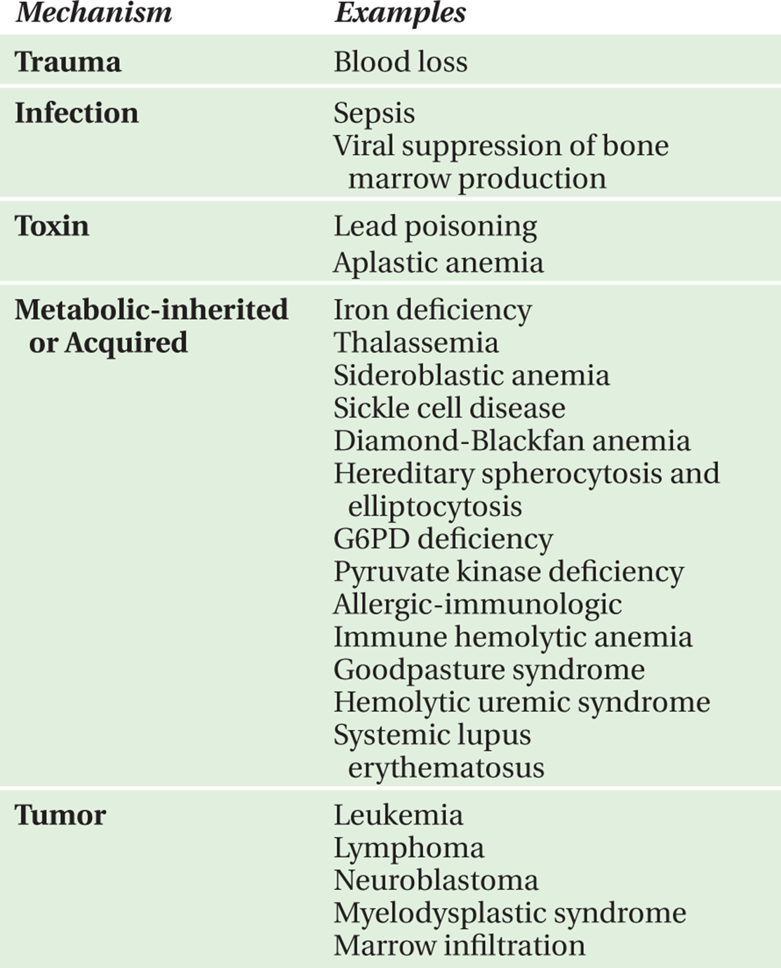
TABLE 10-3. Etiology of anemia by red blood cell morphology.
Microcytic Anemia
Iron deficiency
Lead poisoning
Thalassemias
Hemoglobinopathies
Sideroblastic anemia
Normocytic Anemia
Low reticulocyte count
Anemia of chronic disease
Transient erythoblastopenia of childhood
Diamond-Blackfan anemia
Infection
Myelosuppression from medication side effect
Renal disease
Leukemia
Marrow infiltration
Aplastic anemia
Elevated reticulocyte count
Blood loss
Pulmonary hemosiderosis
Goodpasture syndrome
Immune hemolytic anemia
Hemoglobinopathies
Hereditary spherocytosis and elliptocytosis
G6PD deficiency
Pyruvate kinase deficiency
Hemolytic-uremic syndrome
Thrombotic thrombocytopenic purpura
Kasabach-Merritt syndrome (hemangioma thrombocytopenia syndrome)
Macrocytic Anemia
Low reticulocyte count
Folate or vitamin B12 deficiency
Aplastic anemia
Diamond-Blackfan anemia
Transient erythroblastopenia of childhood
Elevated reticulocyte count
Hemolytic anemia and high reticulocyte count
Autoimmune hemolytic anemia
Secondary folate deficiency in hemolytic anemia
QUESTIONS TO ASK AND WHY
• Is there hemodynamic instability or need for emergent intervention?
—The most important question to ask is whether a child’s pallor is secondary to severe anemia with hemodynamic compromise that requires emergent intervention and stabilization with fluid resuscitation until transfusion of packed RBCs. This question also helps to determine whether severe anemia is secondary to acute external hemorrhage that may require immediate surgical intervention. One can also elucidate if a child’s pallor is secondary to decreased blood flow from acute thrombosis or systemic shock further guiding therapy.
• What are the child’s dietary habits?
—Because the most common cause of pallor is iron deficiency anemia, important clarifying questions pertain to diet. Diets containing large quantities of cow’s milk may raise concern for iron deficiency due to lack of iron intake and loss of appetite for iron-rich foods.
aCan present as life-threatening emergencies
• Is there a family history of anemia, splenectomy, or gallbladder surgery?
—Many RBC membrane disorders are inherited. Some disorders, such as hereditary spherocytosis and elliptocytosis, frequently necessitate splenectomy due to RBC sequestration. Inherited diseases causing hemolysis, such as sickle cell disease, may be recognized in some families by a history of gallbladder surgery for gallstones.
• Are there signs of systemic illness, fatigue, growth failure, weight loss, or lymphadenopathy?
—The presence of these signs or symptoms should raise concern for malignancy.
• Was there exposure to certain medications or toxins?
—Questions regarding exposure to toxins or medications should be asked as these may cause anemia by hemolysis (e.g., sulfonamide or nitrofurantoin exposure in a patient with glucose- 6-phosphate dehydrogenase [G6PD deficiency]) or by suppressing bone marrow production of RBCs (e.g., trimethoprim/sulfamethoxazole).
• Did pallor develop quickly or gradually?
— Information about the progression of anemia provides clues about the cause. Gradual onset suggests iron deficiency while acute onset with jaundice suggests hemolysis. In cases of gradual development of pallor, additional questions should focus on sources of iron loss or blood loss, such as history of heavy menstruation, blood in stool or melena, or multiple blood draws for diagnostic testing.
SUGGESTED READINGS
1. Poncz, M. Pallor. In: Schwartz MW, ed. Pediatric Primary Care: A Problem-Oriented Approach. 2nd ed. Chicago, London, Boca Raton, Littleton, MA: Year Book Medical Publishers; 1987.
2. Segel GB, Hirsh MG, Feig SA. Managing anemia in pediatric office practice: Part I. Pediatr Rev. 2002;23:75-84.
3. Shah S. Pallor. In: Fleisher GR, Ludwig S, eds. Textbook of Pediatric Emergency Medicine. 6th ed. Philadelphia: Lippincott Williams & Wilkins; 2010.
4. Bizzaro M, Colson E, Ehrenkranz R. Differential diagnosis and management of anemia in the newborn. Pediatr Clin North Am. 2004;51:1087-1107.