
Approach to the Problem
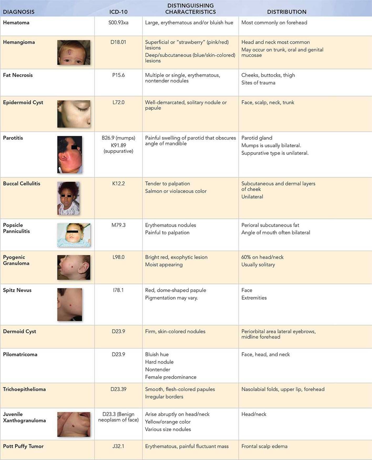
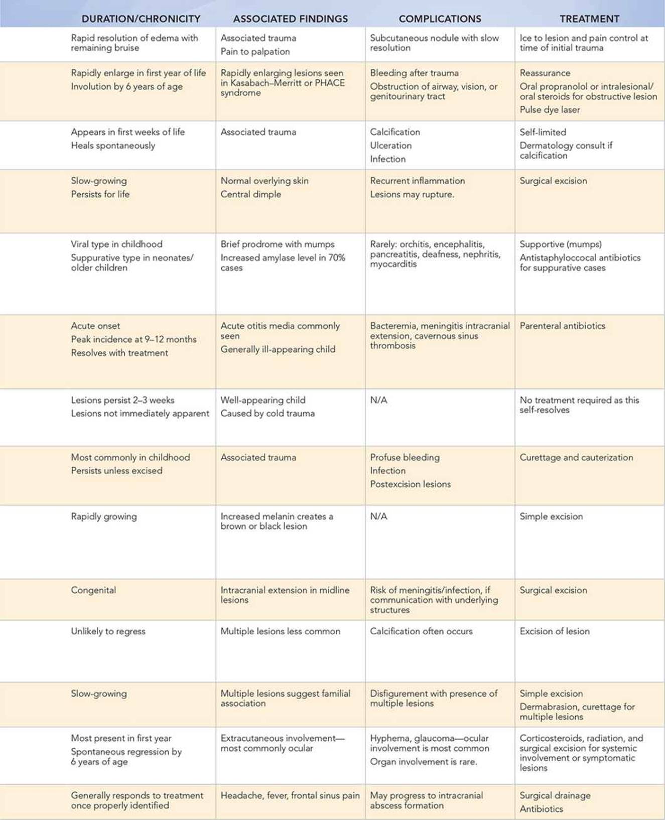
Facial lumps cause concern for parents though many of these lesions are benign and self-limited. The more common pediatric facial lesions include dermoid cyst, epidermoid cyst, superficial and deep (subcutaneous) hemangioma, pilomatricoma, buccal cellulitis, pyogenic granuloma, Spitz nevus, and suppurative parotitis. Less common lesions include trichoepitheliomas, Pott puffy tumor, panniculitis, fat necrosis, juvenile xanthogranuloma, and mumps. Although many of these facial lumps do not require immediate therapy, it is important to correctly diagnose and identify those lesions requiring urgent medical attention.
Key Points in the History
• Trauma is usually associated with hematomas.
• Hemangiomas, juvenile xanthogranulomas, and dermoid cysts are present at birth or appear in early infancy.
• Epidermoid cysts can appear at any age but appear more commonly after puberty.
• Prolonged exposure to cold in the area of swelling suggests panniculitis. Fat necrosis often occurs secondary to cold trauma.
• Constitutional symptoms of fever and malaise may suggest mumps, suppurative parotitis, buccal cellulitis, or Pott puffy tumor.
• A history of recurrent parotid swelling or a family history of parotid swelling may indicate juvenile recurrent parotitis, a nonsuppurative parotid inflammation of unknown etiology.
• The history of an unimmunized child with parotid inflammation strongly suggests mumps.
• Chronic, nonpainful swelling of the parotid gland may be seen in patients with HIV infection.
Key Points in the Physical Examination
• Swelling that obscures the angle of the jaw suggests parotid inflammation or parotitis.
• Children with mumps are rarely ill-appearing.
• Suppurative parotitis, most commonly caused by Staphylococcus aureus, is associated with an ill-appearing child with purulent discharge from Stensen duct.
• Buccal cellulitis, panniculitis, hematoma, parotitis, and Pott puffy tumor produce painful lumps.
• Swelling associated with erythema of the overlying skin suggests buccal cellulitis, panniculitis, fat necrosis, or suppurative parotitis.
• Friable lesions are characteristic of pyogenic granulomas.
• A Spitz nevus is a rapidly growing dome-shaped, erythematous papule commonly found on the face and extremities.
• Lumps with an associated bluish hue suggest a pilomatricoma or deep (subcutaneous) hemangioma.
• A pilomatricoma is typically a rock-hard papule due to its propensity to calcify.
• Fluctuant, tender, erythematous swelling over the frontal sinus suggests a frontal osteomyelitis with associated subperiosteal abscess, a condition known as Pott puffy tumor.
|
PHOTOGRAPHS OF SELECTED DIAGNOSES |
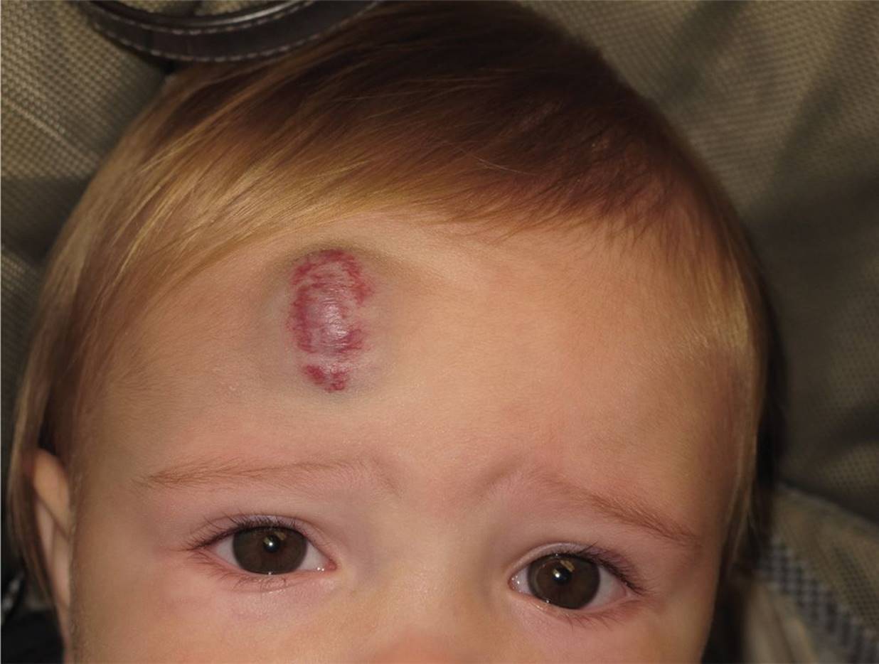
Figure 11-1 Mixed hemangioma. A rapidly growing mass with superficial and subcutaneous components. (Courtesy of Andrea L. Zaenglein, MD.)
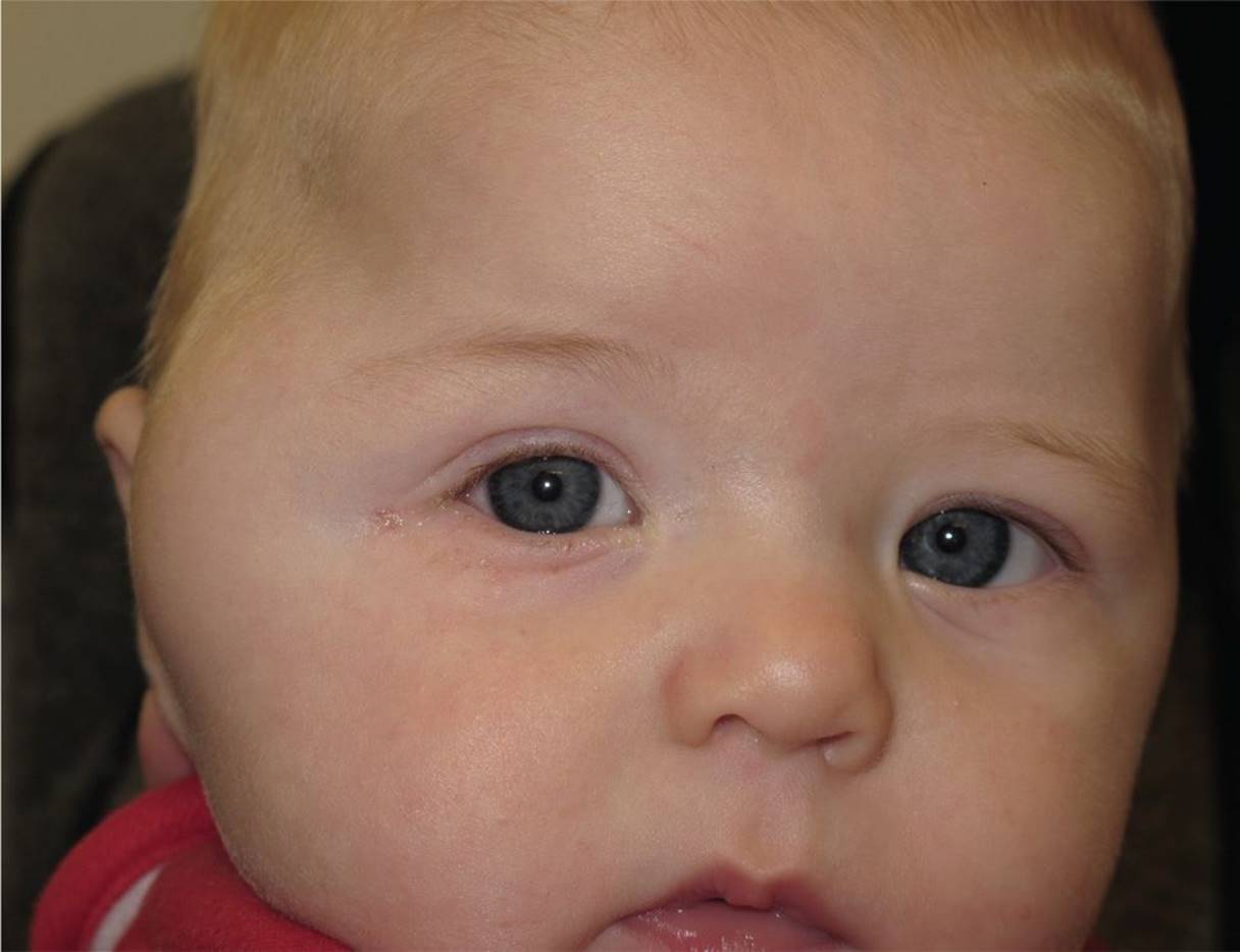
Figure 11-2 Deep or subcutaneous hemangioma. Note the normal overlying skin. (Courtesy of Andrea L. Zaenglein, MD.)
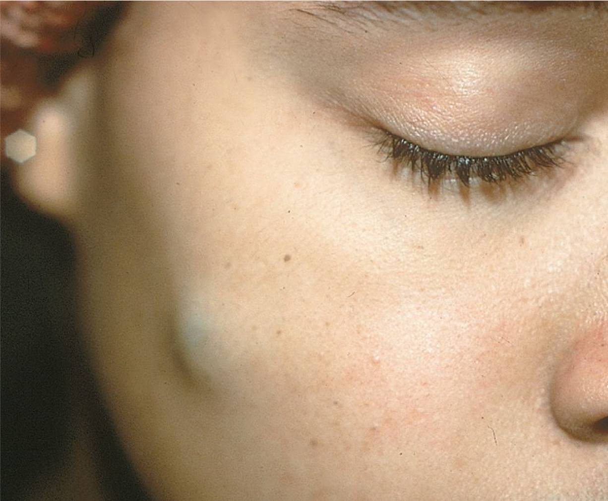
Figure 11-3 Epidermoid cyst. A well-demarcated solitary nodule on the face. (Used with permission from Goodheart HP. Goodheart’s Photoguide of Common Skin Disorders. Philadelphia, PA: Lippincott Williams & Wilkins; 2003:4.)
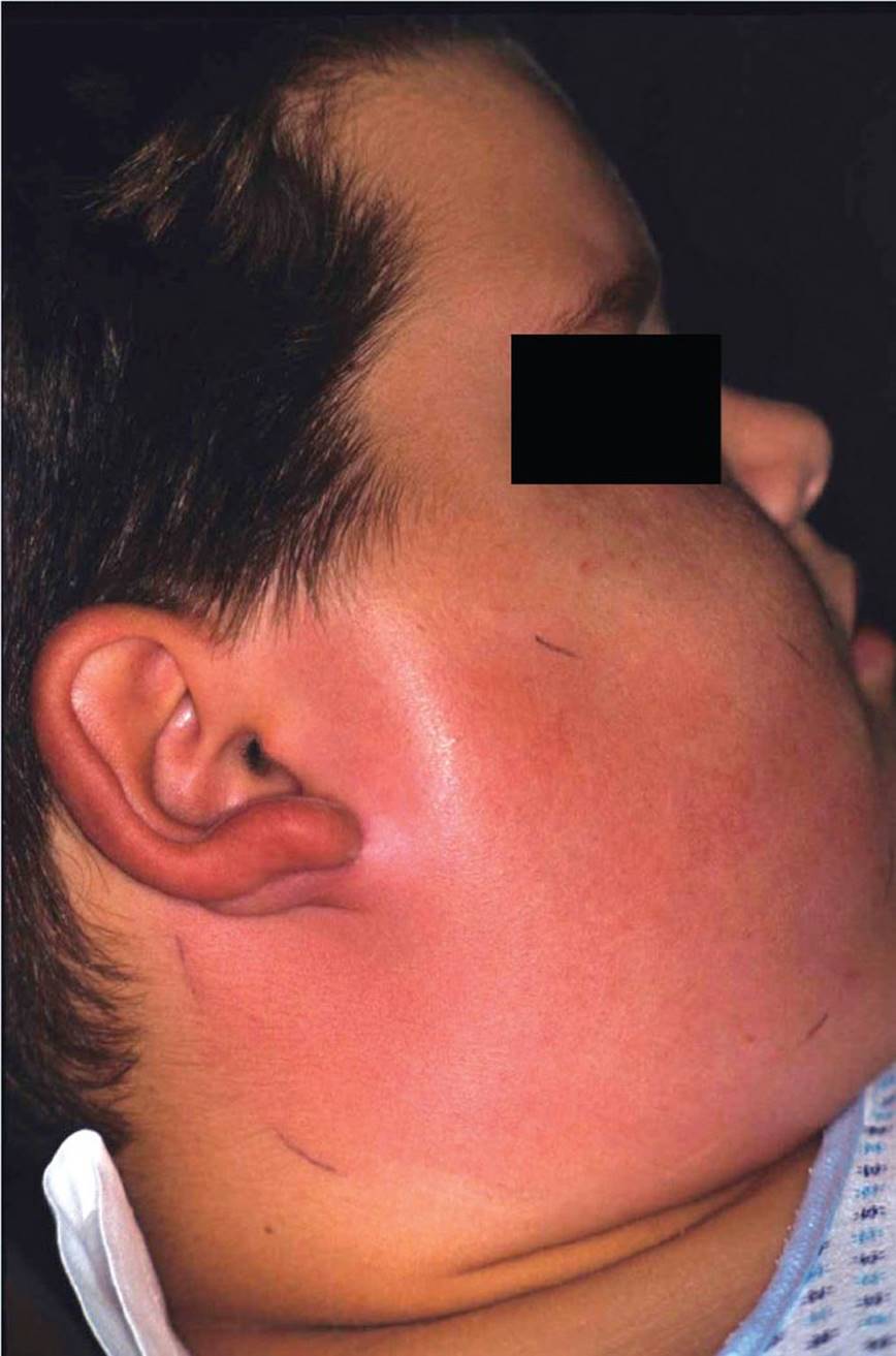
Figure 11-4 Parotitis. Dramatic edema, erythema, and induration of the face overlying the parotid gland. (Courtesy of Kathleen Cronan, MD.)
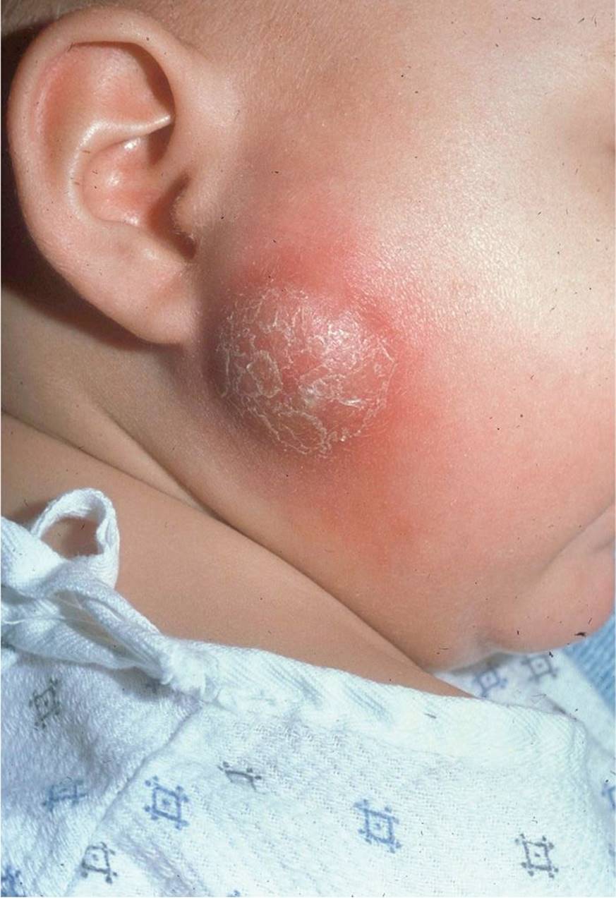
Figure 11-5 Parotid abscess. A well-demarcated fluctuant mass overlying the parotid gland. (Courtesy of the late Peter Sol, MD.)
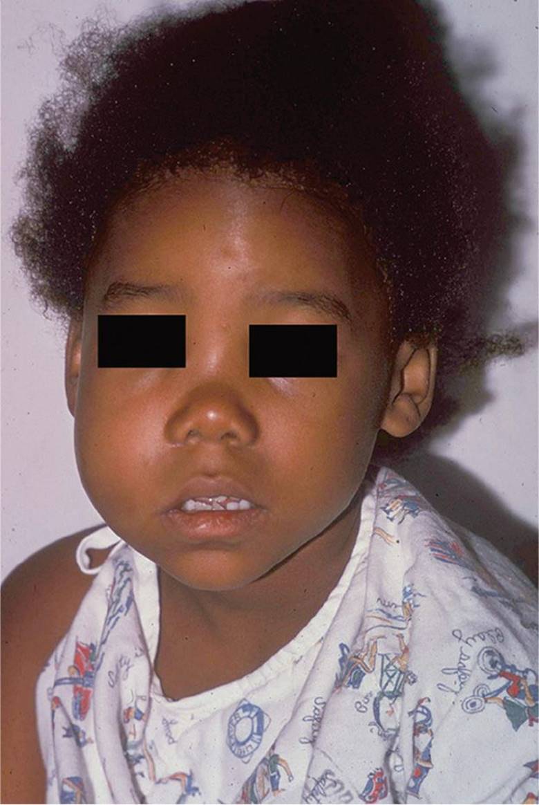
Figure 11-6 Buccal cellulitis. Diffuse unilateral facial swelling associated with dental caries. (Courtesy of the late Peter Sol, MD.)
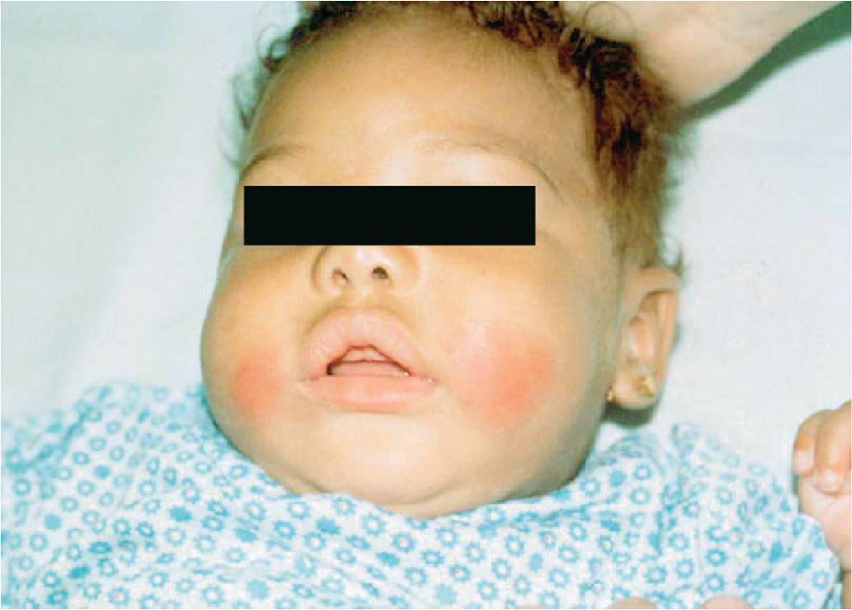
Figure 11-7 Panniculitis. Bilateral, erythematous firm subcutaneous nodules in the cheeks of this infant caused by cold trauma (popsicle). (Courtesy of Kathleen Cronan, MD.)

Figure 11-8 Pyogenic granuloma. Isolated erythematous nodule that easily bleeds. Note the annular “band-aid sign” around the lesion, showing evidence that a band-aid had been used for the bleeding. (Courtesy of Andrea L. Zaenglein, MD.)
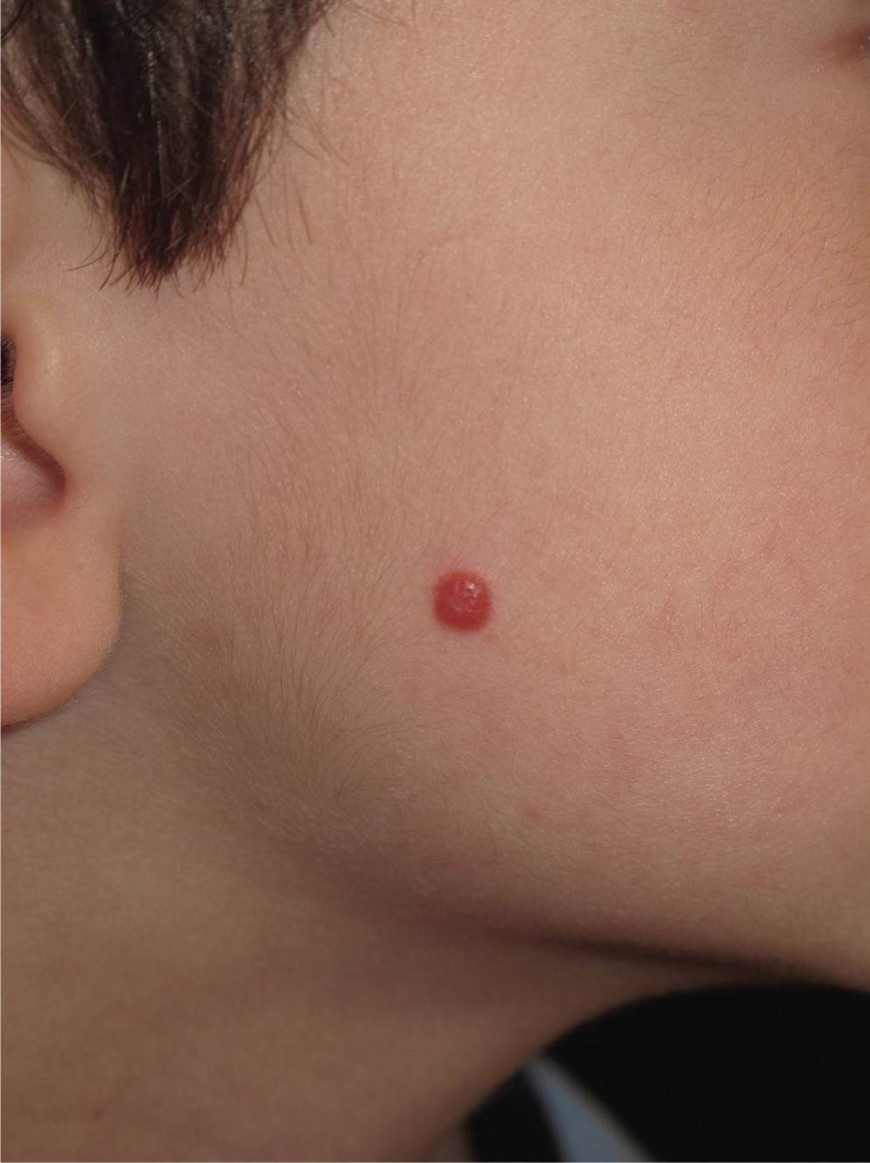
Figure 11-9 Spitz nevus. Erythematous, solitary, dome-shaped lesion on the cheek of this child. (Courtesy of Andrea L. Zaenglein, MD.)
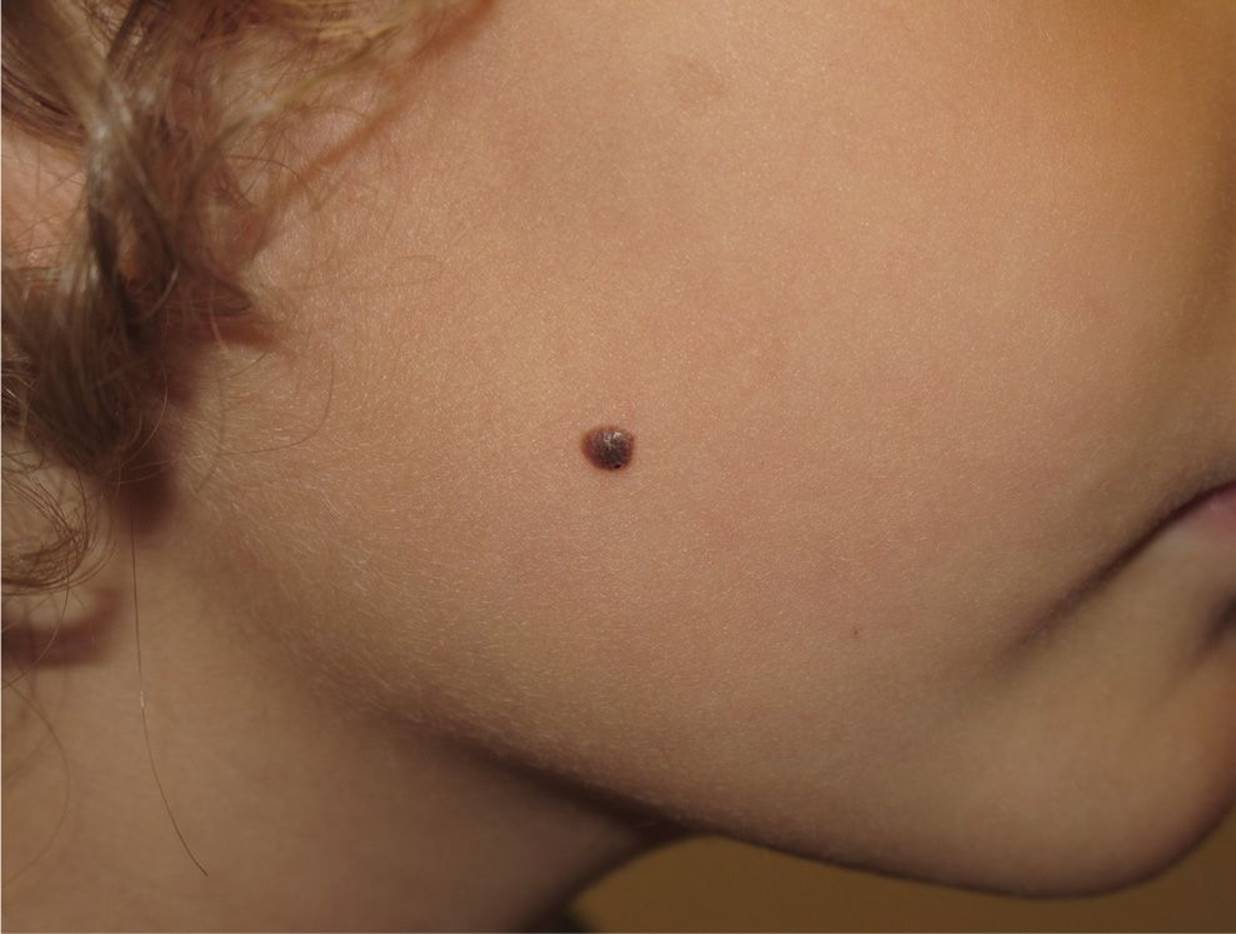
Figure 11-10 Pigmented Spitz nevus. Deeply pigmented, solitary, dome-shaped nodule. (Courtesy of Andrea L. Zaenglein, MD.)
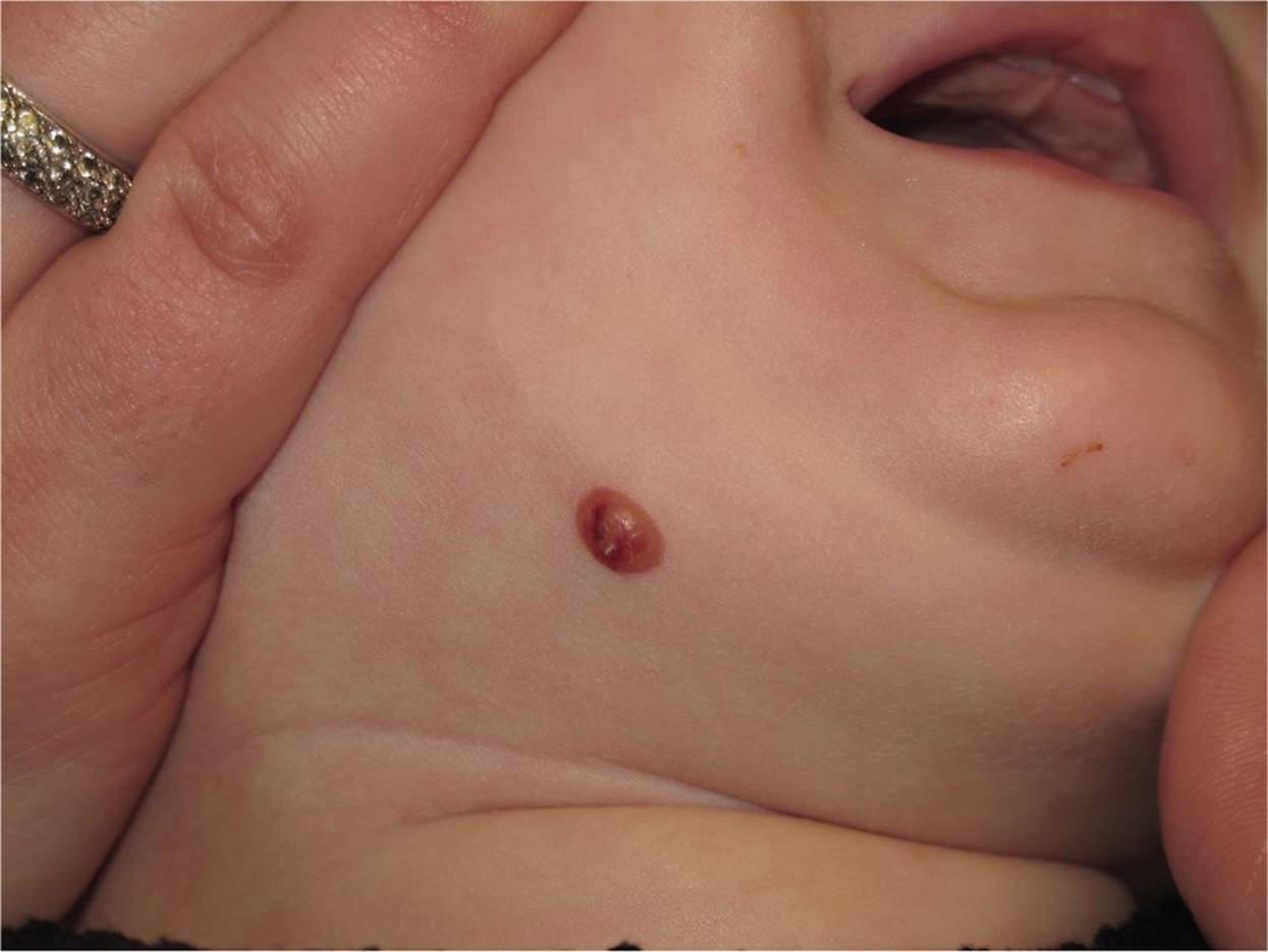
Figure 11-11 Juvenile xanthogranuloma. Isolated orange/brown firm papule on the chin of this child. (Courtesy of Andrea L. Zaenglein, MD.)
DIFFERENTIAL DIAGNOSIS
Other Diagnoses to Consider
• Metastasis of malignant tumor
• Idiopathic neuroma
• Multiple mucosal neuromas (multiple endocrine neoplasia type 2B)
• Lymphocytoma cutis
When to Consider Further Evaluation or Treatment
• Enlarging hemangiomas at risk for obstructing the nose or eye should be referred to a dermatologist.
• Hemangiomas in a “beard” distribution require evaluation by a dermatologist due to the risk of airway complications.
• Lesion consistent with fat necrosis may require dermatology referral if calcification or ulceration occurs.
• Epidermoid cysts with recurring inflammation may require surgical excision.
• Prompt medical attention is required if parotid or buccal mucosal swelling is accompanied by fever or an ill appearance.
• Pyogenic granulomas with frequent or profuse bleeding require urgent surgical excision.
• Dermoid cysts communicating with underlying structures may evolve into serious infections requiring antibiotics.
• A deeply pigmented Spitz nevus warrants a dermatology evaluation to rule out a more significant lesion such as malignant melanoma.
• Multiple xanthogranulomas on the face should prompt a referral to ophthalmology to assess for extracutaneous involvement.
• Pott puffy tumor can lead to significant morbidity if not diagnosed and treated promptly.
SUGGESTED READINGS
Haggstrom A, Drolet B, Baselga E. Prospective study of infantile hemangiomas: Clinical characteristics predicting complications and treatment. Pediatrics. 2006;118(3):882–887.
Kinsey EN, Wilson BB. Pyogenic granuloma. Consultant. 2012;52:82.
Krowchuk D, Mancini A. Pediatric Dermatology. 2nd ed. Elk Grove Village, IL: American Academy of Pediatrics; 2012:321–339, 417–419.
Miller T, Frieden IJ. Hemangiomas: New insights and classification. Pediatr Ann. 2005;34(3):179–190.
Nahlieli O, Shachem R, Shlesinger M, et al. Juvenile recurrent parotitis: A new method of diagnosis and treatment. Pediatrics. 2004;114(1):9–12.
Pickering LK, Baker CJ, Kimberlin DW, et al., eds. Red Book: 2012 Report of the Committee on Infectious Diseases. 29th ed. Elk Grove Village, IL: American Academy of Pediatrics; 2012:653–668.
Suwan PT, Mogal S, Chaudhary S. Pott’s puffy tumor: An uncommon clinical entity. Case Reports in Pediatrics. 2012;2012, Article ID 386104, 4 pages.
Pride H, Yan A, Zaenglein A. Requisites in Pediatric Dermatology. London/GB: Elsevier; 2008:120–136.