
Approach to the Problem
Mouth sores and patches are commonly found on careful oral examination of pediatric patients. Lesions may be asymptomatic or may lead to ulceration, pain, and decreased oral intake. Oral lesions may be categorized as anatomic, traumatic, or infectious. Many of these lesions are isolated to the oral cavity and mucosa, but certain systemic illnesses and conditions can present with oral sores or patches as part of a constellation of symptoms. Diagnosis is primarily clinical. Management is often limited to supportive care, including pain control and ensuring adequate hydration.
Key Points in the History
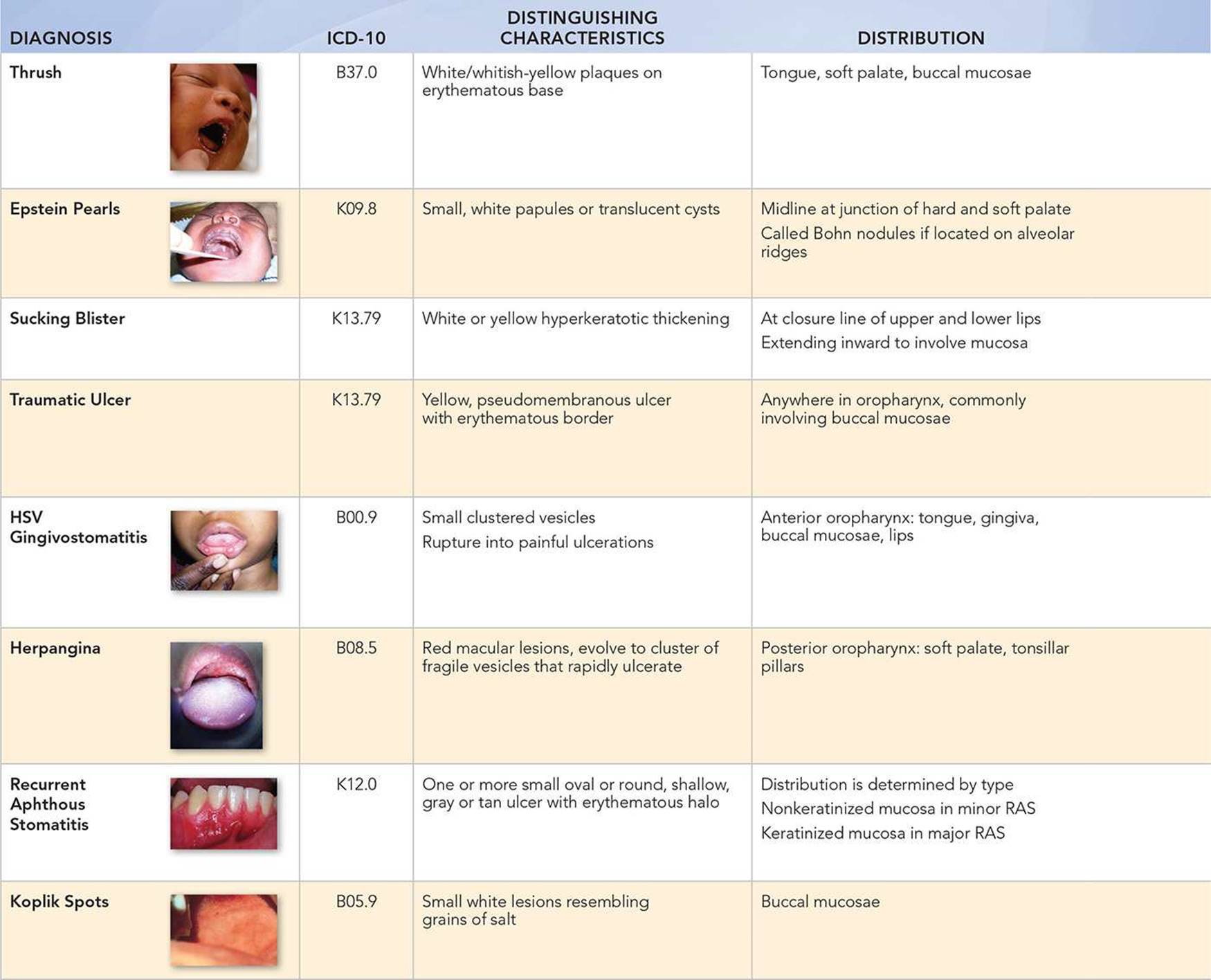
• Oral candidiasis is the most common oral fungal infection in infants and children, with Candida albicans being the most frequently identified species. When infections are persistent in children older than 6 months, the clinician should consider an underlying defect in the systemic immune system.
• Epstein pearls are asymptomatic and self-resolving, keratin-filled, epithelial-lined cysts that are extremely common in newborns.
• A sucking blister, also referred to as a sucking pad or callus, is a hyperkeratotic thickening at the closure line of the upper and lower lips caused by the mechanical effects of sucking, and may be white or pigmented. These lesions, found in newborns and young infants, usually disappear by 3 to 6 months of age.
• Traumatic oral lesions arise from mouthing objects in the period of infancy, and accidental biting or injury from objects placed in the oral cavity in older children. Pain is the predominant symptom, occurring 24 to 48 hours after the initial injury.
• Small gingival vesicles that progress into painful ulcerations following high fever, irritability, and malaise should prompt consideration of primary herpetic gingivostomatitis caused by herpes simplex virus (HSV) type 1 as a diagnosis.
• Herpangina also produces oral ulcerations and follows a prodrome that includes malaise, sore throat, and low-grade fever. It is caused by coxsackievirus group A, usually in the summer and early fall. The oral ulcerations are isolated in herpangina. When oral ulcerations occur in conjunction with palmar and plantar papulovesicles, hand, foot, and mouth disease should be strongly considered.
• Recurrent aphthous stomatitis (RAS) is the most common inflammatory ulcerative condition of the oral mucosa in patients in North America, with up to 20% of the population affected during childhood or early adulthood. Its cause is unknown. It is categorized into major and minor forms based on size and location of ulcers.
• Other viruses can cause enanthems, and should be considered in the diagnosis of oral lesions when prodromal symptoms are present. Examples include Koplik spots associated with measles, and ulcerations found in infectious mononucleosis and varicella.
Key Points in the Physical Examination
• Lesions of oral candidiasis include white or whitish-yellow plaques and erythema of the tongue, soft palate, or buccal mucosae. When plaques are scraped off, there is often underlying raw, erythematous mucosa, which may bleed. These mucosal changes may help the clinician differentiate the white plaques from milk residue seen on the tongue of infants.
• Epstein pearls, found in newborns, are white, nodular lesions typically found on the alveolar ridge or midline of the hard palate.
• Drooling and tenderness may be predominant features on physical examination when ulcerative lesions are present.
• Location of lesions can help differentiate between HSV gingivostomatitis and herpangina. HSV lesions are located in both the anterior and the posterior oropharynx, and lesions of herpangina are located predominantly in the posterior oropharynx, sparing the lips and gingiva.
• Both HSV and herpangina present with painful, small, grouped vesicles that eventually ulcerate. Gingival erythema, friability, and edema are commonly seen in HSV gingivostomatitis, but not in herpangina.
• RAS may produce ulcers of nonkeratinized mucosa (unattached gingiva) and keratinized surfaces. Lesions of major RAS are larger and can cause scarring.
• Koplik spots occur early in the course of measles, before other cutaneous signs, and are often missed.
|
PHOTOGRAPHS OF SELECTED DIAGNOSES |

Figure 24-1 Epstein pearls. Whitish cysts visible on the midline palate of a neonate. (Courtesy of Paul S. Matz, MD.)

Figure 24-2 Aphthous stomatitis. This tender lesion is visible on the gingival mucosa. (Courtesy of T.P. Croll, DDS.)
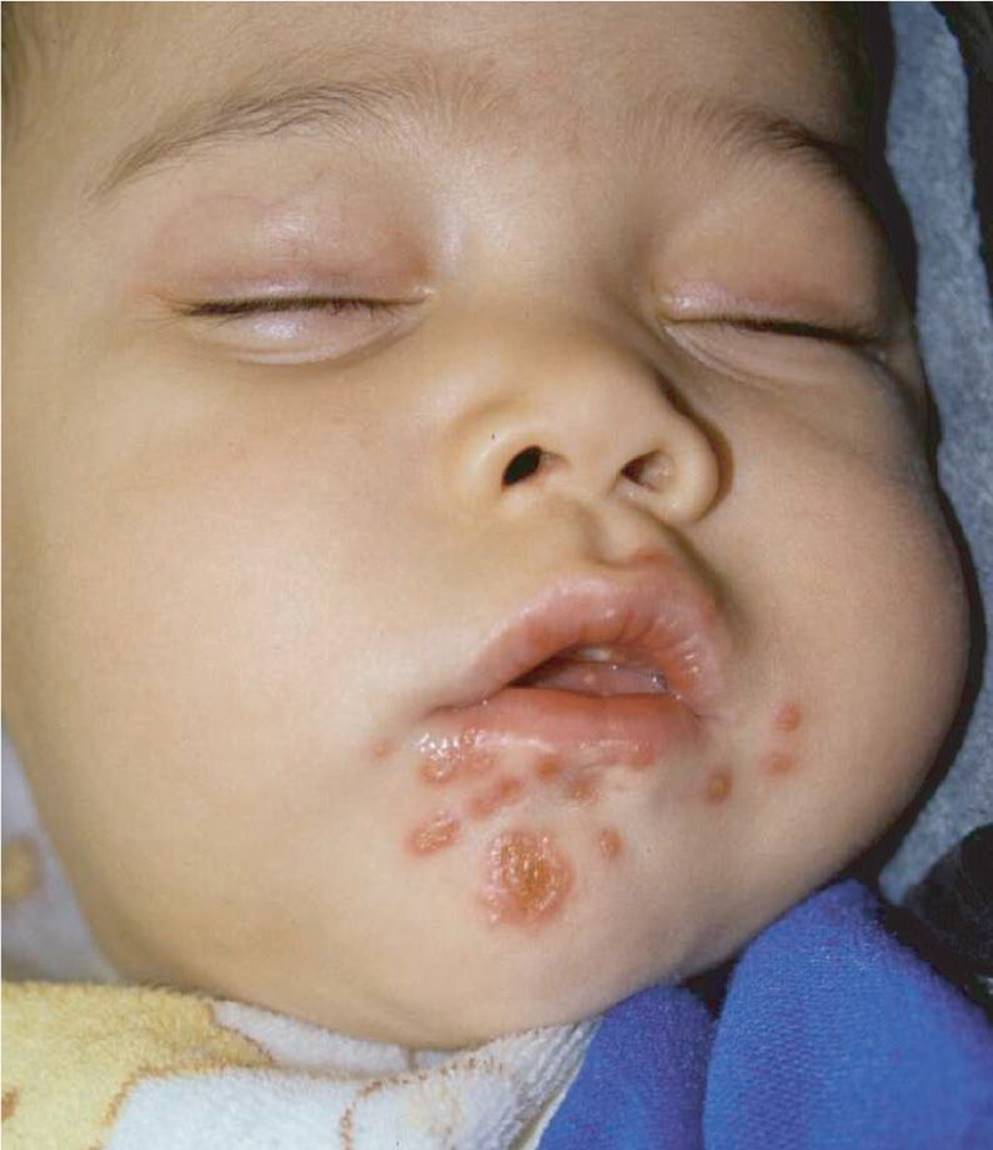
Figure 24-3 HSV stomatitis. An infant with extraoral HSV lesions. (Courtesy of George A. Datto, III, MD.)

Figure 24-4 HSV stomatitis. Lesions are visible on the tongue and labial mucosa. (Courtesy of Paul S. Matz, MD.)

Figure 24-5 Herpangina. Posterior pharyngeal lesions distinguish this illness from HSV gingivostomatitis. (Courtesy of Paul S. Matz, MD.)

Figure 24-6 Koplik spots. Fine, white spots with red halos seen on the buccal mucosa. (Used with permission from The Wellcome Trust, National Medical Slide Bank, London, UK.)

Figure 24-7 Oral thrush. An infant with numerous whitish tongue plaques. (Courtesy of Paul S. Matz, MD.)
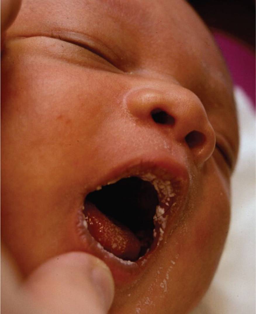
Figure 24-8 Oral thrush. Note the whitish plaques on the labial mucosa. (Courtesy of George A. Datto, III, MD.)
DIFFERENTIAL DIAGNOSIS

Other Diagnoses to Consider
• Mucous cysts (mucoceles, ranulas)
• Hemangiomas
• Intraoral abscess
• Leukoplakia
• Erythema multiforme
When to Consider Further Evaluation or Treatment
• Atypical presentation or unexpected clinical course of a common superficial ulcerative lesion should prompt further workup to investigate underlying immunologic or rheumatologic conditions.
• Ulcerative lesions with granulation may require biopsy by a specialist to verify the diagnosis based on clinical suspicion.
• Dental evaluation is indicated if dental trauma is suspected as an underlying cause of a mouth sore or ulceration.
• Severe pain or dehydration may require systemic therapy and intravenous fluids and warrants inpatient management.
SUGGESTED READINGS
Gonsalves WC, Chi AC, Neville BW. Common oral lesions: Part I. Superficial mucosal lesions. Am Fam Physician. 2007;75(4):501–506.
Hebert AA, Del Carmen Lopez M. Oral lesions in pediatric patients. Adv Dermatol. 1997;12:169–194.
Krol DM, Keels MA. Oral conditions. Pediatr Rev. 2007;28:15.
Patel NJ, Sciubba JS. Oral lesions in young children. Pediatr Clin N Am. 2003;50:469–486.
Witman PM, Rogers RS. Pediatric oral medicine. Dermatol Clin. 2003;21:157–170.