
Approach to the Problem
Visible discoloration of the teeth can be caused by extrinsic factors, which stain the outside of the tooth. Alternatively, discoloration can be caused by intrinsic changes in the tooth, which are triggered during or after tooth development. Because the inner dentin of a tooth is darker than the white enamel covering a tooth, pits caused by thinning of the enamel can appear as dark areas. Problems with dental health such as trauma, genetics, exposures to chemicals and antibiotics, systemic diseases, malnutrition, or poor dental care can all contribute to discoloration.
Key Points in the History
• The risk of developing dental caries depends on multiple overlapping factors. Caries risk is influenced by family history, diet and feeding style, dental hygiene, and fluoride use. The highest caries risk is seen in children with complex medical needs, those from disadvantaged families, and in babies who bottle feed through the night. Dental caries risk assessment and counseling should be part of early childhood care.
• Children’s vitamins, especially those containing iron, can lead to external tooth stains. External tooth staining can also be seen from cigarettes or other tobacco products, coffee, and red wine.
• Dental trauma can damage primary teeth and influence the development of preeruptive permanent teeth. Even mild trauma, depending on timing and location, can lead to significant changes in tooth appearance.
• Systemic fluoride given during tooth formation helps teeth develop strong enamel. Topical fluoride throughout life helps protect teeth by preventing and repairing damage to enamel. However, excessive fluoride exposure during tooth development can lead to fluorosis. Despite some cosmetic consequences, most degrees of fluorosis strengthen the teeth against caries. Common sources of fluoride exposure are from fluoridated municipal water, toothpastes and rinses (if swallowed), and from dental treatments. Fluoride exposure after tooth development, past age 8, will not cause fluorosis.
• Significant systemic illness can alter a tooth’s appearance. Children with cystic fibrosis or renal insufficiency, for example, may have mottled teeth or hypoplastic enamel.
• Stains occur from tetracycline antibiotics, including doxycycline, when they are incorporated into a tooth during development. These antibiotics will not stain fully formed permanent teeth. Pregnant women, and children younger than 8 years old, should avoid tetracyclines.
Key Points in the Physical Examination
• Milk bottle caries causes the most damage to the upper teeth and is associated with staining, caries, and destruction of the primary teeth.
• Most fluorosis causes easily overlooked subtle discoloration, including tiny white spots or streaks. Moderate to severe fluorosis affects the appearance of all teeth.
• Staining from iron vitamin drops is dark brown or black. It will not come off with routine toothbrushing, but can be removed by a dentist.
• White, opaque spots or patches can be caused by early caries or from altered mineralization related to illness, malnutrition, or trauma. They may also be idiopathic. These spots are sometimes referred to by the technically incorrect terms of hypocalcification or demineralization.
• Discoloration affecting all teeth is most likely related to systemic illness or exposures, and warrants thorough evaluation.
• Tetracycline staining may be brown or gray, and may affect the entire tooth. The staining is intrinsic to the tooth and cannot be buffed off. The best cosmetic result may be reached by covering the tooth with a veneer.
|
PHOTOGRAPHS OF SELECTED DIAGNOSES |
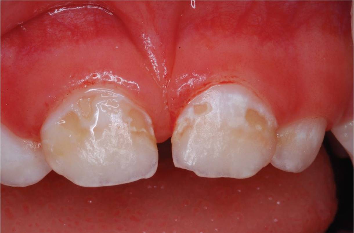
Figure 26-1 Dental caries. The upper incisors show significant caries and decalcification, along with gingivitis. (Courtesy of Theodore P. Croll, DDS.)
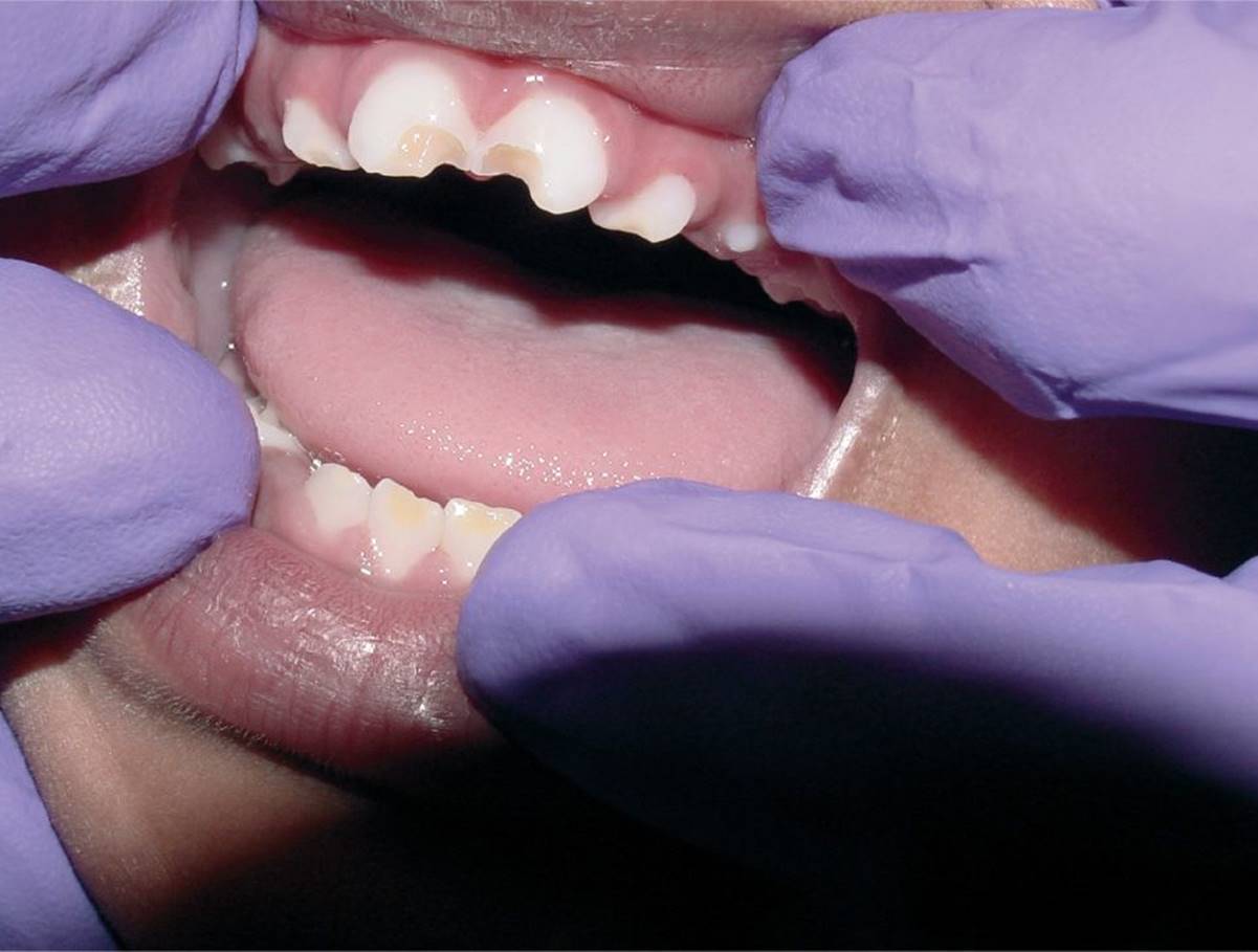
Figure 26-2 Moderate milk bottle caries. Brown discoloration and decay of the upper incisors. (Courtesy of Michael Lemper, DDS.)
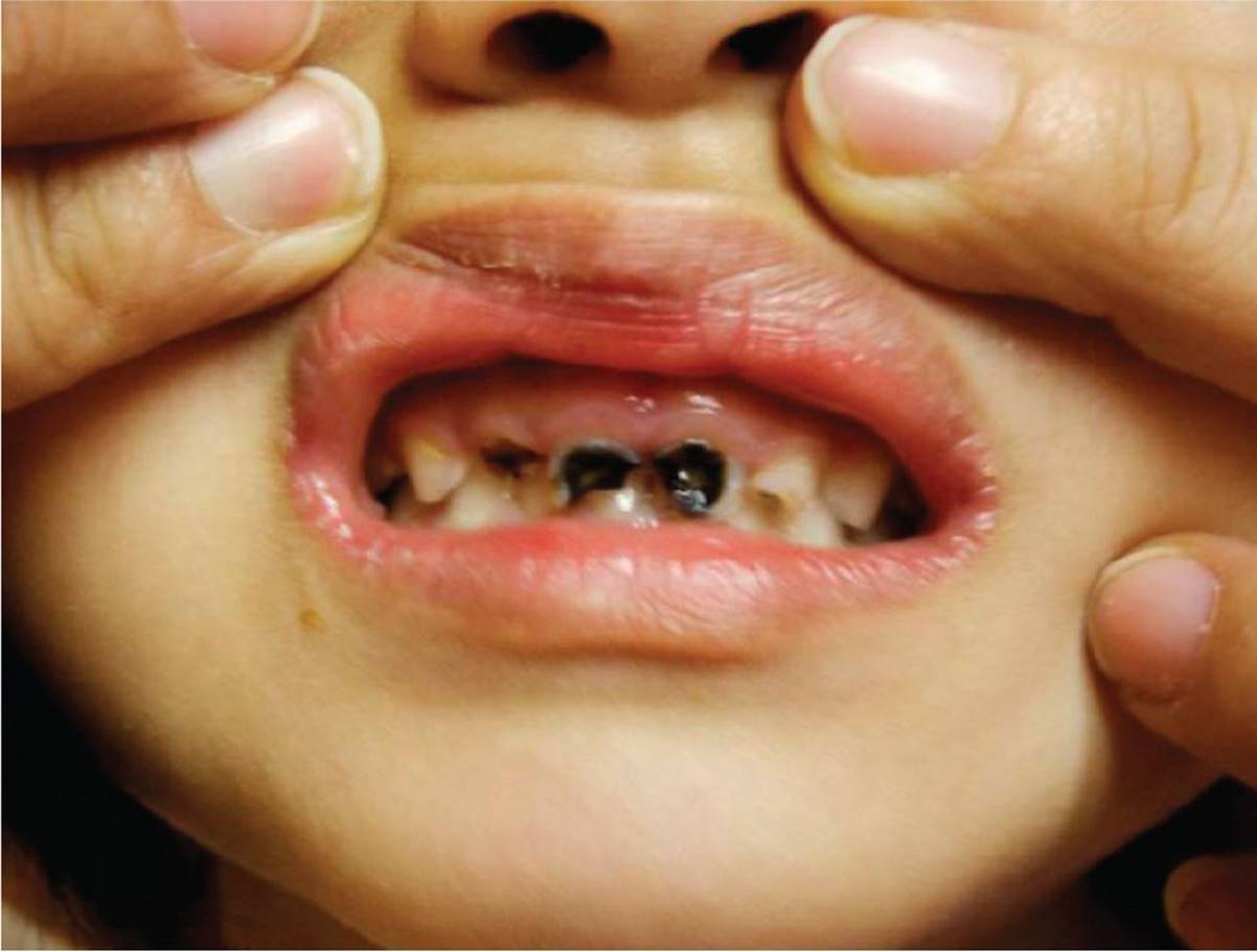
Figure 26-3 Severe milk bottle caries. Severe erosion of the upper incisors, associated with black staining. (Courtesy of Philip Siu, MD.)

Figure 26-4 Moderate fluorosis. All teeth are affected by staining from excessive systemic fluoride during tooth formation. (Courtesy of Theodore P. Croll, DDS.)
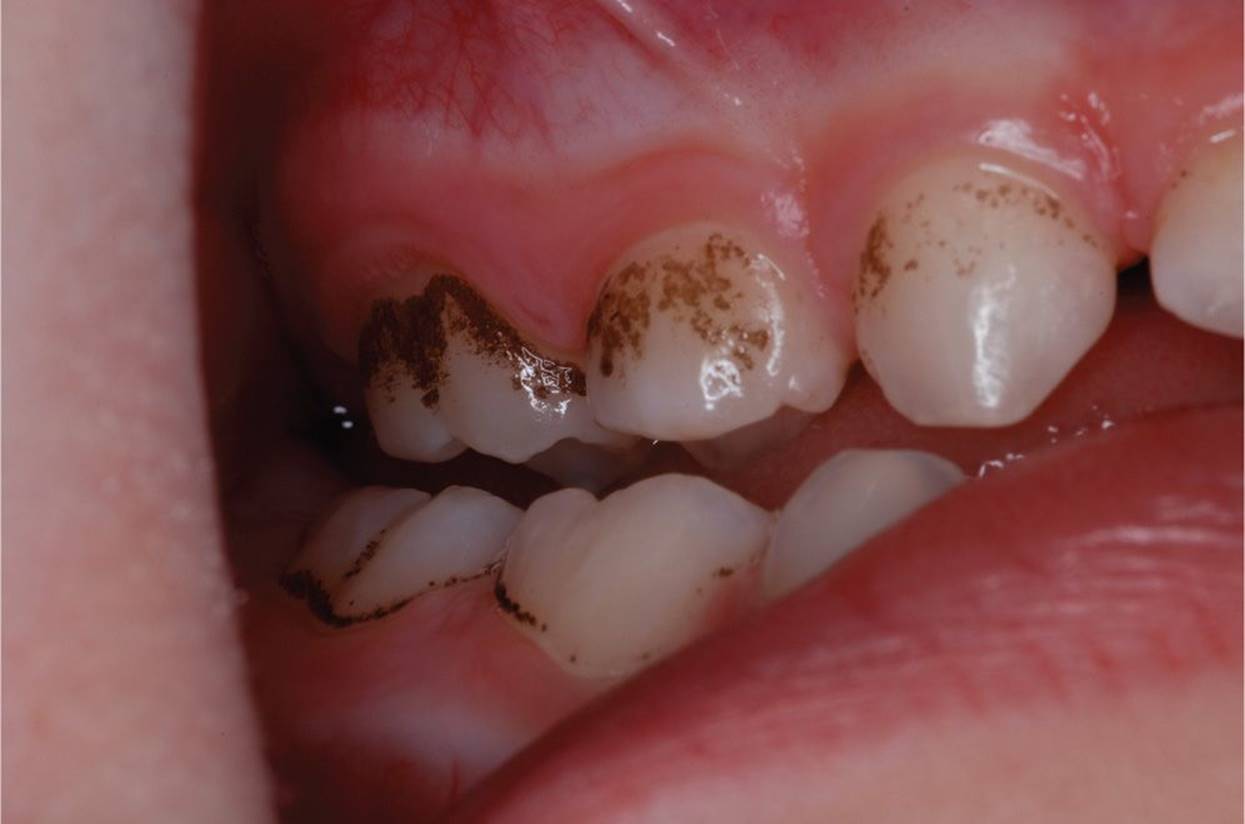
Figure 26-5 Iron staining. Iron solutions or multivitamin solutions that contain iron can stain teeth, as shown here most prominently near the gums. (Courtesy of Theodore P. Croll, DDS.)
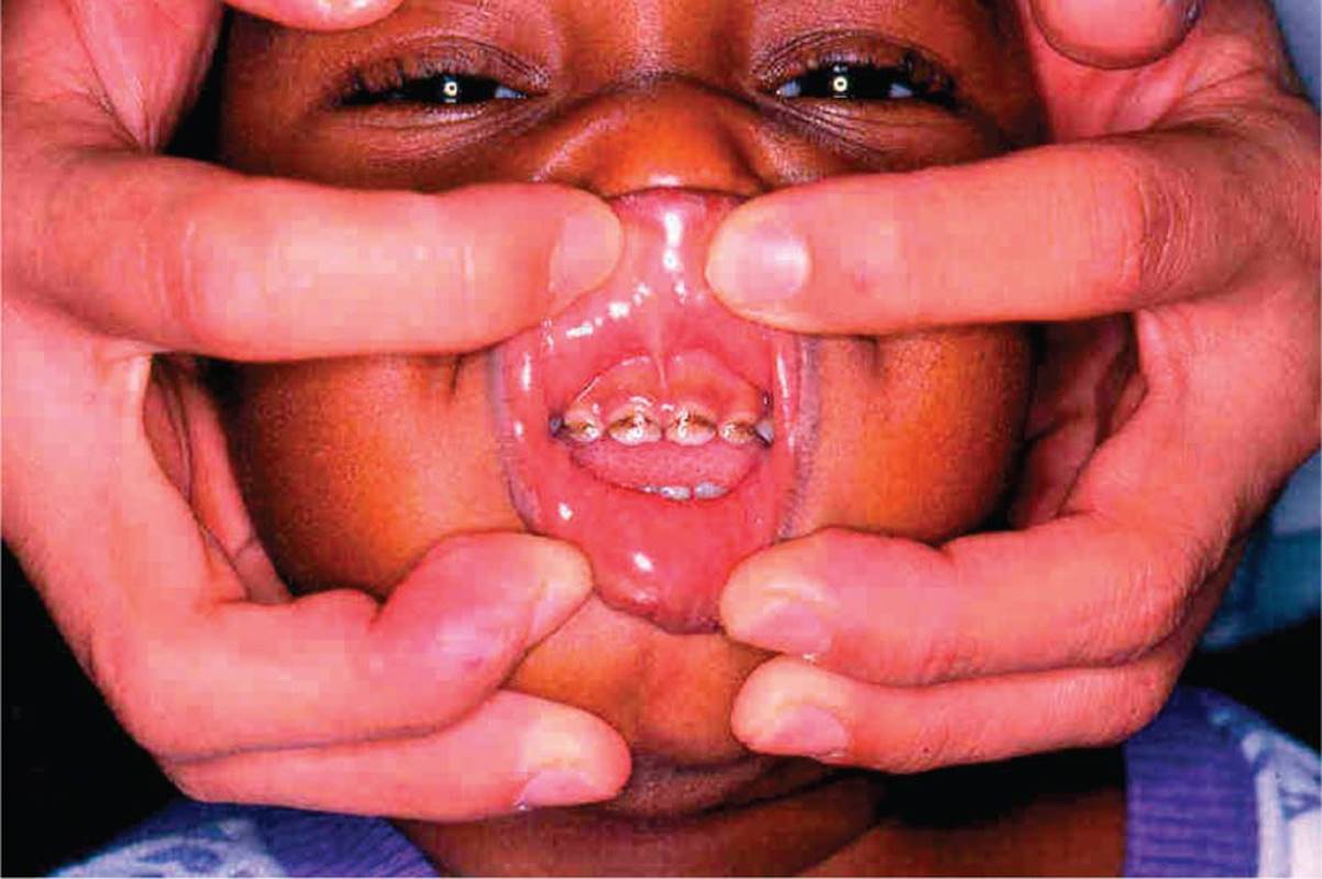
Figure 26-6 Tetracycline staining. Brown, band-like staining visible on all four maxillary incisors. (Courtesy of Jan Edwin Drutz, MD.)

Figure 26-7 Enamel hypoplasia. Brown dentin is visible through hypoplastic enamel of the upper incisors, in this case probably related to trauma affecting the developing tooth. (Courtesy of Theodore P. Croll, DDS.)
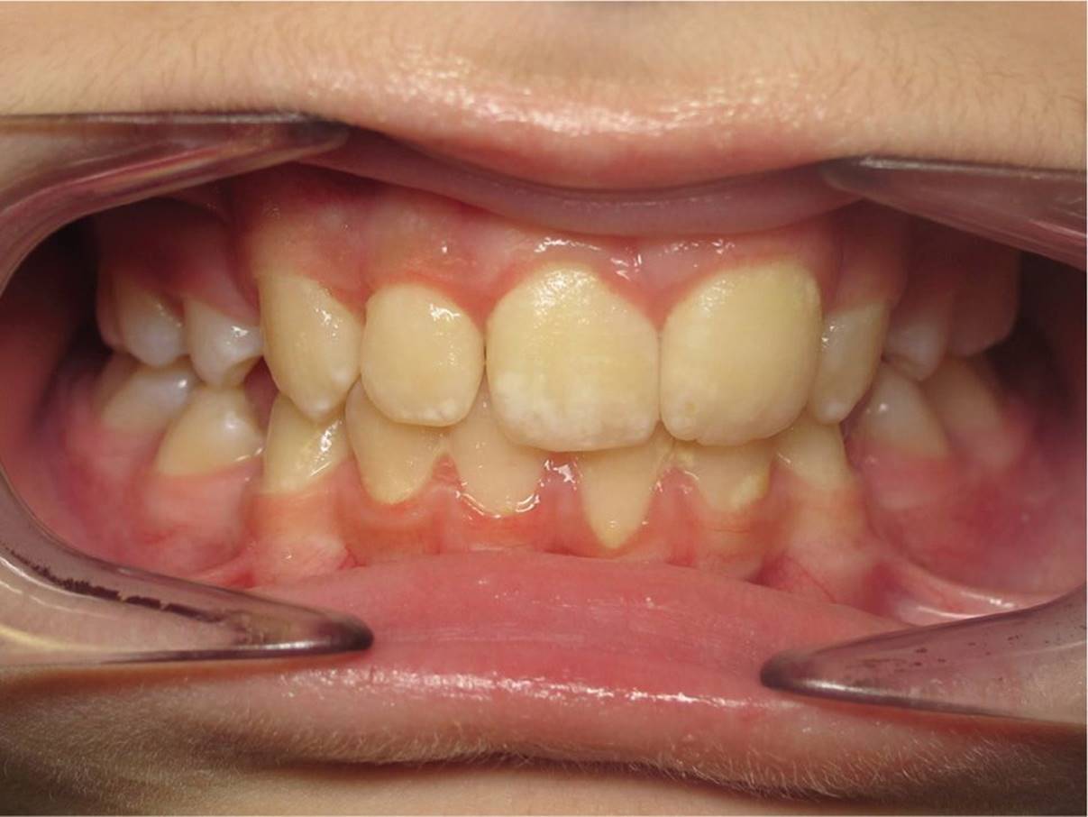
Figure 26-8 Hypocalcification. Caused by dysregulated mineralization that is often idiopathic, these white “chalk marks” are most prominent on the upper teeth. (Courtesy of Jason Kaplan, DDS, MS.)
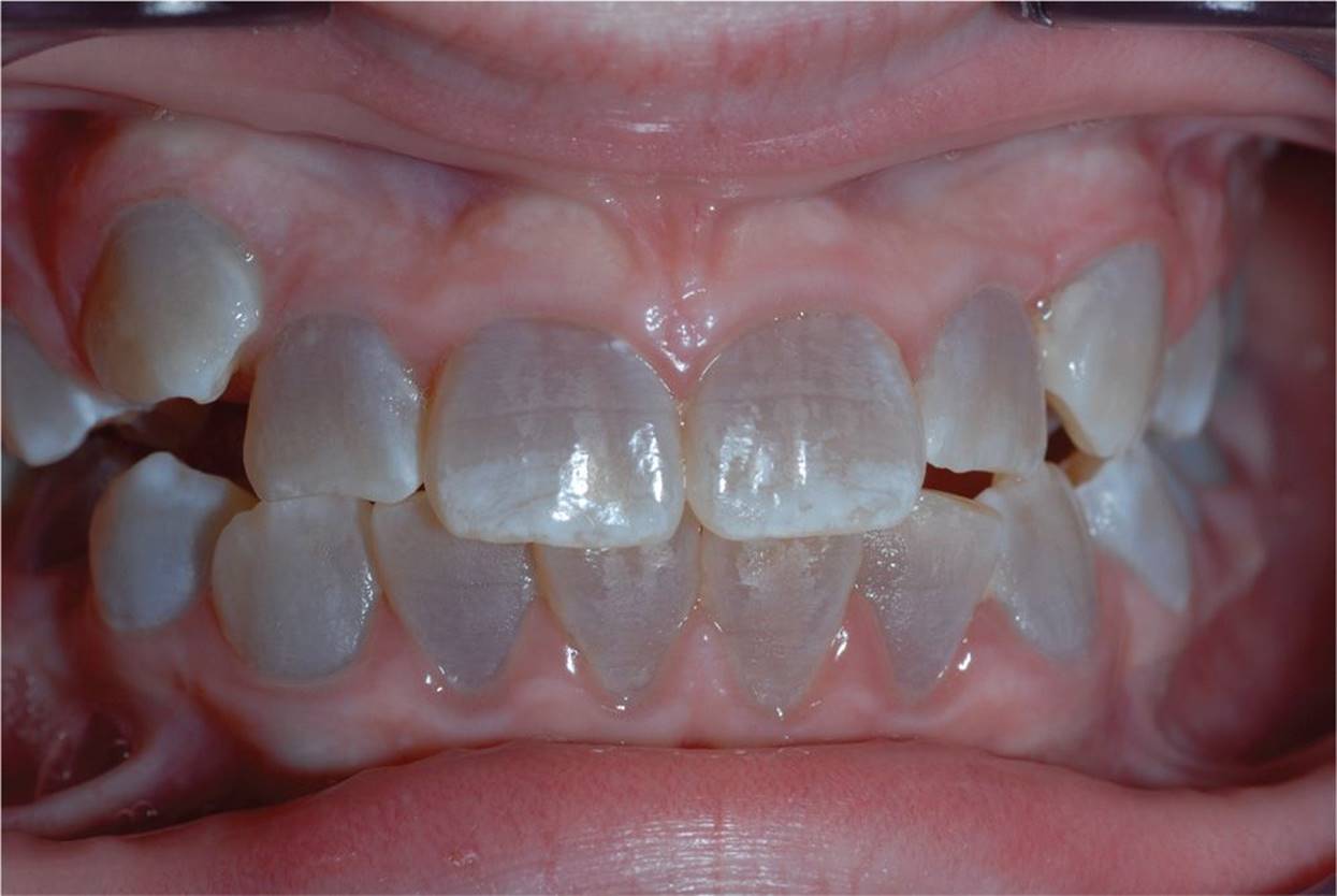
Figure 26-9 Dentinogenesis imperfecta. This genetic disorder of tooth development causes blue-gray or yellow-brown discoloration of all of the teeth. (Courtesy of Theodore P. Croll, DDS.)
DIFFERENTIAL DIAGNOSIS
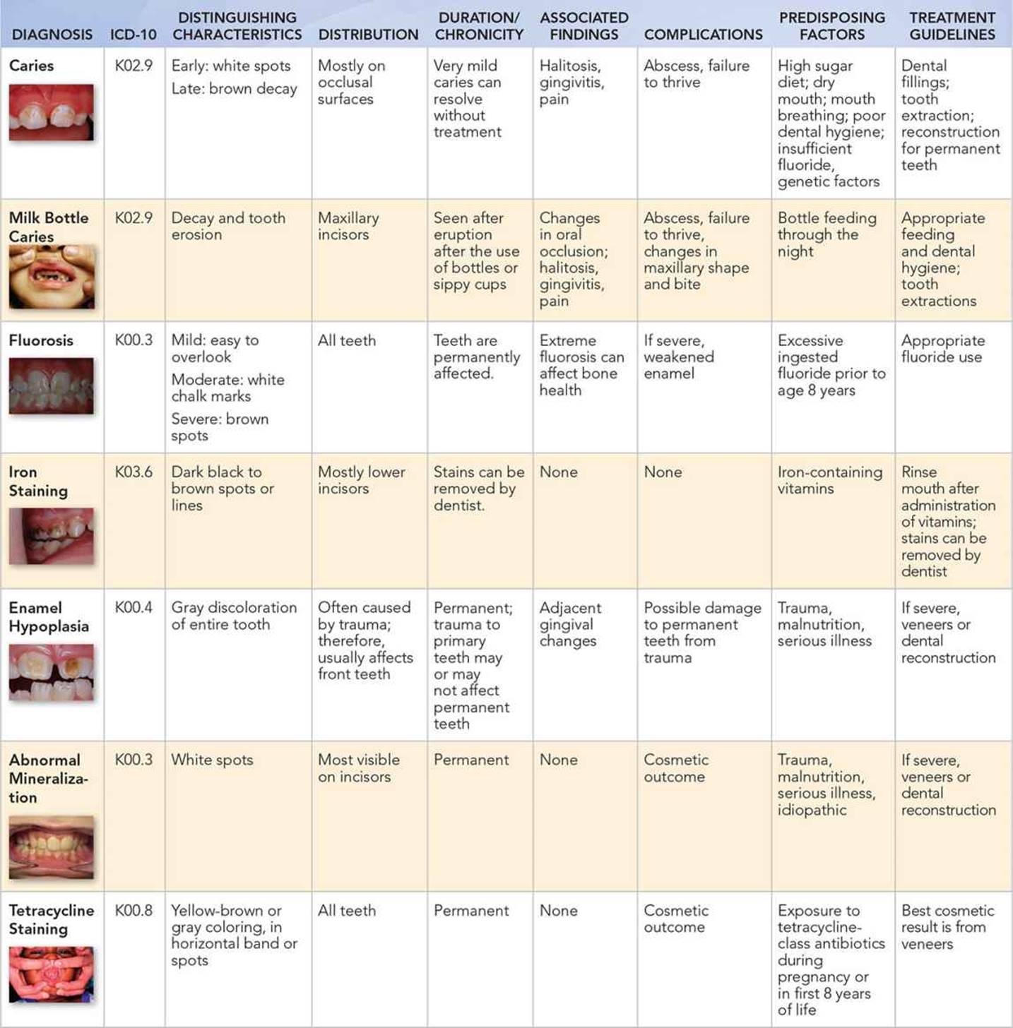
Other Diagnoses to Consider
• Rickets, parathyroid disorders, and other disorders of calcium and vitamin D metabolism
• Kidney failure
• Cystic fibrosis
• Ectodermal dysplasias
• Disorders of tooth development, including amelogenesis imperfecta and dentinogenesis imperfecta
When to Consider Further Evaluation or Treatment
• Even without staining, the American Dental Association and the American Academy of Pediatrics recommend children establish a dental home at age 12 months.
• Significant dental disease can contribute to malnutrition and a poor quality of life. Children, particularly those with multiple risk factors, may need early referral to prevent the progression of dental disease.
• Dental findings may be a clue to serious, systemic illness, especially when accompanied by other signs and symptoms. For instance, children with malabsorption syndromes may present with abdominal complaints and poor growth in addition to poorly mineralized or discolored teeth. Other illnesses can increase the risk of caries, including diabetes and Sjogren syndrome.
SUGGESTED READINGS
Croll T. The ASDC Kid’s Mouth Book. Chicago, IL: The American Society of Dentistry for Children; 1999:144.
Norwood KW, Slayton RL. Oral health care for children with developmental disabilities. Pediatrics. 2013;131:614–619.
Schafer TE, Adair SM. Prevention of dental disease: The role of the pediatrician. Pediatric Clin N A. 2000;47:1021–1042.
Section on Pediatric Dentistry and Oral Health. Preventive oral health intervention for pediatricians. Pediatrics. 2008;122(6):1387–1394.