
Approach to the Problem
It is important to consider the age of the patient and his or her stage of pubertal development when assessing the potential cause of breast enlargement, as it is common for an individual’s breasts to develop at different rates. Disorders involving breast swelling most often present during the pubertal years, although they may occur in infancy and early childhood. Many patients may be uncomfortable disclosing breast concerns during an initial patient history; therefore, related abnormalities may be only coincidentally identified during routine physical examination. A breast examination should be performed as part of every well child care visit.
Some of the most common breast concerns, such as breast asymmetry and gynecomastia, may not be true diseases but rather normal physiological variants. Breast enlargement and swelling may also be congenital, infectious, or hormonal in etiology. In male patients, the most common cause of breast tissue development is benign physiologic gynecomastia.
Key Points in the History
• Breast tissue development occurring in females less than 8 years of age is generally considered to be abnormal, although there may be racial or ethnic variations noted.
• Benign gynecomastia often begins as unilateral breast enlargement and involves the right side twice as often as the left.
• Breast tissue in gynecomastia tends to be tender and/or painful soon after the onset of breast enlargement.
• Development of gynecomastia either before puberty begins or after puberty is completed would not be consistent with benign physiologic gynecomastia.
• In a teenager with gynecomastia, the history should include a list of any medications or recreational drugs that the patient might be using, as drugs may be the cause of gynecomastia. Some drugs associated with gynecomastia include anabolic steroids, marijuana, heroin, isoniazid (INH), metronidazole, dilantin, ketoconazole, ranitidine, and omeprazole.
• Rapid, painful growth of breast tissue in females in early adolescence is seen with a diagnosis of juvenile breast hypertrophy.
• A history of significant chest wall trauma in the prepubertal female may predispose to underdevelopment of the breast on the side of the trauma and thus contribute to breast asymmetry.
• A history of breastfeeding, nipple piercing, or breast trauma often precedes the development of a breast infection.
Key Points in the Physical Examination
• There is considerable variability in the size, shape, and consistency of breasts among individuals.
• An individual’s breasts may develop at different rates, which can result in a different Tanner stage for each breast.
• Although breast asymmetry may be a normal physiological variant, a physical exam ination that includes inspection and palpation should be done to rule out a mass lesion in the larger breast.
• Erythema, warmth, and tenderness of the breast would support the diagnosis of an infectious process, such as mastitis or breast abscess.
• In the evaluation of premature thelarche, the patient must be examined for other signs of pubertal development, such as the presence of pubic hair, axillary hair, or a growth spurt.
• In an overweight patient, fatty tissue may be confused with breast development. A physical examination that includes palpation is necessary to determine whether breast tissue is present.
• Physiologic gynecomastia in males, if it occurs, usually presents when a male is in Tanner stages II–III for pubic hair and genitalia.
|
PHOTOGRAPHS OF SELECTED DIAGNOSES |

Figure 33-1 Breast asymmetry. Adolescent female with Tanner stage III breast development and significantly asymmetric breasts. (Courtesy of Mary L. Brandt, MD.)

Figure 33-2 Breast asymmetry. Note the left breast is smaller, although with a normal appearance. (Courtesy of Jeff Friedman, MD.)

Figure 33-3 Gynecomastia. Thirteen-year-old boy with gynecomastia Tanner stage II breasts. (Courtesy of D’Juanna White-Satcher, MD, MPH.)

Figure 33-4 Gynecomastia. Fourteen-year-old boy with cosmetically significant benign gynecomastia—both breasts in Tanner stage III. (Courtesy of Lior Heller, MD.)
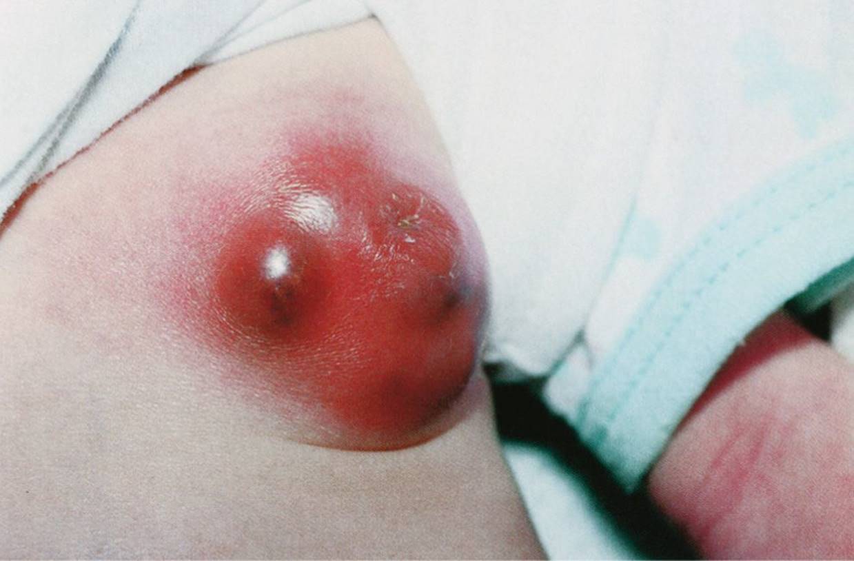
Figure 33-5 Mastitis. Ten-day-old female infant with swelling and erythema of the left breast. (Used with permission from Fleisher GR, Ludwig S, Baskin MN, et al. Pictorial Review of Pediatrics. Baltimore, MD: Lippincott Williams & Wilkins; 1998:185.)

Figure 33-6 Neonatal breast hypertrophy. Two-week-old female infant with right breast hypertrophy. (Courtesy of D’Juanna White-Satcher, MD, MPH.)

Figure 33-7 Tuberous breast. Note hypoplasia and sagging of breast tissue at chest wall on right breast. (Courtesy of David A. Horvath, MD.)
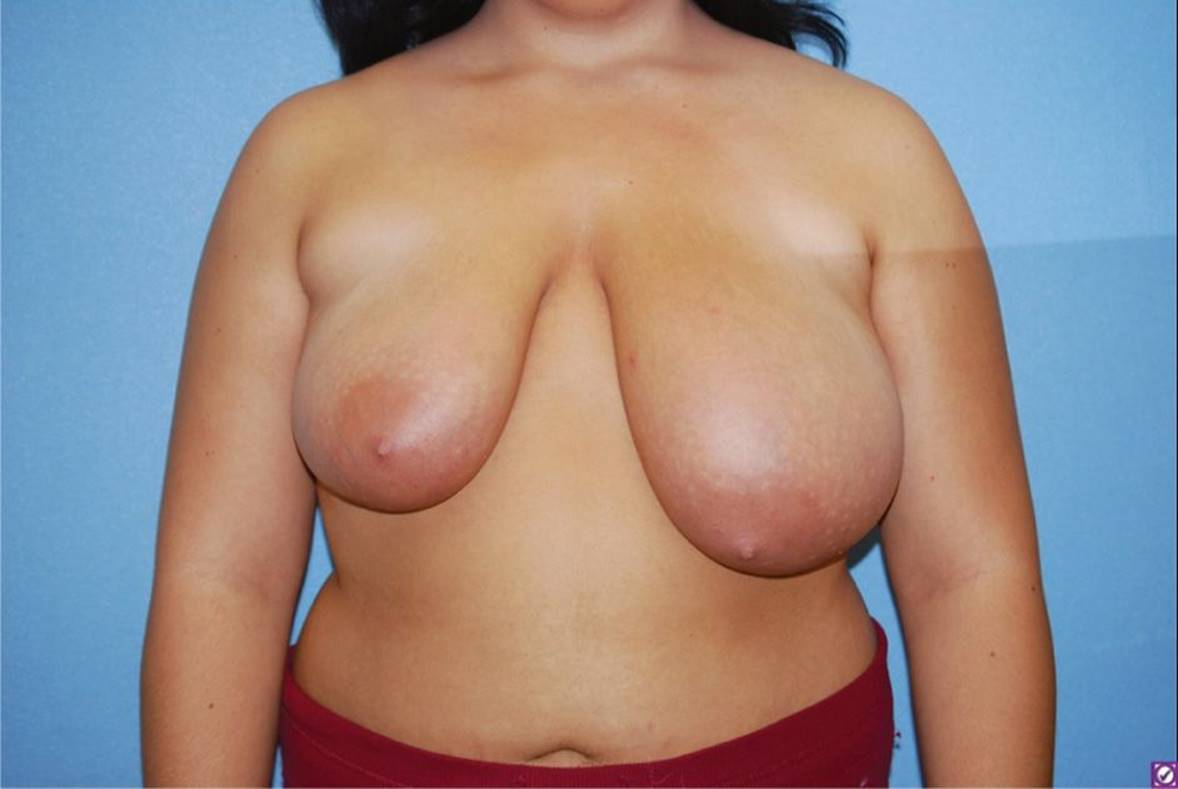
Figure 33-8 Juvenile breast hypertrophy. Note pendulous left breast in this 13-year-old girl. Also, the patient presents with asymmetric breasts. (Courtesy of Jeff Friedman, MD.)
DIFFERENTIAL DIAGNOSIS
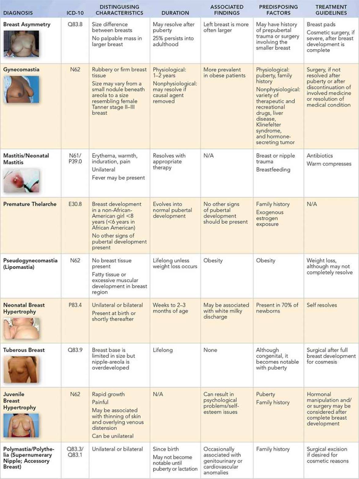
Other Diagnoses to Consider
• Fibroadenoma
• Trauma
• Foreign body
• Breast cyst
• McCune–Albright syndrome
• Klinefelter syndrome
• Neoplastic disorders
When to Consider Further Evaluation or Treatment
• If surgical intervention is being considered, it should be delayed until breast development is complete, usually after 18 years of age.
• Patients with breast tissue enlargement greater than 4 cm before the onset of puberty should be evaluated for potential underlying pathology.
• In a patient with isolated premature thelarche, one should consider obtaining a bone age, especially if a growth spurt has been noted.
• In a patient with premature thelarche who has other secondary signs of sexual maturation, further evaluation for precocious puberty is recommended.
SUGGESTED READINGS
Diamantopoulos S, Bao Y. Gynecomastia and premature thelarche: a guide for practitioners. Pediatr Rev. 2007;28(9):e57–e66.
DiVastas A, Weldon C, Labow B. The breast: examination and lesions. In: Emana SJ, Laufer MR, Goldstein DP, eds. Pediatric and Adolescent Gynecology. 6th ed. Philadelphia, PA: Lippincott Williams & Wilkins; 2011:405–419.
Greydanus D, Matytsina L, Gains M. Breast disorders in children and adolescents. Prim Care Clin Office Pract. 2006;33(2):455–502.
Macdonald HR. Breast disorders. In: Neinstein LS, ed. Adolescent Health Care: A Practical Guide. 5th ed. Philadelphia, PA: Lippincott Williams & Wilkins; 2007:754–763.