
Approach to the Problem
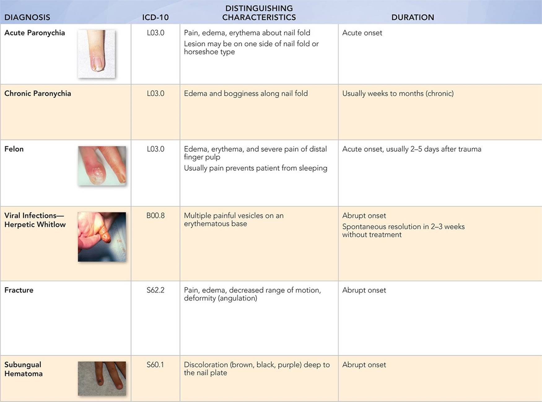
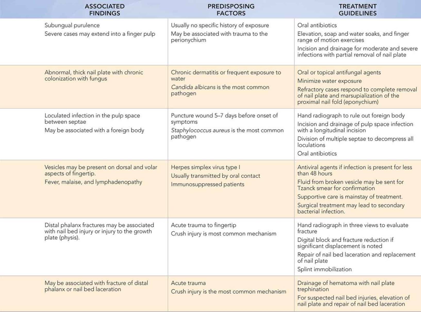
Fingertip swelling in a child is usually a consequence of trauma or infection. Traumatic injuries with a crush mechanism (e.g., digit crushed between door and door frame) are common in toddlers. These injuries may damage the fingertip soft tissues (nail fold, pulp), distal phalanx, and nail bed. Infections of the distal digit may be precipitated by trauma (felon) or occur spontaneously (paronychia) and may be caused by bacteria, viruses, or fungi.
Key Points in the History
• Crush injury is the most common etiology of fingertip trauma.
• Penetrating injuries from splinters, shards of glass, or minor puncture wounds may lead to an abscess of the distal finger pulp (felon).
• Thumb sucking or nail biting habits may predispose to infections (paronychia).
• Herpetic whitlow may occur as a complication of oral lesions due to herpes simplex virus.
Key Points in the Physical Examination
• Severe pain and multiple clear vesicles on an erythematous base are suggestive of a herpetic whitlow.
• Pain, erythema, and globular swelling of the distal pulp are consistent with a felon.
• Angulation deformity of the distal phalanx because of a fracture may be best detected by examining the finger flexion cascade, or flexion of all the digits together.
|
PHOTOGRAPHS OF SELECTED DIAGNOSES |

Figure 44-1 The perionychium and its associated structures. From Hunt TR, Wiesel SW. Operative Techniques in Hand, Wrist, and Forearm Surgery. 1st ed. Philadelphia, PA: Lippincott Williams & Wilkins; 2010.

Figure 44-2 Acute paronychia. Visible swelling, erythema, and discharge along the nail fold, yet the nail itself is intact. (Courtesy of Mary L. Brandt, MD.)
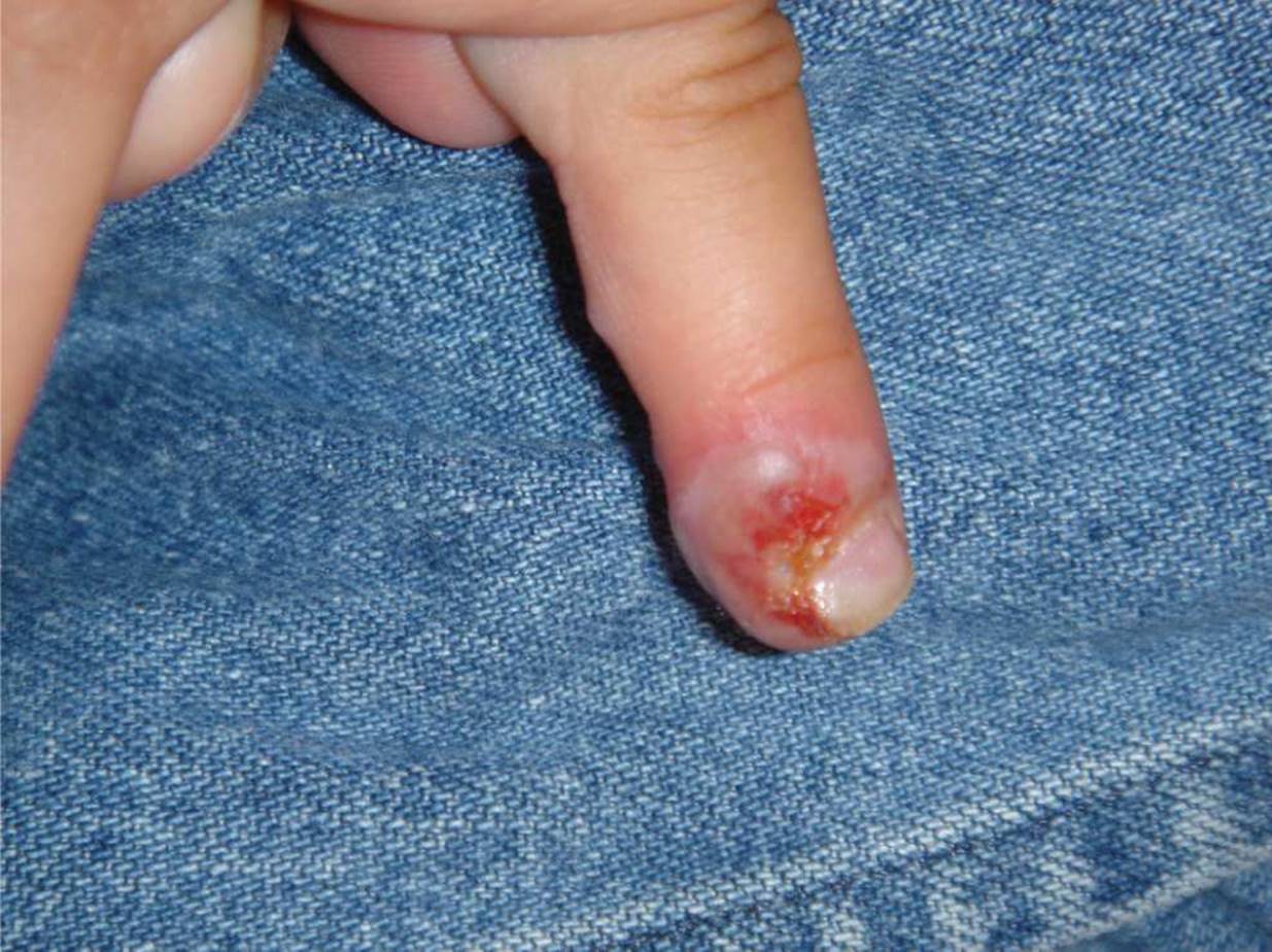
Figure 44-3 Acute paronychia. Swelling, erythema, and discharge along the lateral nail edge. (Courtesy of Larry H. Hollier, Jr, MD, FACS.)
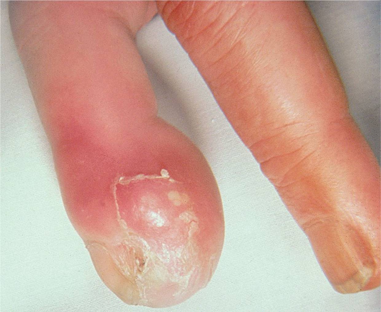
Figure 44-4 Felon. (Used with permission from Greenberg MI. Greenberg’s Atlas of Emergency Medicine. Philadelphia, PA: Lippincott Williams & Wilkins; 2005:458.)
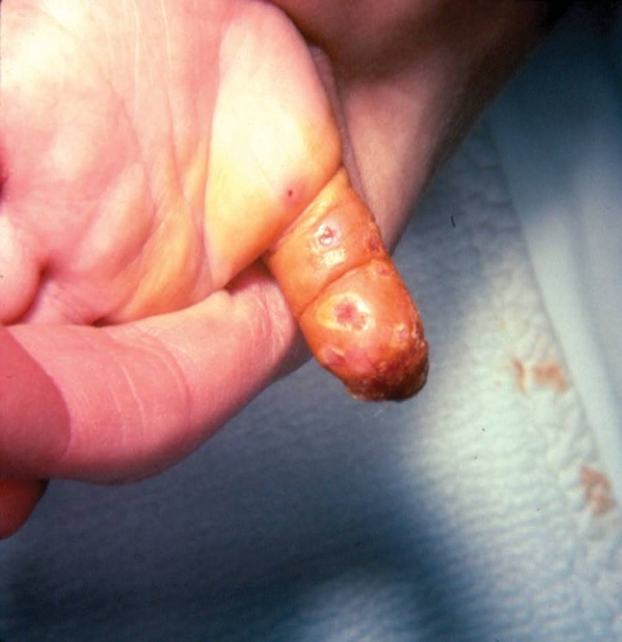
Figure 44-5 Herpetic whitlow. Ulcers where previously there were vesicles along the ventral thumb. (Courtesy of Mark A. Ward, MD.)
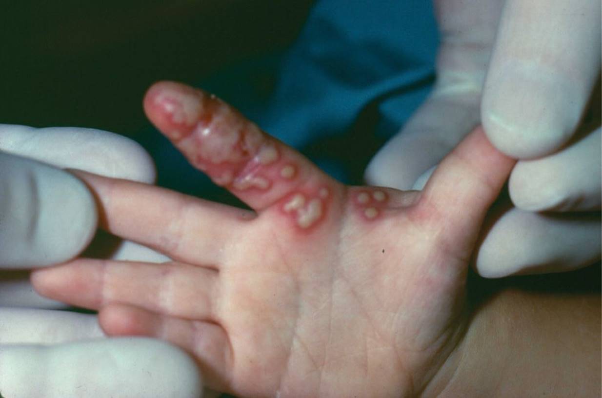
Figure 44-6 Herpetic whitlow. Grouped vesicles on an erythematous base along the ventral surface of the finger. (Courtesy of Mark A. Ward, MD.)

Figure 44-7 Subungual hematoma. A 2-year-old child with a subungual hematoma that resulted from a fall with a plate in his hand. (Courtesy of Julie A. Boom, MD.)
DIFFERENTIAL DIAGNOSIS
Other Diagnoses to Consider
• Subungual melanoma
• Ligamentous injury (sprain) to the distal interphalangeal joint
• Atypical mycobacterial (Mycobacterium marinum) or fungal infections
• Flexor tendon sheath infections (tenosynovitis)
• Extensor tendon injury (mallet finger)
• Flexor tendon injury (jersey finger)
When to Consider Further Evaluation or Treatment
• Distal phalanx fractures that are associated with nail bed lacerations, suspected injury to the growth plate, extensor tendon injuries (mallet finger), or displacement should prompt a referral to a pediatric hand surgeon.
• Acute paronychia that does not resolve with a treatment course of oral antibiotics may require formal incision and drainage of the nail fold.
• Consider atypical organisms such as M. marinum for infections that do not resolve with standard surgical and antimicrobial treatments.
SUGGESTED READINGS
Clark DC. Common acute hand infections. Am Fam Physician. 2003;68(11):2167–2176.
Doraiswamy NV, Baig H. Isolated finger injuries in children—incidence and aetiology. Injury. 2000;31(8):571–573.
Hart RG, Kleinert HE. Fingertip and nail bed injuries. Emerg Med Clin North Am. 1993;11(3):755–765.
Ljungberg E, Rosberg HE, Dahlin LB. Hand injuries in young children. J Hand Surg. 2003;28B(4):376–380.
Rockwell PG. Acute and chronic paronychia. Am Fam Physician. 2001;63(6):1113–1116.
Shmerling RH. Finger pain. Prim Care. 1988;15(4):751–766.
Zitelli BJ, Davis HW. Atlas of Pediatric Physical Diagnosis. 4th ed. Philadelphia, PA: Mosby; 2002.