
Approach to the Problem
Appreciation of the differences between the lower extremities of the normal newborn as compared with those of the older child is important in the detection of abnormalities. When compared with older children, normal newborns have greater mobility of the hip, varus alignment of the knee, flatter feet, greater ankle range of motion, and less defined bony prominences.
Lower extremity abnormalities can involve the hip, leg, foot and/or toes and can be positional or structural. While many abnormalities are isolated, some occur with other lower extremity abnormalities or in association with congenital disorders such as myelomeningocele or a congenital myopathy.
It is helpful to think about lower extremity abnormalities as intrinsic or extrinsic. Intrinsic abnormalities are due to characteristics of the infant such as genetic conditions like achondroplasia or osteogenesis imperfecta. Conversely, extrinsic abnormalities are due to in utero conditions such as breech positioning that limit fetal movement, a critical contributor to proper musculoskeletal development. Intrinsic abnormalities are more likely to be due to underlying pathology, whereas most extrinsic abnormalities are positional and resolve spontaneously or respond to surgical correction.
Key Points in the History
• Female gender, breech positioning, and family history of developmental dysplasia of the hip (DDH) are significant risk factors for DDH.
• 80% of cases of DDH occur in females.
• DDH is more common in first-born infants owing to the relative inelasticity of the primigravid uterus and abdominal wall.
• Native Americans and Laplanders are the ethnic groups at highest risk for DDH, with an incidence of up to 50 in 1,000 live births.
• History of diminished fetal movement for significant periods of time as reported by the mother may be associated with lower extremity abnormalities caused by intrauterine mechanical factors or intrinsic disease of the fetus such as a myopathy, chromosomal abnormality, or myelomeningocele.
• Congenital talipes equinovarus (clubfoot) occurs two to four times more often in males than in females.
• Maternal smoking during pregnancy is a significant risk factor for clubfoot.
• Though polydactyly is most likely to be an isolated trait, it can be inherited in an autosomal dominant manner with variable penetrance.
• Some teratogenic medications taken during pregnancy such as warfarin, methotrexate, and thalidomide are associated with limb anomalies.
Key Points in the Physical Examination
• A positive exam for DDH is the palpable “clunk” elicited by the Barlow maneuver (detects the hip subluxing or dislocating from the acetabulum) or Ortolani maneuver (detects reduction of the subluxed or dislocated hip).
• Benign, high-pitched soft tissue hip “clicks” are common and should not be confused with true clunks.
• Asymmetry of the inguinal, gluteal, and posterior thigh creases, although nonspecific, is common in DDH. Up to 20% to 30% of normal newborns may have thigh crease asymmetry.
• The malposition seen with metatarsus varus (metatarsus adductus) and positional calcaneovalgus foot are most often correctable with passive motion.
• Metatarsus adductus is distinguished from skewfoot (angled flat foot) by the ability to palpate the head of the talus in the arch of the skewfoot.
• The malposition in talipes equinovarus (clubfoot) is fixed and cannot be passively corrected.
|
PHOTOGRAPHS OF SELECTED DIAGNOSES |

Figure 5-1 Developmental dysplasia of the hip. Asymmetry of skin folds are common findings in DDH. (Used with permission from Anatomical Chart Co.)

Figure 5-2 Hip flexion contracture. A newborn presents with bilateral hip flexion contracture after breech presentation. (Courtesy of Gerardo Cabrera-Meza, MD.)

Figure 5-3 Physiologic bowing of the legs. Normal newborn knee alignment is 10–15 degrees varus, creating the bowlegged appearance. (Courtesy of Gerardo Cabrera-Meza, MD.)

Figure 5-4 Talipes calcaneovalgus. The right foot is hyperdorsiflexed with the dorsal surface contacting the anterior tibial surface. (Courtesy of Gerardo Cabrera-Meza, MD.)
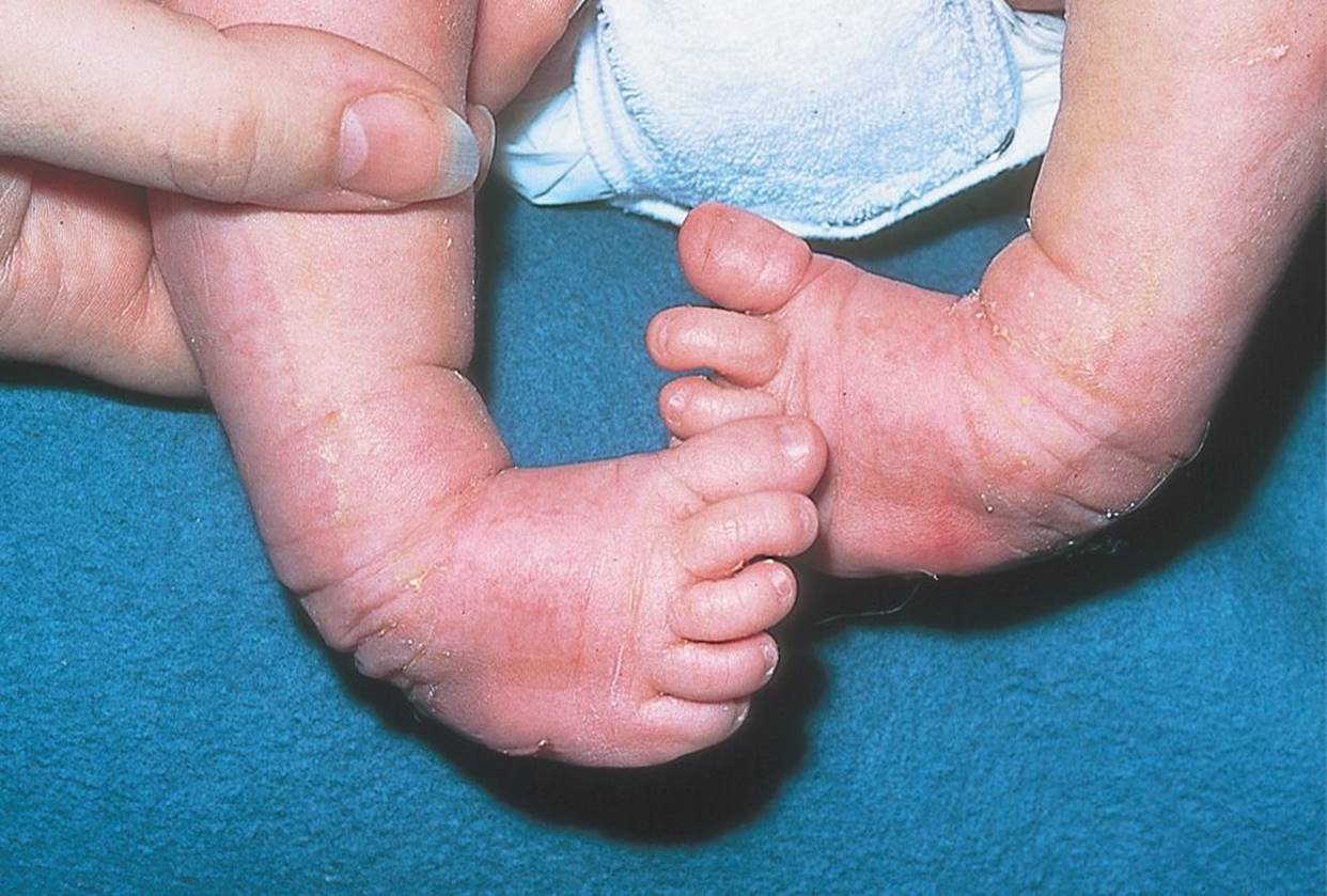
Figure 5-5 Talipes equinovarus (clubfoot). The heel is varus, and the forefoot is adducted and inverted. (Used with permission from Jim Stevenson/SPL/Science Source/Photo Researchers.)

Figure 5-6 Metatarsus adductus. (Used with permission from Frank J Frassica, Paul D Sponseller, John H Wilckens. 5-Minute Orthopaedic Consult. 2nd edition. Philadelphia, PA: Lippincott Williams & Wilkins; 2006.)

Figure 5-7 Congenital vertical talus (rocker bottom foot). The forefoot is dorsiflexed and abducted with a convex plantar surface. (Courtesy of Gerardo Cabrera-Meza, MD.)
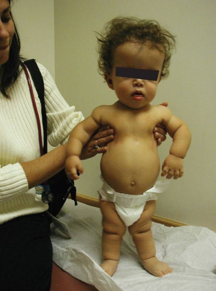
Figure 5-8 Achondroplasia. This infant with achondroplasia has tibial bowing, frontal bossing, rhizomelia (the proximal limb segment is shorter than the distal segment), and brachydactyly (short fingers). (Courtesy of Paul S. Matz, MD.)
DIFFERENTIAL DIAGNOSIS


Other Diagnoses to Consider
• Arthrogryposis multiplex l
• Congenital myopathies (most commonly nemaline myopathy)
• Fibular hemimelia
• Klippel–Trenaunay–Weber syndrome
• Skeletal dysplasias (most commonly osteogenesis imperfecta and achondroplasia)
When to Consider Further Evaluation or Treatment
• Any infant with a positive exam for DDH (i.e., clunk on Barlow or Ortolani maneuver) should be referred directly to a pediatric orthopedic surgeon.
• All breech-born females without a positive exam should be referred for hip ultrasonography at 6 weeks of age in view of the high incidence of DDH and variable physical exam sensitivity.
• Any leg-length discrepancy should be further evaluated with plain radiography (at or after 4 months of age) for underlying cause and qualitative bone abnormalities.
• Talipes calcaneovalgus that cannot be passively flexed to neutral should be imaged with plain radiography to exclude congenital vertical talus (rocker bottom foot) and then referred for casting.
• Metatarsus adductus that cannot be passively corrected to neutral (i.e., inflexible metatarsus adductus) should be referred to a pediatric orthopedic surgeon for serial casting.
• All patients with rocker bottom feet should undergo MRI of the brain and spine to evaluate for possible underlying neuromuscular disorder.
SUGGESTED READINGS
American Academy of Pediatrics, Committee on Quality Improvement, Subcommittee on Developmental Dysplasia of the Hip. Clinical practice guideline: Early detection of developmental dysplasia of the hip. Pediatrics. 2000;105(4 Pt 1):896–905.
Furdon SA and Donlon CR. Examination of the newborn foot: Positional and structural abnormalities. Adv Neonatal Care. 2002;2(5):248–258.
Gore AI and Spencer JP. The newborn foot. Am. Fam. Physician. 2004;69(4):865–872.
Kasser JR. The foot. Lovell & Winter’s Pediatric Orthopaedics. 6th ed. Vol 2. Philadelphia, PA: Lippincott Williams & Wilkins; 2006:1258–1328.
Schoenecker PL and Rich MM. The lower extremity. Lovell & Winter’s Pediatric Orthopaedics. 6th ed. Vol 2. Philadelphia, PA: Lippincott Williams & Wilkins; 2006:1158–1211.
Weinstein SL. Developmental hip dysplasia and dislocation. Lovell & Winter’s Pediatric Orthopaedics. 6th ed. Vol 2. Philadelphia, PA: Lippincott Williams & Wilkins; 2006:988–1037.