
Approach to the Problem
The most common cause of genital irritation and bleeding in a prepubertal girl beyond the neonatal period is vulvovaginitis, and hygiene-related problems are often implicated. Other causes of postneonatal genital bleeding include genital warts, trauma, vaginal foreign body, hemangioma, tumors, and urethral prolapse. Dermatologic conditions—psoriasis; lichen sclerosis; impetigo; and seborrheic, contact, and atopic dermatitis—commonly cause rashes, pain, itching, bleeding, and fissures in the anogenital area. The distribution of the individual lesions is important for differentiating a generalized dermatitis from localized infections, trauma, and congenital lesions. The differential diagnosis of perineal sores and lesions includes child sexual abuse, which must be addressed by an experienced clinician.
Key Points in the History
• When approaching child sexual abuse, most of the medical history, review of systems, and context and content of the child’s disclosure can be obtained from adults who accompany the child without the child present. The child should be interviewed without the caretakers’ presence, if necessary for medical management. Questioning should be nonleading, open-ended, and carefully documented.
• The key to diagnosis of sexual abuse is the clear history of sexual contact provided by the child, whereas the diagnosis of straddle injury is supported by a clear history of blunt genital impact particularly during a fall onto an object.
• Midline fusion defects and hemangiomas should be recognized within the first few months of life if not detected at birth.
• The history of painful oral plus genital lesions suggests herpes simplex virus infection or Behçet syndrome.
• The history of dermatologic or allergic conditions involving other body sites should be considered because the anogenital rash, itching, pain, bleeding, or lesions may be the result of the same generalized condition.
• A history of maternal, congenital, or acquired syphilis with inadequate treatment precedes the condyloma lata of secondary syphilis.
• Genital itching typically accompanies candidal dermatitis and/or vaginitis, lichen sclerosis, and genital warts.
• Pain typically accompanies trauma (which may result from rubbing or itching), lesions from viral infections (caused by herpes, varicella, Epstein–Barr, coxsackie, or influenza), and Behçet syndrome.
Key Points in the Physical Examination
• The evaluation of a child who presents with a chief complaint of sexual abuse is often done best at a local or regional sexual abuse center. Clinicians should explain the examination in advance to the child who should be reassured that examination of the genital area by a physician is all right and that it will not be painful. A gentle, deliberate manner is appropriate, and physical force should not be used.
• The most common physical findings in cases of sexual abuse are normal or nonspecific anogenital examinations. When sexual abuse injuries are found, they are typically near the posterior midline within the vaginal vestibule and involve the hymen.
• Injuries from straddle trauma are typically unilateral or asymmetrical and anterior or anterolateral in location.
• Lesions associated with bleeding include acute straddle or sexual abuse injuries, hemangiomas, lichen sclerosis, and genital warts. Unlike most of the other lesions, hemangiomas and failure of midline fusion should be completely unchanged when reexamined 2 to 4 weeks later.
• Oral ulcerations with genital ulcerations suggest herpes simplex virus infection or Behçet syndrome.
• Lesions in nongenital areas may be found in some individuals with perineal hemangiomas, genital warts, or both.
• Molluscum contagiosum does not involve mucous membranes or palms and soles, but a rash of the palms and soles may accompany condyloma lata.
• Bilateral, diffuse labial redness is usually from vulvovaginitis, but it may also accompany lichen sclerosis.
• Hemangiomas typically blanch with pressure, but most other lesions do not.
|
PHOTOGRAPHS OF SELECTED DIAGNOSES |

Figure 56-1 Sexual abuse, acute. Acute laceration in posterior wall of the vaginal vestibule extending to hymenal membrane found in a 7-year-old girl who reported penile vaginal penetration by an 11-year-old stepbrother occurring a few hours ago. (Courtesy of Allan R. De Jong, MD.)

Figure 56-2 Sexual abuse, acute. Same child as in Figure 56-1 demonstrating the acute laceration. (Courtesy of Allan R. De Jong, MD.)
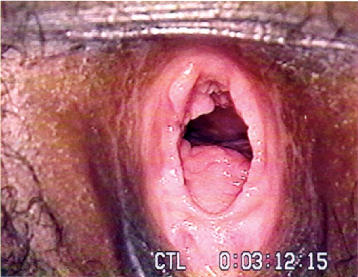
Figure 56-3 Sexual abuse, nonacute. Deep, wide posterior midline hymenal cleft extending to the vaginal wall (examined in prone knee chest position). Cleft represents healed complete hymenal tear or transection in a 15-year-old girl who disclosed multiple acts of penile vaginal penetration. (Courtesy of Allan R. De Jong, MD.)

Figure 56-4 Sexual abuse, nonacute. Two deep, posterior midline hymenal clefts extending to the vaginal wall. Clefts represent healed complete hymenal tears or transections in a 11-year-old girl who described a single act of penile vaginal penetration accompanied by pain and bleeding 2 weeks previously. (Courtesy of Allan R. De Jong, MD.)
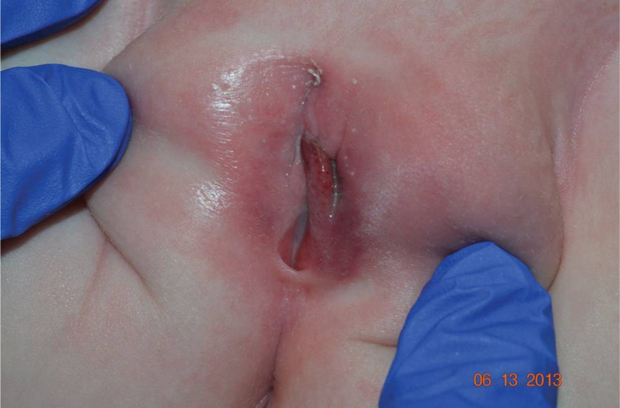
Figure 56-5 Straddle injury. Crush or blunt impact injury with typical asymmetric pattern to the left labia minora with marked swelling and labia majora with both external blue bruising and red bruising of medial surface in a 2-year-old girl who fell straddling the edge of a sandbox. (Courtesy of Allan R. De Jong, MD.)

Figure 56-6 Genital warts. Multiple genital warts are seen in vaginal vestibule of 6-year-old girl who disclosed only digital penetration. Blood vessels in the irregular masses create the red stippling of the wart surface. (Courtesy of Allan R. De Jong, MD.)
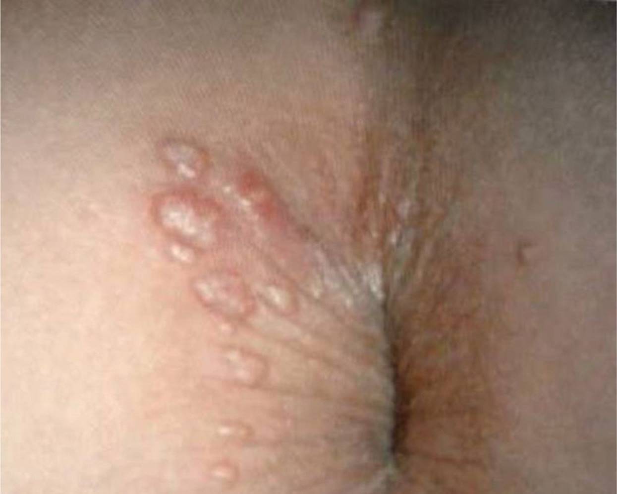
Figure 56-7 Molluscum contagiosum. Dome-shaped individual lesions and clustered lesions with shiny or pearly surface on perineum of 5-year-old girl referred for “genital warts.” She had similar lesions on arms and trunk. (Courtesy of Allan R. De Jong, MD.)
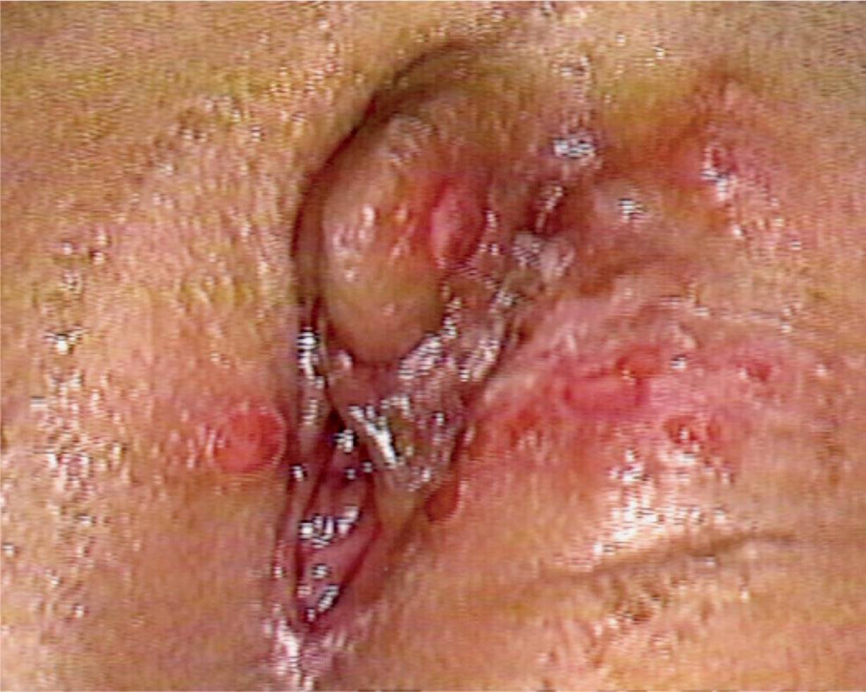
Figure 56-8 Herpes simplex virus infection. Multiple, painful, erythematous ulcerations on the labia of a 4-year-old girl who reported penile genital contact with an adult relative. Culture was positive for herpes simplex virus type II. (Courtesy of Allan R. De Jong, MD.)

Figure 56-9 Lichen sclerosus et atrophicus. Characteristic subepidermal hemorrhages in a 4-year-old girl with a 3-week history of genital itching and intermittent dysuria. Note lesions extend over clitoral prepuce with fissuring due to friability in midline superior to clitoris. Lesions showed only slight improvement at follow-up weeks later. (Courtesy of Allan R. De Jong, MD.)

Figure 56-10 Hemangioma. Superficial, red capillary hemangioma of the posterior commissure and perineal body. Initially mistaken as an abrasion, it blanched with pressure. (Used with permission from Bays J. Conditions mistaken for child sexual abuse. In: Reece R, Ludwig S, eds. Child Abuse: Medical Diagnosis and Management. 2nd ed. Philadelphia, PA: Lippincott Williams & Wilkins; 2001:290.)

Figure 56-11 Failure of midline fusion. Midline, pale indented defect with prominent vascularity and the appearance of mucosa that was initially mistaken for trauma. (Used with permission from Bays J. Conditions mistaken for child sexual abuse. In: Reece R, Ludwig S, eds. Child Abuse: Medical Diagnosis and Management. 2nd ed. Philadelphia, PA: Lippincott Williams & Wilkins; 2001:292.)
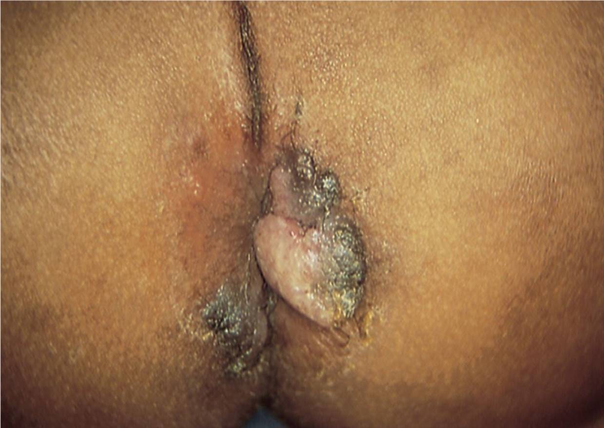
Figure 56-12 Condyloma lata. Pale hypertrophic plaque of condyloma lata in a 2-year-old girl with previously untreated primary syphilis. (Used with permission from De Jong AR, Finkel MA. Medical findings in child sexual abuse. In: Reece R, Ludwig S, eds. Child Abuse: Medical Diagnosis and Management. 2nd ed. Philadelphia, PA: Lippincott Williams & Wilkins; 2001:259.)
DIFFERENTIAL DIAGNOSIS

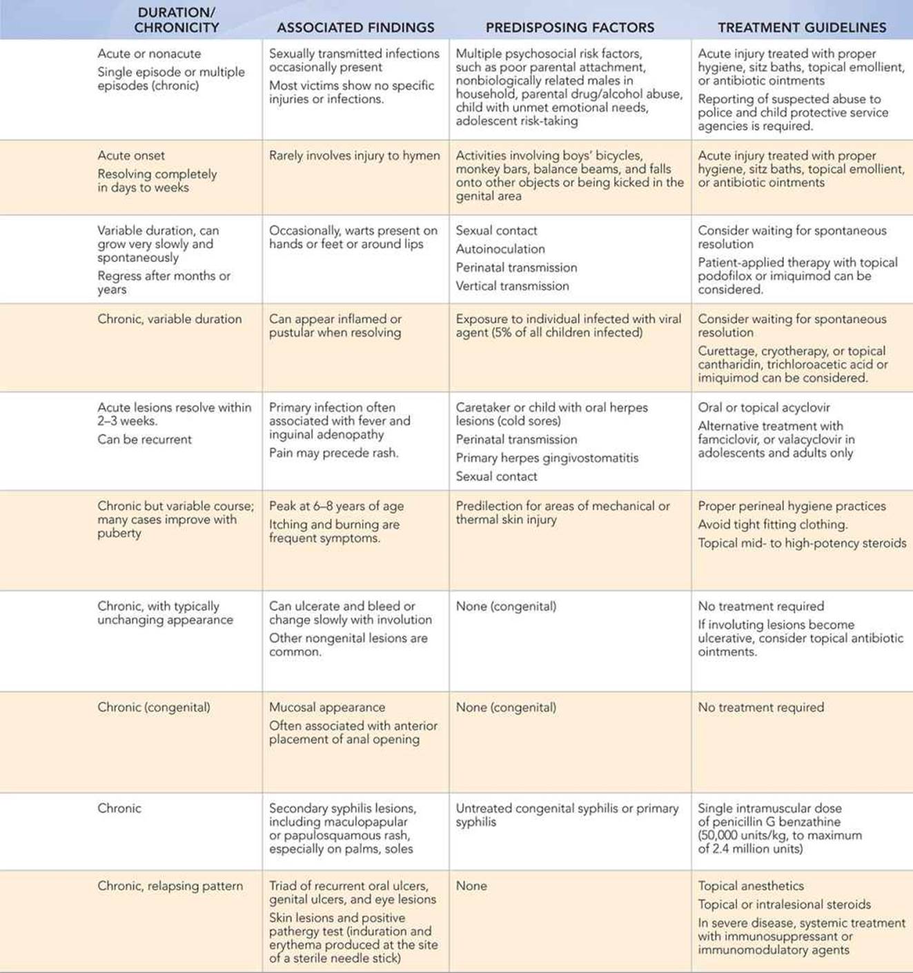
Other Diagnoses to Consider
• Poor hygiene
• Seborrheic, psoriatic, atopic, or contact dermatitis
• Impetigo, streptococcal vulvovaginitis
• Labial adhesions or agglutination
• Ulcerations from varicella, herpes zoster, influenza, Epstein–Barr, or coxsackievirus infection
• Stevens–Johnson syndrome
• Crohn disease
When to Consider Further Evaluation or Treatment
• Sexual abuse requires a multidisciplinary approach. Medical care providers should initiate medical evaluation and treatment, including counseling, but suspected cases should be reported to the state child protective services agency and to local police departments for further investigation.
• The general treatment of genital injuries, infections, and inflammation includes excellent perineal hygiene, sitz baths with warm water only, and topical lubricant ointments.
• Avoidance of irritants including soaps, application of topical aluminum acetate solution, and several days of low-dose topical steroids are helpful for specific and nonspecific vulvovaginitis.
• The diagnosis of genital warts, genital infections with herpes virus, and genital syphilis should result in evaluation for other sexually transmitted infections and consideration of possible sexual abuse.
SUGGESTED READINGS
Baldwin DD, Landa HM. Common problems in pediatric gynecology. Urol Clin North Am. 1995;22:161–176.
De Jong AR, Finkel MA. Medical findings in child sexual abuse. In: Reece R, Ludwig S, eds. Child Abuse: Medical Diagnosis and Management. 2nd ed. Philadelphia, PA: Lippincott Williams & Wilkins; 2001:207–286.
Emans SJH, Laufer MR, Goldstein DP, eds. Pediatric and Adolescent Gynecology. 5th ed. Philadelphia, PA: Lippincott-Raven Publishers; 2005:565–684, 939–975, 1024–1036.
Frasier L. Medical conditions that mimic sexual abuse. In: Kaplan R, Adams JA, Starling SP, Giardino AP, eds. Medical Response to Child Sexual Abuse. St. Louis, MO: STM Learning, Inc.; 2011:145–166.
Quint EH, Smith YR. Vulvar disorders in adolescent patients. Pediatr Clin North Am. 1999;46:593–606.