THYROID PHYSIOLOGY
Introduction
The normal thyroid gland secretes sufficient amounts of the thyroid hormones¾triiodothyronine (T3) and tetraiodothyronine (T4, thyroxine)¾to normalize growth and development, body temperature, and energy levels. These hormones contain 59% and 65% (respectively) of iodine as an essential part of the molecule. Calcitonin, the second type of thyroid hormone, is important in the regulation of calcium metabolism and is discussed in Chapter 42.
Iodide Metabolism
The recommended daily adult iodide (I-)* intake is 150 mcg (200 mcg during pregnancy).
Iodide, ingested from food, water, or medication, is rapidly absorbed and enters an extracellular fluid pool. The thyroid gland removes about 75 mcg a day from this pool for hormone synthesis, and the balance is excreted in the urine. If iodide intake is increased, the fractional iodine uptake by the thyroid is diminished.
*In this chapter, the term "iodine" denotes all forms of the element; the term "iodide" denotes only the ionic form, I-.
Biosynthesis of Thyroid Hormones
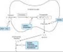
Once taken up by the thyroid gland, iodide undergoes a series of enzymatic reactions that convert it into active thyroid hormone (Figure 38-1). The first step is the transport of iodide into the thyroid gland by an intrinsic follicle cell basement membrane protein called the sodium/iodide symporter (NIS). This can be inhibited by such anions as thiocyanate (SCN-), pertechnetate (TcO4-), and perchlorate (ClO4-). At the apical cell membrane a second I- transport enzyme called pendrin controls the flow of iodide across the membrane. Pendrin is also found in the cochlea of the inner ear and if deficient or absent, a syndrome of deafness and goiter, called Pendred's syndrome, ensues. At the apical cell membrane, iodide is oxidized by thyroidal peroxidase to iodine, in which form it rapidly iodinates tyrosine residues within the thyroglobulin molecule to form monoiodotyrosine (MIT) and diiodotyrosine (DIT). This process is called iodide organification. Thyroidal peroxidase is transiently blocked by high levels of intrathyroidal iodide and blocked more persistently by thioamide drugs.
Two molecules of DIT combine within the thyroglobulin molecule to form L-thyroxine (T4). One molecule of MIT and one molecule of DIT combine to form T3. In addition to thyroglobulin, other proteins within the gland may be iodinated, but these iodoproteins do not have hormonal activity. Thyroxine, T3, MIT, and DIT are released from thyroglobulin by exocytosis and proteolysis of thyroglobulin at the apical colloid border. The MIT and DIT are deiodinated within the gland, and the iodine is reutilized. This process of proteolysis is also blocked by high levels of intrathyroidal iodide. The ratio of T4 to T3 within thyroglobulin is approximately 5:1, so that most of the hormone released is thyroxine. Most of the T3 circulating in the blood is derived from peripheral metabolism of thyroxine (see below, Figure 38-2).
|
|
Figure 38-1. Biosynthesis of thyroid hormones. The sites of action of various drugs that interfere with thyroid hormone biosynthesis are shown. |
|
|
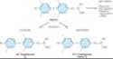
Figure 38-2. Peripheral metabolism of thyroxine. (Modified from Greenspan FS: Thyroid gland. In: Greenspan FS, Gardner D [editors]: Basic & Clinical Endocrinology, 7th ed. McGraw-Hill, 2004.) |
Transport of Thyroid Hormones
T4 and T3 in plasma are reversibly bound to protein, primarily thyroxine-binding globulin (TBG). Only about 0.04% of total T4 and 0.4% of T3 exist in the free form. Many physiologic and pathologic states and drugs affect T4, T3, and thyroid transport. However, the actual levels of free hormone generally remain normal, reflecting feedback control.
Peripheral Metabolism of Thyroid Hormones
The primary pathway for the peripheral metabolism of thyroxine is deiodination. Deiodination of T4 may occur by monodeiodination of the outer ring, producing 3,5,3¢-triiodothyronine (T3), which is three to four times more potent than T4. Alternatively, deiodination may occur in the inner ring, producing 3,3¢,5¢-triiodothyronine (reverse T3, or rT3), which is metabolically inactive (Figure 38-2). Drugs such as amiodarone, iodinated contrast media, b blockers, and corticosteroids, and severe illness or starvation inhibit the 5¢-deiodinase necessary for the conversion of T4 to T3, resulting in low T3 and high rT3 levels in the serum. The pharmacokinetics of thyroid hormones are listed in Table 38-1. The low serum levels of T3 and rT3 in normal individuals are due to the high metabolic clearances of these two compounds.
Evaluation of Thyroid Function
The tests used to evaluate thyroid function are listed in Table 38-2.
A. THYROID-PITUITARY RELATIONSHIPS
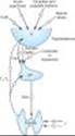
Control of thyroid function via thyroid-pituitary feedback is also discussed in Chapter 37. Briefly, hypothalamic cells secrete thyrotropin-releasing hormone (TRH) (Figure 38-3). TRH is secreted into capillaries of the pituitary portal venous system, and in the pituitary gland, TRH stimulates the synthesis and release of thyroid-stimulating hormone (TSH). TSH in turn stimulates an adenylyl cyclase-mediated mechanism in the thyroid cell to increase the synthesis and release of T4 and T3. These thyroid hormones act in a negative feedback fashion in the pituitary to block the action of TRH and in the hypothalamus to inhibit the synthesis and secretion of TRH. Other hormones or drugs may also affect the release of TRH or TSH.
B. AUTOREGULATION OF THE THYROID GLAND
The thyroid gland also regulates its uptake of iodide and thyroid hormone synthesis by intrathyroidal mechanisms that are independent of TSH. These mechanisms are primarily related to the level of iodine in the blood. Large doses of iodine inhibit iodide organification (Wolff-Chaikoff block, see Figure 38-1). In certain disease states (eg, Hashimoto's thyroiditis), this can inhibit thyroid hormone synthesis and result in hypothyroidism. Hyperthyroidism can result from the loss of the Wolff-Chaikoff block in susceptible individuals (eg, multinodular goiter).
C. ABNORMAL THYROID STIMULATORS
In Graves' disease (see below), lymphocytes secrete a TSH receptor-stimulating antibody (TSH-R Ab [stim]), also known as thyroid-stimulating immunoglobulin (TSI). This immunoglobulin binds to the TSH receptor and turns on the gland in the same fashion as TSH itself. The duration of its effect, however, is much longer than that of TSH. TSH receptors are also found in orbital fibrocytes, which may be stimulated by high levels of TSH-R Ab [stim].
|
|
Figure 38-3. The hypothalamic-pituitary-thyroid axis. Acute psychosis or prolonged exposure to cold may activate the axis. Hypothalamic TRH stimulates pituitary TSH release, while somatostatin and dopamine inhibit it. TSH stimulates T4 and T3 synthesis and release from the thyroid, and they in turn inhibit both TRH and TSH synthesis and release. Small amounts of iodide are necessary for hormone production, but large amounts inhibit T3 and T4 production and release. (Solid arrows, stimulatory influence; dashed arrows, inhibitory influence. H, hypothalamus, AP, anterior pituitary.) |
I. BASIC PHARMACOLOGY OF THYROID & ANTITHYROID DRUGS
THYROID HORMONES
Chemistry
The structural formulas of thyroxine and triiodothyronine as well as reverse triiodothyronine (rT3) are shown in Figure 38-2. All of these naturally occurring molecules are levo (L) isomers. The synthetic dextro (D) isomer of thyroxine, dextrothyroxine, has approximately 4% of the biologic activity of the L isomer as evidenced by its lesser ability to suppress TSH secretion and correct hypothyroidism.
Pharmacokinetics
Thyroxine is absorbed best in the duodenum and ileum; absorption is modified by intraluminal factors such as food, drugs, and intestinal flora. Oral bioavailability of current preparations of L-thyroxine averages 80% (Table 38-1). In contrast, T3 is almost completely absorbed (95%). T4 and T3 absorption appears not to be affected by mild hypothyroidism but may be impaired in severe myxedema with ileus. These factors are important in switching from oral to parenteral therapy. For parenteral use, the intravenous route is preferred for both hormones.
In patients with hyperthyroidism, the metabolic clearances of T4 and T3 are increased and the half-lives decreased; the opposite is true in patients with hypothyroidism. Drugs that induce hepatic microsomal enzymes (eg, rifampin, phenobarbital, carbamazepine, phenytoin, imatinib, protease inhibitors) increase the metabolism of both T4 and T3 (Table 38-3). Despite this change in clearance, the normal hormone concentration is maintained in euthyroid patients as a result of compensatory hyperfunction of the thyroid. However, patients receiving T4 replacement medication may require increased dosages to maintain clinical effectiveness. A similar compensation occurs if binding sites are altered. If TBG sites are increased by pregnancy, estrogens, or oral contraceptives, there is an initial shift of hormone from the free to the bound state and a decrease in its rate of elimination until the normal hormone concentration is restored. Thus, the concentration of total and bound hormone will increase, but the concentration of free hormone and the steady-state elimination will remain normal. The reverse occurs when thyroid binding sites are decreased.
Mechanism of Action
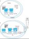
A model of thyroid hormone action is depicted in Figure 38-4, which shows the free forms of thyroid hormones, T4 and T3, dissociated from thyroid-binding proteins, entering the cell by active transport. Within the cell, T4 is converted to T3 by 5¢-deiodinase, and the T3 enters the nucleus, where T3 binds to a specific T3 receptor protein, a member of the c-erb oncogene family. (This family also includes the steroid hormone receptors and receptors for vitamins A and D.) The T3 receptor exists in two forms, a and b. Differing concentrations of receptor forms in different tissues may account for variations in T3 effect on different tissues.
Most of the effects of thyroid on metabolic processes appear to be mediated by activation of nuclear receptors that lead to increased formation of RNA and subsequent protein synthesis, eg, increased formation of Na+/K+ ATPase. This is consistent with the observation that the action of thyroid is manifested in vivo with a time lag of hours or days after its administration.
Large numbers of thyroid hormone receptors are found in the most hormone-responsive tissues (pituitary, liver, kidney, heart, skeletal muscle, lung, and intestine), while few receptor sites occur in hormone-unresponsive tissues (spleen, testes). The brain, which lacks an anabolic response to T3, contains an intermediate number of receptors. In congruence with their biologic potencies, the affinity of the receptor site for T4 is about ten times lower than that for T3. The number of nuclear receptors may be altered to preserve body homeostasis. For example, starvation lowers both circulating T3 hormone and cellular T3 receptors.
|
|
Figure 38-4. Model of the interaction of T3 with the T3 receptor. A: Inactive phase¾the unliganded T3 receptor dimer bound to the thyroid hormone response element (TRE) along with corepressors acts as a suppressor of gene transcription. B: Active phase¾T3 and T4 circulate bound to thyroid-binding proteins (TBPs). The free hormones are transported into the cell by a specific transport system. Within the cytoplasm, T4 is converted to T3 by 5'-deiodinase; T3 then moves into the nucleus. There it binds to the ligand-binding domain of the thyroid receptor (TR) monomer. This promotes disruption of the TR homodimer and heterodimerization with retinoid X receptor (RXR) on the TRE, displacement of corepressors, and binding of coactivators. The TR-coactivator complex activates gene transcription, which leads to alteration in protein synthesis and cellular phenotype. (TR-LBD, T3 receptor ligand-binding domain; TR-DBD, T3 receptor DNA-binding domain; RXR-LBD, retinoid X receptor ligand-binding domain; RXR-DBD, retinoid X receptor DNA-binding domain; T3, triiodothyronine; T4, tetraiodothyronine, L-thyroxine; 5'DI, 5'deiodinase. (Modified and reproduced, with permission, from Greenspan FS, Gardner DG [editors]: Basic & Clinical Endocrinology, 7th ed. McGraw-Hill, 2004.) |
Effects of Thyroid Hormones
The thyroid hormones are responsible for optimal growth, development, function, and maintenance of all body tissues. Excess or inadequate amounts result in the signs and symptoms of hyperthyroidism or hypothyroidism, respectively (Table 38-4). Since T3 and T4 are qualitatively similar, they may be considered as one hormone in the discussion that follows.
Thyroid hormone is critical for nervous, skeletal, and reproductive tissues. Its effects depend on protein synthesis as well as potentiation of the secretion and action of growth hormone. Thyroid deprivation in early life results in irreversible mental retardation and dwarfism¾symptoms typical of congenital cretinism.
Effects on growth and calorigenesis are accompanied by a pervasive influence on metabolism of drugs as well as carbohydrates, fats, proteins, and vitamins. Many of these changes are dependent upon or modified by activity of other hormones. Conversely, the secretion and degradation rates of virtually all other hormones, including catecholamines, cortisol, estrogens, testosterone, and insulin, are affected by thyroid status.
Many of the manifestations of thyroid hyperactivity resemble sympathetic nervous system overactivity (especially in the cardiovascular system), although catecholamine levels are not increased. Changes in catecholamine-stimulated adenylyl cyclase activity as measured by cAMP are found with changes in thyroid activity. Possible explanations include increased numbers of b receptors or enhanced amplification of the b receptor signal. Other clinical symptoms reminiscent of excessive epinephrine activity (and partially alleviated by adrenoceptor antagonists) include lid lag and retraction, tremor, excessive sweating, anxiety, and nervousness. The opposite constellation of symptoms is seen in hypothyroidism (Table 38-4).
Thyroid Preparations
See the Preparations Available section at the end of this chapter for a list of available preparations. These preparations may be synthetic (levothyroxine, liothyronine, liotrix) or of animal origin (desiccated thyroid).
Thyroid hormones are not effective and can be detrimental in the management of obesity, abnormal vaginal bleeding, or depression if thyroid hormone levels are normal. Anecdotal reports of a beneficial effect of T3 administered with antidepressants have not been confirmed with a controlled study.
Synthetic levothyroxine is the preparation of choice for thyroid replacement and suppression therapy because of its stability, content uniformity, low cost, lack of allergenic foreign protein, easy laboratory measurement of serum levels, and long half-life (7 days), which permits once-daily administration. In addition, T4 is converted to T3 intracellularly; thus, administration of T4 produces both hormones. Generic levothyroxine preparations provide comparable efficacy and are more cost-effective than branded preparations.
Although liothyronine (T3) is three to four times more potent than levothyroxine, it is not recommended for routine replacement therapy because of its shorter half-life (24 hours), which requires multiple daily doses; its higher cost; and the greater difficulty of monitoring its adequacy of replacement by conventional laboratory tests. Furthermore, because of its greater hormone activity and consequent greater risk of cardiotoxicity, T3 should be avoided in patients with cardiac disease. It is best used for short-term suppression of TSH. Because oral administration of T3 is unnecessary, use of the more expensive mixture of thyroxine and liothyronine (liotrix) instead of levothyroxine is never required.
The use of desiccated thyroid rather than synthetic preparations is never justified, since the disadvantages of protein antigenicity, product instability, variable hormone concentrations, and difficulty in laboratory monitoring far outweigh the advantage of low cost. Significant amounts of T3 found in some thyroid extracts and liotrix may produce significant elevations in T3 levels and toxicity. Equivalent doses are 100 mg of desiccated thyroid, 100 mcg of levothyroxine, and 37.5 mcg of liothyronine.
The shelf life of synthetic hormone preparations is about 2 years, particularly if they are stored in dark bottles to minimize spontaneous deiodination. The shelf life of desiccated thyroid is not known with certainty, but its potency is better preserved if it is kept dry.
ANTITHYROID AGENTS
INTRODUCTION
Reduction of thyroid activity and hormone effects can be accomplished by agents that interfere with the production of thyroid hormones, by agents that modify the tissue response to thyroid hormones, or by glandular destruction with radiation or surgery. Goitrogens are agents that suppress secretion of T3 and T4 to subnormal levels and thereby increase TSH, which in turn produces glandular enlargement (goiter). The antithyroid compounds used clinically include the thioamides, iodides, and radioactive iodine.
1. Thioamides
Introduction
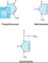
The thioamides methimazole and propylthiouracil are major drugs for treatment of thyrotoxicosis. In the United Kingdom, carbimazole, which is converted to methimazole in vivo, is widely used. Methimazole is about ten times more potent than propylthiouracil.
The chemical structures of these compounds are shown in Figure 38-5. The thiocarbamide group is essential for antithyroid activity.
|
|
Figure 38-5. Structure of thioamides. The thiocarbamide moiety is shown in color. |
Pharmacokinetics
Propylthiouracil is rapidly absorbed, reaching peak serum levels after 1 hour. The bioavailability of 50-80% may be due to incomplete absorption or a large first-pass effect in the liver. The volume of distribution approximates total body water with accumulation in the thyroid gland. Most of an ingested dose of propylthiouracil is excreted by the kidney as the inactive glucuronide within 24 hours.
In contrast, methimazole is completely absorbed but at variable rates. It is readily accumulated by the thyroid gland and has a volume of distribution similar to that of propylthiouracil. Excretion is slower than with propylthiouracil; 65-70% of a dose is recovered in the urine in 48 hours.
The short plasma half-life of these agents (1.5 hours for propylthiouracil and 6 hours for methimazole) has little influence on the duration of the antithyroid action or the dosing interval because both agents are accumulated by the thyroid gland. For propylthiouracil, giving the drug every 6-8 hours is reasonable since a single 100 mg dose can inhibit iodine organification by 60% for 7 hours. Since a single 30 mg dose of methimazole exerts an antithyroid effect for longer than 24 hours, a single daily dose is effective in the management of mild to moderate hyperthyroidism.
Both thioamides cross the placental barrier and are concentrated by the fetal thyroid, so that caution must be employed when using these drugs in pregnancy. Because of the risk of fetal hypothyroidism, both thioamides are classified as pregnancy category D (evidence of human fetal risk based on adverse reaction data from investigational or marketing experience). Of the two, propylthiouracil is preferable in pregnancy because it is more strongly protein-bound and, therefore, crosses the placenta less readily. In addition, methimazole has been, albeit rarely, associated with congenital malformations. Both thioamides are secreted in low concentrations in breast milk but are considered safe for the nursing infant.
Pharmacodynamics
The thioamides act by multiple mechanisms. The major action is to prevent hormone synthesis by inhibiting the thyroid peroxidase-catalyzed reactions and blocking iodine organification. In addition, they block coupling of the iodotyrosines. They do not block uptake of iodide by the gland. Propylthiouracil and (to a much lesser extent) methimazole inhibit the peripheral deiodination of T4 and T3 (Figure 38-1). Since the synthesis rather than the release of hormones is affected, the onset of these agents is slow, often requiring 3-4 weeks before stores of T4 are depleted.
Toxicity
Adverse reactions to the thioamides occur in 3-12% of treated patients. Most reactions occur early, especially nausea and gastrointestinal distress. An altered sense of taste or smell may occur with methimazole. The most common adverse effect is a maculopapular pruritic rash (4-6%), at times accompanied by systemic signs such as fever. Rare adverse effects include an urticarial rash, vasculitis, a lupus-like reaction, lymphadenopathy, hypoprothrombinemia, exfoliative dermatitis, polyserositis, and acute arthralgia. Hepatitis (more common with propylthiouracil) and cholestatic jaundice (more common with methimazole) can be fatal; although asymptomatic elevations in transaminase levels also occur.
The most dangerous complication is agranulocytosis (granulocyte count < 500 cells/mm3), an infrequent but potentially fatal adverse reaction. It occurs in 0.1-0.5% of patients taking thioamides, but the risk may be increased in older patients and in those receiving high-dose methimazole therapy (> 40 mg/d). The reaction is usually rapidly reversible when the drug is discontinued, but broad-spectrum antibiotic therapy may be necessary for complicating infections. Colony-stimulating factors (eg, G-CSF; see Chapter 33) may hasten recovery of the granulocytes. The cross-sensitivity between propylthiouracil and methimazole is about 50%; therefore, switching drugs in patients with severe reactions is not recommended.
2. Anion Inhibitors
Monovalent anions such as perchlorate (ClO4-), pertechnetate (TcO4-), and thiocyanate (SCN-) can block uptake of iodide by the gland through competitive inhibition of the iodide transport mechanism. Since these effects can be overcome by large doses of iodides, their effectiveness is somewhat unpredictable.
The major clinical use for potassium perchlorate is to block thyroidal reuptake of I- in patients with iodide-induced hyperthyroidism (eg, amiodarone-induced hyperthyroidism). However, potassium perchlorate is rarely used clinically because it is associated with aplastic anemia.
3. Iodides
Introduction
Prior to the introduction of the thioamides in the 1940s, iodides were the major antithyroid agents; today they are rarely used as sole therapy.
Pharmacodynamics
Iodides have several actions on the thyroid. They inhibit organification and hormone release and decrease the size and vascularity of the hyperplastic gland. In susceptible individuals, iodides can induce hyperthyroidism (jodbasedow phenomenon) or precipitate hypothyroidism.
In pharmacologic doses (> 6 mg/d), the major action of iodides is to inhibit hormone release, possibly through inhibition of thyroglobulin proteolysis. Improvement in thyrotoxic symptoms occurs rapidly¾within 2-7 days¾hence the value of iodide therapy in thyroid storm. In addition, iodides decrease the vascularity, size, and fragility of a hyperplastic gland, making the drugs valuable as preoperative preparation for surgery.
Clinical Use of Iodide
Disadvantages of iodide therapy include an increase in intraglandular stores of iodine, which may delay onset of thioamide therapy or prevent use of radioactive iodine therapy for several weeks. Thus, iodides should be initiated after onset of thioamide therapy and avoided if treatment with radioactive iodine seems likely. Iodide should not be used alone, because the gland will escape from the iodide block in 2-8 weeks, and its withdrawal may produce severe exacerbation of thyrotoxicosis in an iodine-enriched gland. Chronic use of iodides in pregnancy should be avoided, since they cross the placenta and can cause fetal goiter. In radiation emergencies, the thyroid-blocking effects of potassium iodide can protect the gland from subsequent damage if administered before radiation exposure.
Toxicity
Adverse reactions to iodine (iodism) are uncommon and in most cases reversible upon discontinuance. They include acneiform rash (similar to that of bromism), swollen salivary glands, mucous membrane ulcerations, conjunctivitis, rhinorrhea, drug fever, metallic taste, bleeding disorders and, rarely, anaphylactoid reactions.
4. Iodinated Contrast Media
The iodinated contrast agents¾diatrizoate orally and iohexol orally or intravenously¾are valuable in the treatment of hyperthyroidism, although they are not labeled for this indication. These drugs rapidly inhibit the conversion of T4 to T3 in the liver, kidney, pituitary gland, and brain. This accounts for the dramatic improvement in both subjective and objective parameters. For example, a decrease in heart rate is seen after only 3 days of administration of 0.5-1 g/d of oral contrast media. T3 levels often return to normal during this time. The prolonged effect of suppressing T4 as well as T3 suggests that inhibition of hormone release due to the iodine released may be an additional mechanism of action. Fortunately, these agents are relatively nontoxic. They provide useful adjunctive therapy in the treatment of thyroid storm and offer valuable alternatives when iodides or thioamides are contraindicated. Surprisingly, these agents may not interfere with 131I retention as much as iodides despite their large iodine content. Their toxicity is similar to that of the iodides, and their safety in pregnancy is undocumented.
5. Radioactive Iodine
131I is the only isotope used for treatment of thyrotoxicosis (others are used in diagnosis). Administered orally in solution as sodium 131I, it is rapidly absorbed, concentrated by the thyroid, and incorporated into storage follicles. Its therapeutic effect depends on emission of b rays with an effective half-life of 5 days and a penetration range of 400-2000 um. Within a few weeks after administration, destruction of the thyroid parenchyma is evidenced by epithelial swelling and necrosis, follicular disruption, edema, and leukocyte infiltration. Advantages of radioiodine include easy administration, effectiveness, low expense, and absence of pain. Fears of radiation-induced genetic damage, leukemia, and neoplasia have not been realized after more than 50 years of clinical experience with radioiodine. Radioactive iodine should not be administered to pregnant women or nursing mothers, since it crosses the placenta to destroy the fetal thyroid gland and is excreted in breast milk.
6. Adrenoceptor-Blocking Agents
Beta blockers without intrinsic sympathomimetic activity (eg, metoprolol, propranolol, atenolol) are effective therapeutic adjuncts in the management of thyrotoxicosis since many of these symptoms mimic those associated with sympathetic stimulation. Propranolol has been the b blocker most widely studied and used in the therapy of thyrotoxicosis. Beta blockers cause clinical improvement of hyperthyroid symptoms but do not alter thyroid hormone levels.
II. CLINICAL PHARMACOLOGY OF THYROID & ANTITHYROID DRUGS
HYPOTHYROIDISM
Introduction
Hypothyroidism is a syndrome resulting from deficiency of thyroid hormones and is manifested largely by a reversible slowing down of all body functions (Table 38-4). In infants and children, there is striking retardation of growth and development that results in dwarfism and irreversible mental retardation.
The etiology and pathogenesis of hypothyroidism are outlined in Table 38-5. Hypothyroidism can occur with or without thyroid enlargement (goiter). The laboratory diagnosis of hypothyroidism in the adult is easily made by the combination of a low free thyroxine and elevated serum TSH (Table 38-2).
The most common cause of hypothyroidism in the USA at this time is probably Hashimoto's thyroiditis, an immunologic disorder in genetically predisposed individuals. In this condition, there is evidence of humoral immunity in the presence of antithyroid antibodies and lymphocyte sensitization to thyroid antigens. Certain medications can also cause hypothyroidism (Table 38-5).
Management of Hypothyroidism
Except for hypothyroidism caused by drugs (Table 38-5), which can be treated in some cases by simply removing the depressant agent, the general strategy of replacement therapy is appropriate. The most satisfactory preparation is levothyroxine, administered as either a branded or generic preparation. Treatment with combination levothyroxine plus liothyronine has not been found to be superior to levothyroxine alone. Infants and children require more T4 per kilogram of body weight than adults. The average dosage for an infant 1-6 months of age is 10-15 mcg/kg/d, whereas the average dosage for an adult is about 1.7 mcg/kg/d. Older adults (> 65 years of age) may require less thyroxine for replacement. There is some variability in the absorption of thyroxine, so this dosage will vary from patient to patient. Since interactions with certain foods (eg, bran, soy) and drugs (Table 38-3) can impair its absorption, thyroxine should be administered on an empty stomach (eg, 30 minutes before meals or 1 hour after meals). Its long half-life of 7 days permits once daily dosing. Children should be monitored for normal growth and development. Serum TSH and free thyroxine should be measured at regular intervals and TSH maintained within an optimal range of 0.5-2.5 mU/L. It takes 6-8 weeks after starting a given dose of thyroxine to reach steady-state levels in the bloodstream. Thus, dosage changes should be made slowly.
In long-standing hypothyroidism, in older patients, and in patients with underlying cardiac disease, it is imperative to start treatment with reduced dosages. In such adult patients, levothyroxine is given in a dosage of 12.5-25 mcg/d for 2 weeks, increasing the daily dose by 25 mcg every 2 weeks until euthyroidism or drug toxicity is observed. In older patients, the heart is very sensitive to the level of circulating thyroxine, and if angina pectoris or cardiac arrhythmia develops, it is essential to stop or reduce the dose of thyroxine immediately. In younger patients or those with very mild disease, full replacement therapy may be started immediately.
The toxicity of thyroxine is directly related to the hormone level. In children, restlessness, insomnia, and accelerated bone maturation and growth may be signs of thyroxine toxicity. In adults, increased nervousness, heat intolerance, episodes of palpitation and tachycardia, or unexplained weight loss may be the presenting symptoms. If these symptoms are present, it is important to monitor serum TSH (Table 38-2), which will determine whether the symptoms are due to excess thyroxine blood levels. Chronic overtreatment with T4, particularly in elderly patients, can increase the risk of atrial fibrillation and accelerated osteoporosis.
Special Problems in Management of Hypothyroidism
A. MYXEDEMA AND CORONARY ARTERY DISEASE
Since myxedema frequently occurs in older persons, it is often associated with underlying coronary artery disease. In this situation, the low levels of circulating thyroid hormone actually protect the heart against increasing demands that could result in angina pectoris or myocardial infarction. Correction of myxedema must be done cautiously to avoid provoking arrhythmia, angina, or acute myocardial infarction. If coronary artery surgery is indicated, it should be done first, prior to correction of the myxedema by thyroxine administration.
B. MYXEDEMA COMA
Myxedema coma is an end state of untreated hypothyroidism. It is associated with progressive weakness, stupor, hypothermia, hypoventilation, hypoglycemia, hyponatremia, water intoxication, shock, and death.
Management of myxedema coma is a medical emergency. The patient should be treated in the intensive care unit, since tracheal intubation and mechanical ventilation may be required. Associated illnesses such as infection or heart failure must be treated by appropriate therapy. It is important to give all preparations intravenously, because patients with myxedema coma absorb drugs poorly from other routes. Intravenous fluids should be administered with caution to avoid excessive water intake. These patients have large pools of empty T3 and T4binding sites that must be filled before there is adequate free thyroxine to affect tissue metabolism. Accordingly, the treatment of choice in myxedema coma is to give a loading dose of levothyroxine intravenously¾usually 300-400 mcg initially, followed by 50-100 mcg daily. Intravenous T3 can also be used but may be more cardiotoxic and more difficult to monitor. Intravenous hydrocortisone is indicated if the patient has associated adrenal or pituitary insufficiency but is probably not necessary in most patients with primary myxedema. Opioids and sedatives must be used with extreme caution.
C. HYPOTHYROIDISM AND PREGNANCY
Hypothyroid women frequently have anovulatory cycles and are therefore relatively infertile until restoration of the euthyroid state. This has led to the widespread use of thyroid hormone for infertility, although there is no evidence for its usefulness in infertile euthyroid patients. In a pregnant hypothyroid patient receiving thyroxine, it is extremely important that the daily dose of thyroxine be adequate because early development of the fetal brain depends on maternal thyroxine. In many hypothyroid patients, an increase in the thyroxine dose (about 30-50%) is required to normalize the serum TSH level during pregnancy. Because of the elevated maternal TBG levels and, therefore, elevated total T4 levels, adequate maternal thyroxine dosages warrant maintenance of TSH between 0.5 and 3.0 mU/L and the total T4 at or above the upper range of normal.
D. SUBCLINICAL HYPOTHYROIDISM
Subclinical hypothyroidism, defined as an elevated TSH level and normal thyroid hormone levels, is found in 4-10% of the general population but increases to 20% in women older than age 50. The consensus of expert thyroid organizations concluded that thyroid hormone therapy should be considered for patients with TSH levels greater than 10 mU/L while close TSH monitoring is appropriate for those with lower TSH elevations.
E. DRUG-INDUCED HYPOTHYROIDISM
Drug-induced hypothyroidism (Table 38-3) can be satisfactorily managed with levothyroxine therapy if the offending agent cannot be stopped. In the case of amiodarone-induced hypothyroidism, levothyroxine therapy may be necessary even after discontinuance because of amiodarone's very long half-life.
HYPERTHYROIDISM
INTRODUCTION
Hyperthyroidism (thyrotoxicosis) is the clinical syndrome that results when tissues are exposed to high levels of thyroid hormone (Table 38-4).
1. Graves' Disease
Introduction
The most common form of hyperthyroidism is Graves' disease, or diffuse toxic goiter. The presenting signs and symptoms of Graves' disease are set forth in Table 38-4.
Pathophysiology
Graves' disease is considered to be an autoimmune disorder in which helper T lymphocytes stimulate B lymphocytes to synthesize antibodies to thyroidal antigens. The antibody described previously (TSH-R Ab [stim]) is directed against the TSH receptor site in the thyroid cell membrane and has the capacity to stimulate growth and biosynthetic activity of the thyroid cell. Spontaneous remission occurs but some patients require years of antithyroid therapy.
Laboratory Diagnosis
In most patients with hyperthyroidism, T3, T4, FT4, and FT3 are elevated and TSH is suppressed (Table 38-2). Radioiodine uptake is usually markedly elevated as well. Antithyroglobulin, thyroid peroxidase, and TSH-R Ab [stim] antibodies are usually present.
Management of Graves' Disease
The three primary methods for controlling hyperthyroidism are antithyroid drug therapy, surgical thyroidectomy, and destruction of the gland with radioactive iodine.
A. ANTITHYROID DRUG THERAPY
Drug therapy is most useful in young patients with small glands and mild disease. Methimazole or propylthiouracil is administered until the disease undergoes spontaneous remission. This is the only therapy that leaves an intact thyroid gland, but it does require a long period of treatment and observation (12-18 months), and there is a 50-68% incidence of relapse.
Methimazole is preferable to propylthiouracil (except in pregnancy) because it can be administered once daily, which may enhance adherence. Antithyroid drug therapy is usually begun with divided doses, shifting to maintenance therapy with single daily doses when the patient becomes clinically euthyroid. However, mild to moderately severe thyrotoxicosis can often be controlled with methimazole given in a single morning dose of 20-40 mg initially for 4-8 weeks to normalize hormone levels. Maintenance therapy requires 5-15 mg once daily. Alternatively, therapy is started with propylthiouracil, 100-150 mg every 6 or 8 hours until the patient is euthyroid, followed by gradual reduction of the dose to the maintenance level of 50-150 mg once daily. In addition to inhibiting iodine organification, propylthiouracil also inhibits the conversion of T4 to T3, so it brings the level of activated thyroid hormone down more quickly than does methimazole. The best clinical guide to remission is reduction in the size of the goiter. Laboratory tests most useful in monitoring the course of therapy are serum FT3, FT4, and TSH levels.
Reactivation of the autoimmune process may occur when the dosage of antithyroid drug is lowered during maintenance therapy and TSH begins to drive the gland. In some cases TSH release can be prevented by the daily administration of 50-150 mcg of levothyroxine with 5-15 mg of methimazole or 50-150 mg of propylthiouracil for the second year of therapy. The relapse rate with this program is probably comparable to the rate with antithyroid therapy alone, but the risk of hypothyroidism and overtreatment is avoided.
Reactions to antithyroid drugs have been described above. A minor rash can often be controlled by antihistamine therapy. Because the more severe reaction of agranulocytosis is often heralded by sore throat or high fever, patients receiving antithyroid drugs must be instructed to discontinue the drug and seek immediate medical attention if these symptoms develop. White cell and differential counts and a throat culture are indicated in such cases, followed by appropriate antibiotic therapy.
B. THYROIDECTOMY
A near-total thyroidectomy is the treatment of choice for patients with very large glands or multinodular goiters. Patients are treated with antithyroid drugs until euthyroid (about 6 weeks). In addition, for 10-14 days prior to surgery, they receive saturated solution of potassium iodide, 5 drops twice daily, to diminish vascularity of the gland and simplify surgery. About 80-90% of patients will require thyroid supplementation following near-total thyroidectomy.
C. RADIOACTIVE IODINE
Radioiodine therapy utilizing 131I is the preferred treatment for most patients over 21 years of age. In patients without heart disease, the therapeutic dose may be given immediately in a range of 80-120 uCi/g of estimated thyroid weight corrected for uptake. In patients with underlying heart disease or severe thyrotoxicosis and in elderly patients, it is desirable to treat with antithyroid drugs (preferably methimazole) until the patient is euthyroid. The medication is then stopped for 5-7 days before the appropriate dose of 131I is administered. Iodides should be avoided to ensure maximal 131I uptake. Six to 12 weeks following the administration of radioiodine, the gland will shrink in size and the patient will usually become euthyroid or hypothyroid. A second dose may be required in some patients. Hypothyroidism occurs in about 80% of patients following radioiodine therapy. Serum FT4 and TSH levels should be monitored regularly. When hypothyroidism develops, prompt replacement with oral levothyroxine, 50-150 mcg daily, should be instituted.
D. ADJUNCTS TO ANTITHYROID THERAPY
During the acute phase of thyrotoxicosis, b-adrenoceptor blocking agents without intrinsic sympathomimetic activity are extremely helpful. Propranolol, 20-40 mg orally every 6 hours, will control tachycardia, hypertension, and atrial fibrillation. Propranolol is gradually withdrawn as serum thyroxine levels return to normal. Diltiazem, 90-120 mg three or four times daily, can be used to control tachycardia in patients in whom b blockers are contraindicated, eg, those with asthma. Other calcium channel blockers may not be as effective as diltiazem. Adequate nutrition and vitamin supplements are essential. Barbiturates accelerate T4 breakdown (by hepatic enzyme induction) and may be helpful both as sedatives and to lower T4levels.
2. Toxic Uninodular Goiter & Toxic Multinodular Goiter
These forms of hyperthyroidism occur often in older women with nodular goiters. FT4 is moderately elevated or occasionally normal, but FT3 or T3 is strikingly elevated. Single toxic adenomas can be managed with either surgical excision of the adenoma or with radioiodine therapy. Toxic multinodular goiter is usually associated with a large goiter and is best treated by preparation with methimazole or propylthiouracil followed by subtotal thyroidectomy.
3. Subacute Thyroiditis
During the acute phase of a viral infection of the thyroid gland, there is destruction of thyroid parenchyma with transient release of stored thyroid hormones. A similar state may occur in patients with Hashimoto's thyroiditis. These episodes of transient thyrotoxicosis have been termed spontaneously resolving hyperthyroidism. Supportive therapy is usually all that is necessary, such as b-adrenoceptor blocking agents without intrinsic sympathomimetic activity (eg, propranolol) for tachycardia and aspirin or nonsteroidal anti-inflammatory drugs to control local pain and fever. Corticosteroids may be necessary in severe cases to control the inflammation.
4. Special Problems
Thyroid Storm
Thyroid storm, or thyrotoxic crisis, is sudden acute exacerbation of all of the symptoms of thyrotoxicosis, presenting as a life-threatening syndrome. Vigorous management is mandatory. Propranolol, 1-2 mg slowly intravenously or 40-80 mg orally every 6 hours, is helpful to control the severe cardiovascular manifestations. If propranolol is contraindicated by the presence of severe heart failure or asthma, hypertension and tachycardia may be controlled with diltiazem, 90-120 mg orally three or four times daily or 5-10 mg/h by intravenous infusion (asthmatic patients only). Release of thyroid hormones from the gland is retarded by the administration of saturated solution of potassium iodide, 10 drops orally daily, or iodinated contrast media, 1 g orally daily. The latter medication will also block peripheral conversion of T4 to T3. Hormone synthesis is blocked by the administration of propylthiouracil, 250 mg orally every 6 hours. If the patient is unable to take propylthiouracil by mouth, a rectal formulation* can be prepared and administered in a dosage of 400 mg every 6 hours as a retention enema. Methimazole may also be prepared for rectal administration in a dose of 60 mg daily. Hydrocortisone, 50 mg intravenously every 6 hours, will protect the patient against shock and will block the conversion of T4 to T3, rapidly bringing down the level of thyroactive material in the blood.
Supportive therapy is essential to control fever, heart failure, and any underlying disease process that may have precipitated the acute storm. In rare situations, where the above methods are not adequate to control the problem, plasmapheresis or peritoneal dialysis has been used to lower the levels of circulating thyroxine.
*To prepare a water suspension propylthiouracil enema, grind eight 50 mg tablets and suspend the powder in 90 mL of sterile water.
Ophthalmopathy
Although severe ophthalmopathy is rare, it is difficult to treat. Management requires effective treatment of the thyroid disease, usually by total surgical excision or 131I ablation of the gland plus oral prednisone therapy (see below). In addition, local therapy may be necessary, eg, elevation of the head to diminish periorbital edema and artificial tears to relieve corneal drying. Smoking cessation should be advised to prevent progression of the ophthalmopathy. For the severe, acute inflammatory reaction, a short course of prednisone, 60-100 mg orally daily for about a week and then 60-100 mg every other day, tapering the dose over a period of 6-12 weeks, may be effective. If steroid therapy fails or is contraindicated, irradiation of the posterior orbit, using well-collimated high-energy x-ray therapy, will frequently result in marked improvement of the acute process. Threatened loss of vision is an indication for surgical decompression of the orbit. Eyelid or eye muscle surgery may be necessary to correct residual problems after the acute process has subsided.
Dermopathy
Dermopathy or pretibial myxedema will often respond to topical corticosteroids applied to the involved area and covered with an occlusive dressing.
Thyrotoxicosis during Pregnancy
Ideally, women in the childbearing period with severe disease should have definitive therapy with 131I or subtotal thyroidectomy prior to pregnancy in order to avoid an acute exacerbation of the disease during pregnancy or following delivery. If thyrotoxicosis does develop during pregnancy, radioiodine is contraindicated because it crosses the placenta and may injure the fetal thyroid. In the first trimester, the patient can be prepared with propylthiouracil and a subtotal thyroidectomy performed safely during the mid trimester. It is essential to give the patient a thyroid supplement during the balance of the pregnancy. However, most patients are treated with propylthiouracil during the pregnancy, and the decision regarding long-term management can be made after delivery. The dosage of propylthiouracil must be kept to the minimum necessary for control of the disease (ie, < 300 mg/d), because it may affect the function of the fetal thyroid gland. Methimazole is a potential alternative, although there is concern about a possible risk of fetal scalp defects.
Neonatal Graves' Disease
Graves' disease may occur in the newborn infant, either due to passage of maternal TSH-R Ab [stim] through the placenta, stimulating the thyroid gland of the neonate, or to genetic transmission of the trait to the fetus. Laboratory studies reveal an elevated free thyroxine, a markedly elevated T3, and a low TSH¾in contrast to the normal infant, in whom TSH is elevated at birth. TSH-R Ab [stim] is usually found in the serum of both the child and the mother.
If caused by maternal TSH-R Ab [stim], the disease is usually self-limited and subsides over a period of 4-12 weeks, coinciding with the fall in the infant's TSH-R Ab [stim] level. However, treatment is necessary because of the severe metabolic stress the infant experiences. Therapy includes propylthiouracil in a dose of 5-10 mg/kg/d in divided doses at 8-hour intervals; Lugol's solution (8 mg of iodide per drop), 1 drop every 8 hours; and propranolol, 2 mg/kg/d in divided doses. Careful supportive therapy is essential. If the infant is very ill, oral prednisone, 2 mg/kg/d in divided doses, will help block conversion of T4 to T3. These medications are gradually reduced as the clinical picture improves and can be discontinued by 6-12 weeks.
Subclinical Hyperthyroidism
Subclinical hyperthyroidism is defined as a suppressed TSH level (below the normal range) in conjunction with normal thyroid hormone levels. Cardiac toxicity (eg, atrial fibrillation), especially in older persons, is of greatest concern. The consensus of thyroid experts concluded that hyperthyroidism treatment is appropriate in those with TSH less than 0.1 mU/L, while close monitoring of the TSH level is appropriate for those with less TSH suppression.
Amiodarone-Induced Thyrotoxicosis
Approximately 3% of patients receiving amiodarone will develop hyperthyroidism. Two types of amiodarone-induced thyrotoxicosis have been reported: iodine-induced (type I), which often occurs in persons with underlying thyroid disease (eg, multinodular goiter); and an inflammatory thyroiditis (type II) that occurs in patients without thyroid disease due to leakage of thyroid hormone into the circulation. Treatment of type I requires thioamides while type II responds best to glucocorticoids. Since it is not always possible to differentiate between the two types, thioamides and glucocorticoids are often administered together. If possible, amiodarone should be discontinued; however, rapid improvement does not occur due to its long half-life.
NONTOXIC GOITER
Nontoxic goiter is a syndrome of thyroid enlargement without excessive thyroid hormone production. Enlargement of the thyroid gland is often due to TSH stimulation from inadequate thyroid hormone synthesis. The most common cause of nontoxic goiter worldwide is iodide deficiency, but in the USA, it is Hashimoto's thyroiditis. Other causes include germline or acquired mutations in genes involved in hormone synthesis, dietary goitrogens, and neoplasms (see below).
Goiter due to iodide deficiency is best managed by prophylactic administration of iodide. The optimal daily iodide intake is 150-200 mcg. Iodized salt and iodate used as preservatives in flour and bread are excellent sources of iodine in the diet. In areas where it is difficult to introduce iodized salt or iodate preservatives, a solution of iodized poppyseed oil has been administered intramuscularly to provide a long-term source of inorganic iodine.
Goiter due to ingestion of goitrogens in the diet is managed by elimination of the goitrogen or by adding sufficient thyroxine to shut off TSH stimulation. Similarly, in Hashimoto's thyroiditis and dyshormonogenesis, adequate thyroxine therapy¾150-200 mcg/d orally¾will suppress pituitary TSH and result in slow regression of the goiter as well as correction of hypothyroidism.
THYROID NEOPLASMS
Neoplasms of the thyroid gland may be benign (adenomas) or malignant. The primary diagnostic test is a fine needle aspiration biopsy and cytologic examination. Benign lesions may be monitored for growth or symptoms of local obstruction, which would mandate surgical excision. Management of thyroid carcinoma requires a total thyroidectomy, postoperative radioiodine therapy in selected instances, and lifetime replacement with levothyroxine. The evaluation for recurrence of some thyroid malignancies often involves withdrawal of thyroxine replacement for 4-6 weeks¾accompanied by the development of hypothyroidism. Tumor recurrence is likely if there is a rise in serum thyroglobulin (ie, a tumor marker) or a positive 131I scan when TSH is elevated. Alternatively, administration of recombinant human TSH (Thyrogen) can produce comparable TSH elevations without discontinuing thyroxine and avoiding hypothyroidism. Recombinant human TSH is administered intramuscularly once daily for 2 days. A rise in serum thyroglobulin or a positive 131I scan will indicate a recurrence of the thyroid cancer.
PREPARATIONS AVAILABLE
THYROID AGENTS
Levothyroxine [T4] (generic, Levoxyl, Levo-T, Levothroid, Levolet, Novothyrox, Synthroid, Unithroid)
Oral: 0.025, 0.05, 0.075, 0.088, 0.1, 0.112, 0.125, 0.137, 0.15, 0.175, 0.2, 0.3 mg tablets
Parenteral: 200, 500 mcg per vial (100 mcg/mL when reconstituted) for injection
Liothyronine [T3] (generic, Cytomel, Triostat)
Oral: 5, 25, 50 mcg tablets
Parenteral: 10 mcg/mL
Liotrix [a 4:1 ratio of T4:T3] (Thyrolar)
Oral: tablets containing 12.5, 25, 30, 50, 60, 100, 120, 150, 180 mcg T4 and one fourth as much T3
Thyroid desiccated [USP] (generic, Armour Thyroid, Thyroid Strong, Thyrar, S-P-T)
Oral: tablets containing 15, 30, 60, 90, 120, 180, 240, 300 mg; capsules (S-P-T) containing 120, 180, 300 mg
ANTITHYROID AGENTS
Diatrizoate sodium (Hypaque)
Parenteral: 25% (150 mg iodine/mL); 50% (300 mg iodine/mL) (unlabeled use); 250 g powder for reconstitution (oral use is unlabeled)
Iodide (131I) sodium (Iodotope, Sodium Iodide I 131 Therapeutic)
Oral: available as capsules and solution
Iohexol (Omnipaque)
Parenteral: 140, 180, 240, 300, 350 mg iodine/mL (unlabeled use)
Methimazole (Tapazole)
Oral: 5, 10 mg tablets
Potassium iodide
Oral solution (generic, SSKI): 1 g/mL
Oral solution (Lugol's solution): 100 mg/mL potassium iodide plus 50 mg/mL iodine
Oral syrup (Pima): 325 mg/5 mL
Oral controlled action tablets (Iodo-Niacin): 135 mg potassium iodide plus 25 mg niacinamide hydroiodide
Oral potassium iodide tablets (generic, IOSAT, RAD-Block, Thyro-Block): 65, 130 mg
Propylthiouracil [PTU] (generic)
Oral: 50 mg tablets
Thyrotropin; recombinant human TSH (Thyrogen)
Parenteral: 0.9 mg per vial
REFERENCES
General
Greenspan FS: Thyroid gland. In: Greenspan FS, Gardner DG (editors). Basic & Clinical Endocrinology, 7th ed. McGraw-Hill, 2004.
American Thyroid Association (http://thyroid.org/).
Guidelines
Anonymous: American Association of Clinical Endocrinologists medical guidelines for clinical practice for the evaluation and treatment of hyperthyroidism and hypothyroidism. Endocr Pract 2002;8:457.
Cooper DS et al: Management guidelines for patients with thyroid modules and differentiated thyroid cancer. Thyroid 2006;16:109.
Gharib H et al: Consensus statement #1: Subclinical thyroid dysfunction: A joint statement on management from the American Association of Clinical Endocrinologists, the American Thyroid Association, and The Endocrine Society. Thyroid. 2005;15:24.
Surks MI et al: Subclinical thyroid disease: Scientific review and guidelines for diagnosis and management. JAMA. 2004;291:228.
US Department of Health and Human Services: Potassium iodide as a thyroid blocking agent in radiation emergencies. December 2001 (http://www.fda.gov/cder/guidance/index.htm).
Hypothyroidism
Dong BJ et al: Bioequivalence of generic and brand-name levothyroxine products in the treatment of hypothyroidism. JAMA 1997;277:1205.
Escobar-Morreale HF et al: Treatment of hypothyroidism with combinations of levothyroxine plus liothyronine. J Clin Endocrinol Metab 2005;90:4946.
Gruters A et al: Long-term consequences of congenital hypothyroidism in the era of screening programs. Endocrinol Metab 2002;16:369.
Khouzam HR et al: Thyroid hormone therapy: A review of their effects in the treatment of psychiatric and medical conditions. Comp Ther 2004;30:148.
Toft A: Which thyroxine? Thyroid 2005;15:124.
Wartofsky L, Dickey RA: The evidence for a narrower thyrotropin reference range is compelling. J Clin Endocrinol Metab. 2005;90:5483.
Wiersinga QM: Thyroid hormone replacement therapy. Horm Res 2001;56(Suppl 1):74.
Hyperthyroidism
Abraham P et al: Antithyroid drug regimen for treating Graves' hyperthyroidism. Cochrane Database Syst Rev 2005;CD003420 (http://www.thecochranelibrary.com).
Abraham P et al: A systematic review of drug therapy for Graves' hyperthyroidism. Eur J Endocrinol 2005;153:489.
Braga M, Cooper DS: Oral cholecystographic agents and the thyroid. J Clin Endocrinol Metab 2001;86:1853.
Cooper DS: Antithyroid drugs. N Engl J Med 2005;352:905.
Cooper DS: Hyperthyroidism. Lancet 2003;362:459.
El-Kaissi S, Frauman AG, Wall JR: Thyroid-associated ophthalmopathy: A practical guide to classification, natural history and management. Intern Med J 2004;34:482.
Jongjaroenprasert W et al: Rectal administration of propylthiouracil in hyperthyroid patients: Comparison of suspension enema and suppository form. Thyroid 2002;12:627.
Mestman JH: Hyperthyroidism in pregnancy. Best Pract Res Clin Endocrinol Metab 2004;18:267.
Nontoxic Goiter, Nodules, & Cancer
Hegedus L: Clinical practice. The thyroid nodule. N Engl J Med 2004;351:1764.
Lawrence W Jr, Kaplan BJ: Diagnosis and management of patients with thyroid nodules. J Surg Oncol 2002;80:157.
The Effects of Drugs on Thyroid Function
Arafah BM: Increased need for thyroxine in women with hypothyroidism during estrogen therapy. N Engl J Med 2001;344:1743.
Basaria S, Cooper DS: Amiodarone and the thyroid. Am J Med 2005;118:706.
Caraccop M et al: Long-term follow-up of 106 multiple sclerosis patients undergoing interferon-b 1a or 1b therapy: Predictive factors of thyroid disease development and duration. J Clin Endocrinol Metab 2005;90:4133.
Prummel MF, Laurberg P: Interferon-a and autoimmune thyroid disease. Thyroid 2003;13:547.