I. BASIC PHARMACOLOGY
INTRODUCTION
Calcium and phosphate, the major mineral constituents of bone, are also two of the most important minerals for general cellular function. Accordingly, the body has evolved a complex set of mechanisms by which calcium and phosphate homeostasis are carefully maintained (Figure 42-1). Approximately 98% of the 1-2 kg of calcium and 85% of the 1 kg of phosphorus in the human adult are found in bone, the principal reservoir for these minerals. These functions are dynamic, with constant remodeling of bone and ready exchange of bone mineral with that in the extracellular fluid. Bone also serves as the principal structural support for the body and provides the space for hematopoiesis. Thus, abnormalities in bone mineral homeostasis can lead not only to a wide variety of cellular dysfunctions (eg, tetany, coma, muscle weakness) but also to disturbances in structural support of the body (eg, osteoporosis with fractures) and loss of hematopoietic capacity (eg, infantile osteopetrosis).
Calcium and phosphate enter the body from the intestine. The average American diet provides 600-1000 mg of calcium per day, of which approximately 100-250 mg is absorbed. This figure represents net absorption, because both absorption (principally in the duodenum and upper jejunum) and secretion (principally in the ileum) occur. The amount of phosphorus in the American diet is about the same as that of calcium. However, the efficiency of absorption (principally in the jejunum) is greater, ranging from 70% to 90%, depending on intake. In the steady state, renal excretion of calcium and phosphate balances intestinal absorption. In general, over 98% of filtered calcium and 85% of filtered phosphate is reabsorbed by the kidney. The movement of calcium and phosphate across the intestinal and renal epithelia is closely regulated. Intrinsic disease of the intestine (eg, nontropical sprue) or kidney (eg, chronic renal failure) disrupts bone mineral homeostasis.
Two hormones serve as the principal regulators of calcium and phosphate homeostasis: the peptide parathyroid hormone (PTH) and the steroid vitamin D (Figure 42-2). Vitamin D is a prohormone rather than a true hormone, because it must be further metabolized to gain biologic activity. PTH stimulates the production of the active metabolite of vitamin D, 1,25(OH)2D. 1,25(OH)2D, on the other hand, suppresses the production of PTH. 1,25(OH)2D stimulates the intestinal absorption of calcium and phosphate. 1,25(OH)2D and PTH promote both bone formation and resorption in part by stimulating the proliferation and differentiation of osteoblasts and osteoclasts. Both PTH and 1,25(OH)2D enhance renal retention of calcium, but PTH promotes renal phosphate excretion. Fibroblast growth factor 23 (FGF23) is a newly discovered hormone that stimulates renal phosphate excretion and inhibits renal production of 1,25(OH)2D. Other hormones¾calcitonin, prolactin, growth hormone, insulin, thyroid hormone, glucocorticoids, and sex steroids¾influence calcium and phosphate homeostasis under certain physiologic circumstances and can be considered secondary regulators. Deficiency or excess of these secondary regulators within a physiologic range does not produce the disturbance of calcium and phosphate homeostasis that is observed in situations of deficiency or excess of PTH and vitamin D. However, certain of these secondary regulators¾especially calcitonin, glucocorticoids, and estrogens¾are useful therapeutically and are discussed in subsequent sections.
In addition to these hormonal regulators, calcium and phosphate themselves, other ions such as sodium and fluoride, and a variety of drugs (bisphosphonates, plicamycin, and diuretics) also alter calcium and phosphate homeostasis.
|
|
Figure 42-1. Some mechanisms contributing to bone mineral homeostasis. Direct actions are shown and feedback may alter the net effect. Calcium and phosphorus concentrations in the serum are controlled principally by two hormones, 1,25(OH)2D3 (D) and parathyroid hormone (PTH), through their action on absorption from the gut and from bone and on excretion in the urine. Both hormones increase input of calcium and phosphorus from bone into the serum; both hormones also stimulate bone formation; vitamin D also increases absorption from the gut. Vitamin D decreases urinary excretion of both calcium and phosphorus, whereas PTH reduces calcium but increases phosphorus excretion. Calcitonin (CT) is a less critical hormone for calcium homeostasis, but in pharmacologic concentrations CT can reduce serum calcium and phosphorus by inhibiting bone resorption and stimulating their renal excretion. FGF23 is a recently discovered hormone that stimulates renal excretion of phosphate. Feedback may alter the effects shown; for example, vitamin D usually increases urinary calcium excretion because of effects on calcium absorption from the gut and effects on PTH. |
|
|
|
Figure 42-2. The hormonal interactions controlling bone mineral homeostasis. 1,25(OH)2D is produced by the kidney under the control of PTH, which stimulates its production, and FGF23, which inhibits its production. 1,25(OH)2D in turn inhibits the production of PTH by the parathyroid glands. 1,25(OH)2D is the principal regulator of intestinal calcium and phosphate absorption. Both PTH and 1,25(OH)2D regulate bone formation and resorption, each capable of stimulating both processes. This is accomplished by their stimulation of preosteoblast proliferation and differentiation into osteoblasts, the bone forming cell. PTH and 1,25(OH)2D stimulate the expression of RANKL by the osteoblast, which, with MCSF, stimulates the differentiation and subsequent activation of osteoclasts, the bone resorbing cell. FGF23, fibroblast growth factor; MCSF, macrophage colony-stimulating factor; OPG, osteoprotegerin; RANK, receptor for activation of nuclear factor-kB. |
|
PRINCIPAL HORMONAL REGULATORS OF BONE MINERAL HOMEOSTASIS
PARATHYROID HORMONE
Parathyroid hormone (PTH) is a single-chain peptide hormone composed of 84 amino acids. It is produced in the parathyroid gland in a precursor form of 115 amino acids, the remaining 31 amino terminal amino acids being cleaved off before secretion. Within the gland is a calcium-sensitive protease capable of cleaving the intact hormone into fragments. Biologic activity resides in the amino terminal region such that synthetic 1-34 PTH is fully active. Loss of the first two amino terminal amino acids eliminates most biologic activity.
The metabolic clearance of intact PTH is rapid, with a half-time of disappearance measured in minutes. Most of the clearance occurs in the liver and kidney. The biologically inactive carboxyl terminal fragments produced during metabolism of the intact hormone have a much lower clearance, especially in renal failure. This accounts in part for the very high PTH values often observed in the past in patients with renal failure when measured by radioimmunoassays directed against the carboxyl terminal region of the molecule. However, most PTH assays currently in use measure the intact hormone by a double antibody method, so that this circumstance is less frequently encountered in clinical practice. PTH regulates calcium and phosphate flux across cellular membranes in bone and kidney, resulting in increased serum calcium and decreased serum phosphate. In bone, PTH increases the activity and number of osteoclasts, the cells responsible for bone resorption. However, this stimulation of osteoclasts is not a direct effect. Rather, PTH acts on the osteoblast (the bone-forming cell) to induce a membrane-bound protein called RANK ligand (RANKL). This factor acts on osteoclasts and osteoclast precursors to increase both the numbers and the activity of osteoclasts. This action increases bone turnover or bone remodeling, a specific sequence of cellular events initiated by osteoclastic bone resorption and followed by osteoblastic bone formation. Although both bone resorption and bone formation are enhanced by PTH, the net effect of excess PTH is to increase bone resorption. PTH in low and intermittent doses increases bone formation without first stimulating bone resorption. This action appears to be indirect, involving other growth factors such as IGF-1. This has led to the recent approval of recombinant PTH 1-34 (teriparatide) for the treatment of osteoporosis. In the kidney, PTH increases the ability of the nephron to reabsorb calcium and magnesium but reduces its ability to reabsorb phosphate, amino acids, bicarbonate, sodium, chloride, and sulfate. Another important action of PTH on the kidney is its stimulation of 1,25-dihydroxyvitamin D (1,25[OH]2D) production.
VITAMIN D
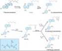
Vitamin D is a secosteroid produced in the skin from 7-dehydrocholesterol under the influence of ultraviolet irradiation. Vitamin D is also found in certain foods and is used to supplement dairy products. Both the natural form (vitamin D3, cholecalciferol) and the plant-derived form (vitamin D2, ergocalciferol) are present in the diet. These forms differ in that ergocalciferol contains a double bond (C22-23) and an additional methyl group in the side chain (Figure 42-3). In humans, this difference apparently is of limited physiologic consequence (although ergocalciferol is less potent), and the following comments apply equally well to both forms of vitamin D.
Vitamin D is a prohormone that serves as precursor to a number of biologically active metabolites (Figure 42-3). Vitamin D is first hydroxylated in the liver to form 25-hydroxyvitamin D (25[OH]D). This metabolite is further converted in the kidney to a number of other forms, the best-studied of which are 1,25-dihydroxyvitamin D (1,25[OH]2D) and 24,25-dihydroxyvitamin D (24,25[OH]2D). Of the natural metabolites, only vitamin D and 1,25(OH)2D (as calcitriol) are available for clinical use (Table 42-1). Moreover, a number of analogs of 1,25(OH)2D are being synthesized to extend the usefulness of this metabolite to a variety of nonclassic conditions. Calcipotriene (calcipotriol), for example, is being used to treat psoriasis, a hyperproliferative skin disorder. Doxercalciferol and paricalcitol have recently been approved for the treatment of secondary hyperparathyroidism in patients with chronic kidney disease. Other analogs are being investigated for the treatment of various malignancies. The regulation of vitamin D metabolism is complex, involving calcium, phosphate, and a variety of hormones, the most important of which is PTH, which stimulates the production of 1,25(OH)2D by the kidney.
Vitamin D and its metabolites circulate in plasma tightly bound to a carrier protein, the vitamin D-binding protein. This a-globulin binds 25(OH)D and 24,25(OH)2D with comparable high affinity and vitamin D and 1,25(OH)2D with lower affinity. In normal subjects, the terminal half-life of injected calcifediol is 23 days, whereas in anephric subjects it is 42 days. The half-life of 24,25(OH)2D is probably similar. Tracer studies with vitamin D have shown a rapid clearance from the blood. The liver appears to be the principal organ for clearance. Excess vitamin D is stored in adipose tissue. The metabolic clearance of calcitriol in humans indicates a rapid turnover, with a terminal half-life measured in hours. Several of the 1,25(OH)2D analogs are bound poorly by the vitamin D-binding protein. As a result, their clearance is very rapid, with a terminal half-life measured in minutes. Such analogs have little of the hypercalcemic, hypercalciuric effects of calcitriol, an important aspect of their use for the management of conditions such as psoriasis and hyperparathyroidism.
The mechanism of action of the vitamin D metabolites remains under active investigation. However, calcitriol is well established as the most potent agent with respect to stimulation of intestinal calcium and phosphate transport and bone resorption. Calcitriol appears to act on the intestine both by induction of new protein synthesis (eg, calcium-binding protein) and by modulation of calcium flux across the brush border and basolateral membranes by a means that does not require new protein synthesis. The molecular action of calcitriol on bone has received less attention. However, like PTH, calcitriol can induce RANK ligand in osteoblasts and proteins such as osteocalcin, which may regulate the mineralization process. The metabolites 25(OH)D and 24,25(OH)2D are far less potent stimulators of intestinal calcium and phosphate transport or bone resorption. However, 25(OH)D appears to be more potent than 1,25(OH)2D in stimulating renal reabsorption of calcium and phosphate and may be the major metabolite regulating calcium flux and contractility in muscle. Specific receptors for 1,25(OH)2D exist in target tissues. However, the role and even the existence of separate receptors for 25(OH)D and 24,25(OH)2D remain controversial.
The receptor for 1,25(OH)2D exists in a wide variety of tissues¾not just bone, gut, and kidney. In these "nonclassic" tissues, 1,25(OH)2D exerts a number of actions including regulation of parathyroid hormone secretion from the parathyroid gland, insulin secretion from the pancreas, cytokine production by macrophages and T cells, and proliferation and differentiation of a large number of cells, including cancer cells. Thus, the clinical utility of 1,25(OH)2D analogs is likely to expand.
|
|
Figure 42-3. Conversion of 7-dehydrocholesterol to vitamin D3 and metabolism of D3 to 1,25(OH)2D3 and 24,25(OH)2D3. Control of the latter step is exerted primarily at the level of the kidney, where low serum phosphorus, low serum calcium, and high parathyroid hormone favor the production of 1,25(OH)2D3. The inset shows the side chain for ergosterol. Ergosterol undergoes similar transformation to vitamin D2 (ergocalciferol), which, in turn, is metabolized to 25(OH)D2, 1,25(OH)2D2, and 24,25(OH)2D2. In humans, corresponding D2 and D3 derivatives have equivalent effects and potency. They are therefore referred to in the text without a subscript. |
INTERACTION OF PTH & VITAMIN D
A summary of the principal actions of PTH and vitamin D on the three main target tissues¾intestine, kidney, and bone¾is presented in Table 42-2. The net effect of PTH is to raise serum calcium and reduce serum phosphate; the net effect of vitamin D is to raise both. Regulation of calcium and phosphate homeostasis is achieved through a variety of feedback loops. Calcium is the principal regulator of PTH secretion. It binds to a novel ion recognition site that is part of a Gq protein-coupled receptor called the calcium sensing receptor (CaR) and links changes in intracellular free calcium concentration to changes in extracellular calcium. As serum calcium levels rise and bind to this receptor, intracellular calcium levels increase and inhibit PTH secretion. Phosphate regulates PTH secretion directly and indirectly by forming complexes with calcium in the serum. Because it is the ionized free concentration of calcium that is detected by the parathyroid gland, increases in serum phosphate levels reduce the ionized calcium and lead to enhanced PTH secretion. Such feedback regulation is appropriate to the net effect of PTH to raise serum calcium and reduce serum phosphate levels. Likewise, both calcium and phosphate at high levels reduce the amount of 1,25(OH)2D produced by the kidney and increase the amount of 24,25(OH)2D produced. The high calcium works directly and indirectly by reducing PTH secretion. The high phosphate works directly and indirectly by increasing FGF23 levels. Since 1,25(OH)2D raises serum calcium and phosphate, whereas 24,25(OH)2D has less effect, such feedback regulation is again appropriate. 1,25(OH)2D itself directly inhibits PTH secretion (independently of its effect on serum calcium) by a direct action on PTH gene transcription. This provides yet another negative feedback loop. The ability of 1,25(OH)2D to inhibit PTH secretion directly is being exploited using calcitriol analogs that have less effect on serum calcium because of their lesser effect on intestinal calcium absorption. Such drugs are proving useful in the management of secondary hyperparathyroidism accompanying chronic kidney disease and may be useful in selected cases of primary hyperparathyroidism.
SECONDARY HORMONAL REGULATORS OF BONE MINERAL HOMEOSTASIS
INTRODUCTION
A number of hormones modulate the actions of PTH and vitamin D in regulating bone mineral homeostasis. Compared with that of PTH and vitamin D, the physiologic impact of such secondary regulation on bone mineral homeostasis is minor. However, in pharmacologic amounts, a number of these hormones have actions on the bone mineral homeostatic mechanisms that can be exploited therapeutically.
CALCITONIN
The calcitonin secreted by the parafollicular cells of the mammalian thyroid is a single-chain peptide hormone with 32 amino acids and a molecular weight of 3600. A disulfide bond between positions 1 and 7 is essential for biologic activity. Calcitonin is produced from a precursor with MW 15,000. The circulating forms of calcitonin are multiple, ranging in size from the monomer (MW 3600) to forms with an apparent molecular weight of 60,000. Whether such heterogeneity includes precursor forms or covalently linked oligomers is not known. Because of its heterogeneity, calcitonin is standardized by bioassay in rats. Activity is compared to a standard maintained by the British Medical Research Council (MRC) and expressed as MRC units.
Human calcitonin monomer has a half-life of about 10 minutes with a metabolic clearance of 8-9 mL/kg/min. Salmon calcitonin has a longer half-life and a reduced metabolic clearance (3 mL/kg/min), making it more attractive as a therapeutic agent. Much of the clearance occurs in the kidney, although little intact calcitonin appears in the urine.
The principal effects of calcitonin are to lower serum calcium and phosphate by actions on bone and kidney. Calcitonin inhibits osteoclastic bone resorption. Although bone formation is not impaired at first after calcitonin administration, with time both formation and resorption of bone are reduced. In the kidney, calcitonin reduces both calcium and phosphate reabsorption as well as reabsorption of other ions, including sodium, potassium, and magnesium. Tissues other than bone and kidney are also affected by calcitonin. Calcitonin in pharmacologic amounts decreases gastrin secretion and reduces gastric acid output while increasing secretion of sodium, potassium, chloride, and water in the gut. Pentagastrin is a potent stimulator of calcitonin secretion (as is hypercalcemia), suggesting a possible physiologic relationship between gastrin and calcitonin. In the adult human, no readily demonstrable problem develops in cases of calcitonin deficiency (thyroidectomy) or excess (medullary carcinoma of the thyroid). However, the ability of calcitonin to block bone resorption and lower serum calcium makes it a useful drug for the treatment of Paget's disease, hypercalcemia, and osteoporosis.
GLUCOCORTICOIDS
Glucocorticoid hormones alter bone mineral homeostasis by antagonizing vitamin D-stimulated intestinal calcium transport, by stimulating renal calcium excretion, and by blocking bone formation. Although these observations underscore the negative impact of glucocorticoids on bone mineral homeostasis, these hormones have proved useful in reversing the hypercalcemia associated with lymphomas and granulomatous diseases such as sarcoidosis (in which production of 1,25[OH]2D is increased) or in cases of vitamin D intoxication. Prolonged administration of glucocorticoids is a common cause of osteoporosis in adults and stunted skeletal development in children.
ESTROGENS
Estrogens can prevent accelerated bone loss during the immediate postmenopausal period and at least transiently increase bone in the postmenopausal woman. The prevailing hypothesis advanced to explain these observations is that estrogens reduce the bone-resorbing action of PTH. Estrogen administration leads to an increased 1,25(OH)2D level in blood, but estrogens have no direct effect on 1,25(OH)2D production in vitro. The increased 1,25(OH)2D levels in vivo following estrogen treatment may result from decreased serum calcium and phosphate and increased PTH. Estrogen receptors have been found in bone, and estrogen has direct effects on bone remodeling. Recent case reports of men who lack the estrogen receptor or who are unable to produce estrogen because of aromatase deficiency noted marked osteopenia and failure to close epiphyses. This further substantiates the role of estrogen in bone development, even in men. The principal therapeutic application for estrogen administration in disorders of bone mineral homeostasis is the treatment or prevention of postmenopausal osteoporosis. However, long-term use of estrogen is being discouraged because of its deleterious side effects. Rather, selective estrogen receptor modulators (SERMs) have been developed to retain the beneficial effects on bone while minimizing these deleterious side effects on breast, uterus, and the cardiovascular system (see Box: Newer Therapies for Osteoporosis).
NEWER THERAPIES FOR OSTEOPOROSIS
Bone undergoes a continuous remodeling process involving bone resorption and formation. Any process that disrupts this balance by increasing resorption relative to formation results in osteoporosis. Inadequate sex hormone production is a major cause of osteoporosis in men and women. Estrogen replacement therapy at menopause is a well-established means of preventing osteoporosis in the female, but many women fear its adverse effects, particularly the increased risk of breast cancer from continued estrogen use (the well-demonstrated increased risk of endometrial cancer is prevented by cycling with a progestin) and do not like the persistence of menstrual bleeding that often accompanies this form of therapy. Medical enthusiasm for this treatment has waned with the demonstration that it does not protect against heart disease. Raloxifene is the first of the selective estrogen receptor modulators (SERMs; see Chapter 40) to be approved for the prevention of osteoporosis. Raloxifene shares some of the beneficial effects of estrogen on bone without increasing the risk of breast or endometrial cancer (it may actually reduce the risk of breast cancer). Although not as effective as estrogen in increasing bone density, raloxifene has been shown to reduce vertebral fractures.
Nonhormonal forms of therapy for osteoporosis with proven efficacy in reducing fracture risk have also been developed. Bisphosphonates such as alendronate, risedronate, and ibandronate have been conclusively shown to increase bone density and reduce fractures over at least 5 years when used continuously at a dosage of 10 mg/d or 70 mg/wk for alendronate and 5 mg/d or 35 mg/wk for risedronate, 2.5 mg/d or 150 mg/mo for ibandronate. Side-by-side trials between alendronate and calcitonin (another approved nonestrogen drug for osteoporosis) indicated a greater efficacy of alendronate. Bisphosphonates are poorly absorbed and must be given on an empty stomach or infused intravenously. At the higher oral doses used in the treatment of Paget's disease, alendronate causes gastric irritation, but this is not a significant problem at the doses recommended for osteoporosis when patients are instructed to take the drug with a glass of water and remain upright. The most recently approved drug for osteoporosis is teriparatide, the recombinant form of PTH1-34. Unlike other approved drugs for osteoporosis, teriparatide stimulates bone formation rather than inhibiting bone resorption. However, teriparatide must be given daily by subcutaneous injection. Its efficacy in preventing fractures appears to be at least as great as that of the bisphosphonates. In all cases, adequate intake of calcium and vitamin D needs to be maintained.
Thus, we now have several well-validated, efficacious forms of treatment for this common debilitating disease.
NONHORMONAL AGENTS AFFECTING BONE MINERAL HOMEOSTASIS
BISPHOSPHONATES
The bisphosphonates are analogs of pyrophosphate in which the P-O-P bond has been replaced with a nonhydrolyzable P-C-P bond (Figure 42-4). Etidronate, pamidronate, and alendronate have now been joined by risedronate, tiludronate, ibandronate, and zoledronate for clinical use. The bisphosphonates owe at least part of their clinical usefulness and toxicity to their ability to retard formation and dissolution of hydroxyapatite crystals within and outside the skeletal system. They localize to regions of bone resorption and so exert their greatest effects on osteoclasts. However, the exact mechanism by which they selectively inhibit bone resorption is not clear.
The results from animal and clinical studies indicate that less than 10% of an oral dose of these drugs is absorbed. Food reduces absorption even further, necessitating their administration on an empty stomach. Because it causes gastric irritation, pamidronate is not available as an oral preparation. However, with the possible exception of etidronate, all currently available bisphosphonates have this complication. Nearly half of the absorbed drug accumulates in bone; the remainder is excreted unchanged in the urine. Decreased renal function, esophageal motility disorders, and peptic ulcer disease are the main contraindications to the use of these drugs. The portion bound to bone is retained for months, depending on the turnover of bone itself.
Etidronate and the other bisphosphonates exert a variety of effects on bone mineral homeostasis. In particular, bisphosphonates are useful for the treatment of hypercalcemia associated with malignancy, for Paget's disease, and for osteoporosis (See Box: Newer Therapies for Osteoporosis). Contrary to expectations, alendronate appears to increase bone mineral density well beyond the 2-year period predicted for a drug whose effects are limited to blocking bone resorption. The bisphosphonates exert a variety of other cellular effects, including inhibition of 1,25(OH)2D production, inhibition of intestinal calcium transport, metabolic changes in bone cells such as inhibition of glycolysis, inhibition of cell growth, and changes in acid and alkaline phosphatase.
Amino bisphosphonates such as alendronate have recently been found to block farnesyl pyrophosphate synthase, an enzyme in the mevalonate pathway that appears to be critical for osteoclast survival. Statins, which block mevalonate synthesis, stimulate bone formation at least in animal studies. Thus, the mevalonate pathway appears to be important in bone cell function and provides new targets for drug development. These effects vary depending on the bisphosphonate being studied (ie, only amino bisphosphonates have this property) and may account for some of the clinical differences observed in the effects of the various bisphosphonates on bone mineral homeostasis. However, with the exceptions of the induction of a mineralization defect by higher than approved doses of etidronate and gastric and esophageal irritation by pamidronate and by high doses of alendronate, these drugs have proved to be remarkably free of adverse effects when used at the doses recommended for the treatment of osteoporosis. Higher doses used in the treatment of hypercalcemia have been associated with renal deterioration and osteonecrosis of the jaw. Esophageal irritation can be minimized by taking the drug with a full glass of water and remaining upright for 30 minutes.
|
|
Figure 42-4. The structure of pyrophosphate and of the first three bisphosphonates¾etidronate, pamidronate, and alendronate¾that were approved for use in the USA. |
CALCIMIMETICS
Cinacalcet is the first representative of a new class of drugs that activates the calcium sensing receptor (CaR). CaR is widely distributed but has its greatest concentration in the parathyroid gland. Cinacalcet blocks PTH secretion by this mechanism and is approved for the treatment of secondary hyperparathyroidism in chronic kidney disease and for the treatment of parathyroid carcinoma.
PLICAMYCIN (MITHRAMYCIN)
Plicamycin is a cytotoxic antibiotic (see Chapter 55) that has been used clinically for two disorders of bone mineral metabolism: Paget's disease and hypercalcemia. The cytotoxic properties of the drug appear to involve its binding to DNA and interruption of DNA-directed RNA synthesis. The reasons for its usefulness in the treatment of Paget's disease and hypercalcemia are unclear but may relate to the need for protein synthesis to sustain bone resorption. The doses required to treat Paget's disease and hypercalcemia are about one tenth the amounts required to achieve cytotoxic effects.
THIAZIDES
The chemistry and pharmacology of this family of drugs are covered in Chapter 15. The principal application of thiazides in the treatment of bone mineral disorders is in reducing renal calcium excretion. Thiazides may increase the effectiveness of parathyroid hormone in stimulating reabsorption of calcium by the renal tubules or may act on calcium reabsorption secondarily by increasing sodium reabsorption in the proximal tubule. In the distal tubule, thiazides block sodium reabsorption at the luminal surface, increasing the calcium-sodium exchange at the basolateral membrane and thus enhancing calcium reabsorption into the blood at this site. Thiazides have proved to be useful in reducing the hypercalciuria and incidence of stone formation in subjects with idiopathic hypercalciuria. Part of their efficacy in reducing stone formation may lie in their ability to decrease urine oxalate excretion and increase urine magnesium and zinc levels (both of which inhibit calcium oxalate stone formation).
FLUORIDE
Fluoride is well established as effective for the prophylaxis of dental caries and has been under investigation for the treatment of osteoporosis. Both therapeutic applications originated from epidemiologic observations that subjects living in areas with naturally fluoridated water (1-2 ppm) had less dental caries and fewer vertebral compression fractures than subjects living in nonfluoridated water areas. Fluoride is accumulated by bones and teeth, where it may stabilize the hydroxyapatite crystal. Such a mechanism may explain the effectiveness of fluoride in increasing the resistance of teeth to dental caries, but it does not explain new bone growth.
Fluoride in drinking water appears to be most effective in preventing dental caries if consumed before the eruption of the permanent teeth. The optimum concentration in drinking water supplies is 0.5-1 ppm. Topical application is most effective if done just as the teeth erupt. There is little further benefit to giving fluoride after the permanent teeth are fully formed. Excess fluoride in drinking water leads to mottling of the enamel proportionate to the concentration above 1 ppm.
Because of the paucity of agents that stimulate new bone growth in patients with osteoporosis, fluoride for this disorder has been examined (see Osteoporosis, below). Results of earlier studies indicated that fluoride alone without adequate calcium supplementation produced osteomalacia. More recent studies, in which calcium supplementation has been adequate, have demonstrated an improvement in calcium balance, an increase in bone mineral, and an increase in trabecular bone volume. However, studies of the ability of fluoride to reduce fractures reach opposite conclusions. Adverse effects observed¾at the doses used for testing fluoride's effect on bone¾include nausea and vomiting, gastrointestinal blood loss, arthralgias, and arthritis in a substantial proportion of patients. Such effects are usually responsive to reduction of the dose or giving fluoride with meals (or both). At present, fluoride is not approved by the Food and Drug Administration for use in osteoporosis.
II. CLINICAL PHARMACOLOGY
INTRODUCTION
Disorders of bone mineral homeostasis generally present with abnormalities in serum or urine calcium levels (or both), often accompanied by abnormal serum phosphate levels. These abnormal mineral concentrations may themselves cause symptoms requiring immediate treatment (eg, coma in malignant hypercalcemia, tetany in hypocalcemia). More commonly, they serve as clues to an underlying disorder in hormonal regulators (eg, primary hyperparathyroidism), target tissue response (eg, chronic kidney disease), or drug misuse (eg, vitamin D intoxication). In such cases, treatment of the underlying disorder is of prime importance.
Since bone and kidney play central roles in bone mineral homeostasis, conditions that alter bone mineral homeostasis usually affect one or both of these tissues secondarily. Effects on bone can result in osteoporosis (abnormal loss of bone; remaining bone histologically normal), osteomalacia (abnormal bone formation due to inadequate mineralization), or osteitis fibrosa (excessive bone resorption with fibrotic replacement of resorption cavities and marrow). Biochemical markers of skeletal involvement include changes in serum levels of the skeletal isoenzyme of alkaline phosphatase and osteocalcin (reflecting osteoblastic activity) and urine levels of hydroxyproline and pyridinoline cross-links (reflecting osteoclastic activity). The kidney becomes involved when the calcium ´ phosphate product in serum exceeds the point at which ectopic calcification occurs (nephrocalcinosis) or when the calcium ´ oxalate (or phosphate) product in urine exceeds saturation, leading to nephrolithiasis. Subtle early indicators of such renal involvement include polyuria, nocturia, and hyposthenuria. Radiologic evidence of nephrocalcinosis and stones is not generally observed until later. The degree of the ensuing renal failure is best followed by monitoring the decline in creatinine clearance. On the other hand, chronic kidney disease can be a primary cause of bone disease because of altered handling of calcium and phosphate, decreased 1,25(OH)2D production, and secondary hyperparathyroidism.
ABNORMAL SERUM CALCIUM & PHOSPHATE LEVELS
HYPERCALCEMIA
Introduction
Hypercalcemia causes central nervous system depression, including coma, and is potentially lethal. Its major causes (other than thiazide therapy) are hyperparathyroidism and cancer with or without bone metastases. Less common causes are hypervitaminosis D, sarcoidosis, thyrotoxicosis, milk-alkali syndrome, adrenal insufficiency, and immobilization. With the possible exception of hypervitaminosis D, the latter disorders seldom require emergency lowering of serum calcium. A number of approaches are used to manage the hypercalcemic crisis.
Saline Diuresis
In hypercalcemia of sufficient severity to produce symptoms, rapid reduction of serum calcium is required. The first steps include rehydration with saline and diuresis with furosemide. Most patients presenting with severe hypercalcemia have a substantial component of prerenal azotemia owing to dehydration, which prevents the kidney from compensating for the rise in serum calcium by excreting more calcium in the urine. Therefore, the initial infusion of 500-1000 mL/h of saline to reverse the dehydration and restore urine flow can by itself substantially lower serum calcium. The addition of a loop diuretic such as furosemide not only enhances urine flow but also inhibits calcium reabsorption in the ascending limb of the loop of Henle (see Chapter 15). Monitoring central venous pressure is important to forestall the development of heart failure and pulmonary edema in predisposed subjects. In many subjects, saline diuresis suffices to reduce serum calcium levels to a point at which more definitive diagnosis and treatment of the underlying condition can be achieved. If this is not the case or if more prolonged medical treatment of hypercalcemia is required, the following agents are available (discussed in order of preference).
Bisphosphonates
Pamidronate, 60-90 mg, infused over 2-4 hours, and zoledronate, 4 mg, infused over at least 15 minutes, have been approved for the treatment of hypercalcemia of malignancy and have largely replaced the less effective etidronate for this indication. The effects generally persist for weeks, but treatment can be repeated after a 7-day interval if necessary and if renal function is not impaired. Some patients experience a self-limited flu-like syndrome after the infusion. Repeated doses of these drugs have been linked to renal deterioration and osteonecrosis of the jaw, but this adverse effect is rare.
Calcitonin
Calcitonin has proved useful as ancillary treatment in a large number of patients. Calcitonin by itself seldom restores serum calcium to normal, and refractoriness frequently develops. However, its lack of toxicity permits frequent administration at high doses (200 MRC units or more). An effect on serum calcium is observed within 4-6 hours and lasts for 6-10 hours. Calcimar (salmon calcitonin) is available for parenteral and nasal administration.
Gallium Nitrate
Gallium nitrate is approved by the FDA for the management of hypercalcemia of malignancy and is undergoing trials for the treatment of advanced Paget's disease. This drug acts by inhibiting bone resorption. At a dosage of 200 mg/m2 body surface area per day given as a continuous intravenous infusion in 5% dextrose for 5 days, gallium nitrate proved superior to calcitonin in reducing serum calcium in cancer patients. Because of potential nephrotoxicity, patients should be well hydrated and have good renal output before starting the infusion.
Plicamycin (Mithramycin)
Because of its toxicity, plicamycin (mithramycin) is not the drug of first choice for the treatment of hypercalcemia. However, when other forms of therapy fail, 25-50 mcg/kg given intravenously usually lowers serum calcium substantially within 24-48 hours. This effect can last for several days. This dose can be repeated as necessary. The most dangerous toxic effect is sudden thrombocytopenia followed by hemorrhage. Hepatic and renal toxicity can also occur. Hypocalcemia, nausea, and vomiting may limit therapy. Use of this drug must be accompanied by careful monitoring of platelet counts, liver and kidney function, and serum calcium levels.
Phosphate
Giving intravenous phosphate is probably the fastest and surest way to reduce serum calcium, but it is a hazardous procedure if not done properly. Intravenous phosphate should be used only after other methods of treatment (bisphosphonates, calcitonin, and saline diuresis with furosemide) have failed to control symptomatic hypercalcemia. Phosphate must be given slowly (50 mmol or 1.5 g elemental phosphorus over 6-8 hours) and the patient switched to oral phosphate (1-2 g/d elemental phosphorus, as one of the salts indicated below) as soon as symptoms of hypercalcemia have cleared. The risks of intravenous phosphate therapy include sudden hypocalcemia, ectopic calcification, acute renal failure, and hypotension. Oral phosphate can also lead to ectopic calcification and renal failure if serum calcium and phosphate levels are not carefully monitored, but the risk is less and the time of onset much longer. Phosphate is available in oral and intravenous forms as sodium or potassium salt. Amounts required to provide 1 g of elemental phosphorus are as follows:
Intravenous:
In-Phos: 40 mL
Hyper-Phos-K: 15 mL
Oral:
Fleet Phospho-Soda: 6.2 mL
Neutra-Phos: 300 mL
K-Phos-Neutral: 4 tablets
Glucocorticoids
Glucocorticoids have no clear role in the acute treatment of hypercalcemia. However, the chronic hypercalcemia of sarcoidosis, vitamin D intoxication, and certain cancers may respond within several days to glucocorticoid therapy. Prednisone in oral doses of 30-60 mg daily is generally used, although equivalent doses of other glucocorticoids are effective. The rationale for the use of glucocorticoids in these diseases differs, however. The hypercalcemia of sarcoidosis is secondary to increased production of 1,25(OH)2D, possibly by the sarcoid tissue itself. Glucocorticoid therapy directed at the reduction of sarcoid tissue results in restoration of normal serum calcium and 1,25(OH)2D levels. The treatment of hypervitaminosis D with glucocorticoids probably does not alter vitamin D metabolism significantly but is thought to reduce vitamin D-mediated intestinal calcium transport. An action of glucocorticoids to reduce vitamin D-mediated bone resorption has not been excluded, however. The effect of glucocorticoids on the hypercalcemia of cancer is probably twofold. The malignancies responding best to glucocorticoids (ie, multiple myeloma and related lymphoproliferative diseases) are sensitive to the lytic action of glucocorticoids. Therefore part of the effect may be related to decreased tumor mass and activity. Glucocorticoids have also been shown to inhibit the secretion or effectiveness of cytokines elaborated by multiple myeloma and related cancers that stimulate osteoclastic bone resorption. Other causes of hypercalcemia¾particularly primary hyperparathyroidism¾do not respond to glucocorticoid therapy.
HYPOCALCEMIA
Introduction
The main features of hypocalcemia are neuromuscular¾tetany, paresthesias, laryngospasm, muscle cramps, and convulsions. The major causes of hypocalcemia in the adult are hypoparathyroidism, vitamin D deficiency, chronic kidney disease, and malabsorption. Neonatal hypocalcemia is a common disorder that usually resolves without therapy. The roles of PTH, vitamin D, and calcitonin in the neonatal syndrome are under active investigation. Large infusions of citrated blood can produce hypocalcemia by the formation of citrate-calcium complexes. Calcium and vitamin D (or its metabolites) form the mainstay of treatment of hypocalcemia.
Calcium
A number of calcium preparations are available for intravenous, intramuscular, and oral use. Calcium gluceptate (0.9 mEq calcium/mL), calcium gluconate (0.45 mEq calcium/mL), and calcium chloride (0.68-1.36 mEq calcium/mL) are available for intravenous therapy. Calcium gluconate is the preferred form because it is less irritating to veins. Oral preparations include calcium carbonate (40% calcium), calcium lactate (13% calcium), calcium phosphate (25% calcium), and calcium citrate (21% calcium). Calcium carbonate is often the preparation of choice because of its high percentage of calcium, ready availability (eg, Tums), low cost, and antacid properties. In achlorhydric patients, calcium carbonate should be given with meals to increase absorption or the patient switched to calcium citrate, which is somewhat better absorbed. Combinations of vitamin D and calcium are available, but treatment must be tailored to the individual patient and individual disease, a flexibility lost by fixed-dosage combinations. Treatment of severe symptomatic hypocalcemia can be accomplished with slow infusion of 5-20 mL of 10% calcium gluconate. Rapid infusion can lead to cardiac arrhythmias. Less severe hypocalcemia is best treated with oral forms sufficient to provide approximately 400-1200 mg of elemental calcium (1-3 g calcium carbonate) per day. Dosage must be adjusted to avoid hypercalcemia and hypercalciuria.
Vitamin D
When rapidity of action is required, 1,25(OH)2D3 (calcitriol), 0.25-1 mcg daily, is the vitamin D metabolite of choice, because it is capable of raising serum calcium within 24-48 hours. Calcitriol also raises serum phosphate, although this action is usually not observed early in treatment. The combined effects of calcitriol and all other vitamin D metabolites and analogs on both calcium and phosphate make careful monitoring of these mineral levels especially important to avoid ectopic calcification secondary to an abnormally high serum calcium ´ phosphate product. Since the choice of the appropriate vitamin D metabolite or analog for long-term treatment of hypocalcemia depends on the nature of the underlying disease, further discussion of vitamin D treatment is found under the headings of the specific diseases.
HYPERPHOSPHATEMIA
Hyperphosphatemia is a common complication of renal failure but is also found in all types of hypoparathyroidism (idiopathic, surgical, and pseudo-), vitamin D intoxication, and the rare syndrome of tumoral calcinosis. Emergency treatment of hyperphosphatemia is seldom necessary but can be achieved by dialysis or glucose and insulin infusions. In general, control of hyperphosphatemia involves restriction of dietary phosphate plus the use of phosphate-binding gels such as sevelamer and of calcium supplements. Because of their potential to induce aluminum-associated bone disease, aluminum-containing antacids should be used sparingly and only when other measures fail to control the hyperphosphatemia.
HYPOPHOSPHATEMIA
A variety of conditions are associated with hypophosphatemia, including primary hyperparathyroidism, vitamin D deficiency, idiopathic hypercalciuria, vitamin D-resistant rickets, various other forms of renal phosphate wasting (eg, Fanconi's syndrome), overzealous use of phosphate binders, and parenteral nutrition with inadequate phosphate content. Acute hypophosphatemia may lead to a reduction in the intracellular levels of high-energy organic phosphates (eg, ATP), interfere with normal hemoglobin-to-tissue oxygen transfer by decreasing red cell 2,3-diphosphoglycerate levels, and lead to rhabdomyolysis. However, clinically significant acute effects of hypophosphatemia are seldom seen, and emergency treatment is generally not indicated. The long-term effects of hypophosphatemia include proximal muscle weakness and abnormal bone mineralization (osteomalacia). Therefore, hypophosphatemia should be avoided during other forms of therapy and treated in conditions such as hypophosphatemic rickets, of which it is a cardinal feature. Oral forms of phosphate available for use are listed above in the section on hypercalcemia.
SPECIFIC DISORDERS INVOLVING THE BONE MINERAL-REGULATING HORMONES
PRIMARY HYPERPARATHYROIDISM
This rather common disease, if associated with symptoms and significant hypercalcemia, is best treated surgically. Oral phosphate and bisphosphonates have been tried but cannot be recommended. Asymptomatic patients with mild disease in general do not get worse and may be left untreated. The calcimimetic agent cinacalcet, the first representative of a new class of drugs that act through the calcium sensing receptor, has been approved for secondary hyperparathyroidism and is in clinical trials for the treatment of primary hyperparathyroidism. If such drugs prove efficacious, medical management of this disease will need to be reconsidered.
HYPOPARATHYROIDISM
In the absence of PTH (idiopathic or surgical hypoparathyroidism) or an abnormal target tissue response to PTH (pseudohypoparathyroidism), serum calcium falls and serum phosphate rises. In such patients, 1,25(OH)2D levels are usually low, presumably reflecting the lack of stimulation by PTH of 1,25(OH)2D production. The skeletons of patients with idiopathic or surgical hypoparathyroidism are normal except for a slow turnover rate. A number of patients with pseudohypoparathyroidism appear to have osteitis fibrosa, suggesting that the normal or high PTH levels found in such patients are capable of acting on bone but not on the kidney. The distinction between pseudohypoparathyroidism and idiopathic hypoparathyroidism is made on the basis of normal or high PTH levels but deficient renal response (ie, diminished excretion of cAMP or phosphate) in patients with pseudohypoparathyroidism.
The principal therapeutic concern is to restore normocalcemia and normophosphatemia. Under most circumstances, vitamin D (25,000-100,000 units three times per week) and dietary calcium supplements suffice. More rapid increments in serum calcium can be achieved with calcitriol, although it is not clear whether this metabolite offers a substantial advantage over vitamin D itself for long-term therapy. Many patients treated with vitamin D develop episodes of hypercalcemia. This complication is more rapidly reversible with cessation of therapy using calcitriol rather than vitamin D. This would be of importance to the patient in whom such hypercalcemic crises are common.
NUTRITIONAL VITAMIN D DEFICIENCY OR INSUFFICIENCY
Vitamin D deficiency (generally defined as 25(OH)D levels < 10 ng/mL), once thought to be rare in this country, is being recognized more often, especially in the pediatric and geriatric populations on vegetarian diets and with reduced sunlight exposure. Vitamin D insufficiency, generally defined as 25(OH)D levels between 10 ng/mL and 32 ng/mL, is very frequently found in most populations, and is associated with decreased bone mineral density and predisposition to falls and fractures in the elderly. This problem can be avoided by daily intake of 800-1200 units of vitamin D and treated by higher dosages (4000 units per day or 50,000 units per week for several weeks). No other metabolite is indicated. The diet should also contain adequate amounts of calcium and phosphate.
CHRONIC KIDNEY DISEASE
Introduction
The major problems of chronic kidney disease that impact on bone mineral homeostasis are the loss of 1,25(OH)2D production, the retention of phosphate that reduces ionized calcium levels, and the secondary hyperparathyroidism that results. With the loss of 1,25(OH)2D production, less calcium is absorbed from the intestine and less bone is resorbed under the influence of PTH. As a result hypocalcemia usually develops, furthering the development of hyperparathyroidism. The bones show a mixture of osteomalacia and osteitis fibrosa.
In contrast to the hypocalcemia that is more often associated with chronic kidney disease, some patients may become hypercalcemic from two causes (in addition to overzealous treatment with calcium). The most common cause of hypercalcemia is the development of severe secondary (sometimes referred to as tertiary) hyperparathyroidism. In such cases, the PTH level in blood is very high. Serum alkaline phosphatase levels also tend to be high. Treatment often requires parathyroidectomy.
A less common circumstance leading to hypercalcemia is development of a form of bone disease characterized by a profound decrease in bone cell activity and loss of the calcium buffering action of bone (adynamic bone disease). In the absence of kidney function, any calcium absorbed from the intestine accumulates in the blood. Therefore, such patients are very sensitive to the hypercalcemic action of 1,25(OH)2D. These individuals generally have a high serum calcium but nearly normal alkaline phosphatase and PTH levels. The bone in such patients may have a high aluminum content, especially in the mineralization front, which blocks normal bone mineralization. These patients do not respond favorably to parathyroidectomy. Deferoxamine, an agent used to chelate iron (see Chapter 58), also binds aluminum and is being used as therapy for this disorder. However, with less use of aluminum-containing phosphate binders, most cases of adynamic bone disease are not associated with aluminum deposition but are attributed to overzealous suppression of PTH secretion.
Vitamin D Preparations
The choice of vitamin D preparation to be used in the setting of chronic kidney disease depends on the type and extent of bone disease and hyperparathyroidism. Individuals with vitamin D deficiency or insufficiency should first have their 25(OH)D levels restored to normal (above 32 ng/mL) with vitamin D. 1,25(OH)2D3 (calcitriol) rapidly corrects hypocalcemia and at least partially reverses the secondary hyperparathyroidism and osteitis fibrosa. Many patients with muscle weakness and bone pain gain an improved sense of well-being.
Two analogs of calcitriol, doxercalciferol and paricalcitol, are approved for the treatment of secondary hyperparathyroidism of chronic kidney disease. Their principal advantage is that they are less likely than calcitriol to induce hypercalcemia for any given reduction in PTH. Their greatest impact is in patients in whom the use of calcitriol may lead to unacceptably high serum calcium levels.
Regardless of the drug used, careful attention to serum calcium and phosphate levels is required. A calcium ´ phosphate product (in mg/dL units) less than 55 is desired with both calcium and phosphate in the normal range. Calcium adjustments in the diet and dialysis bath and phosphate restriction (dietary and with oral ingestion of phosphate binders) should be used along with vitamin D metabolites. Monitoring serum PTH and alkaline phosphatase levels is useful in determining whether therapy is correcting or preventing secondary hyperparathyroidism. Although not generally available, percutaneous bone biopsies for quantitative histomorphometry may help in choosing appropriate therapy and following the effectiveness of such therapy, especially in cases suspected of having adynamic bone disease. Unlike the rapid changes in serum values, changes in bone morphology require months to years. Monitoring serum levels of the vitamin D metabolites is useful to determine compliance, absorption, and metabolism.
INTESTINAL OSTEODYSTROPHY
A number of gastrointestinal and hepatic diseases result in disordered calcium and phosphate homeostasis that ultimately leads to bone disease. The bones in such patients show a combination of osteoporosis and osteomalacia. Osteitis fibrosa does not occur, as in renal osteodystrophy. The common features that appear to be important in this group of diseases are malabsorption of calcium and malabsorption of vitamin D. Liver disease may, in addition, reduce the production of 25(OH)D from vitamin D, although its importance in all but patients with terminal liver failure remains in dispute. The malabsorption of vitamin D is probably not limited to exogenous vitamin D. The liver secretes into bile a substantial number of vitamin D metabolites and conjugates that are reabsorbed in (presumably) the distal jejunum and ileum. Interference with this process could deplete the body of endogenous vitamin D metabolites as well as limit absorption of dietary vitamin D.
In mild forms of malabsorption, vitamin D (25,000-50,000 units three times per week) should suffice to raise serum levels of 25(OH)D into the normal range. Many patients with severe disease do not respond to vitamin D. Clinical experience with the other metabolites is limited, but both calcitriol and calcifediol have been used successfully in doses similar to those recommended for treatment of renal osteodystrophy. Theoretically, calcifediol should be the drug of choice under these conditions, because no impairment of the renal metabolism of 25(OH)D to 1,25(OH)2D and 24,25(OH)2D exists in these patients. Both calcitriol and 24,25(OH)2D may be of importance in reversing the bone disease. However, calcifediol is no longer available. As in the other diseases discussed, treatment of intestinal osteodystrophy with vitamin D and its metabolites should be accompanied by appropriate dietary calcium supplementation and monitoring of serum calcium and phosphate levels.
OSTEOPOROSIS
Osteoporosis is defined as abnormal loss of bone predisposing to fractures. It is most common in postmenopausal women but also occurs in men. The annual cost of fractures in older women and men in the USA was estimated at $13.8 billion in 1996 and would be much higher today. It may occur as a side effect of long-term administration of glucocorticoids or other drugs; as a manifestation of endocrine disease such as thyrotoxicosis or hyperparathyroidism; as a feature of malabsorption syndrome; as a consequence of alcohol abuse and cigarette smoking; or without obvious cause (idiopathic). The ability of some agents to reverse the bone loss of osteoporosis is shown in Figure 42-5. The postmenopausal form of osteoporosis may be accompanied by lower 1,25(OH)2D levels and reduced intestinal calcium transport. This form of osteoporosis is due to estrogen deficiency and can be treated with estrogen (cycled with a progestin in women with a uterus to prevent endometrial carcinoma). However, concern that estrogen increases the risk of breast cancer and fails to reduce the development of heart disease has reduced enthusiasm for this form of therapy.
As noted above, estrogen-like SERMs (selective estrogen receptor modulators, Chapter 40) have been developed that prevent the increased risk of breast and uterine cancer associated with estrogen while maintaining the benefit to bone. Raloxifene is such a drug approved for treatment of osteoporosis. Like tamoxifen, it appears to reduce the risk of breast cancer. Raloxifene protects against spine fractures but not hip fractures¾unlike bisphosphonates and teriparatide, which protect against both. Raloxifene does not prevent hot flushes and imposes the same increased risk of thrombophlebitis as estrogen. To counter the reduced intestinal calcium transport associated with osteoporosis, vitamin D therapy is often used in addition to dietary calcium supplementation. There is no clear evidence that pharmacologic doses of vitamin D are of much additional benefit beyond cyclic estrogens and calcium supplementation. However, in several large studies, vitamin D supplementation (800 IU/d) has been shown to be useful. In addition, calcitriol and its analog 1a(OH)D3 have been shown to increase bone mass and reduce fractures in several recent studies. Use of these agents for osteoporosis is not FDA-approved, although they are used in other countries.
Despite early promise that fluoride might be useful in the prevention or treatment of postmenopausal osteoporosis, this form of therapy remains controversial. A new formulation of fluoride (slow release, lower dose) appears to avoid much of the toxicity of earlier formulations and may reduce fracture rates. However, this formulation has not been approved by the FDA. Teriparatide, the recombinant form of PTH 1-34, has recently been approved for treatment of osteoporosis. Teriparatide is given in a dosage of 20 mcg subcutaneously daily. Like fluoride, teriparatide stimulates new bone formation, but unlike fluoride, this new bone appears structurally normal and is associated with a substantial reduction in the incidence of fractures.
Calcitonin is approved for use in the treatment of postmenopausal osteoporosis. It has been shown to increase bone mass and reduce fractures, but only in the spine. It does not appear to be as effective as bisphosphonates or teriparatide.
Bisphosphonates are potent inhibitors of bone resorption. They increase bone density and reduce the risk of fractures in the hip, spine, and other locations. Alendronate, risedronate, and ibandronate are approved for the treatment of osteoporosis, using either daily dosing schedules: alendronate 10 mg/d, risedronate 5 mg/d, ibandronate 2.5 mg/d; weekly schedules: alendronate 70 mg/wk, risedronate 35 mg/wk; or monthly schedules: ibandronate 150 mg/mo. These drugs are effective in men as well as women and for various causes of osteoporosis.
|
|
Figure 42-5. Typical changes in bone mineral density with time after the onset of menopause, with and without treatment. In the untreated condition, bone is lost during aging in both men and women. Fluoride and PTH promote new bone formation and can increase bone mineral density in subjects who respond to it throughout the period of treatment. In contrast, estrogen, calcitonin, and bisphosphonates block bone resorption. This leads to a transient increase in bone mineral density because bone formation is not initially decreased. However, with time, both bone formation and bone resorption are decreased and bone mineral density reaches a new plateau. |
X-LINKED & AUTOSOMAL DOMINANT HYPOPHOSPHATEMIA
These disorders are manifested by the appearance of rickets and hypophosphatemia in children, although they may first present in adults. X-linked hypophosphatemia is caused by mutations in a gene encoding a protein called PHEX, which appears to be an endopeptidase. Mutations in the gene responsible for the autosomal dominant form target FGF23. The current concept is that FGF23 blocks the renal uptake of phosphate and blocks 1,25(OH)2D3 production. Mutations in PHEX inactivate it, and FGF23 levels increase. Similarly, mutations in FGF23 that resist hydrolysis, as seen in patients with the autosomal form of hypophosphatemic rickets, also have elevated FGF23 levels.
Initially, it was thought that FGF23 was a direct substrate for PHEX, but this no longer appears to be the case. In either disease, intact and biologically active FGF23 accumulates, leading to phosphate wasting in the urine and hypophosphatemia.
Phosphate is critical to normal bone mineralization; when phosphate stores are deficient, a clinical and pathologic picture resembling vitamin D-deficient rickets develops. However, such children fail to respond to the usual doses of vitamin D used in the treatment of nutritional rickets. A defect in 1,25(OH)2D production by the kidney has also been noted, because the serum 1,25(OH)2D levels tend to be low in comparison with the degree of hypophosphatemia observed. This combination of low serum phosphate and low or low-normal serum 1,25(OH)2D provides the rationale for treating such patients with oral phosphate (1-3 g daily) and calcitriol (0.25-2 mcg daily). Reports of such combination therapy are encouraging in this otherwise debilitating disease.
VITAMIN D-DEPENDENT RICKETS TYPES I & II
These distinctly different autosomal recessive diseases present as childhood rickets that does not respond to conventional doses of vitamin D. Type I vitamin D-dependent rickets, now known as pseudovitamin D deficiency rickets, is due to an isolated deficiency of 1,25(OH)2D production caused by mutations in 25(OH)D-1a-hydroxylase. This condition can be treated with vitamin D (4000 units daily) or calcitriol (0.25-0.5 mcg daily). Type II vitamin D-dependent rickets, now known as hereditary vitamin D resistant rickets, is caused by mutations in the gene for the vitamin D receptor, which disrupt the functions of this receptor and lead to this syndrome. The serum levels of 1,25(OH)2D are very high in type II but not in type I vitamin D-dependent rickets. Treatment with large doses of calcitriol has been claimed to be effective in restoring normocalcemia in some patients, presumably those with a partially functional vitamin D receptor. Some patients are totally refractory to vitamin D. One recent report indicates a reversal of resistance to calcitriol when 24,25(OH)2D was given. These diseases are rare.
NEPHROTIC SYNDROME
Patients with nephrotic syndrome can lose vitamin D metabolites in the urine, presumably by loss of the vitamin D-binding protein. Such patients may have very low 25(OH)D levels. Some of them develop bone disease. It is not yet clear what value vitamin D therapy has in such patients, because therapeutic trials with vitamin D (or any other vitamin D metabolite) have not yet been carried out. Because the problem is not related to vitamin D metabolism, one would not anticipate any advantage in using the more expensive vitamin D metabolites in place of vitamin D itself.
IDIOPATHIC HYPERCALCIURIA
People with idiopathic hypercalciuria, characterized by hypercalciuria and nephrolithiasis with normal serum calcium and PTH levels, have been subdivided into three groups: (1) hyperabsorbers, patients with increased intestinal absorption of calcium, resulting in high-normal serum calcium, low-normal PTH, and a secondary increase in urine calcium; (2) renal calcium leakers, patients with a primary decrease in renal reabsorption of filtered calcium, leading to low-normal serum calcium and high-normal serum PTH; and (3) renal phosphate leakers, patients with a primary decrease in renal reabsorption of phosphate, leading to stimulation of 1,25(OH)2D production, increased intestinal calcium absorption, increased ionized serum calcium, low-normal PTH levels, and a secondary increase in urine calcium. There is some disagreement about this classification, and many patients are not readily categorized. Many such patients present with mild hypophosphatemia, and oral phosphate has been used with some success to reduce stone formation. However, a clear role for phosphate in the treatment of this disorder has not been established.
Therapy with hydrochlorothiazide, up to 50 mg twice daily, or chlorthalidone, 50-100 mg daily, is recommended. Loop diuretics such as furosemide and ethacrynic acid should not be used because they increase urinary calcium excretion. The major toxicity of thiazide diuretics, besides hypokalemia, hypomagnesemia, and hyperglycemia, is hypercalcemia. This is seldom more than a biochemical observation unless the patient has a disease such as hyperparathyroidism in which bone turnover is accelerated. Accordingly, one should screen patients for such disorders before starting thiazide therapy and monitor serum and urine calcium when therapy has begun.
An alternative to thiazides is allopurinol. Some studies indicate that hyperuricosuria is associated with idiopathic hypercalcemia and that a small nidus of urate crystals could lead to the calcium oxalate stone formation characteristic of idiopathic hypercalcemia. Allopurinol, 300 mg daily, may reduce stone formation by reducing uric acid excretion.
OTHER DISORDERS OF BONE MINERAL HOMEOSTASIS
PAGET'S DISEASE OF BONE
Paget's disease is a localized bone disease characterized by uncontrolled osteoclastic bone resorption with secondary increases in bone formation. This new bone is poorly organized, however. The cause of Paget's disease is obscure, although some studies suggest that a slow virus may be involved. The disease is fairly common, although symptomatic bone disease is less common. The biochemical parameters of elevated serum alkaline phosphatase and urinary hydroxyproline are useful for diagnosis. Along with the characteristic radiologic and bone scan findings, these biochemical determinations provide good markers by which to follow therapy.
The goal of treatment is to reduce bone pain and stabilize or prevent other problems such as progressive deformity, hearing loss, high-output cardiac failure, and immobilization hypercalcemia. Calcitonin and bisphosphonates are the first-line agents for this disease. Treatment failures may respond to plicamycin. Calcitonin is administered subcutaneously or intramuscularly in doses of 50-100 MRC units every day or every other day. Nasal inhalation at 200-400 units per day is also effective. Higher or more frequent doses have been advocated when this initial regimen is ineffective. Improvement in bone pain and reduction in serum alkaline phosphatase and urine hydroxyproline levels require weeks to months. Often a patient who responds well initially loses the response to calcitonin. This refractoriness is not correlated with the development of antibodies.
Sodium etidronate, alendronate, risedronate, and tiludronate are the bisphosphonates currently approved for clinical use in this condition in the USA. However, other bisphosphonates, including pamidronate, are being used in other countries. The recommended dosages of bisphosphonates are etidronate, 5 mg/kg/d; alendronate, 40 mg/d; risedronate, 30 mg/d; and tiludronate, 400 mg/d. Long-term (months to years) remission may be expected in patients who respond to these agents. Treatment should not exceed 6 months per course but can be repeated after 6 months if necessary. The principal toxicity of etidronate is the development of osteomalacia and an increased incidence of fractures when the dosage is raised substantially above 5 mg/kg/d. The newer bisphosphonates such as risedronate and alendronate do not share this side effect. Some patients treated with etidronate develop bone pain similar in nature to the bone pain of osteomalacia. This subsides after stopping the drug. The principal side effect of alendronate and the newer bisphosphonates is gastric irritation when used at these high doses. This is reversible on cessation of the drug.
The use of a potentially lethal cytotoxic drug such as plicamycin in a generally benign disorder such as Paget's disease is recommended only when other less toxic agents (calcitonin, alendronate) have failed and the symptoms are debilitating. Insufficient clinical data on long-term use of plicamycin are available to determine its usefulness for extended therapy. However, short courses involving 15-25 mcg/kg/d intravenously for 5-10 days followed by 15 mcg/kg intravenously each week have been used to control the disease.
ENTERIC OXALURIA
Patients with short bowel syndromes associated with fat malabsorption can present with renal stones composed of calcium and oxalate. Such patients characteristically have normal or low urine calcium levels but elevated urine oxalate levels. The reasons for the development of oxaluria in such patients are thought to be twofold: first, in the intestinal lumen, calcium (which is now bound to fat) fails to bind oxalate and no longer prevents its absorption; second, enteric flora, acting on the increased supply of nutrients reaching the colon, produce larger amounts of oxalate. Although one would ordinarily avoid treating a patient with calcium oxalate stones with calcium supplementation, this is precisely what is done in patients with enteric oxaluria. The increased intestinal calcium binds the excess oxalate and prevents its absorption. One to 2 g of calcium carbonate can be given daily in divided doses, with careful monitoring of urinary calcium and oxalate to be certain that urinary oxalate falls without a dangerous increase in urinary calcium.
PREPARATIONS AVAILABLE
VITAMIN D, METABOLITES, AND ANALOGS
Calcitriol
Oral (generic, Rocaltrol): 0.25, 0.5 mcg capsules, 1 mcg/mL solution
Parenteral (generic, Calcijex): 1, 2 mcg/mL for injection
Cholecalciferol [D3] (vitamin D3, Delta-D)
Oral: 400, 1000 IU tablets
Doxercalciferol (Hectoral)
Oral: 2.5 mcg capsules
Ergocalciferol [D2] (vitamin D2, Calciferol, Drisdol)
Oral: 50,000 IU capsules; 8000 IU/mL drops
Parenteral: 500,000 IU/mL for injection
Paricalcitol (Zemplar)
Oral: 1, 2, 4 mcg capsules
Parenteral: 5 mcg/mL for injection
CALCIUM
Calcium acetate [25% calcium] (PhosLo)
Oral: 668 mg (167 mg calcium) tablets; 333.5 mg (84.5 mg calcium), 667 mg (169 mg calcium) capsules
Calcium carbonate [40% calcium] (generic, Tums, Cal-Sup, Os-Cal 500, others)
Oral: Numerous forms available containing 260-600 mg calcium per unit
Calcium chloride [27% calcium] (generic)
Parenteral: 10% solution for IV injection only
Calcium citrate [21% calcium] (generic, Citracal)
Oral: 950 mg (200 mg calcium), 2376 mg (500 mg calcium)
Calcium glubionate [6.5% calcium] (Calcionate, Calciquid)
Oral: 1.8 g (115 mg calcium)/5 mL syrup
Calcium gluceptate [8% calcium] (Calcium Gluceptate)
Parenteral: 1.1 g/5 mL solution for IM or IV injection
Calcium gluconate [9% calcium] (generic)
Oral: 500 mg (45 mg calcium), 650 mg (58.5 mg calcium), 975 mg (87.75 mg calcium), 1 g (90 mg calcium) tablets
Parenteral: 10% solution for IV or IM injection
Calcium lactate [13% calcium] (generic)
Oral: 650 mg (84.5 mg calcium), 770 mg (100 mg calcium) tablets
Tricalcium phosphate [39% calcium] (Posture)
Oral: 1565 mg (600 mg calcium) tablets (as phosphate)
PHOSPHATE AND PHOSPHATE BINDER
Phosphate
Oral (Fleet Phospho-soda): solution containing 2.5 g phosphate/5 mL (816 mg phosphorus/5 mL; 751 mg sodium/5 mL)
Oral (K-Phos-Neutral): tablets containing 250 mg phosphorus, 298 mg sodium
Oral (Neutra-Phos): For reconstitution in 75 mL water, packet containing 250 mg phosphorus; 164 mg sodium; 278 mg potassium
Oral (Neutra-Phos-K): For reconstitution in 75 mL water, packet containing 250 mg phosphorus; 556 mg potassium; 0 mg sodium
Parenteral (potassium or sodium phosphate): 3 mmol/mL
Sevelamer (Renagel)
Oral: 403 mg capsules
OTHER DRUGS
Alendronate (Fosamax)
Oral: 5, 10, 35, 40, 70 mg tablets; 70 mg/75 mL oral solution
Calcitonin-Salmon
Nasal spray (Miacalcin): 200 IU/puff
Parenteral (Calcimar, Miacalcin, Salmonine): 200 IU/mL for injection
Cinacalcet (Sensipar)
Oral: 30, 60, 90 mg tablets
Etidronate (Didronel)
Oral: 200, 400 mg tablets
Parenteral: 300 mg/6 mL for IV injection
Gallium nitrate (Ganite)
Parenteral: 500 mg/20 mL vial
Ibandronate (Boniva)
Oral: 2.5, 150 mg tablets
Pamidronate (generic, Aredia)
Parenteral: 30, 60, 90 mg/vial
Plicamycin (mithramycin) (Mithracin)
Parenteral: 2.5 mg per vial powder to reconstitute for injection
Risedronate (Actonel)
Oral: 5, 30, 35 mg tablets
Sodium fluoride (generic)
Oral: 0.55 mg (0.25 mg F), 1.1 mg (0.5 mg F), 2.2 mg (1.0 mg F) tablets; drops
Teriparatide (Forteo)
Subcutaneous: 250 mcg/mL from prefilled pen (3 mL)
Tiludronate (Skelid)
Oral: 200 mg tablets (as tiludronic acid)
Zoledronic acid (Zometa)
Parenteral: 4 mg/vial
REFERENCES
Andress DL: Vitamin D treatment in chronic kidney disease. Semin Dial 2005;18:315.
Berenson JR et al: American Society of Clinical Oncology clinical practice guidelines: The role of bisphosphonates in multiple myeloma. J Clin Oncol 2002;20:3719.
Cho HY et al: A clinical and molecular genetic study of hypophosphatemic rickets in children. Pediatr Res 2005;58:329.
Clines GA, Guise TA: Mechansims and treatment for bone metastases. Clin Adv Hematol Oncol 2004;2:295.
Fowler VG et al: Daptomycin versus standard therapy for bacteremia and endocarditis caused by Staphylococcus aureus. N Engl J Med 2006;355:653.
Gaugris S et al: Vitamin D inadequacy among post-menopausal women: A systematic review. Quart J Med 2005;98:667.
Hodsman AB et al: Parathyroid hormone and teriparatide for the treatment of osteoporosis: A review of the evidence and suggested guidelines for use. Endocr Rev 2005;26:688.
Hutton E: Evaluation and management of hypercalcemia. JAAPA 2005;18:30.
Ishida M et al: Management of calcium, phosphorus, and bone metabolism in dialysis patients using sevalamer hydrochloride and vitamin D therapy. Ther Apher Dial 2005;9(Suppl 1):S16.
Liberman et al: Effect of oral alendronate on bone mineral density and the incidence of fractures in postmenopausal osteoporosis. N Engl J Med 1995;333:1437.
McClung MR et al: Effect of risedronate on the risk of hip fracture in elderly women. N Engl J Med 2001;344:333.
McIntyre CW et al: A prospective study of combination therapy for hyperphosphataemia with calcium-containing phosphate binders and sevelamer in hypercalcaemic haemodialysis patients. Nephrol Dial Transplant 2002;17:1643.
Merigo E et al: Jaw bone necrosis without previous dental extractions associated with the use of bisphosphonates (pamidronate and zoledronate): A four-case report. J Oral Pathol Med 2005;34:613.
Morony S et al: The inhibition of RANKL causes greater suppression of bone resorption and hypercalcemia compared with bisphosphonates in two models of humoral hypercalcemia and malignancy. Endocrinology 2005;146:3235.
Neer RM et al: Effect of parathyroid hormone (1-34) on fractures and bone mineral density in postmenopausal women with osteoporosis. N Engl J Med 2001;344:1434.
Orwoll E et al: Alendronate for the treatment of osteoporosis in men. N Engl J Med 2000;343:604.
Park S, Johnson MA: Living in low-latitude regions in the United States does not prevent poor vitamin D status. Nutr Rev 2005;63:203.
Pettifor JM: Rickets and vitamin D deficiency in children and adolescents. Endocrinol Metab Clin North Am 2005;34:537.
Quarles LD: Cinacalcet HCl: A novel treatment for secondary hyperparathyroidism in stage 5 chronic kidney disease. Kidney Int Suppl 2005;Jul(96):S24.
Rizzoli R: A new treatment for post-menopausal osteoporosis: strontium ranelate. J Endocrinol Invest 2005;28:50.
Rosen CJ: Clinical practice. Postmenopausal osteoporosis. N Engl J Med 2005;353:595.
Steddon SJ, Cunningham J: Calcimimetics and calcilytics¾fooling the calcium receptor. Lancet 2005;365:2237.
Salusky IB: Are new vitamin D analogues in renal bone disease superior to calcitriol? Pediatr Nephrol 2005;20:393.
Stewart AF: Clinical practice. Hypercalcemia associated with cancer. N Engl J Med 2005;352:373.
Strewler GJ: FGF23, hypophosphatemia, and rickets: Has phosphatonin been found? Proc Natl Acad Sci U S A 2001;98:5945.
Tfelt-Hanson J, Brown EM: The calcium-sensing receptor in normal physiology and pathophysiology: A review. Crit Rev Clin Lab Sci 2005;42:35.
Wilkins CH, Birge SJ: Prevention of osteoporotic fractures in the elderly. Am J Med 2005;118:1190.
Zisman AL et al: Inhibition of parathyroid hormone: A dose equivalency study of paracalcitol and doxercalciferol. Am J Nephrol 2005;25:591.