
The ability to reproduce is one of the properties distinguishing living from non-living matter. The more primitive the animal, the simpler the process of reproduction. In human beings the process is one of sexual reproduction, in which the male and female organs differ anatomically and physiologically, and the new individual develops from the fusion of two different sex cells (gametes).
Adult males and females produce specialised reproductive germ cells, called gametes. The male gametes are called spermatozoa and the female gametes are called ova. They contain the genetic material, or genes, on chromosomes, which pass inherited characteristics on to the next generation. Other body cells possess 46 chromosomes arranged in 23 pairs but the gametes contain only 23, one from each pair. Gametes are formed by meiosis (p. 432). At fertilisation, the fusion of an ovum and a spermatozoon, the resulting cell is called a zygote, and now possesses the full complement of 46 chromosomes.
The zygote embeds itself in the wall of the uterus where it grows and develops during the 40-week gestation period before birth.
The functions of the female reproductive system are:
• formation of ova
• reception of spermatozoa
• provision of suitable environments for fertilisation and fetal development
• parturition (childbirth)
• lactation, the production of breast milk, which provides complete nourishment for the baby in its early life.
The functions of the male reproductive system are:
• production of spermatozoa
• transmission of spermatozoa to the female.
The later sections of this chapter describe disorders of the reproductive systems.
Female reproductive system

Learning outcomes
After studying this section, you should be able to:
 describe the main structures of the external genitalia
explain the structure and function of the vagina
describe the location, structure and function of the uterus and the uterine tubes
discuss the process of ovulation and the hormones that control it
outline the changes that occur in the female at puberty, including the physiology of menstruation
describe the structure and function of the female breast.
The female reproductive organs, or genitalia, include both external and internal organs (Fig. 18.1).

Figure 18.1 The female reproductive organs. Faint lines indicate the positions of the lower ribs and the pelvis.
External genitalia (vulva) 
The external genitalia (Fig. 18.2) are known collectively as the vulva, and consist of the labia majora and labia minora, the clitoris, the vaginal orifice, the vestibule, the hymen and the vestibular glands (Bartholin’s glands).
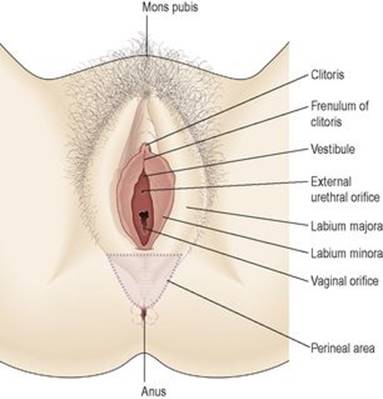
Figure 18.2 The external genitalia of the female.
Labia majora
These are the two large folds forming the boundary of the vulva. They are composed of skin, fibrous tissue and fat and contain large numbers of sebaceous glands. Anteriorly the folds join in front of the symphysis pubis, and posteriorly they merge with the skin of the perineum. At puberty, hair grows on the mons pubis and on the lateral surfaces of the labia majora.
Labia minora
These are two smaller folds of skin between the labia majora, containing numerous sebaceous glands.
The cleft between the labia minora is the vestibule. The vagina, urethra and ducts of the greater vestibular glands open into the vestibule.
Clitoris
The clitoris corresponds to the penis in the male and contains sensory nerve endings and erectile tissue, but it has no reproductive significance.
Hymen
The hymen is a thin layer of mucous membrane that partially occludes the opening of the vagina. It is normally incomplete to allow for passage of menstrual flow.
Vestibular glands 
The vestibular glands (Bartholin’s glands) are situated one on each side near the vaginal opening. They are about the size of a small pea and have ducts, opening into the vestibule immediately lateral to the attachment of the hymen. They secrete mucus that keeps the vulva moist.
Blood supply, lymph drainage and nerve supply
Arterial supply
This is by branches from the internal pudendal arteries that branch from the internal iliac arteries and by external pudendal arteries that branch from the femoral arteries.
Venous drainage
This forms a large plexus which eventually drains into the internal iliac veins.
Lymph drainage
This is through the superficial inguinal nodes.
Nerve supply
This is by branches from pudendal nerves.
Perineum
The perineum is the area extending from the base of the labia minora to the anal canal. It is roughly triangular and consists of connective tissue, muscle and fat. It gives attachment to the muscles of the pelvic floor (p. 416).
Internal genitalia
The internal organs of the female reproductive system (Figs 18.3 and 18.4) lie in the pelvic cavity and consist of the vagina, uterus, two uterine tubes and two ovaries.

Figure 18.3 The female reproductive organs in the pelvis.
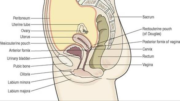
Figure 18.4 Lateral view of the female reproductive organs in the pelvis and their associated structures.
Vagina 
The vagina is a fibromuscular tube lined with stratified squamous epithelium (Fig. 3.14, p. 35), connecting the external and internal organs of reproduction. It runs obliquely upwards and backwards at an angle of about 45° between the bladder in front and rectum and anus behind. In the adult, the anterior wall is about 7.5 cm long and the posterior wall about 9 cm long. The difference is due to the angle of insertion of the cervix through the anterior wall.
Structure of the vagina
The vagina has three layers: an outer covering of areolar tissue, a middle layer of smooth muscle and an inner lining of stratified squamous epithelium that forms ridges or rugae. It has no secretory glands but the surface is kept moist by cervical secretions. Between puberty and the menopause, Lactobacillus acidophilus bacteria are normally present, which secrete lactic acid, maintaining the pH between 4.9 and 3.5. The acidity inhibits the growth of most other micro-organisms that may enter the vagina from the perineum.
Blood supply, lymph drainage and nerve supply
Arterial supply
An arterial plexus is formed round the vagina, derived from the uterine and vaginal arteries, which are branches of the internal iliac arteries.
Venous drainage
A venous plexus, situated in the muscular wall, drains into the internal iliac veins.
Lymph drainage
This is through the deep and superficial iliac glands.
Nerve supply
This consists of parasympathetic fibres from the sacral outflow, sympathetic fibres from the lumbar outflow and somatic sensory fibres from the pudendal nerves.
Functions of the vagina
The vagina acts as the receptacle for the penis during sexual intercourse (coitus), and provides an elastic passageway through which the baby passes during childbirth.
Uterus
The uterus is a hollow muscular pear-shaped organ, flattened anteroposteriorly. It lies in the pelvic cavity between the urinary bladder and the rectum (Fig. 18.4).
In most women, it leans forward (anteversion), and is bent forward (anteflexion) almost at right angles to the vagina, so that its anterior wall rests partly against the bladder below, and forming the vesicouterine pouch between the two organs.
When the body is upright, the uterus lies in an almost horizontal position. It is about 7.5 cm long, 5 cm wide and its walls are about 2.5 cm thick. It weighs from 30 to 40 grams. The parts of the uterus are the fundus, body and cervix (Fig. 18.3).
Fundus
This is the dome-shaped part of the uterus above the openings of the uterine tubes.
Body
This is the main part. It is narrowest inferiorly at the internal os where it is continuous with the cervix.
Cervix (‘neck’ of the uterus)
This protrudes through the anterior wall of the vagina, opening into it at the external os.
Structure
The walls of the uterus are composed of three layers of tissue: perimetrium, myometrium and endometrium (Fig. 18.5).

Figure 18.5 A section of the uterus.
Perimetrium
This is peritoneum, which is distributed differently on the various surfaces of the uterus (Fig. 18.4).
Anteriorly it lies over the fundus and the body where it is folded on to the upper surface of the urinary bladder. This fold of peritoneum forms the vesicouterine pouch.
Posteriorly the peritoneum covers the fundus, the body and the cervix, then it folds back on to the rectum to form the rectouterine pouch (of Douglas).
Laterally, only the fundus is covered because the peritoneum forms a double fold with the uterine tubes in the upper free border. This double fold is the broad ligament, which, at its lateral ends, attaches the uterus to the sides of the pelvis.
Myometrium
This is the thickest layer of tissue in the uterine wall. It is a mass of smooth muscle fibres interlaced with areolar tissue, blood vessels and nerves.
Endometrium
This consists of columnar epithelium containing a large number of mucus-secreting tubular glands. It is divided functionally into two layers:
• The functional layer is the upper layer and it thickens and becomes rich in blood vessels in the first half of the menstrual cycle. If the ovum is not fertilised and does not implant, this layer is shed during menstruation.
• The basal layer lies next to the myometrium, and is not lost during menstruation. It is the layer from which the fresh functional layer is regenerated during each cycle.
The upper two-thirds of the cervical canal is lined with this mucous membrane. Lower down, however, the mucosa changes, becoming stratified squamous epithelium, which is continuous with the lining of the vagina itself.
Blood supply, lymph drainage and nerve supply
Arterial supply
This is by the uterine arteries, branches of the internal iliac arteries. They pass up the lateral aspects of the uterus between the two layers of the broad ligaments. They supply the uterus and uterine tubes and join with the ovarian arteries to supply the ovaries.
Venous drainage
The veins follow the same route as the arteries and eventually drain into the internal iliac veins.
Lymph drainage
Deep and superficial lymph vessels drain lymph from the uterus and the uterine tubes to the aortic lymph nodes and groups of nodes associated with the iliac blood vessels.
Nerve supply
The nerves supplying the uterus and the uterine tubes consist of parasympathetic fibres from the sacral outflow and sympathetic fibres from the lumbar outflow.
Supporting structures
The uterus is supported in the pelvic cavity by surrounding organs, muscles of the pelvic floor and ligaments that suspend it from the walls of the pelvis (Fig. 18.6).

Figure 18.6 The main ligaments supporting the uterus. Only one side is shown.
Broad ligaments
These are formed by a double fold of peritoneum, one on each side of the uterus. They hang down from the uterine tubes as though draped over them and at their lateral ends they are attached to the sides of the pelvis. The uterine tubes are enclosed in the upper free border and near the lateral ends they penetrate the posterior wall of the broad ligament and open into the peritoneal cavity. The ovaries are attached to the posterior wall, one on each side. Blood and lymph vessels and nerves pass to the uterus and uterine tubes between the layers of the broad ligaments.
Round ligaments
These are bands of fibrous tissue between the two layers of broad ligament, one on each side of the uterus. They pass to the sides of the pelvis then through the inguinal canal to end by fusing with the labia majora.
Uterosacral ligaments
These originate from the posterior walls of the cervix and vagina and extend backwards, one on each side of the rectum, to the sacrum.
Transverse cervical (cardinal) ligaments
These extend one from each side of the cervix and vagina to the side walls of the pelvis.
Pubocervical fascia
This extends forward from the transverse cervical ligaments on each side of the bladder and is attached to the posterior surface of the pubic bones.
Functions of the uterus
After puberty, the endometrium of the uterus goes through a regular monthly cycle of changes, the menstrual cycle, under the control of hypothalamic and anterior pituitary hormones (see Ch. 9). The purpose of the menstrual cycle is to prepare the uterus to receive, nourish and protect a fertilised ovum. The cycle is usually regular, lasting between 26 and 30 days. If the ovum is not fertilised a new cycle begins with a short period of bleeding (menstruation).
If the ovum is fertilised the zygote embeds itself in the uterine wall. The uterine muscle grows to accommodate the developing baby, which is called an embryo during its first 8 weeks, and a fetus for the remainder of the pregnancy. Uterine secretions nourish the ovum before it implants in the endometrium, and after implantation the rapidly expanding ball of cells is nourished by the endometrial cells themselves. This is sufficient for only the first few weeks and the placenta is the organ that takes over thereafter (see Ch. 5). The placenta, which is attached to the fetus by the umbilical cord, is also firmly attached to the wall of the uterus, and provides the route by which the growing baby receives oxygen and nutrients, and gets rid of its wastes. During pregnancy, which normally lasts about 40 weeks, the muscular walls of the uterus are prevented from contracting and expelling the baby early by high levels of the hormone progesterone secreted by the placenta. At the end of pregnancy (at term) the hormone oestrogen, which increases uterine contractility, becomes the predominant sex hormone in the blood. Additionally, oxytocin is released from the posterior pituitary, and also stimulates contraction of the uterine muscle. Control of oxytocin release is by positive feedback (see also Fig. 9.5, p. 212). During labour, the uterus forcefully expels the baby by means of powerful rhythmical contractions.
Uterine tubes
The uterine (Fallopian) tubes (Fig. 18.3) are about 10 cm long and extend from the sides of the uterus between the body and the fundus. They lie in the upper free border of the broad ligament and their trumpet-shaped lateral ends penetrate the posterior wall, opening into the peritoneal cavity close to the ovaries. The end of each tube has fingerlike projections called fimbriae. The longest of these is the ovarian fimbria, which is in close association with the ovary.
Structure
The uterine tubes have an outer covering of peritoneum (broad ligament), a middle layer of smooth muscle and are lined with ciliated epithelium.
Blood supply, lymph drainage and nerve supply
These are as for the uterus.
Functions
The uterine tubes move the ovum from the ovary to the uterus by peristalsis and ciliary movement. The mucus secreted by the mucosa provides ideal conditions for movement of ova and spermatozoa. Fertilisation of the ovum usually takes place in the uterine tube, and the zygote is propelled into the uterus for implantation.
Ovaries 
The ovaries (Fig. 18.3) are the female gonads (glands producing sex hormones and the ova), and they lie in a shallow fossa on the lateral walls of the pelvis. They are 2.5 to 3.5 cm long, 2 cm wide and 1 cm thick. Each is attached to the upper part of the uterus by the ovarian ligament and to the back of the broad ligament by a broad band of tissue, the mesovarium. Blood vessels and nerves pass to the ovary through the mesovarium (Fig. 18.7).

Figure 18.7 A section of an ovary showing the stages of development of one ovarian follicle.
Structure
The ovaries have two layers of tissue.
Medulla
This lies in the centre and consists of fibrous tissue, blood vessels and nerves.
Cortex
This surrounds the medulla. It has a framework of connective tissue, or stroma, covered by germinal epithelium. It contains ovarian follicles in various stages of maturity, each of which contains an ovum. Before puberty the ovaries are inactive but the stroma already contains immature (primordial) follicles, which the female has from birth. During the childbearing years, about every 28 days, one ovarian follicle (Graafian follicle) matures, ruptures and releases its ovum into the peritoneal cavity. This is called ovulation and it occurs during most menstrual cycles (Figs 18.7 and 18.8). Following ovulation, the ruptured follicle develops into the corpus luteum (meaning ‘yellow body’), which in turn will leave a small permanent scar of fibrous tissue called the corpus albicans (meaning ‘white body’) on the surface of the ovary.
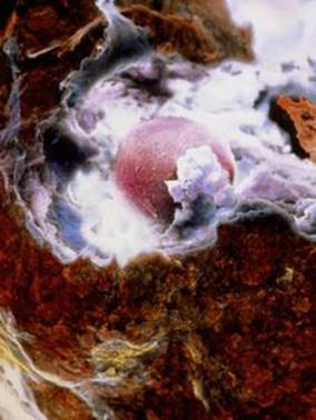
Figure 18.8 The moment of ovulation: scanning electron micrograph of an ovum (pink) emerging through the surface of the ovary (brown).
Blood supply, lymph drainage and nerve supply
Arterial supply
This is by the ovarian arteries, which branch from the abdominal aorta just below the renal arteries.
Venous drainage
This is into a plexus of veins behind the uterus from which the ovarian veins arise. The right ovarian vein opens into the inferior vena cava and the left into the left renal vein.
Lymph drainage
This is to the lateral aortic and preaortic lymph nodes. The lymph vessels follow the same route as the arteries.
Nerve supply
The ovaries are supplied by parasympathetic nerves from the sacral outflow and sympathetic nerves from the lumbar outflow.
Functions
The ovary is the organ in which the female gametes are stored and develop prior to ovulation. Their maturation is controlled by the hypothalamus and the anterior pituitary gland, which releases gonadotrophins (follicle stimulating hormone, FSH, and luteinising hormone, LH), both of which act on the ovary. In addition, the ovary has endocrine functions, and releases hormones essential to the physiological changes during the reproductive cycle. The source of these hormones, oestrogen and progesterone, is the follicle itself. During the first half of the cycle, while the ovum is developing within the follicle, the follicle secretes increasing amounts of oestrogen. However, after ovulation, the corpus luteum secretes primarily progesterone, with some oestrogen (Fig 18.9). The significance of this is discussed under the menstrual cycle (see below).

Figure 18.9 Female reproductive hormones and target tissues.
Puberty in the female
Puberty is the age at which the internal reproductive organs reach maturity, usually between the ages of 12 and 14. This is called the menarche, and marks the beginning of the childbearing period. The ovaries are stimulated by the gonadotrophins from the anterior pituitary: follicle stimulating hormone and luteinising hormone.
A number of physical and psychological changes take place at puberty:
• the uterus, the uterine tubes and the ovaries reach maturity
• the menstrual cycle and ovulation begin (menarche)
• the breasts develop and enlarge
• pubic and axillary hair begins to grow
• increase in height and widening of the pelvis
• increased fat deposited in the subcutaneous tissue, especially at the hips and breasts.
The reproductive cycle
This is a series of events, occurring regularly in females every 26 to 30 days throughout the childbearing period of about 36 years (Fig. 18.10). The cycle consists of a series of changes taking place concurrently in the ovaries and uterine walls, stimulated by changes in blood concentrations of hormones (Fig. 18.10B and D). Hormones secreted during the cycle are regulated by negative feedback mechanisms.
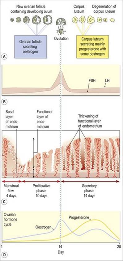
Figure 18.10 Summary of one female reproductive cycle: A. Ovarian cycle; maturation of follicle and development of corpus luteum. B. Anterior pituitary cycle; LH and FSH levels. C. Uterine cycle; menstrual, proliferative and secretory phases. D. Ovarian hormone cycle; oestrogen and progesterone levels.
The hypothalamus secretes luteinising hormone releasing hormone (LHRH), which stimulates the anterior pituitary to secrete (see Table 9.1, p. 211):
• follicle stimulating hormone (FSH), which promotes the maturation of ovarian follicles and the secretion of oestrogen, leading to ovulation. FSH is therefore predominantly active in the first half of the cycle. Its secretion is suppressed once ovulation has taken place, to prevent other follicles maturing during the current cycle
• luteinising hormone (LH), which triggers ovulation, stimulates the development of the corpus luteum and the secretion of progesterone.
The hypothalamus responds to changes in the blood levels of oestrogen and progesterone. It is stimulated by high levels of oestrogen alone (as happens in the first half of the cycle) but suppressed by oestrogen and progesterone together (as happens in the second half of the cycle).
The average length of the cycle is about 28 days. By convention the days of the cycle are numbered from the beginning of the menstrual phase, which usually lasts about 4 days. This is followed by the proliferative phase (about 10 days), then by the secretory phase (about 14 days).
Menstrual phase
When the ovum is not fertilised, the corpus luteum starts to degenerate. (In the event of pregnancy, the corpus luteum is supported by human chorionic gonadotrophin (hCG) secreted by the developing embryo). Progesterone and oestrogen levels therefore fall, and the functional layer of the endometrium, which is dependent on high levels of these ovarian hormones, is shed in menstruation (Fig. 18.10C). The menstrual flow consists of the secretions from endometrial glands, endometrial cells, blood from the degenerating capillaries and the unfertilised ovum.
High circulating levels of ovarian progesterone and oestrogen inhibit the anterior pituitary, blocking the release of FSH and LH, and should pregnancy occur then rising oestrogen and progesterone levels therefore prevent the maturation and release of another ovum. After degeneration of the corpus luteum, however, falling levels of oestrogen and progesterone lead to resumed anterior pituitary activity, rising FSH levels and the initiation of the next cycle.
Proliferative phase
At this stage an ovarian follicle, stimulated by FSH, is growing towards maturity and is producing oestrogen, which stimulates proliferation of the functional layer of the endometrium in preparation for the reception of a fertilised ovum. The endometrium thickens, becoming very vascular and rich in mucus-secreting glands. Rising levels of oestrogen are responsible for triggering a surge of LH approximately mid-cycle. This LH surge triggers ovulation, marking the end of the proliferative phase.
Secretory phase
After ovulation, LH from the anterior pituitary stimulates development of the corpus luteum from the ruptured follicle, which produces progesterone and some oestrogen. Under the influence of progesterone, the endometrium becomes oedematous and the secretory glands produce increased amounts of watery mucus. This is believed to assist the passage of the spermatozoa through the uterus to the uterine tubes where the ovum is usually fertilised. There is a similar increase in the secretion of watery mucus by the glands of the uterine tubes and by cervical glands that lubricate the vagina.
The ovum may survive in a fertilisable form for a very short time after ovulation, probably as little as 8 hours. The spermatozoa, deposited in the vagina during intercourse, may be capable of fertilising the ovum for only about 24 hours although they can survive for several days. This means that the period in each cycle during which fertilisation can occur is relatively short. Observable changes in the woman’s body occur around the time of ovulation. Cervical mucus, normally thick and dry, becomes thin, elastic and watery, and body temperature rises by a small but measurable amount immediately following ovulation. Some women experience abdominal discomfort in the middle of the cycle, thought to correspond to rupture of the follicle and release of its contents into the abdominal cavity.
If the ovum is not fertilised, the corpus luteum degenerates and dies, menstruation occurs and a new cycle begins.
If the ovum is fertilised there is no breakdown of the endometrium and no menstruation. The fertilised ovum (zygote) travels through the uterine tube to the uterus where it becomes embedded in the wall and produces human chorionic gonadotrophin (hCG), which is similar to anterior pituitary luteinising hormone. This hormone keeps the corpus luteum intact, enabling it to continue secreting progesterone and oestrogen for the first 3 to 4 months of the pregnancy, inhibiting the maturation of further ovarian follicles (Figure 18.11). During that time the placenta develops and produces oestrogen, progesterone and gonadotrophins. 


Figure 18.11 Summary of the stages of development of the ovum and the associated hormones.
This is summarised in Figure 18.11. Box 18.1 summarises the reproductive functions of oestrogen and progesterone.
Box 18.1 Reproductive functions of oestrogen and progesterone
Oestrogen
• Development of secondary sexual characteristics at puberty
• Stimulates and supports thickening of uterine lining during proliferative phase
• Triggers LH surge mid-cycle, stimulating ovulation
• Stimulates anterior pituitary secretion of FSH and LH in first half of cycle
Progesterone
• Stimulates and supports thickening and increased glandular development of uterine lining during secretory phase
• With oestrogen, inhibits secretion of FSH and LH from the anterior pituitary in second half of cycle
Menopause
The menopause (climacteric) usually occurs between the ages of 45 and 55 years, marking the end of the childbearing period. It may occur suddenly or over a period of years, sometimes as long as 10 years, and is caused by a progressive reduction in oestrogen levels, as the number of functional follicles in the ovaries declines with age. The ovaries gradually become less responsive to FSH and LH, and ovulation and the menstrual cycle become irregular, eventually ceasing. Several other phenomena may occur at the same time, including:
• short-term unpredictable vasodilation with flushing, sweating and palpitations, causing discomfort and disturbance of the normal sleep pattern
• shrinkage of the breasts
• axillary and pubic hair become sparse
• atrophy of the sex organs
• episodes of uncharacteristic behaviour, e.g. irritability, mood changes
• gradual thinning of the skin
• loss of bone mass predisposing to osteoporosis (p. 421)
• slow increase in blood cholesterol levels that increase the risk of cardiovascular disease in postmenopausal women to that in males of the same age.
Similar changes occur after bilateral irradiation or surgical removal of the ovaries.
Breasts 
The breasts or mammary glands are accessory glands of the female reproductive system. They exist also in the male, but in only a rudimentary form.
Structure
The mammary glands or breasts (Fig 18.12) consist of varying amounts of glandular tissue, responsible for milk production, supported by fatty tissue and fibrous connective tissue that anchor the breast to the chest wall.
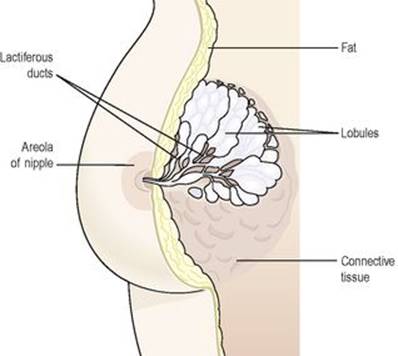
Figure 18.12 Structure of the breast.
Each breast contains about 20 lobes, each of which contains a number of glandular structures called lobules, where milk is produced. Lobules open into tiny lactiferous ducts, which drain milk towards the nipple. Supporting fatty and connective tissues run through the breast, surrounding the lobules, and the breast itself is covered in subcutaneous fat. In the lactating breast, glandular tissue proliferates to support milk production, and recedes again after lactation stops.
The nipple
This is a small conical eminence at the centre of the breast surrounded by a pigmented area, the areola. On the surface of the areola are numerous sebaceous glands (Montgomery’s tubercles), which lubricate the nipple during lactation.
Blood supply, lymph drainage and nerve supply
Arterial supply
The breasts are supplied with blood from the thoracic branches of the axillary arteries and from the internal mammary and intercostal arteries.
Venous drainage
This is formed by an anastomotic circle round the base of the nipple from which branches carry the venous blood to the circumference, and end in the axillary and mammary veins.
Lymph drainage
(see Fig. 6.1, p. 128). This is mainly into the superficial axillary lymph vessels and nodes. Lymph may drain through the internal mammary nodes if the superficial route is obstructed.
Nerve supply
The breasts are supplied by branches from the 4th, 5th and 6th thoracic nerves, which contain sympathetic fibres. There are numerous somatic sensory nerve endings in the breast, especially around the nipple. When these touch receptors are stimulated by sucking, impulses pass to the hypothalamus and the flow of the hormone oxytocin is increased, promoting the release of milk.
Functions
In the female, the breasts are small and immature until puberty. Thereafter they grow and develop under the influence of oestrogen and progesterone. During pregnancy these hormones stimulate further growth. After the baby is born the hormone prolactin (p. 211) from the anterior pituitary stimulates the production of milk, and oxytocin (p. 212) from the posterior pituitary stimulates the release of milk in response to the stimulation of the nipple by the sucking baby, by a positive feedback mechanism.
Male reproductive system
Learning outcomes
After studying this section, you should be able to:
describe the structure and function of the testes
outline the structure and function of the spermatic cords
describe the secretions that pass into the spermatic fluid
explain the process of ejaculation
list the main changes occurring at puberty in the male.
The male reproductive system is shown in Figure 18.13.

Figure 18.13 The male reproductive organs and their associated structures.
Scrotum
The scrotum is a pouch of deeply pigmented skin, fibrous and connective tissue and smooth muscle. It is divided into two compartments each of which contains one testis, one epididymis and the testicular end of a spermatic cord. It lies below the symphysis pubis, in front of the upper parts of the thighs and behind the penis.
Testes 
The testes (Fig. 18.14A and B) are the reproductive glands of the male and are the equivalent of the ovaries in the female. They are about 4.5 cm long, 2.5 cm wide and 3 cm thick and are suspended in the scrotum by the spermatic cords. They are surrounded by three layers of tissue.
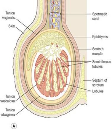
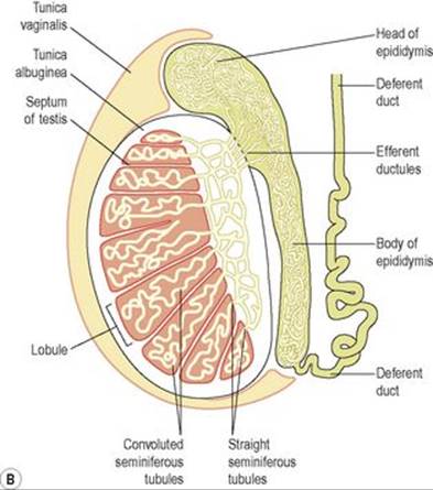
Figure 18.14 The testis: A. Section of the testis and its coverings. B. Longitudinal section of a testis and deferent duct.
Tunica vaginalis
This is a double membrane, forming the outer covering of the testes, and is a downgrowth of the abdominal and pelvic peritoneum. During early fetal life, the testes develop in the lumbar region of the abdominal cavity just below the kidneys. They then descend into the scrotum, taking with them coverings of peritoneum, blood and lymph vessels, nerves and the deferent duct. The peritoneum eventually surrounds the testes in the scrotum, and becomes detached from the abdominal peritoneum. Descent of the testes into the scrotum should be complete by the 8th month of fetal life.
Tunica albuginea
This is a fibrous covering beneath the tunica vaginalis that surrounds the testes. Ingrowths form septa, dividing the glandular structure of the testes into lobules.
Tunica vasculosa
This consists of a network of capillaries supported by delicate connective tissue.
Structure
In each testis are 200 to 300 lobules, and within each lobule are 1 to 4 convoluted loops composed of germinal epithelial cells, called seminiferous tubules. Between the tubules are groups of interstitial cells(of Leydig) that secrete the hormone testosterone after puberty. At the upper pole of the testis the tubules combine to form a single tubule. This tubule, about 6 m in its full length, is repeatedly folded and tightly packed into a mass called the epididymis. It leaves the scrotum as the deferent duct (vas deferens) in the spermatic cord. Blood and lymph vessels pass to the testes in the spermatic cords.
Functions
Spermatozoa (sperm) are produced in the seminiferous tubules of the testes, and mature as they pass through the long and convoluted epididymis, where they are stored. The hormone controlling sperm production is FSH from the anterior pituitary (p. 211). A mature sperm (Fig. 18.15) has a head, a body, and a long whip-like tail used for motility. The head is almost completely filled by the nucleus, containing its DNA. It also contains the enzymes required to penetrate the outer layers of the ovum to reach, and fuse with, its nucleus. The body of the sperm is packed with mitochondria, to fuel the propelling action of the tail that powers the sperm along the female reproductive tract.
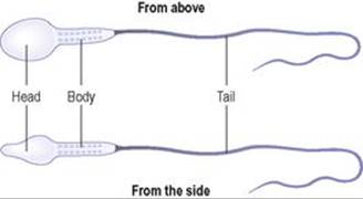
Figure 18.15 A spermatozoon.
Successful spermatogenesis takes place at a temperature about 3°C below normal body temperature. The testes are cooled by their position outside the abdominal cavity, and the thin outer covering of the scrotum has very little insulating fat. 
Spermatic cords
The spermatic cords suspend the testes in the scrotum. Each cord contains a testicular artery, testicular veins, lymphatics, a deferent duct and testicular nerves, which come together to form the cord from their various origins in the abdomen. The cord, which is covered in a sheath of smooth muscle and connective and fibrous tissues, extends through the inguinal canal (p. 416) and is attached to the testis on the posterior wall.
Blood supply, lymph drainage and nerve supply
Arterial supply
The testicular artery branches from the abdominal aorta, just below the renal arteries.
Venous drainage
The testicular vein passes into the abdominal cavity. The left vein opens into the left renal vein and the right into the inferior vena cava.
Lymph drainage
This is through lymph nodes around the aorta.
Nerve supply
This is provided by branches from the 10th and 11th thoracic nerves.
The deferent duct
This is some 45 cm long. It passes upwards from the testis through the inguinal canal and ascends medially towards the posterior wall of the bladder where it is joined by the duct from the seminal vesicle to form the ejaculatory duct(Fig. 18.16).

Figure 18.16 Section of the prostate gland and associated reproductive structures on one side.
Seminal vesicles 
The seminal vesicles are two small fibromuscular pouches lined with columnar epithelium, lying on the posterior aspect of the bladder (Fig. 18.16).
At its lower end each seminal vesicle opens into a short duct, which joins with the corresponding deferent duct to form an ejaculatory duct.
Functions
The seminal vesicles contract and expel their stored contents, seminal fluid, during ejaculation. Seminal fluid, which forms 60% of the bulk of the fluid ejaculated at male orgasm, contains nutrients to support the sperm during their journey through the female reproductive tract.
Ejaculatory ducts
The ejaculatory ducts are two tubes about 2 cm long, each formed by the union of the duct from a seminal vesicle and a deferent duct. They pass through the prostate gland and join the prostatic urethra, carrying seminal fluid and spermatozoa to the urethra (Fig. 18.16).
The ejaculatory ducts are composed of the same layers of tissue as the seminal vesicles.
Prostate gland
The prostate gland (Fig. 18.16) lies in the pelvic cavity in front of the rectum and behind the symphysis pubis, surrounding the first part of the urethra. It consists of an outer fibrous covering, a layer of smooth muscle and glandular substance composed of columnar epithelial cells.
Functions
The prostate gland secretes a thin, milky fluid that makes up about 30% of semen, and gives it its milky appearance. It contains a clotting enzyme, which thickens the semen in the vagina, increasing the likelihood of semen being retained close to the cervix.
Urethra and penis 
Urethra
The male urethra provides a common pathway for the flow of urine and semen, the combined secretions of the male reproductive organs. It is about 19 to 20 cm long and consists of three parts. The prostatic urethra originates at the urethral orifice of the bladder and passes through the prostate gland. The membranous urethra is the shortest and narrowest part and extends from the prostate gland to the bulb of the penis, after passing through the perineal membrane. The spongiose or penile urethra lies within the corpus spongiosum of the penis and terminates at the external urethral orifice in the glans penis.
There are two urethral sphincters (Fig. 18.17). The internal sphincter consists of smooth muscle fibres at the neck of the bladder above the prostate gland. The external sphincter consists of skeletal muscle fibres surrounding the membranous part.
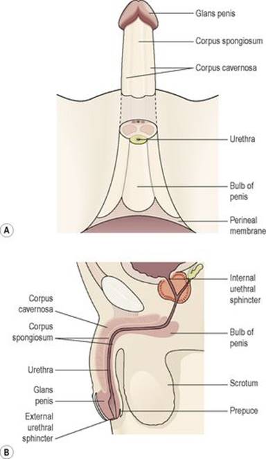
Figure 18.17 The penis: A. Viewed from below. B. Viewed from the side.
Penis 
The penis (Fig. 18.17) has a root and a body. The root lies in the perineum and the body surrounds the urethra. It is formed by three cylindrical masses of erectile tissue and smooth muscle. The erectile tissue is supported by fibrous tissue and covered with skin and has a rich blood supply.
The two lateral columns are called the corpora cavernosa and the column between them, containing the urethra, is the corpus spongiosum. At its tip it is expanded into a triangular structure known as the glans penis. Just above the glans the skin is folded upon itself and forms a movable double layer, the foreskin or prepuce. Arterial blood is supplied by deep, dorsal and bulbar arteries of the penis, which are branches from the internal pudendal arteries. A series of veins drain blood to the internal pudendal and internal iliac veins. The penis is supplied by autonomic and somatic nerves. Parasympathetic stimulation leads to filling of the spongy erectile tissue with blood, caused by arteriolar dilation and venoconstriction, which increases blood flow into the penis and obstructs outflow. The penis therefore becomes engorged and erect, essential for sexual intercourse.
Ejaculation
During ejaculation, which occurs at male orgasm, spermatozoa are expelled from the epididymis and pass through the deferent duct, the ejaculatory duct and the urethra. The semen is propelled by powerful rhythmical contraction of the smooth muscle in the walls of the deferent duct; the muscular contractions are sympathetically mediated. Muscle in the walls of the seminal vesicles and prostate gland also contracts, adding their contents to the fluid passing through the genital ducts. The force generated by these combined processes leads to emission of the semen through the external urethral sphincter (Fig. 18.18). 
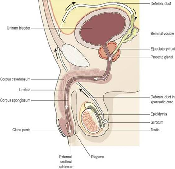
Figure 18.18 Section of the male reproductive organs. Arrows show the route taken by spermatozoa during ejaculation.
Sperm comprise only 10% of the final ejaculate, the remainder being made up of seminal and prostatic fluids, which are added to the sperm during male orgasm, as well as mucus produced in the urethra. Semen is slightly alkaline, to neutralise the acidity of the vagina. Between 2 and 5 ml of semen are produced in a normal ejaculate, and contain between 40 and 100 million spermatozoa per ml. If not ejaculated, sperm gradually lose their fertility after several months and are reabsorbed by the epididymis.
Puberty in the male
This occurs between the ages of 10 and 14. Luteinising hormone from the anterior lobe of the pituitary gland stimulates the interstitial cells of the testes to increase the production of testosterone. This hormone influences the development of the body to sexual maturity. The changes occurring at puberty include:
• growth of muscle and bone and a marked increase in height and weight
• enlargement of the larynx and deepening of the voice – it ‘breaks’
• growth of hair on the face, axillae, chest, abdomen and pubis
• enlargement of the penis, scrotum and prostate gland
• maturation of the seminiferous tubules and production of spermatozoa
• the skin thickens and becomes oilier.
In the male, fertility and sexual ability tend to decline gradually with ageing. The secretion of testosterone gradually declines, usually beginning at about 50 years of age. There is no period comparable to the female menopause.
Sexually transmitted infections
Learning outcomes
After studying this section, you should be able to:
list the principal causes of sexually transmitted infections
explain the effects of sexually transmitted infections.
This is common in all cultures and an increasing problem in many countries.
Micro-organisms responsible for sexually transmitted infections are unable to survive outside the body for long periods and have no intermediate host.
Chlamydia
The organism Chlamydia trachomatis causes inflammation of the female cervix. Infection may ascend through the reproductive tract and cause pelvic inflammatory disease (p. 453). In the male, it may cause urethritis, which may also ascend and lead to epididymitis. Chlamydia infection is often present in conjunction with other sexually transmitted diseases. The same organism causes trachoma, an eye infection that is the primary cause of blindness worldwide (p. 205).
Gonorrhoea
This is caused by Neisseria gonorrhoeae, which infects the mucosa of the reproductive and urinary tracts. In the male, suppurative urethritis occurs and the infection may spread to the prostate gland, epididymis and testes. In the female the infection may spread from vulvar glands, vagina and cervix to the body of the uterus, uterine tubes, ovaries and peritoneum. Healing by fibrosis in the female may cause obstruction of the uterine tubes, leading to infertility. In the male it may cause urethral stricture.
Non-venereal transmission of gonorrhoea may cause neonatal ophthalmia in babies born to infected mothers. The eyes become infected as the baby passes through the vagina.
Syphilis
This disease is caused by Treponema pallidum. There are three clearly marked stages. After an incubation period of several weeks, the primary sore (chancre) appears at the site of infection, e.g. the vulva, vagina, perineum, penis or round the mouth. In the female the primary sore may be undetected if it is internal. After several weeks the chancre subsides spontaneously. The secondary stage, 3 to 4 months after infection, involves systemic symptoms including lymphadenopathy, skin rashes and mucosal ulceration of the mouth and genital tract. There may then be a latent period of between 3 and 10 years. Tertiary lesions (gummas) then develop in many organs, including skin, bone and mucous membranes, and may involve the nervous system, leading to general paralysis and dementia.
Sexual transmission occurs during the primary and secondary stages when discharge from lesions is highly infectious. Congenital transmission from mother to fetus carries a high risk of stillbirth.
Trichomonas vaginalis
These protozoa cause acute vulvovaginitis with irritating, offensive discharge. It is usually sexually transmitted and is commonly present in women with gonorrhoea. Males are often asymptomatic.
Candidiasis
The yeast Candida albicans (see also p. 311) is frequently a commensal in the normal vagina and causes no problems. It is normally prevented from flourishing by vaginal acidity, but in certain circumstances it proliferates, causing candidiasis (thrush). Common precipitating factors include:
• antibiotic therapy, which kills the bacteria that keep vaginal pH low
• pregnancy
• reduced immune function
• diabetes mellitus.
In women, persistent itch is the main symptom, with discharge, swelling and erythema of the vulvar area.
Acquired immune deficiency syndrome (AIDS) and hepatitis B infection
These viral conditions may be sexually transmitted, but there are no local signs of infection. For a description of AIDS and HIV see page 376 and hepatitis B page 325.
Non-specific genital infection
This is increasingly common, especially in males. General symptoms include painful urination and urethral discharge, and there is a wide range of possible causative organisms. Some cases are not sexually transmitted and arise from, for example, urinary tract infection or the presence of a foreign body in the urethra.
Genital herpes
One form of the herpes virus, Herpes simplex 2 (HSV2) is associated with genital infections. Initial infection tends to present as clusters of small, painful ulcers on the external genitalia. There is often fever and headache. Recurrences of the disease occur because the virus establishes itself within the dorsal root ganglion, from where it can be reactivated from time to time.
Diseases of the female reproductive system
Learning outcomes
After studying this section, you should be able to:
describe the causes and consequences of pelvic inflammatory disease
discuss the disorders of the vulva
define the term imperforate hymen
outline the causes and effects of cervical carcinoma
discuss the main pathologies of the uterus and uterine tubes
describe the causes and effects of ovarian disease
describe the causes of female infertility
discuss the principal disorders of the female breast.
Pelvic inflammatory disease (PID)
This condition is usually a consequence of sexually transmitted infections. It usually begins as vulvovaginitis and may spread upwards to the cervix, uterus, uterine tubes and ovaries. Upward spread can also occur when infection is present in the vagina before a surgical procedure, childbirth or miscarriage, especially if some of the products of conception are retained.
Complications of PID include:
• infertility due to obstruction of uterine tubes
• peritonitis
• intestinal obstruction due to adhesions between the bowel and the uterus and/or uterine tubes
• bacteraemia, which may lead to meningitis, endocarditis or suppurative arthritis.
Vulvar dystrophies
Atrophic dystrophy
This is thinning of vulvar epithelium and the formation of fibrous tissue, occurring after the menopause due to oestrogen withdrawal. It predisposes to infection, especially in debilitated women, and to malignant epithelial neoplasia.
Vulvar intraepithelial neoplasia (VIN)
This refers to early neoplastic (cancerous) changes in epithelial cells of the vulva. It is commonest in younger women, often those infected with human papilloma virus, and may proceed to malignancy (see also cervical intraepithelial neoplasia, below).
Imperforate hymen
This congenital abnormality may not be noticed until the onset of menstruation. When complete (imperforate), the hymen forms a barrier in the vagina. Blood accumulates in the vagina, uterus and uterine tubes with each menstrual cycle, and it may enter the peritoneal cavity and cause peritonitis. The uterine tubes may become obstructed by coagulated blood, leading to infertility.
Disorders of the cervix
Cervical carcinoma
Dysplastic changes, referred to as cervical intraepithelial neoplasia (CIN) begin in the deepest layer of cervical epithelium, usually at the junction of the stratified squamous epithelium of the lower third of the cervical canal with the secretory epithelium of the upper two-thirds. Dysplasia may progress to involve the full thickness of epithelium. Not all dysplasias develop into malignant disease, but it is not possible to predict how far development will go, and whether it will remain static or regress. Early detection with a screening programme can allow abnormal tissue to be removed before it becomes malignant. Established malignancy is staged according to how extensive the tumour is. Stage I refers to disease confined to the cervix. Stages II through IV reflect increasing spread, including involvement of the rectum, bladder and structures outwith the pelvis. Early spread is via lymph nodes and local spread is commonly to the uterus, vagina, bladder and rectum. In the late stages spread via the blood to the liver, lungs and bones may occur.
The disease takes 15 to 20 years to develop and it occurs mostly between 35 and 50 years of age. It is likely that a significant proportion of cases are due to the transmission of some sexually transmitted carcinogen. Risk factors include having frequent sexual intercourse with multiple partners from an early age, all of which increase the likelihood of being exposed to a carcinogenic agent. Barrier contraceptives protect against the disease. The human papillomavirus (HPV), which causes genital warts, is strongly associated with this cancer (see also p. 362).
Disorders of the uterine body
Endometritis
This is usually caused by non-specific infection, following childbirth or miscarriage, especially if fragments of membranes or placenta have been retained in the uterus. It may also be caused by an intrauterine contraceptive device (IUD). The inflammation may subside after removal of retained products or the IUD. The infection may spread to surrounding pelvic structures, e.g. uterine tubes, or deeper layers of the uterus.
Endometriosis
This is the growth of endometrial tissue outside the uterus, usually in the ovaries, uterine tubes and other pelvic structures. The ectopic tissue, like the uterine endometrium, responds to fluctuations in sex hormone levels during the menstrual cycle, causing menstrual-type bleeding into the lower abdomen and, in the ovaries, the formation of coloured cysts, ‘chocolate cysts’. There is intermittent pain due to swelling, and recurrent haemorrhage causes fibrous tissue formation. Ovarian endometriosis may lead to pelvic inflammation, infertility and extensive pelvic adhesions, involving the ovaries, uterus, uterine ligaments and the bowel.
Adenomyosis
This is the growth of endometrium within the myometrium. The ectopic tissue may cause general or localised uterine enlargement. The lesions may cause dysmenorrhoea and irregular excessive bleeding (menorrhagia), usually beginning between 40 and 50 years of age.
Endometrial hyperplasia
Hyperplasia of the endometrium is associated with high blood oestrogen levels, e.g. in obesity, oestrogen therapy or an ovarian tumour. Sometimes it is associated with increased risk of malignant change.
Leiomyoma (fibroid, myoma)
These are very common, often multiple, benign tumours of myometrium. They are firm masses of smooth muscle encapsulated in compressed muscle fibres and they vary greatly in size. Large tumours may undergo degenerative changes if they outgrow their blood supply, leading to necrosis, fibrosis and calcification. They develop during the reproductive period and may be hormone dependent, enlarging during pregnancy and when oral contraceptives are used. They tend to regress after the menopause. Large tumours may cause pelvic discomfort, frequency of micturition, menorrhagia, irregular bleeding, dysmenorrhoea and reduced fertility. Malignant change is rare.
Endometrial carcinoma
This occurs mainly in women who have never been pregnant and is most common between 50 and 60 years of age. The incidence is increased when an oestrogen-secreting tumour is present and in women who are obese, hypertensive or diabetic, because they tend to have a high level of blood oestrogen. As the tumour grows, there is often ulceration and vaginal bleeding. Endometrium has no lymphatics, so lymph spread is delayed until there is extensive local spread that involves other pelvic structures. Distant metastases, spread in blood or lymph, develop later, most commonly in the liver, lungs and bones. Invasion of the ureters leads to hydronephrosis and uraemia, commonly the cause of death.
Disorders of the uterine tubes and ovaries
Acute salpingitis
Salpingitis is inflammation of the uterine tubes. It is usually due to infection spreading from the uterus, and only occasionally from the peritoneal cavity. The uterine tubes may be left permanently damaged by fibrous scar tissue, which can cause obstruction and infertility. Infection may spread into the peritoneum and involve the ovaries.
Ectopic pregnancy
This is the implantation of a fertilised ovum outside the uterus, most commonly in a uterine tube. As the fetus grows the tube may rupture and its contents enter the peritoneal cavity, causing acute inflammation (peritonitis) and possibly severe intraperitoneal haemorrhage.
Ovarian tumours
The majority of ovarian tumours are benign, usually occurring between 20 and 45 years of age. The rest occur mostly between 45 and 65 years and are divided between borderline malignancy (low-grade cancer) and frank malignancy.
Ovarian cancer is associated with developed societies, higher socioeconomic groups, and, in some families, a genetic susceptibility. Pregnancy and the use of the contraceptive pill have a protective effect. Most malignancies of the ovary arise from epithelium, but some arise from the germ cells of the ovary, or from stromal cells.
Metastatic ovarian tumours
The ovaries are a common site of metastatic spread from primary tumours in other pelvic organs, the breast, stomach, pancreas and biliary tract.
Female infertility
This common condition may be due to:
• blockage of uterine tubes, often the consequence of pelvic inflammatory disease
• anatomical abnormalities, e.g. retroversion (tilting backwards) of the uterus
• endocrine factors; any abnormalities of the glands and hormones governing the menstrual cycle can interfere with, for example, ovulation or the uterine cycle
• low body weight, e.g. in anorexia nervosa, or severe malnourishment
• endometriosis.
Disorders of the breast
Mastitis (inflammation of the breast)
Acute non-suppurative mastitis
This occurs during lactation and is associated with painful congestion and oedema of the breast. It is of hormonal origin.
Acute suppurative (pyogenic) mastitis
The microbes enter through a nipple abrasion caused by the infant sucking. The most common causative microbes are Staphylococcus aureus and Streptococcus pyogenes usually acquired by the infant while in hospital. The infection spreads along the mammary ducts of a lobe causing localised swelling and redness. If it does not resolve it can become chronic and an abscess may form.
Tumours of the breast
Benign tumours
Most breast tumours (90%) are benign. Fibroadenomas are the commonest type and occur any time after puberty; incidence peaks in the third decade. Some are cystic and some solid, and they usually occur in women nearing the menopause. They may originate from secretory cells, fibrous tissue or from ducts.
Malignant tumours
The most common types of tumour are usually painless lumps found in the upper outer quadrant of the breast. There is considerable fibrosis around the tumour that may cause retraction of the nipple and necrosis and ulceration of the overlying skin. It is increasingly common between 35 and 70 years.
Early spread beyond the breast is via lymph to the axillary and internal mammary nodes. Local invasion involves the pectoral muscles and the pleura. Blood-spread metastases may occur later in many organs and bones, especially lumbar and thoracic vertebrae. The causes of breast cancer are not known, but an important predisposing factor appears to be high oestrogen exposure. Women with an early menarche, a late menopause, and no pregnancies have a higher than normal risk because they experience more menstrual cycles in their lifetimes, and each monthly cycle brings with it the oestrogen surge seen during the proliferative phase (p. 445). A genetic component is also likely, with close relatives of breast cancer sufferers having an elevated risk of developing the disease. In about 15% of cases, the disease is linked to the presence of one of two faulty genes, BRCA1 and BRCA2. Women carrying one of these genes have a very high (80–90%) chance of developing the disease, and there is also increased risk of ovarian and bowel cancer. In women carrying these genes, the average age at which the disease appears is significantly lower than in those without the gene. One per cent of all breast cancer occurs in men.
Diseases of the male reproductive system
Learning outcomes
After studying this section, you should be able to:
outline the causes and effects of penile and urethral infections
describe the main pathologies of the testis
discuss the principal disorders of the prostate gland
list the main causes of male infertility.
Infections of the penis
Inflammation of the glans and prepuce may be caused by a specific or non-specific infection. In non-specific infections, or balanitis, lack of personal hygiene is an important predisposing factor, especially if phimosis is present, i.e. the orifice in the foreskin (prepuce) is too small to allow for its normal retraction. If the infection becomes chronic there may be fibrosis of the foreskin, which increases the phimosis.
Infections of the urethra
Gonococcal urethritis is the most common specific infection. Non-specific infection may be spread from the bladder (cystitis) or be introduced during catheterisation, cystoscopy or surgery. Both types may spread throughout the system to the prostate, seminal vesicles, epididymis and testes. If infection becomes chronic, fibrosis may cause urethral stricture or obstruction, leading to retention of urine.
Epididymis and testes
Infections
Non-specific epididymitis and orchitis are usually due to spread of infection from the urethra, commonly following prostatectomy. The microbes may spread either through the deferent duct (vas deferens) or via lymph.
Specific epididymitis
This is usually caused by gonorrhoea spread from the urethra.
Orchitis (inflammation of the testis)
This is commonly caused by mumps virus, blood-borne from the parotid glands. Acute inflammation with oedema occurs about 1 week after the appearance of parotid swelling. The infection is usually unilateral but, if bilateral, severe damage to germinal epithelium of the seminiferous tubules may result in sterility.
Undescended testis (cryptorchidism)
During embryonic life the testes develop within the abdominal cavity, but descend into the scrotum prior to birth. If they fail to do this and the condition is not corrected, infertility is likely to follow and the risk of testicular cancer is increased.
Hydrocele
This is the most common form of scrotal swelling and is accumulation of serous fluid in the tunica vaginalis. The onset may be acute and painful or chronic. It may be congenital or be secondary to another disorder of the testis or epididymis.
Testicular tumours
Most testicular tumours are malignant, the commonest malignancies in young men. They occur in childhood and early adulthood when the affected testis has not descended or has been late in descending into the scrotum. The tumour tends to remain localised for a considerable time but eventually spreads in lymph to pelvic and abdominal lymph nodes, and more widely in the blood. Occasionally, hormone-secreting tumours develop and may cause precocious development in boys.
Prostate gland
Infections
Acute prostatitis is usually caused by non-specific infection, spread from the urethra or bladder, often following catheterisation, cystoscopy, urethral dilation or prostate surgery. Chronic infection may follow an acute attack. Fibrosis of the gland may occur during healing, causing urethral stricture or obstruction.
Benign prostatic enlargement
Hyperplastic nodules form around the urethra and may obstruct the flow of urine, causing urinary retention. Urethral stricture may prevent the bladder emptying completely during micturition, predisposing to infection, which may spread upwards, causing pyelonephritis and other complications. Prostatic enlargement is common in men over 50, affecting up to 70% of men aged over 70. The cause is not clear, but it may be an acceleration of the ageing process associated with the decline in androgen secretion, which changes the androgen/oestrogen balance.
Malignant prostatic tumours
These are a relatively common cause of death in men over 50. The carcinogen is not known but changes in the androgen/oestrogen balance may be significant or viruses may be involved. Invasion of local tissues is widespread before lymph-spread metastases develop in pelvic and abdominal lymph nodes. Blood-spread metastases in bone are common and bone formation rather than bone destruction is a common feature. Lumbar vertebrae are common sites, possibly due to retrograde spread along the walls of veins. Bone metastases are often the first indication of malignant prostatic tumours.
Breast
Breast tissue in men consists of ducts and stroma only.
Gynaecomastia
This is proliferation of breast tissue in men. It usually affects only one breast and is benign. It is common in adolescents and older men, and is often associated with:
• endocrine disorders, especially those associated with high oestrogen levels
• cirrhosis of the liver (p. 325)
• malnutrition
• some drugs, e.g. chlorpromazine, spironolactone, digoxin
• Klinefelter’s syndrome, a genetic disorder with testicular atrophy and absence of spermatogenesis.
Malignant tumours
These develop in a small number of men, usually in the older age groups.
Male infertility
This may be due to:
• endocrine disorders
• obstruction of the deferent duct
• failure of erection or ejaculation during intercourse
• vasectomy
• suppression of spermatogenesis by, e.g., ionising radiation, chemotherapy and other drugs.
 For a range of self-assessment exercises on the topcs in this chapter, visitwww.rossandwilson.com.