
26
I. OVERVIEW
The ultrafiltrate entering the proximal tubule (PT) from the Bowman space has a composition that is almost identical to that of plasma. It contains over 150 different components, but the major constituents are inorganic ions (Na+, K+, Mg2+, Ca2+, Cl−, HCO3−, H+, and phosphates), sugars, amino acids and peptides, creatinine, and urea. It also contains large amounts of water. The renal tubule's function is to recover >99% of the water and the majority of the solutes before they reach the bladder. Most is recovered within the first few millimeters of the PT, including virtually all organic compounds (sugars, amino acids, peptides, and organic acids) and two thirds of the filtered ions and water. Much of this material is recovered paracellularly by osmosis, made possible by the tubule wall's inherently leaky nature. The PT also actively secretes a number of organic compounds into the tubule lumen for subsequent urinary excretion. Principal sites for reabsorption, secretion, and regulation of various solutes along the nephron are summarized in Figure 27.19.
II. PRINCIPLES
The PT is a high-capacity, “leaky” transport epithelium drawn into a ~50-μm tube (Figure 26.1A). The initial portion of the tube is coiled (the proximal convoluted tubule [PCT]), and it then straightens to form the proximal straight tubule (PST). The PT's primary function is isosmotic fluid reabsorption.
An epithelium's “leakiness” is a reflection of the ease with which solutes and water permeate the tight junctions between adjacent epithelial cells. Leaky epithelia are highly permeable, whereas intercellular junctions in tight epithelia are relatively impermeant (see 4·II·E·2).
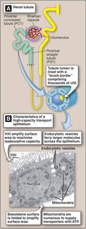
Figure 26.1
Proximal tubule structure.
ATP = adenosine triphosphate.
A. Cellular structure
The PT reabsorbs ~120 L of fluid and solutes per day. The enormity of this load is reflected in the ultrastructure of the epithelial cells that make up its walls, which are packed with mitochondria, and their surface membranes are specialized to amplify surface area (see Figure 26.1B).
1. Metabolism: The apical and basolateral membranes of PT epithelial cells are packed with channels and transporters for retrieval and secretion of inorganic ions and other solutes. Reabsorption is driven by ion gradients generated by adenosine triphosphate (ATP)-dependent pumps, so the cytoplasm is dense with mitochondria to supply the PT's high metabolic needs.
2. Surface area: PT apical and basolateral membranes are extensively modified to increase their surface area. The membrane expanse is required to accommodate high numbers of channels and transporters and also to maximize area for contact between the epithelial cell and tubule contents. The numerous, densely packed microvilli that sprout from the apical surface create a brush border that is structurally and functionally similar to that found in the small intestine (see 31·II).
3. Junctions: Adjacent epithelia cells are connected at their apical surface by tight junctions that have a very loose structure. The junctions are highly permeable to solutes and water and, thus, the epithelium has very low electrical resistance.
C. Reabsorption
Reabsorption involves transferring water and solutes from the tubule lumen to the interstitium. Once in the interstitium, these materials are free to enter the peritubular capillary network by simple diffusion. The principal pathways and mechanisms involved in reabsorption were introduced in Unit I (see 4·III) and are summarized here.
1. Pathways: There are two paths by which materials can cross epithelia (Figure 26.2). The paracellular route lies between two adjacent epithelial cells. The permeability of the paracellular route is determined by tight junction structure. The transcellular route takes a solute through the inside of an epithelial cell and usually requires the assistance of channels or transporters to traverse the apical and basolateral membranes.
2. Motive force: Transepithelial transport is powered by ATP, and virtually all of the energy consumed during reabsorption is used to support Na+-K+ ATPase activity. The consequences of Na+-K+ ATPase activity can be broken down into five partly overlapping steps, all of which contribute to net reabsorption (Figure 26.3).
a. Step 1—Ion gradient: The Na+-K+ pump is located in the basolateral membrane. It exchanges three intracellular Na+ ions for two extracellular K+ ions, creating an inwardly directed gradient for Na+diffusion across both apical and basolateral membranes.
b. Step 2—Voltage gradient: Pumping Na+ ions into the interstitium modifies the potential difference between interstitium and PT lumen. Although the difference is small (~3 mV, lumen negative), it creates a significant driving force for ion movement.
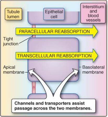
Figure 26.2
Pathways for reabsorption from the tubule lumen.
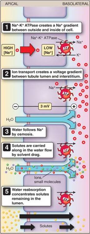
Figure 26.3
Reabsorption mechanisms.
ATP = adenosine triphosphate.
The transepithelial voltage gradient reverses polarity, from lumen-negative to lumen-positive, in the later regions of the PT. Reversal occurs because Cl− is reabsorbed preferentially in the later regions, leaving behind a net positive charge (see below).
c. Step 3—Osmotic gradient: Pumping Na+ ions into the interstitium also creates an osmotic gradient that drives water flow from the tubule lumen across the tight junctions.
d. Step 4—Solvent drag: Water flowing through the intercellular junctions in response to an osmotic gradient creates solvent drag that sweeps ions and small organic molecules along with it.
e. Step 5—Chemical gradient: Water reabsorption concentrates the solutes that are left behind in the tubule lumen, thereby creating a chemical gradient favoring diffusional reabsorption.
D. Peritubular network
The PT's ability to reabsorb large volumes of fluid is only possible with support from the peritubular capillary network, which closely follows the tubule through the kidney (see Figure 25.4). The peritubular network sustains the tubule with O2 and nutrients, but, just as importantly, it also clears recovered fluid from the interstitium before it has a chance to accumulate and reduce the gradients favoring reabsorption. The Starling forces governing fluid movement across the peritubular capillary wall are configured so as to promote reabsorption from the renal interstitium (Figure 26.4; see 19·VII·D). The main force favoring fluid reabsorption is plasma colloid osmotic pressure (πPC). Capillary hydrostatic pressure (PPC) is the principal force opposing reabsorption.
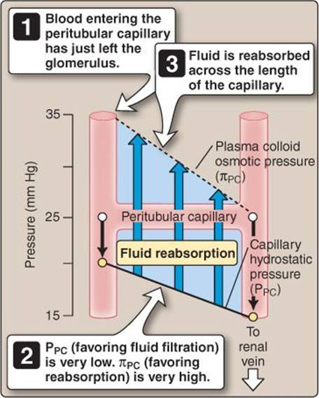
Figure 26.4
Forces controlling fluid reabsorption by peritubular capillaries.
1. Plasma colloid osmotic pressure: πPC averages 25 mm Hg in virtually all other regions of the body, but blood entering the peritubular network has just traversed the glomerulus where ~20% of its fluid was removed by filtration. The plasma proteins are concentrated as a result, which raises πPC to ~35 mm Hg.
2. Capillary hydrostatic pressure: Blood has to pass through an efferent arteriole before reaching peritubular capillaries. Efferent arterioles have a relatively high resistance, which decreases the pressure of blood entering the network to ~20 mm Hg. This is much lower than in other systemic capillary beds (~35 mm Hg). PPC then declines over the length of the capillary. The combination of a high πPC and low PPC means that the driving force for fluid absorption is strongly positive across the entire length of the capillary (Flow ∞ πPC − PPC, or ~15 mm Hg; see Figure 26.4).
III. ORGANIC SOLUTES: REABSORPTION
Plasma is laden with glucose (~4–5 mmol/L), amino acids (~2.5 mmol/L), small peptides, and organic acids (e.g., lactate, pyruvate), all of which are freely filtered into the Bowman space. These compounds represent a significant resource that must be recovered from the filtrate before it reaches the bladder. In practice, >98% of organic compounds are recovered in the early PCT and the remaining ~1%–2% are reabsorbed in the PST (Figure 26.5). Most organics are recovered by apical transporters, traverse cells by diffusion, and then are transported across the baso lateral membrane to the interstitium and vasculature. Transporter involvement means that reabsorption shows saturation kinetics (Figure 26.6).
A. Kinetics
The renal epithelium expresses a finite number of transporters, which limits solute reabsorption. If the glomerulus filters solutes in excess of maximal transporter capacity (Tm), then the transported solute will continue through the tubule and appear in urine. Plasma solute concentrations vary with intake and tissue use, but a healthy nephron is usually well equipped to recover filtered loads within a normal physiologic range. Solutes start appearing in urine in small amounts even before Tm is reached (see Figure 26.6). This region of the titration curve is said to show splay, reflecting transporter and nephron heterogeneity.
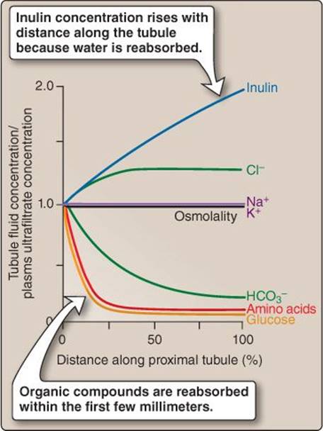
Figure 26.5
Changes in proximal tubule fluid composition with distance from the Bowman capsule.
“Filtered load” is the amount of any substance that filters from the glomerulus and enters the Bowman space per unit time (mg/min). Filtered loads are the products of glomerular filtration rate (GFR) and plasma concentration of the substance in question.
1. Transporters: Nephrons typically contain multiple transporter classes capable of transferring organic solutes across the surface membrane. The combined activity of pathways with different Tm values contributes to splay.
2. Nephrons: Nephrons show anatomic diversity, which causes differences in single-nephron GFR, transporter capacity, and transporter location along the tubule. These differences also contribute to splay.
B. Glucose
Glucose plasma concentrations vary between ~3.8 and 6.1 mmol/L in a healthy person. Glucose filters freely into the tubule, and ~98% is reabsorbed in the early PT. Uptake occurs transcellularly and is transporter mediated (Figure 26.7).
1. Apical: Glucose is recovered using two different Na+ cotransporters. They both harness the transmembrane Na+ gradient to simultaneously absorb Na+ and a glucose molecule. One of these cotransporter classes localizes primarily to the early part of the PCT, the other to the PST.
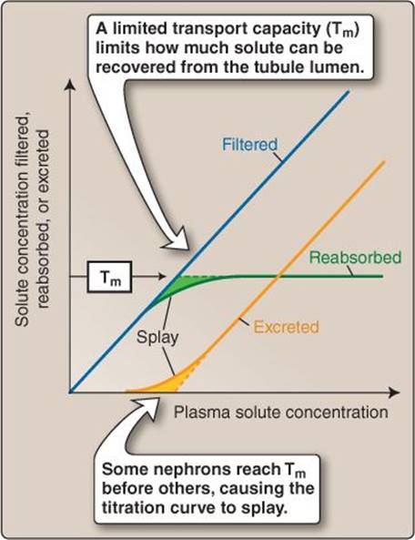
Figure 26.6
Limits to transporter-mediated solute reabsorption.
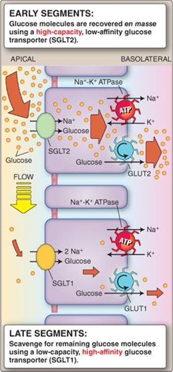
Figure 26.7
Glucose reabsorption strategies.
ATP = adenosine triphosphate;
GLUT1 and -2 = glucose transporter family members 1 and 2; SGLT1 and -2 = sodium-dependent glucose cotransporter family members 1 and 2.
a. Convoluted tubule: The PCT expresses a high-capacity, lowaffinity Na+-glucose cotransporter (SGLT family, SGLT2) designed to recover the bulk of filtered glucose immediately after it enters the tubule.
b. Straight tubule: By the time filtrate reaches the PST, most of the glucose has been reabsorbed. The PST, thus, expresses a high-affinity, low-capacity 2 Na+–glucose cotransporter (SGLT1), designed to recover the last of the glucose before it enters the nephron loop.
2. Basolateral: Glucose uptake by the epithelial cells generates a concentration gradient that drives facilitated diffusion (see 1·V·C·2) via GLUT family glucose transporters (GLUT2 and GLUT1 in the PCT and PST, respectively) across the basolateral membrane to the interstitium.
C. Amino acids
Plasma contains all of the common amino acids, and all are filtered into the renal tubule. The early PT recovers >98% of filtered amino acid load (see Figure 26.5). The amount filtered approaches Tm even under resting conditions, so urine always contains trace amounts of most amino acids. Physiologic increases in plasma amino acid levels easily overwhelm the nephron reabsorptive capacity, and significant amounts are then excreted. There are multiple pathways for amino acids to cross the apical and basolateral membranes.
1. Apical: There are several classes of amino acid transporter in the apical membrane. They generally have broad substrate specificity, so a single species of amino acid may have several recovery options. Anionic (acidic) amino acids are recovered by an excitatory amino acid transporter that exchanges H+, two Na+, and an amino acid for K+. Cationic (basic) amino acids are taken up in exchange for a neutral amino acid. Neutral amino acids are taken up either by a Na+ cotransporter or a H+ cotransporter.
Clinical Application 26.1: Diabetes Mellitus
Plasma glucose concentration can rise to ~10 mmol/L before renal reabsorptive capacity is exceeded in normal, healthy individuals. Once transporter capacity is exceeded, significant quantities of glucose begin to spill over into the urine. The presence of unre-covered glucose within the renal tubule lumen causes an osmotic diuresis, manifesting as polyuria (urine output of >3 L/day). The frequent need to urinate gives rise to the term “diabetes,” which is derived from a Greek verb (diabainein) having a similar meaning. The presence of glucose in urine gives it a sweet taste, providing a ready (albeit somewhat distasteful) means of diagnosing diabetes mellitus in the early days of medicine.
2. Basolateral: The basolateral membrane contains a different set of amino acid transporters whose substrate specificity is broader than that of the apical membrane. Cationic and many neutral amino acids are exchanged for a neutral amino acid plus Na+. Aromatic amino acids cross to the interstitium by facilitated diffusion.
D. Peptides and proteins
The PT has three strategies for recovering peptides and proteins (Figure 26.8): uptake via small-peptide carriers, degradation and then uptake via carriers, and endocytosis.
1. Transport: There are many similarities in the ways the PT handles oligopeptides and glucose. The apical surface contains two peptide transporters: PepT1 and PepT2. Both are H+-peptide cotransporters that transport di- and tripeptides in any of the >8,000 possible amino acid residue combinations. PepT1 is a low-affinity, high-capacity transporter expressed preferentially in the early part of the PT. PepT2 is a low-capacity, high-affinity transporter that scoops up remaining peptides that appear in the PST. Once inside the cell, the peptides are rapidly degraded by proteases and returned to the vasculature as free amino acids.
2. Degradation: The PT brush border resembles that of the small intestine in that it expresses many peptidases. These enzymes degrade large peptides (including hormones) into small peptides or their constituent amino acids, which are then reabsorbed using carriers.
3. Endocytosis: PT epithelial cells express endocytotic receptors (megalin and cubilin) on their apical surface that bind any proteins that might have crossed the glomerular filtration barrier and then internalizes them. Once inside the cell, proteins are digested and released on the basolateral side as free amino acids or small peptides. The PT also expresses receptors that recognize and internalize specific hormones such as somatostatin. The pharmaceutical industry has been exploring the possibility of using these receptors as vehicles for drug delivery.
E. Organic acids
Plasma contains significant quantities of lactate; pyruvate; and other mono-, di-, and tricarboxylates that are freely filtered by the glomerulus and then reabsorbed by the PT using two different Na+cotransporters. One is specific for monocarboxylates (e.g., lactate, pyruvate), the other for di- and tricarboxylates (e.g., citrate, succinate). Monocarboxylates then exit the cell via a basolateral H+-carboxylate cotransporter. Di- and tricarboxylates are exchanged for an organic anion by a member of the organic anion transporter (OAT) family.
IV. ORGANIC SOLUTES: SECRETION
Blood that has traversed the glomerular capillary network still contains a number of metabolic end products that are undesirable and, possibly, toxic. Although these waste products would eventually be excreted during subsequent passes, the kidney supplements its passive filtration and cleansing functions with an active secretory process. Secretion occurs in the late PT and is almost 100% effective in ridding the body of a number of organic anions and cations in a single pass. Uric acid, for example, is a relatively insoluble end product of nucleotide metabolism that is actively secreted by the PT. Other secreted waste products include creatinine, oxalate, and bile salts. Secretion also helps clear exogenous toxins from the body. The secretory transporters have a very broad substrate specificity, which allows them to handle a wide array of potential chemical threats. These pathways also clear a wide range of pharmaceutical drugs from the vasculature (Table 26.1).
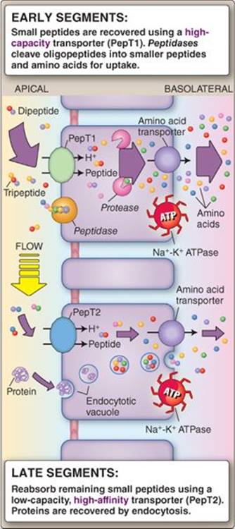
Figure 26.8
Oligopeptide and protein reabsorption.
ATP = adenosine triphosphate.
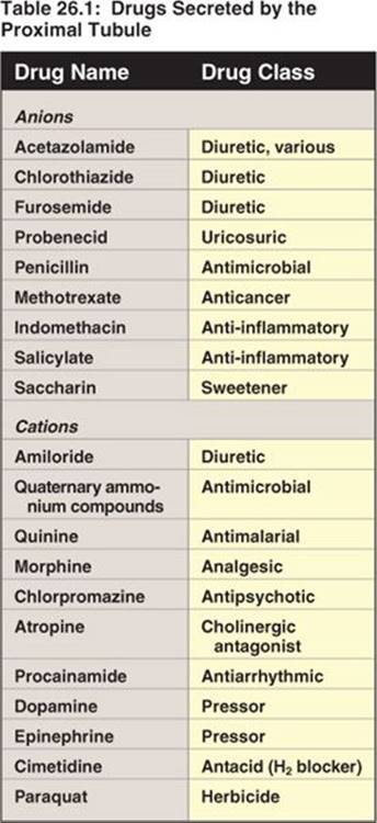
The tendency for the PT to take up pharmaceuticals from the circulation puts it at grave risk because intracellular concentrations can quickly rise to toxic levels. The transporters responsible for uptake have, thus, themselves become high-priority targets for pharmaceutical intervention. Inhibiting the uptake systems not only reduces drug toxicity, but it also decreases the rate of drug elimination from the body and, thus, reduces dosing frequency.
A. Kinetics
Secretion is transporter mediated and, therefore, shows saturation kinetics, as demonstrated using para-aminohippurate (PAH) in Figure 26.9. PAH is a hippuric acid derivative used in studies of renal plasma flow (see 25·V·D) that is both filtered from the glomerulus and secreted from the PT via the OAT pathways mentioned above.
1. Filtration: PAH is freely filtered by the glomerulus in amounts that are directly proportional to GFR. Filtration removes ~20% of total plasma PAH.
2. Secretion: Blood entering the peritubular network still contains 80% of the original arterial PAH load. All but 10% is taken up by transporters in the basolateral membrane of the late PT and secreted into the tubule lumen. PAH excretion rises accordingly. Transporter capacity is finite, however, so the secretion curve flattens and plateaus as plasma PAH concentration approaches Tm. The secretion curve exhibits splay due to transporter and nephron heterogeneity, as discussed above in reference to glucose reabsorption.
B. Transporters
PT epithelia express a number of different broad-specificity transporters for organic anions and cations. Organic anions are taken up from the blood by several members of the OAT family. OAT1 exchanges an organic ion for a dicarboxylate such as α-ketoglutarate. A related family of organic cation transporters takes up amine and ammonia compounds from blood. Anions and cations are then both extruded into the tubule lumen by one of a number of multidrug-resistant proteins (MRPs). MRPs are members of the ATP-binding cassette pump superfamily. Organic anions can also cross the apical membrane by one of a number of OATs.
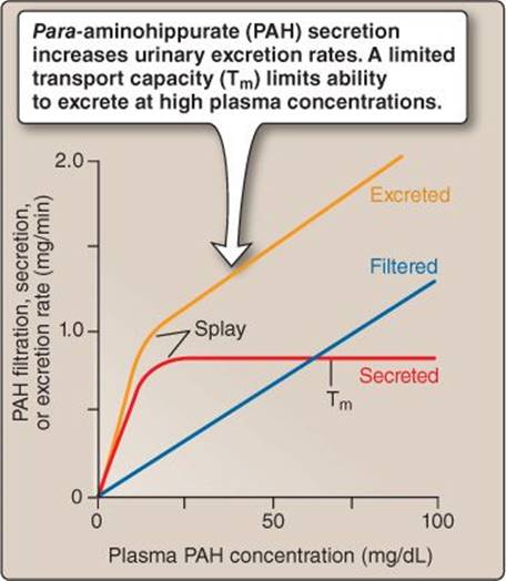
Figure 26.9
Effects of plasma para-aminohippurate (PAH) concentration on secretion and excretion rates.
Clinical Application 26.2: Gout
Organic anion transporters are one of a number of transporter families involved in reabsorption and excretion of uric acid. Most mammals metabolize urate to allantoin, but primates lost the necessary enzyme (uricase) during evolution. Unlike allantoin, urate is relatively insoluble, and when blood concentrations rise, it forms crystals that are often deposited in joints. The result is a painful inflammatory arthritis known as gout. Gout treatment options include drugs that inhibit the transporters that normally reabsorb urate as it passes down the tubule, thereby increasing excretion rates.
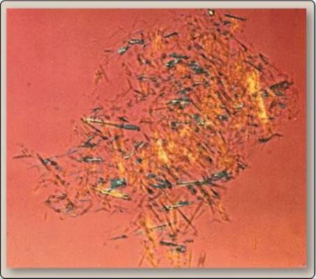
Uric acid crystals in synovial fluid from a patient with chronic gout.
V. UREA
Urea is a small organic molecule comprising two amide groups joined by a carbonyl group. It is formed in the liver1 and excreted in urine as a way of disposing of unwanted amino acids and nitrogen (Figure 26.10). Normal plasma concentrations average 2.5–6.0 mmol/L. The PT reabsorbs ~50% of the filtered load, largely via the paracellular route. Two forces drive movement. The first is solvent drag, created by the large volumes of water being reabsorbed in the PT. Loss of water from the tubule lumen secondarily concentrates solutes in the tubule lumen, which enhances the driving force for urea diffusion across the epithelium. The kidney ultimately excretes ~40% of the filtered urea load, but it first serves an important role in helping concentrate urine. The pathways involved are discussed in Chapter 27 (see 27·V·D).
Urea is the principal means by which nitrogenous wastes are excreted from the body, and, thus, plasma urea levels are a useful indicator of renal health and function. Clinical laboratories cite urea levels in the form of blood urea nitrogen (BUN). Normal BUN values are in the range of 7–18 mg/dL.
 1The role of urea in nitrogen excretion and details of the urea cycle are discussed at length in LIR Biochemistry, 5e, p. 253.
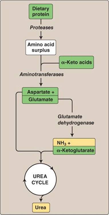
Figure 26.10
Urea formation.
VI. PHOSPHATE AND CALCIUM
Plasma contains a total of ~1.0–1.5 mmol/L inorganic phosphorus (Pi) and ~2.1–2.8 mmol/L Ca2+. Both are critically important for normal cell function. Pi is a component of RNA and DNA, powers metabolism in the form of ATP, and is found associated with numerous lipids and proteins. Ca2+ is a vital second messenger that activates enzymes, initiates muscle contraction, and triggers neurotransmitter secretion. About half of total plasma phosphorus and calcium exists in ionized form (as HPO42−, H2PO4−, and Ca2+), the remainder being complexed with proteins and other molecules. Plasma contains only a tiny fraction of total body phosphorus and calcium, however. The vast majority of phosphorus (>80%) and calcium (>99%) is locked in hydroxyapatite crystals in a mineral vault called bone. Plasma Pi and Ca2+ concentrations are regulated by similar mechanisms. Total body concentrations of both ions represent a precise balance between bone deposition and resorption, intestinal secretion and absorption, and renal filtration and reabsorption. All three processes are regulated by parathyroid hormone ([PTH] discussed in more detail in Chapters 27 and 35).
A. Phosphate
The kidney tubule reabsorbs ~90% of the filtered Pi load, of which ~80% is reclaimed in the PT and the remaining 10% in the distal convoluted tubule (DCT). The PT is the principal site of Pi regulation, effected through PTH and plasma Pi concentrations (see Figure 27.19).
1. Reabsorption: Pi is reabsorbed using two apical Na+-Pi cotransporters (Na+-Pi IIa and Na+-Pi IIc) as shown in Figure 26.11. The mechanism by which Pi crosses the basolateral membrane is under investigation.
2. Regulation: PTH blocks Pi recovery from the tubule lumen by promoting endocytosis and subsequent degradation of the apical Pi transporters. In the absence of a recovery pathway, Pi then passes through the tubule and is excreted. Low dietary intake causes cotransporters to be inserted into the apical membrane, thereby increasing the PT's ability to reabsorb filtered Pi.
B. Calcium
Plasma free Ca2+ concentrations are tightly regulated in the range of 1.0–1.3 mmol/L, and virtually all filtered Ca2+ is reabsorbed during passage through the nephron (see Figure 27.19). The PT recovers ~65%, largely via the paracellular route. The motive force is partly solvent drag and, in the later stages of the PT where the lumen is positively charged with respect to blood, the transepithelial voltage difference. Most of the remaining 35% of filtered load is reabsorbed in the thick ascending limb ([TAL] ~25%) and the DCT (~8%). The DCT is the main site of Ca2+ regulation (see 27·III·C).
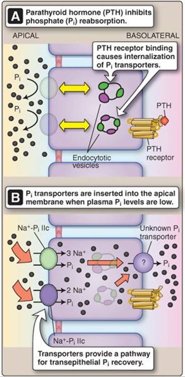
Figure 26.11
Regulation of phosphate reabsorption.
VII. MAGNESIUM
Mg2+ is a vital cofactor required for the normal function of hundreds of enzymes, its positive charge helping stabilize protein structural integrity. It also regulates ion flow through ion channels, so physiologic decreases in plasma free concentrations cause membrane hyperexcitability, arrhythmias, and muscle tetany. The majority of total body Mg2+ is complexed in bone or associated with proteins and other small molecules. Plasma concentrations are normally maintained in a range of ~0.75–1.00 mmol/L, of which ~60% is in the free form. Mg2+ is a common ingredient in most foods, so ~2%–5% of the filtered load typically is excreted in urine to balance daily intake. The PT recovers ~15% of the filtered load. Reabsorption occurs paracellularly by solvent drag and diffusion. Reabsorption is favored by the small lumen-positive potential difference that exists across the more distal regions of the PT epithelium. The bulk of filtered Mg2+ (~70%) is recovered in the TAL, which is also the principal site of Mg2+ homeostatic regulation (see 27·III·B and Figure 27.19).
VIII. POTASSIUM
K+ is unique among electrolytes in that even modest changes in plasma K+ concentrations can be life threatening, causing potentially fatal cardiac dysrhythmias and arrhythmias (see Clinical Application 2.1). Plasma concentrations are tightly regulated within the range of 3.5–5.0 mmol/L. K+ is filtered freely across the glomerulus, so the nephron handles a daily load of ~0.6–0.9 mol. The PT reabsorbs ~80% of the filtered load, primarily via the paracellular route (Figure 26.12). As is the case for Ca2+ and Mg2+, absorption occurs as a result of solvent drag, and by diffusion that is enhanced by a transepithelial voltage gradient. Another 10% is recovered in the TAL (see 27·II·B), but regulation of K+ reabsorption (and excretion) occurs primarily in the distal segments (see 27·IV·C and Figure 27.19).
IX. BICARBONATE AND HYDROGEN IONS
One of the kidney's most important functions is to help maintain extracellular fluid (ECF) pH at ~7.40. Metabolism generates immense quantities of volatile acid (H2CO3) that is expelled via the lungs and another ~50–100 mmol/day of nonvolatile acid (sulphuric, phosphoric, nitric, and other minor acids; see 3·IV·A) that must be excreted by the kidneys. Although all portions of the nephron are involved in acid–base homeostasis to some degree (see Figure 27.19), the PT is a principal site for HCO3− recovery and H+ secretion.
A. Bicarbonate
Excreting HCO3− causes the ECF to become acidic, so the first goal of pH homeostasis is to recover 100% of the filtered HCO3− load. The PT recovers ~80% of total. Because HCO3− is anionic, it cannot diffuse freely across membranes, so the PT secretes molar amounts of H+ into the tubule lumen to titrate the HCO3− and then uses carbonic anhydrase (CA) to convert the H2CO3 to CO2 and H2O. Both molecules are then recovered by simple diffusion. Reclamation is a four-step process (numbers below correspond to steps shown in Figure 26.13):
1. H+ is transported into the tubule lumen by an apical Na+-H+ ex-changer (NHE3). The exchange is powered by the transmembrane Na+ gradient.
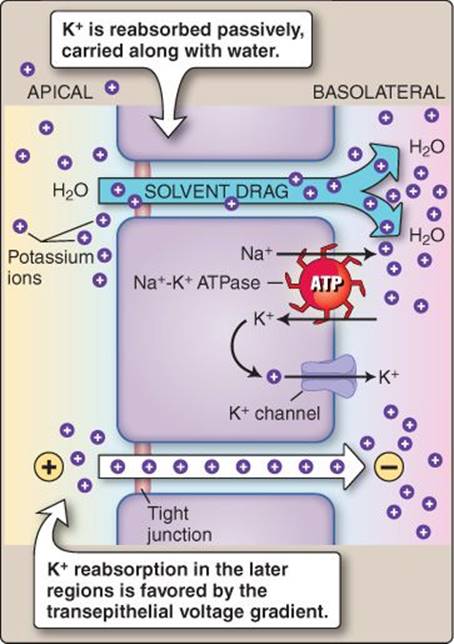
Figure 26.12
Potassium reabsorption pathways in the proximal tubule. ATP = adenosine triphosphate.
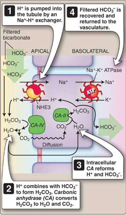
Figure 26.13
Bicarbonate reabsorption pathway in the proximal tubule. ATP = adenosine triphosphate.
2. H+ combines with luminal HCO3− to form H2CO3, which dissociates to form H2O and CO2. The reaction is catalyzed by CA-IV, which is expressed on the epithelium's apical surface:

3. CO2 diffuses into the cell and combines with H2O to reform HCO3− and H+. The reaction is catalyzed by intracellular CA-II.
4. HCO3− is reabsorbed across the basolateral membrane to the interstitium and then into the vasculature, although the mechanism is unclear. H+ is pumped back into the tubule lumen to repeat the reabsorption cycle.
HCO3− reabsorption causes a slight acidification of the tubule contents, from pH 7.4 at the glomerulus to ~pH 6.8 in the late PT.
Acetazolamide is a CA inhibitor that blocks HCO3− and Na+ reabsorption by the PT, causing diuresis. The drug acts on both the apical (CA- IV) and intracellular form (CA- II) of the enzyme. As a class, the CAinhibitors are relatively ineffective diuretics because the more distal regions of the tubule compensate for their effects on PT function.1 The main indication for CA inhibitor use is in patients with metabolic alkalosis, because the drugs impair the tubule's ability to reabsorb HCO3− and, thereby, cause excess base to be excreted in urine.
B. Hydrogen ions
The PT is a principal site for H+ secretion, although final determination of urine pH and regulation of ECF pH occurs in the distal segments (see 27·V·E). H+ is secreted by the NHE3 Na+-H+ exchanger mentioned above, and by a H+ pump (Figure 26.14).
1. Sodium–hydrogen ion exchange: The NHE3 Na+-H+ exchanger uses the Na+ gradient created by the basolateral Na+-K+ ATPase to power H+ secretion. The dependence on the Na+ gradient means that its ability to concentrate H+ in the lumen is limited, but it has a very high capacity that accounts for ~60% of net H+ secretion in the PT.
The NHE3 exchanger is also a principal pathway by which the PT recovers Na+ from the tubule lumen (see below).
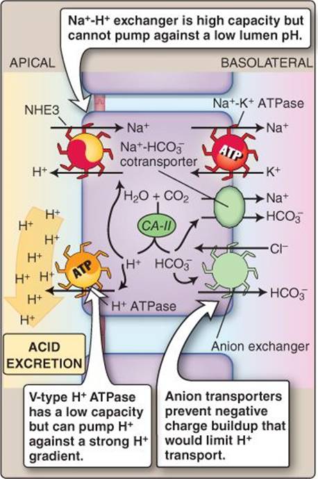
Figure 26.14
Acid secretion by the proximal tubule. ATP = adenosine triphosphate; CA- II = carbonic anhydrase II.
2. Proton pump: The PT also actively secretes H+ into the tubule using a vacuolar-type H+ pump (V-type H+ ATPase). The H+ pump accounts for ~40% of net secretion in the PT and is capable of establishing a strong H+-concentration gradient across the apical membrane. The pump is electrogenic, meaning that it causes a negative charge to build within the cell. This charge can become limiting to further transport, so H+ secretion is balanced by HCO3− movement across the basolateral membrane via a Na+-HCO3− cotransporter and an anion exchanger (see Figure 26.14).
1For more information on acetazolamide use, see LIR Pharmacology, 5e, p. 287.
C. Nonvolatile acid
Ideally, the H+ excess created by nonvolatile acid formation would be transported to the kidney and then dumped into the tubule and excreted without further ado. In practice, the amount of nonvolatile acid generated is large, and the ability of available H+ transporters to pump H+ against a concentration gradient is limited. The V-type H+ ATPase mentioned above can create a lumen pH of ~pH 4.0 at best (i.e., 0.1 mmol/L H+), which is insufficient to handle the daily acid excess. Two different workarounds have evolved to allow H+ to be excreted in the quantities required to maintain pH balance. The first is to simultaneously excrete urinary buffers (titratable acids) that limit a rise in free H+ concentration even as acid is being pumped into the tubule lumen. The second is to attach H+ to ammonia (NH3) and excrete it as an ammonium ion (NH4+).
1. Titratable acids: The plasma filtrate contains several buffers, and the PT secretes several more. These include hydrogen phosphate (pK = 6.8), urate (pK = 5.8), creatinine (pK = 5.0), lactate (pK = 3.9), and pyruvate (pK = 2.5). Collectively, these buffers are known as “titratable acids” that complex with and, thereby, limit rises in tubule H+ concentration. Hydrogen phosphate's pK makes it a more effective urinary buffer than the other titratable acids Hydrogen phosphate accepts H+ to become dihydrogen phosphate (Figure 26.15):

The PT reabsorbs ~80% of filtered phosphate, but the remaining 20% remains to buffer lumen pH during nonvolatile H+ excretion.
2. Ammonia: Plasma does not normally contain NH3, but PT cells are able to synthesize it from glutamine, which is converted to NH3 and α-ketoglutarate. NH3 is lipid soluble, so it diffuses out of the cell into the tubule lumen and combines with H+ to form NH4+. Some NH4+ is formed inside the PT cells and moved into the tubule lumen by the Na+-H+ exchanger, which is able to bind NH4+ in place of H+(Figure 26.16).
3. New bicarbonate: Excreting ~50–100 mmol of nonvolatile acid generated every day creates a sizeable deficit in the body's buffer systems. This must be matched precisely by the formation of new buffer or ECF would rapidly become acidotic. Excreted buffer is replaced by generation of “new” HCO3−. Some is formed de novo, and some is created from α-ketoglutarate after NH3 is formed from glutamine. α-Ketoglutarate is metabolized to glucose and then to CO2 and H2O. CA then catalyzes H2CO3 formation, which dissociates to yield HCO3− and H+. The newly formed HCO3− diffuses into blood and is ultimately used to buffer nonvolatile acid at its formation site within tissues.
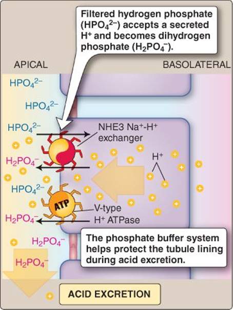
Figure 26.15
Phosphate buffer system. ATP = adenosine triphosphate.
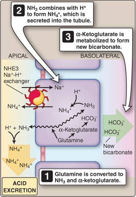
Figure 26.16
Excreting acid in the form of the ammonium ion.
X. SODIUM, CHLORIDE, AND WATER
Plasma Na+ concentration is maintained at between ~136 and 145 mmol/L, primarily as a way of controlling how water distributes between the three body compartments (intracellular, interstitial, and plasma; see 3·III·B). Na+ moves freely across the glomerular filtration barrier, so the daily filtered load exceeds 25 mol. Approximately 99.6% of the filtered load is reabsorbed during passage through the renal tubule, the bulk (~67%) being recovered by the PT (see Figure 27.19). Cl− follows Na+ across the epithelium, driven inward by sodium's positive charge. Reabsorption of Na+, Cl−, and organic solutes creates a strong osmotic potential that also drives water from the tubule lumen toward the interstitium. The net effect of these and all of the other reabsorptive and secretory processes described in the previous sections is that the fluid reabsorbed by the PT is isosmotic and has a composition that resembles plasma. There are regional differences in the way that Na+ and Cl− are reabsorbed between the early and late regions of the PT, however.
A. Early proximal convoluted tubule
Early PT epithelial cells are specialized to recover virtually all useful organic solutes and HCO3− in association with Na+, which leads to significant transcellular Na+ reabsorption. Some of this Na+ then leaks backward paracellularly (Figure 26.17).
1. Transcellular: The primary force driving reabsorption is the basolateral Na+-K+ ATPase, which establishes a Na+ gradient that drives Na+-coupled glucose, amino acid, organic acid, and phosphate reabsorption from the tubule. Large quantities of Na+ also enter cells via the NHE3 Na+-H+ exchanger. Na+ is then moved to the interstitium by the Na+-K+ ATPase and, to a lesser degree, by a basolateral Na+-HCO3− cotransporter. Cotransport is driven by high intracellular HCO3− concentrations following reabsorption and de novo synthesis.
2. Paracellular: The cotransporters that recover organic solutes from the plasma filtrate are electrogenic, leaving an excess of negative charges in the tubule lumen. These charges create a ~3-mV difference between tubule and interstitium, which creates a significant force that drives paracellular Cl− reabsorption. The paracellular route also permits significant amounts of reabsorbed Na+ (~30%) to leak backward from the interstitium to tubule lumen. Movement is driven by the voltage gradient.
B. Proximal straight tubule
The fluid entering the PST has been stripped of all useful organic solutes and most HCO3−, but contains relatively high concentrations of Cl−. Na+ and Cl− are reabsorbed via both transcellular and paracellular routes.
1. Transcellular: The late PT takes up Na+ in exchange for H+, which creates a transcellular Na+ flux. This region of the PT also contains a Cl−-base exchanger (CFEX) that allows for significant transcellular Cl− uptake. CFEX exchanges Cl− for formate, oxalate, OH−, or HCO3−.
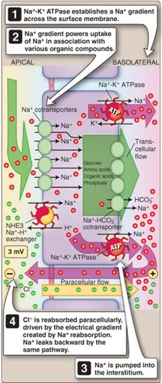
Figure 26.17
Pathways for Na+ reabsorption and backflow in the early proximal convoluted tubule. ATP = adenosine triphosphate.
2. Paracellular: High luminal Cl− concentrations drive diffusion of Cl− out of the lumen via the paracellular route. This leaves an excess of positive charge in the lumen that favors Na+ reabsorption, so Na+follows Cl− across the tight junctions and into the interstitium.
Chapter Summary
• The proximal tubule (PT) recovers ~67% of the fluid and up to 100% of some solutes that are filtered into the renal tubule by the glomerulus. PT epithelial cells possess apical microvilli that increase surface area, and the junctions between cells are leaky to maximize free flow of water and dissolved solutes.
• The proximal tubule reabsorbs fluid isosmotically. Transcellular absorption is powered mainly by the transmembrane Na+ gradient established by a basolateral Na+-K+ ATPase. Reabsorption also occurs by diffusion via tight junctions (paracellular absorption) and paracellular solvent drag.
• Reabsorbed fluid is returned to the vasculature via the peritubular network. Blood reaches the peritubular capillaries by way of the glomerulus. Glomerular filtration concentrates the plasma proteins and, thereby, increases plasma colloid osmotic pressure. The efferent arteriole has a high resistance that lowers capillary hydrostatic pressure. These features together create a situation in which fluid uptake from the interstitium is strongly favored, which facilitates reabsorption.
• The proximal tubule (PT) recovers almost 100% of filtered glucose and amino acids, principally via Na+ cotransport. The PT also recovers small peptides by H+ cotransport. Larger peptides and proteins are degraded to small peptides and are then reabsorbed or taken up by endocytosis.
• The proximal tubule actively secretes a number of organic acids, toxins, and drugs using organic anion or cation transporters or multidrug resistance proteins.
• Phosphate is recovered from the proximal tubule (PT) by Na+-phosphate cotransporters. Reabsorption is regulated by parathyroid hormone. Ca2+ reabsorption by the PT occurs paracellularly.
• Mg2+ reabsorption by the proximal tubule is minimal (~15% of filtered load) and occurs paracellularly.
• Approximately 80% of the filtered K+ load is recovered in the proximal tubule.
• The lungs and kidneys together are responsible for maintaining the pH of extracellular fluids within a narrow range (pH 7.35–7.45). Lungs excrete the daily load of volatile acids (CO2) generated during metabolism. Kidneys excrete nonvolatile acids (sulphuric, phosphoric, nitric, and other minor acids).
• pH homeostasis begins in the proximal tubule (PT) with recovery of 80% of filtered HCO3−, the body's primary pH buffer. Excretion of nonvolatile acid requires that buffers be excreted also to control luminal free H+concentration. The primary urinary buffers are phosphate and ammonium, the latter newly synthesized from glutamine in the PT.
• Na+ reabsorption by the proximal tubule (PT) is driven by the basolateral Na+-K+ ATPase through cotransport with organic solutes and in exchange for H+. Cl− absorption occurs principally in the late PT by the paracellular route or by a Cl−-base exchanger. Water reabsorption occurs by osmosis, driven by influx of Na+, Cl−, and solutes.