
29
I. OVERVIEW
The gastrointestinal (GI) system is a complex tube bounded by the mouth at one end and the anus at the other. Food enters the mouth; travels through the esophagus, stomach, small intestine (duodenum, jejunum, and ileum), large intestine (ascending, transverse, and descending colon), and rectum; and then exits via the anus (Figure 29.1). This tube's primary function is the absorption of dietary nutrients. To maximize nutrient absorption, secretions are added to food from the salivary glands, stomach, liver, gallbladder, and pancreas to convert complex molecules into simpler ones. This conversion, called digestion, is effected by enzymes and H+. Dietary contents and secretions are mixed and propelled along the tube (motility) from one specialized compartment to another by coordinated peristaltic contractions and relaxations of the tube walls (Figure 29.2). Two other important GI functions include storage (e.g., food is stored in the stomach and fecal matter in the colon) and excretion of undigested materials and biliary waste products.
II. GASTROINTESTINAL LAYERS
The intestinal tract is composed of multiple layers, each possessing a distinct function. Depending on structure–function relations, a particular layer's prominence changes along the length of the tube. Moving from the lumen to the outside of the tube, the layers include epithelium, lamina propria, muscularis mucosa, submucosa, submucosal plexus, circular muscle, myenteric plexus, longitudinal muscle, and serosa (Figure 29.3).
A. Mucosa
The epithelium, lamina propria, and muscularis mucosa together form the mucosa. The epithelium is a single cell layer forming a continuous lining of the GI tract. GI epithelial cells are shed and replaced every 2–3 days. The apical side of the epithelium faces the GI lumen, and the basolateral side faces the interstitium and vasculature. Apical surfaces may be enhanced with villi (thumblike projections) and crypts (invaginations) to increase surface area and maximize contact between epithelium and intestinal contents (Figure 29.4). Absorptive areas (e.g., small intestine) contain numerous apical enhancements. Areas primarily involved with motility (e.g., esophagus) do not. The lamina propria is a loose connective tissue composed of elastin and collagen fibers that contains sensory nerves, blood and lymph vessels, and some secretory glands. The muscularis mucosa is a thin layer of smooth muscle that further increases surface area by creating mucosal ridges and folds.
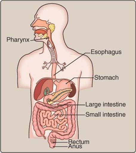
Figure 29.1
Gastrointestinal tract.

Figure 29.2
Peristalsis.
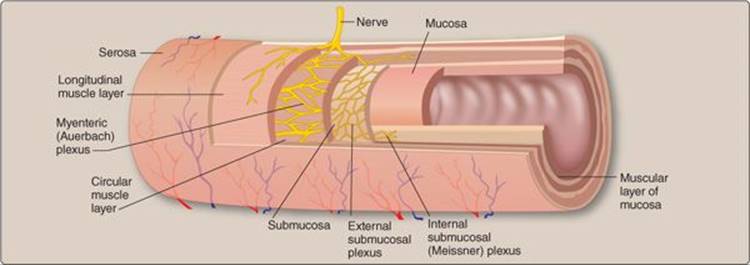
Figure 29.3
Gastrointestinal tract layers.
B. Submucosa
The submucosa is a thicker layer with a similar composition to the lamina propria. It incorporates blood vessels and bundles of nerves that collectively form a submucosal plexus (Meissner plexus), which is an integral part of the enteric nervous system (ENS). The ENS is described in more detail below.
C. Muscularis externa
The muscularis externa comprises circular muscle, the myenteric plexus, and longitudinal muscle layers. The two smooth muscle layers are named based on their orientation. The circular muscle layer is arranged in rings and pinches the tube when it contracts. The longitudinal layer is arranged in parallel and shortens the tube when it contracts. The ENS coordinates circular and longitudinal muscle contraction to mix intestinal contents and move them between compartments. Circular muscle also forms sphincters, which regulate flow of food from one compartment to the next by modulating lumen diameter. The myenteric plexus (Auerbach plexus), also part of the ENS, is located between the circular and longitudinal muscle layers.
D. Serosa
The serosa comprises an outermost layer of connective tissue and a layer of squamous epithelial cells. Some portions of the GI tract (e.g., the esophagus) do not have a serosal layer but rather connect directly to the adventitia, which is connective tissue that blends into the abdominal or pelvic wall.

Figure 29.4
Surface area enhancements.
III. INNERVATION AND NEUROTRANSMITTERS
GI function is regulated by three divisions of the autonomic nervous system (ANS): the parasympathetic nervous system (PSNS), sympathetic nervous system (SNS), and the ENS.
A. Parasympathetic nervous system
Parasympathetic innervation is derived from the vagus (medulla oblongata) and pelvic–splanchnic nerves (S2–S4) and has both motor and sensory components (Figure 29.5). The sensory components respond to stretch, pressure, temperature, and osmolarity and participate in vagovagal reflexes. Vagovagal reflexes occur when the vagus nerve (cranial nerve X) participates in both afferent sensation and efferent responses without central nervous system involvement. Primary neurotransmitters used directly or indirectly by the PSNS are acetylcholine (ACh), gastric-releasing peptide, and substance P (Table 29.1). In general, signals from the PSNS stimulate GI secretions and motility, which facilitates digestion and absorption of nutrients.
B. Sympathetic nervous system
Sympathetic nerves originate in the thoracic (T5–T12) and lumbar (L1–L3) regions and synapse in one of three ganglia: celiac, superior mesenteric, or inferior mesenteric for the lower GI system (Figure 29.6). The upper GI tract (e.g., salivary glands) is innervated by SNS nerves that synapse within the superior cervical ganglion (see Figure 29.6). Unlike the PSNS, the SNS component does not contain a direct sensory arm and generally decreases GI secretions and motility when active. The primary SNS neurotransmitters are norepinephrine and neuropeptide Y (see Table 29.1).
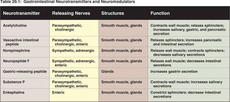

Figure 29.5
Parasympathetic innervation. CN = cranial nerve.
C. Enteric nervous system
PSNS and SNS nerves usually synapse with components of the ENS. Although the ENS is modulated by these extrinsic neural inputs, it can operate autonomously via intrinsic regulation and sensory reflexes. ENS nerves are organized into myenteric and submucosal plexuses.
1. Plexuses: The myenteric plexus forms a dense parallel neuronal configuration that primarily regulates intestinal smooth muscle and participates in tonic and rhythmic contractions. Some myenteric neurons also synapse with neurons in the submucosal plexus or directly on secretory cells. The submucosal plexus primarily regulates intestinal secretions and the local absorptive environment but also can synapse on blood vessels, circular and longitudinal muscle, and the muscularis mucosa. ENS neurons are supported by enteric glial cells, which structurally and functionally resemble astrocytes in the brain.
2. Reflexes: Many GI reflex actions are regulated solely by neural circuits in which a mechanoreceptor or chemoreceptor is stimulated in the mucosa and transmits the signal back to neurons in the submucosal plexus, which stimulates other neurons in the submucosal or myenteric plexus that regulate endocrine or secretory cells.
3. Neurotransmitters: There are a number of neurotransmitters and regulatory molecules used in ENS communication (see Table 29.1). Enkephalins constrict circular muscle around sphincters. In the submucosal plexus, secretory neurons primarily use vasoactive intestinal peptide (VIP) and ACh as neurotransmitters, whereas sensory nerves use substance P. In the myenteric plexus, motor neurons use ACh and nitric oxide, sensory neurons use substance P, and the interneurons use ACh and serotonin (5-hydroxytryptamine). These enteric neurotransmitters are also used elsewhere in the body and are important pharmacologically. For example, a person on serotonin reuptake inhibitors may experience decreased GI motility as a side effect because these drugs alter serotonin levels.

Figure 29.6
Sympathetic innervation.
Clinical Application 29.1: Chagas Disease
Submucosal and myenteric plexus neuropathy can impair motility. For example, a protozoan infestation (Trypanosoma cruzi, often delivered by kissing bugs) of these plexus neurons can lead to Chagas disease. Among other pathologies, Chagas disease causes distention and structural enlargements of the esophagus and colon because regions with neuropathy can constrict but not relax muscular layers. The asymptomatic sections continue to deliver food, which is retained just proximal to the constricted area. Such retention stretches these areas and, over time, enlarges and contorts them.
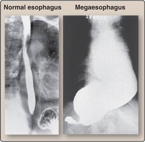
Megaesophagus.

IV. NONNEURAL SIGNALING MOLECULES
In addition to neurotransmitters, hormones and paracrine signaling molecules also regulate and control GI function.
A. Hormones
GI peptide hormones include cholecystokinin (CCK), gastrin, glucose-dependent insulinotropic peptide ([GIP] formerly known as gastric inhibitory peptide), motilin, and secretin (Table 29.2). Endocrine cell types are located in different densities in various locations throughout the stomach and intestines (Figure 29.7). Gastrin is secreted in the stomach antrum and then tapers off in the small intestine. CCK, secretin, GIP, and motilin are primarily secreted in the duodenum and jejunum, and CCK and secretin continue to be secreted in the ileum, albeit to a lesser degree.
B. Paracrines
GI paracrines are both released and act locally. The primary paracrine GI signaling molecules are histamine, prostaglandins, and somatostatin (Table 29.3). Of these, only somatostatin is a peptide. Histamine is classified as a monoamine, and prostaglandins are eicosanoid-signaling molecules. Histamine is released in the stomach, whereas both prostaglandins and somatostatin are more widespread in their release and actions.
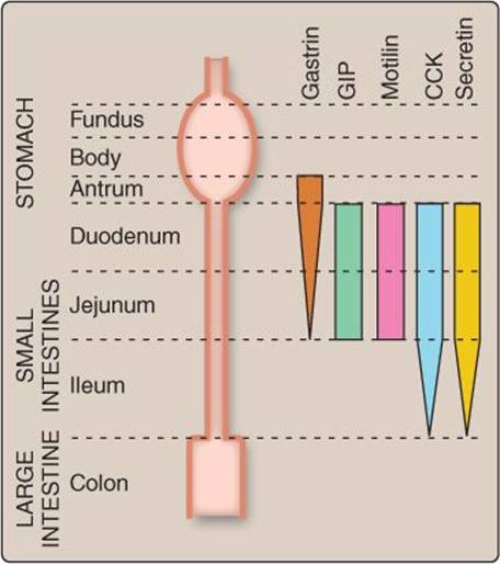
Figure 29.7
Principal sites of gastrointestinal hormone release. CCK = cholecystokinin; GIP = glucose-dependent insulinotropic peptide.

Prostaglandins are cyclooxygenase products derived from arachidonic acid. Prostaglandins have an important role in maintaining mucosal integrity and, thus, cyclooxygenase inhibitors (i.e., aspirin and other nonsteroidal anti-inflammatory drugs) can cause stomach irritation.1
V. DIGESTIVE PHASES
Stomach and duodenal function can be divided into three discrete phases: cephalic, gastric, and intestinal.
A. Cephalic phase
The cephalic phase is triggered by the thought of food or conditions suggestive of previous food intake (e.g., classical conditioning to eat after hearing a dinner bell). Chemoreceptors and mechanoreceptors in the oral and nasal cavities and throat that are stimulated by tasting, chewing, swallowing, and smelling food also contribute. The cephalic phase is primarily neural and causes ACh and VIP release. ACh and VIP stimulate secretion by the salivary glands, stomach, pancreas, and intestines.
Clinical Application 29.2: Feeding Tubes and Intravenous Feeding
Patients with swallowing disorders or on mechanical ventilation require nutrient delivery past obstructed areas. Feeding tubes (e.g., nasogastric [NG] and nasoduodenal [ND] tubes) are used to provide nutritional support for these patients. NG tubes deliver food directly to the stomach, whereas ND tubes deliver food directly to the duodenum. Feeding tubes thereby bypass the majority of the digestive phase initiation cues. This requires the feeding tube formula to be prepared in a manner that will not require upper gastrointestinal (GI) processing of food. Nutrients can also be directly infused intravenously, which bypasses the entire GI system. Care must be taken to include all required nutrients, although less total kcals are necessary insofar as ~7% of energy consumed by mouth is used to digest and absorb nutrients.

Nasogastric tube.
 1 For a discussion of the gastrointestinal effects of nonsteroidal anti-inflammatory drugs, see LIR Pharmacology, 5e, p. 531.
B. Gastric phase
The gastric phase begins when food and oral secretions enter the stomach. It coincides with distention and stomach contents (amino acids and peptides) and elicits neural, hormonal, and paracrine GI responses. A good example of this combination of signaling molecules is in gastric acid secretion, which includes ACh (neural), gastrin (hormonal), and histamine (paracrine).
C. Intestinal phase
The intestinal phase begins when stomach contents enter the duodenum. It is linked to digested constituents of proteins and fats as well as H+ and initiates primarily hormonal but also paracrine and neural responses. CCK, gastrin, secretin, and GIP are all secreted during this phase.
Chapter Summary
• Absorption is the process of transporting dietary contents across the gastrointestinal barrier into the body.
• To prepare nutrients for absorption, the body mechanically and chemically breaks down food into smaller, simpler particles. The chemical breakdown of food is digestion, and the mechanical breakdown of food involves smooth (i.e., as in mixing) or skeletal (i.e., as in chewing) muscle contractions.
• Secretion is the act of transporting molecules or fluid from the body to the gastrointestinal lumen. Secretion facilitates digestion by delivering enzymes and water and protects the endothelial surface by secreting HCO3− and mucus.
• The autonomic nervous system innervates the entire gastrointestinal (GI) system. The parasympathetic nervous system most often facilitates secretion and motility, whereas the sympathetic nervous system decreases these functions. The enteric nervous system can operate independently and is involved with reflexes and the majority of GI functions.
• Gastrointestinal hormones include cholecystokinin, which is released from I cells and participates in pancreatic and biliary secretions; gastrin, which is released from G cells and primarily functions in H+secretion; glucose-dependent insulinotropic peptide, which is released from K cells and primarily functions to increase insulin release and decrease H+ secretion; motilin, which primarily functions to increase motility; and secretin, which is released from S cells and primarily functions to increase water and HCO3− secretion and decrease H+.
• Gastrointestinal (GI) paracrines include histamines, which are derived from enterochromaffin-like cells and mast cells and have many functions such as increasing H+ production; prostaglandins, which have many functions including decreasing H+ production and maintaining GI barrier properties; and somatostatin, which decreases GI secretions.
• The phases of digestion (cephalic, gastric, and intestinal) allow for preparation and timing and regulation feedback. The cephalic is primarily a feedforward regulation, and the gastric and intestinal phases are feedback mechanisms.