
40
I. OVERVIEW
I like a look of Agony,
Because I know it's true
–Men do not sham Convulsion,
Nor simulate, a Throe –
The Eyes glaze once – and that is Death –
Impossible to feign
The Beads upon the Forehead
By homely Anguish strung.
Emily Dickinson
We are all destined to die.
Living depends on a delicate homeostatic balancing act. During life, we rely on our organ systems to compensate for changes in innumerable internal parameters, including PO2 and PCO2, pH, electrolyte levels, and body temperature. Ultimately, however, all these compensatory systems slowly falter and then fail. At the cellular level, this process is known as senescence and apoptosis. At the organismal level, we know it as aging and death.
The Centers for Disease Control and Prevention periodically publishes a list of the leading causes of death in the United States (Table 40.1). The list does not include “old age” because it is based on death certificates, which require physicians to identify a specific causal event (e.g., heart failure). From a physiologic perspective, however, corporeal death usually reflects a long series of individual, aging-related cell deaths. Cell by cell, all organs age and, eventually, fail. Which organ falls off the homeostatic tightrope first may be a matter of chance or may be determined by an underlying disease or lifestyle choice. In this final chapter, we consider various causes and consequences of individual organ failure. There are many other causes of death (e.g., accidents and trauma) as shown in Table 40.1, but, regardless, death of the individual occurs when the cerebral hemispheres are O2 deprived and the cortex dies, either because of cardiovascular failure, respiratory failure, renal failure, or multiple organ system failure. The Eyes glaze once—and that is Death.

Figure 40.1
Emily Dickinson (American poet, 1830–1886).

II. AGING AND DEATH
Gerontology is a relatively new discipline dealing with old issues (and issues encountered by older adults). Although researchers have forwarded many ideas as to why cells and organ systems inevitably lose functionality and fail, there are no solutions to the age-old problem of why we die. Preprogrammed cell death (apoptosis) is probably just one of many contributing factors. Regardless, the human lifespan is limited to ~120 years. Medical advances over the past 100 years may have increased mean life expectancy but not the upper limit (Figure 40.2), suggesting that failure and death may be genetically predetermined.
Apoptosis is a process whereby cells and their contents spontaneously fragment into membrane-bound apoptotic bodies that are rapidly engulfed by phagocytes. Apoptosis may be triggered by intrinsic factors, including genetic programming and cell damage, and by extracellular factors, such as toxins and growth factors. Apoptosis is a normal and necessary process for continued tissue health and homeostasis.

Figure 40.2
Mean life expectancy in the United States, 1900–2000.
A. Physiologic aging
Individual physiologic responses to aging vary widely and can be significantly impacted by physical fitness and underlying disease, but aging is generally accompanied by progressive decreases in cell numbers and in the functionality and responsiveness of remaining cells in most organs. Coinciding with these changes are overall decreases in tissue compliance. The effects of aging on the cardiovascular system, for example, include reduced myocardial sensitivity to adrenergic agonists, which is why maximal heart rate (HR) attained during exercise is estimated from 220 minus age in years. Arteries stiffen with age due to elastin cross-link breakage (see 4·IV·B), increased collagen deposition, and calcification, causing a compensatory increase in arterial blood pressure (see 19·V·C·3). Similar changes occur throughout the body's various tissues. The deep wrinkles that develop in Caucasian skin are the most obvious external indicator of aging, but wrinkles develop largely through photoaging (tissue damage induced by ultraviolet light), not by the intrinsic aging process.
B. Cell death
Death has many causes, but the final common pathway for most diseases and failing organ systems is O2 deprivation resulting from inadequacy of perfusion (shock; see Sections III and IV below). All organs are dependent on O2for continued survival. O2 restriction caused by interruption of local blood supply (ischemia) or reduced arterial O2 levels (hypoxemia) initiates a sequence of biochemical events known as an ischemic cascade. Significant events include switchover to anaerobic metabolism, dissipation of ion gradients, Ca2+-induced toxicity, mitochondrial breakdown, apoptosis, and necrosis (Figure 40.3).
1. Anaerobic metabolism: O2 deprivation forces cells to convert from primarily aerobic to exclusively anaerobic metabolism to generate adenosine triphosphate (ATP). The transition is the metabolic equivalent of switching over to an emergency gas-powered generator during a domestic power outage. The generator keeps a few vital systems running, but output is limited by the size of the generator and the capacity of the gas tank (glycogen stores). Also, the exhaust fumes can be deadly in the absence of adequate ventilation. Anaerobic “exhaust” comes in the form of lactic acid formation, which causes acidosis. Lactic acid is produced even in healthy individuals during intense muscle activity (see 39·III·A·2), but local levels remain relatively low because the circulation limits build up. If the biologic power outage reflects perfusion failure, however, lactic acid levels build rapidly, and intracellular pH falls, which further compromises cell function.

Figure 40.3
Ischemic cascade. ATP = adenosine triphosphate; Na+i = intracellular Na+ concentration; Ca2+i = intracellular Ca2+ concentration.
2. Ion gradients: Falling ATP levels limit the ability of ion pumps (e.g., Na+-K+ ATPase and Ca2+ ATPase) to maintain transmembranous ion gradients (Figure 40.4). Membrane potential depolarizes, and intracellular Ca2+concentration rises as a result. In excitable cells, depolarization triggers cationic influx via voltage-dependent Na+ and Ca2+ channels and K+ efflux via K+ channels, which effectively collapses the ion gradients within seconds. These ion movements raise intracellular fluid osmolality, causing water to enter by osmosis.
3. Calcium toxicity: Ca2+ influx and release from intracellular stores activates a number of signaling pathways that ultimately destroy the cell. These include ATPases, lipases, endonucleases, and Ca2+-activated proteases such as calpains. Calpains are regulatory proteases under normal circumstances. When activated by ischemia-induced rises in intracellular Ca2+ concentration, calpains destroy the cytoskeleton and, with help from Ca2+- dependent lipases, digest the plasma and intracellular membranes (Figure 40.5). The cell swells, lyses, and dies (necrosis).
Necrosis is pathologic cell or tissue death, culminating in lysis and release of cellular contents. These materials trigger an inflammatory response that typically causes extensive cellular damage. This contrasts with apoptosis, in which damaged and dying cells stimulate phagocytosis, and their contents remain contained within membranes.
4. Excitotoxicity: Excitotoxicity is an aggressive positive feedback pathway that makes the brain highly vulnerable to O2 deprivation. Ischemia-induced increases in intracellular Ca2+ concentration cause synaptic vesicles to fuse with the synaptic membrane, releasing their contents into the synaptic cleft (see 5·IV·C). These vesicles often contain glutamate, which is the brain's principal excitatory neurotransmitter. Postsynaptic glutamate receptors (e.g., N-methyl, D-aspartate receptors) are Ca2+ permeable, so intracellular Ca2+ levels rise even faster in neurons than they do in nonexcitable tissues (see Table 5.2). Thus, the ischemic cascade is accelerated in brain tissue.
5. Mitochondrial breakdown: Reduced O2 availability impairs mitochondrial function and increases reactive oxygen species (ROS) accumulation. ROS include the superoxide anion (O2·−), hydrogen peroxide (H2O2), and the hydroxyl radical (OH·), all produced by the mitochondrial electron transport chain (Figure 40.6). ROS are extremely damaging to cells because they react with and break molecular bonds in lipids, proteins, and DNA. Cells normally aggressively defend themselves against ROS using enzymes (e.g., superoxide dismutase and peroxidase) and ROS scavengers (e.g., vitamins C and E). During ischemia, however, rising ROS levels increase mitochondrial membrane permeability, causing organellar swelling and release of electron-chain constituents that initiate apoptosis. If cell necrosis does not occur within the first few minutes, apoptotic pathways impel cellular suicide over a prolonged timescale, but the end result is the same nevertheless.

Figure 40.4
Dissipation of transmembrane ion gradient during ischemia. All ion concentrations are given in mmol/L. ECF = extracellular fluid; ICF = intracellular fluid; ATP = adenosine triphosphate.
Clinical Application 40.1: Therapeutic Hypothermia
Most patients (95%) who suffer a cardiac arrest outside of a hospital do not survive, even with attempted resuscitation. Death occurs largely due to neurologic damage sustained during ischemic cascade progression and exacerbated by distribution of inflammatory mediators when the circulation is restored (reperfusion injury). The chances of surviving myocardial infarction and avoiding neurological damage have improved significantly in the past decade through use of therapeutic hypothermia (TH), during which body temperature is reduced to 32°C–33°C for 12–24 hr following the ischemic event. Target temperatures are achieved by infusing a patient with chilled intravenous fluids, often combined with surface cooling. TH is beneficial because it reduces the extent of mitochondrial breakdown and limits inflammatory mediator release during an ischemic cascade.

Figure 40.5
Nuclear and mitochondrial swelling and membrane deterioration in an ischemic cardiac myocyte. Inset shows normal myocyte ultrastructure.
C. Brain death
Brain death is death. Although our bodily tissues can be sustained artificially following brain death, every trait we associate with being human, including personality, intellect, and awareness of self and others, is a function of the brain. Therefore, when the brain dies, we die. Verifying brain death clinically requires that a set of neurologic tests be performed. The tests are designed to establish a complete and irreversible loss of critical brain functions and reflexes, even though spinal reflexes may persist. Assessing brain function includes testing for the absence of a pupillary light reflex (see 8·II·C) or caloric reflex (response to irrigating the ear canal with warm or cold water; see Clinical Application 9.3). Both assess brainstem function. Establishing brain death also requires that a patient be provided with 100% O2 and then disconnected from a ventilator and observed for 8–10 minutes to confirm the complete absence of spontaneous respiration even as arterial PCO2 climbs >60 mm Hg (apnea test). Reflex increases in respiratory effort induced by hypercapnia are one of the most basic and essential brain functions (see 24·III·C). Some patients may survive a severe ischemic event and progress to a persistent vegetative state(PVS). PVS patients retain sufficient autonomic brainstem function to preserve basic cardiovascular and pulmonary reflexes, yet show no signs of awareness or comprehension. PVS patients typically die from multiorgan failure, infection, or other causes within 2 to 5 years.
III. SHOCK CLASSIFICATIONS
All tissues in the body, including the heart and vasculature, are dependent on the cardiovascular system to deliver O2 in amounts sufficient to meet their metabolic needs. The brain has a high dependence on O2, and loss of consciousness occurs within seconds of interrupting blood flow. Tissues with low O2 demands can tolerate ischemia for longer periods, but ultimately all tissues die when O2 deprived. Inadequacy of flow and O2 delivery results in shock. There are three main types of shock: hypovolemic, cardiogenic, and distributive.

Figure 40.6
Reactive O2 species produced by the mitochondrial electron (e−) chain.
A. Hypovolemic
Hypovolemic shock is caused by a decrease in circulating blood volume. When blood volume decreases, the extent to which the left ventricle (LV) is filled during diastole (i.e., LV preload; see 18·III·D) decreases also, which compromises cardiac output (CO) as shown in Figure 40.7. When CO falls, mean arterial pressure (MAP) falls also, which reduces the amount of oxygenated blood reaching tissues. Hypovolemic shock can be further divided into two categories: hemorrhagic shock and shock caused by loss of extracellular fluid (ECF).
1. Hemorrhagic: Hemorrhagic shock results from loss of whole blood from the vasculature (extravasation). Blood loss to the external environment typically occurs as a result of trauma (see Figure 40.7) but can also occur upon rupture of esophageal or stomach varices. A bone fracture or ruptured abdominal aortic aneurism can also cause significant blood loss to internal compartments.
2. Fluid loss: Hypovolemic shock can also result from ECF volume contraction, due either to fluid loss to the external environment or to the interstitium and abdominal cavities (“third spacing”). Fluid is lost to the environment during sweating, vomiting, episodes of diarrhea, and following significant skin burns (see 16·III·B). Third spacing occurs when plasma protein concentrations fall, either as a result of liver failure and impaired ability to synthesize plasma proteins or increased capillary permeability to proteins.
Plasma proteins create an osmotic potential (πc) that is the main force holding fluid in the vasculature, as defined by the Starling law of the capillary:
Q = Kf [(Pc − Pif) − (πc − πif)]
where Q is net fluid flow across the capillary wall, Kf is a filtration coefficient, Pc is capillary hydrostatic pressure, Pif is interstitial fluid pressure, and πif is interstitial colloid osmotic pressure (see 19·VII·D).

Figure 40.7
Hypovolemic shock.
B. Cardiogenic
Cardiogenic shock is caused by cardiac pump failure. There are four general causes: dysrhythmia, mechanical issues, cardiomyopathies, and extracardiac issues.
1. Dysrhythmia: Cardiogenic shock can result from atrial or ventricular dysrhythmias. Dysrhythmias prevent or impair coordinated contraction of one or more cardiac chambers, which reduces CO. Ventricular tachycardia and fibrillation cause complete loss of CO and prove rapidly fatal unless the arrhythmia is corrected by cardioversion using an external electrical defibrillator (see 17·V·D).
Clinical Application 40.2: Sepsis
Sepsis is a clinical syndrome reflecting a systemic inflammatory response to infection. It is characterized by bacteremia, fever, tachycardia, and increased respiratory rate. Although sepsis can be caused by a variety of organisms, it is often seen in association with Gram-negative infections, in which a bacterial cell wall component (lipopolysaccharide [LPS]) triggers an inflammatory cascade.1 LPS binds to and is recognized by a receptor on the surface of phagocytes, which respond by releasing cytokines and initiating an inflammatory response and fever. Vascular endothelial cells respond to bloodborne cytokines by releasing more cytokines and chemokines, thereby further amplifying the inflammatory response. They also initiate blood coagulation. This inflammatory cascade also includes neutrophil activation and release of reactive O2 species, causing extensive and widespread vascular damage. Resistance vessels and veins lose their resting tone, thereby increasing vascular capacity. Capillary permeability may be increased also, allowing plasma proteins and fluids to leak into the interstitium. Sepsis mortality rates can be as high as 50%, increasing to 90% when shock develops. Treatment options include antibiotics to address the underlying infection, intravenous fluids to help maintain effective circulating blood volume, and vasopressors to increase vascular tone.
2. Mechanical: Incompetent and stenotic heart valves both reduce cardiac efficiency and challenge the ability of the myocardium to maintain a basal CO. Septal defects that allow left-to-right ventricular backflow can also precipitate cardiogenic shock.
3. Cardiomyopathy: The causes and consequences of heart disease are considered in more detail below. Myocardial infarction (MI) that damages >40% of the LV wall is one of the most common causes of cardiogenic shock and death.
4. Extracardiac: Extracardiac causes of shock include pulmonary embolism (PE), advanced pulmonary hypertension, tamponade, and pericarditis. PE and pulmonary hypertension limit right ventricular (RV) output, which limits LV preload. Massive PE can effectively bring the circulation to a halt and result in instant death (Figure 40.8). Tamponade is caused by fluid accumulation (e.g., blood or a pericardial effusion) between the pericardium and heart wall. The presence of fluid prevents normal ventricular filling (Figure 40.9). Inflammation-induced pericardial thickening can similarly limit ventricular filling.

Figure 40.8
Consequences of pulmonary embolism.
 1For more information on differences between Gram-positive and Gram-negative bacteria, see LIR Microbiology, 3e, p. 51.

Figure 40.9
Tamponade effects on ventricular filling.
C. Distributive
Most arterial and venous vessels have a resting tone that is controlled by the sympathetic nervous system (SNS) as a way of limiting cardiovascular capacity to ~5 L (see 20·V). Distributive, or vasodilatory, shock occurs when the SNS loses control over the vasculature, and its capacity increases exponentially through vasodilation. MAP dissipates rapidly as blood flows into dilated resistance vessels and becomes trapped in capillary beds and veins (Figure 40.10). Distributive shock has many causes. The most common include sepsis (see Clinical Application 40.2), systemic inflammatory response syndrome, and anaphylaxis.
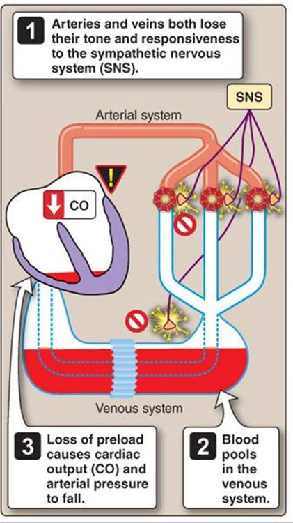
Figure 40.10
Distributive shock.
IV. SHOCK STAGES
The progression of shock can be divided into three stages, beginning with the initial causal event and then progressing in a sequential manner through preshock, shock, and end-organ failure. The following discussion uses hemorrhagic shock as an example to illustrate how the body responds to the initial event and how the systems that attempt to compensate for loss of MAP can create positive feedback spirals that may ultimately hasten failure and lead to death.
A. Preshock
Hemorrhage depletes blood volume and drains the venous reservoir. Hemorrhage depletes veins preferentially because the heart continues transferring blood from the venous compartment to arteries and their dependent capillaries until the venous compartment is depleted. Loss of preload causes MAP to begin to fall, triggering an SNS-mediated baroreceptor reflex (see Figure 20.14 and 20·III). The SNS redirects blood flow away from nonessential organs, increases cardiac inotropy and heart rate (Figure 40.11), and mobilizes blood reservoirs. These pathways are summarized in Figure 40.12.
1. Redirection of flow: Flow to nonessential organs is reduced by selective SNS-mediated constriction of resistance vessels. Systemic vascular resistance (SVR) rises as blood flow is directed away from splanchnic, cutaneous, and muscle vascular beds. Reduced flow to the kidney triggers renin release from the juxtaglomerular apparatus (JGA) and activates the long-term fluid retention pathways (see below). Two key components (angiotensin IIand antidiuretic hormone) are vasoactive and potentiate SNS-mediated vasoconstriction (see 28·III).
2. Cardiac efficiency: SNS stimulation of the myocardium increases HR and contractility to help compensate for loss of preload (see Figure 40.11). Epinephrine release from adrenal glands during SNS activation contributes to tachycardia and increased inotropy during preshock.
3. Venoconstriction: SNS stimulation of veins increases their tone and decreases their capacity, forcing blood back to the heart. Venous return (VR) is aided by a steepening of the pressure gradient between capillary beds and the right atrium.
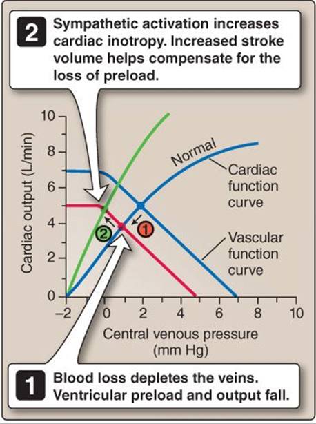
Figure 40.11
Reflex increases in cardiac inotropy during hypovolemia.
4. Transcapillary refill: The baroreceptor reflex helps preserve flow to critical organs during the first few minutes after a hemorrhage. It also buys time that allows fluid to migrate from the interstitium to the vasculature, a process is known as transcapillary refill. Transcapillary refill is a primary survival mechanism that relies on Starling forces to recruit interstitial fluid (see above). Resistance vessel constriction reduces capillary hydrostatic pressure and allows plasma colloid osmotic pressure (πc) to drive fluid movement from the interstitium into the vasculature (see Figure 19.28). Fluid influx dilutes the plasma proteins and reduces πc, but transcapillary refill can still replace ~75% of lost blood volume during hemorrhage.
B. Shock
The baroreceptor reflex effectively compensates for decreases in circulating blood volume of ~10%, and, thus, the only early sign of imminent shock may be mild tachycardia. Once blood volume drops by greater than ~10%, however, compensatory mechanisms are no longer adequate to sustain perfusion in critical circulations, and signs of shock become overt. These signs include hypotension; cold, clammy skin; decreased urine output; and a rise in plasma lactate levels.
Most organ systems have functional reserves that permit homeostasis even as system capacity is reduced. The efficacy of cardiovascular reserves explains why an individual can donate a unit of blood with little or no detrimental effect on MAP.
1. Hypotension: When blood volume drops below ~10%, increases in HR and inotropy alone are unable to compensate for loss of preload, and systolic blood pressure drops to 90 mm Hg or below. Intense SNS-mediated constriction of the vasculature limits blood outflow from the arterial system and keeps diastolic blood pressure high, and, thus, MAP is maintained at a level that allows blood to reach the critical cerebral and coronary circulations. These circulations are regulated primarily through autoregulatory mechanisms (e.g., CO2, K+, and lactate release), and, thus, are not directly influenced by SNS activity.
2. Skin: Intense SNS activation raises SVR by effectively shutting off flow to the vascular beds that occupy the lowest positions on the circulatory hierarchy, including the splanchnic and cutaneous circulations. The intensity of SNS activation is clearly apparent in the skin, which becomes cold and clammy. Cooling is due to intense cutaneous vasoconstriction, which reduces flow to <6 mL/min. Blood drains from the skin, and its temperature cools accordingly. SNS activation also stimulates sweat glands. Because sweat is a modified blood filtrate, when blood flow is curtailed, output is minimal. The skin becomes slightly clammy to the touch.
3. Urinary output: The renal glomerular afferent and efferent arterioles are both resistance vessels. During intense SNS activation, flow through both is restricted severely, and glomerular hydrostatic pressure (PGC) falls dramatically (Figure 40.13; see 25·IV·F). PGC determines glomerular filtration rate (GFR), so flow through the tubule and urine production slows also to <30 mL of urine output per hour (oliguria).

Figure 40.12
Pathways that preserve arterial pressure during preshock.
4. Metabolic acidosis: Plasma lactate levels are normally 0.5–1.5 mmol/L, but hypoxia forces many tissues to rely on anaerobic metabolism, and, hence, lactate levels rise. A plasma lactate of >4 mmol/L is consistent with shock, although lactate levels can rise under other circumstances also (e.g., ketoacidosis and anaerobic exercise).
C. System failure
The actions described above may be insufficient to ensure patient survival, even though arterial pressure may renormalize for an hour or two. Blood pressure alone may not reliably reflect adequacy of perfusion in early shock because the central nervous system (CNS) cardiovascular control centers have the ability and determination to maintain MAP at levels that ensure continued flow to the cerebral circulation to the very last. In cases of severe hemorrhage, this is accomplished by holding SVR at levels that compromise organs that occupy lower positions on the circulatory hierarchy (see 20·II·F), including the kidneys and gastrointestinal (GI) system. Once the invisible line demarcating reversible from irreversible shock has been crossed, a positive feedback spiral begins that leads inevitably to organ failure and death (Figure 40.14).
The need to restore circulating blood volume as soon as possible after trauma is a major reason for the widespread use of mobile trauma teams and Medivac helicopters. Rapid-response units allow medical personnel to reach the scene of an accident and administer intravenous fluids to a patient within the critical window before irreversible tissue damage occurs (a period of variable duration often referred to in Emergency Medicine as the “golden hour”).

Figure 40.13
Effects of intense sympathetic activation on glomerular blood flow.
1. Cardiac depression: If MAP drops below 60 mm Hg, the myocardium becomes ischemic through inadequacy of coronary perfusion. Ischemia impairs myocardial contractility, and, therefore, pressure falls further. So begins a positive feedback cycle that results in acute heart failure. During hemorrhage, one or more myocardial-depressant factors may be released from ischemic tissues that further challenge cardiac function.
2. Sympathetic escape: The SNS cannot maintain intense vasoconstriction for prolonged periods, so SVR eventually falls. Resistance vessel dilation (“sympathetic escape”) may be due to SNS neurotransmitter depletion, α-adrenergic receptor desensitization, or chronically elevated metabolite concentrations overriding central control. Venoconstrictor influence ultimately fails also, thereby impairing VR and preload (see Figure 40.10).
3. Acidemia: Lactic acid and high PaCO2 (due to inadequacy of tissue perfusion and pulmonary and renal dysfunction) together cause significant acidemia. Acidemia impairs myocyte function and further challenges the ability of the myocardium and vasculature to sustain CO and SVR, respectively. MAP falls yet further as a result.

Figure 40.14
Positive feedback pathways that cause cardiovascular system failure.
4. Increased blood viscosity: When blood is moving slowly, red blood cells (RBCs), and other blood components adhere to each other, which raises blood viscosity (see 19·III·C). The process is exacerbated by acidemia and involves not only RBCs, but also leukocytes and platelets, causing blood “sludging.” Sludging increases resistance to flow through the vasculature, and, because MAP cannot rise to compensate, tissue perfusion falls as a consequence. In time, the microvessels (i.e., capillaries, arterioles) become plugged with clots.
5. Cellular deterioration: With prolonged hypoxemia, cell integrity breaks down, triggering an inflammatory response. Inflammatory mediators increase vascular permeability and plasma exudes into the interstitial space at the expense of blood volume. Deterioration of the GI epithelial lining breaches the barrier separating the gut contents from the vasculature, allowing microorganisms to gain access to the circulation. The likelihood of septic shock when (and if) circulation is restored is increased greatly as a result.
6. Cerebral depression: Prolonged hypoxemia ultimately impacts the brain. Neural activity is depressed, and the cardiovascular and respiratory control centers fail. As sympathetic output wanes, MAP declines. Cerebral perfusion pressure decreases also, and brain death soon follows.
V. HEART FAILURE
Heart failure can be the final common pathway for virtually all forms of cardiac disease, and, therefore, there are many underlying causes (Figure 40.15). Although the time course of failure can vary widely, it may ultimately result in cardiogenic shock.

Figure 40.15
Common causes of heart failure.
A. Causes
There can be considerable overlap in the ways in which the various underlying causes of heart failure impact cardiac performance. The right and left ventricles face different challenges and can fail independently of each other, but the left heart is so dependent on the right (and vice versa) that failure of either side independently elicits similar compensatory mechanisms.
1. Right heart: The right heart is a thin-walled chamber that generates low peak systolic pressures against a low pulmonary vascular resistance (PVR). If flow resistance increases as a result of PE or pulmonary hypertension, for example, it has limited ability to compensate, and, hence, failure develops.
The most common cause of right heart failure is left heart failure.
2. Left heart: The left heart is a thick-walled chamber well adapted to stress associated with generating high peak systolic pressures against a high SVR. Causes of left-heart failure can be grouped according to whether they impair filling (diastolic heart failure, also known as heart failure with preserved ejection fraction), or ejection (systolic heart failure).
a. Diastolic: One common cause of diastolic failure is LV hypertrophy, due either to a chronically increased afterload or a cardiomyopathy. Untreated hypertension and aortic stenosis both impair CO by increasing LV afterload. The myocardium hypertrophies in order to generate the high pressures required to maintain a normal CO (see Clinical Application 18.2). New myofibrils are added in parallel with old myofibrils, causing individual myocytes to increase their girth, thickening the ventricular wall (Figure 40.16). The advantage to a thicker wall is that it helps offset the effects of high intraventricular pressure on wall stress, as described by the law of Laplace (Figure 40.17; see 18·IV). The disadvantages to hypertrophy are twofold. First, myocyte diameter may exceed the diffusional limits for O2, which increases the likelihood of ischemia (see Figure 40.16) and arrhythmias. Second, the ventricle stiffens and becomes increasingly difficult to fill, requiring higher RV ejection pressures. Ultimately, both ventricles fail under such circumstances.
b. Systolic: Systolic heart failure occurs when the LV fails to maintain adequate output. This can be due to impaired contractility or an excessive afterload, but the most common cause of systolic failure is MI, as discussed below.
B. Myocardial infarction
MI, or a “heart attack,” is one of the most common causes of heart failure. An MI typically occurs when an atherosclerotic plaque ruptures and forms a blood clot that occludes a coronary supply vessel. Myocytes that had previously been served by the occluded vessel become ischemic and die, which impairs myocardial contractility.
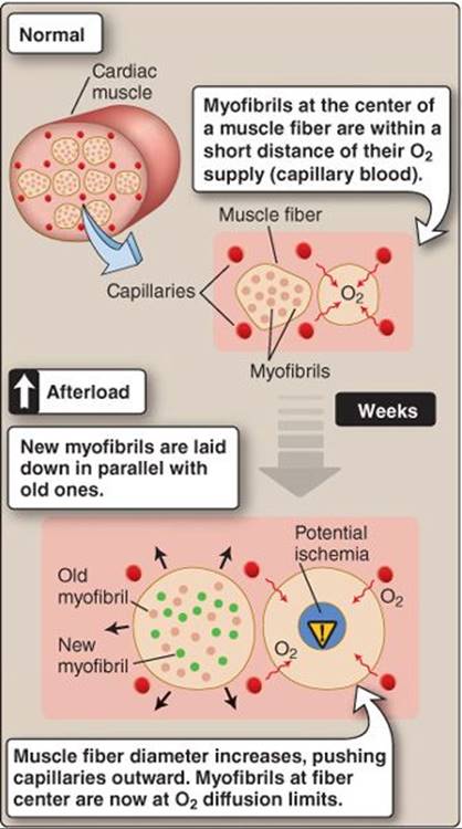
Figure 40.16
Effects of myocardial hypertrophy on O2 delivery to myofibrils.

Figure 40.17
Increases in ventricular wall thickness during cardiac hypertrophy.
The chances of surviving such an event depend on many factors. If the infarcted area is relatively small, short-term responses may allow the patient to survive the initial insult until long-term compensatory mechanisms become active.
C. Compensation
A small infarct triggers short-term and long-term compensatory mechanisms simultaneously. Short-term events help maintain output until the longer-term pathways have had time to activate fully.
1. Short term: The short-term response to myocardial ischemia includes both local and central reflexes.
a. Local: Interruption of blood flow to myocytes causes interstitial metabolite levels (e.g., adenosine, K+, CO2, lactate) to rise. All resistance vessels in the immediate vicinity dilate reflexively through local vascular control mechanisms. Collaterals are tonically constricted normally, but they also participate in the vasodilatory response to rising metabolite levels. Blood flow through collaterals may allow areas peripheral to a focal infarct to survive the initial ischemic event (see 21·III·E).
b. Central: Death of myocytes impairs myocardial contractility, which reduces LV stroke volume and CO (Figure 40.18). MAP falls as a result, triggering a baroreceptor reflex that involves all of same effector mechanisms described in Section IV·A above. If the infarct is small, these pathways may be sufficient to restore MAP.
2. Long term: A decrease in MAP also activates the renin– angiotensin–aldosterone system, regardless of cause (see 20·IV). It takes 24–48 hours for Na+ and water retention mechanisms to expand ECF volume and support the failing myocardium with increased preload. In the days and weeks following an ischemic event, the body begins repairing some of the tissue damage wrought by infarction. Collateral vessels enlarge, and the myocardium hypertrophies to help compensate for the loss of contractility.
D. Preload penalty
The Frank-Starling mechanism is highly effective in compensating for minor decreases in cardiac inotropy (see 18·III·D). Some individuals may suffer a series of these insults and remain unaware for years until compensation causes symptoms (e.g., dyspnea associated with pulmonary congestion, as discussed below). Dyspnea is only one of several penalties associated with preloading, however. Others include limits to the benefits of length-dependent sarcomeric activation, excessive ventricular wall stress, dysrhythmias, valve incompetency, and edema.
1. Length-dependent activation limits: Preloading a healthy heart increases CO through length-dependent activation of sarcomeres (see 13·IV). The end-systolic pressure–volume relationship has a plateau region, however, and, once myocytes have been stretched to a length that optimizes force generation, further increases in preload are ineffective in generating additional force (Figure 40.19).

Figure 40.18
Short-term (sympathetic) response to myocardial infarction.

Figure 40.19
Limits to the beneficial effects of preloading.
2. Wall stress: Preloading dilates the ventricle and increases wall tension, as predicted by the law of Laplace. Wall tension contributes to afterload, so, although preloading does help support output within normal physiologic ranges, high preloads also increase cardiac workload and reduce its efficacy.
3. Dysrhythmias: Excessive preloading stretches the ventricular wall and distorts conduction pathways, which predisposes the myocardium to potentially fatal dysrhythmias and arrhythmias.
4. Valve incompetency: Excessive preloading also stretches and distorts the cartilaginous valve rings and unseats the valves. In practice, this means that the valve leaflets no longer come into close apposition upon closure, and blood flows in a retrograde manner. Regurgitation further increases the workload required of a failing heart.
5. Edema: Central venous pressure (CVP) elevation raises mean capillary pressure and favors fluid filtration from blood into the interstitium. In the systemic vasculature, interstitial fluid excess manifests as swollen ankles and feet. The lower extremities are particularly prone to edema in an erect individual because vascular pressures in these regions are enhanced by gravity. In the lungs, fluid filters from the pulmonary capillaries and collects in the alveolar sacs, where it interferes with gas exchange (pulmonary congestion) as shown in Figure 40.20. Pulmonary edema may cause orthopnea (shortness of breath when lying flat), forcing patients to sleep sitting upright. Gravity helps reduce pulmonary perfusion pressures and, thus, decreases the likelihood of fluid accumulation in the air spaces.
E. System failure
A failing heart becomes locked in a decompensatory spiral in which preload supports output yet ultimately limits efficiency through its effects on wall tension. Unless this cycle is interrupted and managed, it can prove fatal. Thus, the goal of medical intervention is to decrease preload using diuretics while simultaneously supporting the myocardium with inotropes that help it work more efficiently at a lower filling volume.1 In end-stage heart failure, myocytes continue to die one by one, slowly chipping away at contractility and the ability to sustain MAP. Even mild physical exertion causes severe dyspnea because cardiac reserve has dropped to the point where even minimal muscular activity places demands on output that exceed myocardial capabilities, so patients become bedridden (see 21·III·B). Bedrest exacerbates frailty by causing muscle atrophy and decreased bone density. Excessive volume loading causes pulmonary edema and hypoxic respiratory failure. The liver fails due to passive congestion and restricted O2delivery caused by systemic edema. Loss of glomerular pressure precipitates renal failure. Each additional organ loss raises mortality risk by ~20%.
 1For more information on pharmaceutical approaches to treatment of heart failure, see LIR Pharmacology, 5e, p. 193.

Figure 40.20
Pulmonary edema resulting from heart failure. Pc = capillary hydrostatic pressure; πc = plasma colloid osmotic pressure.
VI. RESPIRATORY FAILURE
Respiratory failure occurs when the respiratory system is unable to fulfill one or both of its gas exchange functions, namely O2 uptake or CO2 elimination. Clinically, this manifests as hypoxemic respiratory failure or hypercapnic respiratory failure, respectively. The two failure types represent syndromes (sets of related symptoms) rather than the end result of any specific disease.
A. Causes
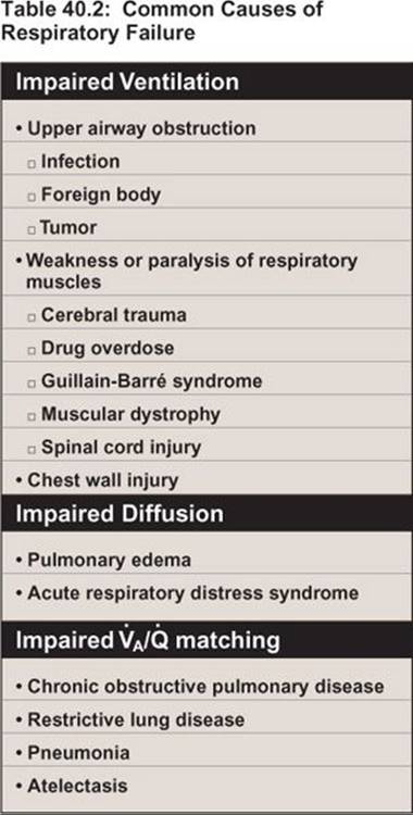
Respiratory failure may develop chronically or acutely, usually as a result of trauma (see Section D below for a discussion of acute respiratory distress syndrome [ARDS]). Conditions causing respiratory failure can be grouped according to whether they impair ventilation (air-pump function and control), diffusion (integrity of the blood–gas interface), or ventilation–perfusion ( ) matching (Table 40.2).
B. Hypoxemic respiratory failure
Hypoxemic respiratory failure is characterized by a PaCO2 <~60 mm. Hypoxemia can be caused by hypoventilation, but, because attaining a normal PaO2 (100 mm Hg) requires that the full area of the blood–gas interface be functional, processes that decrease this area and allow venous blood to pass through the lungs without being arterialized cause some degree of hypoxemia. Thus, hypoxemic respiratory failure usually occurs when air or pulmonary blood is unable to access the interface (i.e., mismatch).
1. Ventilation/perfusion mismatch: Regional mismatch is common in a healthy lung but has minimal impact on overall respiratory function (see 23·IV·B). In disease states, large numbers of alveoli may collapse and seal (atelectasis), or fill with fluid (pulmonary edema), pus (pneumonia), or blood (hemorrhage), all of which effectively prohibit O2 uptake and cause hypoxemia (Figure 40.21).
2. Compensation: Hypoxemia is detected primarily by the aortic and carotid chemoreceptors (see 24·III·C). CNS respiratory control centers respond by increasing minute ventilation, and CNS cardiovascular control centers simultaneously increase CO to help maximize the O2 diffusion gradient across the exchange barrier.
3. Consequences: The physiologic consequences of hypoxemia were discussed in relation to the effects of ascent to altitude (see 24·V·A). Mild hypoxemia causes slight impairment of mental function and visual acuity. When PaO2 drops below ~40–50 mm Hg, patients become confused and prone to personality changes and irritability. Hypoxemia also initiates a positive feedback spiral in which the pulmonary vasculature constricts reflexively and further reduces O2 uptake. Vascular constriction also increases right ventricular afterload and induces pulmonary hypertension, which stresses the RV. These symptoms can usually be reversed clinically by administering O2 to maximize ratios and, at least temporarily, restore PaO2 until the underlying cause of hypoxemia can be evaluated and addressed.

Figure 40.21 mismatch, one common cause of hypoxemia. VA = alveolar ventilation; Q = alveolar perfusion, all lpartial pressure values are given in mm Hg.
C. Hypercapnic respiratory failure
Hypercapnic respiratory failure is indicated by an acute rise in PaCO2 to >~50 mm Hg. Hypercapnia that develops over a course of months is tolerated well, however, so failure may not occur until PaCO2reaches ~70–90 mm Hg. Unlike hypoxemia, hypercapnia can be corrected relatively easily by adjusting alveolar ventilation. Thus, hypercapnic respiratory failure usually only occurs when ventilatory control is impaired. Hypercapnia is usually associated with varying degrees of hypoxemia.
1. Ventilation: Ventilatory failure occurs if the respiratory center or its neural pathways are damaged by stroke, drug overdose, or neuromuscular diseases (e.g., myasthenia gravis), but the most common causes of hypercapnic respiratory failure are impairment of air-pump function (chest wall and respiratory muscles) and chronic airway disorders.
a. Chest wall: Movement of the chest wall can become severely limited by obesity and by abnormal spine curvature. Kyphosis (forward flexion curvature), as shown in Figure 40.22A, and scoliosis (lateral curvature), as shown in Figure 40.22B, are congenital disorders, but the former is also seen in association with arthritis and osteoporosis (see Figure 40.22A). Both can develop into debilitating curvatures that severely limit chest wall excursions and hasten failure in a compromised lung.
b. Muscles: The respiratory muscles (diaphragm and intercostals) increase intrathoracic volume and expand the lungs during inspiration. They are skeletal muscles and, therefore, susceptible to wasting diseases such as muscular dystrophy. They are also subject to fatigue, which is a principal concern when addressing problems underlying respiratory failure. Chronic conditions that reduce chest wall or lung compliance (restrictive lung diseases) increase the work of breathing, and fatigue ultimately reduces contractility and causes hypoventilation and hypercapnia.
c. Airways: Chronic obstructive pulmonary disease and asthma increase airway resistance and can reduce alveolar ventilation and raise PaCO2.
2. Compensation: PaCO2 is monitored by central and peripheral chemoreceptors. The respiratory center responds to acute hypercapnia by increasing ventilation rate, even if such an action causes respiratory muscle fatigue and precipitates a respiratory crisis. During chronic hypercapnia, the chemoreceptors adapt to persistent elevation of PaCO2, and, thus, ventilation rates remain normal. CO2 retention decreases plasma pH (respiratory acidosis), but the kidneys compensate by retaining HCO3−, allowing pH to remain within a normal range even as PaCO2 climbs above ~70–90 mm Hg (see 28·VI·C). Such patients typically have a limited pulmonary reserve, however, and may decompensate quickly if illness creates additional demands on an already compromised system.
3. Consequences: The cerebral vasculature is highly sensitive to PaCO2. Acute CO2 retention causes cerebral vasodilation, which causes headaches and intracranial hypertension. The latter may manifest as a swelling of the optic disc and can cause blindness. High CO2 levels also cause dyspnea and neurologic symptoms, such as involuntary wrist movements and hand tremors.

Figure 40.22
Kyphosis and scoliosis.
D. Acute respiratory distress syndrome
ARDS is the leading cause of respiratory failure in young adults, with mortality rates as high as ~58%. In the military, ARDS was known originally as “shock lung,” reflecting the similarities between the onset of ARDS and septic shock.
1. Causes: ARDS may develop in association with a wide range of conditions, the most common being sepsis, aspiration of stomach contents, near drowning, multiple blood transfusions, trauma, bone fractures, and pneumonia.
2. Stages: ARDS is precipitated by local or circulating inflammatory mediators (e.g., histamine), which trigger an inflammatory cascade that severely damages the alveolar endothelial cells (pneumocytes) and capillary endothelial cells that comprise the blood–gas interface (Figure 40.23; see 22·II·C). There are three discrete stages to ARDS, characterized by exudate formation, hyaline membrane deposition, and fibrosis.
a. Exudates: The initial stages of ARDS (24–48 hr) are characterized by an inflammatory reaction within the lung parenchyma that damages the alveolar epithelium and causes a profound increase in capillary permeability. The lungs fill with a bloody exudate containing plasma proteins and cellular debris (see Figure 40.23A). Chest x-rays typically reveal diffuse bilateral infiltrates that are reminiscent of pulmonary edema but that occur when CVP and left atrial pressure is normal.
b. Hyaline membrane: In the following 2–8 days, hyaline membranes begin to settle from the exudate and cover the alveolar lining (see Figure 40.23B). Hyaline is a fibrous matrix of plasma proteins and disrupted cell debris.
c. Fibrosis: After ~8 days, the interstitium is infiltrated by fibro-blasts, which deposit collagen and other fibrous materials.
3. Consequences: The alveolar infiltrates and hyaline membranes prevent gas exchange, causing hypoxemia. The infiltrates also inactivate surfactant and suppress surfactant production by type II pneumocytes, causing alveolar collapse. Surfactant loss and atelectasis makes the lung extremely stiff and difficult to expand, which is one of the hallmarks of ARDS (Figure 40.24). Atelectasis not only impairs ventilation, it also reduces the area of the blood–gas interface and, thereby, exacerbates hypoxemia. Support from a mechanical ventilator is life saving while the underlying process is addressed.
E. System failure
Hypoxemia associated with acute respiratory failure elicits the same SNS responses as shock, but in the context of an intact and functional vasculature. SNS-stimulated increases in CO and SVR can cause MAP to rise to levels that rupture cerebral blood vessels. Chronic hypoxemia causes a gradual decline in neural function that inhibits the pathways controlling ventilation and blood pressure. The most obvious external sign of hypoxemia is cyanosis, a blue discoloration of the skin and mucous membranes that reflects the color of deoxyhemoglobin (cyanosis occurs when deoxyhemoglobin levels rise to ~5 g/dL). Acute hypercapnia produces respiratory acidosis through CO2retention, but the body's response is dominated by reflex responses to the hypoxemia that accompanies hypercapnia. Acute hypercapnia causes anesthetic-like effects on the CNS (CO2 narcosis). Narcosis appears at a PCO2 of ~90 mm Hg, causing confusion and lethargy. High PCO2 depresses respiratory center function and suppresses ventilatory drive, thereby creating a positive feedback cycle that potentiates CO2 retention and, ultimately, results in coma and death (PCO2 ~130 mm Hg).

Figure 40.23
Acute respiratory distress syndrome. ROS = reactive oxygen species.
VII. KIDNEY FAILURE
Two forms of kidney failure are recognized. Acute kidney injury (AKI) develops abruptly (within 48 hours) but usually can be treated if patients have no other complicating medical issues. Kidney function and risk of failure can be assessed using RIFLE criteria as shown in Table 40.3. Chronic kidney disease (CKD) develops over the course of many years. CKD is characterized by progressive and irreversible loss of nephrons, but the development of dialysis and transplant technologies means that CKD is not necessarily fatal. The mortality rate of patients on dialysis is very high (dialysis increases lifespan by only 4.5 years in 60–64-year-olds), but death usually occurs from cardiovascular disease, infection, or cachexia (a wasting syndrome). AKI is a primary cause of death (~75%) in urgent care facilities, however, where patients may be elderly and have other underlying pathologies.
A. Causes
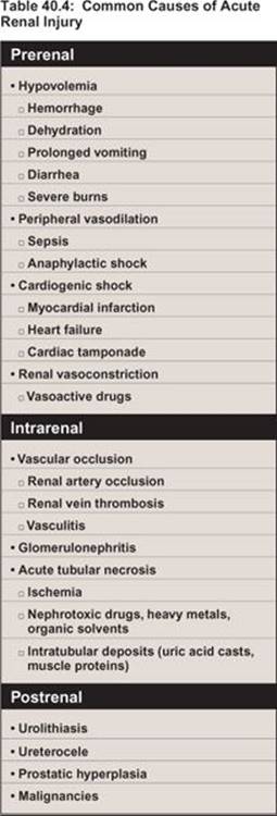
AKI can be precipitated by numerous factors, which are usually grouped according to where in the nephron they act: prerenal, intrarenal, and postrenal (Table 40.4).
1. Prerenal: Prerenal failure is characterized by a profound drop in GFR due to decreased renal blood flow and glomerular perfusion pressure (see Figure 40.13). Prerenal failure usually occurs secondarily to shock and ischemia.
2. Intrarenal: Intrarenal failure occurs when the renal tubule or surrounding interstitium is injured. The most common cause of intrarenal failure is acute tubular nephrosis (ATN). ATN usually results from ischemia, but the tubule may also be injured by drugs and other toxins.
a. Ischemia: The renal epithelium's transport functions create a high ATP dependency and coincident susceptibility to ischemia. The transport epithelium receives O2 via the peritubular capillary network, whose flow is governed by glomerular arterioles (see 26·II·C). Tubule ischemia usually occurs during a hypotensive crisis when both arterioles are constricted, filtration has ceased, and peritubular flow no longer meets the epithelium's basal O2 needs. Ischemic cells may respond by sloughing their apical villi into the tubule lumen, which reduces overall surface area, transporter density, and O2 requirements.
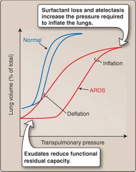
Figure 40.24
Change in lung function accompanying acute respiratory distress syndrome (ARDS).

b. Toxins: The renal tubule's ability to concentrate drugs and toxins makes it very vulnerable to nephrotoxicity. Although all tubule regions are at risk, necrosis most commonly occurs in the proximal tubule, which is responsible for secreting many drugs (see 26·IV).
3. Postrenal: Postrenal failure is caused by urinary outflow obstruction, which can occur at any point within the tubule, collecting system, ureters, bladder, or urethra. Common causes include kidney stones (calculi; see Clinical Application 4.2) and an enlarged prostate (prostatic hyperplasia). Obstruction causes pressure in more proximal tubule segments to build to the point where they negate PUF, and glomerular filtration stops. In time, the affected tubule segments may dilate and atrophy.
B. Consequences
Renal failure is considered to have occurred when GFR is reduced to 25% of normal values. Kidneys lose their ability to control water or electrolyte levels when GFR is so low, which manifests as hypervolemia, hyperkalemia, metabolic acidosis, and accumulation of nitrogenous wastes (azotemia).
C. System failure
An inability to excrete nitrogenous wastes and maintain electrolyte balance negatively impacts all organs, but the neurologic effects dominate. Patients become lethargic, drowsy, and delirious and eventually slip into a coma. Death typically occurs due to cardiac arrhythmia caused by hyperkalemia.
VIII. MULTIPLE ORGAN DYSFUNCTION SYNDROME
Emily Dickinson's generation was well acquainted with the signs of organ system failure and impending death. People usually died at home in the care of family and loved ones. The poem that opened this final chapter accurately notes the effects of acute hypoxemia on the CNS (convulsion) and the consequences of intense SNS-mediated stimulation of sweat glands (The Beads upon the Forehead/By homely Anguish strung). In more recent times, the final throes occur in medical facilities, witnessed mainly by health care professionals. Patients arriving at emergency departments and intensive care units often already have long-standing diseases and have coped with progressive failure of one or more organs for many months or years. They present when an infection or some other seminal event has precipitated multiple organ dysfunction syndrome (a medical crisis involving two or more organ systems), at which point medical intervention is required for continued survival. It is the job of care providers to help restore homeostasis and create an environment in which the body may recover from an acute disease state.
In the final reckoning, however, the physiologic systems that maintain homeostasis and that have been described in the preceding chapters are robust and have remarkable recuperative capabilities. They readily reassert control if given a chance, either on their own or by timely medical intervention as needed. Whether or not they take the chance is the medical mystery that is Life.
Chapter Summary
• Aging is accompanied by a progressive reduction in total cell number and organ functionality. Aging ultimately results in organ failure and death.
• The final common pathway for most instances of cell death is ischemia. Ischemia initiates a series of events that comprise an ischemic cascade. Significant events include acidosis, dissipation of ion gradients, Ca2+ activation of proteases and other degradative enzymes, and mitochondrial lysis.
• Ischemia usually results from lack of perfusion due to circulatory shock. Hypovolemic shock results from hemorrhage or reduced extracellular fluid volume. Cardiogenic shock is caused by loss of cardiac pump function. Distributive shock occurs when the central nervous system loses vascular control, and the resulting systemic vasodilation allows blood to become trapped in capillaries and veins.
• Shock can be divided into three stages: preshock, shock, and organ failure. During preshock, sympathetic nervous system–mediated increases in cardiac and vascular function compensate for falling mean arterial pressure.
• During shock, perfusion of critical circulations (cerebral, coronary) becomes suboptimal, and signs of intense sympathetic nervous system activity become overt (hypotension, decreased urinary output, acidosis).
• System failure occurs when shock becomes irreversible. The cardiovascular system becomes locked in a positive feedback spiral that results in loss of myocardial and vascular contractility; acidemia; blood clotting; cellular deterioration; and, ultimately, loss of cerebral perfusion pressure and brain death.
• Heart failure is the leading cause of death in the United States and is the final common pathway for many cardiac diseases. The right heart typically fails as a result of increased pulmonary vascular resistance. Causes of left-heart failure include filling impairment (diastolic failure), loss of contractility, or an excessive afterload that impairs output (systolic failure).
• Myocardial infarction is a common cause of heart failure. Long-term compensatory mechanisms that support cardiac output though volume retention and invocation of the Frank-Starling mechanismultimately become counterproductive and precipitate failure. Symptoms of congestive heart failure include edema and shortness of breath upon exertion.
• Respiratory failure occurs when the pulmonary system is unable to take up O2 or eliminate CO2 from the body. Hypoxemic respiratory failure usually occurs as a result of impaired alveolar ventilation due to alveolar collapse or accumulation of fluid, pus, or blood in alveoli.
• The central respiratory control centers readily adapt to increases in PaCO2, so hypercapnic respiratory failure usually reflects impairment of air-pump function (respiratory muscles and chest wall).
• Acute respiratory distress syndrome (ARDS) is a leading cause of respiratory failure. ARDS is associated with inflammatory reactions that damage the lung parenchyma and increase pulmonary capillary permeability. Lungs fill with infiltrates and become stiff and difficult to expand.
• Renal failure can be precipitated by inadequacy of perfusion, kidney tubule deterioration, or obstruction of urinary outflow. The kidney's resulting inability to control water and electrolyte levels allows K+levels to rise (hyperkalemia), and death usually results from cardiac arrhythmia.
Study Questions
Choose the ONE best answer.
IX.1 A 23-year-old pregnant woman in her third trimester complains to her friend that her feet and ankles are frequently swollen. The swelling is most likely caused by which of the following?
A. High pedal venous pressures
B. Increased left ventricular preload
C. Hypertension (preeclampsia)
D. Decreased blood viscosity
E. Excessive fluid retention
Best answer = A. The gravid uterus compresses veins returning blood from the lower extremities, causing pedal venous pressures to rise (37·IV·D). Pedal capillary hydrostatic pressure rises as a result, promoting fluid filtration and edema. Filtration is potentiated by a coincident fall in plasma colloid osmotic pressure during pregnancy. Maternal cardiac output increases through fluid retention to increase left ventricular preload, but the additional output is needed to supply the placenta and does not contribute significantly to a rise in venous pressure. Blood viscosity changes and hypertension do not affect the Starling forces significantly under physiologic conditions.
IX.2 Healthy pregnant women and athletes engaged in an aerobic exercise training routine both show which of the following changes?
A. Afterload-induced ventricular hypertrophy
B. Resting heart rate increases
C. Resting diastolic pressure increases
D. Minute ventilation increases
E. Hemoglobin concentration decreases
Best answer = D. Both pregnancy and aerobic training increase minute ventilation to maximize tissue O2 delivery (37·IV·E; 37·VI·D). Hemoglobin (Hb) levels fall during pregnancy, whereas training increases Hb. Ventricular hypertrophy during pregnancy and training occurs in response to a chronically increased preload and stroke volume (SV). Resting heart rate (HR) rises during pregnancy to help meet the increased demands for cardiac output (CO) placed on the maternal cardiovascular system. Resting CO is not changed by training, however, so the SV increase decreases resting HR. Resting diastolic blood pressure falls during pregnancy due to runoff into the low-resistance placental circuit and decreases minimally with aerobic exercise training.
IX.3 The answer choices below compare pairs of cardiovascular system variables. Which of these choices best describes the fetal cardiovascular system?
A. Vascular resistance: systemic > pulmonary
B. Flow: pulmonary vein > descending aorta
C. Atrial pressure: left > right
D. Hemoglobin levels: adult > fetal
E. O2 saturation: inferior vena cava > aorta
Best answer = E. Fetal blood is oxygenated in the placenta, then flows via the umbilical vein at ~85% saturation into the inferior vena cava (~70%), through the heart, and into the aorta (37·V·C). Saturation has fallen to ~65% by venous admixture during passage. The fetal cardiovascular system is characterized by its high pulmonary vascular resistance compared with systemic vascular resistance. Shunts (foramen ovale and ductus arteriosus) direct blood past the high-resistance pulmonary circuit, and, therefore, pulmonary blood flow is lower than aortic flow. Fetal blood is enriched with hemoglobin to enhance its O2-carrying capacity.
IX.4 A 52-year-old female with multiple sclerosis has been admitted to the emergency department on three occasions with mild hypothermia (rectal temperature 34°– 35°C) during fall camping trips. What is the most likely reason that this individual might be experiencing low temperatures during prolonged cold exposure?
A. Decreased cutaneous warmth sensation
B. Decreased cutaneous pain sensation
C. Increased sweat secretion
D. Preoptic hypothalamic lesions
E. Caudal cerebellar lesions
Best answer = D. Multiple sclerosis causes demyelination and decreased axonal conduction. If a sclerotic lesion occurs in the preoptic area, then central sensation and processing of signals from skin temperature receptors may not be regulated appropriately (38·II·B). This can effectively blunt responses to thermal stress and allow body temperature to fluctuate more with ambient temperatures. Lesions occurring in the cerebellum would affect movement and coordination rather than temperature regulation. Individuals with multiple sclerosis can develop peripheral neuropathy, but skin warmth and pain receptors are not directly involved in cold sensation. Sweating occurs during heat exposure, not cold exposure (38·II·D·2).
IX.5 A 6-month-old female is inadvertently exposed to a cold environment after she and her parents were in a rainstorm on a cold day. What thermogenic tissue has a mitochondrial uncoupling protein that can aid her thermoregulation?
A. White adipose tissue
B. Brown adipose tissue
C. Skeletal muscle
D. Cardiac muscle
E. Smooth muscle
Best answer = B. Nonshivering thermogenesis is a process of increasing metabolic rate to generate heat without shivering (38·II·D·4). Brown adipose tissue, which is proportionally higher in infants than in adults, has a specialized mitochondrial uncoupling protein (thermogenin) that generates heat without producing useful work. White adipose tissue does not have this capability. Muscle, primarily skeletal, can participate in nonshivering thermogenesis but does not contain a thermogenin-like protein. Cardiac and smooth muscle do not participate directly in cold responses.
IX.6 In which of the following conditions would a cyclooxygenase inhibitor bring internal temperature back within the 36.5°–37.5°C range?
A. Severe hypothermia
B. Ambient cold stress
C. Ambient heat stress
D. Heat exhaustion
E. Fever
Best answer = E. Nonsteroidal anti-inflammatory medications such as aspirin are cyclooxygenase inhibitors that block prostaglandin synthesis. Their actions include inhibiting prostaglandin EP3-receptor activation in the preoptic hypothalamus, thereby lowering a thermoregulatory set point that has been elevated by pyrogens during fever (38·IV·A). Ambient cold and heat stress challenge thermoregulation, but the internal set point is still within the normal range. Severe hypothermia is defined as an internal temperature <28°C, and heat exhaustion is a hyperthermic condition, but the body again attempts to regulate internal temperature to the set point, which is within normal range (38·IV·B).
IX.7 A 25-year-old woman recently underwent surgery for a strained anterior cruciate ligament. Postsurgical exercises included isometric quadriceps exercises that are held until fatigue (~60 s). Which energy system is primarily used in these exercises?
A. Stored adenosine triphosphate
B. Adenosine triphosphate–creatine phosphate system
C. Lactic acid system
D. Citric acid cycle
E. Oxidative phosphorylation
Best answer = C. The lactic acid system predominates during maximal exercise that fatigues with durations between 30 s to 2.5 min (39·III·A). In this system, glycolysis produces pyruvic acid, which is then shuttled to lactic acid. Adenosine triphosphate (ATP) stores can support exercise for a few seconds and the ATP–creatine phosphate system only extends this time to 8–10 s. Aerobic metabolism (citric acid cycle and oxidative phosphorylation) is the primary energy system used to synthesize ATP during maximal exercise lasting 2.5 min or longer.
IX.8 At the beginning of exercise, a feedforward mechanism increases heart and respiratory rates. What is the best term or receptor responsible for this mechanism?
A. Class III muscle afferents
B. Class IV muscle afferents
C. Arterial baroreceptors
D. Peripheral chemoreceptors
E. Central command
Best answer = E. Central command is the feedforward signal that increases cardiovascular and respiratory system function at the beginning or in the anticipation of exercise (39·IV·B·2). Class III and IV muscle afferents provide feedback regarding stretch, compression, and metabolic status of the muscle. Baroreceptors provide feedback regarding arterial pressures at the aortic arch and carotid arteries. Chemoreceptors provide feedback regarding arterial partial pressure of CO2 and O2, and H+ in similar locations (39·IV·A).
IX.9 An exercise-induced rightward shift in the O2-hemoglobin dissociation curve is likely responsible for increasing which of the following respiratory parameters during aerobic exercise?
A. Alveolar ventilation
B. Excess postexercise oxygen consumption
C. Work of breathing
D. Arteriovenous O2 difference
E. O2-carrying capacity
Best answer = D. The arteriovenous (a–v) O2 difference widens with aerobic exercise through increased O2 offloading by hemoglobin ([Hb] 39·VI·B). This manifests in a decrease in venous O2 content, so that even though arterial levels are unchanged, the a–v difference increases. Increased offloading occurs due to a rightward shift in the Hb-O2 dissociation curve (23·VI·B). Postexercise O2 consumption does not impact O2 usage during exercise. Alveolar ventilation and work of breathing both increase during exercise but are unrelated to the Hb-O2 dissociation curve. Blood's O2-carrying capacity is determined primarily by Hb concentration, not by Hb's O2 affinity.
IX.10 A 21-year-old soldier suffers extensive blood loss from deep wounds inflicted by an improvised explosive device. Which of the following identifies the primary mechanism responsible for maintaining blood volume until fluids can be administered?
A. Venoconstriction
B. Resistance vessel constriction
C. Aldosterone release
D. Decreased renal blood flow
E. Recruitment of interstitial fluid
Best answer = E. Hemorrhage causes central venous pressure to fall, which reduces mean capillary hydrostatic pressure in all circulations (40·IV·A). This causes fluid to move into the vasculature from the interstitium (“transcapillary refill”) under the influence of plasma colloid osmotic pressure, thereby helping support blood volume. Constriction of resistance vessels directs blood away from nonessential organs but does not increase blood volume. Venoconstriction forces blood out of veins but does not affect total blood volume. Aldosterone increases fluid retention by the kidney but only after several hours.
IX.11 A 67-year-old woman is brought to the emergency department in shock. Assessment of her cardiovascular function shows that heart rate is high and cardiac output is increased, whereas left atrial pressure, mean arterial pressure, and systemic vascular resistance are all low. What is the likely cause?
A. Septic shock
B. Hypovolemic shock
C. Cardiogenic shock
D. Cardiac tamponade
E. Pulmonary embolism
Best answer = A. Shock triggers an intense sympathetic response in attempts to raise arterial pressure and restore O2 delivery to the brain (40·IV·B). Such a response includes systemic vasoconstriction to increase systemic vascular resistance (SVR). Inflammatory reactions associated with sepsis damage the vasculature and prevent vasoconstriction, so SVR inevitably falls. Hypovolemic shock reduces cardiac output (CO). In cardiogenic shock (including tamponade), left atrial pressure ([LAP] or preload) would be increased in attempts to support CO. Pulmonary embolism would cause LAP and CO to fall, but SVR would be very high.
IX.12 A 48-year-old man with pneumonia is hospitalized when he develops acute respiratory distress syndrome and requires a mechanical ventilator to support breathing. Why is a mechanical ventilator helpful?
A. It increases cardiac output.
B. Alveolar fluid decreases compliance.
C. Inflammatory exudates impair surfactant.
D. It prevents hyaline membrane formation.
E. It prevents pulmonary fibrosis.
Best answer = C. Acute respiratory distress syndrome (ARDS) patients’ lungs are highly noncompliant and require the assistance of a mechanical ventilator to expand, mainly because the inflammatory exudates inactivate surfactant and inhibit its production (40·VI·D). Fibrosis, which may further reduce compliance over time, is not prevented by ventilation. Ventilation has no effect on hyaline membrane formation, which interferes with gas exchange, and may often decrease left ventricular preload and cardiac output. Fluid in the pulmonary interstitium reduces lung compliance, but not within alveoli (fluid-filled lungs are easier to expand than normal because surface tension effects are negated; 22·IV·B).