
Autoregulation keeps RBF and GFR relatively constant
An important feature of the renal circulation is its remarkable ability to maintain RBF and GFR within narrow limits, although mean arterial pressure may vary between ~80 and 170 mm Hg (Fig. 34-12, middle and bottom panels). Stability of blood flow—known as autoregulation (see p. 481)—is also a property of the vascular beds serving two other vital organs, the brain and the heart. Perfusion to all three of these organs must be preserved in emergency situations, such as hypotensive shock. Autoregulation of the renal blood supply is independent of the influence of renal nerves and circulating hormones, and persists even when one perfuses isolated kidneys with erythrocyte-free solutions. Autoregulation of RBF—and, consequently, autoregulation of GFR, which depends on RBF (see Fig. 34-6D)—stabilizes the filtered load of solutes that reaches the tubules over a wide range of arterial pressures. Autoregulation of RBF also protects the fragile glomerular capillaries against increases in perfusion pressure that could lead to structural damage.

FIGURE 34-12 Autoregulation of RBF and GFR. (Data on RBF from Arendshorst WJ, Finn WF, Gottschalk CW: Autoregulation of renal blood flow in the rat kidney. Am J Physiol 228:127–133, 1975.)
The kidney autoregulates RBF by responding to a rise in renal arterial pressure with a proportional increase in the resistance of the afferent arterioles. Autoregulation comes into play during alterations in arterial pressure that occur, for example, during changes in posture, light to moderate exercise, and sleep. It is the afferent arteriole where the autoregulatory response occurs, and where the resistance to flow rises with increasing perfusion pressure (see Fig. 34-12, top panel). In contrast, efferent arteriolar resistance, glomerular and peritubular capillary resistances, as well as venous resistance all change very little over the range of normal to high renal arterial pressures. However, in the range of low renal perfusion pressures—as in congestive heart failure—efferent arteriolar resistance increases, and thereby minimizes decreases in GFR.
Two basic mechanisms—equally important—underlie renal autoregulation: a myogenic response of the smooth muscle of the afferent arterioles and a tubuloglomerular feedback mechanism.
Myogenic Response
The afferent arterioles have the inherent ability to respond to changes in vessel circumference by contracting or relaxing—a myogenic response (see p. 481). The mechanism of contraction is the opening of stretch-activated nonselective cation channels in vascular smooth muscle. The resultant depolarizing leads to an influx of Ca2+ that stimulates contraction (see p. 477).
Tubuloglomerular Feedback
The juxtaglomerular apparatus (JGA; see p. 727) mediates tubuloglomerular feedback (TGF).  N40-3 The macula densa cells in the thick ascending limb sense an increase in GFR and, in classic feedback fashion, translate this to a contraction of the afferent arteriole, a fall in PGC and RPF, and hence a decrease in GFR.
Experimental evidence for the existence of a TGF mechanism rests on measurements of SNGFR. One introduces a wax block into the lumen of the proximal tubule to block luminal flow (Fig. 34-13A). Upstream from the wax block, a pipette collects the fluid needed to compute SNGFR (see Equation 33-13). Downstream of this wax block, another pipette perfuses the loop of Henle with known solutions and at selected flows. The key observation is that an inverse relationship exists between the late proximal perfusion rate (i.e., fluid delivery to the macula densa) and SNGFR (see Fig. 34-13B).
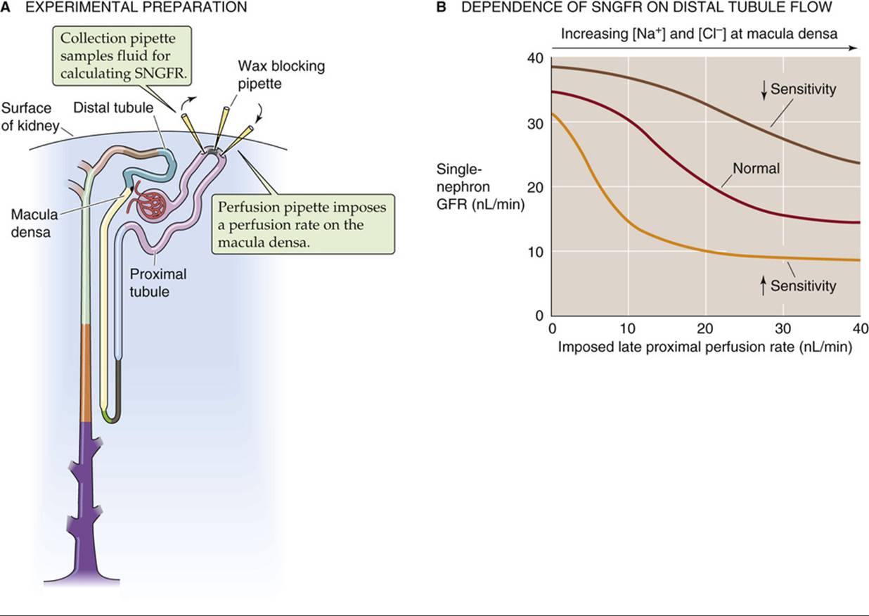
FIGURE 34-13 Tubuloglomerular feedback. (B, Modified from Navar LG: Regulation of renal hemodynamics. Adv Physiol Educ 20:S221–S235, 1998.)
The mechanism of TGF is thought to be the following:
1. An increase in arterial pressure leads to increases in glomerular capillary pressure, RPF, and GFR.
2. Increased GFR leads to an increased delivery of Na+, Cl−, and fluid into the proximal tubule and, ultimately, to the macula densa cells of the JGA.
3. Macula densa cells do not sense flow per se, but the higher luminal [Na+] or [Cl−] resulting from high flow. Increases in luminal [Na+] and [Cl−]—via the apical Na/K/Cl cotransporter (see p. 122) of the macula densa cell—translate to parallel increases in intracellular [Na+] and [Cl−]. Indeed, blocking the Na/K/Cl cotransporter with furosemide (see p. 757) not only blocks the uptake of Na+ and Cl− into the macula densa cells, but also interrupts TGF.
4. The rise in [Cl−]i, in conjunction with a basolateral Cl− channel, leads to a depolarization.
5. The depolarization activates a basolateral nonselective cation channel, which allows Ca2+ to enter the macula densa cell.
6. Increased [Ca2+]i causes the macula densa cell to release paracrine agents, particularly adenosine and ATP, which breaks down to adenosine.
7. Adenosine, binding to A1 adenosine receptors on the smooth-muscle cells, triggers contraction of nearby vascular smooth-muscle cells (see Table 20-8).
8. Increased afferent arteriolar resistance decreases GFR, counteracting the initial increase in GFR.
Several factors modulate the sensitivity of the TGF mechanism (see Fig. 34-13B). Indeed, investigators have suggested that several of these factors may be physiological signals from the macula densa cells or modulators of these signals (Table 34-3).
TABLE 34-3
Modulation of TGF
|
Factors that Increase Sensitivity of TGF |
|
Volume contraction Adenosine PGE2 Thromboxane Hydroxyeicosatetraenoic acid (HETE) ANG II |
|
Factors that Decrease Sensitivity of TGF |
|
Volume expansion ANP NO cAMP PGI2 High-protein diet |
Volume expansion and a high-protein diet increase GFR by reducing TGF
The sensitivity of TGF is defined as the change in SNGFR for a given change in the perfusion rate of the loop of Henle. Thus, sensitivity is the absolute value of the slope of the curves in Figure 34-13B. The set-point of the feedback system is defined as the loop-perfusion rate at which SNGFR falls by 50%.
Both the intrinsic sensitivity of the macula densa mechanism and the initial set-point for changes in flow are sensitive to changes in the ECF volume. Expansion of the ECF decreases the sensitivity (i.e., steepness) of the overall feedback loop. Conversely, volume contraction increases the sensitivity of TGF, thus helping preserve fluid by reducing GFR. Many of these effects may be mediated by ANG II, which is a required cofactor for TGF responses.
Of clinical importance is the observation that a high-protein diet increases GFR and RPF by indirectly lowering the sensitivity of the TGF mechanism. A high-protein diet somehow enhances NaCl reabsorption proximal to the macula densa, so that luminal [NaCl] at the macula densa falls. As a consequence, the flow in the loop of Henle (and thus GFR) must be higher to raise [NaCl] to a given level at the macula densa. A high-protein diet may thereby decrease TGF, increasing glomerular capillary pressure. This sequence of events may lead, particularly in the presence of intrinsic renal disease, to progressive glomerular damage.
Four factors that modulate RBF and GFR play key roles in regulating effective circulating volume
Changes in effective circulating volume (see pp. 554–555) trigger responses in four parallel effector pathways that ultimately modulate either renal hemodynamics or renal Na+ reabsorption. The four effector pathways are (1) the renin-angiotensin-aldosterone axis, (2) the sympathetic nervous system, (3) AVP, and (4) atrial natriuretic peptide (ANP). Beginning on page 836, we discuss the integrated control of effective circulating volume. Here, we focus on the direct actions of the four effector pathways on renal hemodynamics.
Renin-Angiotensin-Aldosterone Axis
In terms of renal hemodynamic effects, the most important part of the renin-angiotensin-aldosterone axis is its middle member, the peptide hormone ANG II (see pp. 841–842). ANG II has multiple actions on renal hemodynamics (Table 34-4). The net effect of ANG II on blood flow and GFR depends on multiple factors. Under normal conditions, the effect of ANG II is primarily to mediate efferent arteriolar constriction, an effect that tends to maintain GFR when renal perfusion is reduced. The reason is that prostaglandins counteract any tendency of ANG II to constrict the afferent arteriole. Indeed, inhibition of prostaglandin production by NSAIDs unmasks the constriction of the afferent arteriole, resulting in a decline in GFR.
TABLE 34-4
Hemodynamic Actions of ANG II on the Kidney
|
SITE |
ACTION |
PROPERTIES |
|
Renal artery and afferent arteriole |
Constriction |
Ca2+ influx through voltage-gated channels |
|
Efferent arteriole |
Constriction |
May depend on Ca2+ mobilization from internal stores |
|
Mesangial cells |
Contraction with reduction in Kf |
Ca2+ influx |
|
TGF mechanism |
Increased sensitivity |
Increased responsiveness of afferent arteriole to signal from macula densa |
|
Medullary blood flow |
Reduction |
May be independent of changes in cortical blood flow |
Modified from Arendshorst WJ, Navar LG: In Schrier RW, Gottschalk CW (eds): Diseases of the Kidney, vol 1, 5th ed. Boston, Little, Brown, 1993, pp 65–117.
Sympathetic Nerves
Sympathetic tone to the kidney may increase either as part of a general response—as occurs with pain, stress, trauma, hemorrhage or exercise—or as part of a more selective renal response to a decrease in effective circulating volume (see pp. 842–843). In either case, sympathetic nerve terminals release norepinephrine into the interstitial space. At relatively high levels of nerve stimulation, both afferent and efferent arteriolar resistances rise, thus generally decreasing RBF and GFR. The observation that the RBF may fall more than the GFR is consistent with a preferential efferent arteriolar constriction. With maximal nerve stimulation, however, afferent vasoconstriction predominates and leads to drastic reductions in both RBF and GFR.
In addition, sympathetic stimulation triggers granular cells to increase their release of renin (see p. 841), raising levels of ANG II, which acts as described above. Finally, sympathetic activation—even at levels too low to reduce RBF and GFR—causes increased reabsorption of Na+ by proximal tubules (see pp. 766–768).
Arginine Vasopressin
In response to increases in the osmotic pressure of the ECF, the posterior pituitary releases AVP—also known as antidiuretic hormone (see p. 843). Although the principal effect of this small polypeptide is to increase water absorption in the collecting duct, AVP also increases vascular resistance. Despite physiological fluctuations of circulating AVP levels, total RBF and GFR remain nearly constant. Nevertheless, AVP may decrease blood flow to the renal medulla, thereby minimizing the washout of the hypertonic medulla; this hypertonicity is essential for forming a concentrated urine.
In amphibians, reptiles, and birds, the generalized vascular effects of AVP are more pronounced than they are in mammals. In humans, severe decreases in effective circulating volume (e.g., shock) cause a massive release of AVP via nonosmotic stimuli (see p. 586). Only under these conditions does AVP produce a systemic vasoconstriction and thus contribute to maintaining systemic blood pressure (see p. 553).
Atrial Natriuretic Peptide
Atrial myocytes release ANP in response to increased atrial pressure and thus effective circulating volume (see p. 843). ANP markedly vasodilates afferent and efferent arterioles, thereby increasing cortical and medullary blood flow, and lowers the sensitivity of the TGF mechanism (see Table 34-3). The net effect is an increase in RPF and GFR. ANP also affects renal hemodynamics indirectly by inhibiting secretion of renin (thus lowering ANG II levels).
Even without affecting GFR, low levels of ANP can be natriuretic by inhibiting Na+ reabsorption by tubules. First, ANP inhibits aldosterone secretion by the adrenal gland (and thereby reduces Na+reabsorption; see pp. 765–766). Second, ANP acts directly to inhibit Na+ reabsorption by the inner medullary collecting duct (see p. 768). At higher levels, ANP decreases systemic arterial pressure and increases capillary permeability. ANP plays a role in the diuretic response to the redistribution of ECF and plasma volume into the thorax that occurs during space flight and water immersion (see p. 1233).
Other vasoactive agents modulate RBF and GFR
Many other vasoactive agents, when infused systemically or applied locally to the renal vasculature, modulate RBF and GFR. Despite considerable research on such hemodynamic actions, the precise role of individual vasoactive agents in the response to physiological and pathophysiological stimuli remains uncertain because of the following three observations: (1) several agents with opposing actions are often released simultaneously; (2) blocking a specific vasoactive messenger may have relatively little effect on renal hemodynamics; and (3) a single vasoactive agent may have different, or even opposing, actions at low and high concentrations. Nevertheless, renal and extrarenal agents may cooperate to provide a full and physiologically adequate response to a wide spectrum of challenges.  N34-7
N34-7
N34-7
Agents that Modify Renal Hemodynamics
Contributed by Gerhard Giebisch, Erich Windhager
Effects of Various Vasoactive Agents on RBF, GFR, and Glomerular Filtration Coefficient (Kf)
|
Agent |
RBF |
GFR |
Kf |
|
Catecholamines |
|||
|
Norepinephrine |
↓ |
NC, ↓ |
↓ |
|
Epinephrine |
↓ |
NC, ↓ |
↓ |
|
Dopamine |
↑ |
↓ |
|
|
Peptides |
|||
|
AVP |
NC or ↓ |
NC |
↓ |
|
Parathyroid hormone |
NC |
NC or ↓ |
↓ |
|
ANP |
NC or ↑ |
NC or ↑ |
NC or ↑ |
|
ANG II |
↓ |
NC or ↓ |
↓ |
|
Bradykinin |
↑ |
NC |
NC or ↓ |
|
Glucagon |
↑ |
↑ |
NA |
|
Endothelin |
↓ |
↓ |
↓ |
|
Arachidonate Metabolites |
|||
|
PGE2, PGI2 |
↑ |
NC or ↑ |
NC or ↓ |
|
Thromboxane A2 |
↓ |
↓ |
↓ |
|
Leukotrienes C4, D4 |
↓ |
↓ |
↓ |
|
20- hydroxyeicosatetraenoic acid (20-HETE) |
↓ |
↓ |
NC |
|
Other |
|||
|
ATP |
↓ |
↓ |
NC |
|
Adenosine |
↓ then ↑ |
↓ |
NC |
|
Histamine |
↑ |
NC |
↓ |
|
Platelet-activating factor |
↓ |
↓ |
↓ |
|
Acetylcholine |
↑ |
NC |
NC or ↓ |
|
NO |
↑ |
NC or ↑ |
↑ |
NC, no change.
Epinephrine
Released by the chromaffin cells of the adrenal medulla (see p. 1030), epinephrine exerts dose-dependent effects on the kidney that are similar to those of norepinephrine (see p. 752).  N34-8
N34-8
N34-8
Effects of Epinephrine on Renal Hemodynamics
Contributed by Gerhard Giebisch, Erich Windhager
Epinephrine increases renal vascular resistance. At lower epinephrine levels, GFR may be maintained despite a significant reduction of RBF, similar to the effects of moderate sympathetic nerve stimulation (see p. 752). This effect implies preferential efferent vasoconstriction. At higher doses, epinephrine decreases both GFR and RBF, similar to the effects of intense sympathetic nerve stimulation.
Dopamine
Dopaminergic nerve fibers terminate in the kidney, and dopamine receptors are present in renal blood vessels. In addition, proximal-tubule cells both produce dopamine and express dopamine receptors. The renal effects of dopamine—vasodilation and inhibition of Na+ reabsorption by tubules—are opposite to the effects of epinephrine and norepinephrine.
Endothelins
The endothelins are peptides with a strong vasoconstrictor action (see p. 480) but a very short half-life. The hemodynamic actions of endothelins are limited to local effects, because little of these hormones escapes into the general circulation. In the kidney, several agents—ANG II, epinephrine, higher doses of AVP, thrombins, and shear stress—trigger release of endothelins from the endothelium of renal cortical vessels and mesangial cells. The endothelins act locally to constrict smooth muscles of renal vessels and thus most likely are a link in the complex network of local messengers between endothelium and smooth muscle. When administered systemically, endothelins constrict the afferent and efferent arterioles and reduce the ultrafiltration coefficient (Kf). The result is a sharp reduction in both RBF and GFR.
Prostaglandins
In the kidney, vascular smooth-muscle cells, endothelial cells, mesangial cells, and tubule and interstitial cells of the renal medulla are particularly important for synthesizing locally acting prostaglandins from arachidonic acid via the cyclooxygenase pathway (see pp. 62–64). The effects of prostaglandins are complex and depend on the baseline vasoconstriction exerted by ANG II. Indeed, prostaglandins appear mainly to be protective and are important under conditions in which the integrity of the renal circulation is threatened. In particular, local intrarenal effects of prostaglandins prevent excessive vasoconstriction, especially during increased sympathetic simulation or activation of the renin-angiotensin system. Accelerated prostaglandin synthesis and release are responsible for maintaining fairly constant blood flow and GFR in conditions of high ANG II levels (e.g., during surgery, following blood loss, or in the course of salt depletion).
Leukotrienes
Probably in response to inflammation, the renal vascular smooth-muscle cells and glomeruli—as well as leukocytes and blood platelets—synthesize several leukotrienes from arachidonic acid via the lipoxygenase pathway (see pp. 64–65). These locally acting vasoactive agents are strong vasoconstrictors; their infusion reduces RBF and GFR.
Nitric Oxide
The endothelial cells of the kidney use nitric oxide synthase (NOS) to generate nitric oxide (NO; see p. 66) from L-arginine. NO has a strong smooth muscle–relaxing effect and—under physiological, unstressed conditions—produces significant renal vasodilation. NO probably defends against excess vasoconstrictor effects of agents such as ANG II and epinephrine. Injecting NOS inhibitors into the systemic circulation constricts afferent and efferent arterioles, increasing renal vascular resistance and producing a sustained fall of RBF and GFR. Moreover, NOS inhibitors blunt the vasodilatation that is triggered by low rates of fluid delivery to the macula densa as part of the TGF mechanism.