
A variety of physiological and pathophysiological stimuli can modulate renal H+ secretion as well as NH3 synthesis. Most of these factors produce coordinated changes in apical and basolateral acid-base transport, as well as in NH3 production.
Respiratory acidosis stimulates renal H+ secretion
The four fundamental pH disturbances are respiratory acidosis and alkalosis, and metabolic acidosis and alkalosis (see Fig. 28-11A). In each case, the initial and almost instantaneous line of defense is the action of buffers—both in the extracellular and intracellular compartments—to minimize the magnitude of the pH changes (see pp. 628–629). However, restoring the pH to a value as close to “normal” as possible requires slower compensatory responses from the lungs or kidneys.
In respiratory acidosis, in which the primary disturbance is an increase in arterial  , the compensatory response is an increase in renal H+ secretion, which translates to increased production of new
  via
via  excretion. The opposite occurs in respiratory alkalosis. These changes in H+ secretion tend to correct the distorted [
 ]/[CO2] ratios that occur in primary respiratory acid-base derangements.
Respiratory acidosis stimulates H+ secretion in at least three ways. First, an acute elevated  directly stimulates proximal-tubule cells to secrete H+, as shown by applying solutions in which it is possible to change
  without altering basolateral pH or [].
  N39-10 Thus, proximal-tubule cells directly sense basolateral CO2. In part, the mechanism is the exocytotic insertion of H pumps into the apical membranes of proximal-tubule cells. Second, acute respiratory acidosis also causes exocytotic insertion of H pumps into the apical membranes of intercalated cells in distal nephron segments. Third, chronic respiratory acidosis leads to adaptive responses that upregulate acid-base transporters. For example, respiratory acidosis increases the activities of apical NHE3 and basolateral NBCe1 in proximal tubule. These adaptive changes allow the kidney to produce a metabolic compensation to the respiratory acidosis (see p. 641).
N39-10
Use of Out-of-Equilibrium Solutions to Probe the Chemosensitivity of the Proximal Tubule
Contributed by Walter Boron
As described in  N28-4, the laboratory of Walter Boron developed a rapid-mixing technique that makes it possible to generate out-of-equilibrium (OOE) CO2/
 solutions with virtually any combination of [CO2], [
 ], and pH—as long as the desired pH is not more than a few pH units from neutrality.
Recently, the laboratory has applied the OOE CO2/ solutions to learn more about how the proximal tubule senses acute acid-base disturbances and translates that information to alter the rate at which the tubule reabsorbs (i.e., moves from the lumen to the basolateral side of the tubule). The approach was to isolate a single proximal tubule and perfuse its lumen with a solution of 5% CO2/22 mM /pH 7.4 as well as 3H-methoxyinulin as a volume marker. By collecting the fluid after it had flowed down the lumen and then analyzing this fluid for [] and [3H-methoxyinulin], the investigators were able to compute the rate of volume reabsorption (JV—that is, the rate at which the tubule moves water from the lumen to the basolateral surface of the tubule, measured in nanoliters per minute per millimeter of tubule length) and the rate of reabsorption (
 —measured in picomoles per minute per millimeter of tubule length). The investigators superfused the basolateral (bl) surface of the tubule with a rapidly flowing solution that was either the “standard” equilibrated 5% CO2/22 mM /pH 7.4 solution or an OOE solution in which they varied—one at a time—[CO2]bl,
  , or pHbl. Thus, they were able to observe the effects of altering basolateral acid-base composition on JV and .
What they found was rather striking. When the investigators raised [CO2]bl from 0 to 4.8 mM—at a fixed  of 22 mM and a fixed pHbl of 7.40—they found that increased in a graded fashion. This result is what one might expect from what we learned about a metabolic compensation to a respiratory acidosis (see p. 641). That is, the kidney ought to respond to a rise in [CO2]bl—the “respiratory” part of a respiratory acidosis—by reabsorbing more and thereby tending to restore blood pH to a more alkaline value. However, the investigators were quite surprised to find that increases in were not accompanied by the expected increases in JV (i.e., the extra NaHCO3 reabsorbed by the proximal tubule should have been accompanied by osmotically obligated water, which should have raised JVappreciably).
When the investigators raised from 0 to 44 mM—at a fixed [CO2]bl of 1.2 mM and a fixed pHbl of 7.40—they found that decreased in a graded fashion. This result is what one might expect for the kidney's response to a metabolic alkalosis caused by an abnormality outside of the kidney. That is, the kidney ought to respond to a rise in —the “metabolic” part of a metabolic alkalosis—by reabsorbing less and thereby tending to restore blood pH to a more acidic value. However, the investigators were quite surprised to find that decreases in were not accompanied by the expected decreases in JV (i.e., because the tubule reabsorbed less NaHCO3, it should also have reabsorbed less osmotically obligated water, so that JV should have fallen appreciably).
Finally, when the investigators raised pHbl from 6.8 to 8.0 mM—at a fixed [CO2]bl of 1.2 mM and a fixed of 22 mM—they found that did not change! One might have expected that a basolateral alkalosis (the “alkalosis” part of a respiratory or metabolic alkalosis) would have caused the tubule to reabsorb less and thereby tend to restore blood pH to a more acidic value. In these experiments, the intracellular pH of the tubule cells changed appreciably, but neither change in pH, intracellular or basolateral, triggered a change in or JV.
These experiments led the investigators to conclude that the proximal tubule cannot sense pH per se. Instead, they propose that the proximal-tubule cell has sensors for both basolateral CO2 and basolateral . In other words, the proximal tubule seems to regulate blood pH by sensing the body's two most important buffers. When activated, the CO2 sensors would trigger an increase in NaHCO3 reabsorption but a compensating decrease in the reabsorption of other solutes. When activated, the sensors would trigger a decrease in NaHCO3 reabsorption but a compensating increase in the reabsorption of other solutes. The compensating effects on the other solutes would serve to stabilize blood pressure.
References
Zhao J, Zhou Y, Boron WF. Effect of isolated removal of either basolateral or basolateral CO2 on reabsorption by rabbit S2 proximal tubule. Am J Physiol Renal Physiol. 2003;285:F359–F369.
Zhou Y, Zhao J, Bouyer P, Boron WF. Evidence from renal proximal tubules that and solute reabsorption are acutely regulated not by pH but by basolateral and CO2. Proc Natl Acad Sci U S A. 2005;102(10):3875–3880 [Epub February 22, 2005].
Metabolic acidosis stimulates both proximal H+ secretion and NH3 production
The first compensatory response to metabolic acidosis is increased alveolar ventilation, which blows off CO2 (see p. 710) and thus corrects the distorted [ ]/[CO2] ratio in a primary metabolic acidosis. The kidneys can also participate in the compensatory response—assuming, of course, that the acidosis is not the consequence of renal disease. Proximal-tubule cells can directly sense an acute fall in basolateral [], which results in a stimulation of proximal H+ secretion. N39-10 In intercalated cells in the distal nephron, metabolic acidosis stimulates apical membrane H pump insertion and activity. The mechanism may be proton-sensitive G protein–coupled receptors on the basolateral membrane of intercalated cells, and an -sensitive soluble adenylyl cyclase (sAC) in the cytosol.
In chronic metabolic acidosis, the adaptive responses of the proximal tubule are probably similar to those outlined above for chronic respiratory acidosis. These include upregulation of apical NHE3 and electrogenic H pumps, as well as basolateral NBCe1 (Fig. 39-7), perhaps reflecting increases in the number of transporters on the surface membranes. The parallel activation of apical and basolateral transporters may minimize changes in pHi, while increasing transepithelial reabsorption. This upregulation appears to involve intracellular protein kinases, including the Src family of receptor-associated tyrosine kinases (see p. 70). Endothelin appears to be essential for the upregulation of NHE3 in chronic metabolic acidosis.

FIGURE 39-7 Effects of chronic acidosis on proximal-tubule function. Enhanced Na citrate reabsorption is a defense against acidosis by conversion of citrate to  . The price paid is enhanced stone formation because luminal citrate reduces stone formation by complexing with Ca2+. Indeed, acidotic patients tend to get calcium-containing kidney stones.
In addition to increased H+ secretion, the other ingredient needed to produce new  is enhanced NH3 production. Together, the two increase
  excretion. Indeed, the excretion of
  into the urine increases markedly as a result of the adaptive response to chronic metabolic acidosis (Fig. 39-8). Thus, the ability to increase NH3 synthesis is an important element in the kidney's defense against acidotic challenges. Indeed, as chronic metabolic acidosis develops, the kidneys progressively excrete a larger fraction of urinary H+ as . As a consequence, the excretion of titratable acid becomes a progressively smaller fraction of total acid excretion.

FIGURE 39-8 Effect of chronic metabolic acidosis on total  excretion into final urine. (Data from Pitts RF: Renal excretion of acid. Fed Proc 7:418–426, 1948.)
The adaptive stimulation of NH3 synthesis, which occurs in response to a fall in pHi, involves a stimulation of both glutaminase and phosphoenolpyruvate carboxykinase (PEPCK). The stimulation of mitochondrial glutaminase increases the conversion of glutamine to and glutamate (see Fig. 39-5A). The stimulation of PEPCK enhances gluconeogenesis and thus the conversion of α-KG (the product of glutamate deamination) to glucose.
Metabolic alkalosis reduces proximal H+ secretion and, in the CCT, may even provoke  secretion
Figure 39-9A illustrates the response of the proximal tubule to metabolic alkalosis. As shown in the upper curve, when the peritubular capillaries have a physiological [], increasing the luminal [] causes H+ secretion to increase steeply up to a luminal [] of ~45 mM. The reason is that the incremental luminal is an additional buffer that minimizes the luminal acidification in the vicinity of the apical H+ transporters.
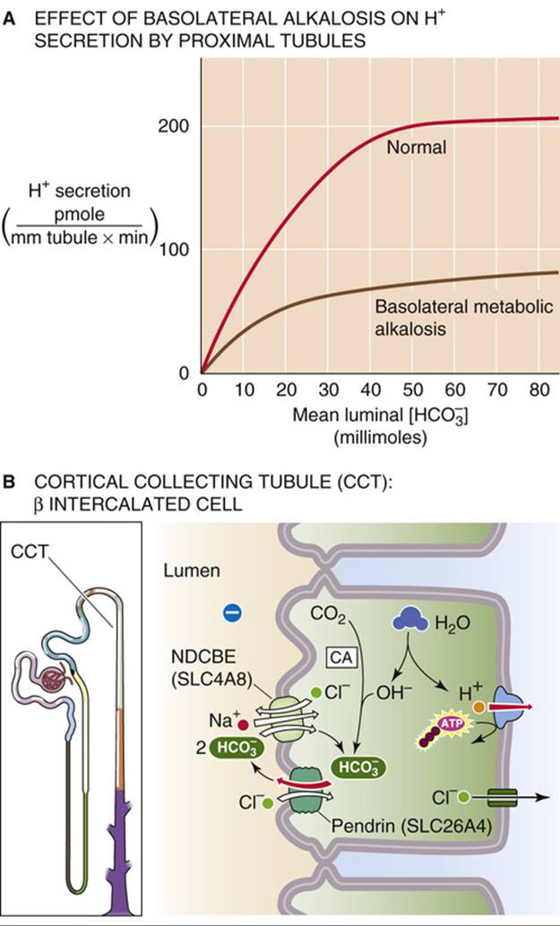
FIGURE 39-9 Effect of chronic metabolic alkalosis on renal acid-base transport. (Data from Alpern RJ, Cogan MG, Rector FC: Effects of extracellular fluid volume and plasma bicarbonate concentration on proximal acidification in the rat. J Clin Invest 71:736–746, 1983.)
As shown in the lower curve in Figure 39-9A, when [ ] in the peritubular blood is higher than normal—that is, during metabolic alkalosis—H+ secretion is lower for any luminal []. The likely explanation is that the proximal-tubule cell directly senses the increase in plasma [], depressing the rates at which NHE3 moves H+ from cell to lumen and NBCe1 moves from cell to blood.
So far, we have discussed the effect of metabolic alkalosis on H+ secretion by the proximal tubule. In the ICT and CCT, metabolic alkalosis can cause the tubule to switch from secreting H+ to secreting into the lumen. The α-intercalated cells in the ICT and CCT secrete H+ by using an apical H pump and a basolateral Cl-HCO3 exchanger, which is AE1 (SLC4A1; see Fig. 39-4D). Metabolic alkalosis, over a period of days, shifts the intercalated-cell population, increasing the proportion of β-intercalated cells (see Fig. 39-9B) N39-5 at the expense of α cells. Because β cells have the opposite apical-versus-basolateral distribution of H pumps and Cl-HCO3 exchangers, they secrete into the lumen and tend to correct the metabolic alkalosis. The apical Cl-HCO3 exchanger in β cells is pendrin (SLC26A4; see Table 5-4).
In contrast to chronic alkalosis, chronic acidosis alters the distribution of intercalated cell types in the distal nephron in favor of acid-secreting α cells (see Fig. 39-4D) over the base-secreting β-intercalated cells.
A rise in GFR increases delivery to the tubules, enhancing reabsorption (glomerulotubular balance for )
Increasing either luminal flow or luminal [] significantly enhances reabsorption, N39-11 probably by raising effective [] (and thus pH) in the microenvironment of H+ transporters in the brush-border microvilli. Because a high luminal pH stimulates NHE3 and the H pumps located in the microvilli of the proximal tubule, increased flow translates to enhanced H+ secretion. This flow dependence, an example of glomerulotubular (GT) balance (see p. 763), is important because it minimizes loss, and thus the development of a metabolic acidosis, when GFR increases. Conversely, this GT balance of reabsorption also prevents metabolic alkalosis when GFR decreases. The flow dependence of reabsorption also accounts for the stimulation of H+ transport that occurs after uninephrectomy (i.e., surgical removal of one kidney), when GFR in the remnant kidney rises in response to the loss of renal tissue.
N39-11
Flow Dependence of  Reabsorption
Contributed by Gerhard Giebisch, Erich Windhager
In the text, we point out that raising either luminal [] or luminal flow increases reabsorption. One likely mechanism is mentioned in the text: The higher the flow or the higher the bulk luminal [], the higher the pH and [] in the unstirred layer that surrounds the microvilli on the apical membrane. In addition, increasing the flow also increases the shear force that acts on the central cilium present on every proximal-tubule cell. It is believed that the more the cilium bends with flow, the greater the signal to increase the reabsorption of solutes (including NaHCO3) and water. This hypothesis would account for at least a portion of the glomerulotubular balance for both reabsorption (see p. 834) and fractional Na+ reabsorption (see p. 763).
Extracellular volume contraction—via ANG II, aldosterone, and sympathetic activity—stimulates renal H+ secretion
A decrease in effective circulating volume stimulates Na+ reabsorption by four parallel pathways (see pp. 838–840), including activation of the renin-angiotensin-aldosterone axis (and thus an increase in ANG II levels) and stimulation of renal sympathetic nerves (and thus the release of norepinephrine). Both ANG II and norepinephrine stimulate Na-H exchange in the proximal tubule. Because the proximal tubule couples Na+ and H+ transport, volume contraction increases not only Na+ reabsorption but also H+ secretion. Similarly, ANG II stimulates acid secretion by α-intercalated cells in the distal nephron. Volume expansion has the opposite effect. On a longer time scale, volume depletion also increases aldosterone levels, thereby enhancing H+ secretion in cortical and medullary collecting ducts (see below). Thus, the regulation of effective circulating volume takes precedence over the regulation of plasma pH. N39-12
N39-12
Effect of Dietary Na+ Intake on Proximal-Tubule NHE3 Activity
Contributed by Gerhard Giebisch, Erich Windhager
Decreased dietary Na+ intake causes a decrease in effective circulating volume (i.e., volume contraction), resulting in increased activity of the apical NHE3. This effect is evident even if one assesses the activity in brush-border membrane vesicles removed from the animal. Consumption of a high-Na+ diet has the opposite effect.
Hypokalemia increases renal H+ secretion
As discussed on page 803, acid-base disturbances can cause changes in K+ homeostasis. The opposite is also true. Because a side effect of K+ depletion is increased renal H+ secretion, K+ depletion is frequently associated with metabolic alkalosis. Several lines of evidence indicate that, in the proximal tubule, hypokalemia leads to a marked increase in apical Na-H exchange and basolateral Na/HCO3 cotransport. As in other cells, in tubule cells the pH falls during K+ depletion (see p. 645). The resulting chronic cell acidification may lead to adaptive responses that activate Na-H exchange and electrogenic Na/HCO3 cotransport, presumably by the same mechanisms that stimulate H+ secretion in chronic acidosis (see Fig. 39-7). In the proximal tubule, K+ depletion also markedly increases NH3 synthesis and excretion, thus increasing urinary H+ excretion as  . Finally, K+ depletion stimulates apical K-H exchange in α-intercalated cells of the ICT and CCT (see p. 799) and enhances H+ secretion as a side effect of K+ retention.
Just as hypokalemia can maintain metabolic alkalosis, hyperkalemia is often associated with metabolic acidosis. A contributory factor may be reduced excretion, perhaps because of lower synthesis in proximal-tubule cells, possibly due to a higher intracellular pH. In addition, with high luminal [K+] in the TAL, K+ competes with for uptake by apical Na/K/Cl cotransporters and K+ channels, thereby reducing reabsorption. As a result, the reduced  levels in the medullary interstitium provide less NH3 for diffusion into the medullary collecting duct. Finally, with high [K+] in the medullary interstitium, K+ competes with for uptake by basolateral Na-K pumps in the medullary collecting duct. The net effects are reduced excretion and acidosis.
Both glucocorticoids and mineralocorticoids stimulate acid secretion
Chronic adrenal insufficiency (see p. 1019) leads to acid retention and, potentially, to life-threatening metabolic acidosis. Both glucocorticoids and mineralocorticoids stimulate H+ secretion, but at different sites along the nephron.
Glucocorticoids (e.g., cortisol) enhance the activity of Na-H exchange in the proximal tubule and thus stimulate H+ secretion. In addition, they inhibit phosphate reabsorption, raising the luminal availability of buffer anions for titration by secreted H+.
Mineralocorticoids (e.g., aldosterone) stimulate H+ secretion by three coordinated mechanisms—one direct and two indirect. First, mineralocorticoids directly stimulate H+ secretion in the collecting tubules and ducts by increasing the activity of the apical electrogenic H pump and basolateral Cl-HCO3 exchanger (see Fig. 39-4D). Second, mineralocorticoids indirectly stimulate H+ secretion by enhancing Na+reabsorption in the collecting ducts (see p. 766), which increases the lumen-negative voltage. This increased negativity may stimulate the apical electrogenic H pump in α-intercalated cells to secrete acid. Third, mineralocorticoids—particularly when administered for longer periods of time and accompanied by high Na+ intake—cause K+ depletion and indirectly increase H+ secretion (see pp. 834–835).
Diuretics can change H+ secretion, depending on how they affect transepithelial voltage, ECF volume, and plasma [K+]
The effects of diuretics on renal H+ secretion  N39-13 vary substantially from one diuretic to another, depending on both the site and the mechanism of action. From the point of view of acid-base balance, diuretics fall broadly into two groups: those that promote the excretion of a relatively alkaline urine and those that have the opposite effect.
N39-13
Effect of Diuretics on Renal H+ Excretion
Contributed by Erich Windhager, Gerhard Giebisch
Box 40-3, as well as Table 40-3, summarizes some of the effects of various classes of diuretics and lists the protein targets of these diuretics in the kidney.
To the first group belong CA inhibitors and K+-sparing diuretics. The CA inhibitors lead to excretion of an alkaline urine by inhibiting H+ secretion. Their greatest effect is in the proximal tubule, but they also inhibit H+ secretion by the TAL and intercalated cells in the distal nephron. K+-sparing diuretics—including amiloride, triamterene, and the spironolactones—also reduce acid excretion. Both amiloride and triamterene inhibit the apical epithelial Na+ channels (ENaCs; see pp. 758–759) in the collecting tubules and ducts, rendering the lumen more positive so that it is more difficult for the electrogenic H pump to secrete H+ ions into the lumen. Spironolactones decrease H+ secretion by interfering with the action of aldosterone.
The second group of diuretics—those that tend to increase urinary acid excretion and often induce alkalosis—includes loop diuretics such as furosemide (which inhibits the apical Na/K/Cl cotransporter in the TAL) and thiazide diuretics such as chlorothiazide (which inhibits the apical Na/Cl cotransporter in the DCT). These diuretics act by three mechanisms. First, all cause some degree of volume contraction, and thus lead to increased levels of ANG II and aldosterone (see pp. 841–842), both of which enhance H+ secretion. Second, these diuretics enhance Na+ delivery to the collecting tubules and ducts, thereby increasing the electrogenic uptake of Na+, raising lumen-negative voltage, and enhancing H+ secretion. Third, this group of diuretics causes K+ wasting; as discussed on pages 834–835, K+ depletion enhances H+ secretion.