Hodgkin's disease is a neoplastic disease characterized by painless, progressive enlargement of lymph nodes, spleen, and other lymphoid tissue resulting from proliferation of lymphocytes, histiocytes, eosinophils, and Reed-Sternberg giant cells. Untreated, Hodgkin's disease follows a variable but relentlessly progressive and, ultimately, fatal course. With appropriate treatment, more than 80% of patients with stage I or II disease survive at least 10 years.
Causes
Unknown; however, viral etiology is suspected (Epstein-Barr virus is a leading candidate)
 Age Alert
Hodgkin's disease is most common in young adults, and more common in males than in females. Incidence peaks in two age-groups: ages 15 to 38 and after age 50—except in Japan, where it occurs exclusively among people over age 50.
Pathophysiology
Hodgkin's disease is characterized by proliferation of a tumor in which only a small proportion of the cells are malignant and most are normal lymphocytes. The characteristic malignant cells—called Reed-Sternberg cells—are most likely multinucleated, giant-cell mutations of the T-lymphocyte. Infiltration of the nodes with eosinophils and plasma cells is associated with lymph node necrosis and fibrosis.
Signs and symptoms
· Painless swelling in one of lymph nodes (usually the cervical region) with a history of upper respiratory infection
· Persistent fever
· Night sweats
· Fatigue
· Weight loss
· Malaise
· Pruritus
· Extremity pain
· Nerve irritation
· Absence of pulse due to rapid enlargement of lymph nodes
· Pericardial friction rub
· Pericardial effusion
· Neck vein engorgement
· Enlargement of retroperitoneal nodes, spleen, and liver
Diagnostic test results
· Lymph node biopsy confirms presence of Reed-Sternberg cells, nodular fibrosis, and necrosis.
· Bone marrow, liver, mediastinal, lymph node, and spleen biopsies reveal histologic presence of cells.
· Chest X-ray, abdominal computed tomography scan, lung scan, bone scan, and lymphangiography detect lymph and organ involvement.
· Hematologic tests show:
§ mild to severe normocytic anemia
§ normochromic anemia
§ elevated, normal, or reduced white blood cell count
§ differential with any combination of neutrophilia, lymphocytopenia, monocytosis, and eosinophilia.
· Elevated serum alkaline phosphatase indicates bone or liver involvement.
Treatment
· Chemotherapy, radiation, or both, appropriate to stage of the disease—based on histologic interpretation and clinical staging
· Concomitant antiemetics, sedatives, or antidiarrheals to combat adverse GI effects
· Autologous bone marrow transplant
· Autologous peripheral blood sternal transfusions
· Immunotherapy
P.247
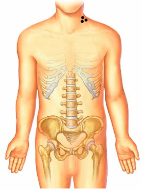
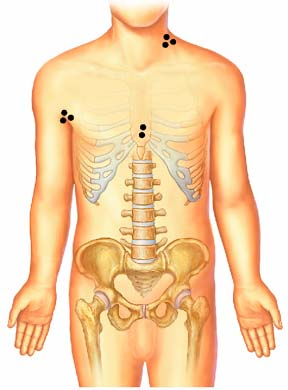
ANN ARBOR STAGING SYSTEM FOR HODGKIN'S DISEASE
|
|
|
|
Stage I · Involvement of single lymph node region or · Involvement of single extralymphatic site (stage IE) |
Stage II · Involvement of 2 or more lymph node regions on same side of diaphragm · May include localized extralymphatic involvement on same side of diaphragm (stage IIE) |
|
|
|
|
Stage III · Involvement of lymph node regions on both sides of diaphragm · May include involvement of spleen (stage IIIS) or localized extranodal disease (stage IIIE) · Hodgkin's disease Stage III1: disease limited to upper abdomen — spleen, splenic hilar, celiac, or portohepatic nodes · Hodgkin's disease Stage III2: disease limited to lower abdomen — periaortic, pelvic, or inguinal nodes |
Stage IV · Diffuse extralymphatic disease (for example, in liver, bone marrow, lung, skin) |