Ther is no thing more precious here than tyme.
Saint Bernard (1090-1153)
The objective of this chapter is to provide a practical approach to acutely ill patients. The emphasis is on diagnosis, not on therapy. In the assessment of acutely ill patients, time is a critical factor. Unlike the assessment of stable patients, the evaluation of acutely ill patients involves not achieving a specific diagnosis but rather identifying a pathophysiologic abnormality that may be identical for several diagnoses. In the evaluation of acutely ill patients, always ask yourself, ''What is the most serious threat to life, and have I ruled it out?'' Remember, also, that your health is important. Exposure to body substances places you at risk. The minimum isolation precaution for an emergency response is the wearing of latex gloves.
When delivering health care in the field, and perhaps even in the hospital, as you approach the apparent patient, always perform a brief evaluation to determine whether you are in a safe environment; if not, protect yourself and your patient to limit exposure to possible injury. This may be a rare situation, but in circumstances in which it is likely that the rescuer could be injured or killed while rendering care, the rescuer should wait until the situation can be made safe. For example, in an automobile accident, a patient trapped in a car in a busy traffic lane should not be given first aid until safety flares or cones can be placed to prevent secondary accidents.
During this evaluation, search for other injured persons who may be hidden from view as you approach the scene of the accident. You should also try to determine the mechanisms of injury and attempt to memorize the scene for later reconsideration in the emergency department and perhaps as a witness for the injured party.
The task for the clinician in approaching most, if not all, patients in acute situations is, first, to ascertain that they are not in cardiopulmonary arrest and do not have major perturbations of their vital signs to the point that their continued viability is threatened. The general approach to these acute, undefined encounters is to consider the patient unstable until you can confirm, through a series of diagnostic steps, that the patient is well enough for you to take the time to perform a more rigorous and complete physical examination and document a complete history.
This strategy involves moving through a series of simple algorithms, which are grouped into two categories termed the primary and secondary surveys. The primary survey is a check for conditions that are an immediate threat to the patient's life. This initial assessment should take no longer than 30 seconds. The primary survey is subdivided into a cardiopulmonary resuscitation (CPR) survey and a key vital functions assessment. The algorithms for the primary survey are shown in Figures 26-1 and 26-2. The secondary survey is a check for conditions that could become life-threatening problems if not recognized and attended to.
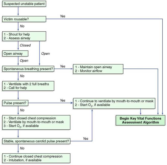
Figure 26-1 Cardiopulmonary resuscitation (CPR) survey algorithm.
The primary and secondary surveys are used for both adult and pediatric patients, as well as for medical and injury-related problems. The treatment process is integrated into the diagnostic process. For example, if the patient is not breathing, ventilations are begun immediately, before you move on to the next diagnostic step in the algorithm.
The first task is to recognize when a patient is acutely ill. An unusual appearance or behavior may be the only sign. These include breathing difficulties, clutching the chest or throat, slurring of speech, confusion, unusual odor to the breath, sweating for no apparent reason, or uncharacteristic skin color (e.g., pale, flushed, or bluish).
Remember that an acutely ill patient is anxious and frightened; a calm and reassuring voice can go a long way toward comforting the patient. It is always easier to care for a relaxed patient than for an anxious one.
Primary Survey
Cardiopulmonary Resuscitation Survey
It should not be assumed that any patient who is not obviously interacting with his or her environment is simply sleeping. For the purpose of this approach, the patient is in cardiopulmonary arrest until it is proved otherwise. As you approach the patient, observe the patient closely, looking for spontaneous breathing or movements. If these are not discernible, stimulate the patient by talking loudly to him or her. If necessary, shout ''ARE YOU OKAY?''
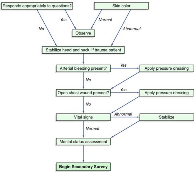
Figure 26-2 Key vital functions assessment algorithm.
If there is no response, obtain an open airway by the chin-lift/head-tilt maneuver and look, listen, and feel (feel air movement against your cheek) for breathing. To open an unconscious victim's airway, hyperextend the head and lift the patient's chin; place one hand on the patient's forehead and the other behind the patient's occiput, and tilt his or her head backward. This maneuver moves the tongue away from the back of the throat, allowing air to pass around the tongue and into the trachea. Caution should be exercised with any patient who has a suspected neck injury. With such a patient, try to open the airway by lifting the chin without tilting the head backward; grasp the lower teeth and pull the mandible forward. If necessary, tilt the head back very slightly. If a patient is wearing dentures, remove them only if they occlude the airway.
If there is no evidence of spontaneous breathing, deliver two full breaths by using mouth- to-mouth ventilation. Next, call for help in any way you can without leaving the patient; it is unlikely that you can manage the entire resuscitation by yourself.
Determine whether there is spontaneous cardiac function by feeling for a carotid pulse or, in an infant, by palpating the precordium for a cardiac impulse. If there is no pulse, begin external chest compressions and intersperse them with ventilations; in other words, begin CPR. Current guidelines (Hazinski et al, 2005) are 30 chest compressions for every two rescue breaths for five cycles (2 minutes). Recheck breathing after every five cycles. The 30:2 ratio is the same for CPR that a single rescuer provides for adults, children, and infants (except newborns). The only exception to this guideline is when two rescuers perform CPR on a child or infant (except newborns), in which they should provide 15 compressions for every two rescue breaths.
Key Vital Functions Assessment Survey
Once it has been determined that the patient does not need CPR or the patient has recovered spontaneous cardiopulmonary activity, ascertain whether key life-sustaining functions are adequate and stable or whether augmentation or other supportive measures are necessary.
In the initial overview of the patient, two observations can save a great deal of time and help avoid unnecessary or untimely interventions. First, if the patient's central nervous system is functioning, as manifested by the patient's ability to respond appropriately to questions, it is unlikely that key vital functions are so deranged as to necessitate immediate intervention. Second, if the patient's skin is warm, dry, and of normal color, it is likely that oxygenation and flow of blood to the periphery are adequate. In shock, peripheral blood flow is shunted centrally; thus, skin changes are early indicators of hypovolemic or cardiogenic (low cardiac output) shock. The key diagnostic skin signs associated with these major acute cardiopulmonary derangements are gray, mottled, or cyanotic color; cold skin temperature; and markedly sweaty skin. The last sign, termed diaphoresis, is caused by activation of the sympathetic nervous system by any major threat to homeostasis.
At this point in the algorithm, if the patient has sustained a possible head injury, immobilize the patient's head and neck by using boards, tape, bulky dressings, or towels or by assigning someone to hold the head immobile. Once the evaluation is complete and imaging studies are performed, if necessary, these restrictions to movement can be removed. However, once a patient is immobilized, removal of these measures requires careful decision-making.
The next two orders of priority are the search for and the management of arterial bleeding and open chest injuries. The latter are termed sucking chest wounds because they allow air to enter the pleural space, leading to collapse of the underlying lung (pneumothorax). Arterial bleeding and a sucking chest wound can cause death in a short time, and both are treated by application of a pressure dressing to occlude the area.
At this point in the algorithm, the patient has been stabilized to the point at which formal vital signs can be obtained. In the field, these include the patient's mental status, respiratory rate and pattern, pulse, blood pressure, and, in some circumstances, body temperature. Mental status can be assessed according to the AVPU system (more traditionally categorized as alert, lethargic, stuporous, or comatose). The AVPU mnemonic for level of consciousness is as follows:
A: patient is alert
V: patient responds to a verbal stimulus P: patient responds to a painful stimulus U: patient is unresponsive
The blood pressure can be estimated by the pulse wave fullness and by assessing which pulses are palpable. If the radial pulse at the wrist is palpable, the systolic blood pressure is at least 80 mm Hg. If the radial pulse is impalpable and only the femoral pulse is perceptible, the systolic blood pressure is 60 to 70 mm Hg. If a vital sign is abnormal, treat the abnormality to bring it back to normal. For example, if the patient is breathing spontaneously at a rate of only five breaths per minute, augment and assist the patient's breathing so that the depth and rate of breathing are normalized. This can be accomplished by applying interspersed mouth- to-mouth ventilations, using a self-inflating bag-valve-mask device, or performing endotracheal intubation and placing the patient on a ventilator. In a similar manner, the blood pressure can be supported by raising the legs, thus emptying the blood stored in the venous system back into the central circulation.
A trauma victim should have a cardiopulmonary examination as well. You are seeking to rule in or rule out a tension pneumothorax (shift of the heart away from the tension, increased breath sounds over the side with the tension pneumothorax, distended neck veins, subcutaneous emphysema), cardiac tamponade (distended neck veins, distant heart sounds, hypotension, pulsus paradoxus, normal breath sounds), and chest wall disruption (paradoxical movement of a flail segment).
Secondary Survey
In the secondary survey, document a history from the patient, the patient's relatives, emergency department personnel, or bystanders. The secondary survey is a systematic method for determining whether other conditions or injuries are present and necessitate attention.
This survey consists of a rapid interview, a check of the vital signs, and a focused physical examination. The mnemonic AMPLE can be helpful in gathering pertinent information:
A: allergies
M: medications currently being taken
P: past medical history
L: last meal
E: events preceding the medical event
A critical piece of information in dealing with a trauma patient is the mechanism of injury. Did the patient sustain blunt trauma, or was a weapon used to cause a penetrating injury? In the case of a vehicular accident, ascertain whether the patient was ejected from the car or was wearing a seat belt and whether there were other injuries or fatalities in the accident. In addition, trauma victims must have all their bones and joints—including the rib cage, pelvis, facial bones, and skull—palpated and gently compressed to determine whether there is a fracture step-off or crepitation; also check for stability of structure and for function. A screening neurologic examination is necessary to determine whether there are focal cranial nerve, motor, or sensory findings. Most patients with multisystem trauma require a rectal examination to determine the presence of blood, tenderness, or upward displacement of the prostate. The latter is a sign of urethral injury.
If alert, an injured patient can direct you to the appropriate body areas to be evaluated during the physical examination. The assessment of the patient involves examination of three main regions: the head and neck, the torso, and the extremities. Can the patient move the neck? Ask the patient to move the neck slowly. Can the shoulders be moved? Ask the patient to take a deep breath and then blow it out. Does this elicit any pain? Is the patient able to move the fingers? Can the arms be bent? Can the patient move the toes? ankles? Can the patient bend the legs? If the patient can move all extremities without experiencing pain, help the patient up to a sitting position slowly. If the patient cannot move a body part or can do so only with pain, reassess the airway, breathing, and circulation, and get immediate assistance. Continue to observe the patient's level of consciousness, breathing, and skin color.
Head and Neck
Look at the victim's face. Evaluate skin color and temperature. Is there evidence of raccoon eyes or Battle's sign? A patient with raccoon eyes is shown in Figure 10-28. Periorbital ecchymoses, or raccoon eyes, are seen 6 to 12 hours after a fracture of the base of the skull. Battle's sign is ecchymosis behind the ear caused by basilar skull or temporal bone fractures; this sign may take 24 to 36 hours to develop. Palpate the head.
Examine the eyes for pupillary size and responsiveness to light. Are the pupils equal? Are the pupils pinpoint? Is there a unilateral dilated pupil? Are the pupils fixed? Table 26-1 reviews the eye signs in a comatose patient.
Is there a discharge from the ears, nose, or mouth?
Inspect the neck. Is the trachea deviated? Suspect a chest injury, such as a tension pneumothorax, if the trachea is not midline. Palpate the neck for crepitus, which is indicative of air under the skin from rupture of the lung.
Abdomen
Inspect the abdomen. Is abdominal distention present? Is there evidence of blunt abdominal trauma, such as an ecchymosis, an abrasion, or an abdominal wound? Cullen's sign is a bluish discoloration around the umbilicus indicative of intra-abdominal bleeding or trauma. Grey Turner's sign is ecchymotic discoloration around the flanks, which is suggestive of retroperitoneal bleeding. Swelling or ecchymosis often occurs late; therefore, its presence is extremely important.
Gently palpate the abdomen, noting the presence of tenderness. If the patient is a woman of childbearing age, always consider the possibility that she may be pregnant.
Inspect the anus and the perineum. Inspect the urethral meatus for blood.
Table 26-1 Eye Signs in a Comatose Patient*
|
Eye Sign |
Possible Causes |
|
Pupils reactive, eyes directed straight ahead, normal oculocephalic reflex (OCR)* |
Toxic/metabolic cause |
|
Pinpoint pupils |
Narcotic poisoning (OCR intact) Pontine or cerebellar hemorrhage (OCR absent) Thalamic hemorrhage Miotic eye drops |
|
Disconjugate deviation of eyes |
Structural brain-stem lesion |
|
Conjugate lateral deviation of eyes |
Ipsilateral pontine infarction Contralateral frontal hemispheric infarction |
|
Unilateral dilated, fixed pupil with no consensual responses |
Supratentorial mass lesion Impending brain herniation Posterior communicating aneurysm |
|
Bilateral midposition pupils, fixed pupils |
Midbrain lesion Impending brain herniation |
|
Raccoon eyes (periorbital ecchymoses), Battle sign |
Fracture of the base of the skull |
|
*Eye signs are difficult to evaluate in patients with artificial lenses, prosthetic eyes, contact lenses, or cataracts or after cataract surgery. *''Doll's eyes'': Rotate the head quickly but gently from side to side. In an unconscious patient with an intact brain stem, the eyes move conjugately in a direction opposite the head turning. |
|
Perform a rectal examination to assess anal sphincter tone, to determine whether blood is present, and to verify that the prostate is in its normal position.
Pelvis
Use the heels of your hands to apply gentle downward pressure on the anterior superior iliac spine and on the symphysis pubis. Is tenderness present? If so, there may be a fracture of the pelvic ring.
Extremities
Inspect and palpate all extremities for evidence of injury. Try to determine whether the patient can move all extremities. Palpate all peripheral pulses.
Back
Inspect the back, looking for obvious signs of injury. This can be done by gently insinuating your hands beneath the back and neck without moving the patient. If this cannot be done, the patient should be gently ''log-rolled'' onto the side. To do this, you need at least four assistants: one to control the head and neck, two to roll the patient onto the side, and one to cautiously move the lower extremities. Figure 26-3 shows this log-roll procedure.
Vital Signs
Reassess vital signs.
Documenting the history from an acutely ill patient conforms closely to documentation of the standard history, but it is abbreviated to allow rapid diagnostic and management decisions to be made. The physical examination of an acutely ill nontrauma patient includes cardiopulmonary and abdominal examinations and evaluation of the peripheral pulses.
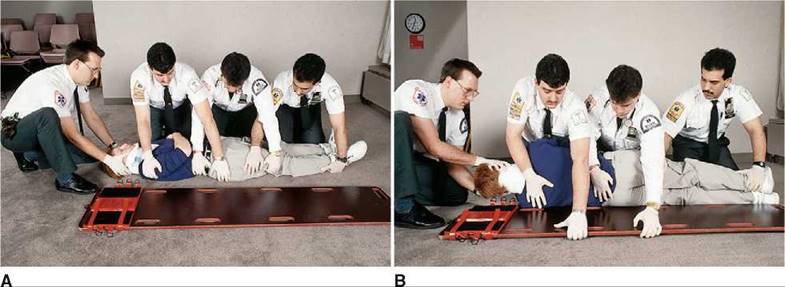
Figure 26-3 Log-roll procedure. A, Positioning for log-rolling. (1) Apply a cervical spine immobilization device and place the patient's arms at the side. Note that one emergency management technician (EMT) maintains cervical immobilization manually throughout this procedure. (2) Three EMTs can be positioned at the side of the patient at the level of the chest, hips, and lower extremities while the long spine board is positioned on one side of the patient. (3) Check the patient's arm on the side of the EMTs for injury before log-rolling the patient, and then align the lower extremities. Note: The EMT at the lower extremities holds the patient's lower leg and thigh region; the EMT at the hips holds the patient's lower legs and places the other hand on top of the patient's buttocks; and the EMT at the chest holds the patient's arms against the body and at the level of the lower buttocks. B, Log-rolling the patient.
(4) On command from the EMT at the head, all EMTs rotate the patient toward themselves, keeping the body in alignment.
(5) The EMTs then reach across with one hand and pull the board beneath the patient's arm. (6) On command from the EMT at the head, they gently roll the patient onto the board and then roll the board to the ground. (7) Strap the patient's torso and extremities securely to the board, and immobilize the head.
The Pediatric Emergency
When assessing an acutely ill child, always consider the similarities and differences between the pediatric age group and adult patients; approach the pediatric emergency as you would an emergency in an adult, but recognize the smaller size of the patient and the difference in the physiologic responses to acute illness and injury. The primary assessment of a child is the same as that of an adult.
The most dangerous life-threatening pediatric emergency is respiratory distress. Respiratory distress in a pediatric patient may arise from a variety of conditions that result from upper or lower airway disease. Common pediatric respiratory problems of the upper airway include croup (laryngotracheobronchitis), epiglottitis, foreign bodies, and bacterial tracheitis. Lower airway obstruction may result from asthma, pneumonia, bronchiolitis, and foreign bodies.
The hallmarks of respiratory distress are tachypnea, nasal flaring, retractions, stridor, cyanosis, head bobbing, prolonged expiration, and grunting. Children with upper airway disease almost always exhibit stridor. In a child with stridor, distinguish between croup and epiglotti- tis; a child with epiglottitis may have a rapid progression to respiratory failure. Fortunately, the incidence of epiglottitis has decreased, presumably because of the Haemophilus influenzae type B (HIB) vaccine. If epiglottitis is suspected, do not examine the airway without being prepared to provide airway stabilization on an emergency basis. Manipulation of the child's airway can lead to complete airway obstruction. Table 26-2 compares some of the important differences between epiglottitis and croup.
The peak time for foreign body aspiration is 1 to 2 years of age. In a child, consider relief of airway obstruction in the following situations:
• Choking is present
• The cough becomes ineffective
• Breathing becomes stridorous
• There is loss of consciousness
• The child becomes cyanotic
Table 26-2 Differentiation Between Epiglottitis and Croup
|
Characteristics |
Epiglottitis |
Croup |
|
Cause |
Haemophilus influenzae type B |
Viral, usually parainfluenza virus |
|
Age of child |
Any age (peak, 3-7 years) |
3 months-3 years |
|
Clinical appearance |
Extremely ill (''toxic'') |
Not extremely ill |
|
Season |
No seasonal predominance |
Autumn and winter |
|
Clinical onset |
Rapid |
Insidious |
|
Upper respiratory tract infection |
Rare |
Common |
|
Fever |
>104° F (40° C) |
<103° F (39.5° C) |
|
Sore throat |
Severe |
Variable |
|
Cough |
Not ''barking''; throughout the day |
''Barking''; during the night |
|
Drooling |
Prominent |
None |
|
Stridor |
On inspiration |
On inspiration and expiration |
|
Position |
Sitting forward with neck extended and mouth open |
Variable |
|
Epiglottis |
Bright red |
Normal |
Immediately place the child face down, with the head lower than the torso, over your arm, which is placed on your thigh. Support the child's head by holding his or her jaw. Deliver five forceful back blows with the heel of your other hand between the child's scapulae. Turn the child onto the back while holding the child's head. Place two fingertips on the middle portion of the sternum, one fingerbreadth below the nipples. Depress the sternum 1 inch. Repeat this maneuver up to five times. Attempt to remove any visible material from the pharynx. Repeat the back blows and chest thrusts until the object is dislodged.
If the child becomes unconscious, check the mouth for a foreign body, and then perform mouth-to-mouth breathing. Gently tilt the child's head back while placing the other fingers under the jaw at the chin, and lift the chin upward. Seal the child's mouth and nose with your mouth. Deliver two breaths, watching the chest rise. Repeat the back blows and chest thrusts. Have someone call for help.
Dehydration is another important pediatric emergency. The most common causes are vomiting and diarrhea. In a child with mild dehydration (<5%), there may be only a slight decrease in mucous membrane moisture. In severe dehydration (15%), the following are commonly found:
• Parched mucous membranes; no tears
• Markedly decreased skin turgor
• Sunken fontanelles
• Sunken eyeballs
• Tachypnea
• Capillary refill* longer than 2 seconds
• Cool and clammy skin
• Orthostatic hypotension: systolic pressure less than 80 mm Hg
• Tachycardia: faster than 130 beats per minute
Immediate intravenous infusion of isotonic fluids should be started in children with severe dehydration.
The secondary assessment outlined earlier and the AVPU mnemonic are just as important for children as for adults. Table 26-3 provides a useful reference for CPR.
*Capillary refill is an assessment of perfusion. It is the time required for a patient's skin color to return to normal after the nail bed has been pressed. The normal refill time is less than 2 seconds.
Table 26-3 Cardiopulmonary Resuscitation Reference Chart
|
Action |
Infant (<1 Year of Age) |
Child (>1 Year of Age) |
Adult |
|
If victim has a pulse, give one breath: |
Every 3 seconds |
Every 3 seconds |
Every 5-6 seconds |
|
If victim has no pulse, locate compression landmark: |
1 fingerbreadth below the nipple line |
Same as in adult |
One finger on sternum |
|
Compressions are performed with: |
Two or three fingers on sternum |
Heel of hand on sternum |
Two hands stacked, with heel of one hand on sternum |
|
Rate of compressions per minute: |
>100 |
100 |
80-100 |
|
Compression depth: |
1/3 to 1/2 depth of chest |
1/3 to 1/2 depth of chest |
1 to 1 1/2 inches (2.5-3.8 cm) |
|
Ratio of compressions to breaths with: |
|||
|
One rescuer |
30:2* |
30:2 |
30:2 |
|
Two rescuers |
15:2* |
15:2 |
30:2 |
|
*3:1 in neonates. |
Bibliography
Barkin RM, Rosen P (eds): Emergency Pediatrics: A Guide to Ambulatory Care, 5th ed. St. Louis, Mosby, 1999.
Capehorn DMW, Swain AH, Goldsworthy LL (eds): A Handbook of Paediatric Accident and Emergency Medicine: A Symptom-Based Guide. Philadelphia, WB Saunders, 1998.
Hazinski MF, Nadkarni VM, Hickey RW, et al: Major changes in the 2005 AHA guidelines for CPR and ECC: Reaching the tipping point for change. Circulation 112(Suppl I):IV-206, 2005.
Henry MC, Stapleton ER (eds): EMT Prehospital Care, 2nd ed. Philadelphia, WB Saunders, 1997.
Howell JM, Altieri M, Jagoda AS, et al (eds): Emergency Medicine. Philadelphia, WB Saunders, 1997. McSwain NE, White RD, Paturas JL, et al: The Basic EMT: Comprehensive Prehospital Patient Care. St. Louis, Mosby Lifeline, 1997.
Revere C, Hasty R: Diagnostic and characteristic signs of illness and injury. J Emerg Nurs 19:2, 1993. Thomas H, O'Connor RE, Hoffmann GL, et al: Emergency Medicine: Self Assessment and Review, 4th ed. St. Louis, Mosby, 1999.