
What is the hardest of all? That which you hold the most simple; seeing with your own eyes what is spread out before you.
Johann Wolfgang von Goethe (1749-1832)
General Considerations
The skin, which is the largest organ of the body, is one of the best indicators of general health. Even a person without medical training is capable of detecting changes in skin color and texture. The trained examiner can detect these changes and at the same time evaluate more subtle cutaneous signs of systemic disease.
Diseases of the skin are common. Approximately one third of the population in the United States has a disorder of the skin that warrants medical attention. Nearly 8% of all adult outpatient visits are related to dermatologic problems. Nonmelanoma skin cancers, basal cell carcinomas, and squamous cell carcinomas are by far the most common malignancies that occur in the United States. One of every three new cancers is a skin cancer, and the vast majority are basal cell carcinomas. About 80% of the new skin cancer cases are basal cell carcinoma, 16% are squamous cell carcinoma, and 4% are melanoma. The American Cancer Society estimates that there are more than 65,000 new cases of basal cell carcinoma annually. Most of these cases occur on the head and neck, which is evidence of the importance of sun exposure as a causative stimulus. Squamous cell carcinoma, the second most common skin cancer after basal cell carcinoma, afflicts more than 200,000 Americans each year. Although in most of these patients the cancer is treated and cured, skin cancer still causes more than 5000 deaths a year.
The incidence of malignant melanoma is rising at a rate faster than that of any other tumor; it has more than tripled among white persons between 1980 and 2004. It was estimated that there were approximately 59,940 new cases of melanoma in 2007: 33,910 in men and 26,030 in women. There were 8110 total deaths estimated in 2007 from melanoma. In 2007, the lifetime risk of developing melanoma (invasive and in situ) for all races was 2.76%; in white persons, the lifetime risk was 3.15%, and in African Americans, it was 0.11%. In 2006, invasive melanoma was the fifth most common cancer diagnosed in men and the sixth most common cancer in women. Six of every seven deaths from skin cancer in the United States are caused by melanoma. The incidence of melanoma has increased 7% per year since the early 1990s. The reasons for this are unclear, but excessive sun exposure is a major factor.
Early detection and treatment of malignant melanoma, as with most cancers, offers the best chance of a cure. Both basal cell carcinoma and squamous cell carcinoma have a better than 95% cure rate if detected and treated early. Among patients with a superficial melanoma (<0.76 mm in depth), the survival rate is more than 99%, whereas among those with a larger lesion (>3.64 mm in depth), the 5-year survival rate is only 42%. The external nature of melanoma gives the examiner an opportunity to detect these small, curable lesions.
The most important function of the skin is to protect the body from the environment. The skin has evolved in humans to be a relatively impermeable surface layer that prevents the loss of water, protects against external hazards, and insulates against thermal changes. It is also actively involved in the production of vitamin D. The skin appears to have the lowest water permeability of any naturally produced membrane. Its barrier to invasion retards potentially noxious agents from entering the body and causing internal damage. This barrier protects against many physical stresses and prohibits the invasion of microorganisms. By observing patients with extensive skin problems, such as burns, clinicians can appreciate the importance of this organ.
Structure and Physiology
The three tissue layers of the skin, depicted in Figure 8-1, are as follows:
• Epidermis
• Dermis
• Subcutaneous tissue
The epidermis is the thin, outermost layer of the skin. It is composed of several layers of keratocytes, or keratin-producing cells. Keratin is an insoluble protein that provides the skin with its protective properties. The stratum corneum is the outermost layer of the epidermis and serves as a major physical barrier. The stratum corneum is composed of keratinized cells, which appear as dry, flattened, anuclear, and adherent flakes. The basal cell layer is the deepest layer of the epidermis and is a single row of rapidly proliferating cells that slowly migrate upward, keratinize, and are ultimately shed from the stratum corneum. The process of maturation, keratinization, and shedding takes approximately 4 weeks. The cells of the basal layer are intermingled with melanocytes, which produce melanin. The number of melanocytes is approximately equal in all people. Differences in skin color are related to the amount and type of melanin produced, as well as to its dispersion in the skin.

Figure 8-1 Cross section through the skin, illustrating the structures in the epidermis and subcutaneous tissues.

Figure 8-2 Types of sweat glands.
Beneath the epidermis is the dermis, which is the dense connective tissue stroma forming the bulk of the skin. The dermis is bound to the overlying epidermis by finger-like projections that project upward into the corresponding recesses of the epidermis. In the dermis, blood vessels branch and form a rich capillary bed in the dermal papillae. The deeper layers of the dermis also contain the hair follicles with their associated muscles and cutaneous glands. The dermis is supplied with sensory and autonomic nerve fibers. The sensory nerves end either as free endings or as special end organs that mediate pressure, touch, and temperature. The autonomic nerves supply the arrector pili muscles, blood vessels, and sweat glands.
The third layer of the skin is the subcutaneous tissue, which is composed largely of fatty connective tissue. This highly variable adipose layer is a thermal regulator, as well as a protection for the more superficial skin layers from bone prominences.
The sweat glands, hair follicles, and nails are termed skin appendages. The evaporation of water from the skin by the sweat glands provides a thermoregulatory mechanism for heat loss. Figure 8-2 illustrates the types of sweat glands.
Within the skin, there are 2 to 3 million small, coiled eccrine glands. The eccrine glands are distributed over the body surface and are particularly profuse on the forehead, axillae, palms, and soles. They are absent in the nail beds and in some mucosal surfaces. These glands are capable of producing more than 6 L of watery sweat in 1 day. The eccrine glands are controlled by the sympathetic nervous system.
The apocrine glands are larger than the eccrine glands. The apocrine glands are found in close association with hair follicles but tend to be much more limited in distribution than are the eccrine glands. The apocrine glands occur mostly in the axillae, the areolae, the pubis, and the perineum. They reach maturity only at puberty, secreting a milky, sticky substance. Apocrine glands are adrenergically mediated and appear to be stimulated by stress.
The sebaceous glands are also found surrounding hair follicles. The sebaceous glands are distributed over the entire body; the largest glands are found on the face and upper back. They are absent on the palms and soles. Their secretory product, sebum, is discharged directly into the lumen of the hair follicle, where it lubricates the hair shaft and spreads to the skin surface. Sebum consists of sebaceous cells and lipids. The production of sebum depends on gland size, which is directly influenced by androgen secretion.

Figure 8-3 Structural relations of the nail: cross section and from above.
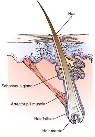
Figure 8-4 The hair follicle and its surrounding structures.
Nails protect the tips of the fingers and toes against trauma. They are derived by keratin- ization of cells from the nail matrix, which is located at the proximal end of the nail plate. The nail plate consists of the nail root embedded in the posterior nail fold, a fixed middle portion, and a distal free edge. The whitish nail matrix of proliferating epithelial cells grows in a semilunar pattern. It extends outward past the posterior nail fold and is called the lunula. The structural relationships of the nail are shown in Figure 8-3.
A hair shaft is a keratinized structure that grows out of the hair follicle. Its lower end, called the hair matrix, consists of actively proliferating epithelial cells. The cells at this end of the follicle, along with those of the bone marrow and gut epithelium, are the most rapidly growing dividing cells in the human body. This is the reason that chemotherapy causes hair loss, along with anemia, nausea, and vomiting. Visible hair is present over the entire body surface except on the palms, soles, lips, eyelids, glans penis, and labia minora. In apparently hairless areas, the hair follicles are small, and the shafts produced are microscopic. Hair follicles show conspicuous morphologic and functional heterogeneity. Follicles and their developing shafts differ from location to location in shaft length, color, thickness, curl, and androgen sensitivity. Some follicles, those in the axilla and inguinal areas, are very sensitive to androgens, whereas others in the eyebrow are insensitive. The arrector pili muscles attach to the follicle below the opening of the sebaceous gland. Contraction of this muscle erects the hair and causes ''goose bumps.'' The structure of a hair follicle is shown in Figure 8-4.
Review of Specific Symptoms
The main symptoms of disease of the skin, hair, and nails are the following:
• Rash or skin lesion
• Changes in skin color
• Itching (pruritus)
• Changes in hair
• Changes in nails
Rash or Skin Lesion
There are some important points to clarify when a patient is interviewed about a new rash or skin lesion. The specific time of onset and location of the rash or skin lesion are critical. A careful description of the first lesions and any changes is vital. The patient with a rash or skin lesion should be asked the following questions:
''Was the rash initially flat? raised? blistered?''
''Did the rash change in character with time?''
‘‘Have there been new areas involved since the rash began?''
''Does the rash itch or burn?''
''Is the lesion tender or numb?''
''What makes the rash better? worse?''
‘‘Was the rash initiated by sunlight?''
''Is the rash aggravated by sunlight?''
''What kind of treatment have you tried?''
''Do you have any joint pains? fever? fatigue?''
‘‘Does anyone near you have a similar rash?''
''Have you traveled recently?'' If so, ''To where?''
''Have you had any contact with anyone who has had a similar rash?''
''Is there a history of allergy?'' If so, ''What are your symptoms?''
Do you have any chronic disease?''
Note whether the patient has used any medications that may have changed the nature of the skin disorder.
Inquire whether the patient uses any prescription medications or over-the-counter drugs. Ask specifically about aspirin and aspirin-containing products. Patients can suddenly develop a reaction to medications that they have taken for many years. Do not ignore a long-standing prescription. Has the patient had any recent injections or taken any new medications? Does the patient use ''recreational'' drugs? Ask the patient about the use of soaps, deodorants, cosmetics, and colognes. Has the patient changed any of these items recently?
A family history of similar skin disorders should be noted. The effect of heat, cold, and sunlight on the skin problem is important. Can there be any contributing factor, such as occupation, specific food allergies, alcohol, or menses? Is there a history of gardening or household repair work? Has there been any contact with animals recently? The interviewer should also remember to inquire about psychogenic factors that may contribute to a skin disorder.
Determine the patient's occupation, if it is not already known. Ascertain avocational and recreational activities. This information is important even if the patient has been exposed to chemicals or similar agents for years. Manufacturers frequently change the basic constituents without notifying the consumer. It may also take years for a patient to become sensitized to a substance.
Changes in Skin Color
Patients may complain of a generalized change in skin color as the first manifestation of an illness. Cyanosis and jaundice are examples of this type of problem. Determine whether the patient is aware of any chronic disease that may be responsible for these changes. Localized skin color changes may be related to aging or to neoplastic changes. Certain medications can also be responsible for skin color changes. Inquire whether the patient is taking or has recently taken any medications.
Pruritus
Pruritus, or itching, may be a symptom of a generalized skin disorder or an internal illness. Ask any patient with pruritus the following questions:
When did you first notice the itching?''
Did the itching begin suddenly?''
Is the itching associated with any rash or lesion on your body?''
‘‘Are you taking any medications?''
''Has there been any change in the sweating or dryness of your skin?''
Have you been told that you have a chronic illness?''
Have you traveled recently?'' If so, To where?''
Diffuse pruritus is observed in biliary cirrhosis and in cancer, especially lymphoma. Pruritus in association with a diffuse rash may be dermatitis herpetiformis. Determine whether the pruritus has been associated with a change in perspiration or dryness of the skin, because either of these conditions may be the cause of the pruritus.
Changes in Hair
Inquire whether there has been a loss of hair or an increase in hair. Ascertain any changes in distribution or texture. If there have been changes, ask the following questions:
When did you first notice the changes?''
Did the change occur suddenly?''
Is the hair loss symmetric?''
Has the change been associated with itching? fever? recent stress?''
Are you aware of any exposure to any toxins? commercial hair compounds?''
Have you changed your diet?''
What medications are you taking?''
Changes in diet and medications are frequently responsible for changes in hair patterns. Hypothyroidism is frequently associated with loss of the lateral third of the eyebrows. Vascular disease in the legs often causes hair loss on the legs. Alternatively, ovarian and adrenal tumors can cause an increase in body hair.
Changes in Nails
Changes in nails may include splitting, discoloration, ridging, thickening, or separation from the nail bed. Ask the patient the following questions:
When did you first notice the nail changes?''
Have you had any acute illness recently?''
Do you have any chronic illness?''
Have you been taking any medications?''
Have you been exposed to chemicals at work or at home?''
Fungal disease causes thickening of the nail. Clues to systemic disease may be found by close examination of the proximal nail fold, the lunula, the nail bed, the nail plate, and the hyponychium. Acute illnesses are associated with lines and ridges in the nail bed and nail. Medications and chemicals are notorious for causing nail changes.
General Suggestions
All patients should be asked whether there have been any changes in moles, birthmarks, or spots on the body. Determine any color changes, irregular growth, pain, scaling, or bleeding. Any recent growth of a flat, pigmented lesion is relevant information.
All patients should be asked whether there are any red, scaly, or crusted areas of skin that do not heal. Has the patient ever had skin cancer? If the patient has had skin cancer, further questioning regarding the body location, treatment, and description is appropriate.
Impact of Skin Disease on the Patient
Diseases of the skin play a profound role in the way the affected patient interacts socially. If located on visible skin surfaces, long-standing skin diseases may actually interfere with the emotional and psychologic development of the individual. The attitude of a person toward self and others may be markedly affected. Loss of self-esteem is common. The adult with a skin disorder often faces limitation of sexual activity. This disruption of intimacy can foster or increase hostility and anxiety in the patient. Skin is a sensitive marker of an individual's emotions. It is known that blushing can reflect embarrassment, sweating can indicate anxiety, and pallor or ''goose bump'' skin may be associated with fear.
Patients with rashes have always evoked feelings of revulsion. Rashes have been associated with impurity and evil. Even today, friends and family may reject the individual with a skin disease. Patients with skin that is red, oozing, discolored, or peeling are rejected not only by family members but perhaps even by their physicians. At other times, skin lesions cause others to stare at the patient, which causes further discomfort. Some skin disorders may be associated with such extreme physical or emotional pain that marked depression may result and, on occasion, lead to suicide.
Skin diseases are often treated palliatively. Because numerous skin disorders have no cure, many patients go through life helpless and frustrated, as do their physicians.
The role of anxiety as a natural stressor in producing rashes is frequently observed. Stress tends to worsen certain skin disorders, such as eczema. This creates a vicious cycle, because the rash then exacerbates the anxiety. Rashes are common symptoms and signs of psychosomatic disorders.
Clinicians should discuss these anxieties with the patient in an attempt to break the cycle. The interviewer who tries to elicit the patient's feelings about the disease allows the patient to ''open up.'' The fears and fantasies can then be discussed. The examiner should also be comfortable in touching the patient for reassurance. This tends to improve the doctor-patient relationship because the patient has a lesser sense of isolation.
Physical Examination
The only equipment necessary for the examination of the skin is a penlight. The examination of the skin consists of only two steps:
Inspection
Palpation
The examination of the skin depends on inspection, but palpation of a skin lesion must also be performed. Although most skin lesions are not contagious, it is prudent to wear gloves to evaluate any skin lesion. This is especially true because of the prevalence of skin disease associated with human immunodeficiency virus (HIV) infection. Palpation of a lesion helps define its characteristics: texture, consistency, fluid, edema in the adjacent area, tenderness, and blanching.
The patient and the examiner must be comfortable during the examination of the skin. The lighting should be adjusted to produce the optimal illumination. Natural light is preferable. Even in the absence of complaints related to the skin, a careful examination of the skin must be performed on all patients because the skin may provide subtle clues of an underlying systemic illness. Examination of the skin may be performed as a separate system approach, or, preferably, the skin should be examined when the other parts of the body are evaluated.
General Principles
The examiner should be suspicious of any lesion that the patient describes as having increased in size or changed in color. The development of any new growth warrants attention.
When examining the skin, the clinician initially determines the general aspects of the skin: the color, moisture, turgor, and texture of the skin.
Any color changes, such as cyanosis, jaundice, or pigmentary abnormalities, should be noted.
Red vascular lesions may be either extravasated blood into the skin, known as petechiae or purpura, or malformed elements of the vascular tree, known as angiomas. When pressure is applied by a glass slide over an angioma, it blanches. This is a useful test to differentiate an angioma from petechiae, which do not change when pressure from a glass slide is applied.
During the physical examination, inspect all pigmented lesions and be aware of the ''ABCD'' warning signs associated with malignant melanoma:
• Asymmetry of shape
• Border irregularity
• Color variation
• Diameter larger than 6 mm
Asymmetry means that half the lesion appears different from the other half. Border irregularity describes a scalloped or poorly circumscribed contour. The color variation refers to shades of tan and brown, black, and sometimes white, red, or blue. A diameter larger than 6 mm, which is the size of a pencil eraser, is considered a danger sign for melanoma.
The clinician should remember this axiom in dermatology: ''There are more errors made by not looking than by not knowing.''
Excessive moisture may be seen in normal individuals, or it may be associated with fevers, emotions, neoplastic diseases, or hyperthyroidism. Dryness is a normal aging change, but it may also be seen in myxedema, nephritis, and certain drug-induced states. Look for excoriations, which might indicate the presence of pruritus as a clue to an underlying systemic illness.
When palpating the skin, evaluate its turgor and texture. Tissue turgor provides a mechanism for estimating the patient's general state of hydration. If the skin over the forehead is pulled up and released, it should promptly reassume its normal contour. In a patient with decreased hydration, this response is delayed.
It is often difficult for the inexperienced examiner to evaluate the texture of the skin because texture is a qualitative parameter. Softness has occasionally been likened to the texture of skin over a baby's abdomen. ''Soft''-textured skin is seen in secondary hypothyroidism, hypopituitarism, and eunuchoid states. ''Hard''-textured skin is associated with scleroderma, myxedema, and amyloidosis. ''Velvety'' skin is associated with Ehlers-Danlos syndrome.
Examination with Patient Seated
When the patient is seated, examine the hair and the skin on the hands and upper extremities. Inspect the Hair
The hair and scalp are evaluated for any lesions. Is alopecia or hirsutism present? Pay attention to the pattern of distribution and texture of hair over the body. In certain diseases, such as hypothyroidism, the hair becomes sparse and coarse. In contrast, patients with hyperthyroidism have hair that is very fine in texture. Loss of hair occurs in many conditions: anemia, heavy metal poisoning, hypopituitarism, and some nutritional disease states, such as pellagra. Increased hair patterns are seen in Cushing's disease, Stein-Leventhal syndrome, and several neoplastic conditions, such as tumors of the adrenal glands and gonads.
Inspect the Nail Beds
Evaluation of the nails can provide important clues about diseases. The nails may be affected in many systemic and dermatologic conditions. Nail-bed changes are usually not pathognomonic for a specific disease. Disorders stemming from renal, hematopoietic, or hepatic conditions may be evident from the nails. The nails should be inspected for shape, size, color, brittleness, hemorrhages underneath, transverse lines or grooves in the nail or nail bed, and an increased white area of the nail bed. Figure 8-5 illustrates some typical nail changes associated with medical diseases.


Figure 8-5 Common nail findings associated with medical diseases.
Beau's lines are transverse grooves or depressions parallel to the lunula. They are often associated with infections (typhus, acute rheumatic fever, malaria, acquired immunodeficiency syndrome [AIDS]); nutritional disorders (protein deficiency, pellagra); circulatory problems (myocardial infarction, Raynaud's disease); dysmetabolic states (diabetes, hypothyroidism, hypocalcemia); digestive diseases (diarrhea, enterocolitis, chronic pancreatitis, sprue); drugs (chemotherapy agents); operations; and alcoholism. Beau's lines are caused by conditions that cause the nail to grow slowly or even cease to grow for short intervals. The point of arrested growth is seen as a transverse groove in the nail. These lines are most commonly seen in the thumbnails and toenails. Figure 8-6A shows a patient's fingers with Beau's lines on the nails. Figure 8-6B shows Beau's lines in the toenails of a patient who had undergone major surgery 5 months earlier.
On occasion, a white transverse line or band results from poisoning or an acute systemic illness. These lines, called Mees' bands, are historically associated with chronic arsenic poisoning. These lines or bands are also parallel to the lunula. By measuring the width of the line and approximating nail growth at 1 mm per week, the examiner may be able to determine the duration of the antecedent acute illness. Figure 8-7 shows the fingernails of a patient who had received chemotherapy several weeks earlier.
Lindsay's nails are also called ''half-and-half nails.'' The proximal portion of the nail bed is whitish, whereas the distal part is red or pink. Chronic renal disease and azotemia are commonly associated with this type of nail abnormality. Figure 8-8 shows Lindsay's nails secondary to chronic renal failure.
Terry's nails are white nail beds to within 1 to 2 mm of the distal border of the nail. These nail findings are most commonly associated with cirrhosis, hypoalbuminemia, chronic congestive heart failure, and adult-onset diabetes mellitus. Figure 8-9 shows classic Terry's nails.
Splinter hemorrhages are formed by extravasation of blood from longitudinal nail bed blood vessels into adjacent troughs. Splinter hemorrhages are very common. It has been estimated that up to 20% of all hospitalized patients have splinter hemorrhages. Their presence is most often related to local, light trauma, but these hemorrhages are commonly associated with systemic disease. Classically associated with subacute bacterial endocarditis, splinter hemorrhages may also be seen in leukemia, vasculitis, infection, rheumatoid arthritis, systemic lupus erythematosus, renal disease, liver disease, and diabetes mellitus, among other diseases. Splinter hemorrhages are depicted in Figure 8-10.
Is koilonychia present? Koilonychia, or ''spoon nail,'' is a dystrophic state in which the nail plate thins and a cuplike depression develops. Spoon nails are most commonly associated with iron-deficiency anemia but may be seen in association with thinning of the nail plate from any cause, including local irritants. Figure 8-11 shows a normal fingernail in comparison with koilonychia secondary to iron-deficiency anemia.
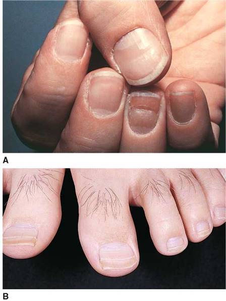
Figure 8-6 Beau's lines. A, Fingernails. B, Toenails.
Inspect the Nails for Clubbing
The angle between the normal nail base and finger is about 160°, and the nail bed is firm. The angle is referred to as Lovibond's angle. When clubbing develops, this angle straightens out to greater than 180°, and the nail bed, when palpated, becomes spongy or floating. As clubbing progresses, the base of the nail becomes swollen, and Lovibond's angle greatly exceeds 180°. The nail has a bullous shape with exaggerated longitudinal and horizontal curvatures. A fusiform enlargement of the distal digit may also occur. In Figure 8-12, a normal finger is compared with a finger with nail clubbing.

Figure 8-7 Mees' bands.

Figure 8-8 Lindsay's nails.

Figure 8-9 Terry's nails.

Figure 8-10 Splinter hemorrhages.

Figure 8-11 Koilonychia.

Figure 8-12 Late-stage clubbing.
To examine for clubbing, the patient's finger is placed on the pulp of the examiner's thumbs, and the base of the nail bed is palpated by the examiner's index fingers. Figure 8-13 illustrates the technique for assessing whether early clubbing is present.
Clubbing of the nails is most commonly acquired but may be inherited as an autosomal dominant trait. When acquired, clubbing is associated with congenital cyanotic heart disease, cystic fibrosis, and acquired pulmonary disease. The most common acquired pulmonary cause is bronchogenic carcinoma. In a patient with chronic obstructive pulmonary disease who manifests clubbing, other causes, including bronchiectasis or bronchogenic carcinoma, should be sought. The initial manifestation of clubbing is a softening of the tissue over the proximal nail fold.
Inspect the Nails for Pitting
Pitting of the nails is commonly associated with psoriasis and psoriatic arthropathy. Involvement of the nail bed and nail matrix by psoriasis causes the nail plate to be thickened and pitted. Nail involvement occurs in about 50% of all patients with psoriasis. Multiple pits in the nail are produced by discrete psoriatic foci in the nail matrix. Minor degrees of pitting are also seen in persons with no other skin complaints. Pitting rarely occurs in toenails. Psoriatic nail pitting is shown in Figure 8-14; Figure 8-15 illustrates a cross section of pitting. Figure 8-16A shows psoriatic nail lesions in another patient. Notice the characteristically dystrophic nail changes caused by subungual hyperkeratosis. Figure 8-16B is a close-up of a typical nail with psoriatic changes.
Inspect the Skin of the Face and Neck
Evaluate the eyelids, forehead, ears, nose, and lips carefully. Evaluate the mucous membranes of the mouth and nose for ulceration, bleeding, or telangiectasis. Is the skin at the nasolabial fold and mouth normal?
Inspect the Skin of the Back
Examine the skin of the patient's back. Are any lesions present?
Examination with Patient Lying Down Inspect the Skin of the Chest, Abdomen, and Lower Extremities
Ask the patient to lie down so that you can complete the examination of the skin. Inspect the skin of the chest and abdomen. Pay particular attention to the skin of the inguinal and genital area. Inspect the pubic hair. Elevate the scrotum. Inspect the perineal area. Evaluate the pretibial areas for the presence of ulcerations or waxy deposits.
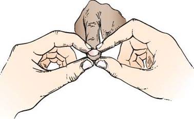
Figure 8-13 Technique for assessing whether clubbing is present.

Figure 8-14 Psoriasis: nail pitting.
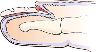
Figure 8-15 Cross section of nail pitting.

Figure 8-16 A and B, Psoriatic nail lesions.
Examine the feet and soles carefully for any skin changes. Spread the toes in order to evaluate the webs between them thoroughly.
Ask the patient to roll onto the left side so that you may examine the skin on the back, gluteal, and perianal areas.
Description of Lesions
If a skin lesion is found, it should be classified as a primary or secondary lesion, and its shape and distribution should be described. Primary lesions arise from normal skin. They result from anatomic changes in the epidermis, dermis, or subcutaneous tissue. The primary lesion is the most characteristic lesion of the skin disorder. Secondary lesions result from changes in the primary lesion. They develop during the course of the cutaneous disease.
The first step in identifying a skin disorder is to characterize the appearance of the primary lesion. In the description of the skin lesion, the clinician should note whether the lesion is flat or raised and whether it is solid or contains fluid. A penlight is often useful to determine whether the lesion is slightly elevated. If a penlight is directed to one side of a lesion, a shadow forms according to the height of the lesion.
The location of the lesion on the body is important. Therefore, the distribution of the eruption is crucial in making a diagnosis. It may be rewarding to inspect a patient's clothing when contact dermatitis or pediculosis (infestation with lice) is suspected. On occasion, occupational exposure may leave traces of contamination with oils or other materials that may be visible on the clothing and help in the assessment.
The three specific criteria for a dermatologic diagnosis are based on morphology, configuration, and distribution, morphology being the most important. The purpose of the following section is to acquaint the reader with the morphologic features of the primary and secondary lesions and the vocabulary associated with them.
Primary and Secondary Lesions
To facilitate reading, the primary lesions are listed here with regard to being flat or elevated and solid or fluid-filled (Figs. 8-17 to 8-20). There is no ''standard'' size of a primary lesion. The dimensions indicated are only approximate. The secondary lesions are grouped according to their occurrence below or above the plane of the skin (Figs. 8-21 and 8-22). Other important lesions are shown and described in Figure 8-23.
|
Primary Skin Lesions: Nonpalpable, Flat |
||
|
Lesion |
Characteristics |
Examples |
|
Macule |
Smaller than 1 cm |
Freckles, moles |
|
Patch |
Greater than 1 cm |
Vitiligo, cafe au lait spots |

Figure 8-17
|
Primary Skin Lesions: Palpable, Solid Mass |
||
|
Lesion |
Characteristics |
Examples |
|
Papule |
Smaller than 1 cm |
Nevus, wart |
|
Nodule |
1-2 cm |
Erythema nodosum |
|
Tumor |
Greater than 2 cm |
Neoplasms |
|
Plaque |
Flat, elevated, superficial papule with surface area greater than height |
Psoriasis, seborrheic keratosis |
|
Wheal |
Superficial area of cutaneous edema |
Hives, insect bite |

Figure 8-18
|
Primary Skin Lesions: Palpable, Fluid Filled |
||
|
Lesion |
Characteristics |
Examples |
|
Vesicle |
Smaller than 1 cm; filled with serous fluid |
Blister, herpes simplex |
|
Bulla |
Greater than 1 cm; filled with serous fluid |
Blister, pemphigus vulgaris |
|
Pustule |
Similar to vesicle; filled with pus |
Acne, impetigo |
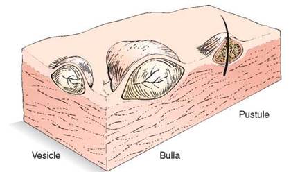
Figure 8-19
|
Special Primary Skin Lesions |
||
|
Lesion |
Characteristics |
Examples |
|
Comedo |
Plugged opening of sebaceous gland |
Blackhead |
|
Burrow |
Smaller than 10 mm, raised tunnel |
Scabies |
|
Cyst |
Palpable lesion filled with semiliquid material or fluid |
Sebaceous cyst |
|
Abscess |
A specific type of primary lesion with localized accumulation of purulent material in the dermis or subcutis; in general, the accumulation is so deep that the pus is not visible from the skin's surface |
|
|
Furuncle |
A specific type of primary lesion that is a necrotizing form of inflammation of a hair follicle |
|
|
Carbuncle |
A coalescence of several furuncles |
|
|
Milia |
Tiny, keratin-filled cysts representing an accumulation of keratin in the distal portion of the sweat gland |
|
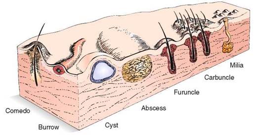
Figure 8-20
Configuration of Skin Lesions
It is not essential for the examiner to make a definitive diagnosis of all skin disease. A careful description of the lesion, the pattern of distribution, and the arrangement of the lesion often points to a group of related disease states with similar manifesting dermatologic signs (e.g., confluent macular rashes, bullous diseases, grouped vesicles, papular rashes on an erythematous base). For example, grouped urticarial lesions with a central depression are suggestive of insect bites. Figure 8-24 lists the terms used to describe the configurations of lesions.
Clinicopathologic Correlations
Skin disorders are frequently perplexing to the examiner. When an examiner sees a rash, the common thought is ''Where do I begin?'' All too often, the examiner may become frustrated and not even attempt to make a diagnosis. Dermatologic terms are complicated, and the names of dermatologic disorders may be intimidating. Often the descriptions of skin disorders in textbooks are more confusing than helpful.
|
Secondary Skin Lesions Below the Skin Plane |
||
|
Lesion |
Characteristics |
Examples |
|
Erosion |
Loss of part or all of the epidermis; surface is moist |
Rupture of a vesicle |
|
Ulcer |
Loss of epidermis and dermis; may bleed |
Stasis ulcer, chancre |
|
Fissure |
Linear crack from epidermis into dermis |
Cheilitis, athlete's foot |
|
Excoriation |
A superficial linear, or ''dug out,'' traumatized area, usually self-induced |
Abrasion, scratch mark |
|
Atrophy |
Thinning of skin with loss of skin markings |
Striae |
|
Sclerosis |
Diffuse or circumscribed hardening of skin |
|
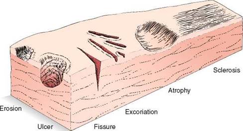
Figure 8-21
|
Secondary Skin Lesions Above the Skin Plane |
||
|
Lesion |
Characteristics |
Examples |
|
Scaling |
Heaped-up keratinized cells; exfoliated epidermis |
Dandruff, psoriasis |
|
Crusting |
Dried residue of pus, serum, or blood |
Scabs, impetigo |
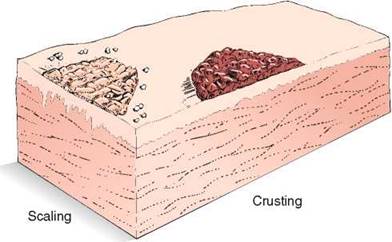
Figure 8-22
|
Vascular Skin Lesions |
||
|
Lesion |
Characteristics |
Examples |
|
Erythema |
Pink or red discoloration of the skin, secondary to dilatation of blood vessels, that blanches with pressure |
|
|
Petechiae |
Reddish-purple; nonblanching; smaller than 0.5 cm |
Intravascular defects |
|
Purpura |
Reddish-purple; nonblanching; greater than 0.5 cm |
Intravascular defects |
|
Ecchymosis |
Reddish-purple; nonblanching; variable size |
Trauma, vasculitis |
|
Telangiectasia |
Fine, irregular dilated blood vessels |
Dilatation of capillaries |
|
Spider angioma |
Central red body with radiating spider-like arms that blanch with pressure to the central area |
Liver disease, estrogens |
|
Miscellaneous Skin Lesions |
||
|
Lesion |
Characteristics |
Examples |
|
Scar |
Replacement of destroyed dermis by fibrous tissue; may be atrophic or hyperplastic |
Healed wound |
|
Keloid |
Elevated, enlarging scar growing beyond boundaries of wound |
Burn scars |
|
Lichenification |
Roughening and thickening of epidermis; accentuated skin markings |
Atopic dermatitis |

Figure 8-23
There are more than 2500 separately named dermatologic diagnoses. Most of these diseases occur in low frequencies; only 10 to 15 common conditions constitute about 50% of all dermatologic diagnoses. If the 50 most common conditions were considered, a diagnosis could be rendered for over 95% of all patients.
In approaching a skin lesion, the examiner must do the following:
1. First, identify the primary lesion.
2. Second, identify its distribution.
3. Third, identify any associated findings.
4. Fourth, consider the age of the patient.
Skin diseases evolve and their manifestations change. A lesion may evolve from a blister to an erosion, from a vesicle to a pustule, or from a papule to a nodule or tumor.
|
Descriptive Dermatologic Terms |
||
|
Lesion |
Characteristics |
Examples |
|
Annular |
Ring-shaped |
Ringworm |
|
Arcuate |
Partial rings |
Syphilis |
|
Bizarre |
Irregular or geographic pattern not related to any underlying anatomic structure |
Factitial dermatitis |
|
Circinate |
Circular |
|
|
Confluent |
Lesions that run together |
Childhood exanthems |
|
Discoid |
Disc-shaped without central clearing |
Lupus erythematosus |
|
Discrete |
Lesions that remain separate |
|
|
Eczematoid |
An inflammation with a tendency to vesiculate and crust |
Eczema |
|
Generalized |
Widespread |
|
|
Grouped |
Lesions that are clustered together |
Herpes simplex |
|
Iris |
Circle within a circle; a bull's-eye lesion |
Erythema multiforme |
|
Keratotic |
Horny thickening |
Psoriasis |
|
Linear |
In lines |
Poison ivy dermatitis |
|
Multiform |
More than one type of shape of lesion |
Erythema multiforme |
|
Papulosquamous |
Papules or plaques associated with scaling |
Psoriasis |
|
Reticulated |
Lacelike network |
Oral lichen planus |
|
Serpiginous |
Snakelike, creeping |
Cutaneous larva migrans |
|
Telangiectatic |
Relatively permanent dilatation of the superficial blood vessels |
Osler-Weber-Rendu disease |
|
Universal |
Entire body involved |
Alopecia universalis |
|
Zosteriform* |
Linear arrangement along a nerve distribution |
Herpes zoster |
|
*Also known as dermatomal. |
||

Figure 8-24

Figure 8-25 Cross section through a wart. Note the thickened epidermis and hyperkeratosis.
There are many common skin disorders or lesions with which the examiner should be familiar. Illustrated in the figures in this chapter are examples of some of these conditions; these cross-sectional diagrams illustrate the locations of these abnormalities in the skin and the involvement of the various skin layers in the pathogenesis of the conditions. The text describes the primary lesions.
A common wart is a common, benign growth usually caused by an infection of an epidermal cell by a virus. These firm nodules with rough, keratinous surfaces range in size from pinhead to pea size and can coalesce to form an extensive bed. There is vacuolation of the epidermis with scaling and an upward growth of the dermal papilla. Figure 8-25 illustrates a cross section through a wart. Two examples of finger warts are pictured in Figure 8-26.
Warts can also occur on the soles of the feet (plantar verruca), where they have a distinctive appearance because of constant pressure. They are very painful owing to the constant pressure, which forces the keratinous material into the deeper tissue. An example of a plantar wart on the heel is shown in Figure 8-27A. Notice the keratotic lesion with a yellow center, within which are visible areas of multiple red to black dots that represent hemorrhage from the tips of the dermal papillae. Classically, there is interruption of the normal skin lines as well. Figure 8-27B shows the excised lesion. Notice the depth of the lesion when viewed horizontally. Because warts are in the epidermis, excision just to the level of the dermis is sufficient for complete removal with minimal to no scarring.
A squamous cell carcinoma is a malignant neoplasm of keratocytes in the epidermis and is locally invasive into the dermis. The tumor results in a scaling, crusting nodule or plaque that can ulcerate and bleed. Squamous cell carcinoma is a potentially dangerous lesion that can infiltrate the surrounding structures and metastasize to lymph nodes and other organs. The causes include ultraviolet radiation, x-radiation, polycyclic hydrocarbons (e.g., tar, mineral oils, pitch, and soot), mucosal diseases (e.g., lichen planus and Bowen's disease), scars, chronic skin disorders, genetic diseases (e.g., albinism and xeroderma pigmentosum), and human papillomavirus. The tumor develops predominantly on areas of skin exposed to sunlight. The latency from carcinogenic exposure to the development of the tumor may be as long as 25 to 30 years. Two examples of squamous cell carcinoma of the skin are pictured in Figure 8-28. Notice that the lesions are ulcerated with firm, raised indurated margins. Figure 11-11 also shows a patient with a squamous cell carcinoma and a malignant melanoma of the ear lobule. A squamous cell carcinoma on the lip of another patient is pictured in Figure 8-29. Notice the round, centrally ulcerating tumor. Figure 8-30 illustrates a cross section through a squamous cell carcinoma.

Figure 8-26 A and B, Warts.
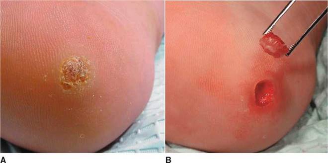
Figure 8-27 Plantar wart. A, Note the keratotic lesion with the yellow center and areas of hemorrhage within. B, After excision.
A basal cell carcinoma is a malignant neoplasm of the basal cells of the epidermis and is the most common skin malignancy. The epidermis is thickened, and the dermis may be invaded by the malignant basal cells. It may manifest as a lesion with a pearly, rolled, well-defined margin and a central ulcerated depression. Although sunlight is an important etiologic factor, basal cell carcinomas are almost always seen on the face and rarely in other sun-exposed areas. They are slow-growing tumors and rarely metastasize, in contrast to squamous cell carcinomas.

Figure 8-28 Squamous cell carcinoma of the skin. A, Ear. B, Face.

Figure 8-29 Squamous cell carcinoma of lip.
They are locally invasive, and when located near the eye or nose, they may invade the cranial cavity. If ulceration, bleeding, and crusting occur, a rodent ulcer is said to be present. Any nonhealing lesion should be carefully evaluated for the possibility of a basal cell carcinoma. Figures 8-31 to 8-33 illustrate the typical features of a basal cell carcinoma.
A melanoma is a malignant neoplasm of the melanocytes of the epidermis. If untreated or unrecognized, a melanoma progresses to fatal metastases. Most melanomas have a prolonged superficial, or horizontal, growth phase in which there is a progressive lateral expansion. With time, the melanoma enters the vertical, or deep, phase by penetrating into the dermis, and metastatic spread may occur.
Malignant melanomas are the most common malignancy seen by dermatologists. The incidence of malignant melanoma is increasing faster than that of any other form of malignancy. Most melanomas have atypical pigmentation in the epidermis, such as shades of red, white, gray, blue, brown, and black, all in a single lesion. There are four types of malignant melanoma: lentigo maligna melanoma, superficial spreading melanoma, nodular malignant melanoma, and acral-lentiginous malignant melanoma. Figure 8-34 shows the typical features of a lentigo maligna melanoma on the face. The lentigo maligna melanoma is seen frequently in the geriatric population. This type of melanoma has a prolonged horizontal growth phase and appears in areas of sun-exposed, sun-damaged skin. The superficial spreading variety (Fig. 8-35) is the most common type of melanoma (70% of all melanomas). Typically, an irregularly colored plaque has sharp notches and variegation of pigment. If it is diagnosed early, the prognosis is excellent, with a 5-year survival rate of 95%. Figure 8-36 illustrates a cross section through a melanoma. Vertical growth and deep invasion follow the spreading phase of superficial malignant melanomas. Figure 8-37 shows another superficial spreading melanoma that has developed vertical growth. The nodular melanoma is the second most common type, seen in approximately 15% of cases of melanoma. Unlike the superficial spreading type, these melanomas are usually black, brown, or dark blue and tend to grow rapidly for months.
Melanomas occur predominantly in white individuals and have a predilection for the back in men and women and for the anterior tibial areas in women. In general, lesions on the back, axillae, neck, and scalp (the so-called BANS area) tend to have a worse prognosis than do melanomas on the extremities. A great contrast between the risk of acquiring melanoma and basal or squamous cell carcinoma is that basal and squamous cell carcinomas occur more frequently in individuals who are exposed to constant sunlight, such as sailors and agricultural workers. Melanomas occur more often in rather fair-skinned individuals who experience brief, intense sun exposure, such as that occurring during vacations in the southern latitudes.
Fewer than 5% of all melanomas occur in the African-American population. The acral- lentiginous melanoma is the most common form in African Americans and occurs on the palms, soles, and nail beds. These melanomas have a short superficial growth phase and an early vertical growth phase and, as such, are associated with a poor prognosis. An acral- lentiginous melanoma on the sole of an African-American patient is pictured in Figure 8-38.
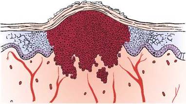
Figure 8-30 Cross section through a squamous cell carcinoma. Note the invasion into the dermis.

Figure 8-31 Cross section through a basal cell carcinoma.

Figure 8-32 Basal cell carcinoma (rodent ulcer).

Figure 8-33 Basal cell carcinoma. Notice the rolled, well-defined margin.

Figure 8-34 Lentigo maligna melanoma.

Figure 8-35 Superficial spreading malignant melanoma.
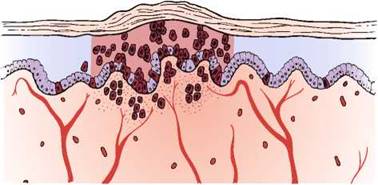
Figure 8-36 Cross section through a melanoma. Note the nests of melanoma cells in the dermis.

Figure 8-37 Superficial spreading malignant melanoma with vertical growth.
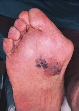
Figure 8-38 Acral-lentiginous melanoma.
Malignant melanoma of the nail apparatus represents 2% to 3% of all melanomas in white individuals and about 20% in dark-skinned individuals. The most common pathologic type is the acral-lentiginous melanoma. It is frequently diagnosed in the sixth decade; women are affected more often than men. The thumb and the great toe are the most common sites. Figure 8-39 shows a classic malignant melanoma of the nail, with a wide longitudinal band and the variegated colors. Figure 8-40 shows a malignant melanoma of the nail bed. Notice that the band width is wider at the base than at the tip, indicating a rapidly growing lesion.
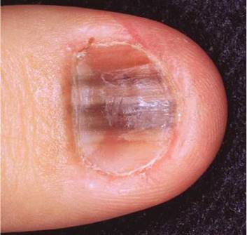
Figure 8-39 Melanoma of the nail.
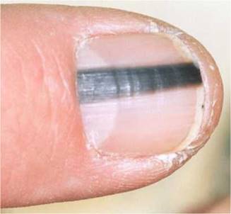
Figure 8-40 Melanoma of the nail bed.

Figure 8-41 Lipoma of the back.
A lipoma is a benign growth of subcutaneous fat and has a rubbery appearance. The epidermis is normal. Frequently, an encapsulated lipoma may grow to a very large size and elevate the overlying dermis and epidermis, as shown in Figure 8-41; a cross section through a lipoma is illustrated in Figure 8-42. The examiner can easily push into the soft tissue tumor. Another example of a lipoma is shown on the arm of the patient in Figure 8-43.

Figure 8-42 Cross section through a lipoma.

Figure 8-43 Lipoma of the arm.

Figure 8-44 Café au lait spot in a patient with neurofibromatosis.
Café au lait spots are patch lesions that are well circumscribed and brownish. They may occur as a solitary birthmark in up to 10% of the normal population. The cafe au lait macule or patch results from an increased number of functionally hyperactive melanocytes. Multiple cafe au lait patches in a patient may suggest neurofibromatosis. Figure 8-44 shows a cafe au lait patch in a patient with neurofibromatosis.
A neurofibroma is a tumor produced by a focal proliferation of neural tissue in the dermis. The epidermis is normal. Neurofibromas may appear as papules or nodules. Cutaneous neurofibromas are soft in consistency. Neurofibromatosis is a disorder in which multiple neurofibromas are present, sometimes as many as several hundred. Although the tumors are benign, the occurrence of these space-occupying lesions may produce severe disfigurement or neurologic disease. Other dermatologic features of neurofibromatosis include multiple cafe au lait patches and axillary freckling. Figure 8-45 shows several neurofibromas in a patient with neurofibromatosis; a cross-sectional view is illustrated in Figure 8-46. Figure 8-47 shows axillary freckling (Crowe's sign) in a patient with neurofibromatosis. Figure 8-48 shows multiple neurofibromas on the face of another patient with neurofibromatosis.
Contact dermatitis is an inflammatory reaction of the skin that is precipitated by contact with an irritant or allergen, such as detergents, acids, alkali, plants, medicines, and solvents. Vesicles in the epidermis and perivascular inflammation result. Figure 8-49 shows contact dermatitis in reaction to poison ivy; the area is illustrated in cross section in Figure 8-50. The characteristic linear distribution of papules, vesicles, and bullae is visible on this patient's calf where the leaves of the plant touched the leg. The distribution of the bullous lesions together with their location is strongly suggestive of the diagnosis, which was confirmed by biopsy.
Psoriasis is one of the most common noninfectious skin disorders. It is frequently inherited and often chronic, and it, too, may affect the joints and nails. The rash is characterized by well- defined, slightly raised, hyperkeratotic (scaling) plaques. If the lesion is scratched, small bleeding points appear, which is a specific sign of the disease. The lesions are frequently symmetric and can be extremely itchy. The stratum corneum thickens, and erythematous plaques with silvery scales result. Within the dermis, there is capillary proliferation with perivascular inflammation. The lesions are characteristically located on the elbows, knees, scalp, and intergluteal cleft. Figure 8-51 shows the typical, symmetric lesions on the knees of an affected patient. Figure 8-52 shows the classic scaling lesions at the intergluteal cleft of another patient. Figure 8-53 illustrates a cross section through an area of psoriasis (see also Fig. 8-15). Figure 8-54 shows psoriasis of the scalp. The lesions commonly extend beyond the hair-bearing areas onto the adjacent skin. Surprisingly, this lesion rarely results in hair loss. Figure 8-55 shows severe, diffuse psoriasis involving more than 85% of the body of a 56-year-old man. This patient suffered from persistent itching, burning, and bleeding with psoriasis for more than 35 years. Psoriasis may produce several nail changes. Pitting of the nail has already been discussed (see Fig. 8-14). There are several other nail changes: oily patches, onycholysis, subungual hyperkeratosis, and splinter hemorrhages.
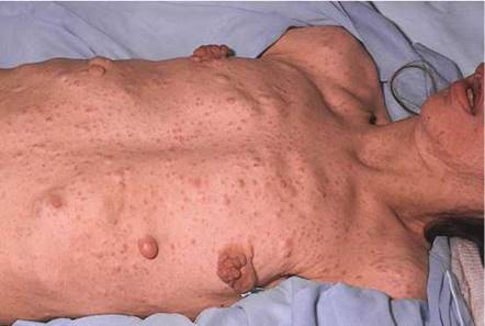
Figure 8-45 Multiple neurofibromas in a patient with neurofibromatosis.

Figure 8-46 Cross section through a neurofibroma. Note that the tumor is a well-delimited mass of loosely packed neural elements.

Figure 8-47 Neurofibromatosis: axillary freckling (Crowe's sign).

Figure 8-48 Multiple neurofibromas on the face of a patient with neurofibromatosis.

Figure 8-49 Contact dermatitis: poison ivy reaction.

Figure 8-50 Cross section through an area of contact dermatitis. Note the perivascular inflammation in the dermis, as well as the vesicles and bullae in the epidermis.
Tinea corporis is ''ringworm'' infection. Fungal infections of the skin produce a scaling, erythematous patch, often with a reddened, raised, serpiginous border. The term tinea indicates the fungal cause, and the second word denotes the area of the body involved: tinea corporis is infection in the body; tinea pedis, in the foot; tinea faciale, in the face; tinea barbae, in the beard and moustache area in men; tinea cruris, in the groin; and tinea capitis, in the head. In all cases, the epidermis is thickened, and the stratum corneum is infiltrated with fungal hyphae.
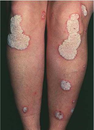
Figure 8-51 Psoriatic lesions on the knees.

Figure 8-52 Psoriatic lesions on the intergluteal cleft.
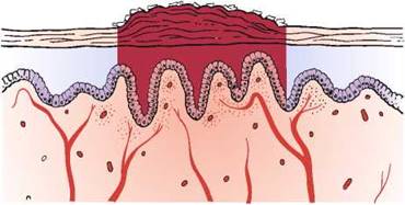
Figure 8-53 Cross section through an area of psoriasis. Note the area of hyperkeratosis.

Figure 8-54 Psoriasis of the scalp.

Figure 8-55 Psoriasis. A through E, Severe psoriasis in a 56-year-old patient.

Figure 8-56 Cross section through an area of tinea corporis. Note the thickened stratum corneum, which is infiltrated by the fungus.

Figure 8-57 Tinea corporis.
The underlying dermis displays mild inflammation. Figures 8-56 and 8-57 illustrate the classic annular lesion of tinea corporis with its raised erythematous border and central clearing. Another example of tinea corporis is pictured in Figure 8-58. Tinea faciale in a child is pictured in Figure 8-59. Tinea cruris is pictured in Figure 8-60. This common pruritic lesion is seen commonly in young men; it is unusual in women. It spreads outward from the groin, down the thigh, leaving postinflammatory pigmentation. The advancing border is well defined, red, scaly, and slightly raised. If untreated, the eruption can spread onto the lower abdomen, as shown, and the buttocks. The foot of a patient with tinea pedis is pictured in Figure 8-61. Notice the maceration with erosions and scaling.
Pityriasis rosea is a common, acute, self-limiting inflammatory disease of unknown cause that usually occurs during the spring. The generalized eruption is preceded by a ''herald patch,'' which is a single lesion resembling that of tinea corporis. In several days, the generalized eruption appears. Papulosquamous plaques appear over the trunk and rarely on the face and distal extremities. Although patients may complain of mild itching, they feel quite well. The full-blown picture develops slowly over 5 to 10 days and lasts for approximately 3 to 6 weeks. Slight hyperkeratosis of the epidermis with moderate dermal perivascular infiltration occurs. Figure 8-62 shows a herald patch and the characteristic lesions of pityriasis rosea; Figure 8-63 illustrates a cross-sectional view. Notice the delicate scale at the border of the annular lesion. Secondary syphilis may manifest with a similar eruption. It is therefore important to order a serologic test for syphilis in any individual with pityriasis rosea.

Figure 8-58 Tinea corporis.
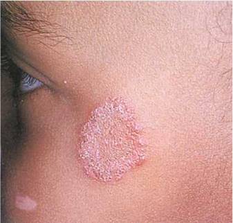

Figure 8-59 Tinea faciale.

Figure 8-60 Tinea cruris.
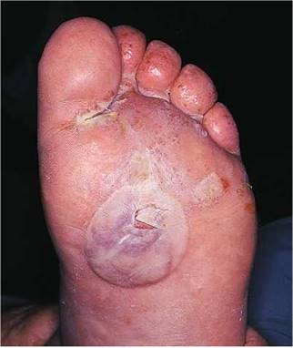
Figure 8-61 Tinea pedis.
Herpes zoster, or shingles, is an intraepidermal vesicular eruption occurring in a dermatomal distribution. Bullae and multinucleated giant cells are present in the epidermis, with perivascular inflammation of the dermis. The condition is caused by activation of the varicella-zoster virus. Groups of vesicles and bullae on erythematous bases are present along the distribution of peripheral nerves. Severe pain often precedes the eruption. Figure 8-64 shows herpes zoster lesions along the T3 distribution in two patients. Usually, the distribution occurs along the spinal or cranial nerves, but it can become generalized, as pictured in Figure 8-65; Figure 8-66 illustrates a cross section. Figure 8-67 is a close-up photograph of the typical vesicles on an erythematous base in a dermatomal distribution.
Herpesvirus infections are frequently encountered in patients with HIV infection; approxi- mately 25% to 50% of these patients have some form of herpetic disease during the course of their illness. Herpesvirus infections are thought to be predictive of future progression from HIV infection to AIDS; the rate of this association with progression is 23% at 2 years and up to 73% at 6 years. When CD4+ T cell counts fall below 100 cells/mm3, the likelihood of a herpesvirus infection approaches 95%. Herpes zoster infections may be severe and fulminant in immunocompromised patients, such as in the patient shown in Figure 10-58.

Figure 8-62 Pityriasis rosea. Note the herald patch.

Figure 8-63 Cross section through a lesion of pityriasis rosea.
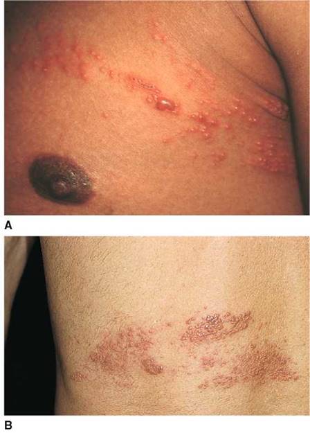
Figure 8-64 A and B, Herpes zoster lesions in T3 distribution.

Figure 8-65 Herpes zoster, generalized.
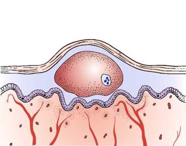
Figure 8-66 Cross section through one of the vesicles in herpes zoster. Note the large epidermal bullae with the classic multinuclear cells.

Figure 8-67 Herpes zoster vesicles.

Figure 8-68 Cross section through an area of acne. Note the rupturing of the sebaceous gland in the dermis as a result of a plugged hair follicle, which results in dermal inflammation.
Acne is a pustular disease affecting the hair follicles and sebaceous glands. In this condition, pustules, papules, and comedones are the primary lesions. There are collections of intradermal and intrafollicular neutrophils. Within the dermis, the hair follicle is occluded by a collection of keratin, sebum, and inflammatory cells. The hair follicle often ruptures into the dermis as a result of increasing pressure, which leads to further dermal inflammation (Figs. 8-68 to 8-71).
Tinea versicolor is a common superficial, noninflammatory, noncontagious fungal skin infection of young adults occurring most often during the summer. Pregnancy, warm climate, corticosteroids, and debilitation seem to be predisposing factors. Persons older than 40 years are rarely affected. The lesion consists of very fine, scaly patches that coalesce as they enlarge. The hypopigmented lesions are frequently seen on seborrheic areas of the body: the neck, the upper trunk, the upper arms, the shoulders, and, on occasion, the groin. The lesions are usually asymptomatic but may be mildly pruritic. Figure 8-72 shows the back of a patient with the classic hypopigmented patches of tinea versicolor. Figure 8-73 shows another patient with tinea versicolor.
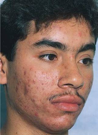
Figure 8-69 Acne.

Figure 8-70 Acne.
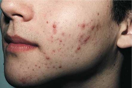
Figure 8-71 Acne.
A ganglion cyst is a chronic, painless lesion on the dorsum of the wrist or ankle. It results from leakage of synovial fluid through the tendon sheath of the capsule of the joint. This fluid eventually becomes encapsulated, and a cyst results. Figure 8-74 shows a ganglion in the wrist, the typical location.
A spider angioma is a common pale red lesion, usually less than 2 cm in diameter, with a pulsating, central arteriole, often raised, surrounded by erythema and radiating ''legs.'' If pressure is exerted on the central body, blanching of the spider's legs occurs. These benign lesions are commonly seen on the face, neck, arms, and upper trunk; they are rarely seen on the lower extremities. Although seen in normal individuals, spider angiomas are found more commonly in pregnant women and in patients with liver disease or vitamin B deficiency. These angiomas frequently become more evident during various times of a woman's menstrual cycle. Figure 8-75 shows a spider angioma on the face of a young woman. Notice the central arteriole with the peripheral blush.

Figure 8-72 Tinea versicolor.

Figure 8-73 Tinea versicolor.
Vitiligo consists of patches of lightened skin resulting from decreased melanin pigmentation. Vitiligo is essentially a large macule that is totally depigmented. The epidermis manifests a complete absence of pigment, whereas the dermis is normal. Vitiligo can occur in any area, but it is commonly found on the neck, knees, elbows, and back of the hands. Figure 8-76 shows extensive vitiligo on the face and neck; a cross section is shown in Figure 8-77. Diffuse vitiligo is pictured in Figure 8-78.
Urticaria is a common condition. The primary lesion is the wheal, or hive. In urticaria, the epidermis is normal. The dermis demonstrates papillary edema. Inflammatory cells may be found surrounding dilated blood vessels. Itching is a common complaint. There are several mechanisms for the development of urticaria, which include both immunologic and nonimmunologic causes. Regardless of the cause, the common factor is the release of substances, such as histamine, that change the vascular permeability and produce dermal edema. Figure 8-79 shows urticaria, and Figure 8-80 depicts the cross section.
Erythema multiforme is an immunologic reaction in the skin triggered by various causative agents, including viruses, bacteria, drugs, and x-radiation. In many cases, the agent cannot be identified. As the name implies, the condition includes a variety of lesions: papules, bullae, plaques, and ''target lesions.'' Target lesions are the diagnostic lesions and have three zones of color: A central, tense bulla, or dark area, is surrounded by a zone of relative pallor that is rimmed by a thin area of erythema. Typically, target lesions are seen on the palms and soles (Fig. 8-81). The epidermis is usually normal. In the dermis, there is a subepidermal separation with inflammatory cells in the papillary dermis. Penicillin and sulfonamides are the drugs most commonly implicated as the cause of this condition. The most severe form of erythema multiforme involves the mucous membranes and is called Stevens-Johnson syndrome. The classic skin lesions of erythema multiforme are shown in Figures 8-82 and 8-83; Figure 8-84 illustrates a cross-sectional view.
Scabies is a common, intensely pruritic skin disorder caused by a mite, Sarcoptes scabiei var. hominis. The female mite burrows into the stratum corneum of the skin and lays her eggs.
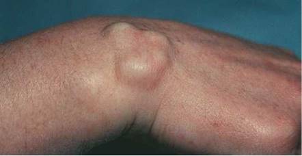
Figure 8-74 Ganglion cyst.

Figure 8-75 Spider angioma.

Figure 8-76 Vitiligo.

Figure 8-77 Cross section through an area of vitiligo. Note the absence of melanocytes and skin pigment.
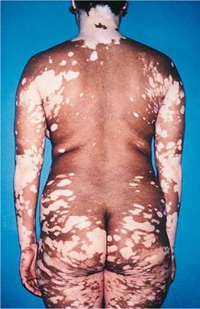
Figure 8-78 Diffuse vitiligo.

Figure 8-79 Urticaria.

Figure 8-80 Cross section through an area of urticaria.

Figure 8-81 Target lesions on palms and hands in a patient with erythema multiforme.

Figure 8-82 Erythema multiforme.
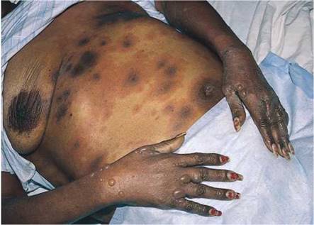
Figure 8-83 Erythema multiforme.
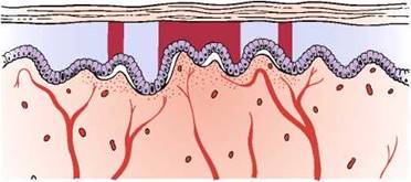
Figure 8-84 Cross section through an area of erythema multiforme. Note the separation of the epidermis from the dermis.

Figure 8-85 Scabies.
A month or longer may pass before the symptom of generalized pruritus develops. The diagnostic physical sign is the burrow, which is a serpiginous, palpable track about 1 cm in length that may end in a papule, nodule, or tiny vesicle. The adult female mite is present in the burrow. The extremely pruritic rash of scabies has a predilection for the web spaces of the fingers and toes, as well as the groin. The buttocks are also frequently involved, as are the genitals of men and the nipples of women. A generalized papular or urticarial eruption may ensue after localized scabies infection. The presence of papules on the genitalia in a patient with intense pruritus should raise the strong suspicion of scabies. Outbreaks of scabies are common in population groups in which HIV infection is prevalent. Figure 8-85 shows the hand of a patient with scabies. Notice the classic eruption between the fingers. Figure 18-16 shows another patient with scabies; the papular rash in the groin and on the penis is clearly seen.
Norwegian scabies is a rare, highly infectious form of scabies that is often seen in immuno- suppressed patients and patients with psychiatric illness; recently, this form of scabies has been seen in patients with AIDS. It is characterized by very thick, white hyperkeratotic scales containing thousands of mites. The dorsal aspects of the hands, the feet, and the extensor surfaces of the elbows and knees are commonly involved. Figure 8-86 shows the hand of a patient with Norwegian scabies. Figure 8-87 shows the diffuse lesions of Norwegian scabies in another patient.
Pyoderma gangrenosum is a cutaneous condition consisting of large, tender, necrotic ulcers having a violaceous, overhanging edge with a purulent base. The lesions are most frequently seen on the face, lower legs, and abdomen. Although most often associated with inflammatory bowel disease, this condition is also seen in association with various blood dyscrasias (especially multiple myeloma), chronic active hepatitis, rheumatoid arthritis, systemic lupus erythematosus, and acute leukemias. Approximately 10% of all patients with ulcerative colitis, however, have cutaneous manifestations, especially pyoderma gangrenosum. The skin lesions of pyoderma gangrenosum are closely linked to the bowel disease; exacerbations of bowel symptoms are associated with extension of existing lesions or development of new ones. Removal of the diseased bowel often leads to improvement in the cutaneous manifestations. Figure 8-88 shows the classic shin lesions of pyoderma gangrenosum in a patient with regional enteritis. See also Figure 17-6, which shows another patient with an exacerbation of ulcerative colitis and pyoderma gangrenosum of the shin.

Figure 8-86 Norwegian scabies.

Figure 8-87 Norwegian scabies.

Figure 8-88 Pyoderma gangrenosum of the shin.
Insect bites are common and should always be considered when a patient complains of a pruritic rash. Papules, vesicles, and wheals amid excoriations suggest the diagnosis. Papules in a grouped or linear arrangement on an arm or face suggest bedbug bites. These insects, which live in crevices in furniture, shun the light and feed at night on exposed areas of the body. They bite and then move around only to bite again. Figure 8-89 shows the linear papules on the arm of a patient who was bitten by bedbugs. Figure 8-90 shows another patient with persistent erythematous papules as a result of bedbug bites. Flea bites were the cause of the pruritic eruption on the feet of the patient shown in Figure 8-91. Notice the excoriations. The lower legs and feet are common sites for flea bites.
Kaposi's sarcoma (KS) is a neoplasm characterized by dark blue-purple macules, papules, nodules, and plaques. The classic form of the disease is a rare, slow-growing neoplasm occurring mostly on the lower extremities, especially the ankles and soles, of elderly men of Mediterranean or Jewish eastern European descent. The male-to-female ratio is 10:1 to 15:1, and the majority of patients are 60 to 80 years of age. Figure 8-92 shows a large reddish-colored plaque, which was slow-growing, on the sole of the foot of a 60-year-old man of eastern European origin.
Currently, KS is the most frequent neoplasm occurring in patients with AIDS. Approximately 35% of patients with AIDS who acquired the disease by sexual contact are affected, as opposed to approximately 5% of patients whose infection was acquired from intravenous drug use. Overall, 24% of all patients with AIDS develop this rapidly progressive form of the disease, also known as the epidemic HIV-associated form. The widely disseminated lesions are present on the legs, trunk, arms, neck, and head. They start as light-colored papules or nodules and coalesce into larger, darker lesions. Unlike the classic form, the epidemic

Figure 8-89 Bedbug bites.
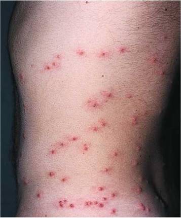
Figure 8-90 Bedbug bites.
HIV-associated form is commonly associated with visceral involvement, frequent oral lesions, and lymphadenopathy. The average length of patient survival from the onset of the disease is 18 months. The skin lesions of epidemic HIV-associated KS are shown in Figure 8-93. Figure 8-93A shows the typical lesions on the arm and chest; Figure 8-93B shows the widely disseminated plaque lesions varying in color from dark red to violet; Figure 8-93C shows a violaceous lesion on the lateral aspect of the lower eyelid; Figure 8-93D shows a large confluent plaque of KS on the hard palate; Figure 8-93E shows a purplish-red, nodular lesion of KS on the gingiva and an infiltrative, violaceous lesion of the nose.
Figure 8-94 demonstrates the rapidity of the growth of epidemic KS. The initial manifestation (Fig. 8-94A) on the back of a 36-year-old gay man was only a few macular lesions of KS; a follow-up photograph, taken only 6 months later, shows the widely disseminated, purplish plaques of KS (Fig. 8-94B).

Figure 8-91 Flea bites.
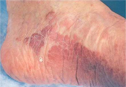
Figure 8-92 Kaposi's sarcoma: a classic plaque.

Figure 8-93 Kaposi's sarcoma: epidemic human immunodeficiency virus (HIV) associated. A and B, Plaque lesions. C, Violaceous lesion affecting the lateral lower eyelid. D, Confluent plaque on the hard palate. E, Nodular lesion on the gingiva and the nose.
Scleroderma, also known as progressive systemic sclerosis, is an important rheumatic disease characterized by hardening of the skin. Vascular changes occur with visceral involvement and involve the microvessels and small arteries. The onset of the disease is often heralded by the development of Raynaud's phenomenon, which is discussed in Chapter 15, The Peripheral Vascular System. The cutaneous manifestations of scleroderma involve tightening of the skin, especially on the face and hands. As a result of tendon contractures, flexion of the fingers results. The fingers of a patient with scleroderma are shown in Figure 8-95; notice that the skin is bound tightly and obscures the superficial vasculature. Skin lines are absent. Figure 8-96 shows the face of the same patient. Notice the tightening and wrinkling of the skin around her mouth and the fixed, expressionless countenance as a result of flattening of the nasolabial folds. The patient had great difficulty in opening her mouth. The skin around the mouth has many furrows radiating outward, creating a mouselike appearance known as mauskopf.

Figure 8-94 A, Early lesions of Kaposi's sarcoma on the back. B, Follow-up photograph, 6 months later, showing rapid development of purplish plaques of Kaposi's sarcoma.
Erythema nodosum is a common reaction associated with streptococcal infections, sarcoidosis, tuberculosis, inflammatory bowel diseases, and fungal diseases. It is infrequently associated with rheumatic disorders. Patients, primarily young women, seek medical attention after the appearance of extremely painful, erythematous nodules on the lower legs, especially over the anterior tibia. The lesions range in size from 1 cm to several centimeters in diameter. The lesions can then coalesce and spread over the entire leg. The lesions of erythema nodosum begin to regress after 1 to 2 weeks. As they disappear, they undergo a series of characteristic color changes: bright erythema to shades of purple, yellow, and green. Figure 8-97 shows the early lesions of erythema nodosum in a 33-year-old woman in whom sarcoidosis was diagnosed 3 months later.

Figure 8-95 Scleroderma: hands.
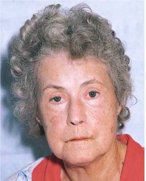
Figure 8-96 Scleroderma: face.

Figure 8-97 Erythema nodosum.
Lichen planus is a relatively common skin disorder of unknown cause. The primary lesion is a polygonal, shiny, flat-topped papule with a violaceous hue. The pruritic lesions can be seen on any part of the body but have a predilection for the front of the wrists and forearms, the backs of the hands, the ankles, the shins, the genitalia, and the lumbar areas. The lesions range in size from 2 mm to more than 1 cm. Figure 8-98A shows the characteristic rash on the arm. Fine reticulated scales are visible (see Fig. 8-98B). Oral lesions are seen in 50% of all patients with lichen planus and consist of a white, lacy network on the buccal mucosa. On occasion, involvement of the mouth may be the only manifestation of lichen planus. The patient usually experiences severe pain as the lesions ulcerate. Figure 12-15 depicts lichen planus of the buccal mucosa. Lichen planus may also involve the genitalia. Figure 8-99 shows lichen planus of the penis. Note the reticular markings on the penis. Figure 18-10 depicts another case of lichen planus of the penis. Note again the fine reticular markings.
Seborrheic dermatitis is a papulosquamous disorder associated with epidermal hyperplasia and scaling. The lesions have a greasy-looking scale in a seborrheic distribution: scalp, eyebrows, nasolabial fold, perioral area, midchest, and groin. Seborrheic dermatitis is one of the most common skin conditions associated with HIV infection; it is estimated that 85% of patients infected with HIV have this skin lesion at some time. In some patients, the development of seborrheic dermatitis is the first sign of HIV infection. Figure 8-100 shows the typical greasy scales of seborrheic dermatitis on the face of a patient with AIDS.
Seborrheic warts are common, benign skin tumors, seen in light-skinned individuals; they occur more frequently with advancing age. Also known as seborrheic keratosis (Fig. 8-101), seborrheic warts may be solitary or multiple lesions. They occur in any area of the body exposed to ultraviolet light. The lesions are well defined and raised and have a fissured surface. The lesions result from a failure of keratinocytes to mature normally, which produces an accumulation of immature cells in the epidermis. Sometimes the lesions may be pedunculated. A similar condition known as dermatosis papulosis nigra is seen in African Americans. Figure 8-102 is a close- up photograph of the characteristic appearance of a seborrheic wart.
A keloid is a hyperproliferative response of fibrous tissue to injury, inflammation, or infection. It has a smooth appearance with a shiny surface and is raised and firm to palpation. It is more commonly seen in dark-skinned individuals. The lesion characteristically spreads beyond the site of the initiating factor. Figure 8-103 shows an extensive keloid on the shoulder.
Nevi are common, localized abnormalities of the skin that may be present at birth or appear within the first few decades of life. Sometimes called ''moles,'' nevi may arise from almost any area of the skin. They are well defined, with a smooth surface and a round shape (Fig. 8-104). Hair may sometimes project from the surface. A strawberry nevus is a vascular tumor or hemangioma that occurs shortly after birth and is red and raised. These grow rapidly and are often seen on the face of a child. They may bleed and ulcerate. Fortunately, most strawberry nevi involute by 6 or 7 years of age. Figure 24-8 shows a strawberry nevus in a child. Figure 24-9 shows a hemangioma in another child.
Blistering, or vesiculobullous, diseases of the skin are rare but important to recognize. Pemphigus vulgaris, pemphigus vegetans, and bullous pemphigoid are autoimmune diseases that affect skin and mucosal surfaces. Pemphigus vulgaris is a vesiculobullous disease of middle age, seen more commonly in Jewish people.

Figure 8-98 A and B, Lichen planus. Notice the fine reticulated white scales.

Figure 8-99 Lichen planus of the penis.
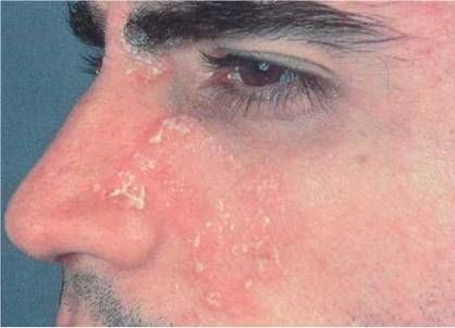
Figure 8-100 Seborrheic dermatitis.
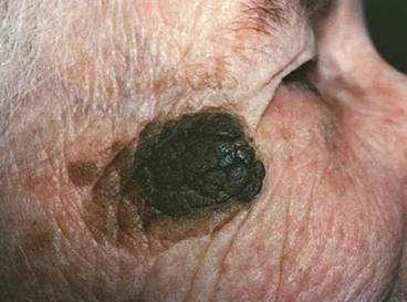
Figure 8-101 Seborrheic keratosis.

Figure 8-102 Seborrheic wart, close-up.

Figure 8-103 Keloid.
The lesions are superficial, flaccid blisters that break easily, leaving the skin denuded and eroded. The broken bullae may crust but do not heal spontaneously. These lesions are nonpruritic but are painful. The disease is caused by the production of antibodies to the intercellular junctions of the epidermis. The defective junctions lead to the formation of traumatic fissures and bullae. Figure 8-105A shows pemphigus vulgaris and broken bullae. Figure 8-105B shows pemphigus vegetans. The lesions may be present on any area of the skin, especially the trunk, umbilicus, intertriginous areas, and scalp. The lesions are frequently found in the mucous membranes of the oral cavity, pharynx, and genitalia. Figure 8-106 shows pemphigus vegetans of the lips.
Bullous pemphigoid is a blistering disorder seen more frequently in elderly patients. It is more common than pemphigus vulgaris. There is no racial predilection, and the disease is not as serious as pemphigus. The lesions are intensely pruritic, tense bullae often on an erythematous base and are symmetric on the limbs, inner aspects of the arms, thighs, and trunk. Oral and mucosal lesions are rarer than those of pemphigus. Figure 8-107 depicts generalized bullous pemphigoid. Figure 8-108 shows bullous pemphigoid in another patient. Notice the tense bullae, which help differentiate this disease from pemphigus. Figure 8-109 is a close-up photograph of the tense bullae of bullous pemphigoid in yet another patient.
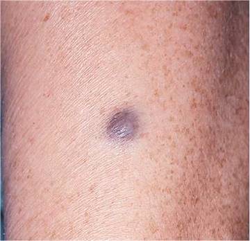
Figure 8-104 Blue nevus.

Figure 8-105 A and B, Pemphigus vulgaris. B, A vegetative form sometimes called pemphigus vegetans.
Atopic dermatitis, a form of eczema, is a common disease associated with other atopic diseases such as asthma and allergic rhinitis. It is characterized by itchy, dry, inflamed skin. The symptoms of atopic dermatitis often begin at a young age. Infants and young children may have eczematous patches on the face, scalp, and extensor surfaces of the extremities. These patches may erode and ooze. Scaling erythematous plaques often develop. As the child grows older, the atopic dermatitis begins to involve the flexural areas such as the neck, antecubital fossae, and popliteal fossae. The pruritic lesions result in excoriations; thickening and lichen- ification of the skin with increased skin markings are common. In the adult, oozing, weeping, and excoriated plaques may become generalized. Although the pathogenesis of atopic dermatitis is unknown, many patients have elevated levels of serum immunoglobulin E. Since the 1970s, the incidence of atopic dermatitis has increased from 4% to 12%; the reasons are unclear. Immune dysregulation appears to play an important role in atopic dermatitis. Up to 50% of children with atopic dermatitis may have evidence of a food sensitivity. Emotional stressors do not cause atopic dermatitis but do exacerbate the symptoms. Figure 8-110 shows classic lesions of atopic dermatitis in the axilla. Figure 8-111 shows atopic dermatitis in another patient. Notice the oozing lesions and excoriations.

Figure 8-106 Pemphigus vegetans of the lips.

Figure 8-107 Bullous pemphigoid.

Figure 8-108 Bullous pemphigoid.
Lyme disease is an infection caused by the spirochete Borrelia burgdorferi that is transmitted by the usually asymptomatic bite of certain ticks of the genus Ixodes. Lyme borreliosis occurs in northeastern, mid-Atlantic, north-central, and far western regions of the United States. Erythema migrans is the clinical, distinctive hallmark of Lyme disease. It is a dynamic lesion whose appearance can change dramatically over a period of days. The rash is recognized in 90% of patients with objective evidence of B. burgdorferi infection. The erythema begins as a red macule or papule at the site of the tick bite that occurred 7 to 10 days earlier. The rash expands as an annular erythematous plaque as the spirochetes spread centrifugally through the skin. Central clearing may or may not be present. Local symptoms of pruritus or tenderness are hardly noticeable. Systemic symptoms are common and include fatigue (54%), myalgia (44%), arthralgia (44%), headache (42%), fever and chills (39%), and stiff neck (35%). Neurologic symptoms are also common. The most common sign is regional lymphadenopathy (23%). Figures 8-112 and 8-113 show the classic erythema migrans of Lyme disease. Note the central lesion in Figure 8-113, which was the area of the tick bite.

Figure 8-109 Bullous pemphigoid; close-up of tense bullae.

Figure 8-110 Atopic dermatitis.

Figure 8-111 Atopic dermatitis.
As indicated previously in this chapter, there are many cutaneous manifestations of AIDS. Three of these common lesions are shown on the face of a patient with AIDS in Figure 8-114. The umbilicated, white papules on and around the lips, nose, and cheek are lesions of mol- luscum contagiosum. The verrucous papule on the upper lip is a wart. The violaceous lesions of KS are present on the lips and chin.
Fungal infections of the nails and hair are very common. The main fungi responsible for hair and nail diseases are dermatophytes of the genera Trichophyton, Microsporum, and Epidermophyton. Onychomycosis is an infection in which fungal organisms invade the nail bed and is the most common nail disorder. It accounts for nearly 50% of all nail problems. Its prevalence in the general population ranges from 2% to 14%. This infection causes progressive changes in the color, structure, and texture of the nail. It rarely resolves spontaneously, and, even with treatment, it may take months to years to clear. Figures 8-115 and 8-116 show examples of distal and lateral subungual onychomycosis, the most common form of nail infection, caused by Trichophyton rubrum. Onychomycosis results in inflammation of the nail bed, which promotes hyperkeratosis of the nail bed epithelium and thickening. This may cause a lifting up of the nail plate, as shown in Figure 8-117.

Figure 8-112 Erythema migrans of Lyme disease.

Figure 8-113 Erythema migrans of Lyme disease: site of the tick bite.
On September 11, 2001, the country and world changed. As a result of the events of that day and the events afterwards, everyone must be aware of the possibility of terrorist activity. Health-care workers must be alert to illness patterns caused by possible acts of terrorism. If you suspect an incident of biologic, chemical, or radiologic terrorism, contact your local state department of health immediately, as well as the Hotline for the Bioterrorism Preparedness and Response Program of the Centers for Disease Control and Prevention at 770-488-7100 or at www.cdc.gov.

Figure 8-114 Cutaneous manifestations of acquired immunodeficiency syndrome (AIDS).

Figure 8-115 Subungual onychomycosis.

Figure 8-116 Subungual onychomycosis.
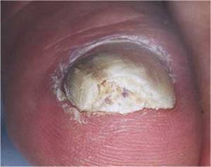
Figure 8-117 Onychomycosis and lifting of the nail plate.
Several of the biologic agents that have been associated with possible terrorist attacks have dermatologic signs; they are anthrax, smallpox, and plague.
Anthrax (from the Greek anthrax, meaning ''coal'') is a disease caused by the spore-forming Bacillus anthracis. Anthrax occurs in three forms: cutaneous (95%), inhalational (5%), and gastrointestinal and oropharyngeal (extremely rare). In the most common cutaneous form, typically, several days after exposure, a raised, inflamed, painless, pruritic papule appears within 1 day. The papule enlarges to approximately 1 to 3 cm, and vesicles may develop around the lesion (Fig. 8-118). Extensive edema of the area is common (Fig. 8-119). Adjacent lymph nodes become swollen and may become tender. The vesicles enlarge and rupture to form an ulcer, with the formation of a tough, adherent, black scab, or eschar, which forms in the center of the developing lesion (Fig. 8-120). In 1 to 2 weeks, the lesion dries with the separation of the eschar, leaving a permanent scar. The mortality rate ranges from 20% if the disease is untreated to very low if treated.

Figure 8-118 Anthrax (intention exposure secondary to bioterrorism), partially treated.

Figure 8-119 Anthrax. Note the early lesion and marked edema.
The inhalation, or pulmonary, form of anthrax usually occurs 1 to 60 days after exposure, although longer incubation periods can follow milder degrees of exposure. During the initial stage of the disease, nonspecific, influenza-like symptoms are common: myalgias, cough, low- grade fever, nonproductive cough, malaise, nausea, vomiting, chills, sweating, headache, and shortness of breath. Usually there follows a period of 1 to 3 days of improvement and then the rapid progression of high fever, severe respiratory distress, and cardiovascular collapse, often leading to shock and death within 24 to 36 hours. Massive mediastinal adenopathy occurs often. Person-to-person spread is not a significant risk because the main lesions are in the mediastinal lymph nodes and surrounding tissue. The mortality rate is 90% to 100% among untreated patients but 30% to 50% if patients are treated, depending on how quickly treatment with antibiotics is initiated.
B. anthracis is susceptible to common antibiotics, including ciprofloxacin, penicillin G, or tetracyclines.
Smallpox is a severe, highly contagious, febrile viral disease caused by a DNA virus of the orthopoxvirus genus. These viruses are among the largest and most complex of all viruses. There is an incubation period of 10 to 12 days after exposure. The illness begins with the symptoms of fever, fatigue, and myalgias. A distinctive erythematous, vesicular rash, centrifugal in distribution, then develops over the next 1 to 2 days, with the lesions appearing early on the face (Fig. 8-121) and arms, with relative sparing of the trunk. As the disease progresses, lesions appear on the trunk. The lesions are uniform in their stage of development and are often umbilicated (Fig. 8-122). After approximately 2 weeks, the lesions form crusts, which fall away after 3 to 4 weeks. The case-fatality rate is 30% to 40% or more. The last naturally occurring case of smallpox was in 1977 in Somalia. In 1980, the World Health Organization officially declared that smallpox was eradicated worldwide as a result of a global vaccination program. Although smallpox was long feared as one of the most devastating infectious diseases, its potential for devastation today is far greater than ever before. In a now highly susceptible, mobile population, medically ignorant of this infection once considered eradicated, smallpox could spread widely and rapidly throughout the world. The presence of even one case would constitute an international health emergency.

Figure 8-120 Anthrax. Note the black eschar.

Figure 8-121 Smallpox. Note the uniform stage of development of the vesicles.
Plague is caused by a gram-negative, non-spore-forming bacillus, Yersinia pestis. Natural human infection usually follows the bite of an infected flea and is less commonly caused by droplet spread from a person or cat with pneumonic plague. Plague occurs in three forms: bubonic, pneumonic, and septicemic. In the case of pneumonic plague, after an incubation period of 1 to 6 days, there is the fulminant onset of high fever, chills, extreme malaise, headache, and myalgias. Within 24 hours, cough with hemoptysis occurs. Dyspnea rapidly develops, and respiratory and cardiovascular collapse ensue. The mortality rate is 100% if the disease is untreated and could be as high as 50% even with treatment.
In the bubonic form of plague, sudden flulike symptoms develop after an incubation period of 2 to 8 days. At about the same time, the patient notices the presence of an oval, elevated, 1-to 10-cm, firm, nonfluctuant mass associated with intense pain in the enlarged regional lymph nodes, known as a bubo. Gangrene of the extremities is one of the common manifestations of plague, accounting for the name of the ''Black Death'' throughout the ages. The mortality rate is 50% to 60% if the disease is untreated. Treatment for all forms of plague is with streptomycin, doxycycline, or ciprofloxacin.
The typical distribution of lesions in six common skin disorders is shown in Figure 8-123.

Figure 8-122 Smallpox. Note the umbilicated lesions on the trunk.

Figure 8-123 Typical distributions of common skin conditions.
Table 8-1 describes the differential diagnosis of common maculopapular diseases. Table 8-2 lists the differentiation of some common eczematous disorders and gives some special hints about their causes derived from the history. Table 8-3 provides information for a differential diagnosis of the vesiculobullous diseases. Table 8-4 classifies some of the common benign tumors by color. Table 8-5 lists some of the common allergens associated with contact dermatitis. Table 8-6 lists the important differences between chickenpox and smallpox.
Table 8-1 Common Maculopapular Diseases*
|
Characteristic |
Psoriasis |
Pityriasis Rosea |
Tinea Versicolor |
Lichen Planus |
Seborrheic Dermatitis |
|
Color |
Dull red |
Pinkish-yellow |
Reddish-brown |
Violaceous |
Pinkish-yellow |
|
Scale |
Abundant |
Fine, adherent |
Fine |
Shiny, adherent |
Greasy |
|
Induration |
1 + |
0 |
0 |
1 + |
1 + |
|
Face lesions* |
Rare |
Rare |
Occasional |
Rare |
Common |
|
Oral lesions* |
0 |
0 |
0 |
2+ |
0 |
|
Nail lesions* |
4+ |
0 |
0 |
Rare |
0 |
*See Figures 8-14, 8-51, 8-52, 8-54, 8-55, 8-62, 8-72, 8-73, 8-98, 8-100, and 12-15.
*0, rarely seen; 1+, occasionally seen; 2+, frequently seen; 4+, nearly always associated.
Table 8-2 Common Eczematous Diseases*
|
Characteristic |
Contact Dermatitis |
Atopic Dermatitis |
Neurodermatitis |
Stasis Dermatitis |
|
History |
Acute, localized to specific area |
History in patient or family member of asthma, hay fever, or eczema |
Chronic, in same areas, associated with anxiety |
Varicosities, past history of thrombophlebitis or cellulitis |
|
Location |
Areas of exposure to allergen |
Eyelids, groin, flexural areas |
Head, lower legs, arms |
Lower legs |
*See Figures 8-49, 8-110, 8-111, and 15-2.
Table 8-3 Vesiculobullous Diseases*
|
Characteristic |
Pemphigus Vulgaris |
Dermatitis Herpetiformis |
Epidermolysis Bullosa* |
Bullous Pemphigoid |
|
Age of patient |
40-60 years |
Children and adults |
Infants and children |
60-70 years |
|
Initial site |
Oral mucosa |
Scalp, trunk |
Extremities |
Extremities |
|
Lesions |
Normal skin at margins |
Erythematous base |
Bullae produced by trauma |
Normal skin at margins |
|
Sites |
Mouth, abdomen, scalp, groin |
Knees, sacrum, back, elbows |
Hands, knees, elbows, mouth, toes |
Trunk, extremities |
|
Groupings* |
0 |
4+ |
1 + |
0 |
|
Weight loss |
Marked |
None |
None |
Minimal |
|
Duration |
1 or more years |
Several years |
Normal lifetime |
Months to years |
|
Pruritus* |
0 |
4+ |
0 |
± |
|
Oral pain* |
4+ |
0 |
± |
± |
|
Palms/soles involved |
No |
No |
Yes |
Yes |
|
Typical lesion |
Flaccid bullae |
Grouped vesicles |
Flaccid vesicles |
Tense bullae |
*See Figures 8-105, 8-106, 8-107, 8-108, and 8-109.
*Refers to a group of inherited diseases.
*0, rarely seen; 1 +, occasionally seen; 4+, nearly always associated; ±, sometimes present.
|
Table 8-4 Common Benign Tumors by Color |
|
|
Color |
Benign Tumor |
|
Skin color |
Warts (see Figs. 8-26 and 8-27) Cysts Keloids (see Fig. 8-103) Nevi (see Fig. 8-104) |
|
Pink or red |
Hemangiomas (see Figs. 24-8 and 24-9) Keloids (see Fig. 8-103) |
|
Brown |
Seborrheic keratoses (see Fig. 8-101) Nevi (see Fig. 8-104) Lentigines (see Fig. 8-101) Dermatofibromas (see Fig. 8-48) |
|
Tannish-yellow |
Xanthomata (see Figs. 14-12 to 14-15) Xanthelasma (see Fig. 14-18) Warts (see Fig. 8-26A) Keloids (see Fig. 8-103) |
|
Dark blue or black |
Seborrheic keratoses (see Fig. 8-101) Hemangiomas (see Figs. 24-8 and 24-9) Blue nevi (see Fig. 8-104) Dermatofibromas (see Fig. 8-48) |
Table 8-5 Allergens Associated with Contact Dermatitis
|
Location |
Possible Allergen |
|
Scalp |
Hair dyes Shampoos Tonics |
|
Eyelids |
Eye makeup Hair sprays |
|
Neck |
Aftershave lotions Perfumes Soaps Washing agents Nickel jewelry |
|
Trunk |
Clothing Washing agents |
|
Axillae |
Deodorants Soaps |
|
Genitalia |
Soaps Contraceptives Deodorants Washing agents |
|
Feet |
Shoes Sneakers Deodorants Socks Washing agents |
|
Hands |
Nickel jewelry Soaps Dyes Plants |
|
Table 8-6 Important Differences Between Chickenpox and Smallpox |
||
|
Characteristics |
Chickenpox |
Smallpox |
|
Onset of symptoms |
Begin with rash |
Begin 2-4 days before rash |
|
Rash |
Evenly distributed all over body |
More dense on face, head, arms, and legs (centrifugal distribution) |
|
Presence on palms and soles |
Almost never |
Common |
|
Development into other lesions |
Rapidly into vesicles (less than 24 hours) |
Slowly into pustules (7-14 days) |
|
Time frame of lesions |
New lesions form and scab at different times |
Lesions form and go through their stages at the same time |
|
Nature of lesions |
Pruritic but relatively soft |
Painful and hard |
|
Patient appears extremely ill and moribund |
Rarely |
Commonly |
Useful Vocabulary
Listed here are the specific roots that are important to understand the terminology related to diseases of the skin.
|
Root |
Pertaining to |
Example |
Definition |
|
kerat(o)- |
horny |
keratoma |
Horny growth |
|
derm(a)- |
skin |
dermatitis |
Inflammation of the skin |
|
trich(o)- |
hair |
trichoid |
Resembling hair |
|
seb(o)- |
sebum |
seborrhea |
Excess flow of sebum |
|
hidr(o)- |
sweat |
hidradenitis |
Inflammation of sweat glands |
|
onych(o)- |
nails |
onychomycosis |
Disease of the nails caused by fungus |
Writing Up the Physical Examination
Listed here are examples of the writeup for the examination of the skin.
• There are oval plaques with well-defined borders and silvery scales symmetrically present on the elbows, knees, scalp, and gluteal cleft. The plaques in the scalp are along the hairline. The scales are large. Examination of the nails reveals pitting of the nail plates. The hair is of normal texture.
• There is an annular lesion 3-4 cm in diameter on the right forearm. Scale is present on the narrow (1- to 2-mm), raised, erythematous border. The central area is slightly hypopigmented. The hair and nails are unremarkable.
• There is a linear bullous eruption along the lateral aspect of the left leg. The eruption consists of bright red edematous papules and bullae. There are no lesions on the palms or soles or in the mouth.
• A wide variety of lesions are seen on the face, shoulders, and back. The predominant lesions are pustules on an inflammatory base. Many pustules are present, and several have become confluent over the chin and forehead. Open and closed comedones are present on the face, especially along the nasolabial folds. Inflammatory papules are present on the lower cheeks and chin. Large abscesses and ulcerated cysts are present over the upper shoulder areas. Numerous scars are present over the face and upper back.
• A diffuse erythematous maculopapular rash is present on the trunk. Some excoriations are present over the shoulders and chest. The hair and nails are unremarkable.
• Examination of the skin reveals several types of lesions. The main lesions are small papules in the antecubital and popliteal fossae. The papules in some areas have become confluent, and plaques are present. On the dorsum of the feet, eczema is present with erythema, weeping, crusting, and scaling. Lichen- ification of the anogenital area, especially the scrotum, is present.
• The skin is slightly cool and dry. Scattered lentigines are present over the trunk. The hair is very fine and soft. There is loss of the lateral one third of the eyebrows. No nail abnormalities are present.
Bibliography
Adams RM: Occupational Skin Disease, 3rd ed. Philadelphia, WB Saunders, 1999.
Ali I, Dawber R: Hirsutism: Diagnosis and management. Hosp Med 65:293, 2004.
Aly R, Maibach HI: Atlas of Infections of the Skin. New York, Churchill Livingstone, 1999.
Callen JP, Paller AS, Greer KE, et al: Color Atlas of Dermatology, 2nd ed. Philadelphia, WB Saunders, 2000.
Cancer Facts & Figures. Atlanta, American Cancer Society, 2007.
DevCan: Probability of developing or dying of cancer software, version 5.1. Statistical Research and Applications Branch, National Cancer Institute, 2003. Available at http://srab.cancer.gov/devcan; accessed June 6, 2008.
Fawcett RS, Linford S, Stulberg DL: Nail abnormalities: Clues to systemic disease. Am Fam Physician 69:1417, 2004.
Freedberg IM, Eisen AZ, Wolff K, et al (eds): Fitzpatrick's Dermatology in General Medicine, 5th ed. New York, McGraw-Hill, 1999.
Friedman-Kien AE, Cockerell CJ: Color Atlas of AIDS, 2nd ed. Philadelphia, WB Saunders, 1996.
Habif TP: Clinical Dermatology: A Color Guide to Diagnosis and Therapy, 4th ed. Philadelphia, Mosby, 2004.
Hordinsky MK, Sawaya ME, Scher RK: Atlas of Hair and Nails. Philadelphia, Churchill Livingstone, 2000.
Jemel A, Tiwari R, Murray T, et al: Cancer Statistics, 2004. CA Cancer J Clin 54:8, 2004.
Jordan RE: Atlas of Bullous Disease. New York, Churchill Livingstone, 2000.
Lebwohl MG: Atlas of the Skin and Systemic Disease. New York, Churchill Livingstone, 1995.
Mandell GL, Fekety R (eds): Atlas of Infectious Diseases, vol 8: External Manifestations of Systemic Infections. Philadelphia, Churchill Livingstone, 1997.
Mandell GL, Mildvan D (eds): Atlas of Infectious Diseases, vol 1: AIDS, 2nd ed. Philadelphia, Churchill Livingstone, 1997.
Nadelman RB, Wormer GP: Lyme borreliosis. Lancet 352:557, 1998.
Safai B, Johnson KG, Myskowski PL, et al: The natural history of Kaposi's sarcoma in the acquired immunodeficiency syndrome. Ann Intern Med 103:744, 1985.
Stone DR, Gorbach SL: Atlas of Infectious Diseases. Philadelphia, WB Saunders, 2000.