
Gianguglielmo Zehender1 , Erika Ebranati1, Lisa Fiaschi1, Massimo Ciccozzi2 and Massimo Galli1
(1)
Dipartimento di Scienze Cliniche e Biomediche “Luigi Sacco”, Sezione di Malattie Infettive, Università degli Studi di Milano, Milan, Italy
(2)
Dipartimento Malattie Infettive, Parassitarie ed Immunomediate, Istituto Superiore di Sanità, Rome, Italy
Gianguglielmo Zehender
Email: gianguglielmo.zehender@unimi.it
Keywords
Hepatitis B virusHBV genotypesHBV mutantsEscape mutantsOrigin evolution of HBV genotypesPhylodynamicsPhylogeographyClinical impact of HBV variationHBV molecular epidemiology
Introduction
Hepatitis B virus (HBV) is a major health problem and chronically infects an estimated 240 million people worldwide [1]. There are approximately 620,000 HBV-related deaths and 4.5 million new HBV infections each year throughout the world. In highly endemic areas such as the central Asian republics, south-eastern Asia, sub-Saharan Africa and the Amazon basin, the HBV carrier rate is >8 %, whereas the prevalence of hepatitis B surface antigen (HBsAg) is <2 % in less endemic regions such as the United States, northern Europe, Australia and parts of South America. The Middle East, some eastern European countries and the Mediterranean basin are considered areas of intermediate endemicity, with carrier rates of between 2 and 8 % [1].
Despite the recent decrease in the rate of new cases, about 7000–8000 new diagnoses are made every year in Europe. The prevalence of HBV infection in Europe varies widely, but is generally higher in south-eastern than in north-western countries. The highest prevalence rates are in Turkey, Romania, Bulgaria, Greece, Albania and southern Italy [2].
The HBV is an enveloped DNA virus with a diameter of 42 nm (the so-called “Dane particle”), which belongs to the Hepadnaviridae family , and infects the hepatocytes of a wide range of animals belonging to several classes of birds (genus Avihepadnavirus) and mammals (genus Orthohepadnavirus). In particular, avian hepadnaviruses have been isolated from various species of ducks, geese, herons, storks, cranes and parrots [3], and orthohepadnaviruses have been discovered in rodents (squirrels, woodchucks), non-human primates (chimpanzees, gorillas, orangutans, gibbons and woolly monkeys) and, more recently, in bats from Myanmar [4].
The double-layered lipoprotein membrane derived from the infected cells making up the viral envelope contains the surface antigen (HBsAg ), a mixture of small (S), medium (PreS2 and S) and large (PreS1, PreS2 and S) proteins that can also be observed circulating freely in the blood of infected subjects as 22-nm spherical and tubular particles.
The role of the S protein in virus attachment has not been conclusively demonstrated; however, this protein contains the major site for the binding of neutralising antibody, designated the a determinant. Two other major determinants of the S protein have also been described; one has either “d” or “y” specificity and the other has “w” or “r”. All combinations of these determinants have been found, resulting in four major subtypes: adw, adr, ayw and ayr and nine minor subtypes, determined by mutually exclusive amino acids substitution in the S region of HBV DNA. Antibodies to the a determinant confer protection to all of these serotypes, whereas antibodies to the subtype determinants do not [5].
The viral nucleocapsid consists of the core protein (HBcAg ) and encloses a single molecule of the viral genome. It consists of a small and circular partially double-stranded DNA of about 3.2 kb whose minus strand, which encompasses four partially overlapping genes (PreS/S, PreC/C, P and X) encoding for at least seven proteins, is incomplete. In particular, the three surface glycoproteins (the small S protein, the middle PreS2 and S protein and the largest Pre S1/PreS2 and S protein), two core antigens (HBcAg and HBeAg), the polymerase and the X protein, a small regulatory protein that is essential for in vivo viral replication [6]. It also plays a central role in hepatocarcinogenesis [7].
After attaching to a hepatocyte as a result of the binding of Pre-S1 with a still unknown specific cell receptor, the viral nucleic acid is transferred to the cell nucleus, where it is completed by cell polymerases and forms covalently closed circular DNA (cccDNA) [8]. This mainly non-integrated cccDNA acts as a template for the production of the four viral transcripts that are necessary for protein production, including an over-length “pre-genomic RNA” (pg-RNA) that gives rise to the core proteins and the viral genome.
After encapsidation, a molecule of pg-RNA is transformed by the viral reverse transcriptase into partially double-stranded circular genomic DNA, and the pg-RNA is degraded by the RNase-H activity of the P protein. Some of the newly produced capsids are not transferred to the cell surface, but return to the nucleus and contribute to the cccDNA reserve [8]. The production of the three surface proteins ensures virion secretion.
In spite of the constrained nature of its genetic evolution, which is due to the partial overlapping of the viral genes [9], the HBV genome is characterised by considerable variability because of the use of an RNA intermediate and reverse transcriptase during replication.
HBV Mutants
Mutations in the HBV genome can occur because of spontaneous errors of viral polymerase, and the action of pressure by the host immune system or by exogenous factors, including passive and/or active immunisation and drug treatment. HBV has a higher frequency of mutations than other DNA viruses because the virus replicates via an RNA intermediate, using a reverse transcriptase that lacks a proof-reading function such as the reverse transcriptase and RNA polymerases of other highly variance viruses. Mutations have been identified in all four HBV genes, but have been most fully characterised in the preC/C gene, the polymerase gene and the preS/S gene [10–12].
Basal Core Promoter , Pre-C/C Gene Mutants
Two major groups of mutations that result in reduced or blocked HBeAg expression have been identified. The most common mutation in the ORF pre-C/C region is a guanine to adenine substitution at nt position 1896 (G1896A) that results in a translational stop codon (TGG to TAG; TAG = stop codon). This codon stops the expression of the e protein, which is processed to produce HBeAg [13]. However, HBV DNA synthesis persists and may cause liver damage, with progression to cirrhosis and cancer. Loss of HBeAg expression can also occur with mutations in the basal core promoter (BCP) region that regulates the expression of both HBeAg and core protein [14]. These mutations have been associated with fulminant hepatitis and severe chronic liver disease [15, 16]. However, fulminant hepatitis can occur in the absence of such mutations [17, 18]. Additional studies are needed to determine the pathogenic basis and clinical sequelae arising from the selection of these mutants [12].
X Gene Mutations
As the X ORF overlaps the BCP completely, promoter mutations can affect the amino acids sequence of the X protein. The most common BCP double mutations occurring at nt 1762 (A1762T) and at nt 1764 (G1764A) can cause changes in the X protein that may affect its ability to transactivate the BCP. In addition, insertions or deletions in the BCP often shift the X gene frame, resulting in truncated forms of the X protein. These shortened X proteins lack the domain in the C terminus that is required for the transactivation activity of HBx antigen [10–12].
Polymerase Gene Mutants
Mutations of the polymerase gene have been associated with resistance to treatment with nucleoside/nucleotides analogues and with viral persistence [10–12, 19, 20]. The most common of these mutations occur at codon 528 (the template binding site of the polymerase) and at codon 552 of the tyrosine, methionine, aspartate, aspartate (YMDD) motif (the catalytic site of the polymerase). Polymerase mutations have been demonstrated to emerge in up 80 % of patients after treatment with nucleoside/nucleotide analogues. These mutations significantly decrease the efficacy of treatment [21]. As the genome of HBV is organised into overlapping reading frames, the selection of polymerase mutants, favouring resistance, can result in the emergence of changes in the overlapping S-gene during long-term antiviral therapy, potentially altering its immunoreactivity [22, 23].
PreS/S Gene Mutants
Isolates with pre-S deletions are often found. There is evidence that a set of mutations (deletion in the pre-S region and in pre-core and BCP mutations) are significantly associated with progressive liver disease and hepatocellular carcinoma (HCC) [24].
Mutations in the S gene can lead to conformational changes in the a determinant, which is located between amino acids 124 and 147 of HBsAg and has a double-loop structure projecting from the surface of the virus; the second loop (amino acids 139–147) is the major target for neutralising anti-HBs.
The prototype of such mutants, the so-called G145R, which shows a point mutation from guanosine to adenosine at nucleotide position 587, resulting in an amino acid substitution from glycine (G) to arginine (R) at position 145 in the a determinant of the surface antigen, was first observed in Italy some 25 years ago [25]. This mutant has been shown to be infectious in experimentally infected chimpanzees [26]. Besides the G145R, other S-gene mutants across the entire a determinant region have been found worldwide. Concern has been expressed that these mutated viruses may allow replication of HBV in the presence of vaccine-induced anti-HBs or anti-HBs contained in hepatitis B immune globulin (HBIG; immunisation escape mutants). In addition, these mutants may not be detected by some commercially available HBsAg assays based on antibodies to the wild-type virus (diagnostic escape mutants ) [27, 28].
Hepatitis B infection with S mutant viruses has been reported to occur in the presence of protective levels of anti-HBs in infants born to HBV-infected mothers who received prophylaxis with HBIG and/or hepatitis B vaccine [25, 29–32] in children who responded to vaccination [33], and in liver transplant recipients who received HBIG for the prophylaxis of relapse of the HBV infection [34]. However, in population-based studies of infants born to HBsAg-positive mothers, S-gene mutant viruses have not been found to be associated with a failure to prevent perinatal HBV transmission [35]. In addition, pre-exposure vaccination of chimpanzees with currently licensed vaccines (not containing pre-S epitopes) conferred protection after intravenous challenge with the G145R HBV [36, 37]. At present, no evidence exists that S-gene mutants have spread in immunised populations or that these mutants pose a threat to hepatitis B immunisation programmes [38]. Further studies and enhanced surveillance to detect the emergence of these mutants and those caused by the onset of resistance to the viral therapy (see above) are a high priority in monitoring the effectiveness of current immunisation strategies.
Hepatitis B surface antigen escape mutants, which cannot be detected by currently available HBsAg assays, are possible carriers of occult HBV infection (OBI) [39].
HBV Genotypes
On the basis of the sequence divergence established by analysing the entire viral genome, HBV has been classified into nine genotypes (A–I) and various subgenotypes (indicated by numbers), with a mean nucleotide difference of ≥8 % between genotypes and ≥4 % between subgenotypes, partially corresponding to the previously described serologically defined subtypes [40].
Hepatitis B virus genotypes have a characteristic ethno-geographic distribution . Some are ubiquitous, such as genotype A, which is present in north-western Europe, North America and Central Africa [40], and genotype D, which has been found throughout the world, although its highest prevalence is in the Mediterranean area, the Middle East and southern Asia, particularly India. Genotypes B and C are only present in Asia; genotype E is found in sub-Saharan Africa [40] and genotype F in South and Central America [40]. Genotype G has been found in France and the USA [41], whereas genotype H seems to be confined to the northern part of Latin America (Table 2.1) [42].
Table 2.1
Subgenotypes, subtypes and geographical origin of HBV
|
Subgenotype |
Subtype |
Geographical origin |
||
|
A |
A1 (Aa, A′) |
adw2, ayw1 |
Africa, Asia, South America |
|
|
A2 (Ae, A-A′) |
adw2, ayw1 |
Northern Europe, North America, South Africa |
||
|
A3 (Ac) |
Cameroon, Gabon, Rwanda |
|||
|
A4 |
Mali, Gambia |
|||
|
A5 |
Nigeria, Rwanda, Cameroon, Haiti (African population) |
|||
|
A6 |
Congo, Rwanda |
|||
|
A7 |
ayw1, adw2, ay |
Cameroon, Rwanda |
||
|
B |
B1 (Bj) |
adw2 |
Japan |
|
|
B2 (Ba) |
adw2, adw3 |
Asia without Japan |
||
|
B3 |
adw2, ayw1 |
Indonesia, Philippines |
||
|
B4 |
ayw1, adw2 |
Vietnam, Cambodia |
||
|
B5 |
Philippines |
|||
|
B6 |
Alaska, Northern Canada, Greenland |
|||
|
B7-B9 |
Indonesia |
|||
|
C |
C1 (Cs) |
adrq+, ayr, adw2, ayw1 |
South East Asia (Vietnam, Myanmar, Thailand, Southern China) |
|
|
C2 (Ce) |
adrq+, ayr |
Far East (Korea, Japan, Northern China) |
||
|
C3 |
adrq–, adrq+ |
Pacific Islands (Micronesia, Melanesia, Polynesia) |
||
|
C4 |
Australia |
|||
|
C5 |
Philippines, Vietnam |
|||
|
C6 |
Indonesia, Philippines |
|||
|
C7 |
Philippines |
|||
|
C8-C16 |
Indonesia |
|||
|
D |
D1 |
ayw2, adw1, ayw1 |
Europe, Middle East, Asia, Tunisia, Egypt |
|
|
D2 |
ayw3, ayw1 |
Europe, Morocco, India |
||
|
D3 |
ayw3, ayw2, ayw4 |
South Africa, Asia, Europe, USA, Northern Canada |
||
|
D4 |
ayw2, ayw3 |
Australia, Japan, Papua New Guinea |
||
|
D5 |
East India, Japan |
|||
|
D6 |
Indonesia |
|||
|
D7 |
Tunisia |
|||
|
D8 |
Niger |
|||
|
D9 |
Eastern India |
|||
|
E |
ayw4, ayw2 |
Sub-Saharan Africa, UK, France, Saudi Arabia |
||
|
F |
F1a |
adw4, ayw4 |
South and Central America |
|
|
F1b |
adw4 |
Argentina, Japan, Venezuela, USA |
||
|
F2 |
adw4 |
South America (Brazil, Venezuela, Nicaragua) |
||
|
F3 |
adw4 |
Venezuela, Panama, Columbia, Bolivia |
||
|
F4 |
adw4 |
Bolivia, France, Argentina |
||
|
G |
adw2 |
USA, Germany, Japan, France, Mexico |
||
|
H |
adw4 |
USA, Japan, Nicaragua |
||
|
I |
I1 |
adw2 |
Laos, Vietnam, Northwest China |
|
|
I2 |
ayw2 |
Laos, Vietnam |
||
|
J |
ayw |
Japan |
||
A ninth genotype (I) has recently been proposed after being found in north-western China [43], Eastern India [44], Laos [45] and Vietnam [45, 46]. Even more recently, a tentative new genotype “J” has been isolated in a single Japanese patient with HCC [47].
The two genotypes responsible for the majority of infections in Europe are genotype A (mainly subgenotype A2) in the north-western part of Europe and genotype D (mainly subgenotypes D1, D2 and D3) in the south-eastern Europe and the Mediterranean area [3].
The Origin of HBV
A number of conflicting hypotheses have been made concerning the origin of HBV. It has been proposed that it originated in the New World and spread to the rest of the world as a result of European colonisation over the last 400 years [48]. This conflicts with the observation of its widespread distribution among wild Old World apes (chimpanzees, orang-utans and gibbons). A second hypothesis proposes a co-divergence of HBV and its (human and non-human) primate hosts over a period of about 10–35 million years [49], but this implies a very slow evolutionary rate that is incompatible with current molecular clock estimates, indicating a faster rate of evolution [50–52]. Moreover, the viruses isolated from non-human primates show the same divergence as those observed among human genotypes, and their phylogenetic patterns show that the relationship between non-human viruses and some human genotypes is closer than that of other human genotypes, suggesting different viral leaps from primates to humans and vice versa and excluding the idea of virus/primate co-divergence [53]. A third hypothesis is that HBV was present in anatomically modern humans and spread as a result of their migrations over the last 100,000 years or so [54].
The main difficulty in reconstructing the HBV phylodynamics is the lack of a consensus in the estimation of the rate of evolution of the virus that also affects the tMRCA estimates and the timescales of HBV evolution [52].
Support for the hypothesis of the long co-evolution of HBV genotypes in humans comes from the two American genotypes F and H, which are inter-related and diverge significantly from the other HBV genotypes. As described above, we know that HBV-F subgenotypes are distributed in different areas and aboriginal tribes [55] in which the prevalence of infection is very high (up to 30 %) [56, 57]. This suggests that they have been generated by the evolution of the virus in small and isolated populations. On the basis of genetic studies, all aboriginal Americans came from a population originating in the region between the Altai and Amur that reached Beringia between 30 and 22 ky and North America 16.6 ky [58]. This founding population probably consisted of fewer than 5000 people [59] divided into bands of probably less than 100, who reached North America in successive waves over a period of 1500 years or more [60]. Moreover, the very high prevalence of HBV subgenotype C2 found in the small Jarawa tribe of the Andaman Islands that remained in complete isolation for thousands of years cannot plausibly be attributed to recent contact with the virus, despite the quite limited divergence from the sequences of the same subgenotype presently circulating in Thailand [61]. However, this apparent discrepancy may find an explanation taking as a reference the sequence belonging to the same genotype recently extracted from the liver cells of a Korean mummy dated 400–500 years ago [62], suggesting that the evolutionary rate of this genotype may be slower than previously expected.
In some of these small, numerically stable and isolated groups, HBV could easily have become hyper-endemic and prevalently transmitted vertically. The resulting immunological tolerance would have reduced the selective pressure of host immunity on the virus, thus justifying a slow evolutionary rate. Very different evolutionary rates have recently been demonstrated in subjects with and without serum HBeAg [63], and HBeAg positivity, associated with slower rates, is a prerequisite for efficient vertical transmission [64].
On the contrary, the penetration of HBV into large, fast-changing, highly mobile and susceptible populations depends on other, mainly horizontal, transmission routes, which may also be responsible for higher evolutionary rates. The rate of vertical transmission is relatively low in populations in which the prevalent HBV genotypes are those associated with high rates of HBeAg negativity (such as genotype D), and horizontal transmissions such as the parenteral (iatrogenic practices and intravenous drug use) and intra-familiar routes play a more important role [64]. The main circulation among immunocompetent HBeAg negative adults justifies a faster evolution of the virus because of stronger selective pressure. Moreover, HBV in these populations is generally characterised by high basic reproduction numbers (R0) of 1–2 estimated in various ways [52, 65, 66], which indicates a very rapid spread of the virus in susceptible communities and once again conflicts with the hypothesis of a slow evolutionary rate.
The Origin and Evolution of Genotype D
The HBV genotype D is one of the two most prevalent genotypes in Europe, in particular in the north-eastern countries and in the Mediterranean basin, including northern Africa, and the Middle East. It is also highly prevalent on the Indian sub-continent and a group of islands in the Indian Ocean with high endemic levels of HBV [67], and has additionally been identified in Oceania [40]. Nine HBV-D subgenotypes (D1–D9) have so far been described (Fig. 2.1), showing a different geographic distribution. Subgenotype D1 is widespread in Greece, Turkey and north Africa [68, 69]; D2 in north-eastern Europe (Russia, Belarus, Estonia) and Albania [70, 71]; and D3 in Italy and Serbia [72, 73]. D4 is the dominant subgenotype in Oceania [40]; D5 in primitive tribes living in India, where a number of different D subgenotypes are also found [74]; D6 in Papua and Indonesia [75]; D7 in Tunisia and Morocco [76, 77]. Finally, the recently described D8 and D9 subgenotypes have been identified in Nigeria and India, and have been recognised as recombinant forms of genotype D with E [78] or C [79].
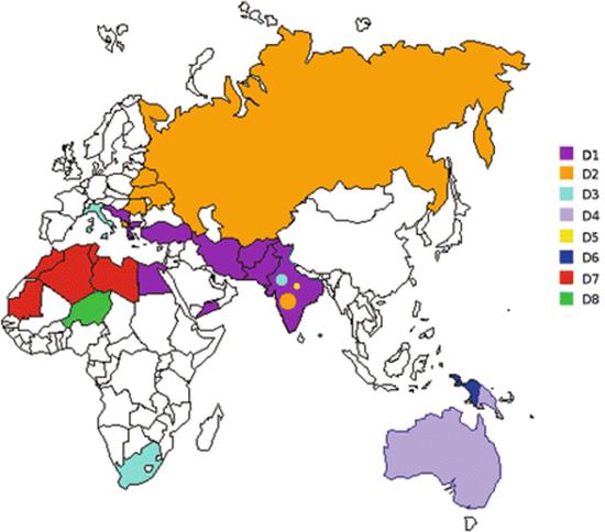
Fig. 2.1
Distribution of hepatitis B virus (HBV) subgenotypes D in Eurasia and the Mediterranean basin. Countries are coloured on the basis of their prevalent subgenotypes (see legend)
A number of preliminary studies agree about a relatively recent origin of genotype D in the early twentieth century [52, 80] in Europe, with two phases of expansion: the first in the 1940s and 1950s and the second occurring from the 1960s until the 1980s, when the growth of the genotype reached a plateau. It was hypothesised that the initial event underlying viral penetration in Europe were the two World Wars at that time when the use of unsafe medical injections was common and there was an increase in the use of blood and blood derivatives for transfusion purposes [81] before HBsAg screening became available in the 1970s [82]. Later, the further expansion was attributable to percutaneous transmission among intravenous drug users (IDUs) and other people exposed to infected blood. In particular, subgenotype HBV-D3 was shown to be associated with percutaneous transmission and drug addiction [72, 83]. The virus isolated from IVDUs is characterised by a number of S and P gene mutations [72], such as the main mutation in residue 125 (S125T) of S protein, which has been described by various authors all over the developed world [40, 52, 80, 84]. A plateau of the epidemic was reached in the 1980s, in relation to the decrease in acute HBV infections in developed countries [52, 85, 86].
A recent and comprehensive reconstruction of the epidemiological history of HBV genotype D obtained using a phylogeographical approach [87] indicates that it originated in the second half of the nineteenth century in India and that subgenotype D5 (an indigenous Indian subgenotype) was probably the first to diverge. The common ancestors left India and reached central Asia in the first decade of the twentieth century, when subgenotypes D1–D3 diverged. Subsequently (between the 1930s and 1940s), they spread to Europe and the Mediterranean area by means of at least two routes: a south-western route (mainly because of the diffusion of subgenotype D1) crossing the Middle East and reaching north Africa and the south-eastern Mediterranean; and a second north-western route (closely associated with D2) that crossed the former Soviet Union and reached eastern Europe and the Mediterranean through Albania [66].
This reconstruction makes it possible to hypothesise that the First and Second World Wars played a crucial role in the global spread of HBV-D from India to the rest of the world, but the further spread of the infection (particularly in south-eastern Europe, the Middle East and northern Africa) was probably sustained by the unsafe use of injections in medical practice. Events such as outbreaks of jaundice following the intravenous injection of arsphenamine (for anti-syphilis treatment) from the early 1920s to the late 1940s, led to the spread of some HBV-D subgenotypes to Europe [88].
In the populations in which HBV-D predominates (which are characterised by a high rate of mutations causing HBeAg negativity), the majority of infections are acquired horizontally, mainly as a result of household contacts or because of the use of unsterilised needles and syringes [89, 90].
The Origin and Evolution of Genotype A
Genotype A is an ubiquitous genotype, largely spread over four continents: Africa, Europe, Asia and America. It has been classified into seven distinct evolutionary groups. Subgenotype A1 is highly prevalent in southern and east Africa (South Africa, Uganda, Malawi, Tanzania, Congo, Somalia) and south Asia (India, the Philippines, Bangladesh, Nepal) [40, 74, 91–93]. Subgenotype A2 is the most widespread in Europe and North America [40, 91] and has also been isolated in South Africa [94]. It has been suggested that genotype A might have originated in Africa and hypothesised the importation of HBV-A2 from Africa to Europe by Portuguese sailors in the sixteenth and seventeenth centuries, and the arrival of A1 in Asia as a consequence of trade and travel between eastern Africa and southern Asia [95]. The more recently described subgenotypes are A3 in Pygmies and Bantus living in Cameroon and Gabon [96, 97]; a “tentative A4” from Mali; and a subgenotype A5 isolated in patients from Nigeria (Fig. 2.2) [98]. Interestingly, HBV-A5 has also been found in Haiti, which suggests that it might have been the dominant subgenotype in an area near the current Nigeria (formerly the Bight of Benin) before the time of the slave trade (between the eighteenth and nineteenth centuries). An HBV-A6 has only been reported in African–Belgian patients [84] and a new “tentative subgenotype A7” has been isolated in Cameroon [99]. It has recently been proposed to classify A3, “tentative A4”, A5 and “tentative A7” within a single subgenotype called “quasi subgenotype A3”, because they share a common ancestor, but none of them meets the criterion necessary for the definition of “subgenotype” (a genetic divergence of 4–8 %) [84] and a new classification, consisting of three subgenotypes (A1, A2 and A4-formerly subgenotype A6) and one West-African quasi-subgenotype (A3) has been proposed [100].
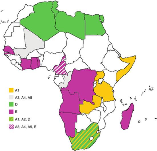
Fig. 2.2
Distribution of HBV genotypes in Africa. Countries are coloured on the basis of their prevalent geno-/subgenotypes (see legend)
Subgenotype A2 is the most prevalent genotype in the developed countries, in particular in the USA and north-west Europe, and in the last few years its prevalence has also been growing in Japan [101]. Several studies have demonstrated a relatively recent penetration of HBV-A in Europe, between the 1960s and 1980s and an association of this subgenotype with people acquiring the infection as a result of sexual transmission, particularly men-having-sex-with-men (MSM) [52, 102].
A single clonal strain has been isolated among high-risk subjects all over the world [2, 103], suggesting a relatively recent distinct worldwide HBV-A2 epidemic among MSM, now also spreading among heterosexuals [103].
It seems that the epidemiological dichotomy of Europe (which makes genotype D the most prevalent genotype in eastern and southern Europe, where HBV is highly endemic, and genotype A the main strain in central and northern Europe, where HBV is less widespread) could be due to the differences in their main routes of transmission, as has been observed in Italy [52]: predominantly parenteral transmission in highly endemic areas (unsafe injections, intra-family transmission), and hetero- and homosexual transmission in less endemic areas [2, 90, 102].
In contrast, HBV-A1 probably originated in Africa and the slave trade and colonisation played a major role in its global dispersion: in particular, the Arabian East African slave trade from Africa to India (until the late nineteenth century), the Belgian colonisation of the Congo (in the first half of the twentieth century), and the European slave trade (until the nineteenth century) [104].
The Origin and Evolution of Genotype E
Genotype E is the most prevalent strain of HBV in central and western Africa (see Fig. 2.2) [105]. It has a very low degree of genetic diversity: the isolates obtained so far form a single monophyletic group [106]. The absence of any significant spread among Afro-Americans, despite the forced immigration of western African slaves [106], indicates that it was probably rare in west Africa at the time of the slave trade and before the nineteenth century. The high prevalence and low degree of genetic divergence of HBV-E suggest its recent explosive spread in west Africa after the end of the slave trade approximately 200 years ago [107]. Recent estimates suggest a penetration of HBV-E in Central West Africa around 60 years ago with an exponential growth of the epidemic between 30 and 40 years ago. These observations support the view that the explosive spread of HBV-E in Africa must have been due to a new and highly efficient route of transmission, probably the unsafe use of needles during numerous mass-vaccination campaigns (against yaws, sleeping sickness, smallpox and measles), which were particularly frequent in west/central Africa between the 1920s and 1960s [107, 108]. In this way, HBV-E brought to the substitution of other, endemic genotypes such as HBV-A5.
The Origin and Evolution of Genotypes F and H
Genotypes F and H are indigenous to America and the most prevalent HBV genotypes in central/south America with the exception of the Afro-Brazilian community, where the most prevalent subgenotype is A1 [109]. Genotype F is the predominant strain among the Amerindians of the Amazon basin [110]. It is classified into four subgenotypes (F1–F4), and further sub-divided into different clades. As shown in Fig. 2.3, F1 is highly prevalent in central America (clade F1a), Alaska and south-east America (clade F1b) [111, 112]; F2 is highly prevalent in Venezuela (clades F2a and b) and is also present in Brazil (clade F2a alone) [111]; F3 is present in central (Panama) and northern Latin America (Colombia and Venezuela); and F4 is present in Bolivia and Argentina (where it co-circulates with F1b) [113]. Genotype H is predominant in Mexico, where it represents more than 50 % of infections, even in some native Mexican populations [114], thus suggesting an ancient origin (pre-Hispanic) of HBV-H in the Aztec population. From a phylogenetic point of view, genotypes F and H share a common ancestor and are highly divergent from Old World HBV genotypes [42], being closer to an isolate obtained from a New World monkey (the woolly monkey). These data suggest that genotypes F and H might have split off a long time ago, and possibly represent the result of a cross-species transfer [42].

Fig. 2.3
Distribution of HBV F subgenotypes in Latin America. Countries are coloured on the basis of their prevalent geno-/subgenotypes (see legend)
The estimation of the phylogeography of HBV-F on a calendar time scale suggested the pre-Columbian origin of HBV-F and their subgenotypes, some of which would have disappeared during the conquests of the sixteenth and seventeenth century as a result of the extreme decline in population numbers. The epidemic would have then expanded because of the rapid increase in the Latin American population since the eighteenth century [113].
A study of the molecular epidemiology and evolutionary dynamics of HBV-F in Colombia demonstrated that HBV-F3 was probably the oldest F subgenotype originating in Venezuela and is most related to genotype H [111].
Recent studies [115] estimated an origin of the Amerindian genotypes (F and H), which probably followed the initial New World settlers coming from Asia 13,000 years ago.
Origin and Evolution of HBV Genotypes B and C
Hepatitis B virus genotypes B and C are the most prevalent genotypes in Asia. Genotype B was identified in 2002 and immediately classified into two subgenotypes: Bj, largely spread in Japan and Ba, present in Asia, but not in Japan. Subgenotype Ba was demonstrated to be a product of recombination between a genotype B and a genotype C PreC/C gene. In 2004, it was proposed to rename them B1 (corresponding to the former Bj) and B2 (former Ba) [40]. Genotype B is characterised by a large spread in Asian and Arctic populations and is highly heterogeneous: nine different subgenotypes have been identified so far. Some of them are recombinant: subgenotype B2, subgenotypes B3 (isolated in Indonesia), B4 (identified in Vietnam) and B5 (identified in the Philippines). Another non-recombinant subgenotype was B6, identified in an indigenous Arctic population and in southwestern China in the Yunnan region. Subgenotypes B7, B8 and B9 were all identified on different islands of Indonesia suggesting a correlation between the B subgenotype and the ethnicity of the infected patients [116].
Genotype C shows the highest level of genetic heterogeneity, being classified into at least 16 different subgenotypes, which display different ethnogeographic distribution. Subgenotypes C1 and C2 (initially named Cs and Ce) are the most widely distributed subgenotypes in Southeast Asia (Vietnam, Myanmar, Southern China, Thailand) and the Far East (Korea, Japan, Northern China) respectively. Subgenotype C4 is found in the aboriginal population of Australia, while all the other subgenotypes are present in the Philippines and Indonesia [116].
On the basis of a recent study, HBV genotype C could be the oldest human genotype, originating about 30.0 ky ago [54]. Accordingly, a recent study detected HBV nucleic acids in a Korean mummy of the sixteenth century, which was clustered with subgenotype C2 [62]. The concordance between the HBV subgenotype and geographic localisation and the great similarity of this strain to the C2 isolates presently circulating [62] suggest a relatively low rate of evolution of HBV, at least of genotype C.
Other Geno-/Subgenotypes of Interest
Genotype G was found in several European countries, in the USA, in Japan and in Mexico [117]. All known isolates have both PreC/C and BCP mutations, preventing the ability of this genotype to give (alone) chronic infections. For this reason, in chronically infected patients, genotype G is always in coinfection with another genotype that can supply the HBeAg [118]. An association between HBV genotype G and sexual transmission, in particular among MSM, has been observed [119], thus justifying the frequent coinfection with subgenotype A2.
Clinical Implications of HBV Variability
HBeAg Expression and Viral Genotypes
The substitution of the G in position 1896 of the PreC/C gene with an A genotype abrogates the expression of HBeAg. Site 1896 is inserted into a stem-loop fundamental as an encapsidation signal, and is paired with the nucleotide in position 1858, which is a C in genotype A, but is a T in several other genotypes. The presence of a T in position 1858 makes the mutation (G to A) in position 1896 more stable. For this reason, several genotypes such as D, E, G and some B and C subgenotypes are more frequently subjected to preC/C mutation. In contrast, BCP mutations are more prominent in HBV genotypes A, F and the other HBV B and C subgenotypes. These data suggest that subjects infected by some HBV genotypes (D, E, G) are more likely to be affected by HBeAg-negative chronic hepatitis [120].
In general, Asian subjects have a longer persistence of HBeAg positivity in their blood. The anti-HBe seroconversion occurs earlier and more frequently in subjects infected with genotype B than in those with genotype C [121]. A possible hypothesis is that genotype B HBV might develop the G1896A mutation, which abolishes the e antigen expression, while genotype C develops BCP mutations that reduce but do not abolish HBeAg expression [120].
In Western Europe, where the predominant genotypes are D and A2, the rate of sustained remission after HBeAg seroconversion is higher in genotype A than in genotype D [120]. Even in this case, the reason may be the higher frequency of PreC/C mutations inducing the reactivation of the viral replication in genotype D than in genotype A, as also suggested by the higher prevalence of HBeAg positivity in the latter [120, 122].
Progression of the Infection
Cirrhosis and HCC are the most significant complications of chronic hepatitis B (CHB) . Given that the viral genotype may influence the natural history of the infection and that viral genotypes are differently distributed throughout the world, the pattern of the development of complications also varies on a geographical basis.
Several studies have suggested that the risk of cirrhosis development in Europe (where genotypes A and D are prevalent) might be higher than in East Asia, where the most prevalent genotypes are B and C, C2 (mean incidence rate of 6.75 vs 2.2 per 100 person-years respectively) [120]. This difference is in part due to the higher incidence of genotype D infection, associated with HBeAg-negative CHB, in Europe than in East Asia.
Accumulating evidence indicated that higher plasma HBV DNA levels, infection with HBV genotype C, in addition to mutations at 1653T, 1753V and A1762T/G1764A are independently associated with the risk of HCC in Asian men. This explains the correlation between HCC development and genotype C rather than genotype B (OR = 1.68 > 2.35 < 3.30) [123]. HBV genotype A1 in Africa is associated with a 4.5-fold increase in the relative risk of developing HCC compared with other genotypes [124].
HBV Genotype and CH Therapy
Several studies have indicated that the loss of HBeAg and anti-HBe seroconversion during interferon treatment are observed more frequently in patients with genotype A than in those with genotype D and, to a lesser degree, in patients with genotype B versus genotype C. In contrast, response rates to nucleoside/tide analogues are similar in the different genotypes. However, a different pattern of resistance-associated mutation may prevail [120] given that mutations at the polymerase coding gene may influence the appearance of mutations in the overlapping surface antigen. For example, the lamivudine and adefovir resistance mutation at position rt181 (from A to T) results in a stop codon in the overlapping surface antigen gene (in position 172). This variant has been associated with enhancing progression to HCC and is more prevalent in Asian patients with genotypes B and C than in European and North American patients with genotypes A and D [125].
Modes of Transmission
As mentioned before, different genotypes are dispersed within populations by different main routes of transmission. In particular, genotypes B and C, which are predominant in populations with a high level of endemicity, vertical/perinatal transmission is the prevalent mode of propagation. On the contrary, in the developed countries, where genotypes A2 and D prevail and the endemicity level is lower/intermediate, the main transmission routes are horizontal and highly efficient, and are sexual (associated with A2) and percutaneous (associated with genotype D). These differences may explain different characteristics in the natural history of infection due to HBV genotypes. For example, the longer persistence of the period of immunotolerance in HBV genotypes B and C may be useful in increasing the probability of transmission from the mother to the children. A HBeAg-positive mother has a probability of virtually 100 % of transmitting the infection to an infant, who has a 90 % risk of developing a chronic infection characterised by a long immune tolerant phase lasting decades, in which HBeAg positivity persists. Similarly, where intrafamilial or percutaneous (through unsafe injections or transfusions) transmission prevails (such as in the Mediterranean area and Africa), genotypes able to give long-lasting HBeAg-negative chronic infections characterised by intermittent flares and remissions, such as genotype D in the Mediterranean area or E in Western and Central Africa, could be selected [64].
The selective action of the prevalent mode of transmission at a population level on the different behavioural characteristics of the genotypes is well described by the differences between two closely related subgenotypes: HBV-A1 and -A2. In fact, subgenotype A1 is highly prevalent: the main mode of transmission at the population level is in the early childhood, whereas HBV-A2 is mainly transmitted through high-risk sexual contact [126]. In Africa, where the prevalence of infection is high, the transmission can easily occur through massive environmental contamination; in this condition, a self-limiting, HBeAg-positive infection is sufficient to infect cohabiting susceptible children. On the contrary, the main sexual transmission of genotype A2 in developed countries may explain its relative initial benignity and the higher rate of chronic infections mainly due to BCP mutations, reducing but not abrogating the HBeAg expression. Similar considerations can be made for genotype F, the most prevalent genotype in South America. This genotype is transmitted in early childhood on a community basis (rather than based on an individual family). Under these conditions, efficient transmission requires a highly dense and close population with a high level of HBV endemicity, to have sufficient numbers of HBeAg-positive transmitters. These conditions are met in Amerindian populations [126].
This observation may account for the common characteristics of genotypes F and A1 natural history as suggested by the high frequency of HCC [124, 127] and the frequent association with HBeAg-positive infection in both cases [128].
References
1.
Ott JJ, Stevens GA, Groeger J, Wiersma ST. Global epidemiology of hepatitis B virus infection: new estimates of age-specific HBsAg seroprevalence and endemicity. Vaccine. 2012;30(12):2212–9. doi:10.1016/j.vaccine.2011.12.116.PubMed
2.
Rantala M, van de Laar MJ. Surveillance and epidemiology of hepatitis B and C in Europe—a review. Euro Surveill. 2008;13(21).
3.
Schaefer S. Hepatitis B virus genotypes in Europe. Hepatol Res. 2007;37(s1):S20–6. doi:10.1111/j.1872-034X.2007.00099.x.PubMed
4.
He B, Fan Q, Yang F, Hu T, Qiu W, Feng Y, Li Z, Li Y, Zhang F, Guo H, Zou X, Tu C. Hepatitis virus in long-fingered bats, Myanmar. Emerg Infect Dis. 2013;19(4):638–40. doi:10.3201/eid1904.121655.PubMedPubMedCentral
5.
Prince AM, Ikram H, Hopp TP. Hepatitis B virus vaccine: identification of HBsAg/a and HBsAg/d but not HBsAg/y subtype antigenic determinants on a synthetic immunogenic peptide. Proc Natl Acad Sci U S A. 1982;79(2):579–82.PubMedPubMedCentral
6.
Zoulim F, Saputelli J, Seeger C. Woodchuck hepatitis virus X protein is required for viral infection in vivo. J Virol. 1994;68(3):2026–30.PubMedPubMedCentral
7.
Tian Y, Yang W, Song J, Wu Y, Ni B. Hepatitis B virus X protein-induced aberrant epigenetic modifications contributing to human hepatocellular carcinoma pathogenesis. Mol Cell Biol. 2013;33(15):2810–6. doi:10.1128/mcb.00205-13.PubMedPubMedCentral
8.
Zoulim F. New insight on hepatitis B virus persistence from the study of intrahepatic viral cccDNA. J Hepatol. 2005;42(3):302–8. doi:10.1016/j.jhep.2004.12.015.PubMed
9.
Mizokami M, Orito E, Ohba K, Ikeo K, Lau JY, Gojobori T. Constrained evolution with respect to gene overlap of hepatitis B virus. J Mol Evol. 1997;44 Suppl 1:S83–90.PubMed
10.
Deny P, Zoulim F. Hepatitis B virus: from diagnosis to treatment. Pathol Biol. 2010;58(4):245–53. doi:10.1016/j.patbio.2010.05.002.PubMed
11.
Harrison TJ. Hepatitis B virus: molecular virology and common mutants. Semin Liver Dis. 2006;26(2):87–96. doi:10.1055/s-2006-939754.PubMed
12.
Locarnini S. Molecular virology of hepatitis B virus. Semin Liver Dis. 2004;24 Suppl 1:3–10. doi:10.1055/s-2004-828672.PubMed
13.
Carman WF, Jacyna MR, Hadziyannis S, Karayiannis P, McGarvey MJ, Makris A, Thomas HC. Mutation preventing formation of hepatitis B e antigen in patients with chronic hepatitis B infection. Lancet. 1989;2(8663):588–91.PubMed
14.
Laskus T, Rakela J, Nowicki MJ, Persing DH. Hepatitis B virus core promoter sequence analysis in fulminant and chronic hepatitis B. Gastroenterology. 1995;109(5):1618–23.PubMed
15.
Liang TJ, Hasegawa K, Rimon N, Wands JR, Ben-Porath E. A hepatitis B virus mutant associated with an epidemic of fulminant hepatitis. N Engl J Med. 1991;324(24):1705–9. doi:10.1056/nejm199106133242405.PubMed
16.
Omata M, Ehata T, Yokosuka O, Hosoda K, Ohto M. Mutations in the precore region of hepatitis B virus DNA in patients with fulminant and severe hepatitis. N Engl J Med. 1991;324(24):1699–704. doi:10.1056/nejm199106133242404.PubMed
17.
Laskus T, Persing DH, Nowicki MJ, Mosley JW, Rakela J. Nucleotide sequence analysis of the precore region in patients with fulminant hepatitis B in the United States. Gastroenterology. 1993;105(4):1173–8.PubMed
18.
Liang TJ, Hasegawa K, Munoz SJ, Shapiro CN, Yoffe B, McMahon BJ, Feng C, Bei H, Alter MJ, Dienstag JL. Hepatitis B virus precore mutation and fulminant hepatitis in the United States. A polymerase chain reaction-based assay for the detection of specific mutation. J Clin Invest. 1994;93(2):550–5. doi:10.1172/jci117006.PubMedPubMedCentral
19.
Blum HE, Galun E, Liang TJ, von Weizsacker F, Wands JR. Naturally occurring missense mutation in the polymerase gene terminating hepatitis B virus replication. J Virol. 1991;65(4):1836–42.PubMedPubMedCentral
20.
Liaw YF, Chu CM. Hepatitis B virus infection. Lancet. 2009;373(9663):582–92. doi:10.1016/s0140-6736(09)60207-5.PubMed
21.
Allen MI, Deslauriers M, Andrews CW, Tipples GA, Walters KA, Tyrrell DL, Brown N, Condreay LD. Identification and characterization of mutations in hepatitis B virus resistant to lamivudine. Lamivudine Clinical Investigation Group. Hepatology. 1998;27(6):1670–7. doi:10.1002/hep.510270628.PubMed
22.
Locarnini SA, Yuen L. Molecular genesis of drug-resistant and vaccine-escape HBV mutants. Antivir Ther. 2010;15(3 Pt B):451–61. doi:10.3851/imp1499.PubMed
23.
Torresi J. The virological and clinical significance of mutations in the overlapping envelope and polymerase genes of hepatitis B virus. J Clin Virol. 2002;25(2):97–106.PubMed
24.
Raimondo G, Costantino L, Caccamo G, Pollicino T, Squadrito G, Cacciola I, Brancatelli S. Non-sequencing molecular approaches to identify preS2-defective hepatitis B virus variants proved to be associated with severe liver diseases. J Hepatol. 2004;40(3):515–9. doi:10.1016/j.jhep.2003.11.025.PubMed
25.
Zanetti AR, Tanzi E, Manzillo G, Maio G, Sbreglia C, Caporaso N, Thomas H, Zuckerman AJ. Hepatitis B variant in Europe. Lancet. 1988;2(8620):1132–3.PubMed
26.
Ogata N, Zanetti AR, Yu M, Miller RH, Purcell RH. Infectivity and pathogenicity in chimpanzees of a surface gene mutant of hepatitis B virus that emerged in a vaccinated infant. J Infect Dis. 1997;175(3):511–23.PubMed
27.
Coleman PF, Chen YC, Mushahwar IK. Immunoassay detection of hepatitis B surface antigen mutants. J Med Virol. 1999;59(1):19–24.PubMed
28.
Mimms L. Hepatitis B virus escape mutants: “pushing the envelope” of chronic hepatitis B virus infection. Hepatology. 1995;21(3):884–7.PubMed
29.
Carman WF, Zanetti AR, Karayiannis P, Waters J, Manzillo G, Tanzi E, Zuckerman AJ, Thomas HC. Vaccine-induced escape mutant of hepatitis B virus. Lancet. 1990;336(8711):325–9.PubMed
30.
Harrison TJ, Hopes EA, Oon CJ, Zanetti AR, Zuckerman AJ. Independent emergence of a vaccine-induced escape mutant of hepatitis B virus. J Hepatol. 1991;13 Suppl 4:S105–7.PubMed
31.
Hsu HY, Chang MH, Ni YH, Lin HH, Wang SM, Chen DS. Surface gene mutants of hepatitis B virus in infants who develop acute or chronic infections despite immunoprophylaxis. Hepatology. 1997;26(3):786–91. doi:10.1002/hep.510260336.PubMed
32.
Zuckerman AJ, Harrison TJ, Oon CJ. Mutations in S region of hepatitis B virus. Lancet. 1994;343(8899):737–8.PubMed
33.
Fortuin M, Karthigesu V, Allison L, Howard C, Hoare S, Mendy M, Whittle HC. Breakthrough infections and identification of a viral variant in Gambian children immunized with hepatitis B vaccine. J Infect Dis. 1994;169(6):1374–6.PubMed
34.
Terrault NA, Zhou S, McCory RW, Pruett TL, Lake JR, Roberts JP, Ascher NL, Wright TL. Incidence and clinical consequences of surface and polymerase gene mutations in liver transplant recipients on hepatitis B immunoglobulin. Hepatology. 1998;28(2):555–61. doi:10.1002/hep.510280237.PubMed
35.
Basuni AA, Butterworth L, Cooksley G, Locarnini S, Carman WF. Prevalence of HBsAg mutants and impact of hepatitis B infant immunisation in four Pacific Island countries. Vaccine. 2004;22(21–22):2791–9. doi:10.1016/j.vaccine.2004.01.046.PubMed
36.
Ogata N, Cote PJ, Zanetti AR, Miller RH, Shapiro M, Gerin J, Purcell RH. Licensed recombinant hepatitis B vaccines protect chimpanzees against infection with the prototype surface gene mutant of hepatitis B virus. Hepatology. 1999;30(3):779–86. doi:10.1002/hep.510300309.PubMed
37.
Purcell RH. Hepatitis B virus mutants and efficacy of vaccination. Lancet. 2000;356(9231):769–70. doi:10.1016/s0140-6736(05)73670-9.PubMed
38.
Mele A, Tancredi F, Romano L, Giuseppone A, Colucci M, Sangiuolo A, Lecce R, Adamo B, Tosti ME, Taliani G, Zanetti AR. Effectiveness of hepatitis B vaccination in babies born to hepatitis B surface antigen-positive mothers in Italy. J Infect Dis. 2001;184(7):905–8. doi:10.1086/323396.PubMed
39.
El Chaar M, Candotti D, Crowther RA, Allain JP. Impact of hepatitis B virus surface protein mutations on the diagnosis of occult hepatitis B virus infection. Hepatology. 2010;52(5):1600–10. doi:10.1002/hep.23886.PubMed
40.
Norder H, Courouce AM, Coursaget P, Echevarria JM, Lee SD, Mushahwar IK, Robertson BH, Locarnini S, Magnius LO. Genetic diversity of hepatitis B virus strains derived worldwide: genotypes, subgenotypes, and HBsAg subtypes. Intervirology. 2004;47(6):289–309. doi:10.1159/000080872.PubMed
41.
Stuyver L, De Gendt S, Van Geyt C, Zoulim F, Fried M, Schinazi RF, Rossau R. A new genotype of hepatitis B virus: complete genome and phylogenetic relatedness. J Gen Virol. 2000;81(Pt 1):67–74.PubMed
42.
Arauz-Ruiz P, Norder H, Robertson BH, Magnius LO. Genotype H: a new Amerindian genotype of hepatitis B virus revealed in Central America. J Gen Virol. 2002;83(Pt 8):2059–73.PubMed
43.
Yu H, Yuan Q, Ge SX, Wang HY, Zhang YL, Chen QR, Zhang J, Chen PJ, Xia NS. Molecular and phylogenetic analyses suggest an additional hepatitis B virus genotype “I”. PLoS One. 2010;5(2):e9297. doi:10.1371/journal.pone.0009297.PubMedPubMedCentral
44.
Arankalle VA, Gandhe SS, Borkakoty BJ, Walimbe AM, Biswas D, Mahanta J. A novel HBV recombinant (genotype I) similar to Vietnam/Laos in a primitive tribe in eastern India. J Viral Hepat. 2010;17(7):501–10. doi:10.1111/j.1365-2893.2009.01206.x.PubMed
45.
Olinger CM, Jutavijittum P, Hubschen JM, Yousukh A, Samountry B, Thammavong T, Toriyama K, Muller CP. Possible new hepatitis B virus genotype, southeast Asia. Emerg Infect Dis. 2008;14(11):1777–80. doi:10.3201/eid1411.080437.PubMedPubMedCentral
46.
Tran TT, Trinh TN, Abe K. New complex recombinant genotype of hepatitis B virus identified in Vietnam. J Virol. 2008;82(11):5657–63. doi:10.1128/jvi.02556-07.PubMed
47.
Tatematsu K, Tanaka Y, Kurbanov F, Sugauchi F, Mano S, Maeshiro T, Nakayoshi T, Wakuta M, Miyakawa Y, Mizokami M. A genetic variant of hepatitis B virus divergent from known human and ape genotypes isolated from a Japanese patient and provisionally assigned to new genotype J. J Virol. 2009;83(20):10538–47. doi:10.1128/jvi.00462-09.PubMedPubMedCentral
48.
Bollyky P, Rambaut A, Grassly N, Carman W, Holmes E. Hepatitis B virus has a recent new world evolutionary origin. Hepatology. 1997;4:765. WB Saunders Co., Independence Square West Curtis Center, Ste 300, Philadelphia, PA 19106-3399.
49.
MacDonald DM, Holmes EC, Lewis JC, Simmonds P. Detection of hepatitis B virus infection in wild-born chimpanzees (Pan troglodytes verus): phylogenetic relationships with human and other primate genotypes. J Virol. 2000;74(9):4253–7.PubMedPubMedCentral
50.
Fares MA, Holmes EC. A revised evolutionary history of hepatitis B virus (HBV). J Mol Evol. 2002;54(6):807–14. doi:10.1007/s00239-001-0084-z.PubMed
51.
Orito E, Mizokami M, Ina Y, Moriyama EN, Kameshima N, Yamamoto M, Gojobori T. Host-independent evolution and a genetic classification of the hepadnavirus family based on nucleotide sequences. Proc Natl Acad Sci U S A. 1989;86(18):7059–62.PubMedPubMedCentral
52.
Zehender G, De Maddalena C, Giambelli C, Milazzo L, Schiavini M, Bruno R, Tanzi E, Galli M. Different evolutionary rates and epidemic growth of hepatitis B virus genotypes A and D. Virology. 2008;380(1):84–90. doi:10.1016/j.virol.2008.07.009.PubMed
53.
Holmes EC. Evolutionary history and phylogeography of human viruses. Annu Rev Microbiol. 2008;62:307–28. doi:10.1146/annurev.micro.62.081307.162912.PubMed
54.
Paraskevis D, Magiorkinis G, Magiorkinis E, Ho SY, Belshaw R, Allain JP, Hatzakis A. Dating the origin and dispersal of hepatitis B virus infection in humans and primates. Hepatology. 2013;57(3):908–16. doi:10.1002/hep.26079.PubMed
55.
Devesa M, Pujol FH. Hepatitis B virus genetic diversity in Latin America. Virus Res. 2007;127(2):177–84. doi:10.1016/j.virusres.2007.01.004.PubMed
56.
Braga WS. [Hepatitis B and D virus infection within Amerindians ethnic groups in the Brazilian Amazon: epidemiological aspects]. Rev Soc Bras Med Trop. 2004;37 Suppl 2:9–13.PubMed
57.
Echevarria JM, Leon P. Epidemiology of viruses causing chronic hepatitis among populations from the Amazon Basin and related ecosystems. Cad Saude Publica. 2003;19(6):1583–91.PubMed
58.
Tamm E, Kivisild T, Reidla M, Metspalu M, Smith DG, Mulligan CJ, Bravi CM, Rickards O, Martinez-Labarga C, Khusnutdinova EK, Fedorova SA, Golubenko MV, Stepanov VA, Gubina MA, Zhadanov SI, Ossipova LP, Damba L, Voevoda MI, Dipierri JE, Villems R, Malhi RS. Beringian standstill and spread of Native American founders. PLoS One. 2007;2(9):e829. doi:10.1371/journal.pone.0000829.PubMedPubMedCentral
59.
Kitchen A, Miyamoto MM, Mulligan CJ. A three-stage colonization model for the peopling of the Americas. PLoS One. 2008;3(2):e1596. doi:10.1371/journal.pone.0001596.PubMedPubMedCentral
60.
Goebel T, Waters MR, O’Rourke DH. The late Pleistocene dispersal of modern humans in the Americas. Science. 2008;319(5869):1497–502. doi:10.1126/science.1153569.PubMed
61.
Murhekar MV, Chakravarty R, Murhekar KM, Banerjee A, Sehgal SC. Hepatitis B virus genotypes among the Jarawas: a primitive Negrito tribe of Andaman and Nicobar Islands, India. Arch Virol. 2006;151(8):1499–510. doi:10.1007/s00705-006-0737-8.PubMed
62.
Kahila Bar-Gal G, Kim MJ, Klein A, Shin DH, Oh CS, Kim JW, Kim TH, Kim SB, Grant PR, Pappo O, Spigelman M, Shouval D. Tracing hepatitis B virus to the 16th century in a Korean mummy. Hepatology. 2012;56(5):1671–80. doi:10.1002/hep.25852.PubMed
63.
Harrison A, Lemey P, Hurles M, Moyes C, Horn S, Pryor J, Malani J, Supuri M, Masta A, Teriboriki B, Toatu T, Penny D, Rambaut A, Shapiro B. Genomic analysis of hepatitis B virus reveals antigen state and genotype as sources of evolutionary rate variation. Viruses. 2011;3(2):83–101. doi:10.3390/v3020083.PubMedPubMedCentral
64.
Hadziyannis SJ. Natural history of chronic hepatitis B in Euro-Mediterranean and African countries. J Hepatol. 2011;55(1):183–91. doi:10.1016/j.jhep.2010.12.030.PubMed
65.
Kretzschmar M, de Wit GA, Smits LJ, van de Laar MJ. Vaccination against hepatitis B in low endemic countries. Epidemiol Infect. 2002;128(2):229–44.PubMedPubMedCentral
66.
Zehender G, Shkjezi R, Ebranati E, Gabanelli E, Abazaj Z, Tanzi E, Kraja D, Bino S, Ciccozzi M, Galli M. Reconstruction of the epidemic history of hepatitis B virus genotype D in Albania. Infect Genet Evol. 2012;12(2):291–8. doi:10.1016/j.meegid.2011.11.009.PubMed
67.
Murhekar MV, Murhekar KM, Sehgal SC. Epidemiology of hepatitis B virus infection among the tribes of Andaman and Nicobar Islands, India. Trans R Soc Trop Med Hyg. 2008;102(8):729–34. doi:10.1016/j.trstmh.2008.04.044.PubMed
68.
Bozdayi G, Turkyilmaz AR, Idilman R, Karatayli E, Rota S, Yurdaydin C, Bozdayi AM. Complete genome sequence and phylogenetic analysis of hepatitis B virus isolated from Turkish patients with chronic HBV infection. J Med Virol. 2005;76(4):476–81. doi:10.1002/jmv.20386.PubMed
69.
Garmiri P, Rezvan H, Abolghasemi H, Allain JP. Full genome characterization of hepatitis B virus strains from blood donors in Iran. J Med Virol. 2011;83(6):948–52. doi:10.1002/jmv.21772.PubMed
70.
Tallo T, Tefanova V, Priimagi L, Schmidt J, Katargina O, Michailov M, Mukomolov S, Magnius L, Norder H. D2: major subgenotype of hepatitis B virus in Russia and the Baltic region. J Gen Virol. 2008;89(Pt 8):1829–39. doi:10.1099/vir.0.83660-0.PubMed
71.
Zehender G, Ebranati E, Bernini F, Lo Presti A, Rezza G, Delogu M, Galli M, Ciccozzi M. Phylogeography and epidemiological history of West Nile virus genotype 1a in Europe and the Mediterranean basin. Infect Genet Evol. 2011;11(3):646–53. doi:10.1016/j.meegid.2011.02.003.PubMed
72.
De Maddalena C, Giambelli C, Tanzi E, Colzani D, Schiavini M, Milazzo L, Bernini F, Ebranati E, Cargnel A, Bruno R, Galli M, Zehender G. High level of genetic heterogeneity in S and P genes of genotype D hepatitis B virus. Virology. 2007;365(1):113–24. doi:10.1016/j.virol.2007.03.015.PubMed
73.
Lazarevic I, Cupic M, Delic D, Svirtlih NS, Simonovic J, Jovanovic T. Distribution of HBV genotypes, subgenotypes and HBsAg subtypes among chronically infected patients in Serbia. Arch Virol. 2007;152(11):2017–25. doi:10.1007/s00705-007-1031-0.PubMed
74.
Banerjee A, Kurbanov F, Datta S, Chandra PK, Tanaka Y, Mizokami M, Chakravarty R. Phylogenetic relatedness and genetic diversity of hepatitis B virus isolates in Eastern India. J Med Virol. 2006;78(9):1164–74. doi:10.1002/jmv.20677.PubMed
75.
Lusida MI, Nugrahaputra VE, Soetjipto, Handajani R, Nagano-Fujii M, Sasayama M, Utsumi T, Hotta H (2008) Novel subgenotypes of hepatitis B virus genotypes C and D in Papua, Indonesia. J Clin Microbiol. 46(7):2160–6. doi:10.1128/jcm.01681-07.
76.
Kitab B, El Feydi AE, Afifi R, Derdabi O, Cherradi Y, Benazzouz M, Rebbani K, Brahim I, Salih Alj H, Zoulim F, Trepo C, Chemin I, Ezzikouri S, Benjelloun S. Hepatitis B genotypes/subgenotypes and MHR variants among Moroccan chronic carriers. J Infect. 2011;63(1):66–75. doi:10.1016/j.jinf.2011.05.007.PubMed
77.
Meldal BH, Moula NM, Barnes IH, Boukef K, Allain JP. A novel hepatitis B virus subgenotype, D7, in Tunisian blood donors. J Gen Virol. 2009;90(Pt 7):1622–8. doi:10.1099/vir.0.009738-0.PubMed
78.
Abdou Chekaraou M, Brichler S, Mansour W, Le Gal F, Garba A, Deny P, Gordien E. A novel hepatitis B virus (HBV) subgenotype D (D8) strain, resulting from recombination between genotypes D and E, is circulating in Niger along with HBV/E strains. J Gen Virol. 2010;91(Pt 6):1609–20. doi:10.1099/vir.0.018127-0.PubMed
79.
Ghosh S, Banerjee P, Deny P, Mondal RK, Nandi M, Roychoudhury A, Das K, Banerjee S, Santra A, Zoulim F, Chowdhury A, Datta S. New HBV subgenotype D9, a novel D/C recombinant, identified in patients with chronic HBeAg-negative infection in Eastern India. J Viral Hepat. 2013;20(3):209–18. doi:10.1111/j.1365-2893.2012.01655.x.PubMed
80.
Michitaka K, Tanaka Y, Horiike N, Duong TN, Chen Y, Matsuura K, Hiasa Y, Mizokami M, Onji M. Tracing the history of hepatitis B virus genotype D in western Japan. J Med Virol. 2006;78(1):44–52. doi:10.1002/jmv.20502.PubMed
81.
Drucker E, Alcabes PG, Marx PA. The injection century: massive unsterile injections and the emergence of human pathogens. Lancet. 2001;358(9297):1989–92. doi:10.1016/s0140-6736(01)06967-7.PubMed
82.
Gerlich WH, Caspari G. Hepatitis viruses and the safety of blood donations. J Viral Hepat. 1999;6 Suppl 1:6–15.PubMed
83.
Zehender G, De Maddalena C, Milazzo L, Piazza M, Galli M, Tanzi E, Bruno R. Hepatitis B virus genotype distribution in HIV-1 coinfected patients. Gastroenterology. 2003;125(5):1559–60. author reply 1660.PubMed
84.
Pourkarim MR, Amini-Bavil-Olyaee S, Lemey P, Maes P, Van Ranst M. Are hepatitis B virus “subgenotypes” defined accurately? J Clin Virol. 2010;47(4):356–60. doi:10.1016/j.jcv.2010.01.015.PubMed
85.
Coppola N, Masiello A, Tonziello G, Pisapia R, Pisaturo M, Sagnelli C, Messina V, Iodice V, Sagnelli E. Factors affecting the changes in molecular epidemiology of acute hepatitis B in a Southern Italian area. J Viral Hepat. 2010;17(7):493–500. doi:10.1111/j.1365-2893.2009.01201.x.PubMed
86.
Wasley A, Samandari T, Bell BP. Incidence of hepatitis A in the United States in the era of vaccination. JAMA. 2005;294(2):194–201. doi:10.1001/jama.294.2.194.PubMed
87.
Zehender G, Ebranati E, Gabanelli E, Shkjezi R, Lai A, Sorrentino C, Lo Presti A, Basho M, Bruno R, Tanzi E, Bino S, Ciccozzi M, Galli M. Spatial and temporal dynamics of hepatitis B virus D genotype in Europe and the Mediterranean Basin. PLoS One. 2012;7(5):e37198. doi:10.1371/journal.pone.0037198.PubMedPubMedCentral
88.
Mortimer PP. Arsphenamine jaundice and the recognition of instrument-borne virus infection. Genitourin Med. 1995;71(2):109–19.PubMedPubMedCentral
89.
Alavian SM, Fallahian F, Lankarani KB. The changing epidemiology of viral hepatitis B in Iran. J Gastrointestin Liver Dis. 2007;16(4):403–6.PubMed
90.
Custer B, Sullivan SD, Hazlet TK, Iloeje U, Veenstra DL, Kowdley KV. Global epidemiology of hepatitis B virus. J Clin Gastroenterol. 2004;38(10 Suppl 3):S158–68.PubMed
91.
Bowyer SM, van Staden L, Kew MC, Sim JG. A unique segment of the hepatitis B virus group A genotype identified in isolates from South Africa. J Gen Virol. 1997;78(Pt 7):1719–29.PubMed
92.
Kramvis A, Weitzmann L, Owiredu WK, Kew MC. Analysis of the complete genome of subgroup A′ hepatitis B virus isolates from South Africa. J Gen Virol. 2002;83(Pt 4):835–9.PubMed
93.
Sugauchi F, Kumada H, Acharya SA, Shrestha SM, Gamutan MT, Khan M, Gish RG, Tanaka Y, Kato T, Orito E, Ueda R, Miyakawa Y, Mizokami M. Epidemiological and sequence differences between two subtypes (Ae and Aa) of hepatitis B virus genotype A. J Gen Virol. 2004;85(Pt 4):811–20.PubMed
94.
Kimbi GC, Kramvis A, Kew MC. Distinctive sequence characteristics of subgenotype A1 isolates of hepatitis B virus from South Africa. J Gen Virol. 2004;85(Pt 5):1211–20.PubMed
95.
Hannoun C, Soderstrom A, Norkrans G, Lindh M. Phylogeny of African complete genomes reveals a West African genotype A subtype of hepatitis B virus and relatedness between Somali and Asian A1 sequences. J Gen Virol. 2005;86(Pt 8):2163–7. doi:10.1099/vir.0.80972-0.PubMed
96.
Kurbanov F, Tanaka Y, Fujiwara K, Sugauchi F, Mbanya D, Zekeng L, Ndembi N, Ngansop C, Kaptue L, Miura T, Ido E, Hayami M, Ichimura H, Mizokami M. A new subtype (subgenotype) Ac (A3) of hepatitis B virus and recombination between genotypes A and E in Cameroon. J Gen Virol. 2005;86(Pt 7):2047–56. doi:10.1099/vir.0.80922-0.PubMed
97.
Makuwa M, Souquiere S, Telfer P, Apetrei C, Vray M, Bedjabaga I, Mouinga-Ondeme A, Onanga R, Marx PA, Kazanji M, Roques P, Simon F. Identification of hepatitis B virus subgenotype A3 in rural Gabon. J Med Virol. 2006;78(9):1175–84. doi:10.1002/jmv.20678.PubMed
98.
Olinger CM, Venard V, Njayou M, Oyefolu AO, Maiga I, Kemp AJ, Omilabu SA, le Faou A, Muller CP. Phylogenetic analysis of the precore/core gene of hepatitis B virus genotypes E and A in West Africa: new subtypes, mixed infections and recombinations. J Gen Virol. 2006;87(Pt 5):1163–73. doi:10.1099/vir.0.81614-0.PubMed
99.
Hubschen JM, Mbah PO, Forbi JC, Otegbayo JA, Olinger CM, Charpentier E, Muller CP. Detection of a new subgenotype of hepatitis B virus genotype A in Cameroon but not in neighbouring Nigeria. Clin Microbiol Infect. 2011;17(1):88–94. doi:10.1111/j.1469-0691.2010.03205.x.PubMed
100.
Pourkarim MR, Amini-Bavil-Olyaee S, Lemey P, Maes P, Van Ranst M. HBV subgenotype misclassification expands quasi-subgenotype A3. Clin Microbiol Infect. 2011;17(6):947–9. doi:10.1111/j.1469-0691.2010.03374.x.PubMed
101.
Fujisaki S, Yokomaku Y, Shiino T, Koibuchi T, Hattori J, Ibe S, Iwatani Y, Iwamoto A, Shirasaka T, Hamaguchi M, Sugiura W. Outbreak of infections by hepatitis B virus genotype A and transmission of genetic drug resistance in patients coinfected with HIV-1 in Japan. J Clin Microbiol. 2011;49(3):1017–24. doi:10.1128/jcm.02149-10.PubMedPubMedCentral
102.
van Houdt R, Bruisten SM, Geskus RB, Bakker M, Wolthers KC, Prins M, Coutinho RA. Ongoing transmission of a single hepatitis B virus strain among men having sex with men in Amsterdam. J Viral Hepat. 2010;17(2):108–14. doi:10.1111/j.1365-2893.2009.01158.x.PubMed
103.
Hahne S, van Houdt R, Koedijk F, van Ballegooijen M, Cremer J, Bruisten S, Coutinho R, Boot H. Selective hepatitis B virus vaccination has reduced hepatitis B virus transmission in the Netherlands. PLoS One. 2013;8(7):e67866. doi:10.1371/journal.pone.0067866.PubMedPubMedCentral
104.
Kramvis A, Paraskevis D. Subgenotype A1 of HBV—tracing human migrations in and out of Africa. Antivir Ther. 2013;18(3 Pt B):513–21. doi:10.3851/imp2657.PubMed
105.
Andernach IE, Nolte C, Pape JW, Muller CP. Slave trade and hepatitis B virus genotypes and subgenotypes in Haiti and Africa. Emerg Infect Dis. 2009;15(8):1222–8. doi:10.3201/eid1508.081642.PubMedPubMedCentral
106.
Mulders MN, Venard V, Njayou M, Edorh AP, Bola Oyefolu AO, Kehinde MO, Muyembe Tamfum JJ, Nebie YK, Maiga I, Ammerlaan W, Fack F, Omilabu SA, Le Faou A, Muller CP. Low genetic diversity despite hyperendemicity of hepatitis B virus genotype E throughout West Africa. J Infect Dis. 2004;190(2):400–8. doi:10.1086/421502.PubMed
107.
Andernach IE, Hubschen JM, Muller CP. Hepatitis B virus: the genotype E puzzle. Rev Med Virol. 2009;19(4):231–40. doi:10.1002/rmv.618.PubMed
108.
Forbi JC, Vaughan G, Purdy MA, Campo DS, Xia GL, Ganova-Raeva LM, Ramachandran S, Thai H, Khudyakov YE. Epidemic history and evolutionary dynamics of hepatitis B virus infection in two remote communities in rural Nigeria. PLoS One. 2010;5(7):e11615. doi:10.1371/journal.pone.0011615.PubMedPubMedCentral
109.
Mello FC, Souto FJ, Nabuco LC, Villela-Nogueira CA, Coelho HS, Franz HC, Saraiva JC, Virgolino HA, Motta-Castro AR, Melo MM, Martins RM, Gomes SA. Hepatitis B virus genotypes circulating in Brazil: molecular characterization of genotype F isolates. BMC Microbiol. 2007;7:103. doi:10.1186/1471-2180-7-103.PubMedPubMedCentral
110.
Castilho Mda C, Oliveira CM, Gimaque JB, Leao JD, Braga WS. Epidemiology and molecular characterization of hepatitis B virus infection in isolated villages in the Western Brazilian Amazon. Am J Trop Med Hyg. 2012;87(4):768–74. doi:10.4269/ajtmh.2012.12-0083.PubMed
111.
Alvarado-Mora MV, Pinho JR. Distribution of HBV genotypes in Latin America. Antivir Ther. 2013;18(3 Pt B):459–65. doi:10.3851/imp2599.PubMed
112.
Venegas M, Alvarado-Mora MV, Villanueva RA, Rebello Pinho JR, Carrilho FJ, Locarnini S, Yuen L, Brahm J. Phylogenetic analysis of hepatitis B virus genotype F complete genome sequences from Chilean patients with chronic infection. J Med Virol. 2011;83(9):1530–6. doi:10.1002/jmv.22129.PubMed
113.
Torres C, Pineiro y Leone FG, Pezzano SC, Mbayed VA, Campos RH. New perspectives on the evolutionary history of hepatitis B virus genotype F. Mol Phylogenet Evol. 2011;59(1):114–22. doi:10.1016/j.ympev.2011.01.010.PubMed
114.
Panduro A, Maldonado-Gonzalez M, Fierro NA, Roman S. Distribution of HBV genotypes F and H in Mexico and Central America. Antivir Ther. 2013;18(3 Pt B):475–84. doi:10.3851/imp2605.PubMed
115.
Godoy BA, Alvarado-Mora MV, Gomes-Gouvea MS, Pinho JR, Fagundes Jr N. Origin of HBV and its arrival in the Americas—the importance of natural selection on time estimates. Antivir Ther. 2013;18(3 Pt B):505–12. doi:10.3851/imp2600.PubMed
116.
Shi W, Zhang Z, Ling C, Zheng W, Zhu C, Carr MJ, Higgins DG. Hepatitis B virus subgenotyping: history, effects of recombination, misclassifications, and corrections. Infect Genet Evol. 2013;16:355–61. doi:10.1016/j.meegid.2013.03.021.PubMed
117.
Kramvis A. Genotypes and genetic variability of hepatitis B virus. Intervirology. 2014;57(3–4):141–50. doi:10.1159/000360947.PubMed
118.
Kato H, Orito E, Gish RG, Bzowej N, Newsom M, Sugauchi F, Suzuki S, Ueda R, Miyakawa Y, Mizokami M. Hepatitis B e antigen in sera from individuals infected with hepatitis B virus of genotype G. Hepatology. 2002;35(4):922–9. doi:10.1053/jhep.2002.32096.PubMed
119.
Bottecchia M, Souto FJ, O KM, Amendola M, Brandao CE, Niel C, Gomes SA. Hepatitis B virus genotypes and resistance mutations in patients under long term lamivudine therapy: characterization of genotype G in Brazil. BMC Microbiol. 2008;8:11. doi:10.1186/1471-2180-8-11.
120.
Kim BK, Revill PA, Ahn SH. HBV genotypes: relevance to natural history, pathogenesis and treatment of chronic hepatitis B. Antivir Ther. 2011;16(8):1169–86. doi:10.3851/imp1982.PubMed
121.
Lin CL, Kao JH. The clinical implications of hepatitis B virus genotype: recent advances. J Gastroenterol Hepatol. 2011;26 Suppl 1:123–30. doi:10.1111/j.1440-1746.2010.06541.x.PubMed
122.
Sanchez-Tapias JM, Costa J, Mas A, Bruguera M, Rodes J. Influence of hepatitis B virus genotype on the long-term outcome of chronic hepatitis B in western patients. Gastroenterology. 2002;123(6):1848–56. doi:10.1053/gast.2002.37041.PubMed
123.
Yang HI, Yeh SH, Chen PJ, Iloeje UH, Jen CL, Su J, Wang LY, Lu SN, You SL, Chen DS, Liaw YF, Chen CJ. Associations between hepatitis B virus genotype and mutants and the risk of hepatocellular carcinoma. J Natl Cancer Inst. 2008;100(16):1134–43. doi:10.1093/jnci/djn243.PubMedPubMedCentral
124.
Kew MC, Kramvis A, Yu MC, Arakawa K, Hodkinson J. Increased hepatocarcinogenic potential of hepatitis B virus genotype A in Bantu-speaking Sub-Saharan Africans. J Med Virol. 2005;75(4):513–21. doi:10.1002/jmv.20311.PubMed
125.
Warner N, Locarnini S. The antiviral drug selected hepatitis B virus rtA181T/sW172* mutant has a dominant negative secretion defect and alters the typical profile of viral rebound. Hepatology. 2008;48(1):88–98. doi:10.1002/hep.22295.PubMed
126.
Araujo NM, Waizbort R, Kay A. Hepatitis B virus infection from an evolutionary point of view: how viral, host, and environmental factors shape genotypes and subgenotypes. Infect Genet Evol. 2011;11(6):1199–207. doi:10.1016/j.meegid.2011.04.017.PubMed
127.
Livingston SE, Simonetti JP, McMahon BJ, Bulkow LR, Hurlburt KJ, Homan CE, Snowball MM, Cagle HH, Williams JL, Chulanov VP. Hepatitis B virus genotypes in Alaska Native people with hepatocellular carcinoma: preponderance of genotype F. J Infect Dis. 2007;195(1):5–11. doi:10.1086/509894.PubMed
128.
Pezzano SC, Torres C, Fainboim HA, Bouzas MB, Schroder T, Giuliano SF, Paz S, Alvarez E, Campos RH, Mbayed VA. Hepatitis B virus in Buenos Aires, Argentina: genotypes, virological characteristics and clinical outcomes. Clin Microbiol Infect. 2011;17(2):223–31. doi:10.1111/j.1469-0691.2010.03283.x.PubMed