
Jittima Manonai1
(1)
Department of Obstetrics and Gynaecology, Faculty of Medicine, Ramathibodi Hospital, Mahidol University, Rama VI Road, Ratchathewi, Bangkok, 10400, Thailand
Jittima Manonai
Email: jittima.man@mahidol.ac.th
Abstract
Pelvic organ prolapse (POP) is a common condition that develops secondary to multiple factors. POP is associated with affected quality of life, loss of productivity, and increased financial burden on the healthcare system. Many studies in literature show that pregnancy and childbirth negatively affect pelvic floor structure and function. Vaginal delivery clearly has an influence on subsequent POP, especially the first childbirth that is critical for the major change of the pelvic floor. Obstetric pelvic floor trauma (levator ani muscle injury) has been linked to the pathogenesis of POP and this condition can be demonstrated using magnetic resonance imaging, 3/4D transperineal ultrasound and 3D endovaginal ultrasound. Pelvic organ prolapse in pregnancy may cause maternal and fetal complications. The reported prevalence of POP after childbirth is 15–48 %. Previous studies define vaginal delivery, usage of forceps and length of the second stage of labour as risk factors for postpartum POP.
Keywords
Pelvic organ prolapsePregnancyChildbirthPuerperiumLevator ani muscle injury
Introduction
Pelvic organ prolapse (POP) is defined as the descent of the vaginal walls, the uterus, or the apex of the vagina [1]. It is a major health issue for women in both developed and developing countries [2–4]. Although the etiology is multi-factorial, injury to the pelvic floor from childbirth is one of the most important risk factors for the development of POP [5, 6]. Several studies reported that POP is more common among parous women than nulliparous women [7–9]. Estimates based on population studies in North America have shown that 50 % of multiparous women develop a mild form of pelvic organ prolapse, while nulliparous women account for only 2 % of prolapse cases [10]. In the literature, pregnancy is one of the most frequently cited lifestyle-related risk factors for POP, accounting for 20–40 % of the risk, with severity increased by gravidity, parity, vaginal delivery and weight of an infant delivered vaginally [11]. It seems that pelvic floor injuries that take place during pregnancy, labour and delivery may predispose to POP later in life.
Vaginal childbirth is found to be associated with prolapse symptoms [12]. The major change in pelvic floor status seems to occur after the first pregnancy and delivery. Furthermore, the first vaginal delivery is the time when women sustain the most significant pelvic floor damage [13]. The relationship between the number of vaginal births and prolapse to or beyond the hymen was examined in a cross-sectional study of women seeking gynecology care. The authors found a 10-fold increase in the odds of prolapse with a single vaginal birth but no significant increase in the odds of this condition for additional vaginal births [14]. This chapter will focus on the effect of pregnancy and childbirth on POP as well as POP during pregnancy and the long-term sequelae of pelvic floor injuries after childbirth.
Pregnancy, Childbirth and Pelvic Organ Prolapse
Pelvic Organ Support and Pelvic Organ Prolapse
Pelvic organ prolapse results from attenuation of the supportive structures, whether by actual tears or breaks or by neuromuscular dysfunction or both. Pelvic support structures include the muscles and connective tissue of the pelvic floor, the fibromuscular tissue of the vaginal wall and the endopelvic connective tissue. Endopelvic connective tissue includes (1) the cardinal/uterosacral complex (2) lateral connective tissue attachment of the anterior vaginal wall to the arcus tendineus fascia pelvis and of the posterior vaginal wall to the fascia of the levator ani and to the posterior arcus tendineus near the ischial spine and (3) less dense areolar connective tissue surrounding retroperitoneal portion of the pelvic organs [15].
The muscles of the pelvis include those of the lateral wall and those of the pelvic floor. The pelvic diaphragm forms the primary supporting structure for the pelvic contents. It is composed of the levator ani and the coccygeus muscles. The levator ani muscles are composed of the pubococcygeus (including the pubovaginalis and pubourethralis), puborectalis and the iliococcygeus [16]. The levator ani assists the anterior abdominal wall muscles in containing the abdominal and pelvic contents. Levator ani muscles are innervated solely by a nerve traveling on the superior (intrapelvic) surface of the muscles. The levator ani nerve originates from S3, S4 and/or S5 and innervates both the coccygeus and the levator ani muscle complex [17].
Effects of Pregnancy and Childbirth on Pelvic Organ Support
The mechanisms by which pregnancy and childbirth lead to failure of pelvic organ support are not completely understood. During pregnancy the urogenital system and pelvic floor itself undergo anatomical and physiological changes. An increase of all hiatal dimensions as well as bladder neck mobility was found from 21 weeks to 37 weeks of gestation in nulliparous pregnant women using three-dimensional and four-dimensional transperineal ultrasonography at rest, during contraction, and during Valsalva maneuver [18]. Pregnancy itself, by means of mechanical changes of pelvic statics and changes in hormones, can be a significant risk factor for pelvic floor damage. During childbirth, the levator ani supports the fetal head while the cervix dilates. Thereafter, the passage of the baby through the birth canal is thought to result in a mechanical distortion that damages the pelvic floor connective tissue and muscular supportive structures, as well as the nerves and vessels that supply these structures [19–22]. The endopelvic fascia and other connective tissue elements of the pelvic floor are at risk of stretching and detachment from their bony attachments during childbirth. Ultimately, these changes may lead to persistent or permanent modifications in the proper function of pelvic floor muscles.
Additionally, difficult or prolonged labour may exceed the stretch limits of the soft tissue, causing imbalance in the reparative and degenerative processes leading to progression of pelvic organ prolapse [23]. After delivery, there is substantial remodeling of the connective tissue components. This is accomplished by an increase in collagen and elastin synthesis in response to mechanical stretch [24]. However, the new tissue that results from healing after childbirth is not as strong as the original tissue that it replaces. Collagen levels, history of collagen disease and childbirth-related pelvic floor trauma are associated with pelvic organ prolapse [25].
Regarding route of delivery, vaginal delivery and emergency cesarean section have been associated with denervation injuries of the levator ani muscle with an incidence of 30 % [26]. Prolapse is noted to be more common in women after a vaginal delivery than after a cesarean section [27]. One study found that nulliparous pregnant women had an increased Pelvic Organ Prolapse Quantification (POP-Q) stage as compared with nonpregnant controls [28]. Thus, both pregnancy and delivery are important causal factors for the development of pelvic organ prolapse. Increasing parity also leads to a linear increase in the probability of developing prolapse [9]. In a British study, women with one child were four-times more likely, and those with two children were eight-times more likely to develop pelvic organ prolapse when compared with nulliparous women [8].
Levator Ani Muscle Injuries and POP
The anterior portion of the levator ani complex, serves to close the urogenital hiatus and pull the urethra, vagina, perineum, and anorectum toward the pubic bone. The horizontally oriented posterior portion serves as a supportive diaphragm behind the pelvic organs. Injury to the levator ani muscle is attributed to an important explanation of the effect of vaginal delivery on the development of POP. Loss of normal levator ani tone, through denervation or direct muscle trauma during vaginal delivery, results in laxity of the urogenital hiatus [29]. Increasing urogenital hiatus size is associated with pelvic organ prolapse and uterine cervix descent at straining was correlated with the hiatus size and levator plate angle at straining [30, 31].
Recently, several studies have been conducted to evaluate the association of levator ani muscle injury and the symptoms and stage of POP [32, 33]. There is evidence that 36 % of women with prolapse have an underlying levator ani muscle avulsion using four-dimensional translabial ultrasound. Women with levator avulsion defects are about twice as likely to have POP of stage II or higher than those without [34]. Several studies confirm that irreversible overdistension of the levator hiatus due to vaginal delivery is related to levator avulsion [35–37] and is a risk factor for POP [38, 39]. Levator avulsion appears to double the risk of significant anterior and apical compartment prolapse, with less effect on posterior compartment prolapse [34]. In addition, a moderate positive correlation was demonstrated between levator ani deficiency score and stage of prolapse [40].
Howard Gainey first described defects in pelvic floor muscles following childbirth in 1943 [41], with a second report from the same author in 1955 [42]. There are various definitions of levator ani muscle injury, according to method and technique of assessment, i.e., clinical palpation, ultrasonography or magnetic resonance imaging [43]. The effect of vaginal childbirth on pelvic floor structures, such as the levator ani and puborectalis muscles, and the pelvic fascial structures, has recently been studied using advanced techniques such as 3- or 4-dimensional ultrasound and magnetic resonance imaging [30, 35, 38, 39, 44, 45].
MRI has previously shown abnormalities in the levator ani in women with stress incontinence and prolapse (Fig. 14.1) [30, 46]. Regarding the MRI studies in nulliparous women and women after their first vaginal birth, 20 % of primiparous women had a visible defect in the pubovisceral or iliococcygeal portion of the levator ani muscle. Major and minor defects in the pubovisceral muscle were seen in 13.7 % and 4.4 %, respectively. These defects were not seen in nulliparous women [44, 47].

Fig. 14.1
Magnetic resonance imaging findings of levator musculator (LM) at the level of the proximal urethra. (a) Control subject, intact LM on both sides. LM subjectively thinner on the right (black arrow) and thicker on the left (white arrow) due to pronounced chemical shift artifact. (b) Similar appearance of the LM on both sides. Minimal chemical shift. (c) No demonstration of striated muscle tissue in the right LM (black arrow); status post-forceps delivery. (d) Loss of the hammock-like configuration of the vagina (white arrow), thin LM on both sides (Reprinted from Tunn et al. [46] with permission from John Wiley and Sons)
Later on, levator ani muscle defects have been found using translabial ultrasound. Vaginal delivery resulted in an increased prevalence and size of a defect in the rectovaginal septum. These defects are associated with symptoms of pelvic organ prolapse and obstructed defecation [48, 49]. In another study by this group [45], it was shown that the avulsion of the levator ani from the pelvic sidewall was demonstrated in one-third of women who had vaginal delivery. This avulsion occurs in 14–22 % of women during the first vaginal delivery using three or four-dimensional translabial ultrasonography (Fig. 14.2) [50–53] by stretching and tearing of the muscle from the insertion on the inferior pubic ramus [54].

Fig. 14.2
Transperineal ultrasound findings of lavator hiatus at rest in rendered volume. PB pubic bone, U urethra, V vagina, A anus, L levator ani muscle. (a) Normal antenatal levator hiatus. (b) Abnormal postnatal levator hiatus. Arrows indicate bilateral LAM avulsion (Reprinted from van Delft et al. [53] with permission from John Wiley and Sons)
Recently, an observational longitudinal cohort study aimed to establish the incidence of levator ani muscle avulsion in primiparous women and to develop a clinically applicable risk prediction model. Nulliparous women at 36 weeks of gestation and 3 months postpartum were recruited. Four-dimensional transperineal ultrasound was performed during both visits. Tomographic ultrasound imaging at maximum contraction was used to diagnose no, minor or major LAM avulsion.
Following vaginal delivery, the overall incidence of LAM avulsion was 21.0 %. Minor and major LAM avulsions were diagnosed in 4.9 % and 16.1 %, respectively.
Risk factors were obstetric anal sphincter injuries (odds ratio 4.4, 95 % CI 1.6–12.1), prolonged active second stage of labour per hour (odds ratio 2.2, 95 % CI 1.4–3.3) and forceps delivery (odds ratio 6.6, 95 % CI 2.5–17.2) [55].
3D endovaginal ultrasonography (EVUS) has been used for visualization of the levator ani muscle injury. The terminology for levator ani defect for EVUS is different and levator ani muscle is divided into three subdivisions: (1) pubovaginalis (puboperinealis + puboanalis), (2) puborectalis, and (3) pubovisceralis (pubococcygeus + iliococcygeus) [56]. Concerning the role of endovaginal ultrasonography on levator ani muscle injury detection, transperineal and endovaginal ultrasound can both be used to analyze hiatus area and anteroposterior diameter with the patient at rest and to diagnose levator avulsion (Fig. 14.3) [43, 57].
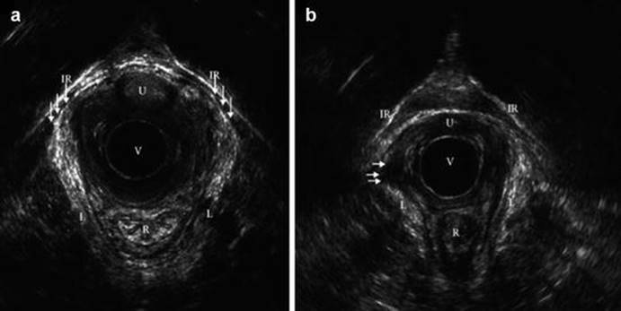
Fig. 14.3
Endovaginal ultrasound findings of levator ani muscle (LAM). IR inferior rami os pubis, L levator ani muscle, R rectum, U urethra, V vagina (with endovaginal probe). (a) A nulliparous woman with intact LAM. (b) A primiparous woman after forceps delivery involving a right mediolateral episiotomy and a third-degree tear with unilateral avulsion injury. Arrows indicate missing LAM on patient’s right side (Reprinted from Schwertner-Tiepelmann et al. [43] with permission from John Wiley and Sons)
A prospective, observational study showed that defects of the pubovisceral muscle were identified with 3D endovaginal ultrasonography in 27 % of women with faecal incontinence that had undergone vaginal delivery. Furthermore, vaginal delivery results in enlargement of the levator hiatus and a lower position of the anorectal junction and bladder neck compared with nulliparous women [58].
Primigravid women were examined using EVUS prior to delivery, early postpartum and 3 months postpartum. The results showed that puborectalis avulsion was found in 15.7 % and 13.3 % of women at 20 h and 13 weeks postpartum, respectively [59].
Pelvic Organ Prolapse in Pregnancy
Prevalence and Natural History
Uterine or cervical prolapse complicating pregnancy is a rare event, with an estimated incidence of 1 in 10,000–15,000 deliveries [60, 61]. An earlier study had identified only one case among more than 13,000 obstetric admissions during a 14-year period [61]. This condition is due to poor cardinal ligament and uterosacral ligament support; therefore, it is categorized as an apical compartment prolapse. However, descent of the uterine cervix may also be aggravated by pregnancy as a result of physiological increases in cortisol and progesterone, which leads to a concomitant softening and stretching of the pelvic tissues, thus causing prolapse during pregnancy [60]. The physiologic changes of pregnancy- in terms of cervical elongation and hypertrophy- with pregnancy-related hormonal changes such as increased progesterone and decreased relaxin, may lead to reduced strength and supportive function of the pelvic floor muscle, can also contribute to prolapse. Even though uterine prolapse frequently complicates pregnancy in multiparous women, POP of nulliparous women has also been reported. Of note, a case report described uterine prolapse in a primiparous woman who had multiple asymptomatic fibroids [60, 62, 63].
Uterine prolapse is equally likely to develop at any time during pregnancy even during labour [64, 65]. This condition can be classified as prolapse that is present before pregnancy and prolapse that occurs during pregnancy. Even though some degree of prolapse is present before pregnancy, most cases with prolapse resolve with progression of the pregnancy, and spontaneous correction can be expected by the end of the second trimester when the uterus becomes an abdominal organ, pulling the cervix up into the vagina [60, 64, 66]. Even if prolapse resolves transiently during pregnancy, the prolapse that preceded pregnancy may persist or even recur after childbirth because the prolapse is secondary to the pelvic floor dysfunction caused by mechanical damage to the pelvic support system [67]. Prolapse that develops during pregnancy is usually first noted in the second and third trimester owing to the considerable changes of pelvic organ support [68].
Maternal and Fetal Complications
Complications of POP in pregnancy are common. This condition could predispose pregnant women to discomfort; cervical ulceration, acute urinary retention, preterm labour and fetal and maternal morbidity [68]. Significant complications may develop during pregnancy and childbirth. Urinary retention and urinary tract infection [69], cervical dystocia and obstructive labour, as well as cervical laceration and infection are documented [70]. Major complications, such as spontaneous abortion, fetal demise, preterm labour, fetal death, maternal sepsis or even death have been reported [66, 71, 72].
Antepartum Complications
The oedematous protruding cervix due to venous obstruction and impaired arterial blood flow in pregnancy is susceptible to mechanical trauma, which could lead to its ulceration and infection. In addition, the ulceration and infection of the oedematous cervix secondary to mechanical trauma may be the cause of the high incidence of abortion [73] and preterm labour [60]. Preterm labour is one of the serious complications of uterine cervical prolapse complicating pregnancy due to impaired blood flow induced by cervical trauma and vascular congestion. Urinary tract infection and acute urinary retention have also been reported as complications of uterine prolapse during pregnancy caused by mechanical obstruction and subsequent infection [72].
Intrapartum Complications
Intrapartum complications of POP in pregnancy include inability for cervical dilatation, cervical dystocia and prolonged or obstructed labour as cervical dilatation may begin outside the introitus, and difficulty is added by oedema or fibrosis of cervix [62]. During pregnancy, cervical lacerations followed by infection are quite common, which may lead to cervical fibrosis. This condition also leads to prolonged labour due to cervical dystocia. When the prolapsed uterus causes obstructed labour, rupture of lower uterine segment and intrapartum fetal death or even maternal death may occur.
A case report described an extensive, irreducible uterine prolapse during labour of a patient without any antenatal care, which resulted in the arrest of labour and stillbirth [74].
Postpartum Complications
Prematurity was the main cause of fetal death after preterm delivery, while infection was the most frequent reason for maternal death in association with POP in the early twentieth century [72]. A review of a total of 170 pregnancies with uterine prolapse (from 1925 to 1940) revealed fetal and maternal mortality during this period to have been 22.1 % and 6.3 %, respectively. And this review reported one case of maternal death due to sepsis [61]. Another complication reported is postpartum bleeding due to uterine atony [75].
Recommendations for Management
Management of POP in pregnancy depends on the severity of prolapse, gestational age and the woman’s preference. Management options range from conservative management with bed rest to aggressive and ambiguous operative procedures, i.e., cervical incision or cesarean hysterectomy. If managed appropriately, the patients without obstetric complication are considered to have favourable outcomes [64], achieving a spontaneous vaginal delivery rate of 84.8 % [70].
· Good genital hygiene is essential to prevent cervical and urinary tract infection [76].
· Local antiseptics should be applied in the event of ulcerations or infected cervix [72].
· Topical magnesium solution has been used to prevent cervical dystocia and lacerations for a prolapsed cervix that is oedematous [77]. The mechanism proposed was due to the osmotic diuretic properties of magnesium.
· Bed rest in a moderately Trendelenburg position can be advised in order to reduce oedema and displacement of the uterus. In addition, this position in combination with good genital hygiene can protect the cervix from local desiccation, trauma, oedema and infection or even preterm labour [62].
· Several authors have also recommended placement of pessaries particularly support pessary, i.e., ring with support or dish pessary after reducing the prolapse. Reduction of the prolapsed uterus during pregnancy will protect the cervix from local trauma and prevent the possibility of incarceration [66, 78].
· Alternatively, in cases where conservative methods have failed or when prolonged bed rest is impossible, minimally invasive surgery in a pregnant woman may be considered. Few cases of laparoscopic uterine suspension during pregnancy were reported with successful outcome [62, 79]. There is a new laparoscopic option for the treatment of uterine prolapse in early pregnancy, namely modified Gilliam suspension.
· Regarding route of delivery in cases of prolapse during pregnancy, normal vaginal delivery can be achieved [80]. Although vaginal delivery with forceps may be an option if required and if the cervix is fully dilated, continued stretching of the lower segment to the point of uterine rupture due to cervical dystocia has been reported [72]. In this situation, cesarean delivery becomes the inevitable choice for women with a thick, oedematous, trapped, and irreducible cervix. Duhrssen’s cervical incision and forceps application for vaginal delivery in a situation that emergency cesarean section is not available has been reported [60, 75].
· Cesarean hysterectomy with subsequent suspension of the vaginal cuff might be a therapeutic option for women who have completed their families [81] particularly in developing countries where access to healthcare is limited [68].
· Prophylactic bilateral uterine artery ligation can be considered to prevent lower uterine segment atony and postpartum hemorrhage [82].
Puerperium and Pelvic Organ Prolapse
During childbirth, the pelvic floor is extended due to direct pressure of the fetal presenting part and maternal pressure efforts. The decline of the levator ani muscle tone is caused either by denervation or by direct muscle trauma. This results in an open urogenital hiatus, which combined with functional and anatomic alterations in the muscles and nerves of the pelvic floor, contributes to the development of POP in the puerperium.
Incidence and Prevalence
There are numerous reports on the incidence, prevalence and degree of pelvic organ prolapse after childbirth. The reported wide range (15 %–48 %) in the prevalence of POP after childbirth is mainly a result of differences in study populations and varying classification of POP [83–86]. Moreover, there are little data that describe the quantification of prolapse in primiparous women at and beyond 6 weeks from childbirth regarding POP-Q system. Incomplete recovery of pelvic organ support in nulliparous women defined using objective measures ranged from 33 to 79 % for women evaluated at various time-points between 6 weeks and 1 year postpartum [87–91].
· The puerperium is the period of time encompassing the first few weeks following childbirth. The duration of this period is considered between 4 and 6 weeks. A study from China reported 100 and 87.5 % rates of POP after vaginal delivery and elective cesarean delivery 6 weeks postpartum. These women had at least stage I prolapse. However, prolapse symptoms were not evaluated [90]. The incidence of POP from this study was higher than a previous study, which demonstrated that 32 % of women who had spontaneous vaginal delivery and 35 % of cesarean delivery group during active labour developed at least stage II prolapse when compared to their 36-week antepartum [88].
· At 3 months after vaginal delivery, predominantly Hispanic primiparous women were evaluated with POP-Q examination and multichannel urodynamic testing was conducted in Dallas, Texas, USA. The results showed that with respect to the cumulative stage of prolapse, 56 % had stage II, and none had stage III prolapse or greater [92].
· At 6 months after vaginal delivery, magnetic resonance imaging was used to quantify the changes that occur in the levator ani muscles. Levator ani signal intensities and thickness, in areas of the urogenital and the levator hiatus were assessed prospectively. The authors reported that recovery of connective tissue and complete pelvic floor muscles contractility takes up to 6 months after vaginal delivery [93]. According to a prospective study conducted in Albuquerque, NM, USA, nulliparous women were recruited and evaluated at 6 months postpartum. At the 6-months postpartum visit, the vaginal birth group was more likely to have a higher stage of prolapse than the cesarean delivery group; the POPQ differences were limited to the anterior vaginal wall [94].
· The prevalence is consistent with data from an observational study in the primigravid women evaluated 6 months postpartum in Barcelona, Spain. In terms of POP-Q system stage, the authors found that 19.4 % of women were assessed as POPQ stage II [95]. These findings are slightly lower than the 31.2 % of stage II prolapse reported by the Pelvic Floor Disorders Network at 6 months postpartum [89].
· At 1 year after delivery. A prospective, observational study was conducted in Wenzhou, Zhejiang, China. Pelvic organ support was assessed at 36–38 weeks of gestation, before the onset of labour, as well as at 6 weeks, 6 months and 1 year postpartum using the POP-Q system. Stage II prolapse was present in 35 and 37 % of women in unlaboured cesarean delivery (UCD) and trial of labour (TOL) at 36–38 weeks of gestation. After delivery, the likelihood of stage II prolapse declined during the first year postpartum in the whole cohort. The TOL group was much less likely to recover from stage II prolapse compared with the UCD. The continued changes in the pelvic floor were shown from 36 to 38 weeks of gestation to 1 year postpartum, therefore, the process by which the reproductive tract returns anatomically to a normal non-pregnant state after delivery might be more than 6 weeks [96].
· 5 years after childbirth. A longitudinal observational cohort study was conducted in the UK to assess the pelvic organ support stage and pelvic floor symptoms in the second trimester, at 14 weeks after delivery, 1 year and 5 years. The results showed that in women who had a vaginal delivery, the change in average POP-Q stage score was significantly increased from baseline score at 14 weeks, 1 year and at 5 years. In the caesarean delivery group the change in average POP-Q stage score from baseline was only significantly increased at 14 weeks postpartum. Prolapse symptoms were not significantly altered from baseline at 14 weeks, 1 and 5 years in both groups. The authors suggest that although pelvic organ support stage and some symptoms worsen after one vaginal delivery, they do not affect condition-specific QOL [91].
· 12 years after childbirth. All of the women who delivered in three maternity units: in Aberdeen (UK), Birmingham (UK) and Dunedin (New Zealand) were surveyed. The main research question was whether delivery mode history was associated with either prolapse symptoms or prolapse signs at 12 years after the index birth. A questionnaire survey of the 7725 women was conducted around 12 years after their index delivery. Women were also invited to a clinical examination to assess any degree of pelvic organ prolapse using the POP-Q system. Compared with women whose births were all spontaneous vaginal deliveries, women who had all births by caesarean section were the least likely to have prolapse (OR 0.11, 95 % CI 0.03–0.38), and there was a reduced risk after forceps or a mixture of spontaneous vaginal delivery and caesarean section. The authors concluded that prolapse symptoms and objective prolapse may not be in concordance [97].
· 20 years after childbirth. A national survey of pelvic floor dysfunction, the SWEPOP (SWEdish Pregnancy, Obesity and Pelvic floor) study was conducted in 2008 to assess pelvic floor function in women 20 years after one single pregnancy terminating either in a vaginal or a surgical delivery. Symptomatic pelvic organ prolapse (sPOP) was diagnosed according to a validated five-item questionnaire. The overall prevalence of sPOP was 12.8 %. The prevalence of sPOP was doubled after vaginal delivery compared with caesarean section, two decades after a single birth. The odds of sPOP 20 years after birth increased by 255 % after vaginal delivery compared with caesarean section [83].
Associated Factors
Mode of Delivery
Several studies have linked vaginal childbirth to pelvic organ prolapse [97, 98]. An observational study was undertaken to evaluate the influence of mode of delivery on pelvic organ support of primigravid women after childbirth. Pelvic organ support was evaluated at 6 months postpartum using the POP-Q system. Specifically, spontaneous vaginal delivery was found to more than treble the risk (OR 3.19; 95 % CI 1.07–9.49), while with instrumental vaginal delivery it increased more than fivefold (OR 5.52; 95 % CI 1.79–17.30). Stage II prolapse was found in only 7.7 % women who had undergone cesarean sections [95]. This finding is similar to other authors [94, 95, 97, 99], who observed a low prevalence of POP after cesarean section.
A cross-sectional study conducted in Turkey confirms such association as well. Vaginal delivery was associated with an odds ratio of 2.92 (95 % confidence interval 1.19–7.17) for prolapse when compared with nulliparity [100]. Moreover, each vaginal delivery increased the risk of POP (odds ratio 1.23; 95 % confidence interval 1.12–1.35) after controlling for all confounding factors [100]. The odds for symptomatic pelvic organ prolapse increased with number of childbirths and were 3.3-fold higher among mothers of 4 than among mothers of 1 [85].
Operative Vaginal Delivery
Operative vaginal delivery or the instrumental vaginal delivery refers to the use of traction devices to assist uterine contractions and maternal expulsive efforts during the second stage of labour to achieve delivery of the fetus. Forceps and vacuums are the most commonly used instruments for this purpose. Forceps delivery was found to increase risk of levator ani muscle avulsion during the first vaginal delivery (OR 6.6, 95 % CI 2.5–17.2) [55]. Forceps delivery increased the odds of POP (OR 1.95, 95 % CI 1.03–3.70) in a cohort study [101]. The result suggests that one additional woman would have development of POP for every eight women who experienced at least one forceps birth (compared with delivering all her children by spontaneous vaginal birth).
Prolonged Second Stage of Labour
The second stage of labour is characterized by progressive descent of the fetal head through a completely dilated cervix. This is achieved by the expulsive forces generated by uterine contractions and maternal effort. During these contractions, intrauterine pressure is high. Maternal pushing can additionally increase intrauterine pressure [102]. Ischemic necrosis of the pelvic tissues (including nerves and muscles) and stretch injuries, leading to permanent denervation of the tissues, can occur if this pressure continues for an extended duration [103]. As a result, a prolonged second stage may increase soft tissue injury and neuromuscular damage to the pelvic floor.
This finding is further supported by the suggestion that prolonged pushing for more than 1 h during the second stage of labour is associated with denervation injuries to the pelvic floor in primiparous women [20]. A study using four-dimensional transperineal ultrasound found that prolonged active second stage of labour increased the risk of levator ani muscle avulsion (OR 2.2, 95 % CI 1.4–3.3) [54]. A small Japanese study identified duration of the second stage of labour of more than 30 min as a risk factor for POP in primiparous women [104].
Conclusion
The development of pelvic organ prolapse has been associated with pregnancy and childbirth. Hormonal changes during pregnancy and mechanical injury to the pelvic floor support, which are direct muscle trauma, disruption of connective tissue support and denervation, are some of the underlying mechanisms for development of pelvic organ prolapse. Over the past two decades, imaging techniques that include ultrasonography and magnetic resonance imaging have revealed mechanisms of injury to the pelvic floor with the time of greatest risk of damage during the first vaginal delivery.
Pelvic organ prolapse in pregnancy is a rare condition. Early recognition is essential in order to avoid possible maternal and fetal risks. In puerperium, continuous changes occur in the pelvic floor from childbirth to 1 year postpartum. Associated factors with pelvic organ prolapse after childbirth are vaginal delivery, forceps delivery and prolonged second stage of labour.
References
1.
Haylen BT, de Ridder D, Freeman RM, Swift SE, Berghmans B, Lee J, et al. An International Urogynecological Association (IUGA)/International Continence Society (ICS) joint report on the terminology for female pelvic floor dysfunction. Int Urogynecol J. 2010;21:5–26.PubMed
2.
Swift SE. The distribution of pelvic organ support in a population of female subjects seen for routine gynecologic health care. Am J Obstet Gynecol. 2000;183:277–85.PubMed
3.
Walker GJ, Gunasekera P. Pelvic organ prolapse and incontinence in developing countries: review of prevalence and risk factors. Int Urogynecol J. 2011;22:127–35.PubMed
4.
Barber MD, Maher C. Epidemiology and outcome assessment of pelvic organ prolapse. Int Urogyn col J. 2013;24:1783–90.
5.
Awwad J, Sayegh R, Yeretzian J, Deeb ME. Prevalence, risk factors, and predictors of pelvic organ prolapse: a community-based study. Menopause. 2012;19:1235–41.PubMed
6.
Chow D, Rodríguez LV. Epidemiology and prevalence of pelvic organ prolapse. Curr Opin Urol. 2013;23:293–8.PubMed
7.
Glazener C, Elders A, Macarthur C, Lancashire RJ, Herbison P, Hagen S, ProLong Study Group, et al. Childbirth and prolapse: long-term associations with the symptoms and objective measurement of pelvic organ prolapse. BJOG. 2013;120:161–8.PubMed
8.
Mant J, Painter R, Vessey M. Epidemiology of genital prolapse: observations from the oxford family planning association study. Br J Obstet Gynaecol. 1997;5:579–85.
9.
Kudish BI, Iglesia CB, Gutman RE, Sokol AI, Rodgers AK, Gass M, et al. Risk factors for prolapse development in white, black, and Hispanic women. Female Pelvic Med Reconstr Surg. 2011;17:80–90.PubMedPubMedCentral
10.
Tinelli A, Malvasi A, Rahimi S, Negro R, Vergara D, Martignago R, et al. Age-related pelvic floor modifications and prolapse risk factors in postmenopausal women. Menopause. 2010;17:204–12.PubMed
11.
Swift S, Woodman P, O’Boyle A, Kahn M, Valley M, Bland D. Pelvic organ Support Study (POSST): the distribution, clinical definition, and epidemiologic condition of pelvic organ support defects. Am J Obstet Gynecol. 2005;192:795–806.PubMed
12.
Uustal Fornell E, Wingren G, Kjølhede P. Factors associated with pelvic floor dysfunction with emphasis on urinary and fecal incontinence and genital prolapse: an epidemiological study. Acta Obstet Gynecol Scand. 2004;83:383–9.PubMed
13.
Kamisan Atan I, Gerges B, Shek K, Dietz H. The association between vaginal parity and hiatal dimensions: a retrospective observational study in a tertiary urogynaecological centre. BJOG. 2014. doi:10.1111/1471-0528.12920.PubMed
14.
Quiroz LH, Munoz A, Shippey SH, Gutman RE, Handa VL. Vaginal parity and pelvic organ prolapse. J Reprod Med. 2010;55:93–8.PubMedPubMedCentral
15.
Gleason JL, Richter HE, Varner RE. Pelvic organ prolapse. In: Barek JS, editor. Berek & novak’s gynecology. 15th ed. Philadelphia: Lippincott Williams & Wilkins; 2012. p. 906–39.
16.
Sokol ER, Genadry R, Anderson JR. Anatomy and embryology. In: Barek JS, editor. Berek & novak’s gynecology. 15th ed. Philadelphia: Lippincott Williams & Wilkins; 2012. p. 62–111.
17.
Barber MD, Bremer RE, Thor KB, Dolber PC, Kuehl TJ, Coates KW. Innervation of the female levator ani muscles. Am J Obstet Gynecol. 2002;187:64–71.PubMed
18.
Stær-Jensen J, Siafarikas F, Hilde G, Bø K, Engh ME. Ultrasonographic evaluation of pelvic organ support during pregnancy. Obstet Gynecol. 2013;122:329–36.PubMed
19.
Snooks SJ, Setchell M, Swash M, Henry M. Injury to innervation of pelvic floor sphincter musculature in childbirth. Lancet. 1984;2:546–50.PubMed
20.
Allen RE, Hosker GL, Smith AR, Warrell DW. Pelvic floor damage and childbirth: a neurophysiological study. BJOG. 1990;97:770–9.
21.
Weidner AC, Jamison MG, Branham V, South MM, Borawski KM, Romero AA. Neuropathic injury to the levator ani occurs in 1 in 4 primiparous women. Am J Obstet Gynecol. 2006;195:1851–6.PubMed
22.
Snooks SJ, Swash M, Henry MM, Setchell M. Risk factors in childbirth causing damage to the pelvic floor innervation. Int J Colorectal Dis. 1986;1:20–4.PubMed
23.
Zong W, Jallah ZC, Stein SE. Reparative mechanical stretch increases extracellular collagenase activity in vaginal fibroblasts. Female Pelvic Med Reconstr Surg. 2010;16:257–62.PubMedPubMedCentral
24.
Goepel C, Johanna Kantelhardt E, Karbe I, Stoerer S, Dittmer J. Changes of glycoprotein and collagen immunolocalization in the uterine artery wall of postmenopausal women with and without pelvic organ prolapse. Acta Histochem. 2011;113:375–8.PubMed
25.
Durnea CM, Khashan AS, Kenny LC, Durnea UA, Smyth MM, O’Reilly BA. Prevalence, etiology and risk factors of pelvic organ prolapse in premenopausal primiparous women. Int Urogy ecol J. 2014;25:1463–70.
26.
South MM, Stinnett SS, Sanders DB, Weidner AC. Levator ani denervation and reinnervation 6 months after childbirth. Am J Obstet Gynecol. 2009;200:519.e1–7.
27.
Chiaffarino F, Chatenoud L, Dindelli M, Meschia M, Buonaguidi A, Amicarelli F, et al. Reproductive factors, family history, occupation and risk of urogenital prolapse. Eur J Obstet Gynecol Reprod Biol. 1999;82:63–7.PubMed
28.
O’Boyle AL, Woodman PJ, O’Boyle JD, Davis GD, Swift SE. Pelvic organ support in nulliparous pregnant and nonpregnant women: a case control study. Am J Obstet Gynecol. 2002;187:99–102.PubMed
29.
Kisli E, Kisli M, Agargun H, Altinokyigit F, Kamaci M, Ozman E, et al. Impaired function of the levator ani muscle in the grand multipara and great grand multipara. Tohoku J Exp Med. 2006;210:365–72.PubMed
30.
Delancey JO, Hurd WW. Size of the urogenital hiatus in the levator ani muscles in normal women and women with pelvic organ prolapse. Obstet Gynecol. 1998;91:364–8.PubMed
31.
Ansquer Y, Fernandez P, Chapron C, Frey C, Bennis M, Roy C, et al. Static and dynamic MRI features of the levator ani and correlation with severity of genital prolapse. Acta Obstet Gynecol Scand. 2006;85:1468–75.PubMed
32.
Lammers K, Futterer JJ, Prokop M, Vierhout ME, Kluivers KB. Diagnosing pobovisceral avulsions: a systematic review of the clinical relevance of a prevalent anatomical defect. Int Urogynecol J. 2012;23:1653–64.PubMedPubMedCentral
33.
DeLancey JOL, Morgan DM, Fenner DE, Kearney R, Guire K, Miller JM, et al. Comparison of levator ani muscle defects and function in women with and without pelvic organ prolapse. Obstet Gynecol. 2007;109:295–302.PubMed
34.
Dietz HP, Simpson JM. Levator trauma is associated with pelvic organ prolapse. BJOG. 2008;115:979–84.PubMed
35.
Shek K, Dietz H. Intrapartum risk factors of levator trauma. Br J Obstet Gynaecol. 2010;117:1485–92.
36.
Khunda A, Shek K, Dietz H. Can ballooning of the levator hiatus be determined clinically? Am J Obstet Gynecol. 2012;206:246.e241–4.
37.
Dietz HP, Bhalla R, Chantarasorn V, Shek KL. Avulsion of the puborectalis muscle is associated with asymmetry of the levator hiatus. Ultrasound Obstet Gynecol. 2011;37(6):723–6.PubMed
38.
Krofta L, Otcenasek M, Kasikova E, Feyereisl J. Pubococcygeus-puborec talis trauma after forceps delivery: evaluation of the levator ani muscle with 3D/4D ultrasound. Int Urognecol J. 2009;20:1175–81.
39.
Dietz H, De Leon J, Shek K. Ballooning of the levator hiatus. Ultrasound Obstet Gynecol. 2008;31:676–80.PubMed
40.
Rostaminia G, White D, Hegde A, Quiroz LH, Davila GW, Shobeiri SA. Levator ani deficiency and pelvic organ prolapse severity. Obstet Gynecol. 2013;121:1017e24.
41.
Gainey HL. Post-partum observation of pelvic tissue damage. Am J Obstet Gynecol. 1943;46:457–66.
42.
Gainey HL. Postpartum observation of pelvic tissue damage: further studies. Am J Obstet Gynecol. 1955;70:800–7.PubMed
43.
Schwertner-Tiepelmann N, Thakar R, Sultan AH, Tunn R. Obstetric levator ani muscle injuries: current status. Ultrasound Obstet Gynecol. 2012;39:372–82.PubMed
44.
Kearney R, Miller J, Ashton-Miller J, Delancey J. Obstetric factors associated with levator ani muscle injury after vaginal birth. Obstet Gynecol. 2006;107:144–9.PubMedPubMedCentral
45.
Dietz H, Lanzarone V. Levator trauma after vaginal delivery. Obstet Gynecol. 2005;106:707–12.PubMed
46.
Tunn R, Paris S, Fischer W, Hamm B, Kuchinke J. Static magnetic resonance imaging of the pelvic floor muscle morphology in women with stress urinary incontinence and pelvic prolapse. Neurourol Urodyn. 1998;17:579–89.PubMed
47.
DeLancey JO, Kearney R, Chou Q, Speights S, Binno S. The appearance of levator ani muscle abnormalities in magnetic resonance images after vaginal delivery. Obstet Gynecol. 2003;101:46–53.PubMedPubMedCentral
48.
Dietz HP, Steensma AB. The role of childbirth in the aetiology of rectocele. BJOG. 2006;113:264–7.PubMed
49.
Dietz HP, Korda A. Which bowel symptoms are more strongly associated with a true rectocele? Aust N Z J Obstet Gynaecol. 2005;45:505–8.PubMed
50.
Shek KL, Dietz HP. The effect of childbirth on hiatal dimensions. Obstet Gynecol. 2009;113:1272–8.PubMed
51.
Valsky DV, Lipschuetz M, Bord A, Eldar I, Messing B, Hochner-Celnikier D, et al. Fetal head circumference and length of second stage of labor are risk factors for levator ani muscle injury, diagnosed by 3-dimensional transperineal ultrasound in primiparous women. Am J Obstet Gynecol. 2009;201:e1–7.PubMed
52.
Chan SS, Cheung RY, Yiu AK, Lee LL, Pang AW, Choy KW, et al. Prevalence of levator ani muscle injury in Chinese women after first delivery. Ultrasound Obstet Gynecol. 2012;39:704–9.PubMed
53.
van Delft K, Sultan AH, Thakar R, Schwertner-Tiepelmann N, Kluivers K. The relationship between postpartum levator ani muscle avulsion and signs and symptoms of pelvic floor dysfunction. BJOG. 2014;121:1164–72.PubMed
54.
Lien KC, Mooney B, DeLancey JOL, Ashton-Miller JA. Levator ani muscle stretch induced by simulated vaginal birth. Obstet Gynecol. 2004;103:31–40.PubMedPubMedCentral
55.
van Delft K, Thakar R, Sultan AH, Schwertner-Tiepelmann N, Kluivers K. Levator ani muscle avulsion during childbirth: a risk prediction model. BJOG. 2014;121:1155–63.PubMed
56.
Shobeiri SA, Leclaire E, Nihira MA, Quiroz LH, O’Donoghue D. Appearance of the levator ani muscle subdivisions in endovaginal three-dimensional ultrasonography. Obstet Gynecol. 2009;114:66–72.PubMed
57.
van Delft KW, Sultan AH, Thakar R, Shobeiri SA, Kluivers KB. Agreement between palpation and transperineal and endovaginal ultrasound in the diagnosis of levator ani avulsion. Int Urogynecol J. 2015;26:33–9.PubMed
58.
Murad-Regadas SM, Fernandes GO, Regadas FS, Rodrigues LV, Pereira Jde J, Dealcanfreitas ID, et al. Assessment of pubovisceral muscle defects and levator hiatal dimensions in women with faecal incontinence after vaginal delivery: is there a correlation with severity of symptoms? Colorectal Dis. 2014;16:1010–8.PubMed
59.
van Delft K, Shobeiri SA, Thakar R, Schwertner-Tiepelmann N, Sultan AH. Intra- and interobserver reliability of levator ani muscle biometry and avulsion using three-dimensional endovaginal ultrasonography. Ultrasound Obstet Gynecol. 2014;43:202–9.PubMed
60.
Brown HL. Cervical prolapse complicating pregnancy. J Natl Med Assoc. 1997;89:346–8.PubMedPubMedCentral
61.
Keettel WC. Prolapse of the uterus during pregnancy. Am J Obstet Gynecol. 1941;42:121–6.
62.
Ishida H, Takahashi K, Kurachi H. Uterine prolapse during late pregnancy in a nulliparous woman. Int Urogynecol J. 2014;25:1739–40.PubMed
63.
Partsinevelos GA, Mesogitis S, Papantoniou N, Antsaklis A. Uterine prolapse in pregnancy: a rare condition an obstetrician should be familiar with. Fetal Diagn Ther. 2008;24:296–8.PubMed
64.
Eddib A, Allaf MB, Lele A. Pregnancy in a woman with uterine procidentia: a case report. J Reprod Med. 2010;55:67–70.PubMed
65.
Daskalakis G, Lymberopoulos E, Anasrasakis E, Kalmantis K, Athanasaki A, Manoli A, et al. Uterine prolapse complicating pregnancy. Arch Gynecol Obstet. 2007;276:391–2.PubMed
66.
Horowitz ER, Yogev Y, Hod M, Kaplan B. Prolapse and elongation of the cervix during pregnancy. Int J Gynecol Obstet. 2002;77:147–8.
67.
Miyano N, Matsushita H. Maternal and perinatal outcome in pregnancies complicated by uterine cervical prolapse. J Obstet Gynaecol. 2013;33:569–71.PubMed
68.
Yogev Y, Horowitz ER, Ben-Horoush A, Kaplan B. Uterine cervical elongation and prolapse during pregnancy: an old unsolved problem. Clin Exp Obstet Gynecol. 2003;30:183–5.PubMed
69.
Tomezsko JE, Sand PK. Pregnancy and intercurrent diseases of the urogenital tract. Clin Perinatol. 1997;24:343–68.PubMed
70.
Piver MS, Spezia J. Uterine prolapse during pregnancy. Obstet Gynecol. 1968;32:765–9.PubMed
71.
Guariglia L, Carducci B, Botta A, Ferrazzani S, Caruso A. Uterine prolapse in pregnancy. Gynecol Obstet Invest. 2005;60:192–4.PubMed
72.
Hill PS. Uterine prolapse complicating pregnancy. A case report. J Reprod Med. 1984;29:631–3.PubMed
73.
Gaetane J, Labriola BF. Prolapse of the uterus complicating pregnancy and labor; review and report of two cases. Obstet Gynecol. 1956;8:278–83.PubMed
74.
Yousaf S, Haq B, Rana T. Extensive uterovaginal prolapse during labor. J Obstet Gynaecol Res. 2011;37:264–6.PubMed
75.
O’Herlihy C, Kearney R. Perinatal repair and pelvic floor injury. In: James DK, Steer PJ, Weiner CP, Gonik B, editors. High risk pregnancy: management options. 3rd ed. Philadelphia: Elsevier Saunders; 2005. p. 1499–501.
76.
Sawyer D, Frey K. Cervical prolapse during pregnancy. J Am Board Fam Pract. 2000;13:216–8.PubMed
77.
Lau S, Rijhsinghani A. Extensive cervical prolapse during labor: a case report. J Reprod Med. 2008;53:67–9.PubMed
78.
Mohamed-Suphan N, Ng RK. Uterine prolapse complicating pregnancy and labor: a case report and literature review. Int Urogynecol J. 2012;23:647–50.PubMed
79.
Matsumoto T, Nishi M, Yokota M, Ito M. Laparoscopic treatment of uterine prolapse during pregnancy. Obstet Gynecol. 1999;93:849.PubMed
80.
Kart C, Aran T, Guven S. Stage IV C prolapse in pregnancy. Int J Gynaecol Obstet. 2011;112:142–3.PubMed
81.
Meydanli MM, Ustun Y, Yalcin OT. Pelvic organ prolapse complicating third trimester pregnancy. A case report. Gynecol Obstet Invest. 2006;61:133–4.PubMed
82.
Cingillioglu B, Kulhan M, Yildirim Y. Extensive uterine prolapse during active labor: a case report. Int Urogyn J. 2010;21:1433–4.
83.
Gyhagen M, Bullarbo M, Nielsen T, Milsom I. Prevalence and risk factors for pelvic organ prolapse 20 years after childbirth: a national cohort study in singleton primiparae after vaginal or caesarean delivery. BJOG. 2013;120:152–60.PubMed
84.
Milsom I, Altman D, Herbison P, Lapitan MC, Nelson R, Sille’n U, et al. Epidemiology of urinary (UI) and faecal (FI) Incontinence and pelvic organ prolapse (POP). In: Abrams P, Cardozo L, Khoury S, Wein A, editors. Incontinence. Paris: Health Publications Ltd; 2009. p. 35–111.
85.
Tegerstedt G, Miedel A, Maehle-Schmidt M, Nyren O, Hammarstrom M. Obstetric risk factors for symptomatic prolapse: a population based approach. Am J Obstet Gynecol. 2006;194:75–81.PubMed
86.
Slieker-ten Hove MC, Pool-Goudzwaard AL, Eijkemans MJ, Steegers-Theunissen RP, Burger CW, Vierhout ME. The prevalence of pelvic organ prolapse symptoms and signs and their relation with bladder and bowel disorders in a general female population. Int Urogynecol J Pelvic Floor Dysfunct. 2009;20:1037–45.PubMedPubMedCentral
87.
O’Boyle AL, O’Boyle JD, Ricks RE, Patience TH, Calhoun B, Davis G. The natural history of pelvic organ support in pregnancy. Int Urogynecol J Pelvic Floor Dysfunct. 2003;14:46–9.PubMed
88.
Sze EH, Sherard 3rd GB, Dolezal JM. Pregnancy, labor, delivery, and pelvic organ prolapse. Obstet Gynecol. 2002;100:981–6.PubMed
89.
Handa VL, Nygaard I, Kenton K, Cundiff GW, Ghetti C, Ye W. Pelvic floor disorders network. Pelvic organ support among primiparous women in the first year after childbirth. Int Urogynecol J Pelvic Floor Dysfunct. 2009;20:1407–11.PubMedPubMedCentral
90.
Zhu L, Bian XM, Long Y, Lang JH. Role of different childbirth strategies on pelvic organ prolapse and stress urinary incontinence: a prospective study. Chin Med J. 2008;121:213–5.PubMed
91.
Elenskaia K, Thakar R, Sultan AH, Scheer I, Onwude J. Effect of childbirth on pelvic organ support and quality of life: a longitudinal cohort study. Int Urogynecol J. 2013;24:927–37.PubMed
92.
Wai CY, McIntire DD, Atnip SD, Schaffer JI, Bloom SL, Leveno KJ. Urodynamic indices and pelvic organ prolapse quantification 3 months after vaginal delivery in primiparous women. Int Urogynecol J. 2011;22:1293–8.PubMedPubMedCentral
93.
Tunn R, DeLancey JO, Howard D, Thorp JM, Ashton-Miller JA, Quint LE. MR imaging of levator ani muscle recovery following vaginal delivery. Int Urogynecol J. 1999;10:300–7.
94.
Rogers RG, Leeman LM, Borders N, Qualls C, Fullilove AM, Teaf D, et al. Contribution of the second stage of labour to pelvic floor dysfunction: a prospective cohort comparison of nulliparous women. BJOG. 2014;121:1145–54.PubMedPubMedCentral
95.
Diez-Itza I, Arrue M, Ibañez L, Paredes J, Murgiondo A, Sarasqueta C. Influence of mode of delivery on pelvic organ support 6 months postpartum. Gynecol Obstet Invest. 2011;72:123–9.PubMed
96.
Chen Y, Li F-Y, Lin X, Chen J, Chen C, Guess M. The recovery of pelvic organ support during the first year postpartum. BJOG. 2013;120:1430–7.PubMed
97.
Glazener C, Elders A, Macarthur C, Lancashire RJ, Herbison P, Hagen S, et al. ProLong Study Group. Childbirth and prolapse: longterm associations with the symptoms and objective measurement of pelvic organ prolapse. BJOG. 2013;120:161–8.
98.
Handa VL, Blomquist JL, Knoepp LR, Hoskey KA, McDermott KC, Munoz A. Pelvic floor disorders 5–10 years after vaginal or cesarean childbirth. Obstet Gynecol. 2011;118:777–84.PubMedPubMedCentral
99.
O’Boyle AL, O’Boyle JD, Calhoun B, Davis GD. Pelvic organ support in pregnancy and postpartum. Int Urogynecol J Pelvic Floor Dysfunct. 2005;16:69–72.PubMed
100.
Yeniel AO, Ergenoglu AM, Askar N, Itil IM, Meseri R. How do delivery mode and parity affect pelvic organ prolapse? Acta Obstet Gynecol Scand. 2013;92:847–51.PubMed
101.
Handa VL, Blomquist JL, McDermott KC, Friedman S, Muñoz A. Pelvic floor disorders after vaginal birth: effect of episiotomy, perineal laceration, and operative birth. Obstet Gynecol. 2012;119:233–9.PubMedPubMedCentral
102.
Rempen A, Kraus M. Pressures on the fetal head during normal labor. J Perinat Med. 1991;19:199–206.PubMed
103.
Lien KC, Morgan DM, Delancey JO, Ashton-Miller JA. Pudendal nerve stretch during vaginal birth: a 3D computer simulation. Am J Obstet Gynecol. 2005;192:1669–76.PubMed
104.
Tsunoda A, Shibusawa M, Kamiyama G, Kusano M, Shimizu Y, Yanaihara T. The effect of vaginal delivery on the pelvic floor. Surg Today. 1999;29:1243–7.PubMed