
Cornelia Betschart1 and Josef Wisser2
(1)
Department of Gynecology, University Hospital Zurich, Frauenklinikstrasse 10, Zurich, 8091, Switzerland
(2)
Department of Obstetrics, University of Zurich, Frauenklinikstrasse 10, Zurich, 8091, Switzerland
Cornelia Betschart (Corresponding author)
Email: cornelia.betschart@usz.ch
Email: cornelia.betschart@gmx.ch
Josef Wisser
Email: josef.wisser@usz.ch
Abstract
Knowledge of physiological and anatomical changes during pregnancy is important for optimal prevention and care. Epidemiological and clinical observational studies have focused on bothersome patient symptoms and changes related to the pelvic floor during pregnancy.
Pelvic floor symptoms are common in pregnant women. These are mostly bladder related symptoms like higher micturition frequency, nocturia and stress urinary incontinence.
Quantitative assessments of the pelvic floor anatomy in pregnancy can be performed by clinical tools like the POP-Q or imaging like ultrasound and magnetic resonance technique.
Objectively, changes in the hiatal area, vaginal location, bladder neck position, anal sphincter muscle thickness and levator ani volume can be identified with ultrasound and MRI in pregnant women.
Keywords
PregnancyPelvic floor muscleLigament laxityAnal sphincterHiatusPelvic Organ Prolapse Quantification (POP-Q)Pelvic floor symptomsUltrasoundMRI
Physiological Changes of the Pelvic Floor During Pregnancy
Pregnancy exerts physiological alterations in virtually all organ systems. Significant alterations are described in the cardiovascular, gastrointestinal, haematological, endocrine and musculoskeletal systems [1]. The connective tissue of the ligaments and cartilage such as in symphysis pubis or ischiosacral junctions loosens. Hormonal changes are significant. The blood oestrogen levels rise to a hyperoestrogenic state, mostly by additive production by the placenta. Also other steroidal hormones including progesterone, dehydroepiandrosterone and cortisone undergo significant changes and hormones like the human chorionic gonadotropin, chorionic adrenocorticotropin, thyreotropin, relaxin growth hormone-variants and placental peptide hormones like neuropeptide-Y, inhibin, activin and atrial natriuretic peptide (ANP) arise.
During pregnancy the urogenital system undergoes anatomical and physiological changes [2]. Blood flow to the pelvic organs increases significantly, the bladder detrusor smooth muscle loses tone and hormones affect the biochemical composition of pelvic floor tissue. Pregnancy is known to significantly affect the instantaneous stiffness and relaxation behaviour of vaginal tissues in rat [3]. For pregnant human pelvic floor tissues accurate histomorphological findings are lacking.
Pelvic floor symptoms in pregnancy include increased urinary frequency, urgency and incontinence. These symptoms are aggravated in the third trimester as the fetal head engages in the pelvis [4]. Urinary retention may occur in the late first trimester with an estimated prevalence of 1 in 3000 to 1 in 8000 of pregnancies. It is mostly found in women with a retroverted uterus or large fibroids where the uterus exerts mechanical compression to the urethra. Often intermittent or continuous catheterisation is indicated until the uterus is large enough and cannot become incarcerated in the sacral hollow. Several reports of acute retention in pregnancy make special note of the fact that passage of a catheter in patients with retention is not difficult, suggesting that compression of the urethra per se may not be the cause of retention [5]. As urodynamically the bladder capacity increases during pregnancy it is assumed that in some women the high progesterone levels not only result to a weakness of the detrusor contractility but also to incapacity to relax the urethral sphincter [5].
During delivery, it is well established that pelvic floor muscle trauma and denervation occur [6–8]. These injuries are associated with stress urinary incontinence [9], defecation disorders [10] and prolapse [11] postpartum or become apparent decades later in life. Abnormalities are more often found in multiparae [6, 7], correlate with a prolonged second stage of labor [7, 12], forceps delivery [7] and high birthweight [6, 9]. These risk factors have cumulative effects. As pelvic organ prolapse is associated with parity in many clinical and epidemiological studies, it is unknown whether pregnancy, parturition, or a combination of these factors contributes to that. The influence of the hormonal changes during pregnancy on the pelvic floor is not easy to assess and up till now there are only few studies on the effects of pregnancy on the pelvic floor.
To elucidate the effect of pregnancy on the different parts of the pelvic floor, assessment of women before, during and after pregnancy would be required.
Unfortunately, there are significant practical problems in implementing such a study design.
Clinical Assessment of Pelvic Floor Changes
Pelvic floor changes during pregnancy can be assessed by measuring the compressing forces through palpation or vaginally placed balloon-type sensors. Invasive electromyographic studies performed in women with prolapse or after deliveries are not suitable for pregnant women, though surface electromyographic assessments are a feasible evaluation tool also in pregnancy. Clinical visual quantification of the location of pelvic organs can be undertaken using the Pelvic Organ Prolapse Quantification system (POP-Q). Imaging techniques like ultrasound or MRI provide high-resolution images of single structures for analysis or as basis for building 3D models. Those finite-element models containing multiple small pieces that maintain the same properties of the original model allow simulation of different stages of vaginal birth, like maximum levator stretch or also the simulation of prolapse. Until now, no pregnancy related finite models mirror the passive forces of uterine growth on the pelvic floor, vagina or bladder.
Pelvic Organ Prolapse Quantification System (POP-Q)
The change of location of the pelvic organs can be assessed with the Pelvic Organ Prolapse Quantifiction system (POP-Q) [13]. It allows assessment of the extent of movement of the anterior and posterior vaginal wall, the cervix or the vaginal vault between rest and strain, as well as interindividual comparisons (Fig. 4.1a). This semi-objective assessment in pregnant women revealed an increased rate of prolapse stages in the pregnant group compared with age-matched non-pregnant controls [14]. This was shown in a retrospective study of 21 pregnant women in the second or third trimester (average 27.8 weeks of pregnancy). POP-Q stages were significantly higher among pregnant subjects compared with control subjects, indicating a higher incidence of pelvic organ prolapse (p < 0.001) (Fig. 4.1b, c). POP-Q points that differed significantly between pregnant and nonpregnant subjects were points Aa, Ba representing the anterior vaginal wall, Ap, Bp for the posterior vaginal wall and the total vaginal length. Measurements at the cervix (point C), the genital hiatus length and the posterior fornix (point D) were not significantly different between the two groups. In the non-pregnant group maximal descent was POP-Q stage 1 in 57 %, whereas in the pregnant group 48 % of the women presented with a POP-Q stage 2. Point Aa, which corresponds to the urethrovesical angle, was significantly more relaxed in pregnant women (Fig. 4.1c).

Fig. 4.1
(a) POP-Q points after Bump RC et al. [13]. (b) All patients in the nonpregnant group had a POP-Q stage of 0 or 1 (c) 47.6 % of the pregnant subjects had POP-Q stage 2 (p < .001). Point Ba (most distal position of the remaining upper anterior vaginal wall) and point Bp (most distal position of the remaining upper posterior vaginal wall) are significantly different
Furthermore, as pregnancy progresses the prolapse stages become more pronounced [15]. O’Boyle et al. demonstrated that there is a significant difference between the first and third trimester for all anterior and posterior vaginal points in the POP-Q as well as for the total vaginal length and the genital hiatus, not so for the apical suspension (point C/D), though.
Apart from using the Pelvic Organ Prolapse Quantification (POP-Q) system, pelvic organ support can be assessed using relevant quality of life questionnaires. One prospective study looked at pelvic floor symptoms in the course of pregnancy and POP-Q stages [16]. Quality of life was assessed with the electronic Personal Assessment Questionnaire-Pelvic Floor (ePAQ-PF). Bother with voiding difficulties and stress urinary incontinence increased during pregnancy. Constipation (p = 0.02) and evacuation subdomains improved significantly (p = 0.009) between the week 20 to week 36 of pregnancy. Fecal incontinence was not present in either trimester of pregnancy. In the sexual domain, the only subdomain that worsened significantly (p = 0.03) was “sex and vaginal symptoms” in the course of pregnancy. None of the pelvic floor symptoms impacted the overall quality of life. Objective parameters like genital hiatus and perineal body length showed significantly higher values in the third trimester compared to the second trimester. In this study, other POP-Q points for the anterior or posterior vaginal wall as well as the cervix location did not differ between the second and third trimester.
Other Findings
More data on developmental histomorphological changes in pelvic floor muscles, ligaments, connective tissue and nerve supply in pregnant women would be beneficial. Biomechanical studies on biological tissues like the pelvic ligaments and vaginal tissue have been performed [17] in postmenopausal women with prolapse.
In 29 female cadaveric specimens a correlation between subjective evaluation and objective strength of ligaments was found [18]: the ileopectineal ligament was significantly stronger than the sacrospinous and the arcus tendineus fascia pelvis. But as the influence of hormones during pregnancy is remarkable, these results can not be translated to pregnant women.
At present there is a lack of data about pregnancy associated pelvic floor damage by contrast with an abundance of studies on intrapartum injuries and age-related changes contributing to the development of prolapse.
Outcome Assessment of Pelvic Floor Muscle Training (PFMT)
Pelvic floor muscle changes during pregnancy can be assessed through palpation of the pelvic floor muscles or vaginally or rectally placed pressure sensors. These measurements also allow to assess therapy outcome of pelvic floor muscle training, the only recommended therapy during pregnancy to prevent further pelvic floor disorders. The concept of pelvic floor muscle training (PFMT) in pregnancy to prevent urinary incontinence has been supported by recent studies, which showed that PFMT can prevent from urinary incontinence (UI) both during and in the immediate postpartum period [19–21]. In a recent Cochrane review, there was evidence of a statistically significant effect of PFMT during pregnancy on prevention of incontinence at 3 and up to 6 months after delivery [22]. Long-term follow up of participants up to 8 years after their initial randomization showed that 35.4 % of women in the PFMT group versus 38.8 % of women in the control group reported urinary incontinence [21]. Mørkved et al. reported in a conference abstract non-significant differences in the long-term outcome, whereby after 6 years the incontinence rate was higher in the group that had PFMT (urinary incontinence in 23 % of PFMT and 17 % of control women), however the sexual satisfaction was higher in women that had PFMT during pregnancy [19, 22].
According to the Cochrane Database of Systematic Reviews [22], PFMT is especially recommended for women with incontinence prior to pregnancy, women with a bladder neck hypermobility in early pregnancy, or postpartum for women that delivered a large baby and/or had a forceps delivery.
A recent study that assessed the outcome of meticulous PFMT with surface electromyography and quality of life questionnaire in pregnant and postpartum women, demonstrated a successful outcome in all three groups. The pelvic floor muscle contractility increased after the training program (p = 0.0001) in the early pregnancy group, in the postpartum group that delivered vaginally with an episiotomy and the postpartum group after an elective caesarean section. Decreases in the scores of both ICIQ-UI SF (P = 0.009) and ICIQ-OAB (P = 0.0003) were also observed after training in all three groups. One point that has to be investigated further is to evaluate if such effects are maintained after stopping training [23].
Imaging Techniques During Pregnancy
Imaging techniques are beneficial to measure changes of the pelvic floor in a non-invasive, reproducible way. Ultrasound and magnetic resonance imaging, as safe and volume based techniques, allow visualisation of structures and function in real time.
Ultrasound
2D Transperineal Ultrasound and Endovaginal 3D Ultrasound
2D ultrasound has a resolution of up to 0.1 mm. The probe held on the perineum in a sagittal position, allows to depict similarly the symphysis, urethra, bladder neck, bladder, vagina and rectum (Fig. 4.2).
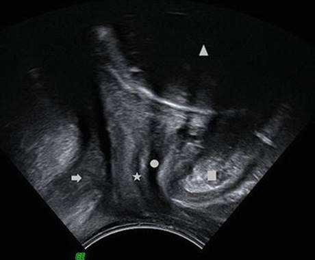
Fig. 4.2
Midsagittal B-mode view of the pelvic floor of a pregnant women in the third trimester. Arrow = rectum, triangle = fetal head, asterisk = vagina, dot = urethra (empty bladder), rectangle = symphysis
Since the introduction of ultrasound in the early 1960s, safety issues were discussed. Ultrasound, physically longitudinal waves, have an influence on biological systems. Physical effects are thermal effects, as energy gets transformed to warmth/heat and second there is a pressure application that can lead to tissue deformation (cavitation). In the B-mode the power is very low (<10 mW/cm2) and the duration of pulses are short (<1 μs). Temperature rises are not measurable for the B-mode ultrasound (US).
3D/4D Transperineal Ultrasound
3D-pelvic floor US generates a volume data set, which can be cut in sequential, orthogonal planes to study structures that are not accessible in the 2D-sonography. The advantage of applying 3D US is that reference planes orthogonal to non-linear structures like the vagina or anal canal can be set time-independently after the examination. Those examinations are also safe, as the applied power is far below the level of biological significance (<100 mW/cm2). Worldwide there are no reports of harmful effects to the fetus through B-mode US which is also the basis for 3D/4D US.
Magnetic Resonance Imaging
Pelvic floor MRI usually performed in a 1.5 or 3 T superconducting magnet, uses proton density T2-weighted scans, 2-D fast-spin proton density with an echo time of 15 ms and a repetition time of 4000 ms, performed at 5-mm intervals in the axial, sagittal and coronal planes in the supine position. Common settings use a slice thickness of 4 mm with a gap of 1 mm. Like ultrasound, magnetic resonance has no harmful effects to the mother and child (Fig. 4.3). Disadvantages are the higher costs, the narrow space of the MR tube and the noise of the machine.
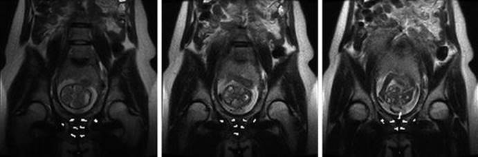
Fig. 4.3
Coronal MRI slides of a women in the second trimester; thickness of slices is 5 mm. The vertical course of the levator ani as thin muscle layer (white arrow head), the bulk of the obturator internus muscle (asterisk) and the internal cervical os in the third picture (arrow) are visible
Pelvic Floor Muscle Anatomy and Function
The female pelvic floor is a complex fibro-muscular-ligamentous unit involved in multiple functions that go beyond the sole support of pelvic organs. Pelvic floor dysfunction globally affects micturition, defecation and sexual activity. Evolutionary modifications like upright walking and the need to deliver fetuses with larger head diameters made the fascial and muscle support of the pelvic floor vulnerable, therefore predisposing women to pelvic organ prolapse and incontinence. The female pelvic floor further undergoes a number of adaptive changes related to pregnancy and endocrine changes.
Animal Studies
Women are not the only upright species that develop pelvic floor disorders during life. Animal studies in primates simulate pregnancy and parturition-related changes. Primates like squirrel monkeys are also known to develop prolapse during their life-time. To study the effects of pregnancy and parturition on the pelvic floor, MRIs of seven female squirrel monkeys were studied prior to pregnancy, 3 days, and 4 months postpartum [24]. No testing was performed during pregnancy to avoid harming the fetus. The bladder neck and cervix position were measured dynamically with abdominal squeezing. The pelvic floor muscles are not completely alike in humans, one difference is a prominent coccygeus muscle. The volume of the coccygeus muscle was greater shortly after parturition than before pregnancy or after recovery. The bladder neck position in the relaxed state and with abdominal pressure descended (p < 0.04) after delivery and descended further (p < 0.001) after recovery. The same happened to the position of the cervix. It seems that parturition-related bladder neck descent in squirrel monkeys is permanent.
Levator Ani Changes in Humans
The appearance of the levator ani (LA) muscle in pregnancy was assessed in 3D-MRI in 84 post-term nulliparas (at 41 weeks of gestation) [25]. This study found a lower levator ani volume in post-term pregnant women of a mean volume of 13.5 ± 3.7 cm3 than in nulligravida, where LA volumes vary between 32.3 and 46.6 cm3 [26–29]. In this investigation on post-term pregnancies the LA muscles appear to be thinned. However, it has to be pointed out that this difference might be partly due to different acquisition techniques.
A 3D ultrasound study [30], aimed to predict delivery outcomes in relation to the levator hiatus area, assessed the LA of 61 nulliparous women between 36 and 40 weeks of pregnancy. The mean hiatal area at rest was 11.81 cm2, at contraction 9.59 cm2, and at Valsalva 16.03 cm2. A correlation between levator dimensions and delivery mode could not be demonstrated. However, an inverse correlation was demonstrated between the area of the hiatus, particularly the one on pelvic floor contraction, and length of total second stage. Women with a smaller hiatal area on pelvic floor contraction, indicating a stronger pelvic floor muscle, had a longer second stage of labor [29].
Interestingly, fetal weight was not associated with LA volume. However, the fetal station was associated with a decreasing levator volume. The lower the fetal station/head, in centimeters to the levator ani, the thinner the LA muscle appeared. After adjusting for maternal BMI, this relationship disappeared.
A longitudinal study could answer the question whether the thinning is an effect of pregnancy or whether imaging has more artefacts in pregnancy due to baby movements or measuring technique.
It is not elucidated yet whether it is beneficial for the pelvic floor health in later life to have a strong and thick pelvic floor musculature before pregnancy, that might lead to a longer second stage of labor, which however, may be associated with a higher rate of pelvic floor trauma, or whether a primarily weaker pelvic floor with a larger hiatus and shorter parturition time has lower negative sequelae later in life.
A randomized controlled trial involving 200 women evaluated the possible prevention of pelvic floor disorders using stretching of pelvic floor muscles with the balloon device Epi-No® in late pregnancy. The rationale is based on sports physiology where an increased muscle extensibility might be obtained by intermittent stretching before the exertion. In this trial a non-significant reduction in levator ani muscle avulsion after training with Epi-No®, beginning at 37 weeks’ gestation (6 % vs. 13 %) was found [31]. It is to mention that this study was insufficiently powered with 200 patients included instead of the 660 women that would have been needed according to a power calculation that was aimed to show a 50 % reduction in the incidence of levator avulsion.
Biomechanical Models
Computational models have been demonstrated to be an effective tool in investigating the processes during the first and second stages of parturition. The pelvic floor and the associated structures are one of the most complex regions of the human body and undergo immense stretching in the course of pregnancy and even more during parturition.
Animal models have the inherent problem of the lack of upright position, which is an important factor in studying the pathophysiology and natural history of prolapse. Interdisciplinary collaborative research, involving bioengineers and clinicians, is essential to investigate and simulate the mechanical effects on the pelvic floor along gravity [32]. Geometrical information is gained from high-resolution images, mostly generated by MRI, and processed by segmentation into a finite element by a mathematical tool. The finite element method discretizes a continuous model into small pieces to investigate their mechanical behaviour under load or stress. This has been done by several authors simulating delivery [33–35] or prolapse [36, 37], though no biomechanical models simulating the changes during pregnancy are available yet.
Anatomical Changes of Ligaments
Landon et al. showed that fascia in pregnant women stretches to a much greater length but had less tensile strength compared with fascia from nonpregnant women [38]. The authors demonstrated that the collagen structure changes and the connective tensile strength decreases during pregnancy. The loosening of connective tissue in ligaments and fascia yields to common symptoms like symphysis pubic dysfunction and pelvic girdle pain. The levels of the hormone relaxin were found to be significantly higher in pregnant women that have bothersome joint pain and laxity [39]. Whether there is a relationship between pelvic girdle pain and specific pelvic floor disorders like prolapse is not elucidated yet.
Bladder and Functional Bladder Neck Anatomy during Pregnancy
Epidemiological studies show an increase in lower urinary tract and pelvic floor symptoms during pregnancy [40]. Pregnancy affects bladder function adversely. Urinary incontinence is seen more often in pregnant women than in matched controls and the mean prevalence of stress urinary incontinence (SUI) during pregnancy can be as high as 41 % (18.6–60 %) and increases with gestational age [41].
It is known from another study that urinary incontinence worsens steadily throughout pregnancy [42], which is dependent on changes of the urethrovesical angle [43].
One study examined the displacement of the vesical neck position during pregnancy [44]. The angle or the urethrovesical junction was measured at rest, during Valsalva and during coughing three times, namely at 12–16 weeks, at 28–32 weeks, and 36–38 weeks of pregnancy. The muscle compliance, as calculated during coughing and during Valsalva remained practically unchanged during the whole pregnancy in this study. The significant increase was noted on examination at 6 weeks postnatally, however also returned to normal values 6 months after childbirth. There was no hysteresis in the muscle activity found in this study during pregnancy that would have indicated a failure of tissue to follow the same course during relaxation as it did during distension. The hysteresis is thought to be the result of shifts in the geometrical structure of the fibers with respect to each other, and can be interpreted as a form of internal friction within the tissue.
Conversely, another match-controlled ultrasound study demonstrated higher anterior wall mobility for the bladder compartment in early pregnancy between 10 and 17 weeks’ gestation than in non-pregnant controls. This mobility increased and showed a higher organ descent in late pregnancy (32–39 weeks) [45]. The apical (uterus) and posterior compartments (bowel) did not descend significantly in this study. This is in concordance with another functional study that assessed the rate of incontinence in early pregnancy. The numbers of incontinent women in the first trimester were significantly higher (20 %) than in the matched non-pregnant group (4 %) [46]. This finding agrees with the statistically significant change of the urethrovesical junction angle during coughing or Valsalva in the pregnant group.
A urodynamic study by Iosif et al. found that in continent and asymptomatic pregnant women, functional urethral length, maximal urethral pressure, and closure pressure increased by an average of 12 cm H2O during pregnancy and returned to normal shortly after delivery [47]. In stress urinary incontinent women, the same authors reported lower function urethral lengths and closure pressures than in the continent pregnant controls [48]. The SUI group did not show the same increase in functional urethral length as the continent pregnant women [48].
Genital Hiatus
The urogenital hiatus varies remarkably among non-pregnant individuals [29]. The ratio between the smallest and highest numbers for the urogenital hiatus has been calculated to be 1.6 for width, 2.3 for length and 2.4 for the urogenital hiatus area. In 10 % there was an absence of visible insertion of the levator ani (LA) into the pubic bone. Women where the LA arises only from the arcus tendineus of the levator ani and not from the pubic bone, might have a larger hiatus. As this finding appeared in nulliparous women, it seems to be a variation in normal insertion and not a pathological finding.
It is yet unknown whether this primarily larger hiatus and the missing pubic insertion have consequences for birth-related injuries.
Recently, an observational study was designed to evaluate the potential effect of pregnancy on pelvic floor function [49]. 3D ultrasound volume data of 688 nulliparae assessed in the late third trimester and again 4 months postpartum were compared with data of 74 nulliparous nonpregnant volunteers. Outcome parameters were the hiatal dimension and the urethral mobility adjusted for BMI and age. The comparison of those two groups revealed a 27 % increase in hiatal area at rest and a 41 % increase on Valsalva comparing the non-pregnant group and the nulliparae of the late third trimester. About 70 % of this difference in hiatal dimension was observed when comparing nonpregnant controls with women 4 months after prelabor or 1st stage caesarean section. For the urethral mobility, similar results were found with the remark that this effect was irreversible postpartum, as the higher urethral mobility persisted and did not recover at 4 months postpartum. Another recent 3D/4D ultrasound study confirmed increased absolute values of hiatal dimensions and increased contractility and distensibility of the levator hiatus at 36 weeks’ gestation compared to 12 weeks’ gestation [50].
In twin pregnancies the hiatal measurements become higher than the ones measured in singleton pregnancies, with changes in coronal diameters reaching significance at rest and during contraction. The sagittal diameters in the course of pregnancy do not differ between twin and singleton mothers. The higher changes in the width of the hiatus suggest that pelvic support undergoes greater changes during twin pregnancy [51].
Anal Sphincter
The sonographic appearance of the anal sphincter in the first trimester of pregnancy [52] has been evaluated using perineal ultrasound with a 7.5-MHz transvaginal probe placed at the posterior fourchette without distending the anal sphincter. The ratio of the anterior and posterior internal anal sphincter (a/p-ratio) was compared in 14 nulliparous women in the first trimester with 26 women after elective caesarean section (Fig. 4.4). There are high interindividual differences in internal anal sphincter thickness. The anterior internal anal sphincter ranges from 1.0 to 3.0 mm in nulliparous pregnant women and 0.5 to 4.2 mm in post-caesarean women, and the posterior internal anal sphincter from 1.3 to 3.0 mm and 0.8 to 4.5 mm, respectively. The a/p-ratio was significantly higher in the nulliparous women (mean 1.03) than in the group after elective cesarean section (mean 0.88), p < 0.01). The two groups differ primarily in the exposure to the hormonal changes of the second half of pregnancy.
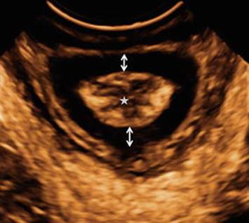
Fig. 4.4
3D ultrasound measurement of the thickness of the anterior and the posterior anal sphincter muscle in the reference plane. The a/p-ratio in this patient is 1.0 (double-head arrow). Asterisk = rectal mucosa
References
1.
Tan EK, Tan EL. Alterations in physiology and anatomy during pregnancy. Best Pract Res Clin Obstet Gynaecol. 2013;27(6):791–802.
2.
Genadry R. A urogynecologist’s view of the pelvic floor effects of vaginal delivery/cesarean section for the urologist. Curr Urol Rep. 2006;7:376–83.CrossRefPubMed
3.
Rahn DD, Ruff MD, Brown SA, Tibbals HF, Word RA. Biomechanical properties of the vaginal wall: effect of pregnancy, elastic fiber deficiency, and pelvic organ prolapse. Am J Obstet Gynecol. 2008;198:590.e1–e9.CrossRef
4.
Yeomans ER, Gilstrap 3rd LC. Physiologic changes in pregnancy and their impact on critical care. Crit Care Med. 2005;33 Suppl 10:S256–8.CrossRefPubMed
5.
FitzGerald MP, Graziano S. Anatomic and functional changes of the lower urinary tract during pregnancy. Urol Clin North Am. 2007;34:7–12.CrossRefPubMed
6.
South MM, Stinnett SS, Sanders DB, Weidner AC. Levator ani denervation and reinnervation 6 months after childbirth. Am J Obstet Gynecol. 2009;200:519.e1–7.CrossRef
7.
Snooks SJ, Setchell M, Swash M, Henry MM. Injury to innervation of pelvic floor sphincter musculature in childbirth. Lancet. 1984;2:546–50.CrossRefPubMed
8.
Weidner AC, Jamison MG, Branham V, South MM, Borawski KM, Romero AA. Neuropathic injury to the levator ani occurs in 1 in 4 primiparous women. Am J Obstet Gynecol. 2006;195:1851–6.CrossRefPubMed
9.
Snooks SJ, Swash M, Mathers SE, Henry MM. Effect of vaginal delivery on the pelvic floor: a 5-year follow-up. Br J Surg. 1990;77:1358–60.CrossRefPubMed
10.
Shafik A, El-Sibai O. Study of the levator ani muscle in the multipara: role of levator dysfunction in defecation disorders. J Obstet Gynaecol. 2002;22:187–92.CrossRefPubMed
11.
Weidner AC, Barber MD, Visco AG, Bump RC, Sanders DB. Pelvic muscle electromyography of levator ani and external anal sphincter in nulliparous women and women with pelvic floor dysfunction. Am J Obstet Gynecol. 2000;183:1390–9.CrossRefPubMed
12.
Allen RE, Hosker GL, Smith AR, Warrell DW. Pelvic floor damage and childbirth: a neurophysiological study. Br J Obstet Gynaecol. 1990;97:770–9.CrossRefPubMed
13.
Bump RC, Mattiasson A, Bø K, Brubaker LP, DeLancey JO, Klarskov P, et al. The standardization of terminology of female pelvic organ prolapse and pelvic floor dysfunction. Obstet Gynecol. 1996;175(1):10–7.
14.
O’Byole AL, Woodman PR, O’Boyle JD, Davis GD, Swift SE. Pelvic organ support in nulliparous pregnant and nonpregnant women: a case control study. Am J Obstet Gynecol. 2002;187:99–102.CrossRef
15.
O’Boyle AL, O’Boyle JD, Ricks RE, Patience TH, Calhoun B, Davis G. The natural history of pelvic organ support in pregnancy. Int Urogynecol J. 2003;14:46–9.CrossRef
16.
Elenskaia K, Thakar R, Sultan AH, Scheer I, Onwude J. Pelvic organ support, symptoms and quality of life during pregnancy: a prospective study. Int Urogynecol J. 2003;24:1085–90.CrossRef
17.
Cosson M, Lambaudie E, Boukerrou M, Lobry P, Crepin G, Ego A. A biomechanical study of the strength of vaginal tissues. Results on 16 post-menopausal patients presenting with genital prolapse. Eur J Obstet Gynecol Reprod Biol. 2004;112:201–5.CrossRefPubMed
18.
Cosson M, Boukerrou M, Lacaze S, Lambaudie E, Fasel J, Mesdagh H, et al. A study of pelvic ligament strength. Eur J Obstet Gynecol Reprod Biol. 2003;109:80–7.CrossRefPubMed
19.
Mørkved S, Rommen K, Schei B, Salvesen KA, Bø K. No difference in urinary incontinence between training and control group six years after cessation of a randomized controlled trial, but improved sexual satisfaction in the training group. Neurourol Urodyn. 2007;26(5):667 [Abstract 50].
20.
Sampselle CM, Miller JM, Mims BL, Delancey JO, Ashton-Miller JA, Antonakos CL. Effect of pelvic muscle exercise on transient incontinence during pregnancy and after birth. Obstet Gynecol. 1998;91:406–12.CrossRefPubMed
21.
Reilly E, Freeman R, Waterfield M, Waterfield A, Steggles P, Pedlar F. Prevention of postpartum stress incontinence in primigravida with increased bladder neck mobility: a randomised controlled trial of antenatal pelvic floor exercises. BJOG. 2002;109:68–76.CrossRefPubMed
22.
Boyle R, Hay-Smith EJ, Cody JD, Morkved S. Pelvic floor muscle training for prevention and treatment of urinary and faecal incontinence in antenatal and postnatal women. Cochrane Database Syst Rev. 2012;(17):CD007471.
23.
Marques J, Botelho S, Pereira LC, Lanza AH, Amorim CF, Palma P, Riccetto C. Pelvic floor muscle training program increases muscular contractility during first pregnancy and postpartum: electromyographic study. Neurourol Urodyn. 2013;32(7):998–1003.CrossRefPubMed
24.
Bracken JN, Reyes M, Gendron JM, Pierce LM, Runge VM, Kuehl TJ. Alterations in pelvic floor muscles and pelvic organ support by pregnancy and vaginal delivery in squirrel monkeys. Int Urogynecol J. 2011;22(9):1109–16.CrossRefPubMedPubMedCentral
25.
Boreham MK, Zaretsky MV, Corton MM, Alexander JM, McIntire DD, Twickler D. Appearance of the levator ani muscle in pregnancy as assessed by 3-D MRI. Am J Obstet Gynecol. 2005;193:2159–64.CrossRefPubMed
26.
Mant J, Painter R, Vessey M. Epidemiology of genital prolapse: observations from the Oxford Family Planning Association Study. BJOG. 1997;104:579–85.CrossRef
27.
Hoyte L, Schierlitz L, Zou K, Flresh G, Fielding JR. Two- and 3-dimensional MRI comparison of levator ani structure, volume and integrity in women with stress incontinence and prolapse. Am J Obstet Gynecol. 2001;185:11–9.CrossRefPubMed
28.
Fielding JR, Dumanli H, Schreyer AG, Okuda S, Gering DT, Zou KH, et al. MR-based three-dimensional modeling of the normal pelvic floor in women: quantification of muscle mass. AJR. 2000;174:657–60.CrossRefPubMed
29.
Tunn R, DeLancey JOL, Howard D, Ashton-Miller JA, Quint LE. Anatomic variations in the levator ani muscle, endopelvic fascia, and urethra in nulliparas evaluated by magnetic resonance imaging. Am J Obstet Gynecol. 2003;188:116–21.CrossRefPubMed
30.
Lanzarone V, Dietz HP. Three-dimensional ultrasound imaging of the levator hiatus in late pregnancy and associations with delivery outcomes. Aust N Z J Obstet Gynaecol. 2007;47(3):176–80.CrossRefPubMed
31.
Shek KL, Chantarasorn V, Langer S, Phipps H, Dietz HP. Does the Epi-No birth trainer reduce levator trauma? A randomised controlled trial. Int Urogynecol J. 2011;22(12):1521–8.CrossRefPubMed
32.
Da Silva-Filho AL, Martins PA, Parente MP, Saleme CS, Roza T, Pinotti M, Mascarenhas T, Natal Jorge RM. Translation of biomechanics research to urogynecology. Arch Gynecol Obstet. 2010;282:149–55.CrossRefPubMed
33.
Hoyte L, Damaser MS, Warfield SK, Chukkapalli G, Majumdar A, Choi DJ, Trivedi A, Krysl P. Quantitiy and distribution of levator ani stretch during simulated vaginal childbirth. Am J Obstet Gynecol. 2008;199:198.e1–e5.CrossRef
34.
Lien KC, Mooney B, DeLancey JO, Ashton-Miller JA. Levator ani muscles stretch induced by simulated vaginal birth. Obstet Gynecol. 2004;103:31–40.CrossRefPubMedPubMedCentral
35.
Parente MP, Jorge RM, Mascarenhas T, Fernandes AA, Martins JA. Deformation oft he pelvic floor muscles during a vaginal delivery. Int Urogynecol J. 2008;19:65–71.CrossRef
36.
Chen L, Ashton-Miller JA, DeLancey JO. A 3D finite element model of anterior vaginal wall support to evaluate mechanisms underlying cystocele formation. J Biomech. 2009;42:1371–7.CrossRefPubMedPubMedCentral
37.
Peña E, Calvo B, Martínez MA, Martins P, Mascarenhas T, Jorge RM, Ferreira A, Doblaré M. Experimental study and constitutive modeling of the viscoelastic mechanical properties of the human prolapsed vaginal tissue. Biomech Model Mechanobiol. 2009;9(1):35–44.CrossRefPubMed
38.
Landon CR, Crofts CE, Smith AR, Trowbridge EA. Mechanical properties on fascia during pregnancy: a possible factor in the development of stress incontinence of urine. Contemp Rev Obstet Gynaecol. 1990;2:40–6.
39.
Keriakos R, Bhatta SR, Morris F, Mason S, Buckley S. Pelvic girdle pain during pregnancy and puerperium. J Obstet Gynaecol. 2011;31(7):572–80.CrossRefPubMed
40.
Wesnes SL, Hunskaar S, Bo K, Rortveit G. The effect of urinary incontinence status during pregnancy and delivery mode on incontinence postpartum. A cohort study. BJOG. 2009;116:700–7.CrossRefPubMedPubMedCentral
41.
Sangsawang B, Sangsawang N. Stress urinary incontinence in pregnant women: a review of prevalence, pathophysiology, and treatment. Int Urogynecol J. 2013;24(6):901–12.CrossRefPubMedPubMedCentral
42.
Thorp JM, Norton PA, Wall LL, Kuller JA, Eucker B, Wells E. Urinary incontinence in pregnancy and the puerperium: a prospective study. Am J Obstet Gynecol. 1999;18:226–73.
43.
Peschers U, Schaer G, Anthuber C, DeLancey JO, Schuessler B. Changes in vesical neck mobility following vaginal delivery. Obstet Gynecol. 1996;88:1001–6.CrossRefPubMed
44.
Wijma J, Weis Potters AE, von der Mark TW, Tinga DJ, Aarnoudse JG. Displacement and recovery of the vesical neck position during pregnancy and after childbirth. Neurourol Urodyn. 2007;26:372–6.CrossRefPubMed
45.
Dietz HP, Eldridge A, Grace M, Clarke B. Does pregnancy affect pelvic organ mobility? Austr Z J Obstet Gynaecol. 2004;44:517–20.CrossRef
46.
Wijma J, Weis Potters AE, de Wolf BT, Tinga DJ, Aarnoudse JG. Anatomical and functional changes in the lower urinary tract during pregnancy. BJOG. 2001;108:726–32.PubMed
47.
Iosif S, Ingemarsson I, Ulmsten U. Urodynamic studies in normal pregnancy and in puerperium. Am J Obstet Gynecol. 1980;137:696–700.CrossRefPubMed
48.
Iosif S, Ulmsten U. Comparative urodynamic studies of continent and stress incontinent women in pregnancy and in the puerperium. Am J Obstet Gynecol. 1981;140:645–50.CrossRefPubMed
49.
Shek KL, Kruger J, Dietz HP. The effect of pregnancy on hiatal dimensions and urethral mobility: an observational study. Int Urogynecol J. 2012;23(11):1561–7.CrossRefPubMed
50.
van Veelen GA, Schweitzer KJ, van der Vaart CH. Ultrasound imaging of the pelvic floor: changes in anatomy during and after first pregnancy. Ultrasound Obstet Gynecol. 2014;44(4):476–80.CrossRefPubMed
51.
Kubotani JS, Araujo Júnior E, Zanetti MR, Passos JP, de Jármy Di Bella ZI, Júnior JE. Assessing the impact of twin pregnancies on the pelvic floor using 3-dimensional sonography: a pilot study. J Ultrasound Med. 2014;33(7):1179–83.CrossRefPubMed
52.
Ochsenbein N, Kurmanavicius J, Huch R, Huch A, Wisser J. Volume sonography of the pelvic floor in nulliparous women after elective cesarean section. Acta Obstet Gynecol Scand. 2001;80:611–5.CrossRefPubMed