
Carl A. Laskin1 , Kenneth I. Cadesky2, Christine A. Clark3 and Karen A. Spitzer3
(1)
Departments of Medicine (Rheumatology) and Obstetrics & Gynecology, (Reproductive Endocrinology and Infertility), LifeQuest Centre for Reproductive Medicine, University of Toronto, 655 Bay Street, Suite 1800, Toronto, ON, Canada, M5G 2K4
(2)
Department of Obstetrics and Gynecology (Reproductive Endocrinology and Infertility), Mount Sinai Hospital; LifeQuest Centre for Reproductive Medicine, Toronto, ON, Canada
(3)
LifeQuest Centre for Reproductive Medicine, Toronto, ON, Canada
Carl A. Laskin
Email: calaskin@gmail.com
Introduction
Definitions
Infertility: The American Society of Reproductive Medicine has recently revised their definition of infertility. This accepted definition is as follows: “Infertility is a disease, defined by the failure to achieve a successful pregnancy after 12 months or more of appropriate, timed unprotected intercourse or therapeutic donor insemination. Earlier evaluation and treatment may be justified based on medical history and physical findings and is warranted after 6 months for women over age 35 years” [1]. The 1-year timeframe is explained by the probability that 85–90 % of healthy young couples will conceive within 12 months, which means that infertility affects 10–15 % of the population.
Fecundity: This is the probability that a single menstrual cycle will result in a live birth whereas fecundability is the probability that a single cycle will result in a pregnancy. The probability of a pregnancy in a single cycle is 20–25 % of otherwise normal couples.
The probability of a pregnancy is dependent upon a number of factors, the most important of which is the woman’s age. In those individuals afflicted with a rheumatic disease, the disease itself or the medications used to treat the underlying condition may adversely affect fertility and fecundity. Bearing these factors in mind, one would suspect that men and women with rheumatic disorders may not have the same fertility as an age-matched general population. Furthermore, one would expect that fecundability and fecundity are likewise impaired in women with rheumatic diseases. These two issues need to be further investigated.
“Fertility 101”
The majority of people with systemic, inflammatory rheumatic diseases are women of childbearing age. It is therefore incumbent upon the attending specialist to discuss family planning with these women at least on a regular basis. It is essential that the woman enter into a pregnancy electively, planned with her attending specialist. A similar approach applies to the couple having difficulty conceiving. Every fertility evaluation should be viewed as a pre-pregnancy evaluation: if the woman’s disease is too active to assure a safe pregnancy, then assisted reproductive technology (ART) should be avoided. Most internists/rheumatologists have little knowledge of reproductive medicine and fertility management. If the rheumatologist’s evaluation suggests that the woman is in a quiescent stage of disease that would maximize the chances of a safe pregnancy, then a referral to a reproductive endocrinology and infertility (REI) specialist will be necessary. The referring rheumatologist should provide important details regarding the patient including the underlying disease entity and the medications currently prescribed. Prior to any management decisions by the REI, a dialogue should be established with the rheumatologist. This will be the best way to ensure that the woman can become pregnant safely using ART.
The Female Menstrual Cycle
Understanding the physiology of the menstrual cycle gives insight into how fertility issues are investigated in women. Figure 12.1 illustrates the events in a normal menstrual cycle. The ovulatory woman will often experience a repeated pattern of premenstrual symptoms followed by recognizable symptoms during menses. The cycles in such women are regular and predictable whereas those in women who may not be ovulating are irregular and unpredictable. The events of the cycle that are the target of investigation are the basal hormone levels: Day 3 follicle stimulating hormone (FSH), luteinizing hormone (LH), and estrogen. The rise in progesterone levels in the latter phase of the cycle is indicative of ovulation. The best method to study the events of a woman’s menstrual cycle would be to monitor the hormone levels and correlate with follicle development using ultrasound. Cycle monitoring provides a dynamic view of the woman’s physiology and may provide clues to understanding the reasons underlying the failure to conceive.
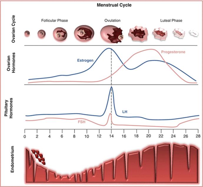
Fig. 12.1
The female menstrual cycle. Each level of the diagram illustrates the response of the various targets to the pituitary hormones, FSH and LH. Production of each of the ovarian hormones, estrogen and progesterone, is dependent upon the growth of the maturing follicle. The development of the endometrium in turn is dependent upon the appropriate production of the ovarian hormones
Fertility Evaluation and Investigation
Aging and Fertility
It becomes very clear when assessing female fertility that there is no substitute for age. The ability to both conceive and maintain a pregnancy is strongly influenced by a woman’s age. Whether career choice or life circumstances, those women who delay childbearing may face infertility by the time they are ready to become pregnant. When a woman reaches 37 years of age, her chances of becoming pregnant decreases significantly with each passing year; indeed, as many as 99 % of women at age 45 are infertile. Although the average age of menopause is 51.8 years, reproductive menopause occurs 6–7 years earlier. Beyond age 37, the remaining oocyte quality declines exponentially as the better quality oocytes have already been released. A decrease in oocyte quality refers to an increased prevalence of aneuploid oocytes owing to dysfunction of the meiotic spindle. This will lead to higher frequency of chromosomal abnormalities, which in turn leads to miscarriage or infertility.
Assessment of the Infertile Couple
The fertility assessment can be conceptualized as “macrofertility” involving the assessment of ovulation, fallopian tubes, uterus, and sperm analysis. When all such studies are found to be normal, the diagnosis of “unexplained infertility” is made. To explore this further involves a “microfertility” assessment. There are literally thousands of steps involved in the interaction of sperm and oocyte. We are currently able to identify about 42 known abnormalities at this level of investigation.
The Office Visit
A targeted, detailed history and physical exam is the starting point for assessment of the woman. The gynecologic and obstetric history should include the menstrual history (onset of menarche; regularity of cycles; dysmenorrhea and dyspareunia); history of pelvic surgery; type and use of contraceptives; and a history of any pregnancy complications and outcomes. Other aspects to the history such as exercise program; weight loss or gain; smoking and alcohol intake; work, travel and dietary preferences may also have significant adverse effects on ability to conceive. The physical exam should include weight and body mass index; thyroid abnormalities; breast abnormalities including secretions; hirsutism and other signs of androgenicity, and a pelvic exam
The male assessment will similarly require a complete history and physical exam. Discovery of an abnormality from the semen analysis requires ruling out systemic illness or medication that adversely affects sperm. Specific points in the history include smoking, alcohol intake, use of recreational drugs, and occupation. Prior reproductive issues in other relationships should be discussed. In addition, a history of erectile dysfunction or difficulty ejaculating may prove to be an underlying cause of the infertility, although this is frequently more difficult to elicit from the patient. A history of varicocele repair, vasectomy, hernia repair, undescended testis, testicular injury or mumps orchitis as well as certain medications such as testosterone, finasteride, or minoxidil may explain abnormalities in the semen analysis.
The last step in the office evaluation is to discuss details of sexual intercourse with the couple. Too often or too infrequent sexual intercourse may explain failed conception. Is the intercourse timed? Are there problems with intercourse? Are they using lubricants, many of which are spermostatic? These are sensitive topics and must be approached with that in mind.
“Macrofertility” Investigations
The investigations in the female are more involved at all levels (Fig. 12.2a, b). Ovulation is assessed through the history as well as hormone levels, including estradiol (E2), LH, FSH, and progesterone, measured at Day 3, midcycle, and after suspected ovulation. The fallopian tubes are assessed using a hysterosalpingogram (HSG) or a sonohystogram (SHG). The former is a much better test for tubal patency while the latter is a good screening test. In addition to the tubal assessment, these two investigations will also delineate the uterine cavity assessing for the presence of polyps or impinging fibroids. With the utilization of 3D imaging, the SHG gives a more detailed view of the uterine cavity compared to the HSG.

Fig. 12.2
Fertility assessment flow chart. This is a simplified view of the fertility assessment in both the male (a) and female (b). FSH follicle stimulating hormone, E2 estradiol, LH luteinizing hormone, AMHanti-Müllerian hormone, TSH thyroid stimulating hormone
The Hormone Evaluation of the Woman
The hormone evaluation includes those hormones that are directly involved in the menstrual cycle. The hormones E2, LH, FSH, and progesterone measured at Day 3 of the woman’s cycle indicate that the woman is indeed at the beginning of her cycle and represent the basal hormone evaluation. The FSH should be at 10 IU/L or lower with a low E2, LH, and progesterone. At midcycle, the LH should rise or surge thereby triggering ovulation. After ovulation, all hormones fall except the progesterone, which should rise (Fig. 12.1). Other hormones that play a role in ovulation indirectly are thyroid stimulating hormone (TSH) and prolactin. TSH should be below 2.5 mU/L. If the prolactin is elevated, it should be measured fasting and include a macroprolactin level, which is a biologically inactive molecule that may account for what appears to be hyperprolactinemia.
Ovarian Reserve
Over the past 20 years, increasing emphasis has been placed on determining the remaining follicular pool (or ovarian reserve): a diminishing ovarian reserve is expected as a woman ages. However, younger women might also be found to have diminished ovarian reserve underlying a fertility disorder. A basal FSH level measured at Day 3 appears to correlate with ovarian reserve: serum FSH is considered elevated when above 10 IU/L and suggests a diminished ovarian reserve; it may also indicate low oocyte quality.
An antral follicle count (AFC) is the number of follicles in the ovaries as determined by ultrasound between days 2–4 of the menstrual cycle. The normal range is not standardized but a count of 12 (the sum total in both ovaries) is usually considered as the lower limit of normal. The AFC appears to be another reliable measure of ovarian reserve and correlates with the probability of pregnancy as well as success in an in vitro fertilization (IVF) cycle.
A more recent test that is the subject of a rapidly growing literature is the anti-Müllerian hormone (AMH) evaluation: AMH is a peptide growth factor produced in females by granulosa cells from pre-antral and antral follicles [2]. High levels of AMH indicate good ovarian reserve. Routine fertility assessment now includes measurement of circulating AMH, based on reports that it is a more reliable indicator of ovarian age than other markers including Day 3 FSH and inhibin [3]. AMH is easier to measure, as levels remain relatively stable throughout the menstrual cycle in contrast to FSH and is less invasive than ultrasound-guided AFC. AMH decreases with declining AFC and increasing age [4].
In summary, an accurate assessment of ovarian reserve should be part of every female fertility evaluation and includes Day 3 FSH, Day 2–4 AFC, and an AMH level, which can be measured at any point in the cycle.
Investigation of the Male
In men, the semen analysis provides the majority of data regarding fertility evaluations (Fig. 12.2a). Routine semen analysis measures volume of the ejaculate, sperm count, motility, and percentage of normal sperm cells. A more detailed sperm analysis utilizes a computer analyzed sperm assessment (CASA) which includes an index of fragmentation of genetic material in the sperm cells called the DNA fragmentation index (DFI). A DFI above 30 % is associated with a fourfold reduction of fertility in natural cycles; some studies suggest a doubling of first trimester miscarriages as well, but this is more controversial [5]. Men may have all sperm parameters normal except for the DFI, highlighting the critical importance of the DFI in the male fertility evaluation. Hormone studies may be helpful in understanding some abnormal results of the sperm analysis
Initial Management
The initial management of a couple with infertility involves identifying and correcting these “macrofertility” abnormalities. Lifestyle issues such as smoking, alcohol intake, stress, diet, exercise, and travel are important management decisions that are under the couple’s control. They should be addressed and may need appropriate counseling. The couple must understand the “correct” window of opportunity and need to know the optimal time in the female partner’s cycle that has the highest probability for conception: that is, they must have intercourse around ovulation. In a woman with a 28-day cycle, having intercourse every 2 days between Days 10 and 20 will generally cover the “fertile” period. When these simple measures are unsuccessful, ovulation induction or superovulation under the care of the REI may be necessary. When there are issues of sperm quality, referral to an andrologist should be made. Improving sperm quality is not always easy or successful but with appropriate guidance by an andrologist, reasonable treatment programs should be attempted. If the above measures are unsuccessful after a reasonable time, ART treatment options are necessary.
Microfertility Management
Most causes of infertility are due to disruptions in microfertility. All ART treatment modalities are based upon superovulation: the administration of fertility drugs (exogenous FSH) stimulates the ovaries to mature a number of follicles as opposed to the single follicle matured in a natural cycle. As the follicles grow and mature they produce estrogen resulting in an increase in the endogenous estrogen level. This may be important to consider when we address such treatment in rheumatic diseases. The least invasive ART protocol is controlled ovarian hyperstimulation followed by intrauterine insemination, which involves the careful administration of exogenous FSH to produce two to three mature follicles. Sperm must then be safely prepared for injection into the uterine cavity. Although this treatment is not associated with a high success rate, outcome is better than the couple simply trying on their own with timed intercourse. A limited number of cycles should be attempted (three to six cycles); if a pregnancy is not achieved, the more invasive IVF technology can be undertaken.
IVF involves a more aggressive stimulation with exogenous FSH leading to the maturation of a larger number of follicles (and higher levels of estrogen). Once the follicles have matured, the oocytes are surgically extracted, fertilized in vitro, and then incubated over 3 or 5 days. Concerns regarding low sperm count or motility can be addressed utilizing intracytoplasmic sperm injection (ICSI) where a single sperm cell is injected directly into the oocyte. Depending upon the management plan, a specific number of embryos (3-day incubation) or blastocysts (5-day incubation) are transferred directly into the uterine cavity. The woman is usually given progesterone until a pregnancy test is performed. If pregnancy has been achieved, then the progesterone is continued until sometime in the latter part of the first trimester (usually 10–12 weeks gestation).
Fertility Management in the Rheumatic Diseases
When the underlying rheumatic disease is active, there may be negative feedback on the hypothalamic–pituitary–ovarian axis in the woman or on spermatogenesis in the male [6, 7] contributing to fertility. In addition, certain drugs used in the treatment of the underlying condition may adversely affect fertility in either gender [8, 9]. The remainder of this chapter will cover specific rheumatic diseases giving special consideration to the impact of the disease and medication on the individual’s fertility, as well as the potential effect of fertility management on the disease course. Fertility issues specific to some of the more common rheumatic disorders such as rheumatoid arthritis (RA), systemic lupus erythematosus (SLE), anti-phospholipid syndrome (APS), ankylosing spondylitis (AS), and scleroderma (SSc) will be discussed below. In addition, some special considerations regarding various medications used in the treatment of specific rheumatic diseases will be discussed.
Rheumatoid Arthritis
Most chronic rheumatic diseases such as RA are not known to directly affect fertility [10]. However, disease activity may impact on ovulatory function through an interruption of the hypothalamic–pituitary–ovarian axis. In addition, medications used in the treatment of RA may adversely affect fertility both in women and in men. Hargreaves reported in 1958 that a group of women with known RA had fewer children than a group without the disease [11]; this observation has been substantiated by others and most recently by Clowse et al. [12]. Variables such as lifestyle, disease activity, or a change in perspective by women and their partners may all contribute to the ultimate size of the family. Recent studies tend to support the original observation from a controlled study by Kay and Bach that fertility is lower in women both before and after the onset of disease [13]; furthermore, menopause occurred earlier in those who ultimately developed RA compared to controls. The major shortcoming of all of these studies suggesting decreased fertility in women with RA is that they are all based on self-report questionnaires and address life events as opposed to a more physiological approach to ovarian function in RA. Until such studies are done, we are left with the limited conclusion that for women with RA, family size tends to be smaller.
Investigation and Management of Infertility
The investigation and management of infertility in RA differs little from that in the general population. However, as in all scenarios with an underlying medical problem in a woman contemplating pregnancy, it is imperative that disease be under optimal control and that all medications are compatible with pregnancy (Chap. 14). The fertility evaluation should be undertaken as outlined above and in (Fig. 12.2a, b). While adjustments to medications need to be made in terms of potential teratogenicity, most medications do not interfere with fertility. There are exceptions. Most nonsteroidal anti-inflammatory drugs (NSAIDs) can be taken prior to conception. It is important to know that NSAIDs can interfere at the ovulation step by inhibiting follicular rupture [14] and affect implantation; therefore, it is advisable to discontinue these medications during a conception cycle. Once an intrauterine pregnancy is documented, NSAIDs can be used until 32 weeks gestation. Ibuprofen and naproxen may be preferable as there is considerable experience with these medications. The other drug to consider is sulfasalazine, which leads to reversible oligospermia and/or asthenospermia demonstrated on semen analysis in approximately 30 % of males on the drug [9]. The low sperm count is reversed in 3 months after discontinuation.
In summary, fertility investigation and management in men and women with RA is directed to controlling disease activity and then ensuring that medications are safe for use before and during pregnancy. With few exceptions, the majority of drugs used in the treatment of RA have little or no impact on fertility.
Systemic Lupus Erythematosus and APS
SLE has its greatest prevalence among women of reproductive age. Unlike RA, SLE seems to be a more hormonally sensitive disorder which leads to significant implications for the treatment of infertility. Medications tend not to be the limiting step in the treatment of a woman with SLE undergoing fertility therapy. Disease manifestations and certain potential risk factors such as the presence of an anti-phospholipid antibody may directly influence the treatment program. In contrast to the generally accepted position that fertility is unaffected, some recent studies suggest that there may be a direct impact of the disease on fertility [15, 16]. As is the case in any medical disorder in a woman contemplating a pregnancy, if there is concern regarding the woman becoming pregnant, then assisting the woman to become pregnant would be contraindicated.
There is a wide body of literature regarding a higher prevalence of various autoantibodies in women with unexplained infertility [17–21]. However, if any of these autoantibodies are associated with infertility, it is unclear which ones may be predictive and there is little if any evidence supporting the routine screening of women or men for the presence of such autoantibodies when investigating infertility. A number of REI specialists routinely assay various autoantibodies in otherwise healthy women being investigated for infertility [17–21] based primarily upon studies in recurrent pregnancy loss, which, in turn, is based on the observation that women with SLE have a higher prevalence of recurrent miscarriage and fetal wastage [22–24]. This extrapolation from the recurrent miscarriage and SLE literature is both questionable and fraught with the risk of erroneous treatment decisions.
Medical Evaluation
The initial evaluation of a couple with infertility where the woman has SLE should be considered a pre-pregnancy evaluation. Once the woman has been medically cleared for pregnancy, the couple can then be referred to an REI specialist for investigation and management of infertility.
Fertility Evaluation
Fertility assessment for patients with SLE does not differ from a routine fertility assessment. Recent papers suggest that there may be diminished ovarian reserve in SLE patients as determined by the measurement of AMH [25, 26]. Lawrenz et al. reported that AMH levels in 33 SLE patients were lower than in healthy controls and concluded that SLE itself may have a negative influence on ovarian reserve, although there were no differences in number of children or miscarriages between the two groups [25]. Mok et al. reported that AMH was significantly lower in women exposed to cyclophosphamide (CYC), suggesting ovarian damage (no effect was seen in women exposed to other immunosuppressive agents). Increasing age and each cumulative 5 g CYC exposure were independently associated with decreased AMH [27]. However, it has also been reported that low levels of AMH are not necessarily predictive of either reduced fecundability or of reduced live birth rate in healthy young women [4]. This latter finding suggests a need for caution before assuming the strength of correlation between AMH and long-term fertility, whether in a general or autoimmune population [2, 28]. Until a comprehensive study of fertility evaluation is undertaken in women with SLE, it is premature to assume that such women have diminished ovarian reserve.
Timing of the fertility evaluation appears to be important. Whether in the male or female lupus patient, active disease may negatively impact fertility whereas inactive or well-controlled disease will lead to a more accurate assessment of fertility in the individual. In a woman with active lupus or chronic renal failure, fertility may be impaired by anovulation through interruption of the hypothalamic–pituitary–ovarian axis; high doses of corticosteroids may interfere with ovulation through a similar mechanism. Whether the disease itself negatively impacts fertility is unclear, and there is considerable variation in opinion [7, 15, 17, 19–21].
Anti-Phospholipid Antibodies and Infertility
Anti-phospholipid antibodies (aPL) have been reported to be associated with recurrent pregnancy loss and adverse pregnancy outcomes [29–33]. However, several recent studies do not find a strong association, if any, between aPL and early pregnancy loss [34, 35]. Recent studies support the association of late pregnancy loss primarily with the lupus anticoagulant (LAC), with little association with anti-cardiolipin antibodies and no association with antibodies to β2-glycoprotein I [36, 37]. There continues to be ongoing controversy regarding an association of aPL with infertility and failed implantation [38–44]. When women with aPL undergoing IVF are treated with heparin +/− ASA +/− corticosteroids, most show no benefit in terms of pregnancy outcome [41–43]. Hornstein et al. in 2000 performed a meta-analysis of seven studies on aPL and IVF outcome [45] including 2,053 women, 703 of whom had at least one positive aPL result: these authors found no association between aPL and either clinical pregnancy or live birth. A further point, based on a pathogenetic mechanism, calls into question the relevance of aPL to female infertility and failed implantation [39]. Finally, the American Society for Reproductive Medicine has issued a statement from the Practice Committee in which they state that there is no indication for the assessment of aPL in otherwise healthy couples undergoing IVF and that therapy to treat such antibodies is not justified based upon the existing data [46].
In summary, despite a few studies implicating aPL in female infertility and implantation failure in IVF, there is a dearth of evidence supporting such an association. Based upon the evidence at this time, there is no reason to routinely screen healthy women undergoing fertility evaluation or treatment for aPL.
Ovulation Induction and IVF
It has been hypothesized that SLE is a disease that can be exacerbated by changes in sex hormone concentrations, particularly elevated estrogen levels. If so, then fertility treatment incorporating ovulation induction or superovulation may present a risk of induction of SLE or a flare of disease in those with known SLE through the generation of endogenous hyperestrogenemia and enhancement of humoral immunity [47]. Although induction of SLE appears to be a rare possibility, flare of established SLE may occur with evidence supporting the occurrence of disease exacerbation in women undergoing fertility therapy [48–50]. However, in the well-selected patient, ovulation induction with or without IVF can be carried out safely.
Identifying the patient in whom ovulation induction is safe involves a careful and complete medical assessment of the woman with respect to her underlying SLE. Clinically the patient should show no evidence of active disease for the 6 months preceding an attempt at pregnancy. Despite the absence of Level I or II evidence, most physicians support the expert recommendations regarding this 6-month time frame. Blood pressure must be normalized, so any anti-hypertensive medication in use must be safe for pregnancy. With this information, a “clinical profile” of the patient can be formulated so that all involved in the patient’s care know which clinical manifestations that appear during ART or later during pregnancy are those of active lupus.
The “laboratory profile” will consist of the patient’s baseline, routine laboratory tests as well as the lupus serology. The patient’s rheumatologist will know if the patient’s clinical disease activity is concordant or discordant with serology. If discordant, then clearly the clinical assessment is critical. Testing for aPL is necessary as those SLE patients positive for this antibody may be at greater risk of a thromboembolic event in the presence of hyperestrogenemia. In those with documented APS with a high risk of thrombosis, it may be inadvisable to undergo ovulation induction especially if it is part of an IVF cycle. This issue will be discussed further below. The clinical and laboratory profiles are helpful to “define” the woman’s disease. In this way all involved will be aware of which manifestations and laboratory parameters to monitor. In addition, a management plan can be formulated in advance should the woman flare during the fertility therapy or during pregnancy.
To complete the assessment prior to undertaking ovulation induction or IVF, medications must be reviewed and the woman stabilized on pregnancy-safe drugs. The ideal patient with SLE deemed to be a good candidate for ovulation induction therapy (with or without IVF) has minor organ disease and is on minimal medication. Women with stable clinical lupus who may have renal disease are also acceptable provided the disease and renal status are stable, with minimal risk during a pregnancy. In contrast, women with complicated lupus on numerous medications as well as higher doses of prednisone, hypertension, renal insufficiency, and/or cytopenias are not suitable candidates for ovulation induction with or without IVF.
Ovarian Hyperstimulation Syndrome
Ovarian hyperstimulation syndrome (OHSS) is an iatrogenic complication of ovulation induction therapy. It is an exaggerated response typically associated with the use of exogenous gonadotropin stimulation. The ovaries are markedly enlarged with numerous maturing follicles. A capillary leak syndrome develops mediated by vascular endothelial growth factor (VEGF), which ultimately results in a third space fluid shift into the peritoneal and/or pleural cavities [51]. OHSS can be mild or of sufficient severity to warrant admission to an ICU. Severe OHSS occurs in approximately 1 % of those undergoing ovulation induction whereas mild to moderate OHSS occurs in 4–10 %. The mortality rate of OHSS is 1/45,000–1/500,000 [7]. OHSS presents a risk of renal compromise and thromboembolism which may be additive in the woman with SLE, especially if she also has aPL. It is therefore prudent to be certain the REI specialist managing the patient is aware of her status and designs a stimulation protocol that minimizes the risk of OHSS.
APS and Fertility Management Including IVF
In those women (with or without underlying SLE) with documented APS, there are definite risks to ovarian induction therapy and IVF due to the hyperestrogenemia. In IVF cycle protocols, combined oral contraceptives are often used at the start of the cycle: this may place the woman at significant risk of a thromboembolic event. In those with previous arterial events, it may be prudent to avoid ovulation induction therapy completely. Alternatively, a minimal stimulation protocol or natural cycle could be used in IVF [52, 53]. Although some centers report a reasonable pregnancy rate with such protocols, many clinics find that they are often accompanied by a much lower pregnancy rate [54]. In those patients where hyperestrogenemia is absolutely contraindicated, eliminating the use of gonadotropins, aromatase inhibitors such as letrozole can be used in cycles using intrauterine insemination. Although letrozole could be used alone in an IVF cycle, this will usually result in a single follicle and single oocyte. This is equivalent to a natural cycle IVF but with more control [55].
In aPL-positive women with a history of venous events, stimulation may be carried out using anticoagulation with heparin or low-molecular-weight heparin, which is then held prior to oocyte recovery, following which it is restarted soon thereafter. In such patients it is critical to avoid OHSS, which in and of itself, presents an added risk to hypercoagulability and thrombosis. With proper attention to management of women with APS including a coordinated approach by the rheumatologist/hematologist and the REI specialist, ovulation induction with or without IVF can be undertaken safely [50, 56, 57].
Ankylosing Spondylitis
Ankylosing spondylitis (AS) is one of the rheumatic disorders that has a predilection for males, with a male to female ratio of 2:1–3:1. The disease has its onset during the reproductive years usually between 20 and 30 years of age. A study surveying a large number of women with AS in the USA, Canada, and Europe found no adverse effects on pregnancy, fertility, or neonatal outcome [58]. In particular, of 649 women with previous pregnancies there were 2.4 pregnancies per woman, of which 1.4 pregnancies occurred during active disease suggesting that fertility was not adversely affected by AS. In contrast to this observation in women, a recent study has found a significantly higher frequency of varicocele in male AS patients than controls (40 vs. 8 %) [59]. Comparing those AS patients with a varicocele to AS patients without, the median of normal sperm forms was 13.5 % compared to 22 % indicating the potential for increased failed conception in those with the varicocele. No other studies have suggested any affect of AS on fertility in men or women [58, 60].
Apart from the lack of effect of the disease on fertility, some medications commonly used to treat AS are associated with subfertility. Sulfasalazine is known to cause reversible azospermia or oligospermia as well as reduced sperm motility and an increase in abnormal forms [61, 62]. In one early study, Birnie et al. found 86 % of men on sulfasalazine for inflammatory bowel disease had an abnormal semen analysis with 72 % having oligospermia [50]. In those men on sulfasalazine having an abnormal semen analysis which was discovered during a fertility assessment, 2–3 months off the drug usually corrected the abnormality. Tumor necrosis factor alpha (TNFα) antagonists have become a mainstay in the treatment of AS. At least two studies have shown no adverse effect of infliximab, etanercept, or adalimumab on male fertility [63–65].
Based upon the above literature, fertility assessment in women with AS usually proceeds routinely. There are no specific adverse effects of the disease on female fertility. However, a recent study by Nukumizu et al. suggests that all men with AS should be assessed for a varicocele [59]. In addition, a routine semen analysis in men on sulfasalazine should identify drug-related abnormalities. In such individuals, the drug should be discontinued for 2–3 months and the semen analysis repeated to determine if the abnormalities are now corrected. Since this may have been the cause of the infertility, no additional treatment needs to be instituted for several months. Should fertility treatment be necessary, there appears to be no contraindication to any intervention provided all medications used to treat the AS are pregnancy-safe.
The fertility evaluation in AS must be a pre-pregnancy evaluation: if pregnancy is not advised, then treatment of a fertility problem must be viewed as contraindicated.
Scleroderma
Scleroderma (SSc) is a rare disease with an incidence of two to ten cases per million usually presenting in the fifth to sixth decades. The ratio of female to male is 3:1 with an even greater female predominance in the reproductive years of 15:1 [66, 67]. Renal crisis remains the most serious complication of this disease; therefore, the major concern when considering pregnancy or fertility management is the extent of organ involvement. To minimize the risk of renal crisis or other major complications such as severe cardiopulmonary manifestations, pregnancy should be avoided in those women with rapidly progressive diffuse disease [68]. To reemphasize, the fertility evaluation in scleroderma must be a pre-pregnancy evaluation. If pregnancy is not advised, then treatment of a fertility problem must be viewed as contraindicated.
Psychosocial Variables
Fertility issues in women with SSc can involve some variables quite distinct from those of other rheumatic diseases. In a survey by Steen et al. of 214 SSc female patients, a significantly higher proportion had never been pregnant compared to a group with RA or normal controls [69]. This study discovered that unique to patients with SSc, a disproportionate number of women with had never married, were sexually inactive, or chose not to have children. Other contributing factors that negatively impact inter-personal relationships are an increased incidence of vaginal dryness and dyspareunia affecting up to 37 % of such patients in addition to joint contractures, which may prevent sexual intercourse [70].
Infertility and “Pre-scleroderma”
An unusual theory applicable only to SSc is that a history of reproductive failure, i.e. infertility or recurrent miscarriage, may predate the onset of SSc [68, 71]. The most recent study by Silman and Black using a postal questionnaire showed twice the rate of miscarriage and three times the rate of infertility (the women with SSc had no pregnancy by age 35) compared to a control population [71]. These authors and others hypothesize that the female predominance of the disease and the peak onset just after the reproductive years is due in part to this adverse reproductive history [71, 72]. The postulated mechanism associating reproductive failure with SSc is that of microchimerism in which there is a transplacental transfer of fetal cells during prior pregnancies or miscarriages that then initiates a chronic graft-versus-host reaction [72–77]. The theory is intriguing and suggests that in the appropriate host, there is a serious consequence to infertility or recurrent miscarriage that extends beyond the reproductive years.
Fertility Evaluation and Treatment
No woman with SSc should undertake fertility therapy without a pre-pregnancy medical assessment by a rheumatologist or internist who is very familiar with the condition: this evaluation becomes the start of the fertility evaluation. Once it is determined that it is safe for the woman to become pregnant, a routine fertility assessment can be undertaken in the woman and her partner. It is important that the REI specialist be cognizant of the unique psychosocial variables that may play a role as female factors in the couple’s infertility as these issues may be the reason underlying the infertility. After consultation between the rheumatologist/internist and the REI specialist, the patient may undergo ovulation induction and even IVF provided no relative or absolute medical contraindications are present. Success rates should be similar to the general population based upon the observations of Steen and Medsger [69]. These investigators found that the overall rate of a successful pregnancy following at least 1 year of failed conception was 37 % compared to 40 % in an RA group which was no different than 43 % seen in normal controls. The miscarriage rate, however, is increased in SSc especially in those with diffuse disease (24 %) compared to women with limited skin involvement (12 %) [77].
In conclusion, women with SSc contemplating a pregnancy require a detailed pre-pregnancy evaluation. This should include an evaluation for the presence of underlying vasculopathy and the extent of such involvement. If a fertility problem is present, only after the woman is cleared by medical evaluation should investigation proceed. Fertility therapy can be implemented as necessary with reasonable expectations on the part of the patients and the physicians. Should the woman become pregnant, in the absence of significant vascular disease, most pregnancies are accompanied by few adverse maternal or neonatal complications.
Alternative Options for the Patient with Complicated Connective Tissue Disease
There will be patients who are advised not to undergo a pregnancy owing to the severity of their underlying medical condition. This is a significant hardship for both the individual and the couple. The rheumatologist has an important role in offering support and referring the patient and/or couple to counseling and support groups when appropriate. Family planning includes a range of options in addition to pregnancy including adoption, gamete donation, and surrogacy utilizing a gestational carrier. In the latter scenario, the IVF cycle proceeds with the resulting embryos being transferred to the woman who has agreed to be the gestational carrier. Utilizing a gestational carrier eliminates the risk of OHSS and pregnancy in a woman with an underlying medical condition. Finally, choosing to be child-free is a family planning option that should also be discussed with patients.
Fertility Preservation
In those cases where it may be possible for a woman to carry a pregnancy at a later date but her current disease status contraindicates doing so at the present time, cryopreservation of oocytes, sperm, or embryos until the disease is inactive may be an option. If the patient has a partner of the opposite sex, the preferred option is to cryopreserve embryos, which have an excellent survival rate upon thawing, exceeding that of cryopreserved oocytes [78]. Where there is a woman without a partner, cryopreservation of oocytes is a viable option provided the woman is well enough to undergo ovulation induction and retrieval of oocytes. Cryopreservation of sperm is an established technology with an excellent survival rate on thaw provided the sperm is of good quality originally.
Prevention of Infertility
CYC therapy can be life-saving for a woman experiencing a major flare of a connective tissue disease such as SLE. However, knowledge of the long-term side effects of infertility and premature ovarian failure will often complicate this management decision. Studies have shown that the woman’s age at the time of treatment and the cumulative dose of CYC administered to either a man or a woman are major risk factors that contribute to CYC-induced gonadal failure [79]. Combined oral contraceptives may be protective for women while testosterone may offer similar benefit to men who are to be treated with CYC. However, there is a paucity of evidence to support such a preventative measure. The prior administration of the long-acting GnRH analogue, leuprolide, may prevent ovarian failure in the female [8]. In males, cryopreservation of sperm is optimal to preserve fertility in those who will require CYC treatment.
Conclusion
It is still maintained that individuals with rheumatic disease have normal fertility. Recent studies have questioned that conclusion but detailed, methodical studies are necessary to confirm or refute the assumption. Women with rheumatic disease who have a fertility problem must be assessed to determine if it is safe for them to undergo a pregnancy. When the disease activity itself renders it inadvisable to enter into a pregnancy, then fertility treatment should be withheld. Appropriate management of a fertility problem in such individuals requires close cooperation and collaboration between the attending rheumatologist and the REI specialist. Once pregnant, the woman should be referred to an obstetrician with experience in dealing with such patients. With close monitoring and appropriate intervention, most women with rheumatic disease should be able to undergo fertility therapy which will ideally result in a successful pregnancy with no adverse maternal or neonatal effects.
References
1.
Practice Committee of American Society for Reproductive Medicine. Diagnostic evaluation of the infertile female: a committee opinion. Fertil Steril. 2012;98:302.CrossRef
2.
Shuhui Loh J, Maheshwari A. Anti-Müllerian hormone − is it a crystal ball for predicting ovarian aging? Hum Reprod. 2011;26:2925–32.CrossRef
3.
La Marca A, Sighinolfi G, Radi D, Argento C, Baraldi E, Artenisio AC, Stabile G, Volpe A. Anti-Müllerian hormone (AMH) as a predictive marker in assisted reproductive technology (ART). Hum Reprod Update. 2010;16:113–30.PubMedCrossRef
4.
Yoo JH, Kim HO, Cha SW, Park SW, Yang KM, Song IO, Koong MK, Kang IS. Age specific serum anti-Müllerian hormone levels in 1,298 Korean women with regular menstruation. Clin Exp Reprod Med. 2011;38:93–7.PubMedCrossRefPubMedCentral
5.
Bungum M. Sperm DNA, integrity assessment: a new tool in diagnosis and treatment of fertility. Obstet Gynecol Int. 2012;2012:531042.PubMedCrossRefPubMedCentral
6.
Shabanova SS, Ananieva LP, Alekberova ZS, Guzov II. Ovarian function and disease activity in patients with systemic lupus erythematosus. Clin Exp Rheumatol. 2008;26:436–41.PubMed
7.
Costa M, Colia D. Treating infertility in autoimmune patients. Rheumatology. 2008;47:iii38–41.PubMedCrossRef
8.
Harward LE, Mitchell K, Pieper C, Copland S, Criscione-Schreiber LG, Clowse ME. The impact of cyclophosphamide on menstruation and pregnancy in women with rheumatologic disease. Lupus. 2013;22:81–6.PubMedCrossRef
9.
O’Morain C, Smethurst P, Dore CJ, Levi AJ. Reversible male infertility due to sulphasalazine: studies in man and rat. Gut. 1984;25:1078–84.PubMedCrossRefPubMedCentral
10.
Gayed M, Gordon C. Pregnancy and rheumatic diseases. Rheumatology. 2007;46:1634–40.PubMedCrossRef
11.
Hargreaves ER. A survey of rheumatoid arthritis in West Cornwall: a report to the Empire Rheumatism Council. Ann Rheum Dis. 1958;17(1):61–75.PubMedCrossRefPubMedCentral
12.
Clowse ME, Chakravarty E, Costenbader KH, Chambers C, Michaud K. Effects of infertility, pregnancy loss, and patient concerns on family size of women with rheumatoid arthritis and systemic lupus erythematosus. Arthritis Care Res (Hoboken). 2012;64:668–74.CrossRef
13.
Kay A, Bach F. Subfertility before and after the development of rheumatoid arthritis in women. Ann Rheum Dis. 1965;24:169–73.PubMedCrossRefPubMedCentral
14.
Uhler ML, Hsu JW, Fisher SG, Zinaman MJ. The effect of nonsteroidal antiinflammatory drugs on ovulation: a prospective, randomized clinical trial. Fertil Steril. 2001;76:957–61.PubMedCrossRef
15.
Cevera R, Balasch J. Bidirectional effects on autoimmunity and reproduction. Hum Reprod Update. 2008;14:359–66.CrossRef
16.
Soares PM, Borba EF, Bonfa E, Hallak J, Al C, Silva CA. Gonad evaluation in male systemic lupus erythematosus. Arthritis Rheum. 2007;56:2352–61.PubMedCrossRef
17.
Gleicher N, Weghofer A, Barad DH. Cutting edge assessment of the impact of autoimmunity on female reproductive success. J Autoimmun. 2012;J38:74–80.CrossRef
18.
Carp HJ, Selmi C, Shoenfeld Y. The autoimmune bases of infertility and pregnancy loss. J Autoimmun. 2012;38:J266–74.PubMedCrossRef
19.
Cline AM, Kutteh WH. Is there a role of autoimmunity in implantation failure after in-vitro fertilization? Curr Opin Obstet Gynecol. 2009;21(3):291–5.PubMedCrossRef
20.
Lockshin MD. Autoimmunity, infertility and assisted reproductive technologies. Lupus. 2004;13:669–72.PubMedCrossRef
21.
Cubillos J, Lucena A, Lucena C, Mendoza JC, Ruiz H, Arango A, Quiroga G, Ferro J, Lucena E. Incidence of autoantibodies in the infertile population. Early Pregnancy. 1997;3:119–24.PubMed
22.
Andrade R, Sanchez ML, Alarcón GS, Fessler BJ, Fernández M, Bertoli AM, Apte M, Vilá LM, Arango AM, Reveille JD, LUMINA Study Group. Adverse pregnancy outcomes in women with systemic lupus erythematosus from a multiethnic US cohort: LUMINA (LVI). Clin Exp Rheumatol. 2008;26:268–74.PubMed
23.
Clowse ME, Magder LS, Witter F, Petri M. The impact of increased lupus activity on obstetric outcomes. Arthritis Rheum. 2005;52:514–21.PubMedCrossRef
24.
Mok CC, Wong RW. Pregnancy in systemic lupus erythematosus. Postgrad Med J. 2001;77: 157–65.PubMedCrossRefPubMedCentral
25.
Lawrenz B, Henes J, Henes M, Neunhoeffer E, Schmalzing M, Fehm T, Kïtter I. Impact of systemic lupus erythematosus on ovarian reserve in premenopausal women: evaluation by using anti-Muellerian hormone. Lupus. 2011;20:1193–7.PubMedCrossRef
26.
Aikawa NE, Sallum AM, Pereira RM, Suzuki L, Viana VS, Bonfá E, Silva CA. Subclinical impairment of ovarian reserve in juvenile systemic lupus erythematosus after cyclophosphamide therapy. Clin Exp Rheumatol. 2012;30:445–9.PubMed
27.
Mok CC, Chan PT, To CH. Anti-Mullerian hormone and ovarian reserve in systemic lupus erythematosus. Arthritis Rheum. 2013;65:206–10.PubMedCrossRef
28.
Hagen CP, Vestergaard S, Juul A, Skakkebaek ND, Andersson A-M, Main KA, Hjollund NH, Ernst E, Bonde JP, Anderson RA, Jensen TK. Low concentration of circulating antimullerian hormone is not predictive of reduced fecundability in young healthy women: a prospective cohort study. Fertil Steril. 2012;98:1602–8.PubMedCrossRef
29.
Christiansen OB. Reproductive immunology. Mol Immunol. 2012. pii:S0161–5890.
30.
Triplett DA, Harris EN. Antiphospholipid antibodies and reproduction. Am J Reprod Immunol. 1989;21:123–31.PubMedCrossRef
31.
Love PE, Santoro SA. Antiphospholipid antibodies: anticardiolipin and the lupus anticoagulant in systemic lupus erythematosus (SLE) and in non-SLE disorders. Prevalence and clinical significance. Ann Intern Med. 1990;112:682–98.PubMedCrossRef
32.
Kutteh WH. Antiphospholipid antibody-associated recurrent pregnancy loss: treatment with heparin and low-dose aspirin is superior to low-dose aspirin alone. Am J Obstet Gynecol. 1996;174:1584–9.PubMedCrossRef
33.
Empson MB, Lassere M, Craig JC, Scott JR. Prevention of recurrent miscarriage for women with antiphospholipid antibody or lupus anticoagulant. Cochrane Database Syst Rev. 2005;18(2):CD002859.
34.
Soh MC, Pasupathy D, Gray G, Nelson-Piercy C. Persistent antiphospholipid antibodies do not contribute to adverse pregnancy outcomes. Rheumatology. 2013;52(9):1642–7.PubMedCrossRefPubMedCentral
35.
Gris J-D, Bouvier S. Antiphospholipid syndrome: looking for a refocusing. Thrombosis Res. 2013;131 Suppl 1:S28–31.CrossRef
36.
Lockshin MD, Kim M, Laskin CA, Guerra M, Branch DW, Merrill J, Petri M, Porter TF, Sammaritano L, Stephenson MD, Buyon J, Salmon JE. Prediction of adverse pregnancy outcome by the presence of lupus anticoagulant, but not anticardiolipin antibody, in patients with antiphospholipid antibodies. Arthritis Rheum. 2012;64:2311–8.PubMedCrossRefPubMedCentral
37.
Clark CA, Laskin CA, Spitzer KA. Anticardiolipin antibodies and recurrent early pregnancy loss: a century of equivocal evidence. Hum Reprod Update. 2012;18:474–84.PubMedCrossRef
38.
Carp HJ, Schoenfeld Y. Anti-phospholipid antibodies and infertility. Clin Rev Allergy Immunol. 2007;32:159–61.PubMedCrossRef
39.
Buckingham KL, Chamley LW. A critical assessment of the role of antiphospholipid antibodies in infertility. J Reprod Immunol. 2009;80:132–45.PubMedCrossRef
40.
Blank M, Shoenfeld Y. Antiphospholipid antibody-mediated reproductive failure in antiphospholipid syndrome. Clin Rev Allergy Immunol. 2010;38:141–7.PubMedCrossRef
41.
Steinvil A, Raz R, Berliner S, Steinberg DM, Zeitser D, Levran D, et al. Association of common thrombophilias and antiphospholipid antibodies with success rate of in vitro fertilisation. Thromb Haemost. 2012;108:1192–7.PubMedCrossRef
42.
Branch DW, Hatasaka HH. Antiphospholipid antibodies and infertility: fact or fallacy. Lupus. 1998;7 Suppl 2:S90–4.CrossRef
43.
Kutteh WH, Yetman DL, Chantilis SJ, Crain J. Effect of antiphospholipid antibodies in women undergoing in-vitro fertilization: role of heparin and aspirin. Hum Reprod. 1997;12:1171–5.PubMedCrossRef
44.
Kutteh WH. Antiphospholipid antibodies and reproduction. J Reprod Immunol. 1997;35: 151–71.PubMedCrossRef
45.
Hornstein MD. Antiphospholipid antibodies in patients undergoing IVF: the data do not support testing. Fertil Steril. 2000;74:635–6.PubMedCrossRef
46.
Practice Committee of the American Society for Reproductive Medicine. Anti-phospholipid antibodies do not affect IVF success. Fertil Steril. 2008;90:S172–3.
47.
Cutolo M, Sulli A, Capellino S, Villaggio B, Montagna P, Seriolo B, Straub RH. Sex hormones influence on the immune system: basic and clinical aspects in autoimmunity. Lupus. 2004;13:635–8.PubMedCrossRef
48.
Bengtsson AA, Rylander L, Hagmar L, Nived O, Sturfelt G. Risk factors for developing systemic lupus erythematosus: a case-control study in southern Sweden. Rheumatology. 2002;41:563–71.PubMedCrossRef
49.
Cooper GS, Dooley MA, Treadwell EL, St Clair EW, Gilkeson GS. Hormonal and reproductive risk factors for development of systemic lupus erythematosus: results of a population-based, case-control study. Arthritis Rheum. 2002;46:1830–9.PubMedCrossRef
50.
Bellver J, Pellicer A. Ovarian stimulation for ovulation induction and in vitro fertilization in patients with systemic lupus erythematosus and antiphospholipid syndrome. Fertil Steril. 2009;92:1803–10.PubMedCrossRef
51.
Geva E, Jaffe RB. Role of vascular endotheolial growth factor in ovarian physiology and pathology. Fertil Steril. 2000;74:429–38.PubMedCrossRef
52.
Heijnen EM, Eijkemans MJ, De Klerk C, Polinder S, Beckers NG, Klinkert ER, Broekmans FJ, Passchier J, Te Velde ER, Macklon NS, Fauser BC. A mild treatment strategy for in-vitro fertilisation: a randomised non-inferiority trial. Lancet. 2007;369:743–9.PubMedCrossRef
53.
Verberg MF, Eijkemans MJ, Macklon NS, Heijnen EM, Baart EB, Hohmann FP, Fauser BC, Broekmans FJ. The clinical significance of the retrieval of a low number of oocytes following mild ovarian stimulation for IVF: a meta-analysis. Hum Reprod Update. 2009;15:5–12.PubMedCrossRef
54.
Nargund G, Waterstone J, Bland J, Parsons J, Campbell S. Cumulative conception and live birth rates in natural (unstimulated) IVF cycles. Hum Reprod. 2001;16:259–62.PubMedCrossRef
55.
Oktay K, Hourvitz A, Sahin G, Oktem O, Safro B, Cil A, Bang H. Letrozole reduces estrogen and gonadotropin exposure in women with breast cancer undergoing ovarian stimulation before chemotherapy. J Clin Endocrinol Metab. 2006;91:3885–90.PubMedCrossRef
56.
Guballa N, Sammaritano L, Schwartzman S, Buyon J, Lockshin MD. Ovulation induction and in vitro fertilization in systemic lupus erythematosus and antiphospholipid syndrome. Arthritis Rheum. 2000;43:550–6.PubMedCrossRef
57.
Huong DL, Wechsler B, Vauthier-Brouzes D, Duhaut P, Costedoat N, Lefebvre G, Piette JC. Importance of planning ovulation induction therapy in systemic lupus erythematosus and antiphospholipid syndrome: a single center retrospective study of 21 cases and 114 cycles. Semin Arthritis Rheum. 2002;32:174–88.PubMedCrossRef
58.
Ostensen M, Ostensen H. Ankylosing spondylitis-the female aspect. J Rheumatol. 1998;25:120–4.PubMed
59.
Nukumizu LA, Gonçalves Saad C, Ostensen M, Almeida BP, Cocuzza M, Gonçalves C, Saito O, Bonfá E, Silva CA. Gonadal function in male patients with ankylosing spondylitis. Scand J Rheumatol. 2012;41:476–81.PubMedCrossRef
60.
Gordon D, Beastall GH, Thomson JA, Sturrock RD. Androgenic status and sexual function in males with rheumatoid arthritis and ankylosing spondylitis. Q J Med. 1986;60:671–9.PubMed
61.
Birnie GG, McLeod TI, Watkinson G. Incidence of sulphasalazine-induced male infertility. Gut. 1981;22:452–5.PubMedCrossRefPubMedCentral
62.
Toovey S, Hudson E, Hendry WF, Levi AJ. Sulphasalazine and male infertility: reversibility and possible mechanism. Gut. 1981;22:445–51.PubMedCrossRefPubMedCentral
63.
Paschou S, Voulgari PV, Vrabie IG, Saougou IG, Drosos AA. Fertility and reproduction in male patients with ankylosing spondylitis treated with infliximab. J Rheumatol. 2009;36:351–4.PubMed
64.
Saougou I, Markatseli TE, Papagoras C, Kaltsonoudis E, Voulgari PV, Drosos AA. Fertility in male patients with seronegative spondyloarthropathies treated with infliximab. Joint Bone Spine. 2013;80:34–7.PubMedCrossRef
65.
Villiger PM, Caliezi G, Cottin V, Förger F, Senn A, Østensen M. Effects of TNF antagonists on sperm characteristics in patients with spondyloarthritis. Ann Rheum Dis. 2010;69:1842–4.PubMedCrossRef
66.
Silman AJ. Epidemiology of scleroderma. Ann Rheum Dis. 1991;50 Suppl 4:846–53.PubMedCrossRefPubMedCentral
67.
Steen VD, Medsger Jr TA. Epidemiology and natural history of systemic sclerosis. Rheum Dis Clin North Am. 1990;16:1–10.PubMed
68.
Miniati I, Guiducci S, Mecacci F, Mello G, Matucci-Cerinic M. Pregnancy in systemic sclerosis. Rheumatology (Oxford). 2008;47 Suppl 3:iii16–8.
69.
Steen VD, Medsger Jr TA. Fertility and pregnancy outcome in women with systemic sclerosis. Arthritis Rheum. 1999;42:763–8.PubMedCrossRef
70.
Sampaio-Barros PD, Samara AM, Marques Neto JF. Gynaecologic history in systemic sclerosis. Clin Rheumatol. 2000;19:184–7.PubMedCrossRef
71.
Silman AJ, Black C. Increased incidence of spontaneous abortion and infertility in women with scleroderma before disease onset: a controlled study. Ann Rheum Dis. 1988;47:441–4.PubMedCrossRefPubMedCentral
72.
Lidar M, Langevitz P. Pregnancy issues in scleroderma. Autoimmun Rev. 2012;11:A515–9.PubMedCrossRef
73.
Jimenez SA, Artlett CM. Microchimerism and systemic sclerosis. Curr Opin Rheumatol. 2005;17:86–90.PubMedCrossRef
74.
Bianchi DW, Romero R. Biological implications of bi-directional fetomaternal cell traffic: a summary of a National Institute of Child Health and Human Development-sponsored conference. J Matern Fetal Neonatal Med. 2003;14:123–9.PubMedCrossRef
75.
Turco AE, Bambara LM. Pregnancy, microchimerism and autoimmunity: an update. Lupus. 2004;13:659–60.PubMedCrossRef
76.
Chakravarty E. Pre-disease pregnancy complications and systemic sclerosis: pathogenic or pre-clinical? Arthritis Res Ther. 2012;14:102–3.PubMedCrossRefPubMedCentral
77.
Steen VD. Pregnancy in scleroderma. Rheum Dis Clin North Am. 2007;33:345–58.PubMedCrossRef
78.
Edgar DH, Gook DA. A critical appraisal of cryopreservation (slow cooling versus vitrification) of human oocytes and embryos. Hum Reprod Update. 2012;18:536–54.PubMedCrossRef
79.
Dooley MA, Nair R. Therapy Insight: preserving fertility in cyclophosphamide-treated patients with rheumatic disease. Nat Clin Pract Rheumatol. 2008;4:250–7.PubMedCrossRef