
Lisa R. Sammaritano1 and Bonnie L. Bermas2
(1)
Division of Rheumatology, Hospital for Special Surgery, Weill Cornell Medical College, 535 East 70th Street, New York, NY 10021, USA
(2)
Division of Rheumatology, Brigham and Women’s Hospital, Harvard Medical School, 75 Francis Street, Boston, MA 02115, USA
Lisa R. Sammaritano
Email: sammaritanol@hss.edu
Introduction
Rheumatic diseases disproportionately affect women during their reproductive years. Advances in effective rheumatology therapies, clarification of pregnancy risk factors, and improved obstetric monitoring and management have given increasing numbers of rheumatic disease patients the option of pursuing pregnancy with a high likelihood of favorable outcome. Knowledge of the immunology and physiology of pregnancy as described in Chaps. 1 and 2provides the groundwork for understanding the specifics unique to individual rheumatic diseases during pregnancy. These specifics are detailed in the chapters that follow, as are other particular issues for these patients with regard to fertility, contraception, medication use, and offspring (both neonatal and long term). This chapter focuses on the pre-conception evaluation and counseling of the rheumatic disease patient, emphasizing a general approach and introducing the issues common to most rheumatic disease patients planning pregnancy. For any rheumatic disease patient, a structured pre-pregnancy assessment of that individual’s risk for maternal and obstetric complications is paramount; equally important, however, is the communication of that risk and prognosis through effective counseling with the patient and her partner. Ideally, every rheumatic disease patient should have coordinated rheumatology and obstetric care with planning that is individualized to her diagnosis and particular clinical and social situation.
Dynamic Interplay of Rheumatic Disease and Physiology of Pregnancy
For any woman, whether or not she has an underlying medical condition, pregnancy brings changes in physiology and the development of new (and occasionally puzzling) symptoms. It is critical that both the physician and the patient understand and anticipate these changes. For the woman with rheumatic disease, pregnancy-related immune changes may impact the course of her underlying disease: examples include the increased risk of systemic lupus erythematosus (SLE) flare during pregnancy as well as the possibility of pregnancy-induced remission in rheumatoid arthritis (RA). Conversely, the immune system dysfunction particular to specific rheumatic diseases can adversely affect pregnancy outcome, as with antiphospholipid antibodies, which may lead to increased risk of miscarriage, fetal loss, and preeclampsia. In addition, transplacental passage of pathogenic autoantibodies can directly affect the fetus, notably anti-Ro/SS-A and La/SS-B antibodies that may cause neonatal lupus erythematosus (NLE). Both severe maternal disease activity and preexisting damage from autoimmune disease can have significant negative effects on maternal and fetal/neonatal outcomes. Lastly, the potential adverse fetal effects of medications, rheumatic and otherwise, must be assessed and communicated to the patient, with appropriate adjustments made before conception.
Physiologic changes induced by pregnancy are described in detail in Chap. 2. Briefly, however, potential interaction with rheumatic disease manifestations occurs in nearly every organ system during pregnancy. Patients who have significant underlying renal or cardiac disease may fail to tolerate the anticipated increase in intravascular volume. A roughly 50 % pregnancy-induced increase in glomerular filtration rate (GFR) generally leads to increased urinary protein measures in patients with preexisting proteinuria which may be confused with reactivation of nephritis or development of preeclampsia. Pregnancy induces a prothrombotic state, based on changes in both procoagulant and fibrinolytic systems; this, together with mechanical factors such as venous stasis, compression by the gravid uterus, or bed rest, leads to an increased risk for thromboembolic disease in all pregnant women, which may be exacerbated by presence of nephrotic syndrome, antiphospholipid antibodies, and active inflammatory disease. Gastroesophageal reflux increases, potentially worsening symptoms in patients with systemic sclerosis. Frequent gum swelling and bleeding may exacerbate oral and dental issues in Sjogren’s syndrome patients. Finally, potentially significant reversible bone loss due to pregnancy and lactation is common, which may be worrisome for patients with preexisting steroid-induced or disease-related osteoporosis.
Normal pregnancy changes and the changes associated with pregnancy complications can mimic autoimmune disease activity, making it challenging to differentiate between pregnancy and disease flare. For example, the facial flushing or hyperpigmentation of normal pregnancy may look like a lupus malar rash, and palmar erythema may be mistaken for cutaneous vasculitis. Hemodilution leads to anemia and lowered platelet counts in many healthy patients, mimicking hematologic manifestations of connective tissue disease (CTD). The erythrocyte sedimentation rate (ESR) increases throughout pregnancy due to an elevation in fibrinogen, and so becomes less useful as a marker of disease activity; similarly, pregnancy-induced elevation in white blood cell count may be confusing in certain conditions such as the vasculitides or adult-onset Stills disease. Diffuse arthralgias and even bland joint effusions are common, and may raise the question of arthritis flare.
Preeclampsia, characterized by hypertension, proteinuria, renal insufficiency, and edema usually requires urgent delivery, but may be difficult to differentiate from flare of lupus nephritis or vasculitis, or scleroderma renal crisis in certain situations. The earliest reported cases of polyarteritis nodosa in pregnancy presented in the third trimester and symptoms were most often initially attributed to preeclampsia, leading to delay in diagnosis and high maternal mortality [1]. HELLP syndrome, characterized by a low platelet count, increased liver function tests, hemolysis, and abdominal pain, and eclampsia, which includes seizures and rarely stroke, can also be mistaken for active SLE or vasculitis. One of the greatest challenges of caring for rheumatic disease patients through pregnancy is ultimately being able to make these distinctions—when possible—between pregnancy and autoimmune disease manifestations.
Pre-pregnancy Assessment
Assessment of rheumatic disease patients considering pregnancy should follow the same general pattern regardless of the specific diagnosis. Determination of risk for a given individual patient should include identification of serious disease-related organ damage that might affect the patient’s ability to safely carry a pregnancy, evaluation of current and recent disease activity, serologic evaluation for identification of autoantibodies associated with adverse fetal or neonatal outcome, and review of current medications and their safety in pregnancy, both rheumatology-specific and other medications (Fig. 3.1).
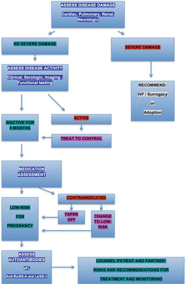
Fig. 3.1
Pre-pregnancy evaluation of the rheumatic disease patient
Severe Disease Damage
Severe manifestations of disease damage may preclude pregnancy and include cardiomyopathy or severe cardiac valve disease, pulmonary issues such as pulmonary hypertension or severe interstitial lung disease, serious neurologic manifestations, or renal insufficiency with significantly decreased clearance (see Table 3.1).
Table 3.1
Common rheumatic disease-related organ damage representing contraindication to pregnancy
|
• Cardiac disease including heart failure, severe valvular disease, or aneurysm |
|
• Pulmonary disease including severe interstitial lung disease or pulmonary hypertension |
|
• Renal disease including severe renal failure/insufficiency |
|
• Recent arterial thrombosis, particularly cerebral vascular accident (CVA) |
|
• Previous life-threatening eclampsia or HELLP syndrome despite prophylactic treatment |
Pulmonary arterial hypertension (PAH) in particular is associated with a high risk of pregnancy-related mortality. Although pregnancy-related mortality for patients with CTD-related PAH has decreased slightly in recent years, from 36 to 28 %, risk is still prohibitive even with current aggressive therapies. Essentially all deaths occur in the postpartum period, most within 72 h of delivery and due to right heart failure [2]. Patients with systemic sclerosis and mixed CTD should have baseline echocardiograms and pulmonary function tests to rule out asymptomatic PAH prior to conceiving.
Preexisting renal disease is a common issue for CTD patients, particularly those with SLE. The most important predictors of permanent renal disease in pregnant women with chronic kidney disease (CKD) are GFR < 40 mL/min/1.73 m2 and proteinuria greater than 1 g/24 h [3]. The risk for poor renal outcome in patients with quiescent renal disease is dependent on impairment in renal function, not the underlying disease diagnosis. One comprehensive review of studies (many dating from the 1980s) of pregnant SLE patients with CKD suggested pregnancy-induced acute kidney injury in 10 %, permanent decline in renal function in 3 %, and end-stage renal failure in 6 % of patients [4]. Another meta-analysis of 37 studies including 2,751 pregnancies in lupus nephritis patients suggests a better renal prognosis, however, with only 2 % patients developing deterioration in renal function and only 1 % requiring dialysis. The patients in these studies had milder disease at outset, with only 11 % having CKD stage 3: the authors suggest that more effective counseling may be limiting the numbers of high-risk lupus nephritis patients choosing to enter into pregnancy, thus improving outcome [5].
In general the potential morbidity and mortality associated with severe chronic disease manifestations make pregnancy unacceptable for patients who have these complications. If patients with severe disease-related damage are intent on having a biological child, it may be appropriate to consider the safety of in-vitro fertilization (IVF) with surrogacy. A patient with underlying disease-related damage who is considered unable to safely carry a pregnancy herself may incur less risk with the more limited duration and physiologic changes associated with IVF (discussed in detail in Chap. 12). Issues surrounding surrogacy, however, may be complex, depending in part on financial and legal constraints. For patients open to consideration of adoption, this is an alternative option.
The Risk of Disease Activity
Once patients are assured they have no identifiable disease-related damage that precludes consideration of pregnancy, the discussion then focuses not on “if” but on the more favorable “when” and “how” to proceed toward pregnancy. It is essential to assess for disease activity and other risk factors: if patients have ongoing active disease, studies and reports from pregnancy in almost all autoimmune diseases suggest this to be a poor time to conceive. Patients should defer pregnancy, use suitable contraception, and be treated aggressively: when disease has been inactive for a period of time usually estimated at 6 months, they may be re-assessed.
While active disease at the time of conception (or within the immediate preceding months) has been shown to increase risk of complications based on available data for most rheumatic disease patients, for lupus in particular there are numerous studies confirming the importance of stable disease at the time of conception and the adverse effect of flare during pregnancy [6, 7]. A high level of lupus disease activity during pregnancy has been associated with a lower chance of live birth, greater chance of perinatal death, and lower chance of full-term delivery [8]. Women with long-standing remission in SLE are more likely to complete their pregnancies uneventfully [9].
Active vasculitis patients are also advised to defer pregnancy until disease is under control, as active disease at conception (or disease onset during pregnancy) increases risk for adverse outcome. Initial severity of disease presentation, however, does not predict activity during pregnancy [10].
Although most patients with systemic sclerosis without severe disease-related damage have successful pregnancies (although often with preterm delivery), patients with early diffuse disease are advised to defer pregnancy for several years: they appear to be at increased risk for developing renal crisis during pregnancy [11].
In general, pregnancy outcome for rheumatoid arthritis (RA) patients is comparable to the general population. Even in rheumatoid arthritis patients, however, disease activity during pregnancy may impact outcome: recent studies suggest a small but significant increase in the risk of lower birth weight and preterm delivery for RA patients who have active disease during pregnancy, with a slight increase in perinatal mortality and higher frequency of Cesarean sections [12]. Disease activity may be associated with the lower birth weight, and prednisone use with risk of preterm delivery [13].
Medication Review
If disease is inactive and there is no indication of severe damage, it is appropriate to next assess the patient’s present medical regimen. When current medications are contraindicated for use in pregnancy, options include taper and discontinuation—if disease permits—or a change to permissible medications. It is ideal to have stable disease on pregnancy-safe medications for 6 months before conceiving. Detailed guidelines for medication use and safety are reviewed in Chap. 14. In general, in addition to prednisone, immunosuppressive medications that are considered permissible during pregnancy include azathioprine and cyclosporine. Use of tacrolimus is increasingly considered an effective alternative for patients who fail azathioprine: studies from the transplant literature suggest safety and tolerability in pregnancy [14] and pilot studies suggest efficacy for lupus nephirits [15], including a case report of successful treatment of lupus nephritis with tacrolimus during lupus pregnancy [16].
For inflammatory arthritis patients, the safety of TNFα-inhibitors during pregnancy continues to be controversial although a number of practitioners feel comfortable continuing these drugs throughout pregnancy. Published reports are reassuring concerning safety with use immediately before conception and in the first trimester: a common approach for women with RA on combination methotrexate and TNFα-inhibitor therapy is to stop the methotrexate and continue the TNFα-inhibitor, waiting 3 months before trying to conceive. Once pregnancy is confirmed, patients should discontinue the TNFα-inhibitor if possible.
Non-rheumatology medications should also be assessed and modified as necessary in conjunction with the obstetrician and other involved specialists. Angiotensin converting enzyme-inhibitors (ACE-I) may be changed to pregnancy-safe substitutes several weeks before attempting to conceive, with careful follow-up of blood pressure and proteinuria after discontinuation. Warfarin should be changed to heparin; low-molecular-weight heparin can be monitored with factor Xa levels, most often in conjunction with a hematologist. In general, it is recommended that the patient change to heparin prior to conception; when this is not possible, the change must be made prior to the sixth week of pregnancy to avoid warfarin embryopathy.
There are particular medications that may have a beneficial effect on pregnancy outcome in specific cases. Women with SLE on hydroxychloroquine (HCQ) have been shown to have lower disease activity scores and to be on lower prednisone doses at delivery [17]. Patients discontinuing HCQ within 3 months of conceiving have a greater risk of flare than those who continue their medications and also require a higher prednisone dose [18]. In addition, HCQ-treated patients may have a lower risk of NLE [19]. If SLE patients or patients with anti-Ro/SS-A or La/SS-B antibodies have no contraindications to HCQ, there may be benefits for both mother and child.
The addition of low-dose aspirin during pregnancy in CTD patients has varying support: it is generally used for preeclampsia prevention in patients with risk factors (hypertension, renal insufficiency, and history of preeclampsia) [20] and is often used in patients with asymptomatic aPL, although no controlled studies support this practice.
Folic acid is advised pre-pregnancy and during the first trimester to reduce risk of neural tube defects; patients on the folate antagonist sulfasalazine are generally advised to further increase their folic acid intake before and during pregnancy.
Assessment of Autoantibodies
Assessment of autoantibodies will help to determine the type and frequency of pregnancy monitoring, the need for potential additional therapy for antiphospholipid obstetric prophylaxis, and to inform both physician and patient regarding risk.
Antiphospholipid Antibodies
Every patient with SLE and as well as patients with other CTD with an adverse obstetric history or a history of thrombosis should be evaluated for presence of antiphospholipid antibodies (aPL). Patients should be counseled regarding risk of aPL in pregnancy, which include miscarriage, fetal loss, preterm birth, intrauterine growth restriction (IUGR), and preeclampsia. Adverse outcome is most strongly associated with lupus anticoagulant (LAC). Other risk factors in patients with positive aPL include history of thrombosis, history of prior fetal loss, and presence of SLE. Patients with low titer aCL and anti-beta 2 glycoprotein I (anti-β2GPI), especially those without a history of prior fetal loss or thrombosis, can be reassured that their risk is relatively low [21].
Treatment for antiphospholipid syndrome (APS) pregnancy prophylaxis is summarized in Chap. 6. In general, many patients will be on low-dose aspirin before conception, with plans for unfractionated or low-molecular-weight heparin after diagnosis of pregnancy if indicated. Current recommendations are to monitor for fetal distress during the third trimester, utilizing non-stress tests, umbilical artery Dopplers, or serial ultrasounds.
Anti-Ro/SS-A and La/SS-B Antibodies
Patients with SLE, RA, undifferentiated connective tissue disease (UCTD), and Sjogrens syndrome should be evaluated for presence of anti-Ro/SS-A and La/SS-B antibodies. Positive patients will require periodic fetal echocardiograms beginning at 16–18 weeks: for high-risk patients, i.e. those with a previous child with NLE, echocardiograms are recommended weekly from weeks 18–26 and every 2 weeks until week 32 (see Chap. 13). Patients should be counseled regarding the spectrum of NLE manifestations, including rash, thrombocytopenia, liver function abnormalities, and risk of congenital heart block (2–3 %), as well as the associated mortality and long-term morbidity in the children with heart block. Risk of congenital heart block in the offspring of an anti-Ro/SS-A positive patient who has already had a child with NLE is higher, about 17 %.
Counseling
Counseling is important for both the patient and her partner: for most patients, knowing what to expect makes a difference. In general, it is recommended that one review the full reproductive spectrum of fertility through lactation during the pre-conception visit. Education should include the risk of pregnancy in general for the given diagnosis and then the risk of pregnancy for the individual patient with her particular risk profile, based on damage, disease activity, renal function, aPL, anti-Ro/SS-A and La/SS-B, and medications. Each patient needs to understand the risk to her own health, the anticipated pregnancy outcome, and the potential risk to offspring (most often these are the complications associated with preterm birth or small size for dates). One should review necessary follow-up and monitoring during pregnancy, and assure availability of neonatal supportive care if early delivery or other complications develop.
Fertility Issues: Can I Get Pregnant?
In patients likely to be at risk for infertility, it is advisable to address fertility issues even before the couple begins attempting to conceive. Although fertility is generally normal in patients with rheumatic diseases—decreased fertility has been suggested but not confirmed in systemic sclerosis [11]—the primary reason for lowered fertility is the use of alkylating agents, generally cyclophosphamide. Most other patients can be reassured that their risk of infertility is similar to that in the general population. The risk of infertility with cyclophosphamide is related to greater age at administration as well as higher cumulative dose. Overall risk of sustained amenorrhea was 62 % for patients over age 31 in early studies [22]; risk may be lower now for patients treated with ovarian protection therapy with synthetic gonadotropin-releasing hormone analogs during the course of their cyclophosphamide treatment [23].
Although fertility in patients is comparable to the general population in the absence of cyclophosphamide treatment, the rate of fertility problems in the general population is not insignificant, at about 20 %. Some of this is age-related, and certainly a patient who is counseled to wait for quiescent disease to conceive may start her pregnancy attempts at a somewhat older age. Given the complexity of the rheumatic disease patient, as well as the fact that they may be treated with less effective (pregnancy safe) second- or third-line medications during this period, it seems reasonable to be proactive and aggressive with referral to reproductive medicine specialists, especially in patients over age 35.
Pregnancy Risk and Outcome: Will My Pregnancy Be Normal?
Although pregnancy outcome in patients with CTD has improved in recent years, many diagnoses are still associated with an increased risk of adverse outcome when compared to the general population. It is important for patients to understand this, in both general and personal terms. Much of the literature has focused on SLE and APS, given the large number of patients as well as the long-recognized increased risk of adverse outcome. A recent national study of complications in pregnancies of patients with the diagnosis of SLE revealed that lupus patients have a two to fourfold increase in pregnancy complications including preeclampsia, preterm labor, and IUGR; medical complications are similarly increased, including thrombosis, major infection, and thrombocytopenia [24]. First pregnancies after SLE diagnosis were followed prospectively in the LUMINA multiethnic SLE cohort: 76.2 % of patients had some complication and there was a small but significant increase in irreversible damage postpartum [25]. In addition to risk of pregnancy-related complications, most but not all studies support some increased risk of flare of lupus during pregnancy including the postpartum period [26]. Most reviews of pregnancies in systemic sclerosis, the vasculitides, and inflammatory myositis find increased rates of preterm birth and/or small-for-gestational-age (SGA) infants (reviewed in detail in Chaps. 8–10).
Patients should be aware that risk of flare during or after the pregnancy exists for most rheumatic diseases, even when disease is quiescent at conception. They should be counseled regarding the need for pre-pregnancy testing and pregnancy monitoring, which will vary by disease, autoantibody status, and individual clinical history. Postpartum risks should also be defined, including risk of postpartum flare or thrombosis.
Delivery Options: Must I Have a Cesarean Section?
In general, Cesarean section is reserved for those with obstetric indications such as previous Cesarean delivery, breech presentation, dystocia, and fetal distress. Rarely, orthopedic impairments due to severe rheumatoid arthritis or bilateral hip replacements with limited hip range of motion may preclude vaginal delivery. Other concerns regarding mode of delivery and anesthesia may be relevant for particular diseases and should be discussed. For example, patients with severe rheumatoid or spondylitic involvement of the cervical spine should be assessed, as with any surgery, for instability should endotracheal intubation be necessary. Patients with history of vasculitis, primarily Takayasu’s, may require baseline vascular imaging pre-pregnancy and anesthesia involvement for hemodynamic monitoring at the time of delivery due to variable hyper and hypo-perfusion. Subglottic stenosis in patients with granulomatous polyangiitis (GPA, formerly Wegener’s granulomatosis) may complicate delivery, requiring temporary tracheotomy to protect the airway (see Chap. 9).
Pregnancy Outcome: What Is the Risk to My Baby?
The major risk to the child is the constellation of complications associated with prematurity and small size, which may include neurologic, ophthalmologic, pulmonary, and gastrointestinal issues, some with long-term implications for disability. Rates of preterm delivery vary depending on diagnosis and disease severity but may be as high as 30–40 % in certain situations. Neonatal lupus is an additional risk in women with anti-Ro/SS-A and La/SS-B antibodies. There are extremely rare case reports of passively acquired neonatal autoimmune syndromes including neonatal APS (thrombosis in the fetus or neonate of a patient with APS) [27], elevated creatine kinase levels in several infants of mothers with myositis [28], and babies with transient skin lesions born to mothers with vasculitis [29].
Long-term outcome of children of mothers with rheumatic disease has only recently been studied, and is described in detail in Chap. 15. A small increase in risk of learning disability in children of women with SLE and APS has been suggested by the literature, although numbers are small and this can be difficult to distinguish from the long-term effects associated with preterm birth.
Postpartum Course: Can I Breastfeed My Baby?
Breastfeeding is possible for many but not all patients. Patients with recurrence of active disease should be treated with the most effective medication, which may preclude breastfeeding. Breastfeeding also may be a concern for the patient with known or suspected osteoporosis, since it may further lower bone density beyond the expected pregnancy-induced decrease and lengthen the time until recovery, increasing risk of fracture.
A number of medications are compatible with breastfeeding. These are detailed in Chap. 14. Corticosteroids in low dose are considered safe, with administration of the dose more than four hours prior to nursing if possible. Aspirin, heparin, warfarin, HCQ, sulfasalazine, and even ibuprofen may be used in patients who are breastfeeding. In general, one would like to avoid immunosuppressive medications during this period, although many rheumatologists feel comfortable with the use of azathioprine or TNF-inhibitors in women who are breastfeeding.
Postpartum risk of flare should be discussed, particularly for patients with rheumatoid arthritis or other inflammatory arthritis, as the need for medication may impact a woman’s ability to breastfeed. A plan should be in place before delivery concerning treatment for postpartum flare if and when it occurs. A common approach is to treat with low-dose corticosteroid while the mother weans the infant, with resumption of disease-modifying or immunosuppressive therapy after weaning. Patients on prophylactic anticoagulation therapy during pregnancy for aPL are generally advised to continue anticoagulation for approximately 6 weeks postpartum to minimize risk of venous thrombosis; those requiring long-term anticoagulation may switch back to warfarin postpartum, as this is compatible with breastfeeding.
Contraception: What Contraception Can I Use While I Am Waiting for the Right Time to Conceive?
One cannot effectively discuss planning of pregnancy without considering availability and safety of contraception. Contraception is discussed in detail in Chap. 11. Oral contraceptives that contain estrogen can be used in most rheumatic diseases including lupus if disease is quiet and if patients do not have antiphospholipid antibodies. For patients who cannot use combined hormonal contraceptives (such as patients with positive antiphospholipid antibodies), the levonorgestrel (progesterone-containing) IUD is a good option. It is recommended even for nulliparous women, risk of infection is low, and it can remain in place for up to 5 years. It significantly decreases menstrual blood flow, a benefit for patients who are on warfarin.
Summary
Pregnancy in rheumatic disease patients is most successful when the rheumatologist, obstetrician, other involved specialists, and patient work together as a team, with identification and understanding of potential risks particular to that individual patient. In general, the best chance for optimal maternal and fetal outcomes depends on planning for pregnancy during a period of well-controlled disease in the rheumatic disease patient without serious end-organ damage who is taking pregnancy-compatible medications.
References
1.
Owen J, Hauth JC. Polyarteritis nodosa in pregnancy: a case report and brief literature review. Am J Obstet Gynecol. 1989;160(3):606–9.PubMedCrossRef
2.
Bédard E, Dimopoulos K, Gatzoulis MA. Has there been any progress made on pregnancy outcomes among women with pulmonary arterial hypertension? Eur Heart J. 2009;30(3): 256–65.PubMedCrossRef
3.
Imbasciati E, Gregorini G, Cabiddu G, Gammaro L, Ambroso G, Del Giudice A, et al. Pregnancy in CKD stages 3 to 5: fetal and maternal outcomes. Am J Kidney Dis. 2007;49: 753–62.PubMedCrossRef
4.
Bramham K, Soh MC, Nelson-Piercy C. Pregnancy and renal outcomes in lupus nephritis: an update and guide to management. Lupus. 2012;21:1271–83.PubMedCrossRef
5.
Smythe A, Oviveira GH, Lahr BD, Bailey KR, Norby SM, Garovic VD. A systematic review and metalaysis of pregnancy outcomes in patients with systemic lupus erythematosusu and lupus nephritis. Clin J Am Soc Nephrol. 2010;5:2060–8.CrossRef
6.
Chakravarty EF, Colon I, Langer ES, Nix DA, El-Sayed YY, Genovese MC, et al. Factors that predict prematurity and preeclampsia in pregnancies that are complicated by SLE. Am J Obstet Gynecol. 2005;192:1897–904.PubMedCrossRef
7.
Andrade R, Sanchez ML, Alarcon GS, Fessler BJ, Fernandez M, Bertoli AM, et al. Adverse pregnancy outcomes in women with systemic lupus erythematosus from a multiethnic US cohort: LUMINA (LVI). Clin Exp Rheumatol. 2008;26:268–74.PubMed
8.
Clowse MEB, Magder LS, Petri M. The impact of increased lupus activity on obstetric outcomes. Arthritis Rheum. 2005;52:514–21.PubMedCrossRef
9.
Le Thi HD, Wechlser B, Vauthier-Brouzes D, Seebacher J, Lefebvre G, Bletry O, et al. Outcome of planned pregnancy in systemic lupus erythematosus: a prospective study on 62 pregnancies. Br J Rheumatol. 1997;36:772–7.CrossRef
10.
Doria A, Bajocchi G, Ronon M, Salvarani C. Pre-pregnancy counseling of patients with vasculitis. Rheumatology (Oxford). 2008;47:iii3–5.CrossRef
11.
Steen V, Chakravarty EF. Pregnancy. In: Scleroderma. Springer, New York, NY, USA; 2012. p. 547–57.
12.
Wallenius M, Skomsvoll JF, Irgens LM, Salvesen KÅ, Nordvåg BY, Koldingsnes W, et al. Pregnancy and delivery in women with chronic inflammatory arthritides with a specific focus on first birth. Arthritis Rheum. 2011;63(6):1534–42.PubMedCrossRef
13.
de Man YA, Hazes JM, van der Heide H, Willemsen SP, de Groot CJ, Steegers EA, et al. Association of higher rheumatoid arthritis disease activity during pregnancy with lower birth weight: results of a national prospective study. Arthritis Rheum. 2009;60(11):3196–206.PubMedCrossRef
14.
Kainz A, Harabacz I, Cowlrick IS, Gadgil S, Hagiwara D. Analysis of 100 pregnancy outcomes in women treated systemically with tacrolimus. Transpl Int. 2000;13(S1):S299–300.PubMedCrossRef
15.
Mok CC, Tong KH, To CH, Siu YP, Au TC. Tacrolimus for induction therapy of diffuse proliferative lupus nephritis: an open-labeled pilot study. Kidney Int. 2005;68(2):813–7.PubMedCrossRef
16.
Alsuwaida A. Successful management of systemic lupus erythematosus nephritis flare-up during pregnancy with tacrolimus. Mod Rheumatol. 2011;21(1):73–5.PubMedCrossRef
17.
Levy RA, Vilela VS, Cataldo MJ, Ramos RC, Duarte JLMB, Tura BR, et al. Hydroxychloroquine (HCQ) in lupus pregnancy: double-blind and placebo-controlled study. Lupus. 2001;10(6): 401–4.PubMedCrossRef
18.
Clowse ME, Magder L, Witter F, Petri M. Hydroxychloroquine in lupus pregnancy. Arthritis Rheum. 2006;54(11):3640–7.PubMedCrossRef
19.
Izmirly PM, Costedoat-Chalumeau N, Pisoni CN, Khamashta MA, Kim MY, Saxena A, et al. Maternal use of hydroxychloroquine is associated with a reduced risk of recurrent anti-SSA/Ro-antibody-associated cardiac manifestations of neonatal lupus. Circulation. 2012;126(1): 76–82.PubMedCrossRefPubMedCentral
20.
Askie LM, Duley L, Henderson-Smait DJ, Stewart LA, on behalf of the PARIS Collaborative Group. Antiplatelet agents for prevention of pre-eclampsia: a meta-analysis of individual patient data. Lancet. 2007;369:1791–98.
21.
Lockshin MD, Kim M, Laskin CA, Guerra M, Branch D, Merrill J, et al. Prediction of adverse pregnancy outcome by the presence of lupus anticoagulant, but not anticardiolipin antibody, in patients with antiphospholipid antibodies. Arthritis Rheum. 2012;64(7):2311–8.PubMedCrossRefPubMedCentral
22.
Boumpas DT, Austin HA, Vaughan EM, Yarboro CH, Klippel JH, Balow JE. Risk for sustained amenorrhea in patients with systemic lupus erythematosus receiving intermittent pulse cyclophosphamide therapy. Ann Intern Med. 1993;119(5):366–9.PubMedCrossRef
23.
Somers EC, Marder W, Christman GM, Ognenovski V, McCune WJ. Use of a gonadotropin-releasing hormone analog for protection against premature ovarian failure during cyclophosphamide therapy in women with severe lupus. Arthritis Rheum. 2005;52(9):2761–7.PubMedCrossRef
24.
Clowse MEB, Jamison M, Myers E, James AH. A national study of the complications of lupus in pregnancy. Am J Obstet Gynecol. 2008;199:127.e1–6.
25.
Andrade RM, McGwin G, Alarcon GS, Sanchez ML, Bertoli AM, Fernandez M, et al. Predictors of postpartum damage accrual in systemic lupus erythematosus: data from LUMINA, a multiethnic US cohort (XXXVIII). Rheumatology. 2006;45:1380–284.PubMedCrossRef
26.
Ruiz-Irastorza G, Lima F, Alves J, Khamashta MA, Simpson J, Hughes GR, et al. Increased rate of lupus flare in pregnancy and the puerperium: a prospective study of 78 pregnancies. Br J Rheumatol. 1996;35:133–8.PubMedCrossRef
27.
Boffa MC, Lachassinne E. Infant perinatal thrombosis and antiphospholipid antibody: a review. Lupus. 2007;16(18):634–41.PubMedCrossRef
28.
Messina S, Fagiolari G, Lamperti C, Cavaletti G, Prelle A, Scarlato G, et al. Women with pregnancy-related polymyositis and high serum CK levels in the newborn. Neurology. 2002;58(3):482–4.PubMedCrossRef
29.
Schlieben DJ, Korbet SM, Kimura RE, Schwartz MM, Lewis EJ. Pulmonary-renal syndrome in a newborn with placental transmission of ANCAs. Am J Kidney Dis. 2005;45(4):758–61.PubMedCrossRef