
Dana Block-Abraham, DO Ahmet Alexander Baschat, MD
You should have come to me before.
—Marlon Brando in The Godfather
KEY POINTS
• Fetal growth restriction may be the result of various pathologies that need to be considered in the differential diagnosis; of these, placental vascular dysfunction is clinically the most relevant.
• Placental insufficiency is associated with fetal responses in almost every organ system, of which only the cardiovascular and behavioral responses are utilized in clinical management.
• An abdominal circumference (AC) of <10th percentile (reference ranges based on a mixed group of high- and low-risk pregnancies) or <2.5th percentile (reference ranges based on normal pregnancies only) is the most sensitive biometric parameter to detect growth delay.
• Umbilical artery (UA) Doppler is the best method to evaluate the fetal compartment of the placental circulation.
• The combination of a small AC, normal anatomy, low or normal amniotic fluid volume, and abnormal umbilical artery Doppler strongly suggests intrauterine growth restriction (IUGR) due to placental insufficiency. However, the possibility of aneuploidy, syndromes, and viral infection should always be considered and fetal karyotyping should be offered.
• Fetal cardiovascular and behavioral deterioration follows a relatively predictable pattern progressing from early to late changes.
• Direction of surveillance and intervention is inaccurate if based on umbilical artery Doppler alone; examination of the cerebral and venous circulation is mandatory if Doppler surveillance is chosen as the primary management tool.
• For arterial vessels, the pulsatility index offers the narrowest reference limits and measurement error; multiple venous Doppler indices have been described without any clear advantage of individual indices.
• Monitoring intervals should be shortened with progressive cardiovascular compromise.
• Once delivery becomes imminent, antenatal steroids should be administered.
• Delivery should be performed with strong evidence of fetal acidemia and/or impending stillbirth. Ductus venosus (DV) index escalation beyond 3 SDs, or absence or reversal of the DV a-wave, are strong evidence of significant fetal compromise. Corroborating evidence from biophysical and computerized heart rate analyses should be sought whenever possible.
• Randomized management studies on venous Doppler for delivery timing of the preterm IUGR fetus are still lacking.
• Diabetes alters several aspects of fetal cardiovascular and behavioral responses, making fetal testing less reliable in diabetic patients.
INTRODUCTION
Disturbance of fetal growth dynamics can result in abnormal weight, body mass, or body proportion at birth. Maternal diabetes is characteristically associated with excessive fetal growth and macrosomia, but diabetic patients may also be at risk for intrauterine growth restriction (IUGR). This risk is related to the degree of maternal vasculopathy and increases with long-standing maternal disease. Although the perinatal management of growth-restricted fetuses with placental insufficiency has evolved markedly over the past five decades, there is scant information on the impact of maternal diabetes on the disease process. The primary focus of this chapter is to review the pathophysiology and perinatal management of growth-restricted fetuses.
Traditionally, abnormal fetal growth was classified by the absolute birth weight as low birth weight (<2500 g), very low birth weight (VLBW, <1500 g), extremely low birth weight (ELBW, <1000 g), or macrosomia (>4000 g). The introduction of population-based birth weight reference ranges was a significant advance, since it allowed the classification of fetal growth patterns by comparing actual birth weight to the expected weight at that gestational age. Lubchenco et al.1 demonstrated that only the classification of neonates by their birth weight percentile allows the detection of growth-restricted neonates at increased risk of adverse health events throughout life.2-4 Accordingly, neonates are now classified as very small for gestational age (VSGA, <3rd percentile), small-for-gestational age (SGA, <10th percentile), appropriate-for-gestational age (AGA, 10-90th percentile), or large-for-gestational age (LGA, >90th percentile).5 The classification of growth disorders is further enhanced by adjusting birth weight reference limits for pre-pregnancy maternal body mass index, race, birth order, and fetal/neonatal gender (growth potential).6 Percentiles that are derived in such a way are superior for the prediction of adverse perinatal outcome compared to conventional reference ranges.7-9 Longitudinal fetal growth patterns, while complicated to study, may help identify more growth abnormalities including the subset of fetuses who remain >10th percentile but have pathologic growth restriction later in pregnancy.10,11
The detection of abnormal body mass or proportions is based on anthropometric measurements and ratios that are relatively independent of gender, race, and, to a certain extent, gestational age (and therefore, also birth weight percentiles).12 The ponderal index [(birthweight (g)/crown-heel length3) x 100]13 has a high accuracy for the identification of IUGR14 and macrosomia15 and correlates more closely with perinatal morbidity and mortality than birth weight percentiles, but may miss the proportionally small and lean growth-restricted neonate.16,17
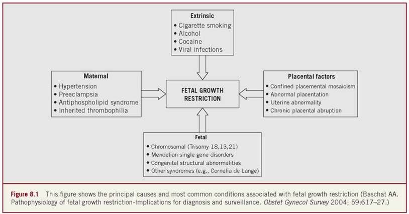
Refining a gold standard that distinguishes between abnormal and physiologic growth patterns at birth is highly desirable for any investigation of the relationship between neonatal size and outcome. From the perspective of the managing perinatologist, only prenatal identification of IUGR is relevant, since it allows appropriate prospective fetal management. Fetal disease, maternal disease, primary placental disease, and extrinsic factors may all interfere with the efficiency of placental nutrient and waste exchange and may therefore result in growth restriction (Figure 8-1). Thus, IUGR is a physical sign rather than a single disease entity, and its impact on outcome is determined by the range of manifestations that are associated with the principal underlying condition. Knowledge of the interactions between etiology, clinical presentation, prognostic factors, and antenatal interventions in pregnancies complicated by growth restriction is required to properly diagnose, assign prognosis, and manage these pregnancies. To formulate a uniform diagnostic and management approach, an understanding of the milestones in normal fetal and placental development and the pathophysiology of disturbed fetal growth is of critical importance.
REGULATION OF FETAL GROWTH
The placenta is the interface between the mother and fetus. Fetal growth is regulated at multiple levels and requires successful placentation for the coordination of key components in the maternal, placental, and fetal compartments.18 Placental adherence in the first trimester initiates a series of important milestones in the three overlapping gestational epochs. The initiation of placental vascular development permits nutrient and oxygen delivery beyond the capacity of simple diffusion, and therefore, poses few limitations to the growing trophoblast. Maternal adaptations to pregnancy predominate in this epoch. Differentiation of placental transport mechanisms and paracrine and endocrine signaling pathways between the mother, placenta, and fetus continues throughout the second trimester. These steps allow placental growth and establishment of efficient and coordinated nutrient transfer, as well as waste and gas exchange, by completion of the second trimester. This is a prerequisite for third trimester exponential fetal growth and differentiation in preparation for extrauterine life.
Placental adherence is established by the formation of anchoring villi by the cytotrophoblast. These villi eventually connect the decidua and uterus. The maternal circulation gains access to the intervillous space via angiogenesis. Increasing quantities of placental secretory products then appear in the maternal circulation, promoting postprandial hyperglycemia, fat deposition, maternal intravascular volume expansion, relative refractoriness of the maternal circulation to vasoactive agents, and increased fasting levels of free fatty acids, triglycerides, and cholesterol. These maternal adaptations increase substrate availability and steadiness of nutrient delivery to the placenta, permitting ongoing placental development. The villous trophoblasts, consisting of maternal microvillous and fetal basal layers, develop as the primary site of nutrient and gas exchange. The efficiency of maternal-fetal exchange depends on four principal factors: (1) the thickness that has to be traversed by diffusible substances, (2) the vascular throughput from the maternal and fetal circulations, (3) the surface area available for exchange, and (4) the elaboration of active transport mechanisms.18
By the 16th week of gestation, the villous trophoblast has progressively thinned down to 4 microns, providing little resistance to diffusion. Vascular throughput of the placenta increases in both the maternal and fetal compartments. Extravillous cytotrophoblast infiltration of the maternal spiral arteries results in progressive loss of the musculoelastic media. This process is paralleled in the fetal compartment by continuous villous vascular branching. Significant reduction in vascular resistance and a rapid increase in the exchange area are achieved by 26 weeks gestation and then continue at a slower rate toward term. Under normal circumstances in the term placenta, up to 600 mL/min of maternal cardiac output are delivered to an exchange area of up to 12 m2. This is matched with a blood flow volume of 200-300 mL/kg/min in the fetal compartment throughout gestation. This magnitude of maternal blood flow is necessary, since maintenance of placental function is energy intensive and consumes as much as 40% of the oxygen and 70% of the glucose supplied to the uterus. Optimal fetal growth and development can only be achieved when the magnitude of maternal nutrient and oxygen delivery to the uterus leaves sufficient surplus for fetal substrate utilization. Perfusion matching between the maternal and fetal compartments is optimized through placental autoregulation. While these developments significantly enhance the efficiency of exchange for diffusible substrate, other substances such as glucose, amino acids, and fatty acids rely on elaboration of active transport mechanisms. Such transport systems develop for each of the nutrient classes and optimize the transfer of glucose, amino acids, and fatty acids across the bilayer.
Each nutrient class has a different role in the fetus. Glucose is the primary oxidative fuel, whereas amino acids are incorporated into proteins. Glucose, and to a lesser extent amino acids, drives the insulin-like growth factor axis and therefore stimulates longitudinal fetal growth. Amino acids are major contributors to protein synthesis and manifest as muscle bulk. Fatty acids are precursors for bioactive compounds including prostaglandins, thromboxanes, and leukotrienes and are also necessary for the maintenance of membrane fluidity and permeability. In addition, long-chain polyunsaturated fatty acids such as arachidonic acid and docosahexanoic acid are essential for normal brain and retinal development. Leptin co-regulates transplacental amino acid and fatty acid transport, and thereby modulates fetal body fat content and proportions. With advancing gestation, the magnitude and efficiency of transfer of these substances increases significantly to provide for placental and fetal growth requirements (Figure 8-2).
Concurrent development of the fetal circulation as a conduit for nutrient and waste delivery is an important cofactor in the fetal growth process. With establishment of a functional circulation, nutrients and oxygen-rich blood from the primitive villous circulation enter the fetus via the umbilical vein. The arrangement of the fetal circulation allows further preferential streaming of these nutrients. The ductus venosus (DV) is the first vascular conduit encountered. Through modulation in DV shunting, 68%- 82% of umbilical venous blood continues to the liver, while the remainder is distributed to the heart.19 Differential directionality of blood streams entering the right atrium ensures that nutrient-rich blood is distributed to the left ventricle, myocardium, and brain, while low-nutrient venous return is distributed to the placenta for re-oxygenation and waste exchange. In addition to the overall distribution of left- and right-sided cardiac output, several fetal organs can modify local blood flow to meet oxygen and nutrient demands by autoregulation.

With achievement of these milestones, the prerequisites for normal placental and fetal growth are met. Healthy metabolic and vascular status of the mother promotes steady and enhanced nutrient delivery to the uterus, and placental transport mechanisms allow for efficient bidirectional exchange of nutrients and waste. Under these circumstances, placental and fetal growth across the three trimesters is characterized by sequential cellular hyperplasia, hyperplasia plus hypertrophy, and lastly hypertrophy alone. Placental growth follows a sigmoid curve that plateaus in mid-gestation and precedes exponential third trimester growth of the fetus. During this exponential fetal growth phase of 1.5% per day, initial weight gain is due to longitudinal growth and muscle bulk and therefore correlates with glucose and amino acid transport, respectively. From 32 weeks onward, fetal fat stores increase from 3.2% to 16% of fetal body weight, accounting for the significant reduction in body water content and preparing the fetus for extrauterine life.18
MECHANISMS OF PLACENTAL INSUFFICIENCY
The precise mechanisms underlying how various conditions interfere with normal placentation and culminate in either pregnancy loss or IUGR are still under investigation. Broadly categorized into maternal, uterine, placental, and fetal, the underlying etiologic disorders affect either nutrient and oxygen delivery to the placenta, nutrient and oxygen transfer across the placenta, fetal uptake of nutrients, or regulation of growth processes producing growth restriction that may be characterized by a reduction in fetal size and, when early and severe enough, cell number. Fetal abnormalities (both chromosomal and/or anatomical) and abnormal placental vascular development cause the preponderance of IUGR in singleton pregnancies.20-24 Generally, the earlier onset of the disease process, the more likely the fetus is to be symmetrically small with a decreased cell number. The etiology of early-onset IUGR is more likely to be a severe maternal vascular disorder, fetal infection, or chromosomal abnormality.25
Early interference with placentation affects all levels of placental and fetal development and culminates in the most severe clinical picture. Early first-trimester interference with angiogenesis may prevent successful placental adherence and therefore result in miscarriage. Once placental adherence is achieved, diffusion initially suffices to fulfill embryonic nutrient demands in the first trimester. At this point, interference with vascular maturation and differentiation may compromise placental and fetal nutrition, resulting in miscarriage or stillbirth. If sufficient supply to the placental mass can be established, further differentiation may be possible. Suboptimal maternal adaptation to pregnancy and deficient nutrient delivery pose limitations at all levels of placental function. If too few placental stem villous arteries and terminal villous capillaries develop, IUGR may ensue.26,27 If trophoblast invasion remains confined to the decidual portion of the myometrium, maternal spiral and radial arteries fail to undergo the physiologic transformation into low-resistance vessels.28,29 Altered expression of vasoactive substances may increase vascular reactivity, and if hypoxia-stimulated angiogenesis cannot overcome these challenges, placental autoregulation becomes deficient. Maternal placental floor infarcts, fetal villous obliteration, and fibrosis each increase placental blood flow resistance, producing maternal- fetal placental perfusion mismatch that decreases the effective exchange area.30-33 Feto-placental flow resistance is increased throughout the vascular bed with progressive vascular occlusion, and eventually metabolically active placental mass is reduced.
If adaptive mechanisms permit ongoing fetal survival, early-onset growth restriction with its many fetal manifestations develops. This spectrum of fetal manifestations is determined by the balance of compensatory and decompensatory responses in various organ systems. If compensatory mechanisms are unsuccessful, permanent fetal damage or stillbirth occurs. With successful compensation, the consequences of nutrient shortage may remain largely subclinical, only to be unmasked through its restrictive effect on exponential fetal growth in the second to third trimesters. In these cases, vascular manifestations may be less pronounced and physical characteristics more apparent; a decrease in adipose tissue or abnormal body proportions at birth may be the only evidence.
CONSEQUENCES OF PLACENTAL DYSFUNCTION
When placental dysfunction compromises nutrient delivery sufficiently to trigger fetal mobilization of hepatic glycogen stores, physical manifestations of growth delay become clinically apparent. Liver size is reduced as hepatic glycogen stores are depleted, resulting in a decrease in the abdominal circumference (AC). In addition to this cardinal sign of growth restriction, a range of fetal manifestations of placental insufficiency have been documented in almost every organ system. Of these, metabolic, endocrine, hematologic, cardiovascular, and behavioral responses are best described. Cardiovascular and central nervous system (CNS) responses are best studied in the context of fetal surveillance, because their noninvasive assessment is readily achieved by multivessel Doppler, gray-scale ultrasound, and fetal heart rate analysis. An appreciation of the variety of fetal manifestations is relevant from the perspective of the managing perinatologist and neonatologist. For the perinatologist, this knowledge illustrates the potential limitations of antenatal surveillance. For the neona- tologist, it allows anticipation of potential complications that may arise from fetal manifestations persisting beyond the transition to extrauterine life. An appreciation of the range of responses also illustrates how long-term consequences of placental insufficiency may never be completely prevented, even with optimal perinatal management.
METABOLIC RESPONSES
Oxygen and glucose consumption by the placenta is unaltered when uterine nutrient delivery is only mildly restricted and the fetal demands are met by an increased fractional extraction. However, when uterine oxygen delivery falls below a critical value (0.6 mmol/min/kg fetal body weight in sheep), fetal oxygen uptake is reduced and is eventually accompanied by fetal hypoglycemia.34 The initially mild hypoglycemia results in a blunted fetal pancreatic insulin response, allowing gluconeogenesis from hepatic glycogen stores.35-38 At this stage, fetal glucose stores and lactate are diverted to the placenta in order to preferentially maintain placental metabolic, endocrine, and nutrient transfer functions. Since hepatic glycogen stores are soon depleted, persistent or declining nutrient deficit results in worsening fetal hypoglycemia and the ability to maintain fetal oxidative metabolism and placental nutrition becomes limited. The use of other fetal energy sources becomes necessary and more widespread metabolic consequences ensue with the significant limitation of oxidative metabolism, downreg- ulation of placental transport mechanisms, and intensifying hypoglycemia. Branched-chain and other essential amino acids are depleted as amino acid transfer becomes limited, and breakdown of endogenous muscle proteins to obtain gluconeogenic amino acids occurs.39-42 Simultaneously, lactate accumulates due to the limited capacity for oxidative metabolism. Placental transfer of fatty acids is maintained unless there is considerable loss of placental substance. The selectivity of transport mechanisms, particularly for essential fatty acids, may suffer. Fetal free fatty acid and triglyceride levels rise due to reduced utilization, and consequently, there is failure to accumulate adipose stores. In this setting of advanced malnutrition, the liver metabolizes the majority of accumulating lactate. However, the fetal brain and heart can also switch their primary nutrient source from glucose to lactate and ketones.43 Cardiac metabolism has the capacity to remove up to 80% of the circulating lactate.44,45 Acid-base balance can be maintained as long as acid production is met by sufficient buffering capacity of fetal hemoglobin and a matching removal rate by fetal organs.
TABLE 8-1 Summary of Metabolic Responses to Placental Insufficiency
|
Substrate |
Change |
|
Glucose |
Decreased proportional to the degree of fetal hypoxemia. |
|
Amino acids |
Significant decrease in branched chain amino acids (valine, leucine, isoleucine) as well as lysine and serine. In contrast, hydroxyproline is elevated. The decrease in essential amino acids is proportional to the degree of hypoxemia. Elevated amniotic fluid glycine/valine ratio. Elevations in amniotic fluid ammonia with a significant positive correlation to the ponderal index. |
|
Fatty acids and triglycerides |
Decrease in long-chain polyunsaturated fatty acids (docosahexanoic and arachidonic acid). Decrease in overall fatty acid transfer only with significant loss of placental substance. Hypertriglyceridemia due to decreased utilization. Lower cholesterol esters. |
|
Oxygen and CO2 |
Degree of hypoxemia proportional to villous damage and correlates significantly with hypercapnia, acidemia, hypoglycemia and hyperlacticemia. |
Therefore, metabolic compromise progresses through degrees of severity. Hypoglycemia, hypoxemia, and decreased levels of essential amino acids occur first. Increasing hypoxemia, overt hypoaminoacidemia, hypercapnia, hypertriglyceridemia, and hyperlacticemia follow. Lactate production is exponentially correlated to the degree of acidemia that generally results from this metabolic state.41,46,47 Amniotic fluid evaluation of the glycine/ valine ratio and ammonia elevation are additional markers of this state of protein-energy malnutrition.48,49 Such severe metabolic alterations are more likely with severe, early-onset IUGR, while those fetuses that manifest growth restriction in the third trimester may only have mild acid-base disturbance and subtle changes in lipid metabolism (Table 8-І).18,50
ENDOCRINE RESPONSES
The immediate effect of decreased fetal glucose and amino acid levels is the downregulation of the principal endocrine growth axis involving insulin, insulin-like growth factor (IGF) I, IGF II, and leptin-coordinated deposition of fat stores.51,52 In addition, there is evidence of pancreatic cellular dysfunction through a decreased insulin/glucose ratio and impaired fetal glucose tolerance.41,53 Elevations in serum glucagon and stimulation of the fetal adrenal axis promote the mobilization of fetal hepatic glycogen stores and peripheral gluconeogenesis in IUGR.54 Corticotropin-releasing hormone, adrenocorticotropic hormone, and cortisol levels are significantly elevated, relating both to the level of hypoglycemia and to the degree of placental vascular compromise.41,55,56 However, cortisol elevation downregulates IGF-I activity and may therefore have additional negative impacts on fetal linear growth and potentially on postpartum catch-up growth.57,58 In addition to the glucocorticoid axis, significant elevations of adrenaline and noradrenaline levels are found in IUGR, while the mineralocorticoid axis appears to remain unaffected.59-61
In the IUGR fetus, disturbances at all levels of the endocrine axis can result in hypothyroidism correlating to the degree of hypoxemia.62,63 Thyroid gland dysfunction may develop as indicated by low levels of thyroxine (T4) and triiodothyronine (T3) despite elevated thyroid-stimulating hormone (TSH) levels. In other instances, central production of TSH may be responsible for fetal hypothyroidism.64 Finally, downregulation of thyroid hormone receptors may limit the biologic activity of circulating thyroid hormones in specific target tissues such as the developing brain.65
There is also evidence of disturbed endocrine regulation of bone formation in IUGR fetuses. Serum levels of active vitamin D and osteocalcin are significantly decreased and may be responsible for the decreased bone mineralization as well as for the decreased bone growth that has been documented in these babies.66,67
HEMATOLOGICAL RESPONSES
Fetal hypoxemia is a trigger for erythropoietin release and stimulation of red blood cell production through both medullary and extramedullary sites, resulting in polycythemia.68-71 The elevation in erythropoietin levels corresponds to the degree of fetal cardiac compromise. Increased extramedullary hematopoiesis may be physiologic until 28 weeks, but can also be induced by prolonged tissue hypoxemia and/or acidosis after this gestational age. Extramedullary sites have larger capillary fenestrations that permit the escape of large nucleated red blood cells (NRBC). Thus, elevated NRBC counts correlate with metabolic and cardiovascular status and are independent markers of poor perinatal outcome.73-76 While polycythemia and elevations of NRBC counts are typical findings in the majority of IUGR fetuses, more complex hematologic abnormalities suggestive of dysfunctional erythropoiesis are observed with advancing compromise. Fetal anemia despite increased NRBC release and overt decrease in red cell progenitors is observed. These findings could reflect downregulation of pro-erythropoietic cytokines, vitamin B12 and ferritin deficiency, or a combination of factors.77-80
Coinciding with the abnormalities in red cell indices, platelet counts also decrease. Although platelet-activating factor is inhibited in the placenta,81 abnormal villous vasculature as indicated by absence or reversal of fetal umbilical artery end-diastolic velocity (see cardiovascular responses below) may pose an overwhelming stimulus for placental platelet activation and aggregation.82 In the presence of such abnormal umbilical waveform patterns, accelerating cardiac deterioration is associated with lower platelet counts and the incidence of thrombocytopenia at birth increases over tenfold.83,84 In addition to villous vascular abnormality, the levels of anemia and hypoxemia are independent risk factors for decreasing platelet counts.85 Increased whole blood viscosity,86,87 decreased red blood cell membrane fluid- ity,88 and platelet aggregation may be important cofactors for accelerating placental vascular occlusion and dysfunction.
Immune dysfunction in IUGR fetuses may develop at the cellular and humoral levels. Decreases in immunoglobulin and absolute β-cell counts have long been recognized.89 Reduction in total white blood cell counts and neutrophil, monocyte, and lymphocyte subpopulations occurs.90 Selective suppression of T-helper and cytotoxic T cells, and smaller ultrasonographic fetal thymus measurements have also been observed.91,92 These abnormalities are related to the degree of acidemia and help explain the higher susceptibility of IUGR babies to infection after delivery.
CARDIOVASCULAR RESPONSES
Doppler ultrasound is the primary tool used for investigating fetal vascular responses to placental insufficiency. Arterial Doppler waveforms reflect vascular resistance and thus provide information on downstream distribution of cardiac output. Since changes in blood flow resistance relate to vascular structure (e.g., placental histology) as well as to vascular tone (e.g., oxygen-related autoregulation), the information gained depends on the vascular bed examined. The most widely used arterial indices are the systolic/diastolic ratio, the resistance index, and the pulsatility index (PI). The PI has a smaller measurement error, narrower reference limits, and the theoretical advantage of ongoing numerical analysis even when end-diastolic velocity is lost.93
The severity of placental vascular dysfunction is reflected in the uterine (maternal compartment) and umbilical (fetal compartment) arteries. The presence of an early diastolic notch in the uterine arteries at 12-14 weeks is the earliest evidence of delayed trophoblast invasion, which is almost certain when “notching” persists beyond 24 weeks.94,95 Reductions in umbilical venous blood flow volume96 and increases in multi-gate-measured intraplacental blood flow resistance97 are the earliest Doppler signs of disturbed fetal villous perfusion. When some 30% of the fetal villous vessels are abnormal, umbilical artery end-diastolic velocity decreases and the Doppler resistance indices become elevated.98 Absence (AEDV) of umbilical artery end-diastolic velocity or reversal of umbilical artery end-diastolic velocity (RDV) can occur when 60%-70% of the villous vascular tree has been damaged.99 Increasing Doppler abnormality in the maternal vascular bed identifies patients at risk for preeclampsia, abruption, and IUGR,100 while abnormal umbilical flows indicate increased risk for fetal hypoxemia and acidemia proportional to the severity of the Doppler abnormality.101,102
In the fetal circulation, changes in blood flows are related to placental blood flow resistance, fetal oxygenation, organ autoregulation, and vascular reactivity. The combination of elevated placental blood flow resistance and impaired transplacental gas transfer has several effects. Venous shunting across the DV increases the proportion of umbilical venous blood that bypasses the liver and ultimately reaches the left side of the heart through the foramen ovale. The parallel arrangement of the fetal circulation dictates unique impacts of placental dysfunction on the relative distributions of right and left ventricular output. Elevation of right ventricular afterload (placental resistance) forces redistribution of cardiac output toward the left ventricle and the relative proportion of left ventricular output rises. Through these mechanisms on the venous and arterial sides of the circulation, the supply of nutrient and oxygen-rich blood to the heart and brain can be increased.
In the compensated state, fetal cardiac output is increased and organ autoregulation is maintained.103,104 Several vascular beds show individual changes in blood flow dynamics. The trunk and cerebral circulations respond differently to hypoxemia. The peripheral arteries constrict in response, and truncal resistance increases as manifested by the elevated umbilical, thoracic, and descending aortic Doppler resistance indices (“hind limb reflex”), which account for most of the increase in right ventricular afterload.105-108 Conversely, the fetal cerebral circulation dilates in response to hypoxemia. Fetal cerebral vasodilation is reflected in the decline of middle cerebral artery Doppler indices (“brain sparing”)109,110 and acts to decrease the left ventricular afterload. This changing balance between the right and left ventricular afterload results in a decline of the cerebro- placental Doppler index ratio and redistribution of the nutrient-rich left ventricular output to the heart and brain. This is corroborated by direct measurements of cardiac output and progressive decreases in amniotic fluid volume after long-standing redistribution.105,106,111-113
Direct evidence of enhanced blood flow to individual organs in response to hypoxemia can be seen in the myocardium,114 adrenal glands,115 spleen,116 and liver,117 whereas blood flow resistance increases in peripheral pulmonary arteries,118 the celiac axis,119 mesenteric vessels,120,121 kidneys,122,123 and the femoral and iliac arteries.124 Overall, these changes complement central blood flow redistribution by enhancing the perfusion of organs vital in fetal life and result in preferential streaming of descending aortic blood flow to the placenta for reoxygenation. In addition, increased levels of endothelin, arginine, vasopressin, norepinephrine, epinephrine, vasoactive intestinal peptide, and atrial natriuretic peptide result in enhanced vascular reactivity that may aggravate the clinical status and increase the complication rate during cordocentesis.125-127
Deteriorations of fetal metabolic and cardiovascular status often coincide and are associated with Doppler evidence of declining forward cardiac function and abnormal organ autoregulation.84,128-130 Examination of fetal cardiovascular status is therefore incomplete without knowledge of cardiac forward function as assessed by venous Doppler. Forward blood flow in the venous system is determined by cardiac compliance, contractility, and afterload, and is characterized by a triphasic flow pattern.131 The venous flow velocity waveform consists of systolic and diastolic peaks (S- and D-waves) that are generated by the descent of the AV-ring during ventricular systole and passive diastolic ventricular filling, respectively. The sudden increase in right atrial pressure with atrial contraction in late diastole causes a variable amount of reverse flow, producing a second trough after the D wave (a-wave). The magnitude of forward flow during atrial systole varies considerably in individual veins. Reversal of flow may be physiologic in the inferior vena cava and hepatic veins, but it is always abnormal in the DV. A decline in forward cardiac function and preload handling marks the onset of cardiovascular decompensation in IUGR fetuses10-105,132 and is manifested in abnormal venous flow velocity waveforms. Abnormal venous flow is characterized by decreasing forward velocities during the a-wave and, to a lesser extent, during the D wave. Multiple venous Doppler indices have been described to characterize this complex waveform without any clear advantage of individual indices.133,134 Impaired preload handling has been documented in the precordial veins (DV, inferior vena cava,135 superior vena cava136), the hepatic veins (right, middle, and left hepatic131,137), and the head and neck veins (jugular veins138 and cerebral transverse sinus139). If the failure to accommodate preload is progressive, umbilical venous pulsations may be observed and are the ultimate reflection of increased central venous pressure.140 With advanced circulatory dysfunction, autoregulation may become exaggerated in the coronary circulation104,114 or nonfunctional in the cerebral and placental circulations.128,129,141 Ongoing deterioration of cardiac function results in holosystolic tricuspid insufficiency and spontaneous fetal heart rate decelerations, and finally is followed by fetal demise.142,143 A summary of vascular responses of IUGR fetuses is provided in Table 8-2.
TABLE 8-2 Summary of Vascular Responses in IUGR Fetuses
|
Doppler Finding |
Physiologic Significance |
|
Uterine artery notching |
Trophoblast invasion remains limited to the myometrial portion of the spiral arteries. Subsequent failure to fully transform into a low resistance, high capacitance vascular bed increases risk for developing IUGR and/or preeclampsia. |
|
Decreased, absent, or reversed umbilical artery end-diastolic velocity |
Abnormal terminal villi and stem arteries result in increased placental vascular resistance and a proportional decrease in the umbilical artery end-diastolic velocity. Associated placental perfusion defects are responsible for impaired feto-maternal gas and nutrient exchange. |
|
Elevation of blood flow resistance in the thoracic aorta and iliac artery |
Hind limb reflex: Diversion of blood flow away from the carcass at the expense of the lower body. Achieved through increase in right ventricular afterload proximal to the umbilical arteries as well as increased blood flow resistance distally. In addition to centralization (see below), descending aortic blood flow is also preferentially distributed to the placenta. |
|
1. Decrease in the cerebroplacental Doppler ratio. 2. Direct measurement of cardiac output. 3. Reversal of end-diastolic velocity in the aortic isthmus. 4. Absence or reversal of umbilical artery end-diastolic velocity. |
Centralization: A measurable shift in the relationship between the right and left ventricular afterload, which results in redistribution of cardiac output in favor of the left ventricle (i.e., the heart and the brain). This can be passively mediated purely by an increase in the placental blood flow resistance and therefore right ventricular afterload. |
|
Decrease in the carotid or middle cerebral artery Doppler index. |
Brain sparing: Cerebral vasodilatation in response to perceived hypoxemia. |
|
Increased superior mesenteric artery Doppler resistance. |
During perceived hypoxemia and/or redistribution of cardiac output blood flow to the gut as a nonessential organ in utero is compromised. |
|
Decreased in the splenic artery Doppler index. |
Splenic artery vasodilatation enhances perfusion of this important hematopoietic organ possibly facilitating an increase in red cell mass. |
|
Decreased Doppler resistance in the celiac axis. |
There may be a reflection of blood flow augmentation in the hepatic and splenic arteries, which are the main branches of this axis. |
|
Increased Doppler resistance in peripheral pulmonary arteries. |
As nonessential organs in fetal life, lung perfusion may be further compromised by increased vascular resistance in the pulmonary circulation ensuring that a greater proportion of right ventricular output bypasses the lungs to reach the placenta. |
|
Increased Doppler resistance in the renal arteries. |
Redistribution and increased renal vascular tone may be the mediators of oliguria and oligohydramnios observed with chronic and/or progressive hypoxemia. |
|
Measured dilation of the ductus venosus with elevated Doppler index accompanied by decreased hepatic artery Doppler index. |
Liver sparing: Preferential arterial blood supply to the fetal liver invoked when increased diversion of umbilical venous blood through the ductus venosus jeopardizes hepatic perfusion. |
|
Decreased Doppler index in the adrenal artery flow velocity waveforms. |
Adrenal sparing: Enhanced adrenal perfusion is triggered as part of the fetal stress response to chronic or acute-on-chronic malnutrition. |
|
Umbilical venous pulsations in association with elevated venous Doppler indices. |
Evidence of inefficient forward delivery of cardiac output with subsequent elevation of central venous pressure that is transmitted all the way back into the umbilical vein. |
|
Normalization of cerebral Doppler indices after a period of “brain sparing.” |
With advanced cardiovascular deterioration, brain autoregulation may become abnormal. Probably in association with a decrease in cardiac function the interval between systolic and diastolic velocities widens resulting in an increase (thus normalization) of the Doppler index. |
|
Sudden ability to visualize and measure coronary blood flow in a setting of deteriorating venous Doppler indices in a premature IUGR fetus. |
Heart sparing: Marked augmentation of coronary blood flow in situations of acute on chronic hypoxemia that is achieved through upregulation of coronary vascular reserve and vasodilatation. |
BEHAVIORAL RESPONSES
Fetal behavioral responses are related to neurodevelopmental status and the impact of ambient oxygen tension on the central regulation of fetal behaviors. Characteristics of the fetal heart rate (FHR) are determined by autonomic control mechanisms superimposed on intrinsic cardiac activity and the effects of oxygen on fetal central regulatory centers. With the maturation of the vasomotor center, reticular activating system, central connections, and increasing processing of peripheral sensory inputs, the characteristics of the fetal heart rate change with advancing gestation. Variations of the heart rate and episodic accelerations coupled to fetal movement each indicate normal functioning of these connections.
Under normal circumstances, successive fulfillment of behavioral milestones progresses from the initiation of gross body movements and fetal breathing to coupling of fetal behavior (e.g., heart rate reactivity) and integration of rest-activity cycles into stable behavioral states (1-4 F). These developments are accompanied by a steadily decreasing FHR baseline (reflecting increasing vagal tone), increasing short- and long-term variability and variation (reflecting increased central processing), and increasing amplitudes of accelerations with advancing gestational age. Once organized behavioral states are established, the diurnal and responsive cyclicity (e.g., to maternal glucose) and their coupling with heart rate reactivity are initiated by 28 weeks of gestation.144 All milestones are then generally completed by 32 weeks, and heart rate reactivity by traditional criteria is present in 80% of fetuses by this time.
Because variations of fetal behavior may be due to several factors including maturational state, behavioral state, and oxygen tension, observation of several variables over a sufficient time period is necessary to separate physiologic from abnormal variation. The five-component biophysical profile score (BPS) system provides a means to quantify fetal behavior by assessing tone, movement, breathing activity, and fetal heart rate reactivity in an observation period of 30 minutes. Amniotic fluid volume measurement has traditionally been a part of the BPS, providing an indirect assessment of fetal renal/vascular status. In the second trimester, amniotic fluid production is primarily related to fetal urine production and therefore to renal perfusion. Through its relationship with vascular status, amniotic fluid volume assessment provides the main longitudinal monitoring component of the BPS and, accordingly, carries a higher weight in the overall grading of the score. Visual FHR analysis has traditionally been used but poses the problems of inter- and intra-observer variability. These are circumvented by computerized analysis of the fetal heart rate (cCTG). The cCTG assesses short-term, long-term, and mean minute variation in addition to traditional FHR parameters and also allows longitudinal observations.
In IUGR fetuses with chronic hypoxemia and mild placental dysfunction, the primary CNS response is a delay in all aspects of CNS maturation.145-149 The detection of these early behavioral responses requires sophisticated and/or computerized research tools and therefore cannot be reliably detected by traditional antenatal monitoring. IUGR fetuses typically have delayed acquisition of behavioral milestones. The combination of delayed central integration of fetal heart rate control, decreased fetal activity, and chronic hypoxemia results in a higher baseline heart rate with lower short- and long-term variation (on computerized analysis) and delayed development of heart rate reactivity in IUGR fetuses.150-153 These maturational differences in fetal heart rate parameters are particularly evident between 28-32 weeks gestation.
Despite the maturational delay of many aspects of CNS function, several centrally regulated responses to acid-base status are preserved. The IUGR fetus maintains behavioral responses to declines in acid-base status. Decreasing global fetal activity initiates the cascade of late behavioral responses to placental insufficiency in the setting of worsening fetal hypoxemia.154 Fetal breathing movement is typically the first behavioral response to cease with increasing hypoxemia. Gross body movements and tone then decrease until they are no longer observed.155,156 Traditional fetal heart rate variables are frequently abnormal by this time. Late decelerations of the fetal heart rate may develop due to a relative drop in oxygen tension that exceeds 8 mm Hg (classical late decelerations). Computerized heart rate parameters, especially the short-term variation, may still be maintained in the normal range (above 3.5 milliseconds). Spontaneous decelerations due to depressed cardiac contractility (cardiac late decelerations) typically herald fetal demise.
The sequential loss of these biophysical variables is determined by the central effects of hypoxemia/acidemia independently of the cardiovascular status.157-161 Reduction of global fetal activity and loss of fetal coupling (loss of heart rate reactivity and fetal breathing movements) are typically observed at a mean pH between 7.10 and 7.20. Abolition of tone and movement is characteristic as pH drops further.155 In contrast, the declining amniotic fluid volume that commonly accompanies the sequential loss of biophysical variables appears to be related to renal blood flow and the degree of vascular redistribution.162,163
MISCELLANEOUS RESPONSES
Several other abnormalities have been described in IUGR fetuses. These include vitamin A, zinc, and copper deficiencies or elevations of the purine nucleotide breakdown product hypoxanthine in correlation with the degree of hypoxemia.164-168 These alterations further illustrate the diverse fetal impacts of placental insufficiency. It is apparent that IUGR is a complex multisystem disease in which the balance and range of compensatory efforts determines the disease manifestation and progression. Although many fetal responses have been presented in a sequential manner in this chapter, our knowledge on their spectrum and relationships continues to evolve. There appears to be no uniform fetal clinical picture. For example, vascular reactivity, blood viscosity, red cell plasticity, and platelet aggregation determine blood flow dynamics in the placental and fetal circulations. Peripheral blood flow dynamics, metabolic milieu, and filling state of the circulation all influence the efficiency of cardiac forward function and delivery of oxygen, nutrients, and waste to their destined sites. Nutrient deprivation, endocrine imbalance, and hypoxemia potentially alter many aspects of organ function and maturation. Deficient body storage limits available nutrient resources after delivery. It is extremely unlikely that all these factors superimposed on the dynamic process of fetal growth would produce a uniform clinical presentation and progression. The fetal presentation may become even more variable when effects of maternal disease are superimposed on the fetal condition. Individual fetal assessment tools do not adequately reflect the range of the possible fetal impacts of placental insufficiency. This poses serious limitations to both diagnosis and management of IUGR fetuses and calls for the integration of multiple fetal assessment modalities.
PROGRESSION TO FETAL COMPROMISE
Longitudinal observation of fetal cardiovascular and biophysical parameters offers insight into disease severity and acceleration and, therefore, has implications for planning fetal surveillance. There are cardinal “early” and “late” changes in each monitoring system that progress in a reasonably predictable sequence in 70%-80% of IUGR fetuses presenting before 34 weeks.163,169,170 Fetal growth restriction that manifests after this gestational age is usually due to milder placental disease producing more subtle cardiovascular abnormalities, while behavioral responses remain related to acid-base status.
When umbilical artery and middle cerebral artery Doppler index deviations are subtle in mild placental insufficiency, a decrease in the cerebroplacental Doppler ratio provides an early and sensitive marker of redistribution of cardiac output and often precedes overt growth delay by up to two weeks.171 With more marked placental disease, the reduction of fetal growth velocity generally mirrors the elevation in umbilical artery blood flow resistance and is followed by decreasing middle cerebral artery impedance and a decline in amniotic fluid index (AFI). At this time traditional heart rate reactivity may also be lost. The nadir of cerebral blood flow resistance is typically reached after a median of two weeks and is followed by an increase in aortic blood flow impedance.172,173 Alterations in blood flow patterns across the aortic isthmus, a conduit between the parallel placental and cerebral circulations in the fetus, have been suggested to occur following umbilical and middle cerebral artery Doppler abnormalities and as an intermediate step in the progression from placental insufficiency-induced fetal hypoxemia to fetal cardiovascular decompensation.174,175 The clinical utility of aortic isthmus flows is currently limited, however. These cardinal “early” cardiovascular responses are considered compensatory since they occur at a time when cardiac function is normal. They are typically accompanied by preferential perfusion of vital organs and the placenta.169,171,176 At this stage, behavioral and FHR responses primarily reflect delayed maturation of central control mechanisms and are premonitory since they require sophisticated examination techniques for their detection.
Accelerating fetal disease and the onset of decompensation become evident through parallel elevations in placental blood flow resistance and precordial venous Doppler indices (inferior vena cava and DV) that are inversely correlated with fetal heart rate variation and variability.159,169,170,173 Absence of umbilical artery end-diastolic velocity is characteristic in this setting. With chronic fetal hypoxemia, global fetal activity declines and breathing movements are lost. When fetal compromise accelerates, there is a further steady rise in umbilical blood flow resistance while venous Doppler indices escalate over a wide range.177 Reversed umbilical artery end-diastolic velocity, overtly abnormal venous Doppler indices, and the development of oligohydramnios are characteristic of this stage of compromise. With ineffective downstream delivery of cardiac output, short-term variation of the FHR becomes abnormal and fetal tone and movements are lost.170,173 Concurrent evaluation of fetal cardiovascular and biophysical variables indicates that Doppler deterioration precedes an abnormal BPS in the majority of IUGR fetuses.163 In the final stages of compromise, cardiac dilatation with holosystolic tricuspid insufficiency, complete fetal inactivity, short-term variation below 3.5 milliseconds, and spontaneous “cardiac” late decelerations of the fetal heart rate can be observed as preterminal events.143,178 This progression may proceed over a median of two weeks but may vary at different gestational ages and with different maternal medical comorbidities.179-182 These cardinal “late” cardiovascular changes require more advanced Doppler examination techniques, while the biophysical abnormalities become readily recognizable on BPS as metabolic compromise progresses.
RELATIONSHIP BETWEEN FETAL RESPONSES AND OUTCOME
Relationships between fetal testing parameters and subsequent outcome determine the relationships between fetal and neonatal risks and, therefore, are critical for the definition of intervention thresholds. Although prevention of long-term morbidity of IUGR is an attractive goal, there is insufficient information on its relationships with prenatal variables to direct management. Many short-term outcomes have been related to fetal status, but only a few presently appear to be of clinical relevance. Fetal acidemia and major neonatal complications have a significant impact on subsequent neurodevelopment, while the combination of fetal and neonatal deaths determines the overall perinatal mortality.183 The likelihood for fetal acidemia and stillbirth are, therefore, the strongest fetal criteria for intervention. In contrast, gestational age-specific expectations for neonatal complications and survival often force conservative clinical management. Although multiple fetal and perinatal relationships have been reported, a practical and comprehensive evaluation of the IUGR fetus can be based on the examination of the umbilical and middle cerebral arteries, precordial veins (inferior vena cava, DV), biophysical parameters, and fetal heart rate analysis.
PREDICTION OF ACIDEMIA
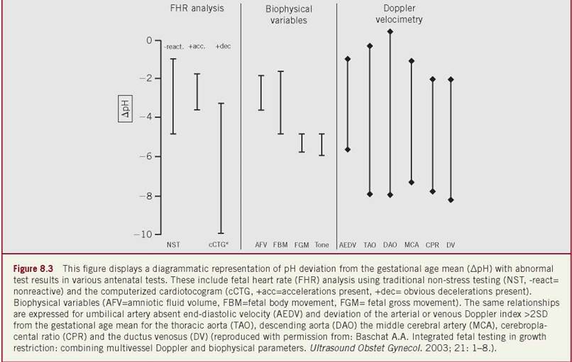
Since Doppler parameters are influenced by several variables (i.e., vascular histology, tone, blood pressure), their relationship with acid-base status is not only variable but also dependent on the vascular system examined and the prevalence of Doppler abnormalities and acidemia in the tested population. Brain sparing in the presence of normal venous Doppler parameters is typically associated with hypoxemia but a normal pH. Elevation of venous Doppler indices, either alone or in combination with umbilical venous pulsations, increases the risk for fetal acidemia. This association is strengthened by serial elevations of the DV Doppler index.177 Depending on the cutoff (2 vs. 3 SD) and the combinations of veins examined, sensitivity for prediction of acidemia ranges from 70% to 90% and specificity from 70% to 80%.184 Because IUGR fetuses preserve their central responses to acid- base status despite their maturational delay, the BPS and arterial pH remain closely related from 20 weeks onward.185 An abnormal BPS score of 4 or less is associated with a mean pH < 7.20, and its sensitivity in the prediction of acidemia is 100% for a score of 2 or less. A combination of multivessel Doppler and BPS is, therefore, complementary in the prediction of acid-base status and critical perinatal outcomes.161 While a nonreactive traditional nonstress test only approaches 50% sensitivity, a computerized documentation of a mean minute variation below 3.5 milliseconds predicts an umbilical artery cord pH < 7.20 with over 90% sensitivity.186 When the relationship between the various testing modalities and fetal acid-base status is compared, biophysical parameters show a closer relationship with pH and Doppler parameters have a wider variance (Figure 8-3).
PREDICTION OF STILLBIRTH
Abnormal venous flow velocity waveforms are the strongest Doppler predictors of stillbirth. Even among fetuses with severe arterial Doppler abnormalities (e.g., absent/reversed umbilical artery end-diastolic velocity), the risk of stillbirth is largely confined to those fetuses that have abnormal venous Dopplers.187 The likelihood of stillbirth increases with the degree of venous Doppler abnormality. Venous Doppler findings that are particularly ominous are absence or reversal of the DV a wave and bipha- sic/triphasic umbilical venous pulsations. In the setting of a 25% stillbirth rate in a preterm severe IUGR population, these Doppler findings have a 65% predictive sensitivity and 90% specificity.188 The duration of the persistent absent or reversed DV a-wave, particularly when occurring for over seven days, is a powerful predictor of stillbirth regardless of gestational age.189 In IUGR fetuses, the BPS deteriorates late and often rapidly, making it unsuitable for the prediction of stillbirth unless daily testing is performed.190
PREDICTION OF NEONATAL COMPLICATIONS
Neonatal complications such as respiratory distress syndrome, bronchopulmonary dysplasia, and intraventricular hemorrhage have several determinants. Gestational age is the primary determinant of neonatal complications, followed by birth weight and the degree of growth restriction. Prediction of neonatal variables by fetal status remains imprecise. However, after accounting for gestational age, the impact of fetal Doppler status on neonatal outcomes becomes apparent. Arterial redistribution and brain sparing are not associated with a significant rise in perinatal morbidity. On the other hand, A 2 SD elevation of the DV Doppler index is associated with a threefold increase in major neonatal complications. Further escalation of DV Doppler indices increases this relative risk to 11-fold. The association between the BPS and neonatal morbidity is well documented in a large cohort of patients. The neonatal complication rate of 35% in the presence of an equivocal score rises to as high as 100% when the score deteriorates further.
PREDICITON OF NEONATAL MORTALITY
Neonatal mortality is determined by multiple factors including gestational age at delivery and the occurrence and severity of neonatal complications. The expected neonatal mortality rate in fetuses with umbilical artery (UA) absent or reduced, A/REDV is variable, ranging between 5% and 18% when the venous Doppler indices are normal. An increase in the DV Doppler index doubles this mortality rate, but the predictive sensitivity is only 38% (specificity 98%) in this setting.20 Although abnormal venous Doppler is associated with a higher rate of neonatal complications, the ultimate impact on their occurrence and on overall neonatal mortality is a function of gestational age at delivery.
The risk for adverse outcome in IUGR fetuses has traditionally been related to the umbilical artery Doppler waveform. Such an approach no longer stratifies the risk appropriately to direct management. Although the risk for adverse outcome is clearly proportional to the degree of UA Doppler abnormality, meta-analysis indicates that DV Doppler effectively identifies those preterm IUGR fetuses at greatest risk for adverse outcome irrespective of the umbilical artery end-diastolic velocity.
SCREENING AND PREDICTION OF FETAL GROWTH RESTRICTION
The ability to screen for or predict IUGR would be particularly useful in the setting of disease prevention. Over the past decade, substantial research has been dedicated to this topic. Various modalities have been investigated, including the evaluation of first- and second-trimester maternal serum analytes and ultrasound parameters. While lower measurements of first-trimester maternal serum pregnancy-associated plasma protein-A (PAPP-A), ADAM12, and placental growth factor (PlGF) have all been associated with small-for-ges- tational age fetuses, none has sufficient individual predictive value for IUGR.191-193 First-trimester ultrasound findings corresponding to SGA fetuses include reduced crown-rump length and slow early fetal growth.194,195 Certain arterial and venous Dopplers in the first trimester also identify fetuses at risk. Elevations in the uterine artery PI, resistance index, and the presence of diastolic notches have been noted in women whose fetuses ultimately develop IUGR.196-198 Abnormal fetal DV and umbilical vein Dopplers have also been linked to IUGR.199,200 In the second trimester, elevations of individual and combined components of the multiple-marker screen are associated with IUGR development. Elevated a-fetoprotein and human chorionic gonadotropin (hCG) are particularly specific in predicting IUGR and may be useful to direct women for ultrasound surveillance for IUGR.201 Any single biomarker or ultrasound finding is not useful in IUGR prediction models, however, and the ultimate combination of serum and ultrasound markers for adequate prediction remains to be elucidated. More advanced techniques including metabolomics and cell-free fetal DNA quantity in the first trimester are also under preliminary investigation.202,203
DIAGNOSTIC APPROACH TO SUSPECTED FETAL GROWTH RESTRICTION
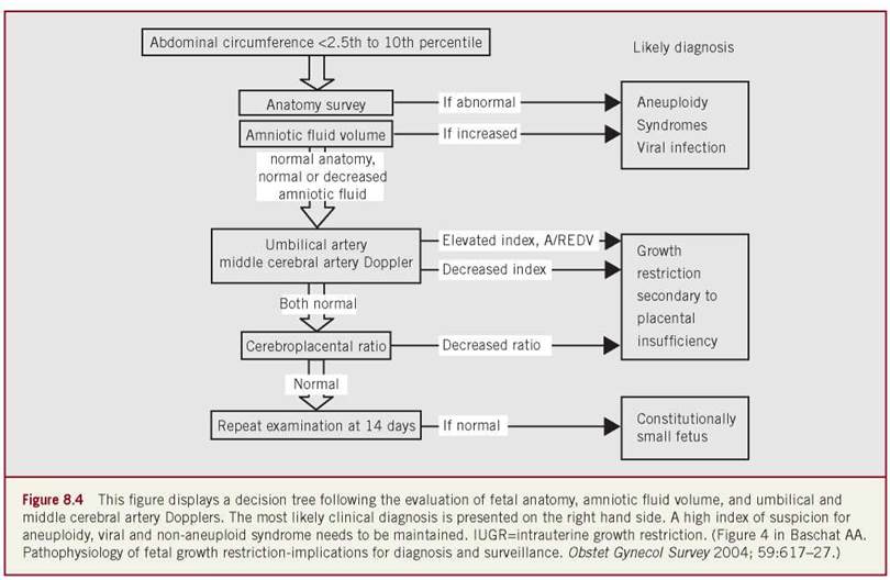
In fetuses with suspected IUGR, a comprehensive diagnostic evaluation is of critical importance, as it is the first step to appropriately direct management. IUGR may be the consequence of various etiologies, and the differential diagnosis always includes maternal disease, placental insufficiency, aneuploidy, nonaneuploid syndromes, and viral infections. Therefore, delayed fetal growth, physical abnormalities, ultrasound markers of aneuploidy, abnormalities of amniotic fluid volume, and disturbed blood flow dynamics may be observed in different combinations based on the underlying disease process. The accurate identification of fetuses that are truly at risk for adverse outcome requires exclusion of small fetuses that are normally grown and those in whom IUGR is due to an underlying condition not amenable to intervention. While maternal disease is readily apparent through a history and physical examination, the accurate evaluation of the possible fetal disorder requires the integration of several diagnostic modalities that evaluate fetal, placental, and amniotic fluid characteristics. Gray-scale ultrasound is the primary diagnostic tool since it allows a detailed fetal anatomic survey, quantification of fetal growth, and assessment of amniotic fluid volume and placental appearance. Although gray-scale ultrasound provides important clues to the presence of IUGR, the liability of preterm delivery and iatrogenic complications is great if the diagnosis is based solely on biometry.204 It is the combination of fetal biometry with Doppler that is the best available tool for the identification of the small fetus at risk for adverse outcome due to placental insufficiency.205-208
An anatomic survey should focus on the exclusion of ane- uploidy markers and overt anomalies. Echogenic bowel, nuchal thickening, abnormal hand positioning, gastroschisis, omphalocele, diaphragmatic hernia, and congenital heart defects are examples of ultrasound findings that may be associated with IUGR. Markers of viral infection are nonspecific but include echogenicity and calcification in organs such as the brain and liver.209 Amniotic fluid volume assessment complements the anatomic survey. Sonographic measurement does not provide an accurate reflection of actual amniotic fluid volume and, by itself, is a poor screening tool for IUGR and fetal acidosis.210-212 Nevertheless, amniotic fluid volume assessment by either the four-quadrant AFI or the maximum vertical pocket, especially if performed serially, provides an important diagnostic as well as prognostic tool. Faced with small fetal size, abundant amniotic fluid volume is an indication of ane- uploidy or fetal infection while normal or decreased amniotic fluid volume is compatible with the diagnosis of placental insufficiency.
Quantification of fetal growth requires accurate knowledge of the gestational age as a reference point to calculate percentile ranks of fetal measurements. An estimated date of confinement (EDC) is based on the last menstrual period when the sonographic estimate of gestational age is within the predictive error (7 days in the first trimester, 14 days in the second trimester, and 21 days in the third trimester). Once the EDC is set by this method or a first-trimester ultrasound, it should not be changed because such a practice interferes with the ability to diagnose IUGR. The fetal AC is related to the hepatic glycogen storage and nutritional state of the fetus, and therefore is the most sensitive and specific measurement for the detection of IUGR.213-215 Its sensitivity is further enhanced by serial measurements at least 14 days apart.216 The most accurate AC is the smallest directly measured circumference obtained at the level of the hepatic vein between fetal respirations.217 If reference ranges for the AC are based on a cross section of small, appropriately grown, preterm and term newborns, the 2.5th percentile is an appropriate cutoff. However, if reference limits are based on healthy women delivering appropriately nourished neonates at term, the <10th percentile cutoff is consistent with IUGR. Concurrent measurement of the head and AC, as well as the femur length, allows calculation of the sonograph- ically estimated fetal weight. An estimated fetal weight below the 10th percentile for gestational age has a lower sensitivity than the AC (85% vs. 98%) but a higher positive predictive value (51% vs. 36%).215 IUGR fetuses with overall growth <10% in combination with measured AC <10% have the worst perinatal outcomes.218
The next diagnostic step evaluates feto-placental vascular function and is critical for the documentation of placental vascular insufficiency. Randomized trials and meta-analyses confirm that the use of umbilical artery Doppler for this purpose significantly reduces perinatal mortality and iatrogenic intervention, since the documentation of placental vascular insufficiency effectively separates IUGR fetuses that require surveillance and possible intervention from constitutionally small fetuses.219-221 An even more comprehensive assessment of feto-placental vascular status can be achieved through the combined examination of the uterine, umbilical, and middle cerebral arteries. Qualitative waveform analysis of the uterine artery (for presence of notching) and the umbilical artery end-diastolic velocity (for positive, absent, or reversed flow) is both simple and effective. For semiquantitative waveform analysis, angle-independent indices are used. Of these, the PI offers the smaller measurement error, narrower reference limits and the possibility for ongoing numerical analysis even when end-diastolic velocity is absent.222,223 In fetuses presenting with IUGR due to placental insufficiency before 34 weeks gestation, the umbilical artery Doppler waveform is frequently abnormal. Beyond this gestational age, umbilical artery waveforms may be normal while cerebral artery “brain sparing” still occurs with perceived hypoxemia.224 Therefore, the middle cerebral/umbili- cal artery Doppler ratio (cerebroplacental ratio) may be abnormal in fetuses with mild placental disease.225 Beyond 34 weeks gestation, a decrease in the middle cerebral artery Doppler index or the cerebroplacental ratio should therefore heighten suspicion for IUGR even if the umbilical artery blood flow is normal.
Once IUGR is confirmed, fetal karyotyping should be offered and further specialized tests such as maternal serology (TORCH titers), thrombophilia studies, or amniotic fluid viral DNA testing may be indicated. After nontreatable fetal conditions and chromosome abnormalities have been ruled out, further antenatal surveillance should be instituted based on the severity of the maternal and/or fetal condition. A schematic representation of the diagnostic algorithm is presented in Figure 8-4.
CLINICAL MANAGEMENT IN SUSPECTED FETAL GROWTH RESTRICTION
Following the diagnosis of placental-based IUGR, intrauterine therapy would be ideal at gestational ages where pregnancy prolongation is beneficial to the fetus. Clinically available therapeutic options remain limited. Maternal hyperoxygenation,226,227 intravascular volume expansion,228 and hyperalimentation229 have been reported. Many issues such as patient selection, efficacy, the impact of therapy on outcome, and the testing required to monitor the fetus during therapy have not yet been adequately clarified to justify clinical application these modalities outside of a research setting.
Elimination of potential external contributors such as stress and smoking, as well as encouragement of lateral positioning when resting, are advocated. Although not of proven efficacy, these steps should maximize the maternal uterine blood flow. Hospitalized bed rest should be considered, which has the advantages of positive enforcement of rest and facilitation of daily testing. The decision for inpatient versus outpatient management should be based on the severity of the maternal and/or fetal condition and the local standard of care.
Although low-dose aspirin (81 mg/d) has not been shown to help severe early-onset IUGR, it may be of benefit in patients with mild placental dysfunction.230,231 In view of its documented safety,232,233 we typically consider low-dose aspirin therapy once the diagnosis of placental-based IUGR is made.
Universally available therapeutic options that can positively affect outcome include the antenatal administration of corticosteroids to hasten fetal lung maturity in the preterm fetus and delivery at an institution with a neonatal care unit able to address the management complexities of the IUGR neonate. A complete course of antenatal corticosteroids should be administered to any IUGR fetus in which delivery is anticipated before 34 weeks. A protective effect of intrauterine “stress” against the effects of prematurity is not supported by large population studies of IUGR neonates and therefore does not warrant the omission of steroid therapy.3,234 In addition, early transfer of a pregnant patient with suspected early-onset IUGR to an experienced perinatal center is strongly suggested in order to assure optimal perinatal therapy. In the absence of any other effective therapies, the main focus in perinatal management is antenatal surveillance in order to minimize fetal risks and to direct the timing of interventions including delivery.
AN INTEGRATED APPROACH TO FETAL SURVEILLANCE IN IUGR
Although IUGR fetuses with placental insufficiency show responses correlating with disease severity in almost every organ system, fetal surveillance is limited to cardiovascular and behavioral evaluation due to its non-invasive nature. The primary assessment tools are Doppler ultrasound, BPS, and fetal heart rate analysis. Since the goal of surveillance is the minimization of perinatal risks to the fetus, it must address two issues: the need for active intervention and the choice of monitoring intervals. The assessment of fetal well-being determines the need for active intervention, while the anticipated rate of progression determines the choice of monitoring intervals. Since the manifestation of fetal disease as well as the rate of progression may be variable, an understanding of the strengths and limitations of individual surveillance tests is important. Arterial Doppler abnormalities typically progress in a characteristic sequence when biophysical and computerized fetal heart rate parameters are still normal. Deteriorating fetal status is reflected in all monitoring systems although the primary manifestation may be variable. The anticipated rate of progression is, therefore, best assessed by Doppler examination of the fetal arterial system. Fetal decompensation can manifest itself in escalation of venous Doppler indices, abnormal BPS, anhydramnios, and an abnormal fetal heart rate.18,163 While any of the monitoring systems has the potential to detect deterioration, there is increasing evidence that a combination of multiple modalities offers the most comprehensive approach to assessment of fetal well-being.161
The need for an accurate assessment of fetal status is of particular importance in circumstances where the thresholds for delivery are high. Although there are surprisingly few randomized management studies that address the issue of delivery timing in IUGR, the growth restriction intervention trial (GRIT) clarifies several important points. In this prospective, randomized, multicenter study of IUGR pregnancies in whom physicians were unsure about delivery timing, more than 500 women were randomized to immediate delivery versus delayed delivery until testing became overtly abnormal. In these pregnancies, timing of delivery had little effect on short-term outcomes although earlier delivery (before 32 weeks) produced a trend toward more disability in early childhood.235,236 The lack of a relationship between timing of delivery and short-term outcomes suggests that the background morbidity as predetermined by gestational age may not be altered by obstetric management, and/or that physicians already deliver at an optimal time to minimize mortality. At the same time, these data suggest that monitoring and management protocols are insufficient to guide delivery prior to damage of brain development and/or fetal death. This stresses the need for excellent surveillance if conservative management is elected. It was previously shown that clinical management of IUGR fetuses with UA AEDV by daily BPS monitoring with strict delivery indications can prevent stillbirth and acidemia at birth. It is presently unknown if management that is based on multivessel Doppler, BPS, cCTG, or integrated fetal testing will improve short-term outcome in placental-based IUGR across the whole disease spectrum, or if benefits are confined to certain gestational ages.
We utilize a surveillance approach to pregnancies with IUGR due to placental disease that combines Doppler ultrasound and BPS (integrated fetal testing). The testing is always supplemented with maternal assessment of fetal movement (“kick counts”). Doppler examination includes evaluation of the umbilical artery, middle cerebral artery, DV, and free umbilical vein flow velocity waveforms. Monitoring frequencies are adjusted to the fetal condition and depend on the anticipated speed of clinical deterioration and the risk for impending acidemia and/or stillbirth. In fetuses with elevated UA pulsatility, positive end-diastolic flow, and absence of any additional abnormality, weekly BPS and fortnightly multivessel Doppler monitoring is performed. With the onset of brain sparing, Doppler monitoring intervals should be shortened to weekly visits. In fetuses with oligohydramnios or UA AEDV, surveillance every three to four days is suggested. Elevation of the DV Doppler index to >2 SD should prompt surveillance every two to three days. With further increase in the DV Doppler index, daily testing becomes necessary and inpatient admission may be prudent based on local practice. Any change in maternal condition, especially the development of preeclampsia, calls for reassessment of fetal status irrespective of the last examination result (Figure 8-5).
The issue of optimal timing of delivery for IUGR fetuses remains unresolved. By principle, the decision to deliver always weighs fetal versus neonatal risks. Typically, decline in neonatal mortality is greatest between 24 and 28 weeks, while morbidity declines progressively thereafter toward 32 weeks.237 Perinatal mortality and morbidity is greatest among IUGR fetuses with abnormal venous Doppler indices irrespective of the umbilical artery Doppler waveform. Therefore, basing delivery decision on the umbilical artery waveform alone appears to no longer be appropriate.238 Delivery is indicated when the risk for fetal acidemia and/ or stillbirth is high. This is the case when the DV Doppler index elevation escalates beyond 3 SDs and DV reversed a-wave is observed with accompanying umbilical venous pulsations. Other indicators are a BPS less than 6, anhydramnios, fetal heart rate variation below 3.5 milliseconds, or overt fetal distress. The interpretation of test results when the three modalities are in disagreement with each other has not been sufficiently studied. However, a vigorous fetus with normal amniotic fluid volume is unlikely to suffer relevant metabolic compromise even if the venous Doppler index is elevated. Conversely, isolated abnormality of the cCTG is unlikely to be of clinical relevance if all other testing is reassuring. Ultimately, the decision to deliver is critically influenced by gestational age. An ongoing study of prenatally identified IUGR fetuses with elevated placental blood flow resistance suggests that the effect of gestational age overshadows all other perinatal variables until approximately 27 weeks, when survival and intact survival first exceed 50%.237
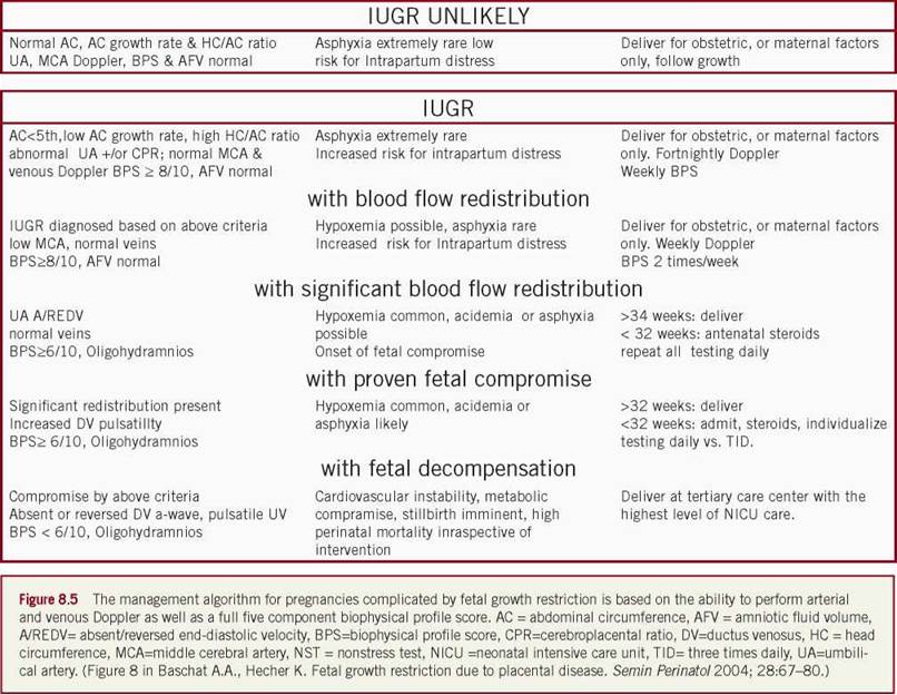
The following approach is therefore suggested. Until 27 weeks gestation and/or with an estimated fetal weight below 500 g, delivery thresholds should be high. Indications for delivery should ideally be based on strong corroborating evidence for fetal compromise from several modalities. Venous Doppler abnormalities with an abnormal BPS provide the strongest evidence in this setting. Patients need to be counseled that the chances for survival and intact survival are poor under these circumstances, even with the highest level of neonatal intensive care. Once these gestational and weight thresholds are passed, improved outcomes can be expected for similar delivery indications. It is currently unknown when this approach can be modified to deliver earlier in order to prevent ongoing fetal compromise. In more than 600 IUGR fetuses delivered prior to 32 weeks, we observed survival and intact survival of 80% after 29 weeks gestation suggesting that this may be a time to individualize intervention thresholds.239 However, in the absence of randomized proof, such practices need to be discussed in a multidisciplinary setting and should be tailored to local practice.
Since the outlined surveillance approach requires multivessel Doppler as well as BPS, it can only be performed at centers that are familiar with both techniques. Modifications of this surveillance protocol need to address the limitations of such an approach. For example, the BPS alone offers little in the prediction of longitudinal progression. Thus, if BPS is the only surveillance tool, daily testing may be required to assure a good outcome. The conclusion of the randomized TRUFFLE study (Trial of Umbilical and Fetal Flow in Europe) will hopefully clarify if delivery triggered by Doppler versus computerized fetal heart rate analysis has a measureable impact on outcome. Ongoing randomized efforts are necessary to refine our understanding of the relationship between fetal testing variables, interventions, and outcomes.
IUGR AND MATERNAL DIABETES—DIAGNOSTIC IMPLICATIONS
There is relatively little information that pertains to patients in whom placental insufficiency and maternal diabetes coexist. From the pathophysiology point of view and the perspective of clinical management, several potential interactions are of importance. In mothers with type 1 DM, those with preconception vascular disease (retinopathy, nephropathy, hypertension) are more likely to develop IUGR.240 Since glucose plays a central role in the regulation of longitudinal fetal growth, an increased supply of glucose modifies several important fetal manifestations of placental insufficiency. Growth is accelerated through upregulation of the insulin-IGF axis and fat deposition is increased through increased leptin concentrations.241 Increased hepatic glycogen storage and liver volume correlate with maternal HbA concentrations and 1c result in larger measurements for the AC.242
Despite the accelerated fetal growth generally evident in diabetic patients, the relationship between uterine and umbilical artery flow velocity waveforms and pregnancy complications is maintained independently of glycemic control. Elevated blood flow resistance in the uterine artery as early as the first trimester is associated with an increased risk for subsequent hypertensive disorders and fetal growth restriction.243,244 Fetal weight is inversely correlated with the umbilical artery Doppler resistance.245 Elevated umbilical artery Doppler indices in diabetic patients identify a subgroup of fetuses at risk for IUGR and/or adverse outcome.246-248 In fetuses with established IUGR, glycemic control also does not appear to affect aortic and middle cerebral artery flow velocity waveforms.249-251 These findings suggest that screening for IUGR by combined assessment of the AC and umbilical and middle cerebral artery Doppler is valid in diabetic patients with one important caveat. Because fetuses of diabetic mothers have larger measurements for the AC, higher percentile cutoffs for the AC may have to be selected (10th, or even 25th). More emphasis may have to be placed on serial growth measurements, and a greater than 20% drop in growth velocity may suggest growth delay even if individual measurements are maintained in the normal range.252
MATERNAL DIABETES-ALTERATIONS IN FETAL RESPONSES
Infants of diabetic mothers are at risk for polycythemia that is mediated through increased erythropoietin concentrations in correlation with the maternal HbA1cvalue.253 Although buffering capacity of fetal blood is increased, the increased red cell mass may have negative impacts on placental blood flow dynamics.
Fetuses of diabetic mothers are also at risk for acidemia and hyperlacticemia that may develop in the presence of normal oxygen tension and independently of placental vascular status.254,255 In these fetuses, metabolic derangement may therefore be obscured by their apparent normal growth and failure to demonstrate blood flow redistribution.256
Maternal diabetes has a significant impact on the development of fetal cardiac function in addition to the known risks of cardiac malformations with elevated HbA, levels. Fetuses of mothers with diabetes have higher combined ventricular outputs but the physiologic decrease in right-to-left ventricular output ratio does not occur.257 Accelerated cardiac growth with interventricular septal hypertrophy or global myocardial hypertrophy may be observed.258-260 In addition to the increased myocardial muscle mass, there is a delayed or absent development of diastolic function.261-263 This may be reflected in abnormal myocardial wall motion or abnormal trans-atrioventricular valve flows. Interventricular wall thickness and hematocrit values significantly and independently affect the ratios between early and active ventricular filling from the mitral and tricuspid valves.264,265 Impaired cardiac forward function may also be manifested by an increased percentage of reverse flow in the inferior vena cava.266 While these abnormalities may be seen with apparently normal glycemic control, they are more pronounced in poorly controlled diabetics. No other differences in venous flow dynamics have yet been reported, although it is of note that there is no increase in umbilical venous volume flow despite the increase in liver volume and body mass.267
The sum of these changes has important implications for the fetal cardiovascular system if the challenge of placental insufficiency is superimposed. Since the relationship between right and left ventricular outputs is abnormal, the ability for central redistribution of cardiac output may be limited. Regulation of organ perfusion, therefore, has to rely more on autoregulation. Diastolic function may decompensate earlier, posing limitations to effective downstream delivery of cardiac output. And lastly, increased venous shunting across the DV may leave a larger volume of liver underperfused with subsequent limitations to organ function.
Maternal diabetes also affects behavioral development and biophysical variables. A delay in the development of behavioral milestones is related to the severity of maternal diabetes.268 Similarly, milestones in fetal heart rate control are delayed. On computerized analysis this is manifested by increased basal heart rates, smaller amplitudes of accelerations with movement, decreased numbers of high variations, and decreased short-term variation.269,270 Even in the traditional NST, a significant negative correlation between the glycemic control and the heart rate variability has been described.271
IUGR AND MATERNAL DIABETES- MANAGEMENT IMPLICATIONS
Although it appears that fetuses with placental insufficiency are most likely to react similarly in the presence of maternal diabetes, it is clear that their compensatory mechanisms are limited. These fetuses are most notable for their risk of having metabolic compromise in the absence of any other signs. There is no information how such fetuses should be managed. Conflicting results from several studies suggest that none of Doppler, BPS, or fetal heart rate analysis appear to be satisfactory if used alone.272-275 In addition to the management scheme outlined in Figure 8-5, the following modifications are suggested: (1) the diagnosis of IUGR should be based on serial examinations even if the AC is above the 10th percentile (see above), (2) monitoring intervals may need to be shortened, especially if there is a change in the maternal condition, and (3) patients with diabetes have to be aware that sudden fetal death can occur without any premonitory signs.
REFERENCES
1. Lubchenco LO, Hansman C, Boyd E. Intrauterine growth as estimated from live born birth-weight data at 24-42 weeks of gestation. Pediatrics. 1963;32:793.
2. Battaglia FC, Lubchenco LO. A practical classification of newborn infants by weight and gestational age. J Pediatr. 1967;71:159-163.
3. Bernstein IM, Horbar Jd, Badger GJ, et al. Morbidity and mortality among very-low-birthweight neonates with intrauterine growth restriction. The Vermont Oxford Network. Am J Obstet Gynecol. 2000;182:198-206.
4. Barker DJ. Fetal growth and adult disease. Br J Obstet Gynaecol. 1992;99:275-276.
5. Hoffman HJ, Stark CR, Lundin FE, et al. Analysis of birthweight, gestational age, and fetal viability, US births, 1968. Obstet Gynecol Surv. 1974;29:651.
6. Gardosi A, Chang A, Kaylan B, et al. Customized antenatal growth charts. Lancet. 1992;339:283-287.
7. Clausson B, Gardosi J, Francis A, et al. Perinatal outcome in SGA births defined by customized versus population-based birthweight standards. BJOG. 2001;108:830-834.
8. Bukowski R, Burgett AD, Gei A, et al. Impairment of fetal growth potential and neonatal encephalopathy. Am J Obstet Gynecol. 2003;188:1011-1015.
9. Odibo AO, Francis A, Cahill AG, et al. Association between pregnancy complications and small-for-gestational-age birth weight defined by customized fetal growth standard versus a population-based standard. J Matern Fetal Neonatal Med. 2011;24(3):411-417.
10. Zhang J, Merialdi M, Platt LD, et al. Defining normal and abnormal fetal growth: promises and challenges. Am J Obstet Gynecol. 2010;202(6):522-528.
11. Roex A, Nikpoor P, van Eerd E, et al. Serial plotting on customised fundal height charts results in doubling of the antenatal detection of small for gestational age fetuses in nulliparous women. Aust N Z J Obstet Gynaecol. 2012;52(1):78-82.
12. Owen P, Farrell T, Hardwick JC, et al. Relationship between customized birthweight centiles and neonatal anthropometric features of growth restriction. BJOG. 2002;109:658-662.
13. Miller HC. Fetal growth and neonatal mortality. Pediatrics. 1972;49:392.
14. Weiner CP, Robinson D. The sonographic diagnosis of intrauterine growth retardation using the postnatal ponderal index and the crown heel length as standards of diagnosis. Am J Perinatol. 1989;6:380-383.
15. Lepercq J, Lahlou N, Timsit J, et al. Macrosomia revisited: pon- deral index and leptin delineate subtypes of fetal overgrowth. Am J Obstet Gynecol. 1999;181:621-625.
16. Walther FJ, Ramaekers LHJ. The ponderal index as a measure of the nutritional status at birth and its relation to some aspects of neonatal morbidity. J Perinat Med. 1982;10:42-47.
17. Ballard JL, Rosenn B, Khoury JC, et al. Diabetic fetal mac- rosomia: significance of disproportionate growth. Pediatrics. 1993;122:115-119.
18. Baschat AA. Fetal responses to placental insufficiency—an update. BJOG. 2004;111:1031-1041.
19. Kiserud T. The ductus venosus. Semin Perinatol. 2001;25:11-20.
20. Snijders RJM, Sherrod C, Gosden CM, et al. Fetal growth retardation: associated malformations and chromosome abnormalities. Am J Obstet Gynecol. 1993;168:547-555.
21. Khoury MJ, Erickson D, Cordero JE, et al. Congenital malformations and intrauterine growth retardation: a population study. Pediatrics. 1988;82:83-90.
22. Sickler GK, Nyberg DA, Sohaey R, et al. Polyhydramnios and fetal intrauterine growth restriction: ominous combination. J Ultrasound Med. 1997;16:609-614.
23. Odegard RA, Vatten LJ, Nilsen ST, et al. Preeclampsia and fetal growth. Obstet Gynecol. 2000;96:950-955.
24. Kupferminc MJ, Peri H, Zwang E, et al. High prevalence of the prothrombin gene mutation in women with intrauterine growth retardation, abruptio placentae and second trimester loss. Acta Obstet Gynecol Scand. 2000;79:963-967.
25. Weiner CP. Pathogenesis, evaluation, and potential treatments for severe, early onset growth retardation. Semin Perinatol. 1989;13:320.
26. Almasry SM, Elfayomy EK. Morphometric analysis of terminal villi and gross morphological changes in the placentae of term idiopathic intrauterine growth restriction. Tissue Cell. 2012;44(4):214-219.
27. Almasry SM, Eldomiaty MA, Elfayomy EK, et al. Structural analysis of human placental stem and terminal villi from normal and idiopathic growth restricted pregnancies. J Mol Histol. 2012;43(3):263-271.
28. Brosens I, Dixon HG, Robertson WB. Fetal growth retardation and the arteries of the placental bed. Br J Obstet Gynaecol. 1977;84:656-663.
29. Meekins JW, Pijnenborg R, Hanssens M, et al. A study of placental bed spiral arteries and trophoblast invasion in normal and severe preeclamptic pregnancies. Br J Obstet Gynaecol. 1994;101:669-674.
30. Aardema MW, Oosterhof H, Timmer A, et al. Uterine artery Doppler flow and uteroplacental vascular pathology in normal pregnancies and pregnancies complicated by pre-eclampsia and small for gestational age fetuses. Placenta. 2001;22:405^11.
31. Ferrazzi E, Bulfamante G, Mezzopane R, et al. Uterine Doppler velocimetry and placental hypoxic-ischaemic lesion in pregnancies with fetal intrauterine growth restriction. Placenta. 1999;20:389-394.
32. Sebire NJ, Talbert D. The role of intraplacental vascular smooth muscle in the dynamic placenta: a conceptual framework for understanding uteroplacental disease. Med Hypotheses. 2002;58:347-351.
33. Kingdom JC, McQueen J, Connell JM, et al. Fetal angiotensin II levels and vascular (type I) angiotensin receptors in pregnancies complicated by intrauterine growth retardation. Br J Obstet Gynaecol. 1993;100:476-482.
34. Jones CT, Ritchie JW, Walker D. The effects of hypoxia on glucose turnover in the fetal sheep. J Dev Physiol. 1983;5:223-235.
35. Nicolini U, Hubinont C, Santolaya J, et al. Maternal-fetal glucose gradient in normal pregnancies and in pregnancies complicated by alloimmunization and fetal growth retardation. Am J Obstet Gynecol. 1989;161:924-927.
36. Economides DL, Nicolaides KH. Blood glucose and oxygen tension levels in small-for-gestational-age fetuses. Am J Obstet Gynecol. 1989;160:385-389.
37. Hubinont C, Nicolini U, Fisk NM, et al. Endocrine pancreatic function in growth-retarded fetuses. Obstet Gynecol. 1991;77:541-544.
38. Van Assche FA, Aerts L, DePrins FA. The fetal endocrine pancreas. Eur J Obstet Gynecol Reprod Biol. 1984;18:267-272.
39. Cetin I, Marconi AM, Corbetta C, et al. Fetal amino acids in normal pregnancies and in pregnancies complicated by intrauterine growth retardation. Early Hum Dev. 1992;29:183-186.
40. Cetin I, Corbetta C, Sereni LP, et al. Umbilical amino acid concentrations in normal and growth-retarded fetuses sampled in utero by cordocentesis. Am J Obstet Gynecol. 1990;162:253-261.
41. Economides DL, Nicolaides KH, Campbell S. Metabolic and endocrine findings in appropriate and small for gestational age fetuses. J Perinat Med. 1991;19:97-105.
42. Paolini CL, Marconin AM, Ronzoni S, et al. Placental transport of leucine, phenylalanine, glycine, and proline in intrauterine growth-restricted pregnancies. J Clin Endocrinol Metab. 2001;86:5427-5432.
43. Vannucci RC, Vannucci SJ. Glucose metabolism in the developing brain. Semin Perinatol. 2000;24:107-115.
44. Fisher DJ, Heymann MA, Rudolph AM. Fetal myocardial oxygen and carbohydrate consumption during acutely induced hypoxemia. Am J Physiol. 1982;242:H657-H661.
45. Spahr R, Probst I, Piper HM. Substrate utilization of adult cardiac myocytes. Basic Res Cardiol. 1985;80 (suppl 1):53-56.
46. Soothill PW, Nicolaides KH, Campbell S. Prenatal asphyxia, hyperlacticaemia, hypoglycaemia, and erythroblastosis in growth retarded fetuses. Br Med J. 1987;294:1051-1053.
47. Owens JA, Falconer J, Robinsin JS. Effect of restriction of placental growth on fetal uteroplacental metabolism. J Dev Physiol. 1987;9:225-238.
48. Wolfe HM, Sokol RJ, Dombrowski MP, et al. Increased neonatal urinary ammonia: a marker for in utero caloric deprivation? Am J Perinatol. 1989;6:4-7.
49. Bernstein IM, Silver R, Nair KS, et al. Amniotic fluid glycine- valine ratio and neonatal morbidity in fetal growth restriction. Obstet Gynecol. 1997;90:933-937.
50. Spencer JA, Chang TC, Crook D, et al. Third trimester fetal growth and measures of carbohydrate and lipid metabolism in umbilical venous blood at term. Arch Dis Child Fetal Neonatal Ed. 1997;76:F21-F25.
51. Fant ME, Weisoly D. Insulin and insulin-like growth factors in human development: implications for the perinatal period. Semin Peinatol. 2001;25:426-435.
52. Ostlund E, Tally M, Fried G. Transforming growth factor-beta1 in fetal serum correlates with insulin-like growth factor-I and fetal growth. Obstet Gynecol. 2002;100(3):567-573.
53. Nicolini U, Hubinont C, Santolaya J, et al. Effects of fetal intravenous glucose challenge in normal and growth retarded fetuses. Horm Metab Res. 1990;22:426-430.
54. Hubinont C, Nicolini U, Fisk NM, et al. Endocrine pancreatic function in growth-retarded fetuses. Obstet Gynecol. 1991;77:541-544.
55. Goland RS, Jozak S, Warren WB, et al. Elevated levels of umbilical cord plasma corticotropin-releasing hormone in growth-retarded fetuses. J Clin Endocrinol Metab. 1993;77:1174-1179.
56. Giles WB, McLean M, Davies JJ, et al. Abnormal umbilical artery Doppler waveforms and cord blood corticotropin-releasing hormone. Obstet Gynecol. 1996;87:107-111.
57. Cianfarani S, Germani D, Rossi L, et al. IGF-I and IGF-binding protein-1 are related to cortisol in human cord blood. Eur J Endocrinol. 1998;138:524-529.
58. Spencer JA, Chang TC, Jones J, et al. Third trimester fetal growth and umbilical venous blood concentrations of IGF-1, IGFBP-1, and growth hormone at term. Arch Dis Child Fetal Neonatal Ed. 1995;73(2):F87-F90.
59. Weiner CP, Robillard JE. Atrial natriuretic factor, digoxin-like immunoreactive substance, norepinephrine, epinephrine, and plasma are in activity in human fetuses and their alteration by fetal disease. Am J Obstet Gynecol. 1988;159:1353-1360.
60. Greenough A, Nicolaides KH, Lagercrantz H. Human fetal sympathoadrenal responsiveness. Early Hum Dev. 1990;23:9-13.
61. Ville Y, Proudler A, Kuhn P, et al. Aldosterone concentration in normal, growth-retarded, anemic, and hydropic fetuses. Obstet Gynecol. 1994;84:511-514.
62. Thorpe-Beeston JG, Nicolaides KH. Fetal thyroid function. Fetal Diagn Ther. 1993;8:60-72.
63. Thorpe-Beeston JG, Nicolaides KH, Snijders RJ, et al. Relations between the fetal circulation and pituitary-thyroid function. Br J Obstet Gynaecol. 1991;98:1163-1167.
64. Nieto-Diaz A, Villar J, MAtorras-Weinig R, et al. Intrauterine growth retardation at term: association between anthropometric and endocrine parameters. Acta Obstet Gynecol Scand. 1996;75:127-131.
65. Kilby MD, Gittoes N, McCabe C, et al. Expression of thyroid receptor isoforms in the human fetal central nervous system and the effects of intrauterine growth restriction. Clin Endocrinol (Oxf). 2000;53:469-477.
66. Verhaeghe J, Van Herck E, Bouillon R. Umbilical cord osteocalcin in normal pregnancies and pregnancies complicated by fetal growth retardation or diabetes mellitus. Biol Neonate. 1995;68:377-383.
67. Namgung R, Tsang RC, Specker BL, et al. Reduced serum osteocalcin and 1,25-dihydroxyvitamin D concentrations and low bone mineral content in small for gestational age infants: evidence of decreased bone formation rates. J Pediatr. 1993;122:269-275.
68. Weiner CP, Williams on RA. Evaltuation of severe growth retardation using cordocentesis—hematologic and metabolic alterations by etiology. Obstet Gynecol. 1989;73:225-229.
69. Thilaganathan B, Athanasiou S, Ozmen S, et al. Umbilical cord blood erythroblast count as an index of intrauterine hypoxia. Arch DIs Child Fetal Neonatal Ed. 1994;70:F192-F194.
70. Maier RF, Gunther A, Vogel M, et al. Umbilical venous erythropoietin and umbilical arterial pH in relation to morphologic placental abnormalities. Obstet Gynecol. 1994;84:81-87.
71. Snijders RJM, Abbas A, Melby O, et al. Fetal plasma erythropoietin concentration in severe growth retardation. Am J Obstet Gynecol. 1993;168:615-619.
72. Girsen A, Makikallio K, Hiilesmaa V, et al. The relationship between human fetal cardiovascular hemodynamics and serum erythropoietin levels in growth-restricted fetuses. Am J Obstet Gynecol. 2007;196:467.e1-467.e6.
73. Thilaganathan B, Nicolaides KH. Erythroblastosis in birth asphyxia. Ultrasound Obstet Gynecol. 1992;2:15.
74. Baschat AA, Gembruch U, Reiss I, et al. Neonatal nucleated red blood cell counts in growth-restricted fetuses: relationship to arterial and venous Doppler studies. Am J Obstet Gynecol. 1999;181:190-195.
75. Bernstein PS, Mnior VK, Divon MY. Nucleated red blood cell counts in small for gestational age fetuses with abnormal umbilical artery Doppler studies. Am J Obstet Gynecol. 1997;177:1079-1084.
76. Baschat AA, Gembruch U, Reiss I, et al. Neonatal nucleated red blood cell count and postpartum complications in growth restricted fetuses. J Perinat Med. 2003;31:323-329.
77. Stallmach T, Karolyi L, Lichtlen P, et al. Fetuses from preeclamptic mothers show reduced hepatic erythropoiesis. Pediatr Res. 1998;43:349-354.
78. Hiett AK, Britton KA, Hague NL, et al. Comparison of hematopoietic progenitor cells in human umbilical cord blood collected from neonatal infants who are small and appropriate for gestational age. Transfusion. 1995;35:587-591.
79. Rondo PH, Abbott R, Rodrigues LC, et al. Vitamin A, folate, and iron concentrations in cord and maternal blood of intra-uterin growth retarded and appropriate birth weight babies. Eur J Clin Nutr. 1995;49:391-399.
80. Abbas A, Snijders RJ, NIcolaides KH. Serum ferritin and cobalamin in growth retarded fetuses. Br J Obstet Gynaecol. 1994;101:215-219.
81. Ohshige A, Yoshimura T, Maeda T, et al. Increased platelet-activating factor-acetylhydrolase activity in the umbilical venous plasma of growth-restricted fetuses. Obstet Gynecol. 1999;93:180-183.
82. Trudinger B, Song JZ, Wu ZH, et al. Placental insufficiency is characterized by platelet activation in the fetus. Obstet Gynecol. 2003;101:975-981.
83. Baschat AA, Kush M, Berg C, et al. Hematologic profile of neonates with growth restriction is associated with rate and degree of prenatal Doppler deterioration. Ultrasound Obstet Gynecol. 2013;41:66-72.
84. Baschat AA, Gembruch U, Reiss I, et al. Absent umbilical artery end-diastolic velocity in growth-restricted fetuses: a risk factor for neonatal thrombocytopenia. Obstet Gynecol. 2000;96:162-166.
85. Van den Hof MC, Nicolaides KH. Platelet count in normal, small, and anemic fetuses. Am J Obstet Gynecol. 1990;162:735-739.
86. Drew JH, Guaran RL, Grauer S, et al. Cord whole blood hyperviscosity: measurement, definition, incidence and clinical features. J Pediatr Child Health. 1991;27:363-365.
87. Steel SA, Pearce JM, Nash G, et al. Correlation between Doppler flow velocity waveforms and cord blood viscosity. Br J Obstet Gynaecol. 1989;96:1168-1172.
88. Lemery DJ, Beal V, Vanlieferinghen P, et al. Fetal blood cell membrane fluidity in small for gestational age fetuses. Biol Neonate. 1993;64:7-12.
89. Singh M, Manerikar S, Malaviya AN, et al. Immune status of low birth weight babies. Indian Pediatr. 1978;15:563-567.
90. Davies N, Snijders R, Nicolaides KH. Intra-uterine starvation and fetal leucocyte count. Fetal Diag Ther. 1991;6:107-112.
91. Thilaganathan B, Plachouras N, Makrydimas G, et al. Fetal immunodeficiency: a consequence of placental insufficiency. Br J Obstet Gynaecol. 1993;100:1000-1004.
92. Cromi A, Ghezzi F, Raffaelli R, et al. Ultrasonographic measurement of thymus size in IUGR fetuses: a marker of the fetal immu- noendocrine response to malnutrition. Ultrasound Obstet Gynecol. 2009;33:421^26.
93. Thompson RS, Trudinger BJ, Cook CM. Doppler ultrasound waveform indices: A/B ratio, pulsatility index and Pourcelot ratio. Br J Obstet Gynaecol. 1988;95:581-588.
94. Harrington K, Carpenter RG, Goldfrad C, et al. Trans vaginal Doppler ultrasound of the uteroplacental circulation in the early prediction of pre-eclampsia and intrauterine growth retardation. Br J Obstet Gynaecol. 1997;104:674-681.
95. Bower S, Kingdom J, Campbell S. Objective and subjective assessment of abnormal uterine artery Doppler flow velocity waveforms. Ultrasound Obstet Gynecol. 1998;12:260-264.
96. Rigano S, Bozzo M, Ferrazzi E, et al. Early and persistent reduction in umbilical vein blood flow in the growth-restricted fetus: a longitudinal study. Am J Obstet Gynecol. 2001;185:834-838.
97. Yagel S, Anteby EY, Shen O, et al. Placental blood flow measured by simultaneous multigate spectral Doppler imaging in pregnancies complicated by placental vascular abnormalities. Ultrasound Obstet Gynecol. 1999;14:262-266.
98. Giles WB, Trudinger BJ, Baird PJ. Fetal umbilical artery flow velocity waveforms and placental resistance: pathological correlation. Br J Obstet Gynaecol. 1985;92:31-38.
99. Morrow RJ, Adamson SL, Bull SB, et al. Effect of placental embolization on the umbilical artery velocity waveform in fetal sheep. Am J Obstet Gynecol. 1989;161:1055-1060.
100. Papageorghiou AT, Yu CK, Cicero S, et al. Second-trimester uterine artery Doppler screening in unselected populations: a review. J Matern Fetal Neonatal Med. 2002;12:78-88.
101. Weiner CP. The relationship between the umbilical artery sys- tolic/diastolic ratio and umbilical blood gas measurements in specimens obtained by cordocentesis. Am J Obstet Gynecol. 1990;162:1198-1202.
102. Bilardo CM, Nicolaides KH, Campbell S. Doppler measurements of fetal and uteroplacental circulations: relationship with umbilical venous blood gases measured at cordocentesis. Am J Obstet Gynecol. 1990;162:115-120.
103. Rizzo G, Arduini D. Fetal cardiac function in intrauterine growth retardation. Am J Obstet Gynecol. 1991;165:876-882.
104. Baschat AA, Gembruch U, Gortner L, et al. Coronary artery blood flow visualization signifies hemodynamic deterioration in growth-restricted fetuses. Ultrasound Obstet Gynecol. 2000;16:425^31.
105. Reed KL, Anderson CF, Shenker L. Changes in intracardiac Doppler flow velocities in fetuses with absent umbilical artery diastolic flow. Am J Obstet Gynecol. 1987;157:774-779.
106. Al Ghazali W, Chita SK, Chapman MG, et al. Evidence of redistribution of cardiac output in asymmetrical growth retardation. Br J Obstet Gynaecol. 1987;96:697-704.
107. Griffin D, Bilardo K, Masini L, et al. Doppler blood flow waveforms in the descending thoracic aorta of the human fetus. Br J Obstet Gynaecol. 1984;91:997-1006.
108. Akalin-Sel T, Nicolaides KH, Peacock J, et al. Doppler dynamics and their complex interrelation with fetal oxygen pressure, carbon dioxide pressure, and pH in growth-retarded fetuses. Obstet Gynecol. 1994;84:439-444.
109. Wladimiroff JW, Tonge HM, Steward PA. Doppler ultrasound assessment of cerebral blood flow in the human fetus. Br J Obstet Gynaecol. 1986;93:471-475.
110. Arbeille P, Maulik D, Fignon A, et al. Assessment of the fetal PO2 changes by cerebral and umbilical Doppler on lamb fetuses during acute hypoxia. Ultrasound Med Biol. 1995;21:861-870.
111. Gramellini D, Folli MC, Raboni S, et al. Cerebral-umbilical Doppler ratio as a predictor of adverse perinatal outcome. Obstet Gynecol. 1992;79:416-420.
112. Fouron JC, Skoll A, Sonesson SE, et al. Relationship between flow through the fetal aortic isthmus and cerebral oxygenation during acute placental circulatory insufficiency in ovine fetuses. Am J Obstet Gynecol. 1999;181:1102-1107.
113. Makikallio K, Jouppila P, Rasanen J. Retrograde net blood flow in the aortic isthmus in relation to human fetal arterial and venous circulations. Ultrasound Obstet Gynecol. 2002;19:147-152.
114. Baschat AA, Gembruch U, Reiss I, et al. Demonstration of fetal coronary blood flow by Doppler ultrasound in relation to arterial and venous flow velocity waveforms and perinatal outcome—the ‘heart-sparing effect.’ Ultrasound Obstet Gynecol. 1997;9:162-172.
115. Tekay A, Jouppila P. Fetal adrenal artery velocimetry measurements in appropriate-for-gestational age and intrauterine growth-restricted fetuses. Ultrasound Obstet Gynecol. 2000;16:419-424.
116. Abuhamad AZ, Mari G, Bogdan D, et al. Doppler flow velocimetry of the splenic artery in the human fetus: is it a marker of chronic hypoxia? Am J Obstet Gynecol. 1995;172:820-825.
117. Kilavuz O, Vetter K. Is the liver of the fetus the 4th preferential organ for arterial blood supply besides brain, heart, and adrenal glands? J Perinat Med. 1999;27:103-106.
118. Rizzo G, Capponi A, Chaoui R, et al. Blood flow velocity waveforms from peripheral pulmonary arteries in normally grown and growth-retarded fetuses. Ultrasound Obstet Gynecol. 1996;8:87-92.
119. Gamsu HR, Vyas S, Nicolaides K. Effects of intrauterine growth retardation on postnatal visceral and cerebral blood flow velocity. Arch Dis Child. 1991;66:1115-1118.
120. Mari G, Abuhamad AZ, Uerpairojkit B, et al. Blood flow velocity waveforms of the abdominal arteries in appropriate- and small-for- gestational-age fetuses. Ultrasound Obstet Gynecol. 1995;6:15.
121. Rhee E, Detti L, Mari G. Superior mesenteric artery flow velocity waveforms in small for gestational age fetuses. J Matern Fetal Med. 1998;7:120-123.
122. Veille JC, Kanaan C. Duplex Doppler ultrasonographic evaluation of the fetal renal artery in normal and abnormal fetuses. Am J Obstet Gynecol. 1989;161:1502-1507.
123. Arduini D, Rizzo G. Fetal renal artery velocity waveforms and amniotic fluid volume in growth-retarded and post-term fetuses. Obstet Gynecol. 1991;77:370-373.
124. Mari G. Arterial blood flow velocity waveforms of the pelvis and lower extremities in normal and growth-retarded fetuses. Am J Obstet Gynecol. 1991;165:143-151.
125. Weiner CP, Robillard JE. Atrial natriuretic factor, digoxin-like immunoreactive substance, norepinephrine, epinephrine, and plasma are in activity in human fetuses and alteration by fetal disease. Am J Obstet Gynecol. 1988;159:1353-1360.
126. Rizzo G, Capponi A, Rinaldo D, et al. Release of vasoactive agents during cordocentesis: differences between normally grown and growth restricted fetuses. Am J Obstet Gynecol. 1996;175:563-570.
127. Rizzo G, Montuschi P, Capponi A, et al. Blood levels of vasoactive intestinal polypeptide in normal and growth retarded fetuses: relationship with acid-base and haemodynamic status. Early Hum Dev. 1995;41:69-77.
128. Rowlands DJ, Vyas SK. Longitudinal study of fetal middle cerebral artery flow velocity waveforms preceding fetal death. Br J Obstet Gynaecol. 1995;102:888-890.
129. Arduini D, Rizzo G, Romanini C. Changes of pulsatility index from fetal vessels preceding the onset of late decelerations in growth- retarded fetuses. Obstet Gynecol. 1992;79:605-610.
130. Hecher K, Campbell S, Doyle P, et al. Assessment of fetal compromise by Doppler ultrasound investigation of the fetal circulation. Arterial, intracardiac, and venous blood flow velocity studies. Circulation. 1995;91:129-138.
131. Hecher K, Campbell S. Characteristics of fetal venous blood flow under normal circumstances and during fetal disease. Ultrasound Obstet Gynecol. 1996;7:68-83.
132. Rizzo G, Capponi A, Rinaldo D, et al. Ventricular ejection force in growth-retarded fetuses. Ultrasound Obstet Gynecol. 1995;5:247-255.
133. Baschat AA, Guclu S, Kush ML, et al. Venous Doppler in the prediction of acid-base status of growth-restricted fetuses with elevated placental blood flow resistance. Am J Obstet Gynecol. 2004;191:277-284.
134. Kiserud T, Eik-Nes SH, Blaas HG, et al. Ductus venosus blood velocity and the umbilical circulation in the seriously growth retarded fetus. Ultrasound Obstet Gynecol. 1994;4:109-114.
135. Rizzo G, Capponi A, Talone PE, et al. Doppler indices from inferior vena cava and ductus venosus in predicting pH and oxygen tension in umbilical blood at cordocentesis in growth-retarded fetuses. Ultrasound Obstet Gynecol. 1996;7:401-410.
136. Fouron JC, Absi F, Skoll A, et al. Changes in flow velocity patterns of the superior and inferior venae cavae during placental circulatory insufficiency. Ultrasound Obstet Gynecol. 2003;21:53-56.
137. Hofstaetter C, Gudmundsson S, Hansmann M. Venous Doppler velocimetry in the surveillance of severely compromised fetuses. Ultrasound Obstet Gynecol. 2002;20:233-239.
138. Weiner Z, Goldberg Y, Shalev E. Internal jugular vein blood flow in normal and growth-restricted fetuses. Obstet Gynecol. 2000;96:167-171.
139. Senat MV, Schwarzler P, Alcais A, et al. Longitudinal changes in the ductus venosus, cerebral transverse sinus and cardioto- cogram in fetal growth restriction. Ultrasound Obstet Gynecol. 2000;16:19-24.
140. Gudmundsson S, Tulzer G, Huhta JC, et al. Venous Doppler in the fetus with absent end-diastolic flow in the umbilical artery. Ultrasound Obstet Gynecol. 1996;7:262-267.
141. Sebire NJ, Talbert DG. The dynamic placenta: a closer look at the pathophysiology of placental hemodynamics in uteroplacental compromise. Ultrasound Obstet Gynecol. 2001;18:557-561.
142. Rizzo G, Arduini D. Fetal cardiac function in intrauterine growth retardation. Am J Obstet Gynecol. 1991;165:876-882.
143. Rizzo G, Capponi A, Pietropolli A, et al. Fetal cardiac and extracardiac flows preceding intrauterine death. Ultrasound Obstet Gynecol. 1994;4:139-142.
144. Manning FA. Fetal biophysical profile. Obstet Gynecol Clin North Am. 1999;26:557-577.
145. Arduini D, Rizzo G, Romanini C, et al. Computerized analysis of behavioral states in asymmetrical growth retarded fetuses. J Perinat Med. 1988;16:357-363.
146. Arduini D, Rizzo G, Caforio L, et al. Behavioral state transitions in healthy and growth retarded fetuses. Early Hum Dev. 1989;19:155-165.
147. Nijhuis IJ, ten Hof J, Nijhuis JG, et al. Temporal organisation of fetal behaviour from 24-weeks gestation onwards in normal and complicated pregnancies. Dev Psychobiol. 1999;34:257-268.
148. Vindla S, James D, Sahota D. Computerised analysis of unstimulated and stimulated behaviour in fetuses with intrauterine growth restriction. Eur J Obstet Gynecol Reprod Biol. 1999;83:37-45.
149. Yum MK, Park EY, Kim CR, et al. Alterations in irregular and fractal heart rate behavior in growth restricted fetuses. Eur J Obstest Gynecol Reprod Biol. 2001;94:51-58.
150. Nijhuis IJ, ten Hof J, Mulder EJ, et al. Fetal heart rate in relation to its variation in normal and growth retarded fetuses. Eur J Obstet Gynecol Reprod Biol. 2000;89:27-33.
151. Henson G, Dawes G, Redman CW. Characterization of the reduced heart rate variation in growth-retarded fetuses. Br J Obstet Gynaecol. 1984;91:751-755.
152. Ribbert LS, Snijders RJ, Nicolaides KH, et al. Relation of fetal blood gases and data from computer-assisted analysis of fetal heart rate patterns in small for gestation fetuses. Br J Obstet Gynaecol. 1991;98:820-823.
153. Smith JH, Anand KJ, Cotes PM, et al. Antenatal fetal heart rate variation in relation to the respiratory and metabolic status of the compromised human fetus. Br J Obstet Gynaecol. 1988;95:980-989.
154. Ribbert LS, Nicolaides KH, Visser GH. Prediction of fetal acidae- mia in intrauterine growth retardation: comparison of quantified fetal activity with biophysical profile score. Br J Obstet Gynaecol. 1993;100:653-656.
155. Vintzileos AM, Felming AD, Scorza WE, et al. Relationship between fetal biophysical activities and umbilical cord blood gas values. Am J Obstet Gynecol. 1991;165:707-713.
156. Manning FA, Snijders R, Harman CR, et al. Fetal biophysical profile score. VI. Correlation with antepartum umbilical venous fetal pH. Am J Obstet Gynecol. 1993;169:755-763.
157. Ribbert LS, Visser GH, MUlder EJ, et al. Changes with time in fetal heart rate variation, movement incidences and haemodynamics in intrauterine growth retarded fetuses: a longitudinal approach to the assessment of fetal well being. Early Hum Dev. 1993;31:195-208.
158. Pillai M, James D. Continuation of normal neurobehavioural development in fetuses with absent umbilical arterial end-diastolic velocities. Br J Obstet Gynaecol. 1991;98:277-281.
159. Rizzo G, Arduini D, Pennestri F, et al. Fetal behavior in growth retardation: its relationship to fetal blood flow. Prenat Diagn. 1987;7:229-238.
160. Arduini D, Rizzo G, Capponi A, et al. Fetal pH value determined by cordocentesis: an independent predictor of the development of antepartum fetal heart rate decelerations in growth retarded fetuses with absent end-diastolic velocity in umbilical artery. J Perinat Med. 1996;24:601-607.
161. Baschat AA. Integrated fetal testing in growth restriction: combining multivessel Doppler and biophysical parameters. Ultrasound Obstet Gynecol. 2003;21:1-8.
162. Ribbert LS, Nicolaides KH, Visser GH. Prediction of fetal acidae- mia in intrauterine growth retardation: comparison of quantified fetal activity with biophysical profile score. Br J Obstet Gynaecol. 1993;100:653-656.
163. Baschat AA, Gembruch U, Harman CR. The sequence of changes in Doppler and biophysical parameters as severe fetal growth restriction worsens. Ultrasound Obstet Gynecol. 2001;18:571-577.
164. Osada H, Watanabe Y, Nishimura Y, et al. Profile of trace element concentrations in the feto-placental unit in relation to fetal growth. Acta Obstet Gynecol Scand. 2002;81:931-937.
165. Frkovic A, Medugorac B, Alebic-Juretic A. Umbilical cord blood zinc levels in normal and pathological pregnancies. Int J Gynaecol Obstet. 1997;57:181-182.
166. Nasrat H, Boxam D, Nicolini U, et al. Midpregnancy plasma zinc in normal and growth retarded fetuses—a preliminary study. Br J Obstet Gynaecol. 1992;99:646-650.
167. Neel NR, Alvarez JO. Chronic fetal malnutrition and vitamin A in cord serum. Eur J Clin Nutr. 1990;44:207-212.
168. Sawa R, Yoneyama Y, Otsubo Y, et al. Fetal plasma hypoxanthine level in growth-retarded fetuses before labor. J Clin Endocrinol Metab. 1997;82:4028-4031.
169. Ferrazzi E, Bozzo M, Rigano S, et al. Temporal sequence of abnormal Doppler changes in the peripheral and central circulatory systems of the severely growth-restricted fetus. Ultrasound Obstet Gynecol. 2002;19:140-146.
170. Hecher K, Bilardo CM, Stigter RH, et al. Monitoring of fetuses with intrauterine growth restriction: a longitudinal study. Ultrasound Obstet Gynecol. 2001;18:564-570.
171. Harrington K, Thompson MO, Carpenter RG, et al. Doppler fetal circulation in pregnancies complicated by preeclampsia or delivery of a small for gestational age baby: 2. Longitudinal analysis. Br J Obstet Gynaecol. 1999;106:453-466.
172. Arduini D, Rizzo G, Romanini C. Changes of pulsatility index from fetal vessels preceding the onset of late decelerations in growth- retarded fetuses. Obstet Gynecol. 1992;79:605-610.
173. Senat MV, Schwarzler P, Alcais A, et al. Longitudinal changes in the ductus venosus, cerebral transverse sinus and cardiotocogram in fetal growth restriction. Ultrasound Obstet Gynecol. 2000;16:19-24.
174. Kennelly MM, Farah N, Turner MJ, et al. Aortic isthmus Doppler velocimetry: role in assessment of preterm fetal growth restriction. Prenat Diagn. 2010;30:395^01.
175. Figueras F, Benavides A, Del Rio M, et al. Monitoring of fetuses with intrauterine growth restriction: longitudinal changes in ductus venosus and aortic isthmus flow. Ultrasound Obstet Gynecol. 2009;33:39-43.
176. James DK, Parker MJ, Smoleniec JS. Comprehensive fetal assessment with three ultrasonographic characteristics. Am J Obstet Gynecol. 1992;166:1486-1495.
177. Bilardo CM, Wolf H, Stigter RH, et al. Relationship between monitoring parameters and perinatal outcome in severe, early intrauterine growth restriction. Ultrasound Obstet Gynecol. 2004;23:119-125.
178. Guzman ER, Vintzileos AM, Martins M, et al. The efficacy of individual computer heart rate indices in detecting acidemia at birth in growth-restricted fetuses. Obstet Gynecol. 1996;87:969-974.
179. Hecher K, Bilardo CM, Stigter RH, et al. Monitoring of fetuses with intrauterine growth restriction: a longitudinal study. Ultrasound Obstet Gynecol. 2001;18:564-570.
180. Baschat AA, Gembruch U, Reiss I, et al. Relationship between arterial and venous Doppler and perinatal outcome in fetal growth restriction. Ultrasound Obstet Gynecol. 2000;16:407^13.
181. Beattie RB, Dornan JC. Antenatal screening for intrauterine growth retardation with umbilical artery Doppler ultrasonography. BMJ. 1989;298:631-635.
182. Hershkovitz R, Kingdom JC, Geary M, et al. Fetal cerebral blood flow redistribution in late gestation: identification of compromise in small fetuses with normal umbilical artery Doppler. Ultrasound Obstet Gynecol. 2000;15:209-212.
183. Soothill PW, Ajayi RA, Campbell S, et al. Relationship between fetal acidemia at cordocentesis and subsequent neurodevelopment. Ultrasound Obstet Gynecol. 1992;2:80-83.
184. Rizzo G, Capponi A, Talone PE, et al. Doppler indices from inferior vena cava and ductus venosus in predicting pH and oxygen tension in umbilical blood at cordocentesis in growth-retarded fetuses. Ultrasound Obstet Gynecol. 1996;7:401-410.
185. Ribbert LS, Snijders RJ, Nicolaides KH, et al. Relationship of fetal biophysical profile and blood gas values at cordocente- sis in severely growth-retarded fetuses. Am J Obstet Gynecol. 1990;163:569-571.
186. Guzman ER, Vintzileos AM, Martins M, et al. The efficacy of individual computer heart rate indices in detecting acidemia at birth in growth-restricted fetuses. Obstet Gynecol. 1996;87:969-974.
187. Baschat AA. Doppler application in the delivery timing of the preterm growth-restricted fetus: another step in the right direction. Ultrasound Obstet Gynecol. 2004;23:111-118.
188. Baschat AA, Gembruch U, Weiner CP, et al. Qualitative venous Doppler waveform analysis improves prediction of critical perinatal outcomes in premature growth-restricted fetuses. Ultrasound Obstet Gynecol. 2003;22:240-245.
189. Turan OM, Turan S, Berg C, et al. Duration of persistent abnormal ductus venosus flow and its impact on perinatal outcome in fetal growth restriction. Ultrasound Obstet Gynecol. 2011;38:295-302.
190. Divon MY, Girz BA, Lieblich R, et al. Clinical management of the fetus with markedly diminished umbilical artery end-diastolic flow. Am J Obstet Gynecol. 1989;161:1523-1527.
191. Carbone JF, Tuuli MG, Bradshaw R, et al. Efficiency of first- trimester growth restriction and low pregnancy-associated plasma protein-A in predicting small for gestational age at delivery. Prenat Diagn. 2012;32:724-729.
192. Pihl K, Larsen T, Krebs L, et al. First trimester maternal serum PAPP-A, beta-hCG and ADAM12 in prediction of small-for- gestational-age fetuses. Prenat Diagn. 2008;28:1131-1135.
193. Benton SJ, Hu Y, Xie F, et al. Can placental growth factor in maternal circulation identify fetuses with placental intrauterine growth restriction? Am J Obstet Gynecol. 2012;206:163.e1-163.e7.
194. Leung TY, Sahota DS, Chan LW, et al. Prediction of birth weight by fetal crown-rump length and maternal serum levels of pregnancy-associated plasma protein-A in the first trimester. Ultrasound Obstet Gynecol. 2008;31:10-14.
195. Kirkegaard I, Henriksen TB, Uldbjerg N. Early fetal growth, PAPP- A, and free p-hCG in relation to risk of delivering a small-for-ges- tational-age infant. Ultrasound Obstet Gynecol. 2011;37:341-347.
196. Melchiorre K, Leslie K, Prefumo F, et al. First-trimester uterine artery Doppler indices in the prediction of small-for-gestation- al-age pregnancy and intrauterine growth restriction. Ultrasound Obstet Gynecol. 2009;33:524-529.
197. Pilalis A, Souka AP, Antsaklis P, et al. Screening for pre-eclampsia and small for gestational age fetuses at the 11-14 weeks scan by uterine artery Dopplers. Acta Obstet Gynecol Scand. 2007;86:530-534.
198. Dugoff L, Lynch AM, Cioffi-Ragan D, et al. First trimester uterine artery Doppler abnormalities predict subsequent intrauterine growth restriction. Am J Obstet Gynecol. 2005;193:1208-1212.
199. Tsuchiya M, Kikuchi A, Takakuwa K, et al. Increased pulsatility of the ductus venosus blood velocity in the first trimester is associated with the delivery of small for gestational age or low birth weight infants. J Obstet Gynaecol Res. 2010;36:1151-1160.
200. Rizzo G, Capponi A, Pietrolucci ME, et al. First-trimester umbilical vein blood flow in pregnancies with low serum pregnancy-associated plasma protein-A levels: an early predictor of fetal growth restriction. Ultrasound Obstet Gynecol. 2010;36:433-438.
201. Odibo AO, Sehdev HM, Stamilio DM, et al. Evaluating the thresholds of abnormal second trimester multiple marker screening tests associated with intrauterine growth restriction. Am J Perinatol. 2006;23:363-367.
202. Horgan RP, Broadhurst DI, Walsh SK, et al. Metabolic profiling uncovers a phenotypic signature of small for gestational age in early pregnancy. J Proteome Res. 2011;10:3360-3373.
203. Illanes S, Parra M, Serra R, et al. Increased free fetal DNA levels in early pregnancy plasma of women who subsequently develop preeclampsia and intrauterine growth restriction. Prenat Diagn. 2009;29:1118-1122.
204. Zeitlin J, Ancel PY, Saurel-Cubizolles MJ, et al. The relationship between intrauterine growth restriction and preterm delivery: an empirical approach using data from a European case-control study. BJOG. 2000;107:750-758.
205. Ott WJ. Intrauterine growth restriction and Doppler ultrasonography. J Ultrasound Med. 2000;19:661-665.
206. Strigini FA, De Luca G, Lencioni G, et al. Middle cerebral artery velocimetry: different clinical relevance depending on umbilical velocimetry. Obstet Gynecol. 1997;90:953-957.
207. Hecher K, Spernol R, Stettner H, et al. Potential for diagnosing imminent risk for appropriate- and small for gestational fetuses by Doppler examination of umbilical and cerebral arterial blood flow. Ultrasound Obstet Gynecol. 1995;5:247-255.
208. Severi FM, Bocchi C, Visentin A, et al. Uterine and fetal cerebral Doppler predict the outcome of third-trimester small-for-gestational age fetuses with normal umbilical artery Doppler. Ultrasound Obstet Gynecol. 2002;19:225-228.
209. Baschat AA, Towbin J, Bowles NE, et al. Is adenovirus a fetal pathogen? Am J Obstet Gynecol. 2003;189:758-763.
210. Chamberlain PF, Manning FA, Morrison I, et al. Ultrasound evaluation of amniotic fluid volume. I. The relationship of marginal and decreased amniotic fluid volumes to perinatal outcome. Am J Obstet Gynecol. 1984;150:245-249.
211. Chauhan SP, Sanderson M, Hendrix NW, et al. Perinatal outcome and amniotic fluid index in the antepartum and intrapartum periods: a meta-analysis. Am J Obstet Gynecol. 1999;181:1473-1478.
212. Magann EF, Chauhan SP, Barrilleaux PS, et al. Amniotic fluid index and single deepest pocket: weak indicators of abnormal amniotic volumes. Obstet Gynecol. 2000;96:737-740.
213. Sabbagha RE. Intrauterine growth retardation. In: Sabbagha RE, ed. Diagnostic Ultrasound Applied to Obstetrics and Gynecology. 2nd ed. Philadelphia, PA: JB Lippincott Co; 1987:112-131.
214. Tamura RK, Sabbagha RE. Percentile ranks of sonar fetal abdominal circumference measurements. Am J Obstet Gynecol. 1980;138:475.
215. Baschat AA, Weiner CP. Umbilical artery Doppler screening for detection of the small fetus in need of antepartum surveillance. Am J Obstet Gynecol. 2000;182:154-158.
216. Divon MY, Chamberlain PF, Sipos L, et al. Identification of the small for gestational age fetus with the use of gestational age-independent indices of fetal growth. Am J Obstet Gynecol 1986;155:1197-1201.
217. Tamura RK, Sabbagha RE, Pan WH, et al. Ultrasonic fetal abdominal circumference: comparison of direct versus calculated measurement. Obstet Gynecol. 1986;67:833.
218. Chauhan SP, Cole J, Sanderson M, et al. Suspicion of intrauterine growth restriction: Use of abdominal circumference alone or estimated fetal weight below 10%. J Matern Fetal Neonatal Med. 2006;19:557-562.
219. McGowan LM, Harding JE, Roberts AB, et al. A pilot randomized controlled trial of two regimens of fetal surveillance for small-for-gestational age fetuses with normal results of umbilical artery Doppler velocimetry. Am J Obstet Gynecol. 2000;182:81-86.
220. Alfirevic Z, Neilson JP. Dopper ultrasound for fetal assessment in high risk pregnancies. Cochrane Database Syst Rev. 2010;1:CD000073.
221. Westergaard HB, Langhoff-Roos J, Lingman G, et al. A critical appraisal of the use of umbilical artery Doppler ultrasound in high- risk pregnancies: use of meta-analyses in evidence-based obstetrics. Ultrasound Obstet Gynecol. 2001;117:466-476.
222. Gosling RG, King DH. Ultrasound angiology. In: Marcus AW, Adamson L, eds. Arteries and Veins. 2nd ed. Edinburgh, Scotland, UK: Churchill Livingstone; 1975:61-98.
223. Thompson RS, Trudinger BJ, Cook CM. Doppler ultrasound waveform indices: A/B ratio, pulsatility index and Pourcelot ratio. Br J Obstet Gynaecol. 1988;95:581-588.
224. Hershkovitz R, Kindgom JC, Geary M, et al. Fetal cerebral blood flow redistribution in late gestation: identification of compromise in small fetuses with normal umbilical artery Doppler. Ultrasound Obstet Gynecol. 2000;15:209-212.
225. Bahado-Singh RO, Kovanci E, Jeffres A, et al. The Doppler cer- ebroplacental ratio and perinatal outcome in intrauterine growth restriction. Am J Obstet Gynecol. 1999;180:750-756.
226. Nicolaides KH, Campbell S, Bradley RJ, et al. Maternal oxygen therapy for intrauterine growth retardation. Lancet. 1987;8539:942-945.
227. Battaglia C, ARtini PG, D’Ambrodio G, et al. Maternal hyperoxygenation in the treatment of intrauterine growth retardation. Am J Obstet Gynecol. 1992;167:430-435.
228. Karsdorp VH, van Vugt JM, Dekker GA, et al. Reappearance of end-diastolic velocities in the umbilical artery following maternal volume expansion: a preliminary study. Obstet Gynecol. 1992;80:679-683.
229. Ronzoni S, Marconi AM, Paolini CL, et al. The effect of a maternal infusion of amino acids on umbilical uptake in pregnancies complicated by intrauterine growth restriction. Am J Obstet Gynecol. 2002;187:741-746.
230. Trudinger BJ, Cook CM, Thompson RS, et al. Low-dose aspirin therapy improves fetal weight in umbilical placental insufficiency. Am J Obstet Gynecol. 1988;159:681-685.
231. Newnham JP, Godfey M, Walters BJ, et al. Low dose aspirin for the treatment of fetal growth restriction: a randomized controlled trial. Aust N Z J Obstet Gynaecol. 1995;35:370-374.
232. CLASP: a randomized trial of low-dose aspirin for the prevention and treatment of preeclampsia among 9364 pregnant women. CLASP (Collaborative Low-dose Aspiring in Pregnancy) Collaborative Group. Lancet. 1994;343:619-629.
233. Kozer E, Nikfar S, Costei A, et al. Aspirin consumption during the first trimester of pregnancy and congenital anomalies: a metaanalysis. Am J Obstet Gynecol. 2002;187:1623-1630.
234. Ley D, Wide-Swensson D, Lindroth M, et al. Respiratory distress syndrome in infants with impaired intrauterine growth. Acta Paediatr. 1997;86:1090-1096.
235. The GRIT Study Group. A randomized trial of timed delivery for the compromised preterm fetus: short term outcomes and Bayesian interpretation. BJOG. 2003;110:27-32.
236. The GRIT study group. Infant well-being at 2 years of age in the Growth Restriction Intervention Trial: multicentered randomized trial. Lancet. 2004;364:513-529.
237. Baschat AA, Galan HL, Bhide A, et al. Viability in early onset IUGR: is it time to reconsider intervention thresholds? Am J Obstet Gynecol. 2003;189:S216.
238. Karsdrop VH, van Vugt JM, van Geijn HP, et al. Clinical significance of absent or reversed end diastolic velocity waveforms in umbilical artery. Lancet. 1994;344:1664-1668.
239. Baschat AA, Cosmi E, Bilardo CM, et al. Predictors of neonatal outcome in early-onset placental dysfunction. Obstet Gynecol. 2007;109:253-261.
240. Howarth C, Gazis A, James D. Associations of type I diabetes mel- litus, maternal vascular disease and complications of pregnancy. Diabet Med. 2007;24:1229-1234.
241. Cetin I, Morpurgo PS, Radaelli T, et al. Fetal plasma leptin concentrations: relationship with different intrauterine growth patterns from 19 weeks to term. Pediatr Res. 2000;48:646-651.
242. Boito SM, Struijk PC, Ursem NT, et al. Assessment of fetal liver volume and umbilical venous volume flow in pregnancies complicated by insulin-dependent diabetes mellitus. BJOG. 2003;110:1007-1013.
243. Haddad B, Uzan M, Tchobroutsky C, et al. Predictive value of uterine Doppler waveform during pregnancies complicated by diabetes. Fetal Diagn Ther. 1993;8:119-125.
244. Van den Elzen HJ, Cohen-Overbeek TE, Grobbee DE, et al. Early uterine artery Doppler velocimetry and the outcome of pregnancy in women aged 35 years and older. Ultrasound Obstet Gynecol. 1995;5:328-333.
245. Santolaya J, Kahn D, Nobles G, et al. Ultrasonographic growth and Doppler hemodynamic evaluation of fetuses of obese women. J Reprod Med. 1994;39:690-694.
246. Dicker D, Goldman JA, Yeshaya A, et al. Umbilical artery veloci- metry in insulin dependent diabetes mellitus (IDDM) pregnancies. J Perinat Med. 1990;18:391-395.
247. Fadda GM, Cherchi PL, D’Antona D, et al. Umbilical artery pul- satility index in pregnancies complicated by insulin-dependent diabetes mellitus without hypertension. Gynecol Obstet Invest. 2001;51:173-177.
248. Johnstone FD, Steel JM, Haddad NG, et al. Doppler umbilical artery flow velocity waveforms in diabetic pregnancy. Br J Obstet Gynaecol. 1992;99:135-140.
249. Reece EA, Hagay Z, Moroder W, et al. Is there a correlation between aortic Doppler velocimetric findings in diabetic pregnant women and fetal outcome? J Ultrasound Med. 1996;15:437-440.
250. Ishimatsu J, Matsuzaki T, Ykushiji M, et al. Blood flow velocity waveforms of the fetal middle cerebral artery in pregnancies complicated by diabetes mellitus. Kurume Med J. 1995;42:161-166.
251. Salvesen DR, Higueras MT, Mansur CA, et al. Placental and fetal Doppler velocimetry in pregnancies complicated by maternal diabetes mellitus. Am J Obstet Gynecol. 1993;168:645-652.
252. Stratton JF, Scanaill SN, Stuart B, et al. Are babies of normal birth weight who fail to reach their growth potential as diagnosed by ultrasound at increased risk? Ultrasound Obstet Gynecol. 1995;5:114-118.
253. Widness JA, Teramo KA, Clemons GK, et al. Direct relationship of antepartum glucose control and fetal erythropoietin in human type 1 (insulin-dependent) diabetic pregnancy. Diabetologia. 1990;33:378-383.
254. Salvesen DR, Brudenell MJ, Nicolaides KH. Fetal polycythemia and thrombocytopenia in pregnancies complicated by maternal diabetes mellitus. Am J Obstet Gynecol. 1992;166:1287-1293.
255. Bradley RJ, Brudenell JM, Nicolaides KH. Fetal acidosis and hyper- lacticaemia diagnosed by cordocentesis in pregnancies complicated by maternal diabetes mellitus. Diabet Med. 1991;8:464-468.
256. Salvesen DR, Higueras MT, Brudenell JM, et al. Doppler veloci- metry and fetal heart rate studies in nephropathic diabetics. Am J Obstet Gynecol. 1992;167:1297-1303.
257. Lisowski LA, Verheijen PM, De Smedt MM, et al. Altered fetal circulation in type-1 diabetic pregnancies. Ultrasound Obstet Gynecol. 2003;21:365-369.
258. Jaeggi ET, Fouron JC, Proulx F. Fetal cardiac performance in uncomplicated and well-controlled maternal type I diabetes. Ultrasound Obstet Gynecol. 2001;17:311-315.
259. Rizzo G, Arduini D, Romanini C. Accelerated cardiac growth and abnormal cardiac flow in fetuses of type I diabetic mothers. Obstet Gynecol. 1992;80:369-376.
260. Rizzo G, Arduini D, Romanini C. Cardiac function in fetuses of type I diabetic mothers. Am J Obstet Gynecol. 1991;164:837-843.
261. Tsyvian P, Malkin K, Artemieva O, et al. Assessment of left ventricular filling in normally grown fetuses, growth-restricted fetuses, and fetuses of diabetic mothers. Ultrasound Obstet Gynecol. 1998;12:33-38.
262. Weiner Z, Zloczower M, Lerner A, et al. Cardiac compliance in fetuses of diabetic women. Obstet Gynecol. 1999;93:948-951.
263. Weber HS, Botti JJ, Baylen BG. Sequential longitudinal evaluation of cardiac growth and ventricular diastolic filling in fetuses of well controlled diabetic mothers. Pediatr Cardiol. 1994;15:184-189.
264. Wong SF, Chan FY, Cincotta RB, et al. Cardiac function in fetuses of poorly-controlled pre-gestational diabetic pregnancies—a pilot study. Gynecol Obstet Invest. 2003;56:113-116.
265. Rizzo G, Pietropolli A, Capponi A, et al. Analysis of factors influencing ventricular filling patterns in fetuses of type I diabetic mothers. J Perinat Med. 1994;22:149-157.
266. Rizzo G, Arduini D, Capponi A, et al. Cardiac and venous blood flow in fetuses of insulin-dependent diabetic mothers: evidence of abnormal hemodynamics in early gestation. Am J Obstet Gynecol. 1995;173:1775-1781.
267. Boito SM, Struijk PC, Ursem NT, et al. Assessment of fetal liver volume and umbilical venous volume flow in pregnancies complicated by insulin-dependent diabetes mellitus. BJOG. 2003;110:1007-1013.
268. Gazzolo D, Scopesi F, Russo A, et al. Doppler velocimetry and behavioral state development in relation to perinatal outcome in pregnancies complicated by gestational diabetes. Early Hum Dev. 1995;41:193-201.
269. Tincello D, White S, Walkinshaw S. Computerized analysis of fetal heart rate recordings in maternal type I diabetes mellitus. BJOG. 2001;108:853-857.
270. Weiner Z, Thaler I, Farmakides G, et al. Fetal heart rate patterns in pregnancies complicated by maternal diabetes. Eur J Obstet Gynecol Reprod Biol. 1996;70:111-115.
271. Kariniemi V, Forss M, Siegberg R, et al. Reduced short-term variability of fetal heart rate in association with maternal hyperglycemia during pregnancy in insulin-dependent diabetic women. Am J Obstet Gnecol. 1983;147:793-794.
272. Salvesen DR, Freeman J, Brudenell JM, et al. Prediction of fetal acidaemia in pregnancies complicated by maternal diabetes by biophysical profile scoring and fetal heart rate monitoring. BJOG. 1993;100:227-233.
273. Bracero LA, Figueroa R, Byrne DW, et al. Comparison of umbilical Doppler velocimetry, nonstress testing, and biophysical profile in pregnancies complicated by diabetes. J Ultrasound Med. 1996;15:301-308.
274. Johnson JM, Lange IR, Harman CR, et al. Biophysical prolife scoring in the management of the diabetic pregnancy. Obstet Gynecol. 1988;72:841-846.
275. Dicker D, Feldberg D, Yeshaya A, et al. Fetal surveillance in insulin- dependent diabetic pregnancy: predictive value of the biophysical profile. Am J Obstet Gynecol. 1988;159:800-804.