
A Growing Concern
Oded Langer, MD, PhD
The greatest barrier to discovery is not ignorance but the illusion of knowledge.
—David Boorstein
Key Points
• Gestational diabetes mellitus (GDM) is considered an early stage in the natural history of type 2 diabetes; both share similar pathophysiology and phenotypic characteristics and can be considered the same disease with different names
• Women with type 2 diabetes may have few or no classic symptoms of hyperglycemia
• Of all cases of type 2 diabetes, 30%-50% are undiagnosed
• Risk factors for type 2 diabetes include previous GDM, obesity, advancing age, weight gain in adulthood, central fat distribution, ethnicity, family history of diabetes, low birth weight, sedentary lifestyle, impaired glucose, and fasting tolerance test
• Microvascular and macrovascular complications have similar rates in type 1 and type 2 diabetes
• Intensified treatment to achieve glycemic control reduces the risk of microvascular complications but the effect of strict glycemic control on the risk of macrovascular disease (especially in well-established type 2 diabetes) is less certain
• Psychosocial (e.g., motivation and capacity for self-care) and clinical factors (e.g., age, presence/absence of coexisting conditions, and presence/absence of hypoglycemia) need to be considered in setting target range for glycemic control
• Lifestyle modification and pharmacological therapy (especially insulin) are recommended initial therapies for most patients
• The treatment approach needs to address cost, side effects, and short and long-term safety
INTRODUCTION
There has historically been no major focus on type 2 diabetes and pregnancy despite the fact that there is a greater prevalence of type 2 than type 1 diabetes in the population. The numbers of persons with type 2 diabetes in the United States will be more than triple by 2050 from the current estimate of 26 million.1 The increasing incidence of type 2 diabetes is largely attributable to lifestyle changes (diet and activity levels) that often lead to obesity. The problem is global, affects affluent and lower-income societies, and carries high societal costs. Commonly associated metabolic abnormalities include hypertension, dyslipidemia, inflammation, hypercoagulation, and endothelial cell dysfunction.2-4
Approximately 30%-50% of all people with type 2 diabetes may be undiagnosed. The subtle onset (sometimes 9-12 years) of the disease may contribute to the delay in diagnosis and, as a result, account for the presence of complications. Women at risk for diabetes are at similar risk for cardiovascular and peripheral vascular diseases leading to early death.5,6 In contrast to the diligence and extraordinary efforts to maximize quality of life for both pregnant and nonpregnant type 1 diabetic women, a comparable endeavor has not been generated for those with type 2 diabetes. Type 2 diabetes is the leading cause of blindness, non- traumatic lower-limb amputation, and chronic kidney disease in the United States.7
The historic negligence in the treatment and management of type 2 diabetes in pregnancy has several explanations. Traditionally, noninsulin-dependent diabetes mellitus (NIDDM) was considered adult-onset diabetes often diagnosed subsequent to a woman’s reproductive life. It has been demonstrated that type 2 diabetes, although more frequent in the second half of life, now occurs earlier. Over the past 10 years, more women of childbearing age, adolescents, and even children have developed type 2 dia- betes.8 WHO data from 1992 showed the prevalence of diabetes in women of childbearing age (20-39 years) to be highest among Native Americans, Micronesians, rural Fijians, and aboriginal Australians all of whom have very high population rates of type 2 diabetes.9 In adolescents, type 2 diabetes has been increasingly noted in native Canadian and American populations, Mexican- Americans, African-Americans, and Japanese.10 The rise in the prevalence of type 2 diabetes in general, and in younger people in particular, has led to an increasing number of women with type 2 diabetes in pregnancy. Furthermore, it is well recognized that approximately 10% of gestational diabetic women are, in fact, type 2 diabetics.11 The current obesity epidemic, associated with an increased prevalence of type 2 and gestational diabetes, has further aggravated this phenomenon.
The traditional linear lifestyle has been usurped by a cyclical lifestyle in which people postpone marriage, marry, and divorce more frequently, delay childbearing, and so forth; it has unmasked a cohort of women with a preponderance of type 2 diabetes. A study of diabetes prevalence in the United States found that, whereas the prevalence of diabetes increased by 33% overall from 1990 to 1998, the prevalence in individuals aged 30-39 years increased by 70%.12 Furthermore, in vitro fertilization of older women has pushed the envelope of first time or older motherhood beyond the traditional term and has created an entirely new set of obstetric criteria for the physician to ponder.13 Therefore, the past decade has seen the emergence of type 2 diabetes in pregnancy with its attending negative implications. Current research indicates that we may be underestimating the true prevalence of type 2 diabetes in pregnancy, and that the adverse maternal and fetal outcomes in previously threatening type 1 diabetic pregnant patients are now as significant in type 2 diabetic women.
RISK FACTORS FOR THE DEVELOPMENT OF TYPE 2 DIABETES
A discussion of gestational diabetes necessitates a review of the pathophysiology of type 2 diabetes as they are similar. Type 2 diabetes occurs in 3%-5% of the U.S. population and affects over 10 million individuals.14 In Mexican-American and other ethnic groups, the prevalence of the disease is much higher, 15%-20%, and in females over age 50 it exceeds 30%. Moreover, the age of onset of type 2 diabetes in Mexican-Americans is younger,15,16 and the risk and severity of microvascular complications has increased.17-20 As observed in Caucasians, the risk of macrovascular complications in Mexican-Americans with type 2 diabetes has also increased.21,22 Both Caucasian and Mexican-Americans have an increased prevalence of dyslipidemia23,24 and hypertension25-28 that represent major cardiovascular risk factors. Moreover, these factors have an additive effect to cause coronary artery disease and stroke. Results from the Diabetes Control and Complications Trial (DCCT) study, as well as from other smaller studies,29-31 have clearly established hyperglycemia as a major risk factor for microvascular and, perhaps, macrovascular complications.
Consequently, great emphasis has been placed on tight gly- cemic control and treatment of cardiovascular risk factors for patients.32 Nonetheless, the average levels of glycemic control, whether observed in university clinics or in community settings did not achieve desired levels of glycemic control in the majority of patients. Similarly, drugs directed for the treatment of chronic hypertension (ace inhibitors) and the statins for treatment of plasma lipid levels are contraindicated in pregnancy; this limits treatment options for these conditions. With these considerations in mind, it is prudent to intervene early in the onset of type 2 diabetes with measures designed to prevent overt glucose intolerance, dyslipidemia, and hypertension.
Known risk factors for the development of type 2 and gestational diabetes include a positive family history of type 2 diabetes and obesity,33,34 insulin resistance and hyperinsulinemia,35-37 impaired glucose tolerance (IGT),38,39 and gestational diabetes as well as ethnicity, that is, Mexican-American.40-44 In high-risk individuals, that is, patients with IGT or gestational diabetes mellitus (GDM), the presence of additional risk factors such as a family history of diabetes, obesity (body mass index [BMI] >27), certain ethnic backgrounds (i.e., American Indian, Mexican- American), and/or associated hypertension and dyslipidemia, the rate of progression toward type 2 diabetes may be as high as 5%-10% per year.8
The annual rate of progression to type 2 diabetes from gestational diabetes is calculated using the mean follow-up period and total incidence of type 2 diabetes in a given year. Studies have attributed an annual progression rate from a low of 0.5% (Stowers et al.45) to a high of 7.5%. The latter figure most likely represents a significant overestimation because of the use of actuarial projections. For most studies, the rate of progression to type 2 diabetes is about 2%-3% per year.46
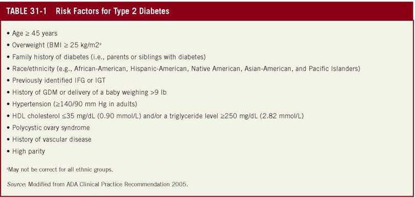
The presence of obesity is a major factor that influences the rate of progression and its presence causes a twofold increase in the incidence of overt type 2 diabetes in patients with previous GDM.47,48 Obesity, excessive weight gain during pregnancy, and/ or failure to lose the excessive weight postpartum, ethnic background (especially Hispanic origin),49,50 severity of fasting hyperglycemia during pregnancy, presence of IGT postpartum,51,52 and positive family history of type 2 diabetes53 have all been shown to increase the incidence of overt type 2 diabetes in women with gestational diabetes. With the above considerations in mind, the person at highest risk to develop type 2 diabetes within the shortest period would be the gestational diabetic woman who is obese, has a positive family history of type 2 diabetes, has a fasting plasma glucose concentration greater than 95 mg/dL during pregnancy, has IGT postpartum, and has an ethnic background shown to be associated with an increased incidence of type 2 diabetes (Table 31-1).
PATHOPHYSIOLOGY OF TYPE 2 DIABETES
Type 2 diabetes is a chronic, progressive, and incompletely understood metabolic disease defined by the presence of chronic hyper- glycemia.54 Although resistance to some actions of insulin and inadequate secretion of insulin for the given metabolic state are the critical abnormalities in type 2 diabetes, several other factors contribute to the hyperglycemic state such as endothelial cell dys- function.3 Insulin resistance is typically present for some years before diagnosis, manifested as diminished stimulation of glucose transport in muscle and adipose tissue and inadequate suppression of glucose production in the liver in response to insulin. However, euglycemia is maintained as long as β cells secrete higher amounts of insulin. Over time, insulin levels decline because of the decreased number of β cells and their diminished secretory capacity.4,54-56
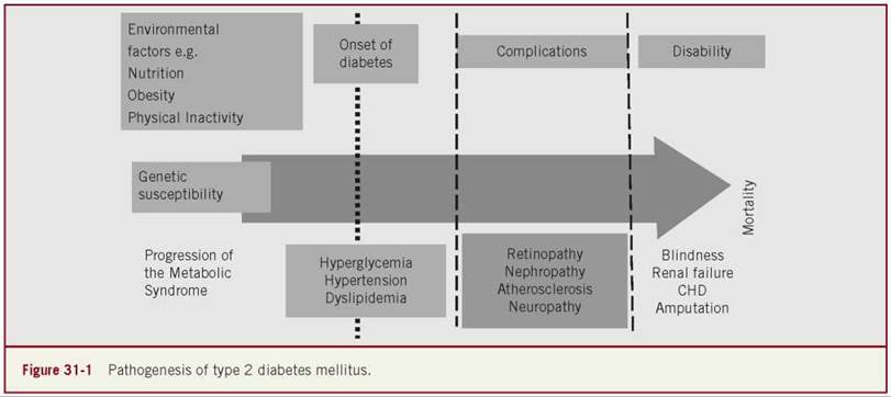
Longitudinal studies involving Pima Indians and other populations have shown a 50% or greater decrease in maximal β-cell function at diagnosis; abnormal postprandial suppression of glucagon secretion also occurs. β-cell failure is mediated by genetic factors and exposure to chronically elevated levels of blood glucose (glucotoxicity) and free fatty acids (lipotoxicity). Older age, amyloid fibrils in islets, and chronically high rates of insulin secretion also perform mechanistic roles. The majority of genetic abnormalities that have been identified in patients with type 2 diabetes are related to β-cell function.57 Insulin resistance and interrelated β-cell dysfunction and failure are the core pathologic defects in type 2 diabetes. Early in the course of the disease, insulin levels are elevated in an attempt to compensate for the increased insulin resistance of muscle/fat and hepatic tissues. As the disease progresses, insulin levels drop as the β-cells decline in function.4,58,59 The disease progresses to hyperglycemia, which, if left untreated, leads to serious complications involving many major organ systems. Once hyperglycemia is identified, a disruption of the normal relationship between β-cell function and insulin sensitivity is established. The United Kingdom Prospective Diabetes Study (UKPDS) showed that type 2 diabetes is a progressive disease that stems from decline in β-cell function (Figure 31-1).60
There is a hepatic defect, which is characterized by excessive basal glucose production despite elevated fasting insulin levels and impaired suppression of hepatic glucose production in response to an incremental increase in plasma insulin concentra- tion.4 A defect in muscle glucose uptake has been demonstrated with leg and forearm catheterization techniques and has been shown to involve the pathways of both glucose oxidation and nonoxidative glucose disposal. In light of these acquired defects, it is obvious that the study of individuals with significant fasting hyperglycemia is unlikely to reveal the basic metabolic defect(s), which characterize the diabetic genotype and which are responsible for the initiation of the demise of glucose tolerance.4
1. Insulin Secretion. Loss of the first phase of insulin secretion develops early (fasting plasma glucose >115 mg/dL) in the natural history of type 2 and impaired second phase insulin secretion is present in the majority of type 2 diabetic individuals with fasting plasma glucose levels in excess of 160-180 mg/ dL.61-63 However, before the onset of IGT, the first and second phases of insulin secretion are increased, and conversion from normal to IGT is associated with the development of severe insulin resistance. A similar sequence of events occurs during the development of IGT in the rhesus monkey.64
2. Insulin Resistance. Impaired insulin action is a characteristic feature of type 2 diabetes. Insulin resistance involves the liver, muscles, and adipose tissue.4,65
Liver. Hepatic insulin resistance is characterized by excessive basal glucose production despite the presence of elevated fasting plasma insulin levels. The increase in fasting plasma glucose concentration in type 2 diabetics with overt fasting hyperglycemia (>126 mg/dL) is closely correlated with the increased rate of basal hepatic glucose production. This observation has led investigators to conclude that increased basal hepatic glucose production is a major, if not the primary, cause of fasting hyperglycemia in type 2 diabetes. Studies have shown that essentially the increased rate of hepatic glucose release results from accelerated gluconeogenesis, without any significant change in glycogenolysis. This increase in gluconeogenesis is due to an increased conversion of lactate, alanine, and glycerol to glucose. In mild type 2 diabetics with fasting plasma glucose concentrations less than 126 mg/dL, the absolute rate of hepatic glucose production is not increased, although evidence suggests that even in these individuals there is a shift from glycogenolysis to gluconeogenesis.66-68
Muscle and Adipose Tissue. Using a variety of techniques, including the insulin clamp, minimal model, and insulin suppression test, numerous studies have documented the presence of moderate-to-severe insulin resistance in peripheral tissues in type 2 diabetic subjects. Muscle has been shown to represent the primary tissue responsible for the defect in insulin action and the involvement of the pathways of both glucose oxidation and nonoxidative glucose disposal. Although fat tissue is resistant to insulin in these subjects, only small amounts of glucose load is disposed of by adipocytes; muscles are primarily responsible for insulin-stimulated glucose uptake in humans. Both glucose oxidation (measured with indirect calorimetry) and nonoxidative glucose disposal are reduced in type 2 diabetes. From the quantitative standpoint, the decrease in nonoxidative glucose disposal is much greater than the defect in glucose oxidation and has been observed more consistently. The rate of nonoxidative glucose disposal agrees closely with the rate of muscle glycogen synthesis, as measured by nuclear magnetic resonance spectroscopy.69In summary, there are disturbances in all major pathways of glucose homeostasis (glycogen synthesis, glucose oxidation, hepatic glucose production) in type 2 individuals with overt fasting hyperglycemia.
At the cellular level, several mechanisms have been implicated in insulin resistance including a decrease in the number of insulin receptors on insulin target tissues, impaired tyrosine kinase activity, diminished glucose transport and phosphorylation, impaired glycogen synthesis, and defects in glycolysis and glucose oxidation. Insulin resistance appears to be the hallmark of type 2 diabetes. It appears early in the natural history of the disease but is offset by the presence of hyperinsulinemia. The insulin resistance is characterized by defects in both muscle and liver. Only later in the natural history of type 2 diabetes, when the compensatory increase in insulin secretion begins to fail does fasting hyperglycemia and overt type 2 diabetes develop.
On the basis of the above review of the pathogenesis of type 2 diabetes, interventions designed to enhance insulin sensitivity would appear to be most effective in the prevention of the disease. As increased physical activity and weight loss also enhance insulin sensitivity, these nonpharmacological interventions should be part of any primary prevention trial for type 2 diabetes.70,71 Recently, Newsom et al.72 reported that a relatively modest single session of exercise in obese adults improved insulin sensitivity the next day, and a reduction in systemic fatty acid uptake in the several hours after exercise may be important for this effect. Once the diabetic state has become fully established and hyperglycemia develops, all aspects of glucose homeostasis are disrupted and it is not possible to ascertain which defects are primary and which are acquired. Hyperglycemia per se can lead to acquired disturbances in insulin secretion.4,73
The Insulin Resistance Syndrome
A contemporary catchphrase in cardiology and diabetes discourse is metabolic syndrome. As many as 47 million Americans may have this cluster of medical conditions. People with the syndrome have nearly a 3% greater risk of developing clogged coronary arteries. Other studies have found that those who have the syndrome are twice as likely to have a heart attack as healthy individuals; four to five factors quadruple the risk of heart attack and raise the risk of diabetes.37,74-76 The syndrome, also known as syndrome X, is a collection of potentially lethal metabolic abnormalities that include glucose intolerance, central obesity, hypertension, dyslipidemia, hyperinsulinemia, hypertriglyceridemia, and hypertension. These disorders serve to identify patients at significant risk for development of both cardiovascular disease (main cause of death in diabetic patients) and type 2 diabetes.77
In clinical practice, no single test diagnoses the condition. A formal definition of the insulin resistance syndrome (IRS) was devised for nonpregnant individuals78 and can be diagnosed if any of the three of the following criteria are present: abdominal obesity (waist circumference >102 cm [40 in] for men and more than 88 cm [35 in] for women; triglyceride levels of 150 mg/dL (1.7 mmol/L) or higher; high-density lipoprotein (HDL) cholesterol lower than 40 mg/dL (1 mmol/L) in men and lower than 50 mg/dL (1.3 mmol/L) in women; blood pressure of 130/85 mm Hg or higher; and fasting glucose level of 110 mg/dL (6.1 mmol/dL) or higher. Two similar definitions were suggested by the American Association of Clinical Endocrinology (AACE) and the WHO. Approximately 25% of populations worldwide meet the criteria for the diagnosis of metabolic syndrome.
It is surprising that despite the acceptance of the syndrome, to date, few studies have addressed its impact in pregnancy. The physiological and metabolic changes accompanying pregnancy have profound effects on the mother. Yet no specific criteria for the pregnant state have been established. It is safe to assume that many GDM women and a fair segment of pregnant nondiabetic women (e.g., obese) would be classified as metabolic syndrome postpartum. Currently, the perinatal impact of the syndrome remains unknown. The criteria for metabolic syndrome in nonpregnancy cannot be accurately used in pregnancy. For example, abdominal circumference is continually increasing; lipid and glucose metabolism changes as part of the normal physiology of pregnancy and pregnancy in and of itself is considered an insulin resistant state. These qualifiers question the validity of the definition of metabolic syndrome during pregnancy. Furthermore, because of the paucity of an accurate definition during pregnancy, it is impossible to measure the short-term outcome of the syndrome. If you cannot measure it, you cannot manage it. The problem of diagnosing without an established diagnosis is putting the cart before the horse. It has led to unnecessary labeling as well as overtreating and/or undertreating these women. We need to address our efforts to create diagnostic thresholds (correlated normality) for lipid, glucose, and other factors associated with type 2 and metabolic syndrome in pregnancy.
An insight into the impact of metabolic syndrome in pregnancy was suggested by Meyers-Seifer and Vohr.79 They investigated lipid levels in women with previous GDM at 5-6 years postpartum. Their study showed that mean total cholesterol, triglycerides, low-density lipoprotein (LDL), glucose, and systolic blood pressure were significantly higher among GDM women compared to nondiabetic controls. In another study, obesity and GDM in a prior pregnancy were found to be a significant risk factor for development of the IRS over time. The authors concluded that early detection of markers of IRS is vital for possible prevention of type 2 diabetes and cardiovascular adverse events in women.80 Bo et al. studied women with normal screening test, abnormal screening test with normal oral glucose tolerance test (OGTT), one abnormal value on the OGTT and gestational diabetes. The prevalence of metabolic syndrome was 0%, 4.9%, 20%, and 18%, respectively. They concluded that metabolic syndrome in mid-pregnancy was an independent predictor of macrosomia in women with any degree of gestational hyperglycemia; the oral glucose challenge test identifies pregnancies with metabolic abnormalities and adverse neonatal outcomes also in the presence of a normal OGTT.81 Enhanced recognition of the population at risk and more aggressive treatment protocols are mandatory if patients are to be spared the development of type 2 diabetes, coronary artery disease, and stroke. The foundation of the treatment needs to be a conceptual framework that addresses diet, exercise, as well as drug therapy.
MATURITY-ONSET DIABETES OF THE YOUNG
Maturity-onset diabetes of the young (MODY) is characterized by nonketotic diabetes, an autosomal dominant inherited disease that affects function of the β cells of the pancreas. MODY is a result of mutations on any one of at least six genes. This heterogeneous group of ailments usually affects children, adolescents, and young adults before age 25. The mutation of the genes not only disrupts β-cell function leading to diabetes mellitus but also causes abnormal functioning of the liver and kidneys. The many factors that influence insulin sensitivity such as infection, puberty, and pregnancy may set in motion the onset of and enhance hyperglycemia in MODY patients.82 Patients with MODY have a family history of diabetes and will often display mild, asymptomatic hyperglycemia. Some will have mild fasting hyperglycemia for many years (no classic symptoms and therefore MODY remains undiagnosed until adulthood), whereas others may demonstrate varying degrees of glucose intolerance for many years before persistent fasting hyperglycemia necessitating pharmacological therapy.83-87 According to current accounts, MODY may be responsible for 1%-5% of all cases of diabetes in the United States and many industrialized nations.88 MODY patients are distinct from type 2 diabetes patients in that family history of the disease can be traced back 3-4 generations, early onset, and absence of obesity. Type 2 diabetic patients are often diagnosed with increasing frequency in adolescence, but the distinguishing feature is obesity.
MODY 2 (glucokinase gene related) is the ailment that displays impaired fasting glucose and IGT especially in children of all racial and ethnic groups with mild hyperglycemia and in women with GDM and a family history of diabetes.89 Heterozygous mutations in glucokinase are associated with mild, nonprogressive hyperglycemia (blood glucose concentration 110-145 mg/dL [6.1-8.0 mmol/L]) and is treated with diet alone.90,91 Approximately 50% of women who are carriers of the muted genes have GDM92; less than 50% have overt diabetes. Those who suffer from overt diabetes are generally obese and older adults. Approximately 2% of carriers require insulin therapy. There are few known diabetes risk factors with MODY 2.93 Reduction in β-cell sensitivity to glucose as well as a defect in postprandial glycogen synthesis in the liver appear to cause hyperglycemia in glucokinase-related MODY patients. The heterozygous mutations are associated with MODY and GDM. They influence a reduction in birth weight of 500 g or more, possibly because of their effect on fetal insulin secretion.94,95 The homozygous mutations cause complete glucokinase deficiency with an accompanying permanent neonatal diabetes characterized by low birth weight, severe diabetes, and the necessity to administer insulin within a few days postpartum.96 There is an increase in the threshold concentration of glucose needed to stimulate insulin secretion from about 90 mg/dL (5.0 mmol/L) to approximately 108-126 mg/dL (6.0-7.0 mmol/L). In addition, patients with MODY 2 have slightly increased basal and postprandial plasma glucose concentrations.94
Mutations in the hepatocyte nuclear factor (HNF)-4a gene (MODY 1) and mutations in the HNF-1a gene (MODY 3) are comparable. MODY 1 and 3 have similar mild elevations in fasting plasma glucose concentrations; they have higher plasma glucose concentrations two hours after glucose administration than do persons with MODY 2.90 MODY 1 and 3 tend to increase over time and are associated with a progressive decrease in insulin secretion. Therefore, these patients tend to need oral hypoglycemic drugs or insulin (30%-40%). Mutations in the gene that encodes insulin promoter factor (IPF)-1 are not usually associated with MODY 4 and most of the information related to this condition is based on data from a single family.97 MODY 5 is the result of mutations in the gene encoding HNF-1P that is characterized by diabetes and renal cysts.98
Today it is possible to identify genes responsible for MODY. Scientists can identify family members who have inherited specific mutations even before onset of carbohydrate intolerance. It is recommended that type 1 diabetic patients with a prominent familial history of the disease undergo genetic diagnosis as an appreciable proportion of these patients have been found to carry the HNF-1a mutation. The diagnosis of this gene mutation rather than of type 1 diabetes may ultimately enhance prognosis for these patients.99,100 Obstetrician see women in adolescence and during their reproductive years, the period of MODY occurrence.
His (her) awareness of this phenomenon and its diagnosis may result in improved short- and long-term outcomes for mothers and infants.
IDENTIFICATION OF TYPE 2 DIABETES IN PREGNANCY
Type 2 diabetes has the highest prevalence among the different diabetic types. It is often asymptomatic in its early stages and can remain undiagnosed for many years. The prevalence of type 2 diabetes in pregnancy has been difficult to ascertain for many reasons. In research studies, pregestational diabetes includes patients with type 1 and type 2 diabetes without differentiation. Specific population subgroups have a much higher prevalence of the disease than the population as a whole. These subgroups have certain attributes or risk factors that either directly cause diabetes or are associated with it. When a pregnant woman is taking insulin, the assumption is that she has type 1 diabetes. This assumption may be valid for countries such as Sweden and Finland where the prevalence of type 1 diabetes is high. However, this would be a mistaken assumption for other countries such as Japan, the United Kingdom, and the United States in which the prevalence of type 2 diabetes is higher than that of type 1 and about 75% of women with prepregnancy diabetes have type 2 diabetes.101
The risk for developing type 2 diabetes increases with age, obesity, and lack of physical activity. In general, type 2 diabetes is more common with a family history of diabetes and in certain racial/ethnic groups. It occurs more frequently in women with prior GDM or polycystic ovary syndrome and/or metabolic syndrome (hypertension, dyslipidemia, IGT, or impaired fasting glucose). The greater number of risk factors increases the odds that the individual will develop type 2 diabetes (Table 31-1).102
Screening for type 2 diabetes should ultimately be based on clinical judgment. Screening should be performed on asymptomatic patients in three-year intervals beginning at age 45, especially in obese women (BMI >25). However, testing for diabetes should be considered at a young age and at more frequent intervals in subjects who are overweight or who have one or more additional risk factors. The fasting plasma glucose is the recommended screening test. Fasting plasma glucose >126 mg/dL (7 mmol/L) is an indication for retesting on a different day for confirmation of the diagnosis. The 75-g OGTT may be necessary for the diagnosis of diabetes when the fasting plasma is normal.
A two-hour post-load of >200 mg/dL (11.1 mmol/L) is considered a positive test but should be confirmed on an alternate day.
Nondiabetic individuals with fasting plasma glucose >100 mg/dL but <126 mg/dL are considered to have impaired fasting glucose, and those with the two-hour value on the OGTT >140 mg/dL (7.8 mmol/L) but >200 mg/dL are defined as having IGT. Both of these categories are referred to as prediabetes. Normal glycemia is defined as plasma glucose <100 mg/dL (5.6 mmol/L) and two-hour post load value of <140 mg/dL on the OGTT.102 Pregnancy is characterized by a lower level of fasting plasma glucose and a higher level of postprandial. Therefore, in pregnancy, patients with fasting glucose levels >100 mg/dL but <126 mg/dL who would be categorized in the nonpregnant state as impaired glucose tolerant, in fact can be a masked type 2 diabetes in pregnancy. Again, this shows the overlap in the diagnosis criteria among normal, GDM, IGT, and type 2 diabetes.
Patients with symptoms of marked hyperglycemia that include polyuria, polydipsia, weight loss, and blurred vision should receive diagnostic testing for diabetes. Other patients need to be screened for potential complications of diabetes or with any other clinical presentation in which diabetes is included in the differential diagnosis. A casual plasma glucose level of >200 mg/dL with symptoms is considered a diagnostic for GDM. The HbA1c is not universally recommended for screening or diagnosis of diabetes (Table 31-2).102
GDM patients are often women who have gone undiagnosed for type 2 diabetes and are brought to the attention of the medical community when screened in pregnancy. In areas where there is efficient universal screening, they are diagnosed before 20 weeks’ gestation and may have fasting hyperglycemia; they may display symptoms of type 2 diabetes when tested postpartum. Inefficient and ineffective screening mechanisms may cause women to go undiagnosed throughout pregnancy. The inability to make accurate estimations of prevalence makes it impossible to determine the frequency of various maternal and fetal complications and the economic and social implications they engender.
PREVALENCE OF TYPE 2 DIABETES IN PREGNANCY
Several studies have measured the prevalence of type 2 diabetes in pregnancy. The prevalence of the disease is based on ethnicity, geographic region, obesity, and dietary habits of a given population. However, the majority of studies showed approximately 10%—15% undiagnosed type 2 diabetes within the GDM population.11,51,103-105 Three studies were population specific and may not be generalizable to the population at large. In a prospective study of 811 pregnancies in the Pima Indians of Arizona, 6.3% were known to have diabetes before pregnancy.106 An additional study of type 2 diabetes (diagnosed before pregnancy) in the Ojibwa-Cree nation of northwestern Ontario, Canada, demonstrated a prevalence of 3.2%.107 A prevalence of 3.4% for type 2 diabetes was found in women of the Tohono O’odham nation in southern Arizona in the first 20 weeks’gestation.108 Engeigau et al.109 in a broad population-based survey performed in the United States suggested an increasing prevalence of type 2 diabetes in pregnancy. They estimated that about 0.2%-0.5% of all pregnancies were complicated by pregestational diabetes (type 1 or type 2) and that type 2 diabetes accounted for 65% in the year of the study, that is, 1995, compared with only 26% in 1980. These studies demonstrated mainly ethnic-specific populations and not a cross section of the population. Therefore, the results of these studies represent the tip of the iceberg but the submerged glacial mass remains unknown.

MATERNAL AND FETAL COMPLICATIONS OF TYPE 2 DIABETES
The increasing prevalence of type 2 diabetes in pregnancy is being recognized as representing at least as significant a risk to both mother and baby as does type 1 diabetes. It may ultimately enhance pregnancy complications for both mother and infant.
Maternal Complications
Type 2 diabetes currently contributes to the increased rate of adult onset loss of vision, renal failure, and amputations more than any other disease. The majority of patients are obese and have hypertension and dyslipidemia resulting in a two- to fivefold higher risk of cardiovascular disease in which 70% of the patients die.110,111 The average delay of 4-9 years till diagnosis of type 2 diabetes translates into approximately 20% of patients who have some evidence of microvascular or neurological diabetic complications at the time of diagnosis.5,112 In the United States, the estimated costs of providing care for diabetes is at least $100 billion annually with half attributable to direct cost.113,114
The complications associated with the disease are a result of its duration in addition to the average level of chronic glycemia. HbA1c is a retrospective measure (10-12 weeks) of the level of glycemic control. As it is a retrospective measure, it can be used as a predictor for maternal medical complications. However, its predictability for fetal disease (excluding anomalies) is minimal.115-117 Two classic studies established the role of intensive therapy in the reduction of long-term complications in nonpregnant patients with type 2 diabetes. They demonstrated that metabolic goals in type 2 patients with HbA1c of <7%, average fasting plasma glucose of 90-130 mg/dL and post-meal plasma glucose levels of <180 mg/dL, will result in significant decreases in patient long-term compli- cations.102,118-120 The intensified management approach (reducing LDL and triglyceride levels, increasing HDL levels), control of hypertension (with two or more medications) will significantly decrease maternal medical complications. However, the level of glycemia required for this effect to occur will not be sufficient to prevent pregnancy-related maternal (preeclampsia) and fetal complications (perinatal mortality, macrosomia, etc.)
There is paucity of studies addressing type 2 diabetes in pregnancy and maternal complications. The majority of studies does not distinguish between type 1 and type 2 diabetes, although they recognize the existence of both in their studies yet combine them for the sake of sample size and paucity of information for each group.121-130
Many studies are based on small sample sizes and report rates of hypertensive disorders such as chronic hypertension and preeclampsia but do not specifically address nephropathy, retinopathy, and neuropathy. In addition, the majority of studies do not include specific data on level of glycemic control (method, definitions). Not surprisingly, there are high rates of chronic hypertension (obesity) and preeclampsia (probably because of severity of the disease and poor glycemic control). In 1989, the St. Vincent Declaration set as one of its targets the improvement in pregnancy outcome compromised by diabetes so that the risks would approach those in the nondiabetic population. Despite this lofty goal, the majority of studies on type 1 diabetes or undistinguished preexisting diabetes reporting national data or data from large centers, failed to demonstrate significant improvement. In general, the rate of medical complications is similar in type 1 and type 2 diabetes.131
The major problem in the evaluation of complication rates for types 1 and 2 diabetes during pregnancy is that data are derived from nonpregnant older patients; women of reproductive age are younger and, therefore, the rate of complications may be different. Research shows that 60% of patients with type 2 diabetes do not increase exercise levels following a diabetes diagnosis and 50% do not alter their diets. The figures result from a survey of 652 patients with type 2 diabetes and 337 physicians from the United Kingdom, United States, Spain, India, Japan, and Brazil. The survey aimed to explore why patients with the type 2 diabetes fail to reach treatment goals. Alarmingly, 75% of the patients surveyed were not concerned about the complications of diabetes. A contributing factor is that only 50% of patients were aware of being told of the risks of complications at their diagnosis. The survey suggests that patients need to repeatedly be made aware of the risk of complications. It is understandable that shock and anxiety at the news of such a diagnosis may make it harder for patients to fully absorb the implications of the condition. Lifestyle interventions are important in type 2 diabetes management in order to reduce the risk of health complications and delay the introduction of stronger diabetes medications such as insulin.
The Diabetes and Aging Study was funded by the National Institute of Diabetes and Digestive and Kidney Diseases and the University of Chicago. They found that patient age and disease duration independently determine the clinical course of the disease among adults aged 60 to 80-plus years. The research analyzed data from the Kaiser Permanente Northern California Diabetes Registry of 72,310 diabetic patients aged 60 years and older at baseline in 2004. The mean patient age was 71 years, and about 15% of the study population was aged 80 years and older. The cohort was ethnically diverse and had equal access to health care; most patients were receiving statins and angiotensin-converting enzyme (ACE) inhibitors appropriately. The study participants were followed for up to seven years (mean follow-up: 5.4 years) for acute hyperglycemic events requiring hospitalization; acute hypoglycemic events requiring emergency department visits or hospitalization; microvascular complications such as severe eye disease, incident end-stage renal disease, peripheral vascular disease, and amputation; nonfatal cardiovascular complications such as myocardial infarction, coronary artery bypass graft surgery, angioplasty, ischemic or hemorrhagic stroke, and congestive heart failure; and fatal complications of any kind.
The cardiovascular complications of diabetes are considered the most common as well as the most serious in patients of all ages, and preventing them by concentrating on glycemic control has been the mainstay of diabetes management. But the above large cohort study showed that among older patients and those with longer disease duration, hypoglycemia rates approached those of coronary artery disease. This finding indicates that the core focus of glycemic control is inappropriate for a substantial number of older diabetes patients. “To the extent that hypoglycemia is an adverse effect of treatment, its emergence as a dominant ‘complication’ raises serious concerns about the acceptable limits of iatrogenesis.” Both patient age and duration of type 2 diabetes had a significant, independent effect on which complications were likely to arise. Most notably, the risk of hypoglycemia rose markedly with increasing age and duration of disease, so that it outpaced both coronary and cerebrovascular events as the most common serious complication in this subset of the population. Hypoglycemia was even fairly frequent among younger patients; it was the fourth most common complication among patients in their 60s and the third most common among patients in their 70s. The rate of hypoglycemic events ranged from a low of 3.0/1000 person-years among the youngest patients with the shortest duration of disease to a high of 19.6/1000 person-years among the oldest patients with the longest duration of disease. The corresponding rates of coronary artery disease events were 8.5 and 24.1 per 1000 person-years. This suggests that intensive glycemic control may not be a helpful treatment goal and may even be harmful to the latter group. In addition, among the oldest patients who had a long duration of diabetes (more than 10 years) the rate of acute hyperglycemic events was only 2.35/1000 person-years. The distinctive clinical course of different patient strata supports recommendations to individualize glycemic targets among older people.132
The TODAY (Treatment Options for type 2 Diabetes in Adolescents and Youth) studies may be preferable as a data source of diabetes in pregnancy. This study controls for age, ethnicity, and similar duration of the disease in the nonpregnant state. The series of studies emphasized the increasing burden of type 2 diabetes in youth and adolescents. In one study, the authors sought to determine the prevalence of retinopathy in 517 youth with type 2 diabetes of 2-8 years duration. They concluded that the prevalence of retinopathy and its association with HbA1c and diabetes duration is similar to that previously reported in youth with type 1 diabetes and in adults with type 2 diabetes of known duration. The mechanism underlying the reduced risk of retinopathy in the most obese individuals is unknown. Follow-up of this cohort will help define the natural history of retinopathy in youth with type 2 diabetes.133 The prevalence of early retinopathy in young people with a mean duration of type 2 diabetes of 4.9 years was 13.7%. This is higher than previously reported in young Pima Indians, in whom retinopathy was detected only after age 20 and who had diabetes for five years. Retinopathy in that study was determined by dilated direct ophthalmoscopy, rather than by standardized fundus photographs assessed by skilled graders.
In the SEARCH study, the prevalence of retinopathy using retinal photography was 17% for type 1 diabetes and 42% for type 2 diabetes. However, participants in the SEARCH study had known diabetes duration of five years (mean duration 7.2 years) and were older (mean age 21 years). In a small Australian study of adolescents with type 2 diabetes, researchers found a retinopathy prevalence of only 4%. Differences in methodology in these studies make direct comparisons difficult. However, retinopathy prevalence in adults who developed diabetes on follow-up in the Diabetes Prevention Program was 15.5% after slightly more than three years of diabetes. As in adults, increased prevalence of retinopathy in TODAY participants was associated with older age, longer diabetes duration, and glycemic control as assessed by HbA1c. The most severely obese individuals had decreased retinopathy. An association of lower weight or BMI with increased retinopathy has been reported previously in adults with type 2 diabetes and has been attributed to poor diabetes control. In one study10 of 207 type 2 diabetics, nonproliferative retinopathy was seen in 28% of women, proliferative retinopathy (undiagnosed before pregnancy) found in 4.3%. A small number of women (1.4%) had overt diabetic nephropathy, whereas preeclampsia was high in both groups (26.5% in the patients with type 1 and 30.9% in women with type 2 diabetes). The risk of diabetic complications from type 2 diabetes in pregnancy already warrants that these women receive careful assessment and follow-up.134
The prevalence of hypertension and microalbuminuria increased over time among adolescents with type 2 diabetes regardless of diabetes treatment. The greatest risk for hypertension was male gender and higher BMI. The risk for microalbuminuria was more closely related to glycemic control. Among adolescents with type 2 diabetes, there is limited information regarding the incidence and progression of hypertension and microalbuminuria. Hypertension and microalbuminuria assessments made during the TODAY clinical trial were analyzed for effect of treatment, glycemic control, gender, and race/ethnicity.
The TODAY trials demonstrated that combination therapy with metformin plus rosiglitazone provided superior durability of glycemic control compared with metformin alone, with significantly lower treatment failure rates (38.6% vs. 51.7%), and metformin plus lifestyle was intermediate. The beneficial change in insulin sensitivity and the resultant lower burden on β-cell function achieved in the first six months with metformin plus rosiglitazone appear to be responsible for its superior glycemic durability over metformin alone and metformin plus lifestyle. However, initial Β-cell reserve and HbA1c at randomization are independent predictors of glycemic durability. Therefore, efforts to preserve β-cell function before significant loss occurs and to reduce HbA1c may be beneficial in the treatment of youth with type 2 diabetes.135 However, the use of rosiglitazone is currently being debated since no study to date has evaluated its safety and efficacy in pregnancy. This leaves us with the option to manage type 2 diabetes with metformin in combination with insulin and/ or sulfonylurea medications; however, this combination, too, has also not been tested in pregnancy.
Two major complications related to level of glycemia during pregnancy are diabetic ketoacidosis and hypoglycemia. Hypoglycemia is associated with type 1 diabetes and less common in GDM and type 2 diabetes. Both complications are described in detail in other chapters. Briefly, diabetic ketoacidosis onset in pregnancy will occur at lower glucose levels, and, in comparison to nonpregnant women, often progresses more rapidly. With early detection of precipitating factors (i.e., infection, intractable vomiting, inadequate insulin management or inappropriate insulin cessation, and steroid administration for fetal lung maturation), prompt hospitalization, and targeted therapy with intensive monitoring, morbidity and mortality can be reduced. Management principles include aggressive volume replacement, initiation of intravenous insulin therapy, correction of acidosis, correction of electrolyte abnormalities, and management of precipitating factors, as well as monitoring of maternal-fetal response to treatment. A chain of events during a diabetic ketoacidotic episode can self-perpetuate into a vicious cycle. The elevated glucose levels in the intravascular space create an osmotic gradient, resulting in marked diuresis that in turn leads to a profound state of dehydration and hypovolemia. Hyperglycemia and acidosis are intensified because it promotes the activation of other counter regulatory stress hormones (i.e., growth hormone, cortisol). Sodium levels can become abnormally low as a result of the osmotic diuresis. In addition, electrolyte salts containing sodium, potassium, and phosphorus become bound to anions from keto acids in the bloodstream and are excreted in the urine. Protein breakdown (as a consequence of the perceived state of starvation) and decreased potassium cellular uptake resulting from the lack of insulin result in normal or elevated serum potassium levels in the presence of diminished total body potassium. To forestall morbidity and mortality, a multidisciplinary approach and continuous monitoring of the maternal response to therapy are critical. After viability, fetal monitoring is also indicated, and it is mandatory that maternal metabolic abnormalities be addressed before considering emergent delivery, because both maternal and fetal conditions will likewise improve.136-141
Euglycemic ketoacidosis was first described by Munro et al. in 1973 and is defined as severe ketoacidosis with a serum bicarbonate of 10 mEq/L or less in the absence of pronounced hyperglycemia (blood glucose <200 mg/dL). This level is twofold higher than the glycemic profile found in nondiabetic women. Therefore, the term euglycemia may be misleading and the complications may occur in mildly controlled patients. The condition can also occur in poorly controlled type 2 and GDM women. True euglyce- mic ketoacidosis is exceedingly rare, occurring in 0.8%—1.1% of all episodes (depending on the defining plasma bicarbonate concentration).142-144 Diabetic ketoacidosis is a rare but serious complication of diabetes in pregnancy with deleterious consequences for both the mother and the fetus. Prompt recognition of precipitating factors, aggressive correction of volume depletion and electrolyte imbalance, and insulin administration are paramount in the management of diabetic ketoacidosis.
Fetal Complications
In 1989, the St. Vincent Declaration set as one of its targets the improvement in pregnancy outcome for women with diabetes so that the risks would approach those of the nondiabetic population. Despite the lofty goal, the majority of studies on type 1 diabetes or undistinguished preexisting diabetes reporting national data or data from large centers, failed to demonstrate significant improvement resulting in an over 20% prematurity rate, 26%—55% rate of large-for-gestational age infants, 15%-35% macrosomia, 4%—9% congenital malformations, and 2%—5% perinatal mortality (Table 31-3).121-145-150
TABLE 31-3 Preexisting Diabetes: Perinatal Outcome
|
PTD |
SGA |
LGA |
Macrosomia |
Neonatal Complication |
CA |
Stillbirth |
Perinatal Mortality |
|
|
Sibai et al. ‘00 (MFMU/NICHD n = 462) |
38% |
6.3% |
34.4% |
15.4% |
48.1% |
NA |
1.8% |
3.9% |
|
Evers et al. ‘04 (118 Hospitals-Netherland n = 289) |
32% |
3.0% |
56% |
25.3% |
80.2% |
8.8% |
NA |
2.8% |
|
Lauenberg et al. ‘03 (Denmark n = 1361) |
NA |
NA |
45% |
35% |
NA |
NA |
1.8 |
NA |
|
Persson et al. ‘96 (Sweden n = 113) |
21% |
3.0% |
26% |
26% |
15% |
3.5% |
NA |
0% |
|
Peck et al. ‘91 (UK n = 133) |
NA |
NA |
38% |
NA |
16% |
5.3% |
NA |
2.4% |
|
Penney et al. ‘03 (Scotland National n = 213) |
NA |
NA |
55% |
NA |
NA |
6.0% |
1.9% |
2.9% |
|
Diabetes in pregnancy group, France (n = 435) |
38% |
NA |
NA |
17.3% |
NA |
4.1% |
3.5% |
4.6% |
|
Jovanovic ‘91 EDPS |
NA |
7.8% |
29% |
NA |
NA |
NA |
NA |
NA |
There is paucity of available data on perinatal mortality in women with type 2 diabetes. However, one study suggested that the rate of perinatal mortality might be higher than that of women with type 1 diabetes.151,152 In the study by Cundy et al.,151 perinatal mortality was calculated over a 12-year period (1985—1997) in women (Maori or recent immigrants from Pacific Island nations) with type 2 diabetes attending a diabetes clinic in Australia. The region-specific nature of the study again may preclude generaliza- bility. Perinatal mortality (late fetal death, 28 weeks to term) was significantly higher than in women with type 1 diabetes. Maternal comorbidities including obesity, higher maternal age, higher frequency of hypertension, and low socioeconomic level may have also contributed to the increased mortality rate. This group presented later for care than did women with type 1 disease and were both smokers and obese. Obesity coupled with type 2 diabetes as well as delayed perinatal care has been associated with an increased risk of late fetal death, fetal macrosomia, and preterm delivery. The risk for the fetus for the above complications is not equal for all fetuses. The risk will be dependent on the glycemic level. In addition, because of the number of undiagnosed type 2 patients in GDM, the fetuses are at greater risk for death.
Other published works report perinatal mortality ranging from 4/1000 to 81/1000. Zhu et al.153 and Coetzee et al.154 demonstrated no significant difference in perinatal mortality between patients with type 2 and type 1 diabetes, whereas the study by Sacks et al.155 reported four perinatal deaths in 113 patients with type 2 and none in the 46 patients with type 1 disease. Sacks et al. also found no significant differences in the rates of macrosomia, caesarean section, shoulder dystocia, and neonatal hypoglycemia between the mothers with type 1 and type 2 diabetes. When confirmation from other studies of these findings and corroboration that the outcomes of women with type 2 are similar to those of women with type 1, similar concern to that afforded women with type 1 diabetes will be shown in women with type 2 diabetes and their infants. In fact, a review of the few studies addressing type 2 diabetes in pregnancy revealed similar adverse outcome rates to type 1 diabetes (Table 31-4).156—160 Recently, Tennant et al.125 reported in a large study that the incidence of fetal death, infant death, and congenital malformation showed no difference between types 1 and 2 diabetics in a North England population. A unique feature in the study was that the perinatal mortality rate was calculated with the exclusion of congenital malformations. Overall, the relative risk for perinatal mortality was two- to fourfold higher than in the nondiabetic population with fetal death (RR 4.56 [95% CI 3.42, 6.1], P < .0001) and infant death (RR 1.86 [95% CI 1.00, 3.46], P = .046). There was no difference in the prevalence of fetal death or infant death between women with type 1 diabetes and women with type 2 diabetes. Moreover, there was no evidence that the relative risk (RR) of fetal and infant death had changed over time (P = .95). The effect of this finding is largely moderated by glycemic control. In our program (unpublished data), we also found similar outcomes in neonatal size, lung complications, and neonatal intensive care unit admissions in type 1 and type 2 diabetic patients (Table 31-5).
Just as infants born of mothers with type 1 diabetes are at increased risk of congenital anomalies, so are infants of women with type 2 diabetes. A prospective study of pregnancies complicated by type 2 diabetes in predominately Hispanic women demonstrated a high rate of congenital anomalies in comparison to those who had not participated in a preconception diabetes care program.161 Fifty-six of the 332 infants (11.7%) were born with major congenital anomalies, whereas the rate of congenital malformations in infants of nondiabetic women born in the same hospital during the same period was <2%. These malformation rates compared to those reported in studies of infants born to women with type 1 diabetes who had not received preconception care. The authors attributed poor glycemic control as the cause of the malformations. In the above study, maternal glycosylated hemoglobin concentrations at initial presentation for care were independently associated with the major malformations (P = .0007).
TABLE 31-4 Type 2 Diabetes Pregnancy Outcomes
|
Coetzee ‘85 |
Dooley ‘98 |
Brydon ‘00 |
Dunne ‘03 |
Ozumba ‘04 |
Gunton ‘02 |
|
|
Mean birth weight |
NA |
4.075 |
NA |
NA |
NA |
3407 |
|
Macrosomia |
NA |
62% |
NA |
9% |
39% |
|
|
LGA |
15% |
NA |
40% |
32% |
NA |
36% |
|
Stillbirth |
37% |
4% |
4% |
1.2% |
14% |
5.3% |
|
Shoulder dystocia |
NA |
15% |
NA |
NA |
NA |
0 |
|
Congenital malformation |
8% |
8% |
12% |
11% |
9.0% |
NA |
|
Jaundice |
53% |
38% |
NA |
NA |
NA |
NA |
|
Hypoglycemia |
12% |
8% |
NA |
NA |
NA |
NA |
|
Polycythemia |
NA |
NA |
NA |
NA |
NA |
NA |
|
Respiratory distress syndrome |
NA |
4% |
NA |
NA |
NA |
NA |
|
NICU |
NA |
NA |
NA |
37% |
NA |
NA |
|
No. of patients |
691 |
26 |
57 |
165 |
122 |
11 |
TABLE 31-5 Pregnancy Outcome in Type 2 Diabetes: The San Antonio Experience (1990-2000)
|
Type 1 |
Type 2 |
P |
|
|
Gestational age delivery |
37 ± 3 |
38 ± 4 |
<0.13 |
|
NICU |
13% |
10% |
<0.001 |
|
Respiratory support |
17% |
7% |
<0.001 |
|
Lung complications |
30% |
23% |
<0.09 |
|
No. of days intubation |
9 ± 14 |
3 ± 8 |
<0.09 |
|
Neonatal hypoglycemia |
18% |
26% |
ns |
|
LGA (>90 percentile) |
18% |
23% |
<0.05 |
|
Macrosomia (>4000 g) |
9% |
14% |
<0.007 |
|
SGA (<10 per.) |
14% |
7% |
<0.05 |
|
Shoulder dystocia |
2% |
6% |
Ns |
No difference in glycemic control was noted between women taking oral hypoglycemic agents during the first eight weeks of gestation and those on insulin or diet, suggesting that the oral hypoglycemic agents were not the cause of the malformations.
A study in the United Kingdom of mostly Indian women with type 2 diabetes reported twice the frequency of congenital malformations compared with type 1 diabetic women (12.2% vs. 6.1%). The authors ascribed poorer attendance for prenatal care, delayed booking for antenatal care, and failure to adhere to a strict regimen of glycemic control during organogenesis as contributing factors to the higher rate of congenital malformations.157
The rate of congenital anomalies in patients with type 1 diabetes can be reduced to that of the general population if good glycemic control is achieved at the time of conception. Although targeted levels of glycemic control have been achieved by women in certain academic centers, the reports from population studies show that clinicians have not been as successful in preventing congenital anomalies.162,163 Population studies performed in Europe and the United States report rates of congenital anomaly and perinatal mortality significantly higher than that in the general population. In a study evaluating preconception education, 61% of the type 1 diabetic women had failed to achieve targeted levels of glycemic control at onset of pregnancy. Another study examining the reasons why women were not proactive in planning for pregnancies with improved glycemic control before conception, found that many socioeconomic variables contributed to their lax behavior.164
Cultural and socioeconomic factors compounded by minority or immigrant status often preclude women with type 2 diabetes in industrialized countries from accessing appropriate care for their disease. In addition to patients’ reticent approach and as aware as they may be of complications because of type 1 diabetes in pregnancy, clinicians are not generally aware of the increased prevalence of type 2 diabetes in pregnancy and the attending complications. They have been trained to accept the dogma that women on diet therapy or oral hypoglycemic agents have “mild” diabetes and are, therefore, at less risk. This fallacy can lead to suboptimum care and follow-up, increasing the chance for poor glycemic control at the onset of pregnancy, and a subsequent increase in congenital anomalies.
Treatment Modalities for Type 2 Diabetes in Pregnancy
A 36-year-old woman at 16 weeks gestation with a two-year history of type 2 diabetes mellitus presents for care. She has no microvascular or macrovascular complications. Her family history is positive for type 2 diabetes and cardiovascular disease in her mother and older brother. On examination, her weight is 99.8 kg (220 lb), with a BMI of 37 and blood pressure 125/85 mm Hg. Her glycosylated hemoglobin level is 9.3%, serum creatinine level 1.0 mg/dL, LDL 88 mg/dL, HDL cholesterol 45 mg/dL, and triglyceride level 190 mg/dL. She does not have microalbuminuria. Her medications include metformin (500 mg twice daily) and glipizide (5 mg twice daily).
This patient presents several areas that require modification to optimize her care. The level of glycemic control is not satisfactory for pregnancy or for the nonpregnant state. She needs to be transferred to insulin therapy as oral agents will not achieve, in this patient, the desired level of glycemic control. The second problem is the inability to administer statins during pregnancy; thus, the only way to improve her status is to encourage diet and behavior modification both dependent on patient compliance. In addition, the hypertension needs to be brought under control with pregnancy-prescribed drugs. This patient will need a complete work-up to rule out potential congenital malformations with a plan for follow-up for fetal growth diversity and surveillance.
The main goal of all treatment modalities in type 2 diabetes is to delay or mitigate the complications of the disease. However, different management approaches need to be employed before conception and during pregnancy as some drugs are contraindicated during pregnancy. In addition, the glycemic goal to optimize pregnancy outcome are different prior and during pregnancy. The two main maternal complications fall into 2 groups: microvascular (retinopathy, nephropathy, and neuropathy) and macrovascular (ischemic heart disease, stroke, and peripheral vascular disease). The randomized UKPDS in nonpregnant individuals clearly demonstrated that intensified therapy (sulfonylureas or insulin) is superior to conventional therapy of diet and exercise alone (to decrease microvascular complications). However, the study showed that diabetes is a progressive disease and the efficacy of the medications decreased after five years. The UKPDS demonstrated that each percentage point reduction in HbA1c reduced microvascular complications by 37%, diabetes-related endpoints by 21%, death related to diabetes by 21%, and myocardial infarction by 14%.116
The traditional approach to treatment of type 2 diabetes and gestational diabetes is based on a step-wise approach from the least invasive to pharmacological therapy. The first step involves diet and lifestyle modification to include exercise to achieve weight loss. Weight reduction of 2.3—4.5 kg (5—10 lb) can result in a significant decrease in the glucose profile.165 The problem with this approach is twofold. This minimal weight reduction will not result in targeted glucose levels that are needed to optimize pregnancy outcome. Furthermore, weight reduction is not recommended in pregnancy even in obese patients. Thus, the hypocaloric diet to the extent of weight reduction may adversely affect the fetus. Ultimately, we seek to program the unborn child for the metabolic world his mother inhabits and the one in which he/she is expected to live and thrive.166,167 Therefore, it is more efficacious to reserve a weight reduction diet for the pre- and postpartum periods. During pregnancy, a diabetic diet should be prescribed based on the principles designed for pregnancy.
The second step is the addition of oral agents if the patient is not yet on that regimen. This step is used mainly in type 2 nonpregnant diabetics; in pregnancy this approach will fail to achieve a level of glycemic control required to enhance pregnancy outcome in the majority of patients. Therefore, in type 2 pregnant women, insulin analog therapy is the preferred management.
Sulfonylureas and metformin are the most commonly used oral hypoglycemic agents. Both drugs lower glycosylated hemoglobin by approximately 1.5%. One problem with oral agents is the debate if these drugs can be used in the first trimester even if it appears safe to use glyburide and metformin throughout pregnancy. Combination therapy with these two drugs can significantly improve the level of glycemic control. However, is the improvement in glycemic control achieved in type 2 diabetes sufficient to protect the fetus from the effect of glucose toxicity? To date, as previously described, there is no data reporting achievement of outcome comparable to nondiabetic individuals. The physician must determine if using this drug will bring his patient with type 2 diabetes to a level of glycemia resulting in a healthy and viable child.
The next step is invoked if oral antidiabetic drugs fail to achieve level of glycemic control or the physician chooses to go directly to insulin therapy. Higher doses of insulin virtually always result in lower glucose levels. The problem is that insulin is not always used in the appropriate dose. The most commonly used therapy is to calculate insulin dose based on 1 unit/kg of body weight and thereafter to increase the dose as needed. Split injection for GDM and type 2 diabetic women is the most effective approach. The insulin pump should be used in type 1 diabetes. Insulin can also be administered in combination with sulfonylurea or metformin thus decreasing the overall insulin dose. However, no data has been forthcoming supporting this management combination.
SUMMARY
Now that we have been forewarned of the growing pandemic of type 2 diabetes and obesity in pregnancy, we need to become forearmed. Over the past few decades there has been no significant improvement in perinatal outcome complicated by diabetes mel- litus (types 1 and 2). The recognition of modifiable risk factors such as maternal glycemic control using self-monitoring blood glucose in combination with pharmacological therapy (intensified therapy) and weight gain in pregnancy should enhance pregnancy outcome. The overemphasis and concentration on the nonmodifiable risk factors in pregnancy is a futile pursuit that may generate lively discussion but paucity of results. The focus needs to be in education for the care provider, that is, enhanced recognition of this growing entity and a heightened awareness of the need for prepregnancy counseling about preconception glycemic control. Another center of attention should be the dissemination of information to patients of the impending maternal and fetal risks of type 2 diabetes in pregnancy. This care would include antenatal care for surveillance of maternal diabetes complications as well as careful obstetric surveillance to improve maternal and fetal outcomes.
The incremental successes in the treatment of type 1 diabetes in the nonpregnant and pregnant states bode well for comparable success in assessing and treating type 2 diabetes in pregnancy The parallel thrusts must be in education for the care provider, that is, enhanced recognition of this growing entity and a heightened awareness of the need for prepregnancy counseling about preconception glycemic control. Another front would be the dissemination of information to patients of the impending maternal and fetal risks of type 2 diabetes in pregnancy coupled with an environment of cultural competence that empowers the patient to seek and receive appropriate care. This care would include antenatal care for surveillance of maternal diabetes complications as well as careful obstetric surveillance to improve maternal and fetal outcomes.
REFERENCES
1. Centers for Disease Control and Prevention. National Diabetes Fact Sheet: National Estimates and General Information on Diabetes and Pre-diabetes in the United States, 2011. Atlanta, GA: Department of Health and Human Services, Centers for Disease Control and Prevention; 2011. http://www.cdc.gov/diabetes/pubs/ pdfndfs_2011.pdf
2. Narayan K, Ali M, Koplan J. Global non-communicable disease— where worlds meet. N Engl J Med. 2010;363:1196—1198.
3. Tahrani A, Bailey C, Del Prato S, Barnett AH. Management of type 2 diabetes: new and future developments in treatment. Lancet. 2011;378:182—197.
4. DeFronzo R. From the triumvirate to the ominous octet: a new paradigm for the treatment of type diabetes mellitus. Diabetes. 2009;58:773—795.
5. Harris M, Klein R, Welborn T, et al. Onset of NIDDM occurs at least 4—7 years before clinical diagnosis. Diabetes Care. 1992;15:815—819.
6. Eastman R, Cowie C, Harris M. Undiagnosed diabetes or impaired glucose tolerance and cardiovascular risk. Diabetes Care. 1997;20:127—128.
7. Emerging Risk Factors Collaboration. Diabetes mellitus, fasting blood glucose concentration, and risk of vascular disease: a collaborative meta-analysis of 102 prospective studies. Lancet. 2010;375:2215—2222. [Erratum, Lancet. 2010;376:958.]
8. Rosenbloom A, Joe J, Young A, et al. Emerging epidemic of type 2 diabetes in youth. Diabetes Care. 1999;22:345—354.
9. Diabetes and impaired glucose tolerance in woman aged 20—39 years. WHO Ad Hoc Diabetes Reporting Group. World Health Stat Q. 1992;45:321—327.
10. Omori Y, Minei S, TestuoT, et al. Current status of pregnancy in diabetic woman: a comparison of pregnancy in IDDM and NIDDM mothers. Diabetes Res Clin Pract. 1994;24(suppl):S273—S278.
11. Conway D, Langer, O. Effects of new criteria for type 2 diabetes on the rate of postpartum glucose intolerance in women with gestational diabetes. Am J Obstet Gynecol. 1999;181(13):610—614.
12. Mokdad A, Ford E, Bowman B, et al. Diabetes trends in the US: 1990—1998. Diabetes Care. 2000;23:1278—1283.
13. Langer O, Langer N. Diabetes in women older than 40 years of age. Obstet Gynecol Clin North Am. 1993;20(2):299—311.
14. Harris M, Hadden W, Knowler W, et al. Prevalence of diabetes and impaired glucose tolerance and plasma glucose levels in U.S. population aged 20—74 yr. Diabetes. 1987;36:523—534.
15. Haffner S, Hazuda H, Mitchell B, et al. Increased incidence of type II diabetes mellitus in Mexican Americans. Diabetes Care. 1991;14:102—108.
16. Stern M, Haffner S. Type II diabetes and complications in Mexican Americans. Diabetes Metab Rev. 1990;6:29—45.
17. Haffner S, Fong D, Stern M, et al. Diabetic retinopathy in Mexican- Americans and non-Hispanic whites. Diabetes. 1988;37:878—874.
18. Haffner S, Mitchell B, Pugh J, et al. Proteinuria in Mexican- Americans and non-Hispanic whites with NIDDM. Diabetes Care. 1989;12:530—536.
19. Pugh J, Tuley M, Hazuda H, et al. The influence of outpatient insurance coverage on the microvascular complications of non-insulin-dependent diabetes in Mexican-Americans. J Diabetes Complications. 1992;6:236—241.
20. Haffner S, Mitchell B, Moss S, et al. Is there an ethnic difference in the effect of risk factors for diabetic retinopathy? Ann Epidemiol. 1993;3:2—8.
21. Ferrannini E, Haffner S, Mitchell B, et al. Hyperinsulinemia: the key feature of cardiovascular and metabolic syndrome. Diabetologia. 1991;34:416^22.
22. Mitchell B, Hazuda H, Haffner S, et al. Myocardial infarction in Mexican-Americans and non-Hispanic whites. The San Antonio Heart Study. Circulation. 1991;83:45—51.
23. West K, Ahuja M, Bennett P, et al. The role of circulating glucose and triglyceride concentrations and their interactions with other “risk factors” as determinants of arterial disease in nine diabetic population samples from the WHO Multinational Study. Diabetes Care. 1983;6:361—369.
24. Fontbonne A, Eschwege E, Cambien F, et al. Hypertriglyceridemia as a risk factor of coronary heart disease mortality in subjects with impaired glucose tolerance or diabetes. Diabetologia. 1989;32:300—304.
25. Haffner S, Stern M, Hazuda H, et al. Cardiovascular risk factors in confirmed prediabetics. JAMA. 1990;263:2893—2898.
26. Kaplan N. The deadly quartet: upper-body obesity, glucose intolerance, hypertriglyceridemia, and hypertension. Arch Inter Mad. 1989;149:1514—1520.
27. Haffner S, Ferrannini E, Hazuda H, et al. Clustering of cardiovascular risk factors in confirmed prehypertensive individuals. Hypertension. 1992;20:38—45.
28. Modan M, Halkin H, Almog S, et al. Hyperinsulinemia: a link between hypertension, obesity and glucose intolerance. J Clin Invest. 1985;75:809—817.
29. The DCCT Research Group. The effect of intensive treatment of diabetes on the development and progression of long-term complications in IDDM. N Engl J Med. 1993;329:977—986.
30. Reichard P, Nilsson B, Rosenqmst U. The effect of long-term intensified insulin treatment on the development of microvascular complications of diabetes mellitus. N Eng J Med. 1993;329:304—309.
31. Wang P, Lau J, Chalmers T. Meta-analysis of effects of intensive blood-glucose control on late complications of type 1 diabetes. Lancet. 1993;341:1306—1309.
32. Lasker R. The DCCT: implications for policy and practice. N Engl J Med. 1993;329:1035—1036.
33. NIH Consensus Development Conference. Health implications of obesity. Ann Intern Med. 1985;103:1073—1077.
34. Saad M, Knowler W, Pettitt D, et al. The natural history of impaired glucose tolerance in Pima Indians. N Engl J Med. 1988;319:1500—1506.
35. DeFronzo R, Bonadonna R, Ferrannini E. Pathogenesis of NIDDM. Diabetes Care. 1992;5:318—366.
36. Bergman R. Lilly Lecture 1989. Toward physiological understanding of glucose tolerance. Minimal-model approach. Diabetes. 1989;38:1512—1526.
37. Reaven G. Banting Lecture 1988. Role of insulin resistance in human disease. Diabetes. 1988;37:1595—1607.
38. O’Sullivan J, Mahan C. Prospective study of patients with chemical diabetes. N Engl J Med. 1968;278:1038—1041.
39. Stern M, Valdez R, Haffner S, et al. Stability over time of modern diagnostic criteria for type 2 diabetes. Diabetes Care. 1993;16:978—983.
40. Couston D. Gestational diabetes. State of the Union. Diabetes Care. 1992;15:716—717.
41. Perss B, Hanson U, Binder C. Follow-up of women with previous GDM. Diabetes. 1991;40(suppl 2):136—141.
42. Harris M. Gestational diabetes may represent discovery of preexisting glucose intolerance. Diabetes Care. 1988;11:402—411.
43. Yudkin J, Alberti K, Moriarty D, et al. Impaired glucose tolerance. Br Med J. 1990;301:397—402.
44. Eriksson K, Lindrarde F. Impaired glucose tolerance in a middle-aged male urban population: a new approach for identifying high risk cases. Diabetologia. 1990;33:526—531.
45. Stowers J, Sutherland H, Kerridge D. Long-range implications for the mother: the Aberdeen experience. Diabetes. 1985;34 (suppl 2):106—110.
46. O’ Sullivan J. Diabetes mellitus after GDM. Diabetes. 1991;40(suppl 2):131—135.
47. Haffner S, Stern M, Mitchell B, et al. Incidence of type II diabetes in Mexican-Americans predicted by fasting insulin and glucose levels, obesity, and body-fat distribution. Diabetes. 1990;39:283—288.
48. Stern M, Gonzalez C, Mitchell B, et al. Genetic and environmental determinants of type 2 diabetes in Mexico City and San Antonio. Diabetes. 1992;41:484—492.
49. Nahum G, Huffaker B. Racial differences in oral glucose screening test results: establishing race-specific criteria for abnormality in pregnancy. Obstet Gynecol. 1993;81:517—522.
50. Hadden D. Geographic and racial variations in the incidence of GDM. Diabetes. 1985;34(suppl 2):8—12.
51. Kjos S, Buchanan T, Greenspoon J, et al. Gestational diabetes mellitus: the prevalence of glucose intolerance and diabetes mel- litus in the first two months post partum. Am J Obstet Gynecol. 1990;163:93—98.
52. Martin A, Simpson J, Ober C, et al. Frequency of diabetes melli- tus in mothers of probands with gestational diabetes. Am J Obstet Gynecol. 1985;151:471—475.
53. American Diabetes Association. Standards of medical care in diabetes-2012. Diabetes Care. 2012;35(suppl 1):S11—S63.
54. DeFronzo, R. Lilly Lecture 1988. The triumvirate: p-cell, muscle, liver. Collusion responsible for NIDDM. Diabetes. 1988;37:667—687.
55. Butler A, Janson J, Bonner-Weir S, et al. Beta-cell deficit and increased beta-cell apoptosis in humans with type 2 diabetes. Diabetes. 2003;52:102—110.
56. Rahier J, Guiot Y, Goebbels R, et al. Pancreatic beta-cell mass in European subjects with type 2 diabetes. Diabetes Obes Metab. 2008;10(suppl 4):32—42.
57. Weyer C, Bogardus C, Mott D, et al. The natural history of insulin secretory dysfunction and insulin resistance in the pathogenesis of type 2 diabetes mellitus. J Clin Invest. 1999;104:787—794.
58. Ward E, Beard J, Porte D. Clinical aspects of islet beta cell function in non-insulin dependent diabetes mellitus. Diabetes Metab Rev. 1986;2:297—314.
59. Ferrannini E., Simonson D, Katz L, et al. The disposal of an oral glucose load in patients with non-insulin-dependent diabetes. Metabolism. 1988;37:79—85.
60. United Kingdom Prospective Diabetes Study. Intensive blood-glucose control with sulphonylureas or insulin compared with conventional treatment and risk of complications in patients with type 2 diabetes. UKPDS Group. Lancet. 1998;352:837—853.
61. Efendic S, Grill V, Luft R, et al. Low insulin response: a marker of prediabetes. Adv Exp Med Biol. 1988;246:167—174.
62. Cerasi E, Luft R, Efendic S. Decreased sensitivity of the pancreatic beta cells to glucose in pre-diabetic and diabetic subjects. Diabetes. 1972;21:224—234.
63. Porte D. Beta cells in type II diabetes mellitus. Diabetes. 1991;40:166—180.
64. O’Rahilly S, Turner R, Matthews D. Impaired pulsatile secretion of insulin in relatives of patients with non-insulin-dependent diabetes. N Engl J Med. 1988;318:1225—1230.
65. DeFronzo RA, Simonson D, Ferrannini E. Hepatic and peripheral insulin resistance: a common feature in type 1 (non-insulin-dependent) and type 2 (insulin-dependent) diabetes mellitus. Diabetologia. 1982;23:313—319.
66. Consoli A, Kennedy F, Miles J, et al. Determination of Krebs cycle metabolic carbon exchange in vivo and its use to estimate individual contributions of gluconeogenesis and glycogenolysis to overall glucose output in man. J Clin Invest. 1987;80:1303—1310.
67. DeFronzo R, Ferrannini E, Simonson D. Fasting hyperglycemia in non-insulin-dependent diabetes mellitus: contributions of excessive hepatic glucose production and impaired tissue glucose uptake. Metabolism. 1988;38:387—395.
68. DeFronzo RA, Ferrannini E. Regulation of hepatic glucose metabolism in humans. Diabetes Metab Rev. 1987;3:415—459.
69. Ramachandran A, Snehalatha C, Mohan V, et al. Decreased insulin sensitivity in offspring whose both parents have type 2 diabetes. Diabetic Med. 1990;7:331—334.
70. Diabetes prevention program research group. Reduction in the incidence of type 2 diabetes with lifestyle intervention or metformin. N Engl J Med. 2002;346(6):393—403.
71. Tuomilehto J, Lindstrom J, Eriksson J, et al. (Finnish Diabetes Prevention Study Group). Prevention of type 2 diabetes mellitus by changes in lifestyle among subjects with impaired glucose tolerance. N Engl J Med. 2001;344(18):1343—1350.
72. Newsom S, Everett A, Hinko A, et al. A single session of low-intensity exercise is sufficient to enhance insulin sensitivity into the next day in obese adults. Diabetes Care. 2013;36(9):2516—2522.
73. Unger R, Grundy S. Hyperglycemia as an inducer as well as a consequence of impaired islet cell function and insulin resistance: implication for the management of diabetes. Diabetologia. 1985;28:119—121.
74. Ford E, Giles W, Dietz W. Prevalence of the metabolic syndrome among US adults: findings from the third National Health and Nutrition Examination Survey. JAMA. 2002;287:356—359.
75. Haffner S, Valdez R, Hazuda H, et al. Prospective analysis of the insulin resistance syndrome (syndrome X). Diabetes. 1992;31:715—722.
76. Reaven G, Lithell H, Landsberg L. Hypertension and associated metabolic abnormalities—the role of insulin resistance and the sympathoadrenal system. N Engl J Med. 1996;334:374—381.
77. Stolar M. Insulin resistance, diabetes, and the adypocyte. Am J Health Syst Pharm. 2002;59(suppl 9):S3—S8.
78. Executive Summary of the Third Report of the National Cholesterol Education Program (NCEP) Expert Panel on Detection, Evaluation, and Treatment of High Blood Cholesterol in Adults (Adult Treatment Panel III). JAMA. 2001;285:2486—2497.
79. Meyers-Seifer C, Vohr B. Lipid levels in former gestational diabetic mothers. Diabetes Care. 1996;19:1351—1356.
80. Verma A, Boney C, Tucker R, et al. Insulin resistance syndrome in women with prior history of gestational diabetes mellitus. J Clin Endocrinol Metab. 2002;87:3227—3235.
81. Bo S, Menato G, Gallo M, et al. Mild gestational hyperglycemia, the metabolic syndrome and adverse neonatal outcomes. Acta Obstet Gynecol Scand. 2004;83:335—340.
82. Fajans S, Bell G, Polansky K. Molecular mechanisms and clinical pathophysiology of maturity-onset diabetes of the young. N Engl J Med. 2001;345(13):971—980.
83. Fajans S. MODY: a model for understanding the pathogeneses and natural history of type II diabetes. Horm Metab Res. 1987;19:591—599.
84. Fajans SS. Scope and heterogeneous nature of MODY. Diabetes Care. 1990;13:49—64.
85. Fajans S, Bell G, Bowden D, et al. Maturity-onset diabetes of the young. Life Sci. 1994;55:413—422.
86. Fajans S, Brown M. Administration of sulfonylureas can increase glucose-induced insulin secretion for decades in patients with maturity-onset diabetes of the young. Diabetes Care. 1993;16:1254—1261.
87. Frayling T, Bulman M, Ellard S, et al. Mutations in the hepatocyte nuclear factor-1a gene are a common cause of maturity-onset diabetes of the young in the UK. Diabetes. 1997;46:7202—7205.
88. Lederman H. Maturity-onset diabetes of the young (MODY) at least ten times more common in Europe than previously assumed? Diabetologia. 1995;38:1482.
89. Bell G, Cuesta-Munoz A, Matschinsky F. Glucokinase. In: Creighton T, editor. Encyclopedia of Molecular Medicine. New York: John Wiley; 2002.
90. Pearson E, Velho G, Clark P, et al. β-cell genes and diabetes: quantitative and qualitative differences in the pathophysiology of hepatic nuclear factor-1a and glucokinase mutations. Diabetes. 2001;50:S101—S107.
91. Owen K, Hattersley A. Maturity-onset diabetes of the young: from clinical description to molecular genetic characterization. Best Pract Res Clin Endocrinol Metab. 2001;15(3):309—323.
92. Ellard S, Beards F, Allen L, et al. A high prevalence of glucokinase mutations in gestational diabetic subjects selected by clinical criteria. Diabetologia. 2000;43:250—253.
93. Velho G, Vaxillaire M, Boccio V, et al. Diabetes complications in NIDDM kindreds linked to the MODY3 locus on chromosome 12q. Diabetes Care. 1996;19:915—919.
94. Byrne M, Sturis J, Clement K, et al. Insulin secretory abnormalities in subjects with hyperglycemia due to glucokinase mutations. J Clin Invest. 1994;93:1120—1130.
95. Hattersley A, Beards F, Ballantyne E, et al. Mutations in the glu- cokinase gene of the fetus result in reduced birth weight. Nat Genet. 1998;19:268—270.
96. Njolstad P, Sovik O, Cuesta-Munoz A, et al. Neonatal diabetes mellitus due to complete glucokinase deficiency. N Engl J Med. 2001;344:1588—1592.
97. Wright N, Metzger D, Borowitz S, et al. Permanent neonatal diabetes mellitus and pancreatic exocrine insufficiency resulting from congenital pancreatic agenesis. Am J Dis Child. 1993;147:607—609.
98. Horikawa Y, Iwasaki N, Hara M, et al. Mutation in hepatocyte nuclear factor-1 beta gene (TCF2) associated with MODY. Nat Genet. 1997;17:384—385.
99. Yamada S, Nishigori H, Onda H, et al. Identification of mutations in the hepatocyte nuclear factor (HNF)-1a gene in Japanese subjects with IDDM. Diabetes. 1997;46:1643—1647.
100. Hathout E, Cickburn B, Mace J, et al. A case of hepatocyte nuclear factor-1a diabetes/MODY 3 masquerading as type 1 diabetes in a Mexican-American adolescent and responsive to a low dose of sulfonylurea. Diabetes Care. 1999;22:867—868.
101. Saxena S, Ambler G, Cole J, et al. Ethnic group differences in overweight and obese children and young people in England: cross sectional survey. Arch Dis Child. 2004;89:30—36.
102. American Diabetes Association. Screening for type 2 diabetes. Diabetes Care. 2004;27(suppl 1):S11—S14.
103. Schaefer-Graf U, Buchanan T, Xiang A, et al. Clinical predictors for a high risk for the development of diabetes mellitus in the early puerperium in women with recent gestational diabetes mellitus. Am J Obstet Gynecol. 2002;186:751—756.
104. Kjos S, Peters R, Xiang A, et al. Predicting future diabetes in Latino women with gestational diabetes. Diabetes. 1995;44:586—591.
105. Metzger B, Cho N, Roston S, et al. Prepregnancy weight and antepartum insulin secretion predict glucose tolerance five years after gestational diabetes. Diabetes Care. 1993;16:1598—1605.
106. Pettitt D, Knowler W, Baird H, et al. Gestational diabetes infant and maternal complications of pregnancy in relation to third- trimester glucose tolerance in the Pima Indians. Diabetes Care. 1980;3:458—484.
107. Harris S, Caulfield L, Sugamori M, et al. The epidemiology of diabetes in pregnant Native Canadians. Diabetes Care. 1997;20:1422—1425.
108. Livingston R, Bachman-Carter K, Frank C, et al. Diabetes mellitus in Tohono O’odham pregnancies. Diabetes Care. 1993;16:318—321.
109. Engeigau M, Herman W, Smith P, et al. The epidemiology of diabetes and pregnancy in the US, 1988. Diabetes Care. 1995;18:1029—1033.
110. Kannel W, McGee D. Diabetes and cardiovascular disease: the Framingham study. JAMA. 1979;241:2035—2038.
111. Panzram G. Mortality and survival in type 2 (non-insulin-dependent) diabetes mellitus. Diabetologia. 1987;30:123—131.
112. UK Prospective Diabetes Study #6. Complications in newly diagnosed type 2 diabetic patients and their association with different clinical and biochemical risk factors. Diabetes Res. 1990;13:1—11.
113. Rubin R, Altman W, Mendelson D. Health care expenditures for people with diabetes mellitus (1992). J Clin Endocrinol Metab. 1994;78:809A—809F.
114. Nathan D. Initial management of glycemia in type 2 diabetes mel- litus. N Engl J Med. 2002;347(17):1342—1349.
115. The Diabetes Control and Complications Trial Research Group. The relationship of glycemic exposure (HbA1C) to the risk of development and progression of retinopathy in the Diabetes Control and Complications Trial. Diabetes. 1995;44:968—983.
116. Stratton I, Adler A, Neil H, et al. Association of glycaemia with macrovascular and microvascular complications of type 2 diabetes (UKPDS 35): prospective observational study. BMJ. 2000;321:405—412.
117. Nathan D, McKitrick C, Larkin M, et al. Glycemic control in diabetes mellitus: have changes in therapy made a difference? Am J Med. 1996;100:157—163.
118. The Diabetes Control and Complications Trial Research Group. The effect of intensive treatment of diabetes on the development and progression of long-term complications in insulin-dependent diabetes mellitus. N Engl J Med. 1993;329:978—986.
119. UK Prospective Diabetes Study (UKPDS) Group. Intensive blood-glucose control with sulphonylureas or insulin compared with conventional treatment and risk of complications in patients with type 2 diabetes (UKPDS 33). Lancet. 1998;352:837—853.
120. UK Prospective Diabetes Study (UKPDS) Group. Effect of intensive blood-glucose control with metformin on complications in overweight patients with type 2 diabetes (UKPDS 34). Lancet. 1998;352:854—865.
121. Sibai B, Caritis S, Hauth J, et al. Risks of preeclampsia and adverse neonatal outcomes among women with pregestational diabetes mellitus. Am J Obstet Gynecol. 2000;182:364—369.
122. Casson I. Pregnancy in women with diabetes—after the CEMACH report, what now? Diabetes Med. 2006;23:481—484.
123. Boulot P, Chabbert-Buffet N, d’Ercole C, et al. French multicentric survey of outcome of pregnancy in women with pregestational diabetes. Diabetes Care. 2003;26:2990—2993.
124. Macintosh M, Fleming K, Bailey J, et al. Perinatal mortality and congenital anomalies in babies of women with type 1 or type 2 diabetes in England, Wales, and Northern Ireland: population-based study. BMJ. 2006;333:117.
125. Tennant P, Glinianaia V, Bilous R, et al. Pre-existing diabetes, maternal glycated haemoglobin, and the risks of fetal and infant death” a population-based study. Diabetologia. 2014;57:285—294. DOI 10.1007/s00125-013-3108-5.
126. Glinianaia S, Tennant P, Bilous R, et al. HbA1C and birthweight in women with pre-conception type 1 and type 2 diabetes: a population-based cohort study. Diabetologia. 2012;55:3193—3203.
127. Bell R, Glinianaia S, Tennant P, et al. Peri-conception hyperglycemia and nephropathy are associated with risk of congenital anomaly in women with pre-existing diabetes: a population-based cohort study. Diabetologia. 2012;55:936—947.
128. Flenady V, Koopmans L, Middleton P, et al. Major risk factors for stillbirth in high-income countries: a systematic review and meta-analysis. Lancet. 2011;377:1331—340.
129. Wahabi H, Alzeidan R, Bawazeer G, et al. Preconception care for diabetic women for improving maternal and fetal outcomes: a systematic review and meta-analysis. BMC Pregnancy Childbirth. 2010;10:63.
130. Peticca P, Keely E, Walker M, et al. Pregnancy outcomes in diabetes subtypes: how do they compare? A province-based study of Ontario, 2005—2006. J Obstet Gynecol Can. 2009;31:487^96.
131. Langer O. Type 2 diabetes in pregnancy: Exposing deceptive appearances. J Matern Fetal Neonatal Med. 2008;21(3):181—189.
132. Huang ES, Laiteerapong N, Liu JY, et al. Rates of complications and mortality in older patients with diabetes mellitus: the diabetes and aging study. JAMA Intern Med. 2014;174:251—258.
133. Today Study Group. Retinopathy in youth with type 2 diabetes participating in the TODAY clinical trial. Diabetes Care. 2013;36:1772—1774.
134. Today Study Group. Rapid rise in hypertension and nephropathy in youth with type 2 diabetes. TODAY clinical trial. Diabetes Care. 2013;36:1735—1741.
135. Today Study Group. Effects of metformin, metformin plus rosigl- itazone, and metformin plus lifestyle on insulin sensitivity and b-cell function in TODAY. Diabetes Care. 2013;36:1749—1757.
136. Sibai B, Viteri O. Diabetic ketoacidosis in pregnancy. Obstet Gynecol. 2014;123:167—178.
137. Madaan M, Aggarwal K, Sharma R, Trivedi S. Diabetic ketoacidosis occurring with lower blood glucose levels in pregnancy: a report of two cases. J Reprod Med. 2012;57:452—455.
138. Darbhamulla S, Shah N, Bosio P. Euglycaemic ketoacidosis in a patient with gestational diabetes. Eur J Obstet Gynecol Reprod Biol. 2012;163:118—119.
139. Guo R, Yang L, Li L, et al. Diabetic ketoacidosis in pregnancy tends to occur at lower blood glucose levels: case-control study and a case report of euglycemic diabetic ketoacidosis in pregnancy. J Obstet Gynaecol Res. 2008;34:324—330.
140. Tarif N, Al Badr W. Euglycemic diabetic ketoacidosis in pregnancy. Saudi J Kidney Dis Transpl. 2007;18:590—593.
141. Pinto M, Villena J. Diabetic ketoacidosis during gestational diabetes. A case report. Diabetes Res Clin Pract. 2011;93:e92—e94.
142. Munro J, Campbell I, McCuish A, et al. Review diabetic ketoacidosis: prevention, treatment, complications. Br Med J. 1973;2(5866):578—580.
143. Joseph F, Anderson L, Goenka N, et al. Starvation-induced true diabetes euglycemic ketoacidosis in severe depression. J Gen Intern Med. 2009;24(1):129—131.
144. Akbay S, Yel A, Yildinmer U, et al. Diabetic ketoacidosis presenting with pseudonormoglycimia in a 15-year old with type 1 diabetes mellitus. J Clin Res Pediatr. 2013;5(2):133—135.
145. Persson B, Hanson U. Fetal size at birth in relation to quality of blood glucose control in pregnancies complicated by pregestational diabetes mellitus. Br J Obstet Gynaecol 1996;103:427^33.
146. Lauenberg J, Mathiesen E, Ovesen P, et al. Audit on stillbirths in women with pregestational type 1 diabetes. Diabetes Care. 2003;26(5):1385—1389.
147. Peck R, Price D, Lang G, et al. Birthweight of babies born to mothers with type 1 diabetes: is it related to blood glucose control in the first trimester? Diabet Med. 1991;8:258—262.
148. Evers I, de Falk H, Visser G. Risk of complications of pregnancy in women with type 1 diabetes: nationwide prospective study in the Netherlands. BMJ. 2004;328:915.
149. Penney G, Mair G, Pearson D. Outcomes of pregnancies in women with type 1 diabetes in Scotland: a national population-based study. BJOG. 2003;110:315—318.
150. Jovanovic-Peterson L, Peterson C, Reed G, et al. Maternal postprandial glucose levels and infant birth weight: the Diabetes in Early Pregnancy Study. Am J Obstet Gynecol. 1991;164:103—111.
151. Cundy T, Gamble G, Townend K, et al. Perinatal mortality in type 2 diabetes mellitus. Diabet Med. 2000;17:33—39.
152. Feig D, Palda V. Type 2 diabetes in pregnancy: a growing concern. Lancet. 2002;359:1690—1692.
153. Zhu I, Nakabayashi M, Takeday Y. Statistical analysis of perinatal outcomes in pregnancy complicated with diabetes mellitus. J Obstet Gynecol Res. 1997;23:555—563.
154. Coetzee E, Jackson W. The management of non-insulin dependent diabetes during pregnancy. Diab Res Clin Pract. 1985;1:281—287.
155. Sacks D, Chen W, Greenspoon J, et al. Should the same glucose values be targeted for type 1 as for type 2 diabetics in pregnancy? Am J Obstet Gynecol. 1997;177:1113—1119.
156. Dooley J, Sugamori M. Pregnancy outcome in aboriginal women with NIDDM in the Sioux Lookout Zone. Int J Circumpolar Health. 1998;57(suppl 1):359—363.
157. Brydon P, Smith T, Proffitt M, et al. Pregnancy outcome in women with type 2 diabetes mellitus needs to be addressed. Int J Clin Pract. 2000;54(7):418—419.
158. Dunne F, Brydon P, Sith K, et al. Pregnancy in women with type 2 diabetes: 12 years outcome data 1990—2002. Diabet Med. 2003;20:734—738.
159. Ozumba B, Obi S, Oli J. Diabetes mellitus in pregnancy in an African population. Int J Gynaecol Obstet. 2004;84(2):114—119.
160. Gunton J, Morris J, Boyce S, et al. Outcome of pregnancy complicated by pre-gestational diabetes — improvement in outcomes. Aust N Z J Obstet Gynaecol. 2002;42(5):478—481.
161. Towner D, Kjos S, Leung B, et al. Congenital malformations in pregnancies complicated by NIDDM. Diabetes Care. 1995;18:1446—1451.
162. Casson I, Clarke C, Howard C, et al. Outcomes of pregnancy in insulin dependent diabetic women results of a five year population cohort study. BMJ. 1997;315:275—278.
163. Hawthorne G, Robson S, Ryall E, et al. Prospective population based survey of outcome of pregnancy in diabetic women: Results of the Northern Diabetic Pregnancy Audit, 1994. BMJ. 1997;315:279—281.
164. Holing E, Shaw C, Brown Z, et al. Why don’t women with diabetes plan their pregnancies? Diabetes Care. 1998;21:889—895.
165. Colstrup M, Mathiesen E, Damm P, et al. Pregnancy in women with type 1 diabetes: have the goals of St. Vincent declaration been met concerning fetal and neonatal complications? J Matern Fetal Neonatal Med. 2013;26:1682—1686.
166. Newnham J. Discovering the mechanisms of fetal programming. Clin Sci. 2002;103:641—642.
167. Barker D. Maternal nutrition, fetal nutrition, and disease in later life. Nutrition. 1997;13(9):807—813.