
César Martín1 and Anna Darnell2
(1)
Radiology Department, UDIAT_CDI, Hospital Parc Taulí, Sabadell, Spain
(2)
Radiology Department, CDI, Hospital Clínic, Barcelona, Spain
César Martín
Email: cmartin@tauli.cat
Anna Darnell (Corresponding author)
Email: andarnel@clinic.ub.es
Keywords
FetusCongenital abnormalitiesMagnetic resonance imagingGastrointestinal tractAbdomenAbdominal wall malformationsUrogenital system
11.1 Anatomy of Fetal Abdomen and Pelvis
Depending on gestational age, it can be difficult to evaluate abdominal and pelvic anatomy. From week 20, most abdominal and pelvic structures can be identified without difficulty. T2-weighted fast spin-echo sequences show the major abdominal, pelvic, and gastrointestinal tract structures clearly. T1-weighted sequences are especially useful in the evaluation of the digestive tract.
The stomach should be seen, even early in gestation, in the left upper abdomen as a fluid-filled cavity that is hyperintense on T2-weighted images and hypointense on T1-weighted images due to the presence of swallowed amniotic fluid. From week 26–27 of gestation, the jejunum and ileum are identified on T2-weighted images as hyperintense serpiginous structures distributed throughout the abdomen but mainly occupying the left hemiabdomen. In advanced pregnancy, the small intestine may not be hypointense on T1-weighted images, especially in the distal loops, where the intestinal content resembles meconium [1]. The presence of meconium makes the colon and the rectum hypointense at T2-weighted images and hyperintense at T1-weighted images [2]. These structures should be evaluated in the axial, coronal, and sagittal planes of the fetal abdomen. The rectal ampulla is best evaluated at T1-weighted images obtained in the midline sagittal plane.
The fetal liver is fairly hypointense on T2-weighted images and of intermediate signal intensity on T1-weighted images. The gallbladder is well visualized in the right side as a hyperintense pear-shaped structure on T2-weighted images. The spleen may be seen on the left side of the upper abdomen lateral to the stomach. Early in gestation its signal intensity at T2 is somewhat greater than that of the liver. As gestation advances, the signal intensity of the spleen decreases. The pancreas and the adrenal glands are more difficult to see, possibly due to their small size and their positions (Fig. 11.1).
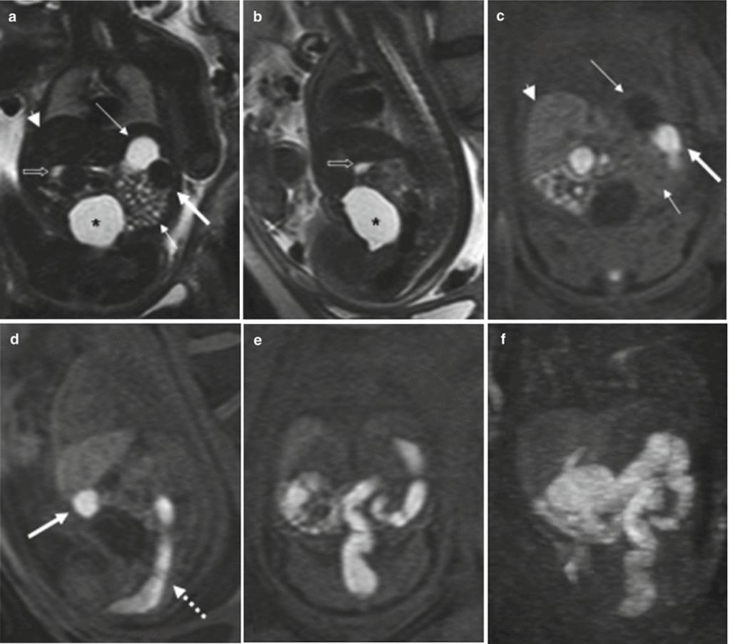
Fig. 11.1
Normal fetal abdominal and pelvic anatomy. Fetus at 32 weeks’ gestation. (a) Coronal and (b) sagittal T2-weighted and corresponding (c) coronal and (d) sagittal T1-weighted images. Stomach (long arrow in a, c), small intestine (short arrow in a, c), colon (thick arrow in a, c, d), rectum (dotted arrow in d), liver (arrowhead in a and c), gallbladder (open arrow in a, b), and bladder (asterisks in a, b). (e) Coronal T1-weighted image and (f) maximum intensity projection (MIP) that allows meconium-based colonography
On T2-weighted images, fetal kidneys are identified as oval structures of intermediate signal intensity on both sides of the spine. Due to the presence of urine, the renal pelvis and calyces are depicted as high-signal structures in the middle of the kidneys. Fetal ureters are not normally seen on T2-weighted images unless dilated, when they are seen as high-signal tubular structures. T1-weighted sequences may help to differentiate dilated ureters from bowel loops, as dilated ureters are seen as low-signal tubular structures, whereas distal bowel loops are hyperintense [3]. The bladder is seen as a fluid-filled round-to-oval structure in the anterior part of the pelvis. It is normal to see changes in bladder volume during MR examination due to fetal micturition. The urethra cannot be seen, but the external genitalia are easily recognized (Fig. 11.2).
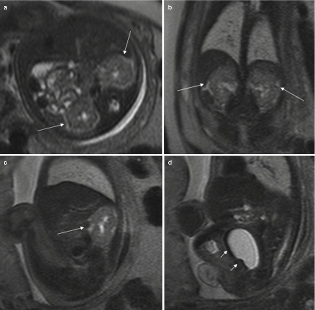
Fig. 11.2
Normal fetal abdominal and pelvic anatomy. Fetus at 31 weeks’ gestation. Fetal axial (a), coronal (b), and sagittal (c, d) T2-weighted images. The fetal kidneys are identified as intermediate signal structures on both sides of the spine (long arrows). The renal calyces and pelvis have high signal intensity due to urine. The fetal bladder is seen as a round-to-oval, high-signal structure in the pelvis (short arrows in d)
The peritoneal cavity, a virtual cavity, is not usually seen except in the presence of ascites. The abdominal wall is easily identified on T2-weighted sequences in all spatial planes. The umbilical cord and its place of insertion are also easily recognizable, especially in the midline sagittal and axial planes. Thick-slab T2-weighted sequences can be very helpful in the evaluation of the umbilical cord.
11.2 Gastrointestinal Tract Anomalies
11.2.1 Stomach Anomalies
In some anomalies, the stomach is not identified in its normal position. In cases of congenital hiatal hernia or diaphragmatic hernias, the stomach may be seen in the thorax (Fig. 11.3). Identification of the stomach in the right side of the fetus or in a central position indicates situs anomalies (Fig. 11.4).

Fig. 11.3
Congenital diaphragmatic hernia. Fetus at 34 weeks’ gestation. (a, b) Coronal T2-weighted and corresponding (c, d) coronal T1-weighted images. The fluid-filled stomach (long arrows in a and c) is seen in the thorax, together with the colon (short arrows in b, d). (e) X-ray after birth shows this anomaly (arrows)

Fig. 11.4
Heterotaxy syndrome. Fetus at 22 weeks’ gestation. (a) Thick-slab coronal T2-weighted image of the fetus shows the fluid-filled stomach in the right upper abdomen (long arrow); the heart is in its normal position (short arrow). (b) Barium studies after birth confirm this anomaly. This child also had polysplenia and malpositioned thoracic and abdominal vessels without heart defects
In cases with polyhydramnios where the stomach is not seen or is very small due to small amounts of gastric fluid, esophageal atresia must be ruled out.
11.2.2 Duodenal Obstruction
Congenital duodenal obstruction can be caused by intestinal malrotation with Ladd’s band, atresia or stenosis, intraluminar diaphragm, annular pancreas, preduodenal portal vein, or volvulus of the middle portion of the intestine, and some fetuses have a combination of these.
In cases of duodenal atresia or marked duodenal obstruction, MR shows dilation of the stomach and the portion of the duodenum proximal to the obstruction as hyperintense structures in T2-weighted sequences [2, 4]. Polyhydramnios is almost always present (Fig. 11.5). When obstruction is due to malrotation with Ladd’s band or volvulus, the loops of the small intestine can be seen lying mostly on the right side of the abdomen while the colon, which is hyperintense on T1-weighted sequences, is on the left (Fig. 11.6). It is important to remember that the stomach and duodenum may be normal sized or even small when the fetus also has esophageal atresia.
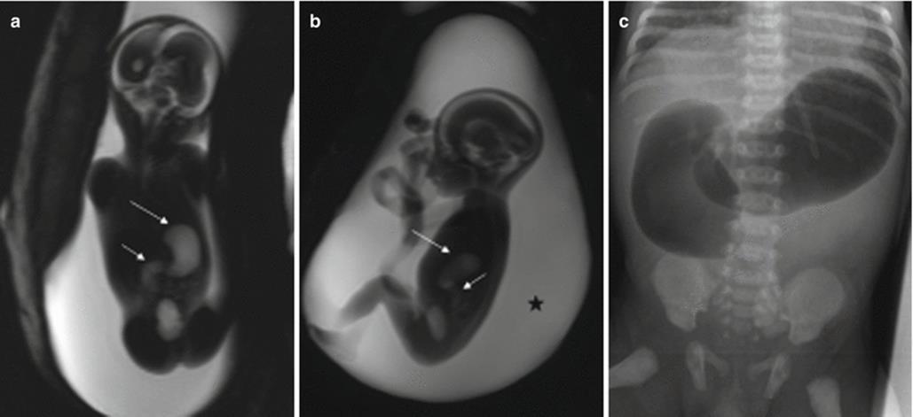
Fig. 11.5
Duodenal atresia. Fetus at 22 weeks’ gestation. Thick-slab coronal (a) and sagittal (b) T2-weighted images of the fetus show gastric (long arrows) and duodenal (short arrows) dilation; polyhydramnios is also present (asterisk). (c) X-ray after birth shows the classical “double bubble” sign that confirms duodenal obstruction, in this case, secondary to duodenal atresia
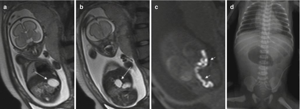
Fig. 11.6
Intestinal malrotation with Ladd’s band. Fetus at 26 weeks’ gestation. (a, b) Coronal T2- weighted images of the fetus show gastric (arrow in a) and duodenal (arrow in b) dilation. On coronal T1-weighted images (c) the hyperintense colon is seen in the left part of the abdomen (short arrows). (d) X-ray after birth confirms duodenal obstruction. At surgery, intestinal malrotation and Ladd’s band causing duodenal occlusion were found
Approximately 50 % of cases of duodenal obstruction are associated with other anomalies, such as other gastrointestinal atresia, genitourinary, heart or skeletal defects, and chromosomal anomalies; trisomy 21 is frequently associated with duodenal obstruction [5–7].
11.2.3 Small Bowel (Jejunum and Ileum) Atresia/Stenosis
Small bowel atresia presents with dilated bowel loops proximal to the site of stenosis. The most frequent site of atresia is the distal ileum, followed by the proximal jejunum; multiple atresia is not uncommon. Vascular impairment during gut development seems to be the most probable cause of this anomaly [8].
MR detects dilation of the small intestine proximal to the atresia; the stomach and duodenum may also be dilated. The location of obstruction may be suggested by the number of dilated bowel loops. The more proximal the lesion, the more likely it is that the fetus will suffer from polyhydramnios. The signal intensity of the contents of the dilated loops varies and it seems to depend mainly on the site of the obstruction; the more distal the obstruction, the lower the signal intensity on T2-weighted images and the higher the signal intensity on T1-weighted images [2, 4, 9]. The post-atretic bowel may be small in caliber or impossible to identify and generally exhibits abnormal signal intensity [4] (Fig. 11.7).
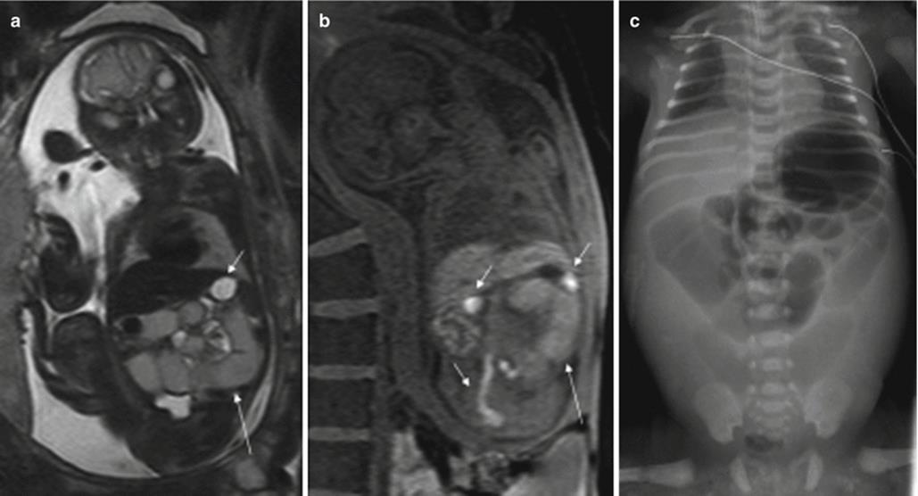
Fig. 11.7
Jejunal atresia. Fetus at 24 weeks’ gestation. (a) Coronal T2-weighted image of the fetus showing dilated proximal small bowel loops of intermediate signal intensity (long arrow); the stomach (short arrow) is not dilated and has higher signal intensity than the small bowel loops. (b) Coronal T1-weighted image shows microcolon (short arrows) and dilated small bowel loops with a higher-than-expected signal intensity (long arrow). (c) X-ray after birth showing dilation of the proximal bowel loops. Jejunal atresia was discovered at surgery
Extraintestinal anomalies are less frequently associated than in duodenal atresia, and the most common associations are with other intestinal anomalies (gastroschisis, intestinal malrotation, meconium ileus) that might be related to the cause of the atresia and to atresias in other locations [5, 10]. A higher incidence of cystic fibrosis has been reported in children with intestinal atresia [11].
Atresia of the small intestine can be very difficult to differentiate from meconium ileus, a condition caused by functional obstruction of the distal ileum by very dense meconium. It is the earliest manifestation of cystic fibrosis. Nearly all newborns with meconium ileus have cystic fibrosis, and 10–15 % of all patients with cystic fibrosis have meconium ileus [12]. The MR findings include dilation of small bowel loops secondary to meconium impaction, microcolon, and polyhydramnios. Ascites may be present whenever the condition is complicated by intestinal perforation. Furthermore, meconium ileus is often associated with gastrointestinal anomalies such as jejunoileal atresia, volvulus, intestinal perforation, and meconium peritonitis.
11.2.4 Meconium Peritonitis
This condition results from intestinal perforation during fetal life. The release of meconium and digestive enzymes into the peritoneal cavity causes chemical peritonitis resulting in an inflammatory reaction and the formation of fibrous tissue that may calcify. Sometimes the inflammatory response seals the perforation spontaneously. In most cases, meconium peritonitis is associated with meconium ileus, intestinal atresia, intestinal volvulus, or intestinal ischemia.
Ascites and dilation of the small intestine can be detected with MR; this is best done using T2-weighted sequences (Fig. 11.8). A meconium pseudocyst may result from a contained perforation. The meconium pseudocyst may exhibit meconium-like signal or high T2 and intermediate T1 signals [4]. Signal intensity can be useful in differentiating meconium pseudocyst from other abdominal cysts. The peritoneal calcifications are difficult to appreciate on MR, as they tend to be small and linear. When seen, they are hypointense in both T1- and T2-weighted sequences. Polyhydramnios may also be present.

Fig. 11.8
Meconium peritonitis. Fetus at 26 weeks’ gestation. (a) Fetal coronal T2-weighted image showing a small amount of ascites (long arrow) and dilation of bowel loops (short arrows). (b, c) CT after birth shows peritoneal calcifications (short arrow) and a meconium pseudocyst (long arrows) in the right side of the abdomen. This child was asymptomatic
11.2.5 Atresia of the Colon, Anorectal Atresia, and Cloacal Malformations
Atresia of the colon and anorectal atresia are uncommon, and up to 70 % of cases are associated with other anomalies, mostly genitourinary and skeletal, especially of the spine, and with chromosomopathies [13, 14]. As in other atresias, vascular impairment seems the most likely etiology. The MR findings for these entities are not well defined. Intestinal loops above the atresia may be dilated, and meconium accumulated above the atresia may cause high signal intensity on T1-weighted sequences [4, 15] (Fig. 11.9). However, this may not be seen when small bowel atresia is also present.
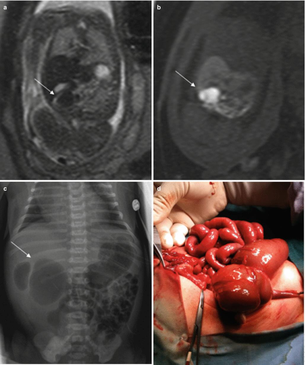
Fig. 11.9
Cecal atresia. Fetus at 22 weeks’ gestation. (a) Fetal coronal T2- and (b) T1-weighted images showing a dilated bowel loop (arrows) in the right hemiabdomen, below the liver; the presence of meconium makes it hypointense on T2- and hyperintense on T1-weighted images. (c) Abdominal X-ray after birth shows a dilated bowel loop (arrow). (d) At surgery, cecal atresia was found
Cloacal malformations represent a spectrum of developmental defects that usually affect female fetuses. In these malformations, the urinary tract, the vagina, and the rectum converge above the level of the perineum, creating a common channel with a single external opening. Cloacal malformations should be suspected in cases with dilation and high position of the distal bowel and abnormalities in the genitourinary system [16]. The typical meconium-filled rectum is not identified in its normal position due to communication between the urinary tract and the rectum (Fig. 11.10).

Fig. 11.10
Duodenal obstruction, anal atresia, and cloacal malformation. Fetus at 30 weeks’ gestation. (a) Coronal T2-weighted image of the fetus showing dilation of the stomach (long arrow) and duodenum (short arrow). (b) Sagittal T2- and (c) T1-weighted images of the fetus show the normal fluid-filled bladder (short arrows) and a cystic tubular structure behind it that corresponds to a fluid-filled vagina (long arrows). Note that the normal hyperintense rectum cannot be identified on T1-weighted image. (d) X-ray after birth shows gastric and duodenal dilation, which was due to a duodenal diaphragm. (e) Image of the fetus after birth with anal atresia. (f) On voiding cystography the vagina was filled with contrast. (g) Colonography through the ostomy shows the colon with a cul-de-sac, but no communication with the urinary tract could be seen
11.3 Abdominal Cysts and Masses
There are many abdominal and pelvic cystic lesions or masses that can be detected prenatally, especially during the third trimester. Some originate in solid organs (liver, spleen, adrenal glands, kidneys…) and are easily identified, but others such as enteric duplication cysts, mesenteric cysts, choledochal cysts, urachal cysts, or ovarian cysts may be difficult to differentiate. T1-weighted sequences can be useful for differentiating these anomalies from dilated intestinal loops and meconium pseudocysts.
11.3.1 Enteric Duplication Cyst
Enteric duplications are uncommon congenital abnormalities that can be found at any point along the gastrointestinal tract. They are believed to be caused by defective recanalization of the gut lumen or failure of the notochord to separate from the endoderm.
Intestinal duplication takes place on the mesenteric side of the intestine, and duplications usually do not communicate with the intestinal lumen. The most common site of this anomaly is the distal portion of the ileum, followed by the distal portion of the esophagus and stomach. Although duplications rarely cause intrauterine intestinal occlusion, they may cause intestinal occlusion or abdominal pain after birth, due to volvulus, invagination, or bleeding of the cyst.
On MR they are seen as rounded intraabdominal masses that are hyperintense on T2-weighted sequences and hypointense on T1-weighted sequences [17] (Fig. 11.11). The rest of the intestinal loops tend to be of normal shape, and polyhydramnios is not usually associated.
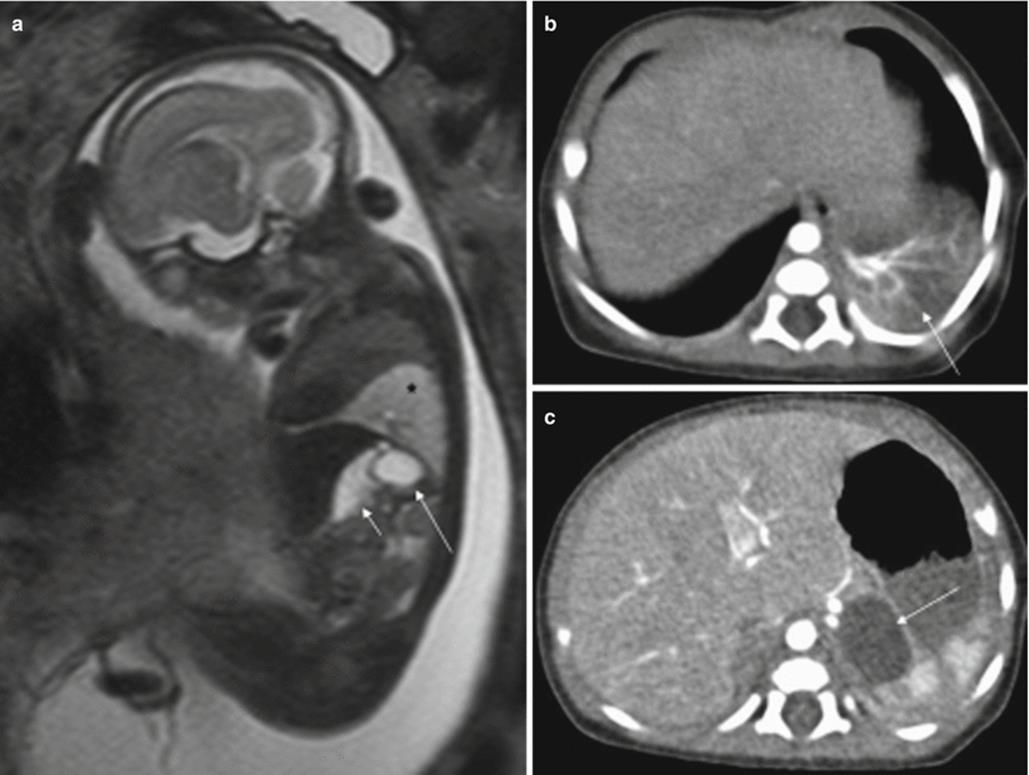
Fig. 11.11
Gastric duplication. Fetus at 23 weeks’ gestation. (a) Fetal sagittal T2-weighted image showing a hyperintense round structure (long arrow) behind the stomach (short arrow). This fetus also had a hyperintense mass in the left lung suggestive of bronchopulmonary sequestration (asterisk). (b, c) CT scan after birth confirms bronchopulmonary sequestration (arrow in b) and gastric duplication (arrow in c)
Duplications are sometimes associated with other anomalies such as bronchopulmonary sequestration or vertebral anomalies (especially in esophageal duplication) [18, 19].
11.3.2 Mesenteric Cyst
Mesenteric cysts are considered lymphatic anomalies, and they are not normally associated with other fetal anomalies. They are generally single unilocular cysts, although they sometimes can be multilocular. Mesenteric cysts are frequently located in the mid-abdomen within the mesentery, omentum, or retroperitoneum. On MR, they are seen as round unilocular fluid-filled structures.
11.3.3 Ovarian Cyst
Prenatal ovarian cysts are common and are usually detected in the third trimester of pregnancy [20]. They are often unilateral, but can also affect both ovaries. In the fetal period, they may be simple cysts or they can complicate with torsion, bleeding, and/or rupture. Approximately 50 % of these cysts disappear during gestation or the first months after birth [20].
MR shows a round intraabdominal structure that tends to be lateral to the midline. Simple cysts are hyperintense on T2-weighted sequences and hypointense on T1-weighted sequences. When complicated by torsion and/or bleeding, the signal intensity of the cysts is heterogeneous and intracystic components or fluid-fluid levels can be detected due to hemorrhage or detritus [15, 21]. Increased signal intensity on T1-weighted images is indicative of hemorrhage [21] (Fig. 11.12).

Fig. 11.12
Ovarian cyst. Fetus at 34 weeks’ gestation. Fetal sagittal (a) and coronal (b) T2-weighted images showing a rounded hyperintense structure (arrows) occupying the middle of the abdomen. In the coronal T1-weighted image (c), the lesion has intermediate signal intensity (arrow), raising suspicion of hemorrhage. (d) US after birth shows a complex cystic lesion with debris that corresponded to a complicated ovarian cyst with hemorrhage
Association with other anomalies is uncommon [20].
11.3.4 Liver and Spleen Masses
These lesions are rare in the prenatal period. Masses in the liver and spleen are usually cysts. In the liver, they may appear within the parenchyma or “hang” from the lower edge. They may be single or multiple. On MR they are seen as round structures within the liver or spleen that are hyperintense on T2-weighted sequences [15], unless they contain blood, in which case fluid-fluid levels can be detected in their interior.
Other masses affecting the liver are hemangioma, hemangioendothelioma, hepatoblastoma, hamartoma, and metastases [22] (Fig. 11.13).

Fig. 11.13
Hepatic hemangioendothelioma. Fetus at 32 weeks’ gestation. Fetal coronal (a), sagittal (b), and axial (c) T2-weighted images showing a mass in the upper left abdomen with intermediate signal intensity (arrows). It is difficult to determine the origin of the mass. (d) Surgery found a hepatic mass hanging from the inferior surface of the left lobe; histology confirmed a hepatic hemangioendothelioma
Hepatosplenomegaly caused by hydrops fetalis, congenital infection, Beckwith-Wiedemann syndrome, or Zellweger syndrome can also be detected prenatally.
Polysplenia can be found in heterotaxy syndromes, but it can be difficult to evaluate with MR [1].
11.3.5 Adrenal Masses
More than 90 % of prenatally diagnosed neuroblastomas arise in the adrenal gland, although they may also be located in the thorax or cervical region [23]. They are normally detected in the right side, almost always in the third trimester [24]. These tumors can disappear before birth, although they can also metastasize, especially to the fetal liver [23, 25]. About half of the tumors are cystic or heterogeneous [23, 26]. The MR appearance of neuroblastoma depends on whether the mass is cystic, solid, or both (Fig. 11.14). Cystic areas are markedly hyperintense on T2-weighted images [27, 28].
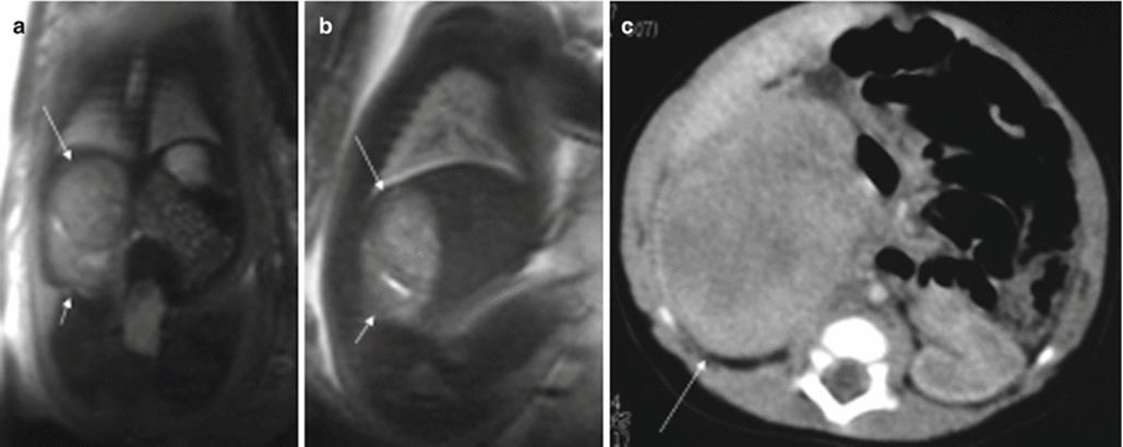
Fig. 11.14
Adrenal neuroblastoma. Fetus at 36 weeks’ gestation. Fetal coronal (a) and sagittal (b) T2-weighted images showing a slightly heterogeneous infradiaphragmatic mass of intermediate signal intensity (long arrows). The mass is above the right kidney, which is displaced posteriorly and downward (short arrows). (c) CT after birth shows this mass (arrow); metaiodobenzylguanidine study after birth was positive
Only half of all adrenal masses are neuroblastomas and should be differentiated from other lesions such as adrenal hemorrhage, renal lesions, or infradiaphragmatic extralobar pulmonary sequestration. Up to 10 % of extralobar sequestrations are infradiaphragmatic and of those more than 90 % of are located on the left side. Unlike neuroblastoma, they may appear in the second trimester [24]. On T2-weighted images they are seen as well-defined high-signal-intensity masses, usually located below the left diaphragm or above the kidney [29, 30]. Hypointense septa may be seen across the lesion [31].
11.4 Abdominal Wall Malformations
Gastroschisis and omphalocele are the two most common congenital abdominal wall defects. Detecting them early during pregnancy allows the best prenatal and postnatal management to be planned.
11.4.1 Gastroschisis
Gastroschisis consists of the herniation of fetal abdominal viscera into the amniotic cavity secondary to a small defect through all the layers of the abdominal wall. Gastroschisis is generally located on the right side of the abdomen with the umbilical cord inserted in its normal position.
Its prevalence is approximately 1.1–5.1 in 10,000 live births with a worldwide increasing trend in recent years [32]. Strong correlations have been found with maternal youth, as well as with drug abuse [33, 34].
MR can detect the evisceration of abdominal structures without difficulties [35–37]. On T2-weighted sequences, eviscerated abdominal structures are clearly visualized floating in the high-signal-intensity amniotic fluid. T1-weighted sequences are useful for determining the position of the large intestine. The axial plane is best to show the defect in the abdominal wall and the position of the umbilical cord (Fig. 11.15).
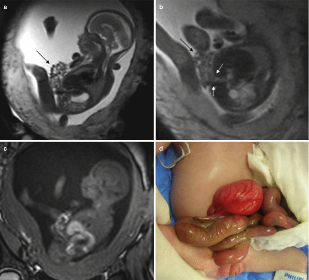
Fig. 11.15
Gastroschisis. Fetus at 20 weeks’ gestation. Fetal sagittal (a) and axial (b) T2-weighted images showing herniation of a large portion of the bowel loops (long arrow) through a right lateral defect (short arrows in b) in the abdominal wall. The bowel loops lie outside the abdominal cavity, and there is no peritoneal lining. The liver is inside the abdomen. (c) Fetal sagittal T1-weighted image. (d) Photograph of the patient after birth showing the bowel loops outside the abdominal cavity without peritoneal lining
Although associated congenital anomalies may be seen in other organ systems, intestinal anomalies or complications are more common, especially intestinal stenosis or atresia, possibly resulting from the direct effect of amniotic fluid on the intestinal loops or from ischemic problems secondary to compromised mesenteric vessels due to the relatively small size of the defect in the abdominal wall [38].
11.4.2 Omphalocele
This is a central defect involving all the layers of the abdominal wall in which the viscera located outside of the abdominal cavity are lined with a membrane consisting of two layers: the peritoneum and the amnion. Embryologic evolution is detained at a point in which the developing intestines are located outside of the abdominal cavity. In omphalocele, the umbilical cord is not inserted in its normal position, rather it inserts into the apex of the omphalocele. The size of this lesion varies considerably.
It occurs in approximately 1.6–2.11 in 10,000 live births [39, 40].
As in gastroschisis, MR usually enables accurate diagnosis and can determine the contents of the omphalocele without difficulties [36, 37]. T2-weighted sequences are the most appropriate for studying omphalocele and T1-weighted sequences are useful for detecting the large intestine. MR also usually shows the linings of the omphalocele and the point of umbilical cord insertion clearly (Fig. 11.16). These two findings are important in differentiating gastroschisis from omphalocele. T2-weighted thick-slab images can be useful in determining whether or not the eviscerated organs have a peritoneal lining as well as the site of umbilical cord insertion. However, early in gestation this is not always possible (Fig. 11.17). Measurement of fetal lung volumes may be useful to predict associated pulmonary hypoplasia in giant omphaloceles [41].
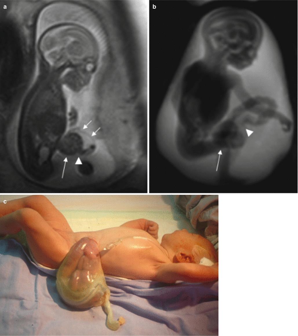
Fig. 11.16
Omphalocele. Fetus at 21 weeks’ gestation. Fetal sagittal (a) and thick-slab sagittal (b) T2-weighted images showing the bowel loops outside the abdominal cavity (long arrows); the loops are lined with peritoneal membranes (short arrows). The umbilical cord (arrowheads) is inserted in the apex of the omphalocele. (c) Photograph of the patient after birth showing the intestinal loops outside the abdominal cavity, the peritoneal membranes, and the position of the umbilical cord
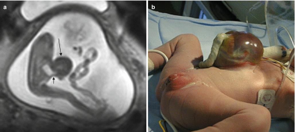
Fig. 11.17
Omphalocele. Fetus at 18 weeks’ gestation. (a) Fetal sagittal T2-weighted image showing the liver (long arrow) and part of the stomach (short arrow) outside the abdominal cavity. The peritoneal membranes and the insertion of the umbilical cord could not be seen. (b) Photograph of the patient after birth shows an omphalocele with the herniated liver
Omphalocele is often associated with other anomalies; it may be part of the OEIS complex, a combination of defects consisting of omphalocele, bladder exstrophy, imperforate anus, and spinal defects [42]. Omphalocele is frequently found in association with chromosomopathies, especially trisomies 18 and 13, and with Beckwith-Wiedemann syndrome [40, 43].
Intestinal complications are less common than in gastroschisis, possibly because the abdominal structures are not in contact with the amniotic fluid and because the defect in the abdominal wall is usually large, making compromised mesenteric blood flow less likely.
11.4.3 Bladder Exstrophy
This entity results from a defect in the midline of the abdominal wall below the umbilical cord in which the anterior vesical wall is absent and the posterior wall is exposed externally.
It occurs in approximately 1 in 30,000 births and affects males 1.5 times more frequently than females [44].
Bladder exstrophy should be suspected at MR when the urinary bladder is not seen in the pelvis, the appearance of the kidneys is normal, and a normal amount of amniotic fluid is present. Although the fetus can empty the bladder through urination, the bladder is rarely emptied in its entirety, and it is unusual for normal bladders not to be seen sometime or other during an MR examination. Irregularities in the anterior abdominal wall below the umbilicus may be detected on MR; however, this finding is not always present. T2-weighted sequences in the midline sagittal plane are ideal for evaluating the absence of the bladder and the irregularities in the abdominal wall [3, 37].
Bladder exstrophy may be part of the OEIS complex and associated genitourinary anomalies, such as epispadias, undescended testes, or bilateral inguinal hernias can be detected in males.
11.5 Ascites
T2-weighted MR sequences can detect even minimal amounts of free fluid within the peritoneum. When significant ascites is present, it can be identified well in any spatial plane, and the intraabdominal structures appear to float within the peritoneal fluid. It is important to determine whether ascites is isolated or forms part of hydrops fetalis.
Isolated ascites usually occurs secondary to an intraabdominal problem rather than to a systemic anomaly, and it is often associated with fetal malformations. Genitourinary anomalies, usually obstructive uropathy, are the most common cause. Isolated ascites may also have a gastrointestinal origin, normally meconium peritonitis, or be caused by liver defects, heart defects, infections, or metabolic anomalies. Ascites is sometimes idiopathic.
Hydrops fetalis is defined as an excess of total body fluid; this term is used when fluid is detected in at least two body cavities or in a single body cavity in the presence of anasarca (Fig. 11.18).
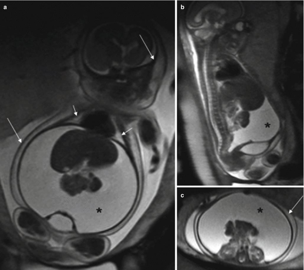
Fig. 11.18
Hydrops fetalis. Fetus at 26 weeks’ gestation. (a) Coronal, (b) sagittal, and (c) axial T2-weighted images of the fetus. There is a large amount of peritoneal fluid (asterisks), together with hydrothorax (short arrows) and subcutaneous edema (long arrows), suggestive of hydrops fetalis; in this case the cause remained unknown
11.6 Urinary System Anomalies
Fetal genitourinary system anomalies are relatively common, accounting for 30–50 % of all structural anomalies at birth [45]. Urogenital tract anomalies may be unilateral or bilateral and isolated or associated with other anomalies, and they sometimes form part of genetic syndromes.
Fetal urination is the most important factor in the production of amniotic fluid, and the amount of amniotic fluid can be used as an indicator of fetal diuresis [45, 46]. The volume of amniotic fluid can easily be evaluated with thick-slab T2-weighted sequences. When the volume of fluid is normal, at least one fetal kidney is working. However, when oligohydramnios is present, once non-urogenital causes have been ruled out, a severe anomaly affecting either both kidneys or outflow from the bladder must be suspected. A severe urogenital tract abnormality that causes oligohydramnios or the associated pulmonary hypoplasia that results from it will affect the viability of the fetus. Fetal MR is especially useful in cases with severe oligohydramnios, a situation that hampers the evaluation of the fetus with US [47].
11.6.1 Renal Agenesis and Ectopic Kidney
Renal agenesis can be unilateral or bilateral. On MR imaging, renal agenesis is suspected when the kidneys cannot be seen in the renal fossae. In cases of unilateral agenesis, the contralateral kidney may be normal or slightly enlarged, the bladder is normally visualized, and the amount of amniotic fluid is normal. There is a high risk of associated anomalies involving the contralateral kidney such as vesicoureteral reflux or obstruction [48]. It is important to look for an ectopic kidney in the rest of the abdomen. Normally, the ectopic kidney is found in the pelvis and is often malrotated. Ectopic kidneys can be difficult to identify on MR imaging because they can look like bowel loops and be misdiagnosed as renal agenesis. Diffusion-weighted imaging can be helpful in demonstrating the empty renal fossa and in locating ectopic kidneys [49] (Fig. 11.19).

Fig. 11.19
Renal ectopia. Fetus at 25 weeks’ gestation. Coronal (a, d) and sagittal (b, e) T2-weighted images of the fetus. The right kidney has a normal morphology and is located in the right renal fossa (arrows in a, b). The left renal fossa is occupied by bowel loops, and the left kidney is identified in the pelvis (arrows in d, e). (c, f) Coronal diffusion-weighted images help to identify the kidneys, which are hyperintense on these sequences: right kidney (arrow in c) and left kidney (arrow in f). Pelvic US after birth confirmed the MR findings
Bilateral renal agenesis is much less common than unilateral agenesis, and it is invariably lethal. MR features of this entity include the absence of both kidneys and the bladder in addition to severe oligohydramnios and pulmonary hypoplasia. In cases of bilateral renal ectopia, the kidneys are not seen in the renal fossae, but the bladder is normally seen and there is no oligohydramnios.
11.6.2 Upper Urinary Tract Dilation
Upper urinary tract dilation is the most common anomaly detected on midtrimester prenatal US studies, with a reported frequency of approximately 2.5 % [50]. In most cases, the excretory system is enlarged, but there is no underlying pathology such as obstruction. Most mild upper urinary tract dilation detected on midtrimester US will resolve prenatally or during the first year of life [50, 51]. However, in some cases, dilation of the upper urinary tract may be an indicator of urinary tract pathologies (ureteropelvic junction anomaly, ureterovesical junction anomaly, outflow obstruction, or duplex systems) or even of extraurinary causes such as pelvic masses that compromise the flow of urine. The severity of obstruction and the time it occurs will determine the extent of damage to the renal parenchyma; in severe cases very little healthy renal parenchyma remains.
The challenge of prenatal diagnosis is to determine which cases of fetal upper urinary tract dilation are due to urinary tract pathologies that would benefit from postnatal surveillance and treatment and which are merely physiologic.
In cases with moderate or severe dilation of the renal pelvis or with mild dilation and other associated anomalies detected on midtrimester US, fetal MR is indicated to detect the possible underlying cause and rule out syndromes with multiple malformations.
11.6.2.1 Ureteropelvic Junction (UPJ) Anomalies
UPJ anomalies are the most common cause of fetal upper urinary tract dilation [52]. They are more common in males and 90 % are unilateral [53]. MR shows dilation of the pelvis with or without dilation of the calyces. The bladder and amount of amniotic fluid are normal [35], and the ureter is not dilated (Fig. 11.20). In the absence of severe renal parenchymal disease, the prognosis of a fetus with unilateral or bilateral UPJ obstruction is usually good.
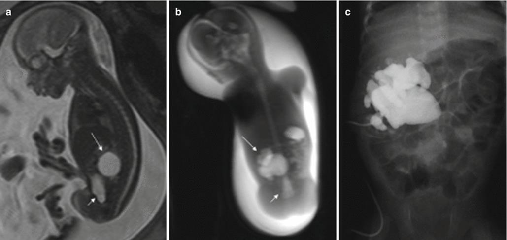
Fig. 11.20
Ureteropelvic junction obstruction. Fetus at 22 weeks’ gestation. (a) Sagittal and (b) coronal thick-slab T2-weighted images of the fetus showing dilation of the renal pelvis and calyces (long arrows). The ureter is not dilated and the bladder is seen in the pelvis (short arrows). (c) Postnatal urography shows the same findings
11.6.2.2 Ureterovesical Junction (UVJ) Anomalies
In UVJ anomalies, hydronephrosis occurs secondary to a narrowing at the lower end of the ureter. Obstruction is often due to a localized region of dysfunction (primary megaureter) and is bilateral in 25 % of cases [54]. On MR imaging, dilation of the ureter is detected with or without pelvic and calyceal dilations (Fig. 11.21). The dilated ureter is tortuous, and it is sometimes difficult to differentiate it from a bowel loop on T2-weighted images; for this reason, it is useful to obtain T1-weighted sequences, which show the dilated ureters as low-signal tubular structures due to the presence of urine [2]. The fetal bladder and amount of amniotic fluid are normal unless the disease is severe and bilateral.
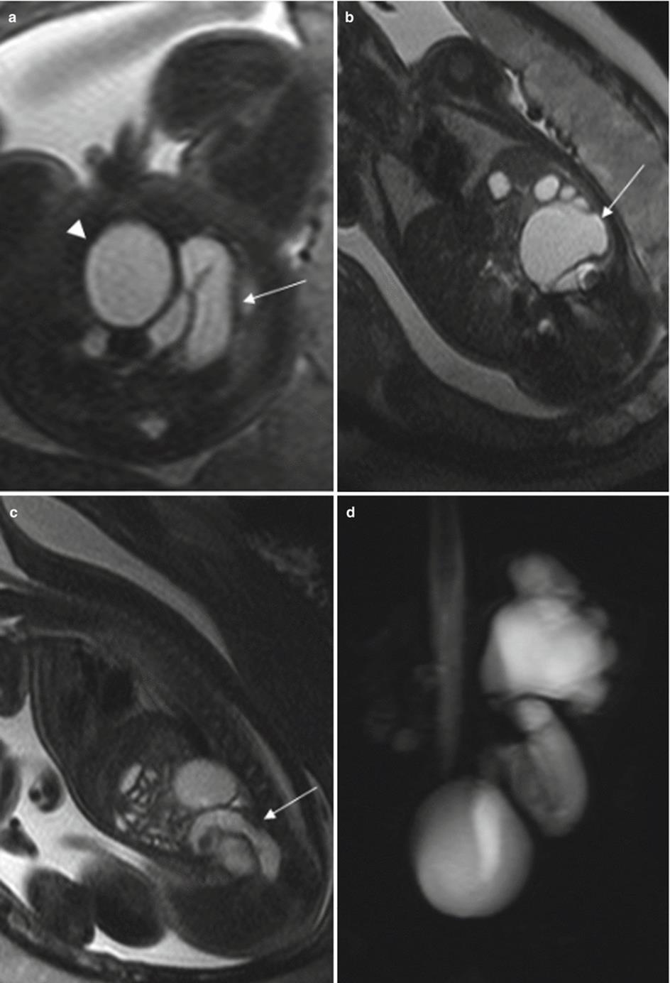
Fig. 11.21
Primary megaureter. Fetus at 32 weeks’ gestation. Fetal axial (a), coronal (b), and sagittal (c) T2-weighted images showing dilation of the left renal pelvis and calyces (arrow in b) and dilation of the left ureter (arrows in a, c). The bladder is normal (arrowhead in a). (d) MR urography after birth shows the same findings
The intrauterine appearance of UVJ anomalies can be identical to that of vesicoureteral reflux and normally cannot be differentiated until postnatal studies are carried out.
11.6.2.3 Posterior Urethral Valves
Bladder outflow obstruction can be complete or partial. Partial obstruction is more frequent and normally manifests later in pregnancy. It is more common in male fetuses due to the presence of posterior urethral valves, which are the most common cause of lower urinary tract obstruction. MR imaging studies show a dilated proximal urethra and bladder. Depending on the grade of obstruction, this entity may be associated with renal dysplasia secondary to bladder dilation and hydronephrosis. It is important to study both kidneys and the amount of amniotic fluid carefully to evaluate renal function.
11.6.2.4 Duplex Systems
Most subjects with duplex systems are asymptomatic, but some present complications that need to be treated and early diagnosis is essential in these cases.
In cases of complete duplication there are two ureters. The upper moiety ureter can be attached to the bladder in the normal position, or it can empty ectopically, often with an associated ureterocele. This is generally associated with hydronephrosis and/or renal dysplasia of the upper moiety of the kidney. Sometimes, the lower moiety of the kidney also presents hydronephrosis, which is usually due to vesicoureteral reflux. MR imaging findings will depend on the anomaly and its complications. If there is hydronephrosis due to ureterocele or ectopic ureteral voiding, the dilated ureter will be depicted as a high-signal tubular structure on T2-weighted sequences and as a low-signal structure on T1-weighted images. The ureterocele is easily identified as a round structure inside the bladder (Figs. 11.22 and 11.23).
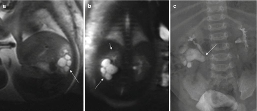
Fig. 11.22
Right duplex system and hydronephrosis of the lower moiety of the kidney. Fetus at 26 weeks’ gestation. (a) Sagittal and (b) coronal T2-weighted images of the fetus show right duplex system and hydronephrosis of the lower moiety (long arrows). The normal upper half of the kidney can be seen (short arrow in b). The left kidney and the fetal bladder (not shown) are normal. (c) Urography after birth confirms those findings. Hydronephrosis of the lower moiety of the kidney is probably secondary to ureteropelvic junction obstruction (arrow) due to a crossover of the upper ureter (Reprinted, with permission, from reference 3.)
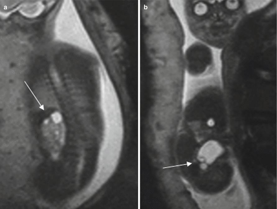

Fig. 11.23
Right duplex system with ureterocele and cystic dysplasia of the upper moiety of the kidney. Fetus at 23 weeks’ gestation. (a, b) Fetal coronal T2-weighted images show the normal-sized right kidney in its normal position, with tiny cysts in the upper pole (arrow in a). The fluid-filled bladder is seen in the pelvis with a ureterocele (arrow in b). (c, d) Coronal T2-weighted MR images after birth show cystic dysplasia of the upper moiety of the kidney (arrow in c), dilation of the right ureter (long arrow in d), and the ureterocele (short arrow in d)
11.6.3 Renal Cystic Diseases
Cystic kidneys are frequently detected on prenatal studies and may be sporadic or have a genetic basis. Multicystic dysplastic kidneys are sporadic. Genetic renal cystic diseases include polycystic renal disease and cystic disease associated with other syndromes. These diseases represent a significant diagnostic challenge; it may be difficult to differentiate among them by imaging, and it can also be difficult to differentiate them from a dilated collecting system.
Polycystic kidney disease has a genetic basis and may have either an autosomal recessive or dominant inheritance pattern. Autosomal recessive polycystic kidney disease (ARPKD) consists of cystic dilation of the renal collecting ducts associated with liver disease (congenital hepatic fibrosis). Clinical and radiologic expressions of ARPKD vary widely depending on the number of collecting ducts affected. Only the perinatal form of this ARPKD can be detected prenatally, with bilateral and symmetrical enlargements of both kidneys. Severe impairment of renal function in ARPKD leads to severe oligohydramnios and pulmonary hypoplasia. MR imaging shows normal-sized or enlarged, normally shaped kidneys occupying most of the abdomen, with tiny corticomedullary cysts (1–2 mm) throughout the periphery. In the most severe cases, there is little amniotic fluid and the bladder is not seen. Associated pulmonary hypoplasia and other malformations such as underdeveloped limbs can also be detected on MR imaging (Fig. 11.24).
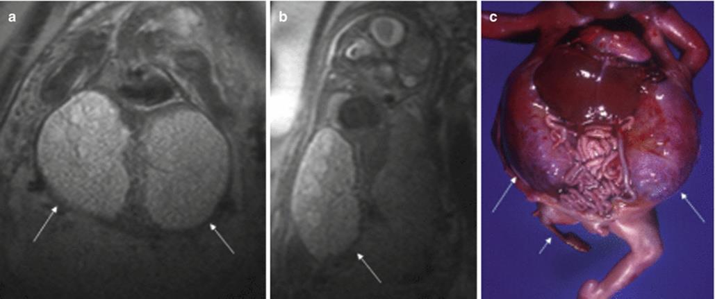
Fig. 11.24
Autosomal recessive polycystic kidney disease. Fetus at 24 weeks’ gestation. (a) Coronal and (b) sagittal T2-weighted images of the fetus show the absence of amniotic fluid, pulmonary hypoplasia, and enlargement of both kidneys with tiny cysts (arrows). (c) Autopsy specimen of this fetus shows enlargement of both kidneys occupying the entire abdomen (long arrows) and severe pulmonary hypoplasia. Note the hypoplastic right lower limb (short arrow) that was not seen with MR or US due to severe oligohydramnios (Reprinted, with permission, from Martín et al. [3])
Autosomal dominant polycystic kidney disease is the most common inherited renal disease, but most symptoms do not usually appear until the fourth or fifth decade of life, so this entity presents in utero only occasionally. On reported intrauterine cases, the most frequent finding is enlarged kidneys with or without cysts and generally with a normal amount of amniotic fluid [55]. These findings can be the same as those found in ARPKD, and the family history is very important to ensure correct diagnosis.
Multicystic dysplastic kidneys are sporadic and frequently develop in conjunction with lower urinary tract malformations that impair urine flow. It seems that severe obstruction of the urinary tract occurring early in gestation (before week 10) affects the normal development of the kidney and causes dysplasia, whereas obstruction later in pregnancy produces hydronephrosis. The impairment of renal function depends on the severity of renal dysplasia, and whether the disease is unilateral or bilateral. MR imaging can depict the entire range, from massive kidneys distended with multiple large cysts to normal or small kidneys with or without cysts [3] (Fig. 11.25). Normally, renal morphology is not conserved, and the normal renal parenchyma is difficult to identify. If cysts are large, they can be identified independently from one another; if they are small, the kidneys show increased signal intensity on T2-weighted images. The affected ureters are not visible. In bilateral disease the bladder is small or absent depending on the severity of the dysplasia. Unilateral multicystic kidneys are often associated with contralateral vesicoureteral reflux. The affected kidney tends to involute with time and prognosis depends on the severity of the contralateral affected kidney and the severity of associated anomalies.
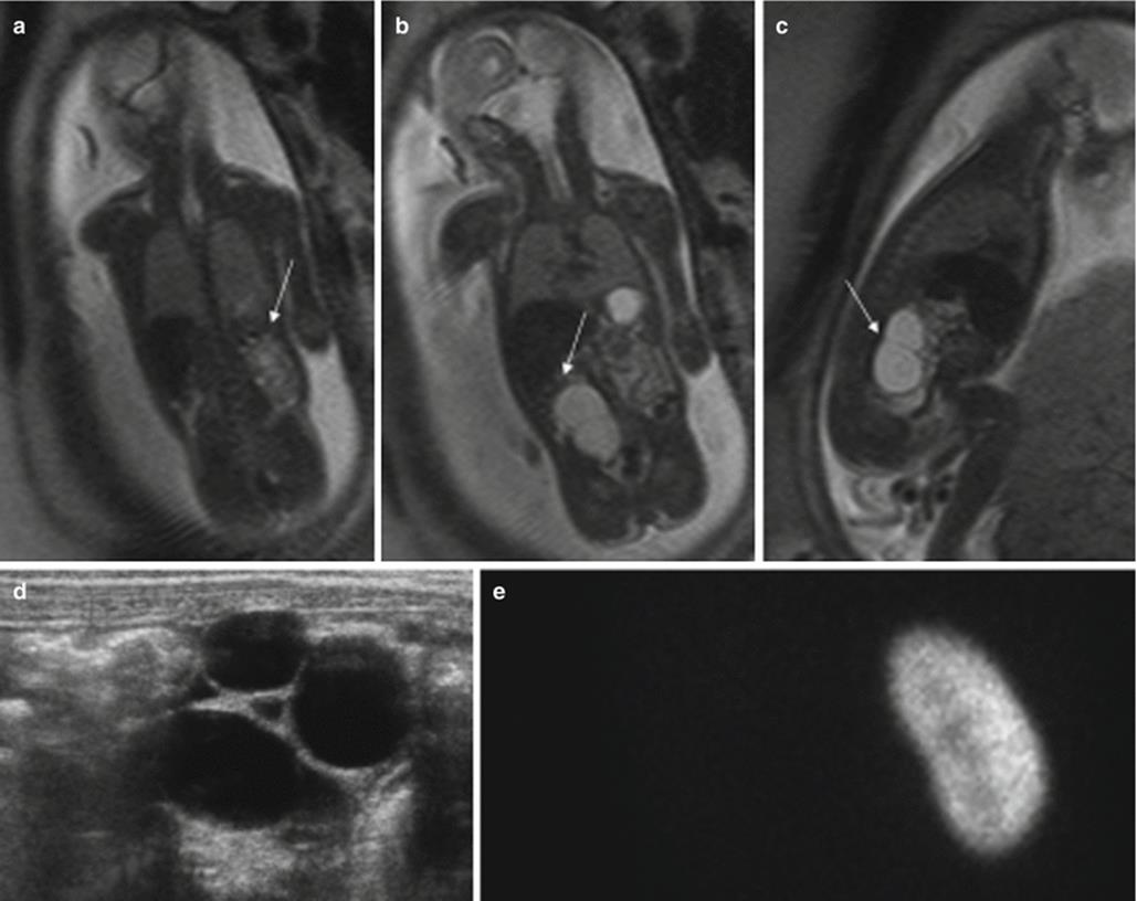
Fig. 11.25
Multicystic dysplastic kidney. Fetus at 22 weeks’ gestation. Fetal coronal (a, b) and (c) sagittal T2-weighted images. The left kidney is normal (arrow in a). The right kidney cannot be identified; instead non-communicating cystic lesions occupy the right renal fossa (arrows in b, c). (d) US after birth shows the cystic lesion replacing the renal parenchyma. (e) DMSA scan performed at 4 years old shows that the right kidney has no functional parenchyma
Renal cystic changes can occur in numerous syndromes. Sometimes, renal cysts are the only symptoms, so genetic syndromes and nonhereditary malformation complexes should be taken into consideration when a renal anomaly is detected.
11.6.4 Severe Bladder Outflow Obstruction
Sometimes oligohydramnios can be caused by an obstruction to the flow of urine out of the bladder, frequently a result of urethra malformation, which commonly occurs in male fetuses. In these cases, there is little or no amniotic fluid, as in cases of severe bilateral renal disease, but normal kidneys and an enlarged bladder can be identified.
Bladder obstruction may be associated with renal dysplasia and pulmonary hypoplasia related to oligohydramnios and bladder enlargement, which worsens the prognosis of the pregnancy. In addition to oligohydramnios, MR imaging shows the bladder as a very large fluid-filled round structure filling the entire abdomen [3]. The kidneys are identified as normal or dysplastic. The differential diagnosis must be done with a rare group of enlarged bladders (megacystis microcolon intestinal hypoperistalsis and prune-belly syndrome) in which the cause is nonobstructive. However, in these cases, the volume of amniotic fluid is generally not affected.
11.6.5 Renal Masses
Simple renal cysts are not common in the prenatal period [56]. On MR imaging, simple renal cysts are seen as well-delineated round structures with high signal intensity on T2-weighted images. The rest of the renal parenchyma is normal.
Fetal renal tumors are rare. The most common types are mesoblastic nephroma, which is benign, and Wilms’ tumor, an extremely rare malignant lesion in newborns. On MR imaging, both tumors can be seen as well-delineated intrarenal masses, with homogeneous signal intensity higher than that of liver and lower than that of water [57]. The diagnosis can only be reached by histology.
Both tumors should be differentiated from other retroperitoneal masses that occur in the fetus such as adrenal neuroblastoma, adrenal hemorrhage, teratoma, or infradiaphragmatic extralobar pulmonary sequestration.
References
1.
Brugger PC, Prayer D (2006) Fetal abdominal magnetic resonance imaging. Eur J Radiol 57:278–293CrossRefPubMed
2.
Saguintaah M, Couture A, Veyrac C et al (2002) MRI of the fetal gastrointestinal tract. Pediatr Radiol 32:395–404CrossRefPubMed
3.
Martín C, Darnell A, Duran C et al (2004) Magnetic resonance imaging of the intrauterine fetal genitourinary tract. Abdom Imaging 29:286–302CrossRefPubMed
4.
Veyrac C, Couture A, Saguintaah M et al (2004) MRI of fetal GI tract abnormalities. Abdom Imaging 29:411–420CrossRefPubMed
5.
Dalla Vecchia LK, Grosfeld JL, West KW et al (1998) Intestinal atresia and stenosis: a 25-year experience with 277 cases. Arch Surg 133:490–497CrossRefPubMed
6.
Haeusler MC, Berghold A, Stoll C et al; EUROSCAN Study Group (2002) Prenatal ultrasonographic detection of gastrointestinal obstruction: results from 18 European congenital anomaly registries. Prenat Diagn 22:616–623
7.
Bailey PV, Tracy TF Jr, Connors RH et al (1993) Congenital duodenal obstruction: a 32-year review. J Pediatr Surg 28:92–95CrossRefPubMed
8.
Werler MM, Sheehan JE, Mitchell AA (2003) Association of vasoconstrictive exposures with risks of gastroschisis and small intestinal atresia. Epidemiology 14:349–354PubMed
9.
Colombani M, Ferry M, Garel C et al (2010) Fetal gastrointestinal MRI: all that glitters in T1 is not necessarily colon. Pediatr Radiol 40:1215–1221CrossRefPubMed
10.
Cragan JD, Martin ML, Moore CA et al (1993) Descriptive epidemiology of small intestinal atresia, Atlanta, Georgia. Teratology 48:441–450CrossRefPubMed
11.
Roberts HE, Cragan JD, Cono J et al (1998) Increased frequency of cystic fibrosis among infants with jejunoileal atresia. Am J Med Genet 78:446–449CrossRefPubMed
12.
Casaccia G, Trucchi A, Nahom A et al (2003) The impact of cystic fibrosis on neonatal intestinal obstruction: the need for prenatal/neonatal screening. Pediatr Surg Int 19:75–78PubMed
13.
Forrester MB, Merz RD (2002) Descriptive epidemiology of anal atresia in Hawaii, 1986-1999. Teratology 66:S12–S16CrossRefPubMed
14.
Martinez-Frias ML, Bermejo E, Rodriguez-Pinilla E (2000) Anal atresia, vertebral, genital, and urinary tract anomalies: a primary polytopic developmental field defect identified through an epidemiological analysis of associations. Am J Med Genet 95:169–173CrossRefPubMed
15.
Martin C, Darnell A, Escofet C et al (2012) Fetal MR in the evaluation of pulmonary and digestive system pathology. Insights Imaging 3:277–293PubMedCentralCrossRefPubMed
16.
Calvo-Garcia MA, Kline-Fath BM, Levitt MA et al (2011) Fetal MRI clues to diagnose cloacal malformations. Pediatr Radiol 41:1117–1128CrossRefPubMed
17.
Levine D, Barnes P, Edelman RR (1999) Obstetric MR imaging. Radiology 211:609–617CrossRefPubMed
18.
Fenton LZ, Williams JL (1996) Bronchopulmonary foregut malformation mimicking neuroblastoma. Pediatr Radiol 26:729–730CrossRefPubMed
19.
Carachi R, Azmy A (2002) Foregut duplications. Pediatr Surg Int 18:371–374CrossRefPubMed
20.
Heling KS, Chaoui R, Kirchmair F et al (2002) Fetal ovarian cysts: prenatal diagnosis, management and postnatal outcome. Ultrasound Obstet Gynecol 20:47–50CrossRefPubMed
21.
Nemec U, Nemec SF, Bettelheim D et al (2012) Ovarian cysts on prenatal MRI. Eur J Radiol 81:1937–1944CrossRefPubMed
22.
Isaacs H Jr (2007) Fetal and neonatal hepatic tumors. J Pediatr Surg 42:1797–1803CrossRefPubMed
23.
Acharya S, Jayabose S, Kogan SJ et al (1997) Prenatally diagnosed neuroblastoma. Cancer 80:304–310CrossRefPubMed
24.
Curtis MR, Mooney DP, Vaccaro TJ et al (1997) Prenatal ultrasound characterization of the suprarenal mass: distinction between neuroblastoma and subdiaphragmatic extralobar pulmonary sequestration. J Ultrasound Med 16:75–83PubMed
25.
Holgersen LO, Subramanian S, Kirpekar M et al (1996) Spontaneous resolution of antenatally diagnosed adrenal masses. J Pediatr Surg 31:153–155CrossRefPubMed
26.
Sauvat F, Sarnocki S, Brisse H et al (2002) Outcome of suprarenal localized masses diagnosed during the perinatal period. Cancer 94:2474–2480CrossRefPubMed
27.
Hamada Y, Ikebukuro K, Sato M et al (1999) Prenatally diagnosed cystic neuroblastoma. Pediatr Surg Int 15:71–74CrossRefPubMed
28.
Maki E, Oh K, Rogers S et al (2014) Imaging and differential diagnosis of suprarenal masses in the fetus. J Ultrasound Med 33:895–904CrossRefPubMed
29.
Huppert BJ, Brandt KR, Ramin KD et al (1999) Single-shot fast spin-echo MR imaging of the fetus: a pictorial essay. Radiographics 19:S215–S227CrossRefPubMed
30.
Dhingsa R, Coakley FV, Albanese CT et al (2003) Prenatal sonography and MR imaging of pulmonary sequestration. AJR Am J Roentgenol 180:433–437CrossRefPubMed
31.
Huang CC, Ko SF, Chung MY et al (2004) Infradiaphragmatic pulmonary sequestration combined with cystic adenomatoid malformation: unusual postnatal computed tomographic features. Abdom Imaging 29:439–442CrossRefPubMed
32.
Mastroiacovo P, Castilla EE (2006) The incidence of gastroschisis: research urgently needs resources. BMJ 332:423–424PubMedCentralCrossRefPubMed
33.
Tan KH, Kilby MD, Whittle MJ et al (1996) Congenital anterior abdominal wall defects in England and Wales 1987-93: retrospective analysis of OPCS data. BMJ 313:903–906PubMedCentralCrossRefPubMed
34.
Torfs CP, Katz EA, Bateson TF et al (1996) Maternal medications and environmental exposures as risk factors for gastroschisis. Teratology 4:84–92CrossRef
35.
Shinmoto H, Kashima K, Yuasa Y et al (2000) MR imaging of non-CNS fetal abnormalities: a pictorial essay. Radiographics 20:1227–1243CrossRefPubMed
36.
Shinmoto H, Kuribayashi S (2003) MRI of the fetal abdominal abnormalities. Abdom Imaging 28:877–886CrossRefPubMed
37.
Daltro P, Fricke BL, Kline-Fath BM et al (2005) Prenatal MRI of congenital abdominal and chest wall defects. AJR Am J Roentgenol 184:1010–1016CrossRefPubMed
38.
Saxena AK, Hülskamp G, Schleef J et al (2002) Gastroschisis: a 15-year, single-center experience. Pediatr Surg Int 18:420–424CrossRefPubMed
39.
Calzolari E, Volpato S, Bianchi F et al (1993) Omphalocele and gastroschisis: a collaborative study of five Italian congenital malformation registries. Teratology 47:47–55CrossRefPubMed
40.
Rankin J, Dillon E, Wright C (1999) Congenital anterior abdominal wall defects in the north of England, 1986-1996: occurrence and outcome. Prenat Diagn 19:662–668CrossRefPubMed
41.
Danzer E, Victoria T, Bebbington MW et al (2012) Fetal MRI-calculated total lung volumes in the prediction of short-term outcome in giant omphalocele: preliminary findings. Fetal Diagn Ther 31:248–253CrossRefPubMed
42.
Chen CP (2007) Syndromes and disorders associated with omphalocele (II): OEIS complex and Pentalogy of Cantrell. Taiwan J Obstet Gynecol 46:103–110CrossRefPubMed
43.
Chen CP (2007) Syndromes and disorders associated with omphalocele (I): Beckwith-Wiedemann syndrome. Taiwan J Obstet Gynecol 46:96–102CrossRefPubMed
44.
International Clearinghouse for Birth Defects Monitoring Systems (1987) Epidemiology of bladder exstrophy and epispadias: a communication from the International Clearinghouse for Birth Defects Monitoring Systems. Teratology 36:221–227CrossRef
45.
Fong KW, Ryan G (1998) The fetal urogenital tract. In: Rumack CM, Wilson SR, Charboneau JW (eds) Diagnostic ultrasound, 2nd edn. Mosby, St. Louis, pp 1093–1121
46.
Hill LM, Sohey R, Nyberg DA (2003) Abnormalities of amniotic fluid. In: Nyberg DA, McGahan JP, Pretrorius DH, Pilu G (eds) Diagnostic imaging of fetal anomalies. Lippincott Williams Wilkins, Philadelphia, pp 59–84
47.
Wenstrom KD, Williamson RA, Weiner CP et al (1991) Magnetic resonance imaging of the fetuses with intracranial defects. Obstet Gynecol 77:529–532PubMed
48.
Atiyeh B, Husmann D, Baum M (1993) Contralateral renal abnormalities in patients with renal agenesis and non cystic renal dysplasia. Pediatrics 91:812–815PubMed
49.
Hörmann M, Brugger PC, Balassy C et al (2006) Fetal MRI of the urinary system. Eur J Radiol 57:303–311CrossRefPubMed
50.
Sairam S, Al-Habib A, Sasson S et al (2001) Natural history of fetal hydronephrosis diagnosed on mid-trimester ultrasound. Ultrasound Obstet Gynecol 7:191–196CrossRef
51.
Chitty LS, Chudleig PM, Pembrey M et al (1999) Renal pathology in fetuses with mild pyelectasis: prediction of outcome. Ultrasound Obstet Gynecol 14(Suppl 1):12
52.
Thomas DFM (1998) Prenatally detected uropathies: epidemiological considerations. Br J Urol 81(Suppl 2):8–12CrossRefPubMed
53.
Kleiner B, Callen PW, Filly FA (1987) Sonographic analysis of the fetus with uretero-pelvic junction obstruction. AJR Am J Roentgenol 148:359–363CrossRefPubMed
54.
Liu HY, Dhillon HK, Yeung CK et al (1994) Clinical outcome and management of prenatally diagnosed primary megaureters. J Urol 152:614–617PubMed
55.
Pretorius DH, Lee ME, Manco-Johnson ML et al (1987) Diagnosis of autosomal dominant polycystic kidney disease in utero and in the young infant. J Ultrasound Med 6:249–255PubMed
56.
McHugh K, Stringer D, Hebert D et al (1991) Simple renal cysts in children: diagnosis and follow-up with US. Radiology 78:383–385CrossRef
57.
Won HS, Jung E, Lee PR et al (2002) Prenatal detection of mesoblastic nephroma by sonography and magnetic resonance imaging. Ultrasound Obstet Gynecol 19:197–199CrossRefPubMed