
António P. Matos1, 2, Fernanda Garozzo Velloni1, 3, Mamdoh AlObaidy1, Rogério Zaia Pinetti3, Richard C. Semelka1 and Miguel Ramalho1, 2
(1)
Department of Radiology, University of North Carolina at Chapel Hill, Chapel Hill, NC, USA
(2)
Department of Radiology, Hospital Garcia de Orta, Almada, Portugal
(3)
Department of Diagnostic Imaging, Federal University of Sao Paulo, Sao Paulo, Brazil
Miguel Ramalho
Email: miguel-ramalho@netcabo.pt
16.1 Introduction
Complications of pregnancy are defined as health problems that occur during pregnancy. Complications during pregnancy can pose a serious risk to both maternal and infant health, and are associated with various adverse outcomes, including miscarriage, hemorrhage, preterm labor, and low birth weight [1]. Four percent of developed world pregnancies are complicated by women’s cardiopulmonary disorders. This is probably related to increased age in pregnancy and increased proportion of childbearing age women with cardiac and pulmonary comorbidities. In fact, the three most common preventable causes of maternal death and disease are obstetric hemorrhage, hypertensive disorders of pregnancy, and pulmonary thromboembolism [2–4].
While different imaging techniques are available for the characterization of chest diseases, computed tomography (CT) is still the most frequently used imaging method. In some groups of patients, such as young or pregnant women, to whom ionizing radiation has proven to be harmful, the use of multiphase CT is being progressively limited. Consequently, CT relies mainly on morphologic evaluation in a single-phase acquisition. Current risk models estimate that a 40-year-old woman who undergoes CT pulmonary angiography has a one in 620 risk of developing a radiation-induced cancer, and this risk doubles for a 20-year-old woman [5].
The radiation-free chest magnetic resonance imaging (MRI) can safely add to the diagnostic value, whether through morphologic/anatomic or functional inputs. Current morphologic sequences in use include balanced steady-state free precession (bSSFP), T1-weighted in-phase and out-of-phase spoiled gradient echo (GRE), Cartesian and radial T1-weighted fat-suppressed 3D GRE, and T2-weighted single-shot fast spin-echo (SS-FSE) [5, 6]. Functional imaging can be performed with gadolinium-based contrast agents (GBCAs) including dynamic contrast-enhanced MRI (DCE-MRI) or perfusion [7–10], or with unenhanced sequences such as diffusion-weighted imaging (DWI) [11, 12]. The unenhanced MRI is safe for the fetus. Although the use of intravascular (IV) GBCAs is a matter of concern, the risk of the administration of GBCAs to pregnant patients remains uncertain for the fetus [13]. It is known that a small amount of GBCAs does cross the placental barrier and is excreted by the fetus to the amniotic fluid where it stays for a long period; however, to date, no adverse effects to the fetus have been documented resulting from the administration of different GBCAs at the period of pregnancy [14, 15]. In our opinion, GBCAs might be considered after a carefully assessment of the risk-benefit ratio.
16.2 MRI Sequences
Chest MR imaging, particularly in the lung, has been historically challenging. The structure of the lung parenchyma is unique and considerably different from other organs and tissues. Major problems result from susceptibility artifacts caused by extensive air-tissue parenchymal interfaces and the low proton density of the aerated lung, both of which are factors that lead to low signal intensity of the normal lung. Another problem is the continuous motion of all components induced by breathing and cardiac cycle, which are most prominent in the lower and anterior sections of the chest. In the past, the lung parenchyma could not be robustly evaluated with MRI. In recent years, MRI advancements, including parallel imaging, increased gradient strength, 3D imaging, and volume interpolation, as well as improvements in respiratory and ECG triggering, have increased image quality, therefore enabling imaging of the lung in clinical settings using both 1.5 T and 3 T MRI systems [5, 6, 11, 16–18]. State-of-the-art MRI has the potential to replace and/or to play a complimentary role to CT for the management of patients with lung and cardiovascular diseases. Current literature suggests that typical pulmonary manifestations on CT can be detected on MRI [19].
Nowadays, a regular chest protocol includes bSSFP sequences (trade marked as TrueFISP, balanced FFE, or FIESTA, depending on the vendor) which are complemented with motion robust SS-FSE T2-weighted sequences and with very short echo time (TE) sequencing, with and/or without fat suppression, in-phase and out-of-phase GRE, and fat-suppressed 3D GRE T1-weighted sequences before and after contrast administration.
An in-depth explanation of the technical aspects of chest MRI is beyond the scope of this chapter [7, 18, 20]; however, some aspects are noteworthy.
The lung parenchyma is composed of multiple small interfaces between air and soft tissue, which creates highly inhomogeneous local magnetic field gradients. This inhomogeneity determines a brief T2* decay in gradient echo sequences, which may be as short as 2 ms on 1.5 T magnets. Therefore, GRE sequences become challenging and require pulse sequences with very short TE. As the inhomogeneity of the magnetic field rises with the increase of B0, even shorter are needed in higher field strengths. Recent 1.5 T MRI systems are equipped with strong gradient systems, which allow short TE, being currently preferred for chest MRI.
The sequences frequently used in lung imaging share some characteristics, such as the very short TE and the use of parallel imaging. Breath-hold sequences are usually performed, albeit free-breathing techniques using radial k-space filling (T1-weighted radial 3D GRE) and motion-resistant sequences (e.g., bSSFP or T2-weighted SS-FSE) are widely available.
Chest MRI examinations often require different and complementary approaches, according to the clinical condition of the patient. Nevertheless, a recent review of lung MRI [6] identified GRE, SS-FSE, and bSSFP sequences as the most relevant sequences in use.
For angiographic evaluation of the pulmonary vessels, different techniques can be used. Enhanced and unenhanced MRI angiography (MRA) techniques are described later under the subheading of Pulmonary Embolism.
Routine functional imaging commonly uses DWI sequences, while perfusion sequences and ventilation studies are utilized in selected centers only. Recently, the use of DWI in the chest has become feasible. DWI probes noninvasively random microscopic motion of water molecules in the body [12]. As water molecules move within tissues, they encounter various restrictions and hindrances. Hence, DWI allows for a functional assessment of microstructure in relation to gross anatomy. The movement of the spinning water molecules causes phase dispersion, which results in a signal intensity loss. This signal intensity loss can be quantified by calculating the apparent diffusion coefficient (ADC) [21].
Noncontrast-enhanced perfusion imaging might be one of the best options in pregnant women; however, it remains experimental. It is either based on arterial spin labeling or Fourier decomposition. Currently, first-pass contrast-enhanced imaging with bolus injection of gadolinium chelates and time-resolved GRE (TR-MRI) sequences covering the whole lung is preferred for lung perfusion [21].
MRI lung ventilation techniques are also experimental. It is performed after inhalation of pure oxygen, an aerosolized contrast agent, or hyperpolarized noble gases. Fourier decomposition MRI based on the low-frequency lung signal oscillation allows visualization of ventilation without contrast agent administration [21].
16.3 Lungs
A number of anatomic and physiologic changes that occur during pregnancy may affect the respiratory system. Some of these changes may predispose the pregnant patient to develop several acute pulmonary disorders, such as aspiration, thromboembolic disease, and pulmonary edema. Additionally, pregnancy can also worsen certain chronic pulmonary diseases, such as asthma and sarcoidosis. If poorly controlled, these pulmonary conditions can adversely affect pregnancy [22]. As a rule, chronic obstructive pulmonary disease secondary to smoking does not cause symptoms until the sixth decade of life, unless the patient has associated α1-antitrypsin deficiency [22].
16.3.1 Acute Diseases
16.3.1.1 Pneumonia
Pneumonia, although infrequent, is the most common non-obstetric infection to cause complications as well as maternal and fetal morbidity and mortality during the peripartum period. Women with pneumonia are more likely to deliver preterm, and their newborns are more likely to have low birth weight and be smaller for gestational age [23]. Pneumonia is more frequent during the first trimester of pregnancy [23].
Pregnant women seem to be more susceptible to organisms that are controlled by cell-mediated immune processes such as viruses, fungi, and mycobacteria. Due to the increasing number of pregnant women with underlying medical disorders, the incidence of pneumonia seems to be increasing [21].
MRI has the potential to replace CT in cases where chest radiography is not definite, especially in pregnant women who have greater sensitivity to the harmful effects of ionizing radiation [24–27].
Disease entities encompassing community-acquired pneumonia, empyema, fungal infections, and chronic bronchitis are demonstrable by MRI [27]. MRI can show areas of consolidation and nodules with sensitivity and specificity comparable to CT [26, 28]. However, ground-glass opacities might be inaccurately assessed using MRI [17]. Ground-glass opacities appear as homogenous, hazy areas of increased T2 hyperintensity that do not obscure bronchovascular margins (Fig. 16.1). Nonetheless, the demonstration of small vascular structures is more difficult with unenhanced MRI than with CT.
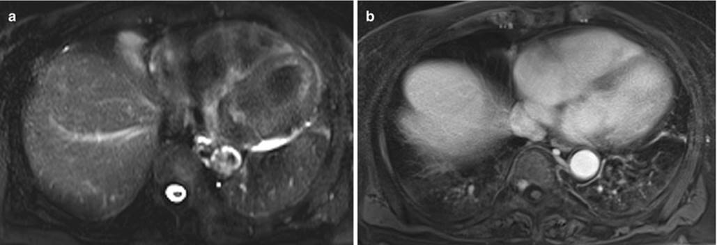
Fig. 16.1
Ground-glass opacity. Axial fat-suppressed fast spin-echo T2-WI (a) and axial free-breathing post-contrast fat-suppressed 3D GRE T1-WI (radial VIBE) (b). In the left lower lobe, a hazy opacity is depicted with high signal intensity on both sequences and without obscuring the bronchovascular markings
Consolidation due to infectious pneumonia (Fig. 16.2) is defined as a region of a normally compressible lung tissue that has been filled with liquid. On MR, consolidation is seen as areas of signal intensity that obscures the margins of vessels and airway walls and appears with high signal intensity on T2-weighted images (T2-WI) (Fig. 16.3), due to the high fluid content. Air bronchogram may be present. Air bronchogram appears as low signal areas following the course of the bronchi within the consolidation (Fig. 16.4). MRI may enable the differentiation of consolidation associated with fibrotic tissue due to the relatively short T2 component of fibrosis [17].

Fig. 16.2
Pneumonia. Chest MRI and follow-up CT performed in a patient with suspected pneumonia. Axial fast spin-echo T2-WI (a), axial fast spin-echo fat-suppressed T2-WI (b), coronal fast spin-echo T2-WI (c), and axial CT image (d). There is a wedge-shaped consolidation in the lower left lobe, with high signal intensity on T2-WI images (a–c), which suggests active inflammation. Note that there is a linear structure converging toward the consolidation (arrow, c), which also shows high signal intensity on T2-WI images, representing thickened wall bronchi with fluid content. A follow-up posttreatment chest CT (d) performed 2 weeks later shows that the consolidation has decreased in size

Fig. 16.3
Pulmonary consolidation (rounded atelectasis). Chest MRI performed to evaluate a lung mass detected on a follow-up x-ray of a patient with pleural effusion. Axial fast spin-echo fat-suppressed T2-WI (a), coronal fast spin-echo T2-WI (b), and axial pre- (c) and post-contrast (d) fat-suppressed 3D GRE T1-WI. Areas of signal intensity that obscures the margins of vessels and airway wall are depicted and are compatible with a consolidation. The typical morphology of a rounded atelectasis in association with pleural effusion is seen. Folding of the visceral pleura (black arrow, b) along with converging of vessels and bronchi into the atelectasis (white arrows, a, c, d) is well shown on MRI images
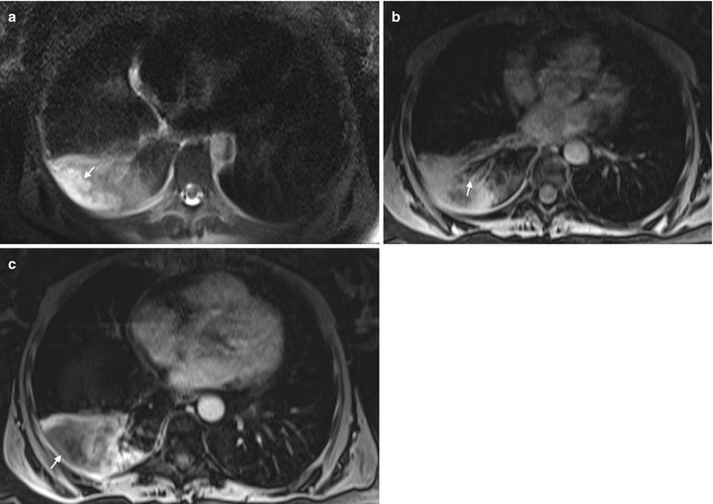
Fig. 16.4
Necrotizing pneumonia. Axial fat-suppressed fast spin-echo T2-WI (a) and axial post-contrast (b, c) fat-suppressed 3D GRE T1-WI. A pulmonary consolidation (arrow) is seen in the right lower lobe with high signal intensity on T2-WI (a) obscuring the bronchovascular margins. Air bronchogram is also depicted (arrow, b). A region of higher T2 signal intensity is seen inside the consolidation, with no enhancement, most consistent with an area of necrosis
The differential diagnoses of consolidation include various substances that may fill the air space, including fluid, blood, pus, and cells. CT attenuation is nonspecific; however, given the MRI ability to characterize different aspects of tissue on the basis of signal intensity, it may be useful for the evaluation of the consolidated parenchyma [17]. For example, focal consolidation in pulmonary infarction due to intra-alveolar blood, or pulmonary hemorrhage that might be related to several diseases such as Goodpasture’s syndrome or invasive aspergillosis, often exhibits high signal intensity on T1-WI due to methemoglobin formation in subacute hemorrhage.
Pleural effusions are a common finding in patients with pneumonia. More than 40 % of patients with bacterial pneumonia and 60 % of patients with pneumococcal pneumonia develop parapneumonic effusions.
Parapneumonic pleural effusions might appear as free-flowing pleural fluid producing sickle-shaped opacity (in most cases posteriorly) with high signal intensity on T2-WI and low signal intensity on T1-WI.
Loculated fluid collections might appear as lenticular opacities in a fixed position and usually develop when frank pus accumulates in the pleural space in the setting of empyema. Significant pleural enhancement is seen after contrast administration. MRI is useful for evaluating the thickening of the pleural membrane when the administration of contrast material is contraindicated, such as in pregnant patients.
16.3.1.2 Tuberculosis
In 2013, 3.3 million cases of tuberculosis were estimated to occur in women worldwide. During pregnancy, tuberculosis is associated with poor outcomes, including increased mortality in both the neonate and the pregnant woman [29].
It has been suggested that MRI might be useful for the diagnostic evaluation of the lung but also to replace CT in assessing lung tuberculosis in women during pregnancy.
Based on a correlation between MRI and CT, the findings in both techniques regarding consolidations, nodules, and cavities correlate well and show comparable results when identifying these characteristics. Consolidations may demonstrate high T2 signal intensity when it is associated with liquefactive necrosis and low signal intensity on T2-weighted and STIR sequences when it is associated with caseating necrosis, probably due to the presence of paramagnetic free radicals in macrophages, which produce T2 shortening or magnetic susceptibility effects on T2-weighted sequences [30].
MRI may depict nodules and micronodules in the pulmonary parenchyma greater than 5 mm in diameter [31]. Three-dimensional GRE and T2-weighted SS-FSE are the most widely used sequences for the detection of pulmonary nodules; however, the three anatomic distributions of the nodules (centrilobular, lymphatic, or random) are not well characterized by MRI. A cavitation is a gas-filled space, within a pulmonary consolidation, mass, or nodule [32]. MRI allows the evaluation of cavity wall thickness and air fluid levels and the identification of intracavitary filling defects [17]. Leutner et al. [33] described the ability of MRI to show various features of opportunistic pneumonia, including cavitation, which are well-known CT findings. MRI and CT have demonstrated a high level of agreement in the detection of lung cavities in tuberculosis. “Tree in bud” and ground-glass opacities are more difficult to detect on MRI [34].
Due to its excellent contrast resolution, MRI seems to be more accurate than unenhanced CT revealing lymph node involvement, pleural abnormalities, and parenchymal caseation.
16.3.1.3 Pulmonary Edema Related to Preeclampsia and Tocolytic Therapy
Preeclampsia is a multisystemic condition, affecting 2–5 % of pregnancies in the United States and with a worldwide prevalence ranging between 3 and 8 %. When severe, various serious complications can increase the mortality in pregnant women. One of them although rare is pulmonary edema, which has an associated mortality rate close to 10 % [35, 36].
Beta (β)-adrenergic agents such as ritodrine and terbutaline are commonly used to inhibit preterm labor and, in this setting, can also cause acute respiratory failure due to pulmonary edema. The etiology of this complication is unclear [22].
There are no specific studies regarding MR evaluation of pulmonary edema; however, the classic CT findings of pulmonary edema, such as septal thickening and areas of consolidation (later stages), may also be seen on MRI. Septal thickening is smooth in contour and appears as interlobular interstitium linear pattern with high signal on T2-WI. Consolidation will appear as described for pneumonias above.
16.3.2 Chronic Diseases
16.3.2.1 Asthma
Asthma affects between 3 and 12 % of pregnant women worldwide, and the prevalence among pregnant women is rising. For unknown reasons, asthma severity appears to be greater in women between the ages of 20–50 years, carrying poorer prognosis than in men [22, 37].
The effect of pregnancy in asthma is incompletely understood and is a matter of debate. However, exacerbations occur in approximately 20 % of all asthmatic pregnant women and are greatly increased in women with severe asthma [37]. Moreover, the effect of pregnancy on asthma symptoms is likely to be consistent during successive pregnancies in an individual patient [22]. Asthma exacerbations require appropriate treatment in order to protect the fetus as far as possible from adverse outcomes. In particular, the fetus is at risk of being born of low birth weight, which may predispose to diseases in later life [37].
Currently, pulmonary function tests (PFT) are considered the most important assessment method and are the most widely used method for diagnosis and management of asthmatics.
Smooth muscle hyperresponsiveness and hypertrophy, increased mucous production, and subepithelial fibrosis are the frequent abnormalities present in asthma. Many of these findings are not evident on standard MRI sequences. New investigational techniques using positive contrast inhaled hyperpolarized noble gases imaging, such as 3He and 129Xe or O2-enhanced MRI (OE-MRI), have shown to be promising in the evaluation of patients with asthma [25, 38]. This technique can assess regional changes caused by airflow obstruction. In a patient with obstruction of medium and small airways caused by asthma, the airspaces distal to the obstruction do not fill with the positive contrast inhaled gas. Therefore, the lungs of healthy patients demonstrate a uniform high signal, whereas the lungs of patients with asthma show wedge-shaped regions of low signal intensity on T1-WI [25]. In patients with asthma, these defects have been shown to increase with provocation by exercise or administration of a bronchoconstrictor (methacholine) and decrease with albuterol. OE-MRI is a nonionizing source of contrast that may have a role in the assessment asthma during pregnancy [25], although further work is required to validate and to extend these findings for pregnant patients.
16.3.2.2 Cystic Fibrosis
Cystic fibrosis is an autosomal recessive disorder that is characterized by exocrine gland dysfunction, with the lungs and pancreas being primarily affected. The pulmonary disease consists of chronic, recurrent infections, bronchiectasis, and airway obstruction. With improved treatment, patients are surviving well into their childbearing years. The advisability of pregnancy should be based on disease stability. As a rule, patients with a significant decline in pulmonary function over a short time period and those with frequent infectious exacerbations are best advised against pregnancy [22].
Chest imaging is essential in the assessment of respiratory disease in cystic fibrosis [39]. Being a chronic disease and affecting young patients, the exposure to ionizing radiation is an important concern. This is especially valid for pregnant women.
MRI and CT are comparable for the depiction of larger morphological changes in cystic fibrosis lung disease such as bronchiectasis, bronchial wall thickening, mucus plugging, and air fluid levels, as well as segment consolidation and architectural destruction (Fig. 16.5). Mucus plugging is well visualized by MRI, even in small airways, due to the high signal intensity of fluid content on T2-WI. This modality has been used successfully to score and follow patients with cystic fibrosis [17, 21, 39, 40].
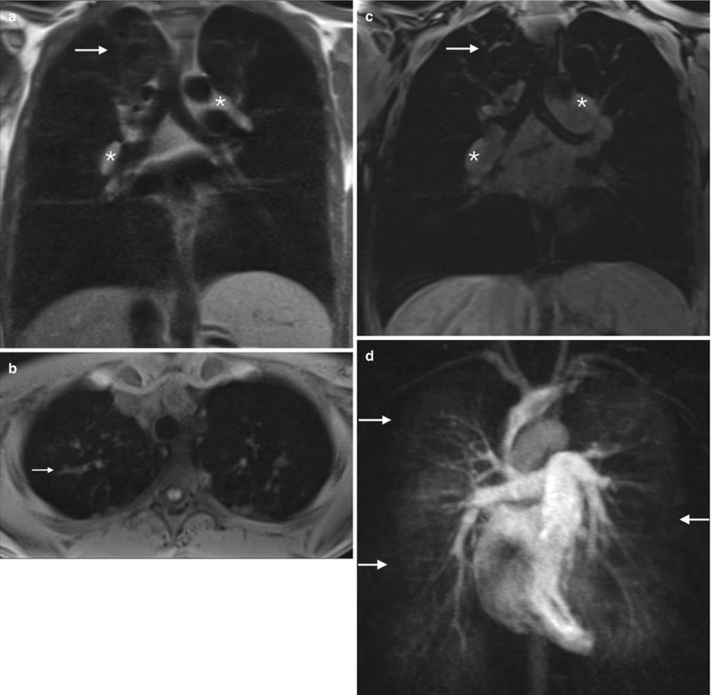
Fig. 16.5
Cystic fibrosis. Chest MRI of a 20-year-old female with cystic fibrosis, admitted with symptoms of pulmonary exacerbation. Coronal spin-echo T1-WI (a), axial (b) and coronal (c) unenhanced 3D GRE T1-WI, and MIP (maximum intension projection) reconstructions of enhanced time-resolved MRA perfusion study image (d). Apical bronchiectasis and mucus plug (arrows, a–c) are depicted. Note the hilar prominent lymphadenopathies (asterisks, a, c). The perfusion study shows areas of decreased perfusion (arrows in d), corresponding to the hypoventilated areas of small airway mucus plugging (arrows, c)
16.3.2.3 Sarcoidosis
Sarcoidosis is a disease of unknown etiology characterized by granulomatous inflammation of body tissues, most commonly the lungs, liver, lymph nodes, eyes, and skin. Patients are often asymptomatic, and the disease would be discovered incidentally on a chest radiograph performed for unrelated reasons [22].
Sarcoidosis in pregnancy is a rare disease associated with an increased risk of adverse obstetrical outcomes. Women with sarcoidosis can carry out successful pregnancies; however, should be made aware of the higher risk of adverse events.
CT is the gold standard imaging method of the lung parenchyma in pulmonary sarcoidosis. However, MRI may be a viable alternative to CT as MRI has been shown to correlate well with CT in this setting [19].
Patterns of sarcoidosis can be described in MRI as parenchymal opacification including ground-glass opacity, consolidation, parenchymal band or scarring (defined as thin linear or discoid regions of signal abnormality), nodules, atelectasis, and focal areas of nodular architectural distortion [19] (Fig. 16.6).
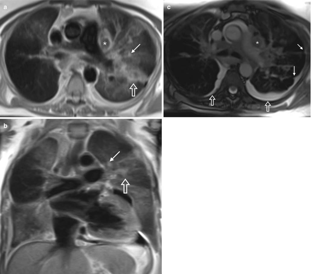
Fig. 16.6
Sarcoidosis. Chest MRI performed in a female patient to follow up for sarcoidosis. Axial (a) and coronal (b) fast spin-echo T2-WI and axial bSSFP (c) images. Areas of ground-glass opacities, more severe at the left side where it tends to consolidate (opened arrows, a, b) and some nodules (arrows, a–c) are depicted, showing the pulmonary sarcoidosis. Lymphadenopathies are well illustrated on both axial images (asterisks, a, c). Pleural retraction (arrows, c) and effusion (opened arrows, c) are also noted
Chung et al. [19] found that in regard to subset scoring, MRI and CT scores seemed to have the strongest correlation for parenchymal opacification and reticulation rather in the setting of lung nodules, where CT performed better. However, they did not include MRI short tau inversion recovery (STIR) images which have been reported to be the most sensitive sequence in the detection of subcentimeter pulmonary nodules [41, 42].
16.3.3 Other Parenchymal Lung Diseases
Other less frequent parenchymal lung diseases may also affect childbearing women, such as lymphangioleiomyomatosis or interstitial lung diseases (Fig. 16.7).

Fig. 16.7
Interstitial pneumonia. Chest MRI performed on a 32-year-old pregnant woman with respiratory symptoms after abnormal findings of the chest x-ray. Axial fast spin-echo fat-suppressed T2-WI (a), coronal fast spin-echo T2-WI (b), and non-enhanced axial fat-suppressed 3D GRE T1-WI (c). A mild-to-moderate interlobular septal thickening is depicted (arrows), with high signal intensity on T2-WI. The septal thickening is less evident on T1-WI. Note the small centrilobular nodules (short arrows, c). Note the minimal right-sided pleural effusion
The relatively limited number of MRI studies that have been clinically performed in these patients is due to the novelty of MRI in this field. Nonetheless, published data suggest at least three possible applications for lung MRI in these settings: (1) visualization and recognition of morphological changes and their patterns, (2) assessment of the inflammatory activity of the disease, and (3) effects of lung morphologic changes on functional parameters such as contrast enhancement and perfusion [24].
16.3.4 Solitary Pulmonary Nodules
Occasionally, a chest x-ray in a pregnant woman, who may not have a previous x-rays for comparison, will show a small lung nodule. The likelihood of malignancy is low for patients under 35 years of age (approximately 5 %), even in cigarette smokers. Additionally, there is little evidence that delaying treatment for some months will result in a reduction of cure rate [43]. Instead, facing these situations may modify attitudes relative to pregnancy.
It has been shown that MRI is useful for detecting and characterizing lung nodules (Fig. 16.8). In fact, MRI has been widely accepted as an ideal screening modality for its nonionizing radiation characteristics [44]. The sensitivity of MRI for nodules greater than 5 mm in diameter is approximately 95 % [45] (Fig. 16.9). The most reliable sequences for detecting lung nodules are STIR, T2-weighted FSE or SS-FSE, and T1-weighted 3D GRE sequences [41, 42, 46].

Fig. 16.8
Breast neoplasm with lung metastases. Chest MRI performed for breast neoplasm staging. Axial fast spin-echo T2-WI (a), axial fast spin-echo fat-suppressed T2-WI (b), and axial pre- (c) and post-contrast (d) fat-suppressed 3D GRE T1-WI. There is an ill-defined speculated mass in the lateral right breast (arrows, a–d) extending from the skin to the underlying pectoralis muscle, better shown on T2-weighted fat-suppressed (b) and T1-weighted post-contrast (d) images. Multiple round-shaped nodules are diffusely spread throughout the lungs, consistent with metastases. These findings show the capability of MRI to detect small nodules
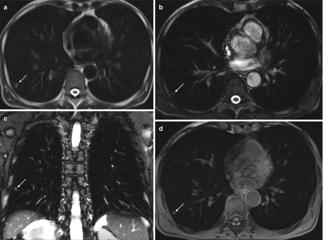
Fig. 16.9
Small metastasis from renal cell carcinoma. Unenhanced chest MRI performed in a patient with history of renal cell carcinoma. Axial fast spin-echo T2-WI (a), axial (b) and coronal bSSFP image (c), and axial fat-suppressed 3D GRE T1-WI (d). A small (5 mm) parenchymal nodule is depicted on all sequences (arrows, a–d), showing the ability of MRI to detect small lung nodules
Many pulmonary nodules, including those of lung cancers, pulmonary metastases, and low-grade malignancies such as carcinoids and lymphomas, appear of low to intermediate signal intensity on T1-WI and of slightly high signal intensity on T2-WI.
DWI allows for differentiation of malignant from benign solitary pulmonary nodules with sensitivity and specificity of 70.0–88.9 % and 61.1–97.0 %, respectively [47–49]. In comparison, the specificity seems to be comparable with that of FDG-PET/CT [47]. Therefore, non-enhanced MR techniques can help in differentiating differentiate malignant from benign SPNs and can be considered as effective as FDG-PET or PET/CT.
Some lung nodules may display specific features, such as pulmonary hamartomas. Pulmonary hamartomas are the third most frequent cause of solitary pulmonary nodule. The presence of fat is the key feature for the diagnosis of this benign lesion. Fat is well depicted on fat suppression and in-phase and out-of-phase imaging (Fig. 16.10).

Fig. 16.10
Hamartoma. MRI performed in a patient for evaluation of a right lung nodule depicted as an incidental finding on x-ray. Axial fast spin-echo T2-WI (a) and T1-WI in-phase (b) and out-of-phase (c) images. The nodule shows heterogeneous high signal intensity on T2-WI (a) and T1-WI in-phase (b) images. Areas of signal loss on T1-WI out-of-phase images (arrows, c) are evident within the nodule, consistent with fatty component
16.3.5 Primary Lung Cancer
Cancer in general during pregnancy is rare and occurs in 1 to every 1000 gestations. The most common types of cancer diagnosed during pregnancy are breast and cervical cancer, lymphoma, melanoma, and leukemia [50].
Primary lung cancer in patients during pregnancy is an extremely rare coincidence.
Lung cancer staging is feasible using MRI. For this purpose, MRI can provide a comprehensive morphologic TNM evaluation. The extent of mediastinal, hilar, and supraclavicular lymph node enlargement can be assessed with excellent soft tissue contrast [24]. Unenhanced MRI is comparable to CT in differentiating between T1–T2 and T3–T4 tumors. MRI seems to be better in evaluating pericardial (T3) and heart (T4) involvement. Contrast-enhanced magnetic resonance angiography (CE-MRA) is also a good imaging method for the evaluation of hilar invasion, with some studies showing higher sensitivity and specificity than CT [51]. MRI might also be superior in the characterization of central masses causing atelectasis [52] (Fig. 16.11).


Fig. 16.11
Central lung cancer. The chest unenhanced MRI performed in a patient to clarify a central left-sided mass. Axial fat-suppressed fast spin-echo T2-WI (a) and axial (b, e, and g) and coronal (c, d, f, and h) and pre- (b–d) and post-contrast (e–h) fat-suppressed 3D GRE T1-WI. A central left-sided primary lung neoplasm is depicted (asterisks, a–c, e, f), with hilar infiltration and left upper lobar atelectasis. On unenhanced T1-WI, note the hilar infiltration (c) and the lower signal of the tumor when compared to the atelectasis (black arrow in c and f), showing the value of unenhanced chest MRI on central lung neoplasms assessment. These findings can be confirmed on enhanced sequences (f). The left-sided mediastinal shift as the rising of the left hemidiaphragm due to left lung volume loss can be also seen. It is also depicted as bilateral pleural effusion (arrows, a) and bilateral lung metastases (opened arrows, d, g, and h), showing that MRI can contribute on lung cancer staging on selected patients
A recently published work by Liu et al. [53] suggested that the DWI-driven ADC measurement of lung cancer is a helpful method to evaluate the pathological grade and tumor cellular density. Therefore, the quantitative analysis of ADC in conjunction with conventional MRI findings could provide more valuable information for the assessment of pulmonary tumor.
MRI assessment of lymph nodes is possible not only referring to morphologic features as CT but also functional evaluation with DWI. Unenhanced STIR sequences combined with DWI can be an accurate noninvasive method of staging lymph nodes [51, 52, 54].
Percutaneous transthoracic needle biopsy has been proven as a minimally invasive, sensitive mean to the tissue diagnosis of lung lesions. Recently, Liu et al. demonstrated that MRI-guided percutaneous transthoracic needle biopsy is safe, feasible, and a highly accurate diagnostic technique for the pathologic diagnosis of pulmonary nodules.
16.4 Mediastinum
Mediastinal tumors in pregnancy can be found incidentally during chest workup for unrelated reasons or, less frequently, found as a result of a symptomatic clinical manifestation. In adults, most mediastinal tumors are primary thymic neoplasms, thyroid masses, or lymphomas [55].
While conventional radiology can assess mediastinal disease, cross-imaging methods made its diagnosis more accurate. Usually, the first-line imaging method is chest CT, ideally with IV iodinated contrast. Besides being a radiation-free method, MRI has a higher soft tissue contrast resolution and is able to characterize most mediastinal masses without the use of GBCAs. To achieve that MRI uses not only the multiparametric morphologic evaluation (T1- and T2-weighted sequences, with and without fat suppression plus or minus GBCAs), but also functional evaluation using DWI.
Recently, Seki et al. [56] showed that DWI alone is equal to CT for differentiating tumors not needing further intervention or treatment from those needing further intervention and treatment. Other studies showed that using unenhanced MRI can be better than CT, particularly in cystic diseases [57, 58]. Therefore, MRI is a good alternative to CT as a first-line modality in evaluating mediastinal masses, especially on pregnant women.
For the sake of simplicity and in order to attempt better differential diagnosis, the authors will follow classic anatomic division of mediastinum (i.e., anterior, middle, and posterior compartments) in this chapter [59].
16.4.1 Anterior Mediastinum
The anterior mediastinum is bounded superiorly by the thoracic inlet; inferiorly by the diaphragm; posteriorly by the pericardium, aorta, and brachiocephalic vessels; and anteriorly by the sternum. Disease of any of the contents of the anterior mediastinum may result in a mass; thus, knowledge of the normal contents of the anterior mediastinum aids in developing a differential diagnosis once a mass has been identified.
The thyroid gland (if it extends into the mediastinum) is traditionally considered an anterior mediastinal compartment structure. The other contents include the adipose tissue, internal mammary vessels, nerves, lymph nodes, and thymus gland [59, 60].
Masses of the anterior compartment account for 50 % of all mediastinal masses [61]. Although more than two thirds of mediastinum tumors are benign, most of anterior compartment masses are malignant [62]. The authors will address the most frequent masses of anterior mediastinum in adults, namely, thymus masses, thoracic goiter, teratoma, and lymphoma.
16.4.1.1 Thymus
Thymus topography makes its imaging using MRI challenging. The recent developments in the MRI field have tackled the constant pulsation of the great vessels, beating of the heart, and movement of the lungs as image degrading factors.
In adulthood, the thymus is atrophic and mainly replaced by fat, being hyperintense in T1- and T2-weighted sequences. When the tissue has almost completely involuted, reticulonodular strands of thymic tissue and occasionally small rounded islands of residual thymus (measuring 7 mm or less) can remain and are considered normal [63].
Hyperplastic thymus can be difficult to distinguish from normal thymus. Some guidelines for making this differentiation are available and include the absence of rounded soft tissue masses >7 mm, absence of a convex contour of the thymus after 19 years of age, absence of soft tissue lobulation, absence of excessive thymic thickness (normal is ≤1.3 cm after 20 years of age), no history of diseases associated with thymic enlargement or hyperplasia, and absence of interval enlargement of thymic tissue during adulthood.
In a prospective study by Inaoka et al. [64], normal or hyperplastic thymus could be differentiated using a non-enhanced on chemical shift imaging (in-phase and out-of-phase T1-weighted sequences) due to the interspersed microscopic fat within the nonneoplastic thymic tissue (Fig. 16.12). These results are particularly interesting in the subset of pregnant patients, since neither ionizing radiation nor IV contrast is applied.

Fig. 16.12
Thymus hyperplasia. Chest MRI performed to evaluate an anterior mediastinal mass in a woman undergoing chemotherapy for breast cancer. Axial fast spin-echo T2-WI (a) and axial GRE in-phase (b) and out-of-phase (c) T1-WI. The mass shows intermediate signal intensity on T2-WI (a) and in-phase T1-WI (b), demonstrating homogeneous signal suppression on out-of-phase T1-WI (c). These features are consistent with hyperplasia due to the presence of microscopic fat within the thymic tissue, virtually excluding thymic tumor
Unenhanced MRI differentiates thymic cysts from solid lesions more readily and consistently than CT. Hyperdense thymic cysts are often misinterpreted as solid lesions on CT [63]. T1 signal of thymic cysts can vary depending on their contents (hemorrhage, lipid, and protein increase in internal T1 intensity), but generally, they are hypointense on T1-WI. Virtually, all thymic cysts are hyperintense on T2-WI, albeit not always as hyperintense as spinal CSF [63] (Fig. 16.13). Congenital cysts tend to be unilocular and have thin walls, whereas acquired cysts are multiloculated and thick walled. The latter usually are inflammatory in nature. When evaluating a thymic cystic lesion, one should be aware that cystic regions might occur in thymic neoplasms such as thymoma and lymphoma.
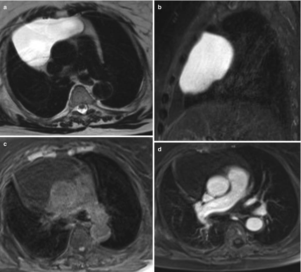
Fig. 16.13
Thymic cyst. Chest MRI performed to evaluate mediastinum enlargement depicted as an incidental finding on an x-ray. Axial fast spin-echo T2-WI (a), sagittal fast spin-echo fat-suppressed T2-WI (b), and axial pre- (c) and post-contrast (d) fat-suppressed 3D GRE T1-WI. There is a large lesion located in the anterior mediastinum, showing high signal intensity on T2-WI (a, b), low signal intensity on T1-WI (c), and absence of enhancement on post-contrast sequence (d), consistent with a cystic lesion, most probably originating from the thymus
Thymomas are histologically classified as less aggressive (types A, AB, and B1 5-year survival rates >94 %) and more aggressive and of poor prognosis types (B2, B3, and thymic carcinoma) [65].
In pregnant women, the possibility of separating these different subtypes with non-enhanced MRI (morphology and functional imaging) is desirable. Based on the clinical settings, lesions with better prognostic outcome could wait till delivery for a treatment decision (e.g., surgery).
Thymoma shows low to intermediate signal intensity on T1-WI and high signal intensity on T2-WI (Fig. 16.14). Signal intensity is heterogeneous in tumors with necrosis, hemorrhage, or cystic change.

Fig. 16.14
Thymoma. Chest MRI performed in a patient with signs and symptoms of myasthenia gravis. Axial fast spin-echo T2-WI (a), axial in-phase (b) and out-of-phase (c) GRE T1-WI, and axial pre- (d) and post-contrast (e) fat-suppressed 3D GRE T1-WI. The mass has mild high signal intensity on T2-WI (a) and intermediate signal on T1-WI (b–d), showing homogeneous enhancement on post-contrast images (e). Note that there is no signal suppression on the out-of-phase T1-WI (c) comparing to the in-phase T1-WI (b), which is compatible of neoplastic tissue instead of hyperplasia
It has been suggested that morphologic MR sequences can differentiate some thymoma subtypes. Round, smoothly marginated encapsulated tumors are typically type A thymomas; and the presence foci within the mass with low signal on T2-weighted sequences (whether due to hemorrhage, flow voids, or calcification) in conjunction with mediastinal lymphadenopathy favors thymic carcinoma [63]. Recently, Seki et al. [56] showed that DWI alone is equal to CT for differentiating tumors not needing further intervention or treatment from those needing further intervention and treatment. Furthermore, comparisons of DWI-driven ADCs for groups A and B and between thymic malignancies and thymomas in group B showed significant differences.
Often discovered incidentally, thymolipoma is a rare benign encapsulated tumor that is usually found in young adults and has no sex predilection. Histologically, it is composed of mature adipose and thymic tissue in variable proportion. In pregnant women, unenhanced MRI is preferred, since it accurately shows a well-defined encapsulated mass that has extensive fat content (high signal intensity on both T1- and T2-weighted sequences and shows obvious drop of signal intensity on fat-suppressed imaging) [55, 60].
16.4.1.2 Thyroid
Intrathoracic, retrosternal goiter accounts for 3–6 % of mediastinal masses, with a majority located in the anterior compartment. The most common type is secondary to goiter with direct downward extension from the neck [60] (Fig. 16.15). An intrathoracic thyroid mass developing from heterotopic thyroid tissue without any connection to the thyroid in the neck is extremely rare [55].

Fig. 16.15
Goiter. Chest MRI performed to evaluate an incidental finding of an anterior mediastinal mass on an x-ray. Axial fast spin-echo fat-suppressed T2-WI (a), coronal pre-contrast fat- suppressed 3D GRE T1-WI (b), axial post-contrast fat-suppressed 3D GRE T1-WI (c), and coronal post-contrast fat-suppressed 3D GRE T1-WI (d). An enlarged thyroid gland reaching the thoracic outlet, with mild high signal intensity on T2-WI (a) and intermediate signal on T1-WI (b). A moderately high signal intensity on T2-WI thyroid nodule is depicted (arrow, a), with strong enhancement after contrast (arrow, c, d). Multiple well-circumscribed lesions abutting the neural foramina at the upper thoracic spine are depicted, with high intensity on T2-WI and no enhancement (short arrows, a). In this patient with history of neurofibromatosis, these findings are most consistent with neurofibromas
Most goiters are euthyroid and are incidentally found, although they can produce life-threatening symptoms because of airway or neurovascular obstruction. No increase in malignancy of thoracic goiter was reported compared to the simple goiter. Malignancy is found in approximately 5 % of thyroid nodules.
A heterogeneous mass extending directly from the thyroid tissue in the neck is the best clue to the diagnosis. Thyroid evaluation is usually performed using head and neck imaging protocols. MRI is able to determine the thyroid origin of the mass and is often preferred to CT due to the improved characterization of heterogeneous nodule structures without the need for IV contrast medium, as iodinated contrast medium interferes with following scintigraphy and radioactive iodine therapy for a period of 1–2 months [66]. In case of thyroid malignancy, MRI can also evaluate cervical lymphadenopathy. Recent studies have shown the possible role of MRI to differentiate malignant from benign nodules. Wu et al. [67] showed that there are significant differences of ADC values between malignant and benign thyroid nodules larger than 1 cm. With multiparametric MRI, including dynamically enhanced MRI sequences and DWI, Sasaki et al. [66] were able to differentiate malignant from benign nodules with sensitivity and specificity of 100 % and 71 %, respectively, and accuracy of 91 %. These results, although promising, require further validation.
16.4.1.3 Teratoma
Germ cell tumors are a heterogeneous group of lesions containing tissues originating from primitive germ cell layers: ectoderm, mesoderm, and endoderm. In adults, germ cell tumors represent 15 % of anterior mediastinum masses. Malignancy is more common in men. Teratoma is the most common germ cell tumor (70 %), contains tissues from all three layers, and occurs in young adults. They are classified into three subtypes: mature, immature, and those with additional malignant components. The vast majority of cases are mature teratomas containing well-differentiated tissue. They most often contain fat and are almost always benign but still have a low malignant potential. On the other hand, although often benign, immature teratomas have their malignant potential proportional to the degree of immature components of neuroectoderm and mesenchymal tissue. Teratomas with additional malignant components are rare lesions in which the teratoma is accompanied by foci of carcinoma, sarcoma, or component of other germ cell tumors [60, 62].
MRI can easily and accurately depict fat-containing teratoma, with high signal intensity on T1- and T2-weighted sequences and drop of signal intensity on fat-suppressed imaging.
As for the other germ cell tumors, a substantial number of teratomas in women are benign, whereas a high proportion of these tumors in men are malignant. Therefore, in our opinion, until proven otherwise, a teratoma in a pregnant woman should be regarded as benign lesion with malignant potential, while surgery might be delayed until delivery.
16.4.1.4 Primary Mediastinal Lymphoma
Enlarged lymph nodes in the mediastinum are usually seen with neoplastic or inflammatory diseases. The former can be primary (mediastinum lymphoma), secondary spreading of other location lymphoma, or secondary to other nonlymphomatous malignancy.
Twenty percent of mediastinal tumors in adults are primary lymphoma, usually occurring in the anterior mediastinum. Approximately 50–70 % of mediastinal lymphomas are due to Hodgkin’s disease, which has no sex predilection and presents with a bimodal age distribution in young adults, as childbearing age women and in patients older than 50 years. Non-Hodgkin’s disease in young and middle-aged adults is classified as large B-cell lymphoma.
Frequently, Hodgkin’s lymphoma appears as a homogeneous soft tissue mass in the anterior mediastinum with surface lobulation and mild-to-moderate contrast enhancement. Occasionally, there may be cystic changes and necrosis [62].
MRI is a valid modality in determining the presence and size of nodes. Morphologic MRI sequences rely only on the size, shape, contours, and homogeneity for characterization. In order to surpass the limited value of morphologic analysis, the standard of reference for lymphoma staging remains FDG-PET/CT. It is known that FDG crosses the human placenta and accumulates in the fetus, locally adding radiation to the ionizing radiation from the CT component of the examination [67].
Whole-body DWI has been successfully used for detecting and staging lymphoma, when compared to FDG-PET/CT. Recently published literature suggests that whole-body DWI should be preferred in patients particularly susceptible to radiation such as pregnant women.
16.4.2 Middle Mediastinum
The middle mediastinum is bounded superiorly by the thoracic inlet, inferiorly by the diaphragm, posteriorly by the posterior portion of the pericardium and posterior tracheal wall, and anteriorly by the anterior portion pericardium. Its contents include the heart pericardium, ascending and transverse aorta, superior and inferior vena cava, brachiocephalic vessels, pulmonary vessels, trachea and main bronchi, lymph nodes, and the phrenic, vagus, and left recurrent laryngeal nerves [59]. Differential diagnoses are based on the contents of this compartment: cardiovascular origin (e.g., ascending aorta aneurysms, pericardial cysts), cysts (bronchogenic, foregut, neurenteric), tracheal lesions, or lymphadenopathies.
Apart from lymphadenopathies (see above), the other differentials for this compartment include benign lesions, most of which are cystic in nature. It has been shown that MRI is superior to CT in depicting cystic lesions. If imaging features identify features of simple cysts, patients can be spared surgery by ruling out the possibility of neoplasm [58].
Because some cysts may have a high attenuation value due to hemorrhage or proteinaceous contents, they may seem solid or indeterminate on unenhanced CT. Multiparametric MRI evaluation shows low hypointensity on T1-WI and hyperintensity on T2-WI. Hemorrhage and proteinaceous contents can increase the T1 signal and decrease the T2 signal (Fig. 16.16). Recently, it has been reported that unenhanced DWI can characterize the internal component of mediastinal lesions with higher confidence level than CT or conventional MRI [58]. Although gadolinium administration is usually precluded in cases of simple cysts, in case of uncertainty, cysts will show no enhancement on CE-MRI (to which subtraction technique can be applied in case of doubted enhancement).
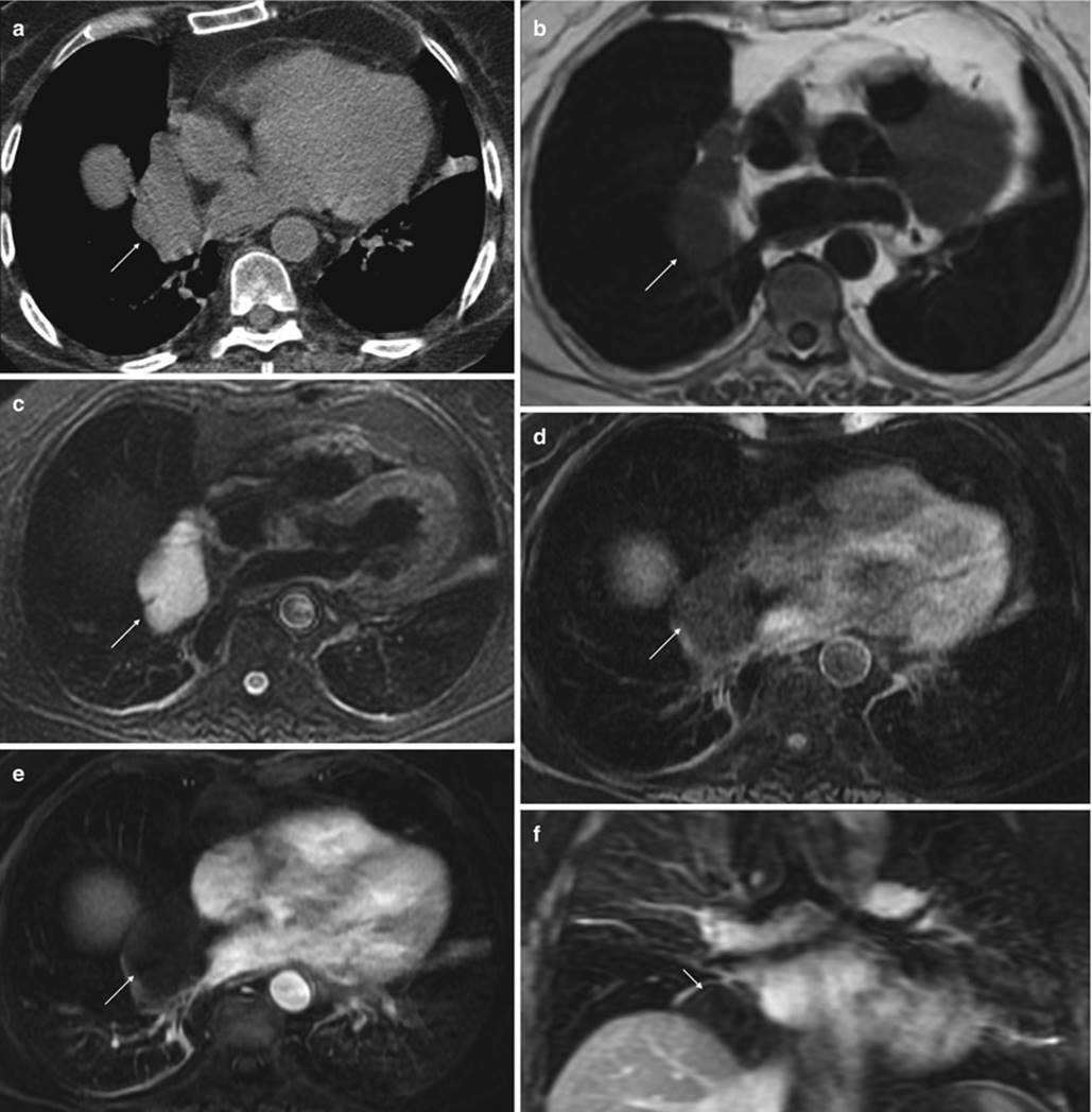
Fig. 16.16
Pericardial cyst. Unenhanced chest CT (a) and chest MRI (b–f) performed to evaluate an incidental finding of a middle mediastinal mass on chest x-ray. The axial unenhanced CT image (a), axial spin-echo T1-WI (“black-blood”) (b), axial fast spin-echo fat-suppressed T2-WI (c), axial pre-(d) and post-contrast (e) fat-suppressed 3D-GRE T1-WI, and coronal post-contrast fat-suppressed 3D GRE T1-WI (f). The CT image depicts a well-marginated right paracardiac nonspecific mass, showing soft tissue density. MRI depicts a mild heterogeneous lesion that has low signal intensity on T1-WI (b, d) and a moderate to severe high intensity signal on T2-WI (c), compatible with a benign cystic lesion (pericardial cyst). The post-contrast sequences confirm the absence of lesion enhancement (e, f), confirming the diagnosis
16.4.3 Posterior Mediastinum
The posterior mediastinum is superiorly limited by the thoracic inlet, inferiorly by the diaphragm, posteriorly by the vertebral column, and anteriorly by the posterior trachea and pericardium. The knowledge of the contents of the posterior mediastinum helps in formulating a differential diagnosis. The contents of the posterior mediastinum include the esophagus, descending aorta, azygos and hemiazygos veins, thoracic duct, vagus and splanchnic nerves, lymph nodes, and fat [62].
Tumors that arise from the intercostal nerve rami or sympathetic chain account for 95 % of the posterior mediastinal masses. Thus, by far, neurogenic tumors are the most frequent tumor of the posterior mediastinum [55].
Seventy to eighty percent of neurogenic tumors are benign, and nearly half are asymptomatic. Generally, they are divided into peripheral nerve tumors (70 % of neurogenic mediastinal tumors), sympathetic ganglion tumors (25 % of neurogenic mediastinal tumors), and mediastinal paragangliomas (rare) (Fig. 16.15) [55].
From the peripheral nerve tumors, schwannomas are the most common (50 % of all mediastinal neurogenic tumors), affecting patients in childbearing age (20–30 years old) [55]. The tumors of the sympathetic ganglion include a spectrum of diseases ranging from purely benign encapsulated ganglioneuromas to malignant ganglioneuroblastomas to aggressively malignant neuroblastomas. Approximately two thirds occur in patients less than 20 years old, and the majority is malignant [68].
MRI can assess the location and extent of intrathoracic neurogenic tumors due to its multiplanar capability and high contrast resolution. The absence of artifacts on MRI caused by high-density bone structures, which often compromise CT scans, facilitates clear visualization of the intracanalicular extension of neurogenic tumors in the paraspinal region.
Generally, schwannomas show low to intermediate signal intensity on T1-WI and intermediate to high signal intensity on T2-WI (Fig. 16.17). Sometimes cystic degeneration can lead to very high signal intensity on T2-WI. If full characterization is absolutely required, gadolinium-enhanced MRI imaging may be performed. Schwannomas show contrast enhancement of the peripheral zone and no enhancement of intralesional cystic areas on T2-WI [68, 69].

Fig. 16.17
Schwannoma. Chest MRI performed in a 29-year-old female patient to clarify an opacity seen at the apex on chest x-ray. Axial (a) and coronal (b) fat-suppressed fast spin-echo T2-WI, axial pre- (c) and post-contrast (d) fast spin-echo T1-WI, coronal post-contrast fat-suppressed 3D GRE T1-WI, and (e) unenhanced axial CT image (f). A well-circumscribed oval-shaped lesion (asterisks) is depicted with heterogeneous high signal intensity on T2-WI (a, b) and heterogeneously strong enhancement on post-contrast T1-WI (d, e). There is extension of this mass along the inferior margin of the left second rib with widening and progression through the adjacent neuroforamen (opened arrows), consistent with a neural origin. Note the lesion’s calcification, depicted on MRI images as signal void area within the lesion on all sequences, confirmed by the CT image (arrows). Findings that are most suggestive of a schwannoma
On the other hand, neurofibromas show a characteristic target appearance on T2-WI (higher signal intensity than the central zone) [69]. Like schwannomas, they homogeneous, low to intermediate signal intensity on T1-WI.
Ganglioneuromas are depicted on both T1- and T2-WI as a homogeneous, intermediate signal intensity and sometimes with a whorled appearance caused by curvilinear or nodular bands of low signal intensity [68].
Neuroblastomas demonstrate homogeneous or heterogeneous signal intensity on all sequences. Cystic (high signal on T2-WI) and hemorrhagic (high signal intensity on T1-WI) areas can be seen on unenhanced MRI. Calcifications can also be perceived as signal voids but suboptimally depicted compared to CT. After contrast administration, the tumor enhances heterogeneously [68].
16.5 Pleura and Chest Wall
MR is an excellent imaging modality for the assessment of primary chest wall tumors (Fig. 16.18), chest wall infections (Figs. 16.19 and 16.20), hernias (Fig. 16.21), and chest wall or diaphragmatic extension of intrathoracic masses. Unenhanced MRI is a good substitute to unenhanced CT. If contrast is deemed necessary, T1-weighted fat-suppressed 3D GRE imaging is especially useful.

Fig. 16.18
Chest wall lipoma. Chest MRI performed in a patient in order to clarify a chest wall tumor. Coronal fast spin-echo T2-WI (a), axial fat-suppressed fast spin-echo T2-WI (b), non-enhanced axial fat-suppressed 3D GRE T1-WI (c), and non-enhanced coronal fat-suppressed 3D GRE T1-WI (d). An expansile left-sided chest wall lesion is noted, with high signal intensity on fast spin-echo T2-WI (asterisk, a) and with signal drop on fat-suppressed sequences (asterisks, b–d), consistent with lipoma. The homogenous appearance and lack of soft tissue areas are compatible with a benign nature

Fig. 16.19
Chest wall infection. Chest MRI performed in a patient with a chest wall painful mass. Axial fat-suppressed fast spin-echo T2-WI (a), axial pre-contrast and post-contrast (c) fat-suppressed free-breathing radial 3D GRE T1-WI, and coronal (d) post-contrast fat-suppressed 3D GRE T1-WI. A left-sided chest wall lesion is depicted involving the chest wall muscles and costal cartilages (asterisk, a, b). This lesion shows high signal intensity on fast spin-echo T2-WI (asterisk, a) and perilesional edema (arrow, a). On post-contrast images, it shows a heterogeneous enhancement (asterisk, c, d). There is an area of necrosis in the thickness of a costal cartilage, showing a high signal intensity on T2-WI (opened arrow, a) and no enhancement on post-contrast T1-WI (opened arrows, d). Note the good image quality of radial 3D GRE despite the free-breathing acquisition

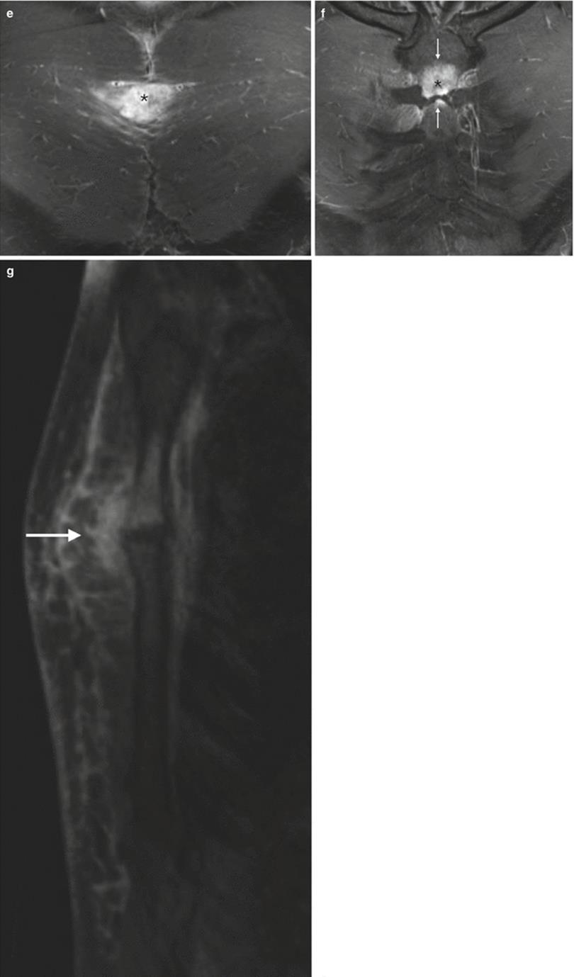
Fig. 16.20
Chest wall infection. Chest wall MRI performed in a patient with fever, to evaluate a presternal mass. Coronal fast spin-echo T2-WI (a), coronal short tau inversion recovery (STIR) (b), axial pre- (c) and post-contrast (d) fat-suppressed radial 3D GRE T1-WI, and coronal (e) and sagittal (f) post-contrast fat-suppressed 3D GRE T1-WI. A lesion centered at the manubriosternal joint shows high signal on T2-WI (asterisks, a, b), surrounded by a significant amount of peripheral edema of the pectoris major (arrows, a, b). This lesion demonstrates mildly high signal intensity on T1-WI (arrow, c) and shows intense enhancement (asterisks, d–f), with articular and bone involvement (arrows, f, g), is compatible with osteomyelitis and septic arteritis

Fig. 16.21
Bochdalek hernia. Chest MRI performed to evaluate an inconclusive chest x-ray, showing a right lower lung lobe lesion. Axial out-of-phase GRE T1-WI (a), coronal bSSFP (b), coronal fast spin-echo T2-WI (c), and coronal post-contrast fat-suppressed 3D GRE T1-WI (d). A right-sided diaphragmatic hernia is depicted, containing part of the right liver lobe (asterisks). Hepatic vessels are seen inside the hernia, assuring the hepatic nature of the hernial content (arrows, b–d)
CT should be preferred in cases of trauma, or in the context of suspected cortical bone neoplastic invasion.
Pleural diseases during pregnancy, apart from pneumothorax (usually in the setting of trauma) and pleural effusion, are rare. Pleural effusion will show high signal intensity T2-WI and low signal intensity T1-WI (see Figs. 16.6, 16.7, and 16.11). Septa can be seen as hypointense lines inside the high signal of pleural effusion on T2-WI. Hemorrhagic and proteinaceous contents generally increase the signal intensity on T1-WI. Pleural enhancement is seen in infectious complications like in cases of empyema, or in the settings of primary and metastatic pleural pathologies.
16.6 Pulmonary Embolism
The hypercoagulable state of pregnancy is a physiologic response preparing for delivery. This fact, among others, predisposes to venous thromboembolism (VTE). VTE is the leading cause of maternal mortality in the developed world. An accurate diagnosis is essential in order to treat this life-threatening condition or to prevent unnecessary treatment, as it is associated with side effects for both the mother and fetus [16, 70]. As a cross-sectional imaging method, it is expected that MRI cannot only exclude or confirm the pulmonary embolism (PE) but also provide an alternative diagnosis clarifying the clinical presentation [15].
MRI of the pulmonary vessels is based on morphologic sequences, to which functional imaging can be added. The goals of the morphological evaluation are mainly to provide information on the vessel diameter and lumen patency. Unlike CT, MRI can additionally provide functional assessment with lung perfusion information, which can be useful in selected clinical settings. Many advanced practices are converging toward a subset of techniques that are fast, reliable, and reproducible.
As it was stated above, GBCAs can be used after assessing the risk-benefit ratio; however, it should be avoided whenever possible, as the risk for the fetus is still unknown. In the following sections, we will discuss noncontrast-enhanced angiography (NCE-MRA) and contrast-enhanced angiography (CE-MRA).
16.6.1 Noncontrast-Enhanced MRA
NCE-MRA is an established technique for the evaluation of pulmonary vessels. Different types of sequences are available for MRA: “black-blood” sequences [fast spin-echo sequences] and “bright-blood” sequences [time of flight (TOF), phase contrast MRA (PC-MRA), and bSSFP sequences].
16.6.1.1 “Black-Blood” Sequences
These techniques are termed black-blood because signal void is created in flowing blood. They consist of either conventional or echo-train spin-echo techniques, usually with ECG gating, exploiting the contrast between rapidly flowing blood and the vessel wall [71, 72]. Black-blood MRA may be useful for the depiction of intraluminal abnormalities such as dissection flaps and tumor thrombus. The RF refocusing pulses cannot repeatedly refocus spins flowing out of the imaging section during the echo train. This signal void is the premise behind black-blood 2D FSE [72]. Therefore, low flow and entry/exit slice phenomena may produce high signal in the vessel lumen, artificially simulating disease. The goal of black-blood techniques is to eliminate as much signal as possible from flowing blood in the vessel lumen (Fig. 16.22). They are particularly helpful in aorta imaging, because it is very accurate in depicting vessel wall disease.
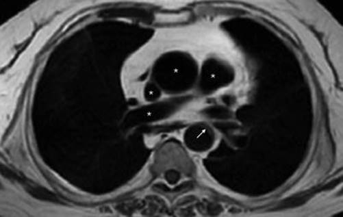
Fig. 16.22
“Back-blood” image. Spin-echo axial T1-WI (“black-blood” technique) at the level of the pulmonary arteries. Note the signal void inside the vessels (asterisks) and the clear depiction of the vessel wall (arrow)
Usually, black-blood techniques are used in combination with other imaging sequences, most often CE-MRA and/or noncontrast-enhanced “bright-blood” techniques.
16.6.1.2 “Bright-Blood” Sequences
The use of TOF in pulmonary angiography is limited due to long acquisition times and to the almost unavoidable motion artifacts, being clinically reserved mainly for neuroimaging angiography [73]. PC-MRA can be used in pulmonary vessels for quantitative blood flow measurements (velocity and gradients), unlike CE-MRA or bSSFP. With PC imaging, it is the flow-induced phase shift of the transverse magnetization that is employed to image the blood vessels with phase contrast techniques. Faster flowing protons experience a greater phase shift than slower moving protons, allowing for the calculation of flow velocity. With PC-MRA, the signal intensity of the blood is proportional to its flow velocity, and the direction of flow can be determined on the basis of the direction of the phase shift [72].
For morphology assessment of pulmonary vessels, the best “bright-blood” sequence is bSSFP. It is a gradient echo technique with a short TE and TR (repetition time) that exploits the coherent transverse magnetization remaining when the TR used is shorter than the T1 and T2 of the tissue. With bSSFP, intraluminal signal generally is very high and relatively homogenous even in cases of turbulent flow because this sequence depends mainly on a function of the T2/T1 ratio [72] (Fig. 16.23).

Fig. 16.23
“Bright-blood” image. Axial (a) and coronal (b) bSSFP (“bright-blood”) images at the level of the pulmonary arteries. Note the high signal intensity of the moving blood inside the vessel (asterisks)
16.6.2 Gadolinium-Enhanced MRA
The use of GBCAs for CE-MRA was introduced in 1994 [74]. The use of contrast media greatly accentuates the signal from inflowing blood via the T1-shortening effect of gadolinium, resulting in improved angiograms [72, 73].
The most commonly used sequence for CE-MRA is a heavily T1-weighted 3D GRE sequence [75, 76]. A three-dimensional acquisition is preferable because it allows thin contiguous image partitions to be obtained during a single breath-hold and 3D reconstructions (Fig. 16.24). Typically, this technique uses a 3D GRE sequence with ultrashort TR and TE, while the flip angle is maintained relatively high at 25–60° to allow for saturation of stationary tissues. This results in reduced signal from the background (stationary) tissue, as there is insufficient time for recovery of longitudinal magnetization (T1 relaxation). Therefore, saturation effects are not problematic with CE-MRA, since this technique exploits the T1-shortening effect of gadolinium with marked increase of intraluminal signal intensity during the first pass of the contrast bolus.
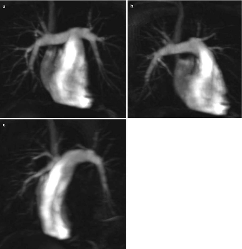
Fig. 16.24
CE-MRA images. Three-dimensional rendering reconstructions of the CE-MRA of the pulmonary arteries, showing a frontal view (a), and right (b) and left (c) pulmonary arteries
A standard breath-hold T1-weighted fat-suppressed 3D GRE sequence can be performed as a complement or a substitute to the previous sequence, with shorter flip angles keeping the short TE and TR. This sequence allows not only an accurate appreciation of the pulmonary vessels but also the aorta, lung parenchyma, pleura, mediastinum, and chest wall (Fig. 16.25). Usually, a standard dose (0.1 mL/kg body weight) of a contrast is administered at a flow rate of 2.5–3 mL per second, followed by a saline flush of 20 mL injected at the same flow rate immediately after contrast administration, to achieve a compact bolus [5].

Fig. 16.25
3D GRE T1-WI. Coronal (a) and axial (b) images of contrast-enhanced 3D GRE T1-WI standard breath-hold sequence
Standard breath-hold fat-suppressed 3D GRE T1-weighted sequences are prone to respiratory motion artifacts, sometimes degrading the quality of image. Patients suffering from PE have difficulty to cooperate with the breath-hold times. Therefore, a newly available work-in-progress free-breathing technique using radial k-space filling (T1-weighted radial 3D GRE) is able to provide high-quality diagnostic images (Fig. 16.26).

Fig. 16.26
Free-breathing radial 3D GRE T1-WI. Axial (a–d) contrast-enhanced 3D GRE T1-WI with radial k-space acquisition (radial 3D GRE). This sequence allows the acquisition in free-breathing technique with a high spatial resolution. It is especially useful in patients that are unable to cooperate with breath-holds. This has a potential application for the evaluation of patients suspected of having PE. The high contrast and the high spatial resolutions allow the evaluation of subsegmental vessels (a, d)
Because signal enhancement and overall image quality of CE-MRA depends on the intra-arterial concentration of the contrast agent, the correct timing of imaging after contrast material injection is fundamental. The time of peak arterial enhancement may vary substantially between patients. The CE-MRA examination needs to be tailored to the individual contrast arrival time.
Some techniques were developed in order to get the best timing possible. One can use a bolus test of 1 mL with a time-resolved bolus-tracking scan to determine the delay time or use a real-time bolus monitoring [6, 77]. The latter method allows the operator to inject the total volume of contrast material, instead of injecting a small amount of contrast material in a separate test bolus scan, and to proceed with the 3D CE-MRA acquisition when the desired signal enhancement in the arterial bed of interest has been detected by the MR system, or by visual feedback by the operating technologist. Real-time MRA fluoroscopic technique also integrates a monitoring phase and an imaging phase into a single pulse sequence. With the MRI fluoroscopic method, monitoring is performed by using a continuous fast two-dimensional (2D) GRE pulse sequence with imaging centered over the vascular bed.
Alternatively, a time-resolved (TR-MRA) 4D acquisition with data view sharing such as a TWIST or TRICKS sequence can be used. The higher time resolution allows for up to nine acquisitions with a few seconds between them in a single breath-hold (Fig. 16.27). This sequence delineates the arterial and venous regional hemodynamics, regional perfusion defects, and cardiac shunts. With TR-MRA, k-space data are shared between successive datasets [6]. The high temporal resolution results in a compromise with respect to spatial resolution. Both can be balanced in order to get a good balance between space and time resolution, in order to keep the acquisition to a single breath-hold [78].


Fig. 16.27
TR-MRA images. Maximum intensity projections of enhanced TR-MRA images of the chest. The numbers on each image correspond to the time (in seconds) of the acquisition delay after the contrast injection. The first image (a) depicts the GBCA arriving through the left brachiocephalic venous trunk and SVC into the right atrium chambers. Note the good enhancement of the arteries and the possibility of evaluation of the lung perfusion
16.6.3 Dynamic CE-MRI and Perfusion
The use of DCE-MRI in order to noninvasively assess the lung perfusion has been studied since the end of the twentieth century [79, 80]. With the latest evolution seen in hardware and sequence development, DCE-MRI allows for the detection of parenchymal hypoperfusion caused by acute and chronic PE and has demonstrated good agreement with single-photon emission tomography (SPECT) and CT angiography with sensitivities and specificities for detecting PE ranging from 83–100 % and 90–100 % [81–84]. Mainly, these techniques use contrast-enhanced 3D TR-MRA sequences for evaluation of lung perfusion (Fig. 16.28), limiting its use during pregnancy.
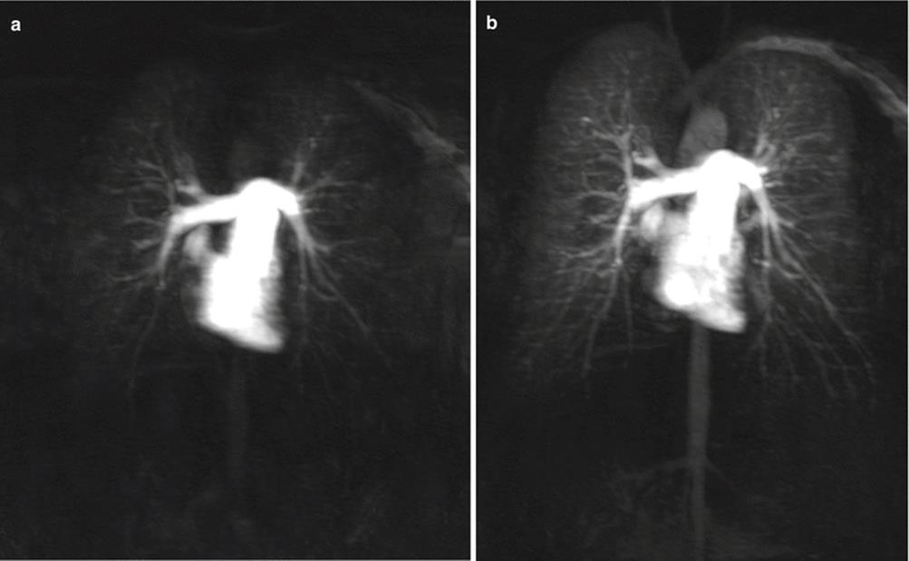

Fig. 16.28
Perfusion study images. Maximum intensity projections of enhanced TR-MRA images of the chest (a–e). Both lung perfusions are normal and symmetric, showing maximum enhancement on figure (b)
New techniques of MRI perfusion are emerging using no GBCAs, being especially useful in pregnant women. The recently published results are promising on the use of these non-enhanced perfusion-weighted MRI functional sequences along with morphologic non-enhanced GRE and bSSFP sequences [84].
16.6.4 Protocol Approach for Pulmonary Thromboembolic Disease
The selected protocol is dependent on the clinical condition (acute PE vs. chronic PE). For imaging pregnant women, the radiologist must choose the least invasive and harmful examination, for both mother and fetus, and which can potentially give the most accurate diagnosis. Keeping this in mind, unenhanced MRI sequences should be preferred in order to safely diagnosis pulmonary embolism. As happens with CT, a short in-room time and reliable protocol should be prescribed. Therefore, in the acute setting, the protocol should include bSSFP (preferable with ECG and respiratory triggering) sequences in two or three planes, which should be done in the first 5 min.
These motion-resistant datasets should be enough to get an overall morphology of pulmonary vessels and exclusion of proximal PE [5, 24]. Revel et al. [83] found that the breath-hold unenhanced bSSFP sequences have high specificity and sensitivity for proximal PE (up to 99.1 % and 100 %, respectively). If still in doubt, one can still assess the benefit of giving IV GBCAs.
PIOPED III is the largest report on MRI detection of PE to date [76]. Its results showed sensitivity between 45 and 100 % at different centers, averaging at 78 %. One of the limitations of this study is related to the use of a single sequence only (CE-MRA). Since the use of clinical MRI is based on a multisequence and multiparametric evaluation of diseases, those results should not be considered reflective of real-life clinical scenarios, as has been showed that the combination of data arising from the unenhanced and enhanced MRI reduce the rate of MRI inconclusiveness [5, 83]. One recent study [5] showed sensitivities for PE detection of 55 % for MR pulmonary angiography, 67 % for triggered bSSFP, and 73 % for 3D GRE MR imaging. Combining all three MR sequences improved overall sensitivity to 84 %. Specificity was 100 % for all detection methods, except for MR pulmonary angiography that had one false-positive case. Because gadolinium has a larger window of visibility than iodine on CT images, it renders timing of data acquisition less critical than on CT and allows the use of different CE-MRA techniques with only one injection of contrast.
The pertinence for the study of chronic PE should be very carefully assessed. Usually, in these clinical settings, the examination can be postponed until delivery. Most treatment options must be deferred in order to minimize the risk to the fetus. If still needed, this evaluation can be performed using contrast-enhanced MRI, which allows for the detection of parenchymal hypoperfusion caused by chronic PE and has demonstrated good agreement with single-photon emission tomography (SPECT) and CT angiography, with sensitivities and specificities for detecting PE ranging from 83–100 % and 90–100 % [82, 85].
16.6.5 Imaging Findings in Acute PE
NCE-MRA sequences, namely, bSSFP, offer the opportunity to directly visualize IV clots as signal voids within the spontaneous high signal intensity of the circulating blood within vessel lumens (Fig. 16.29). As said before, this technique is especially useful in proximal PE. In our experience, we have found bSSFP to be an effective mean of visualizing the central and lobar pulmonary arteries in pregnant patients and evaluating a full image of thoracic disease processes

Fig. 16.29
Acute PE. Unenhanced free-breathing MRI of the chest of a 20-year-old woman suspected of having pulmonary embolism. Axial (a), sagittal (b), and coronal (c) bSSFP images showing an endoluminal hypointense image surrounded by the hyperintense blood flow. This is compatible with a central saddle-shaped pulmonary thrombus. The thrombus (arrows) is depicted in both pulmonary arteries right pulmonary artery on coronal and axial images left pulmonary artery on sagittal image. Note the low signal artifact seen at the aorta in the coronal plane (asterisk, c), not depicted on the other planes, and the pleural (opened arrows, a) and pericardial effusions (opened arrows, b)
In CE-MRA, emboli appear as images indicative of consistent flow impairment (contrast deficiency) or as an abrupt interruption of the signal (Fig. 16.30). The many possibilities of multiplanar reconstruction and of 3D rendered or maximum intensity projection (MIP) images can facilitate the diagnosis and further increase the sensitivity of the method (Fig. 16.31). More importantly, the use of 3D GRE sequences can detect pulmonary embolism in the pulmonary arteries but also secondary signs of PE, including oligemic areas, pulmonary hemorrhage (Fig. 16.32) or infarction (Fig. 16.33), and a full range of chest disease including parenchymal pathologies (see earlier). Because of the larger window of visibility of gadolinium compared to iodine on CT images, pulmonary emboli can often be detected in later acquisitions. The possibility to acquire multiple acquisitions of MRI (due to the lack of ionizing radiations and the long gadolinium intravascular dwell time) raises the confidence in the diagnosis.
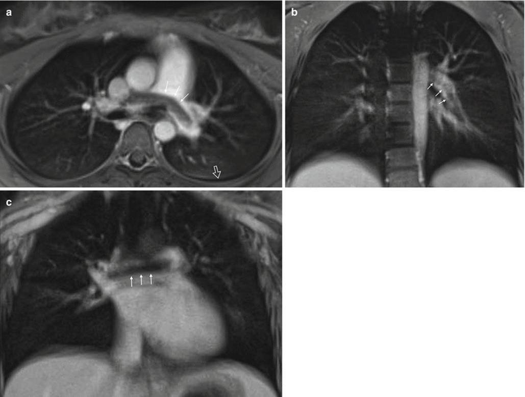
Fig. 16.30
Acute PE. Contrast-enhanced MRI of the chest of the same 20-year-old woman of the previous figure. Axial (a) and coronal (b, c) free-breathing fat-suppressed post-contrast 3D GRE T1-WI (radial VIBE) showing the same central saddle-shaped pulmonary thrombus, as an endoluminal filling defect (arrows, a, c). On the coronal image (b), the thrombus can be more peripherally appreciated. Note the low signal intensity on T1-WI of the pleural effusion described on the previous figure

Fig. 16.31
PE in a 33-year-old female patient. Time-resolved MRA showing an endoluminal defect and the abrupt termination of the affected vessel (white circle). Note the substantial pulmonary oligemia in the right lower lobe

Fig. 16.32
Pulmonary infarction. Reevaluation of a 35-year-old patient with history of pulmonary embolism. Axial 2D GRE T1-WI (a), fat-suppressed fast spin-echo T2-WI (b), and pre- (c) and post-contrast (d) fat-suppressed 3D GRE T1-WI. The images depict a pulmonary heterogeneous lesion, moderate hyperintensity on T2-WI (b, arrow), mild hyperintensity on pre-contrast T1-WI (a, c, arrows), and no enhancement on post-contrast T1-WI (arrow, d). This lesion is compatible with lung infarction, with mild hemorrhagic component. Note the moderate pleural effusion (asterisks)
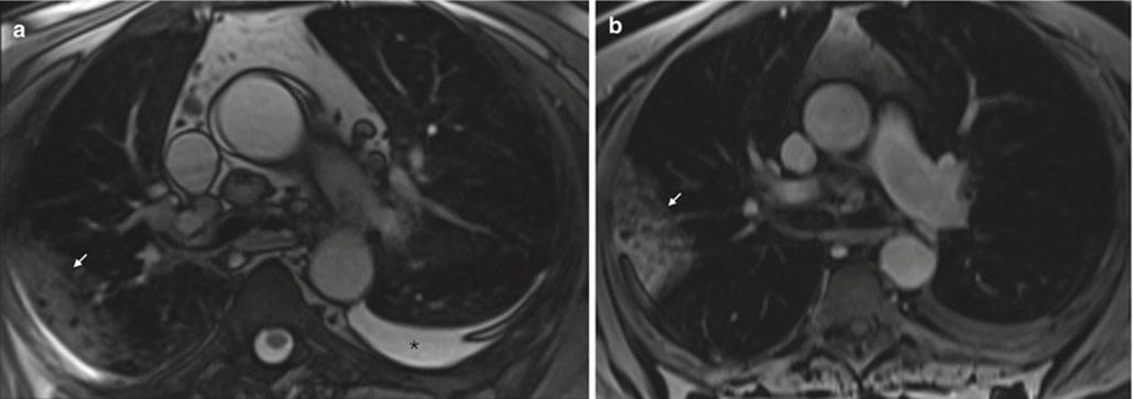

Fig. 16.33
Pulmonary infarction. A 28-year-old patient with a recent diagnosis of PE. Axial bSSFP (a) and post-contrast axial and coronal fat-suppressed 3D GRE T1-WI (b). The images depict a pulmonary wedge-shaped peripheral consolidation (arrows) compatible with pulmonary infarction. Note the mild left pleural effusion (asterisk, a)
Right heart function can be assessed with cine bSSFP imaging, and functional imaging, namely perfusion, can further elucidate on the peripheral vessels.
16.6.6 Imaging Findings in Chronic PE
Typically, after an episode of acute PE pulmonary, the emboli resolve completely; however, in rare occasions, they may follow an aberrant path of organization and recanalization leading to characteristic abnormalities such as intraluminal webs and bands, pouch-like endings of arteries, irregularities of the arterial wall, stenotic lesions, and complete occlusion (Fig. 16.34). Repeated and silent small PE events can also contribute. Chronic thromboembolic pulmonary hypertension (CTEPH) is the major complication of chronic PE. It is a rare disease whose incidence is not precisely known. Estimates have ranged from 0.5–3.8 % of patients after an acute PE and in up to 10 % of those with a history of recurrent PE [86]. This outcome is yet to be established in pregnancy setting.
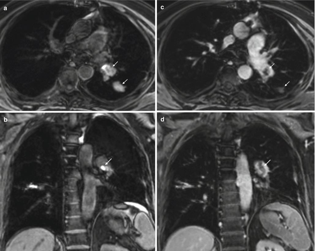
Fig. 16.34
Subacute/chronic pulmonary embolism. Chest MRI performed to reevaluate a patient with a history of pulmonary embolism. Axial and coronal pre- (a, b) and post-contrast (c, d) fat-suppressed 3D GRE T1-WI. The pre-contrast images depict a spontaneous endoluminal thrombus inside the pulmonary arteries, (arrows, a, b). On the post-contrast images, (arrow, c, d). These findings are compatible with subacute/chronic PE
Chronic PE changes, such as irregular vessel walls, thickening due to a concave thrombus adhered to the vessel wall, intraluminal streaks and bands, abnormal proximo-distal tapering, and asymmetry/absence of segmental vessels, as well as peripheral perfusion defects (Fig. 16.35) can be identified by MRI.
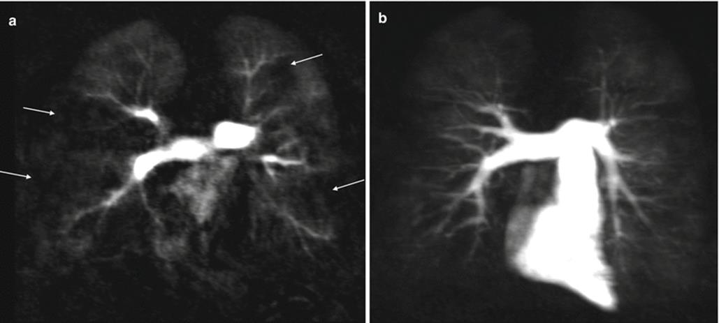
Fig. 16.35
Chronic pulmonary embolism. Chest MRI perfusion study performed to evaluate a patient with a history of chronic PE. Coronal native image (a) and maximum intensity projection (b) of enhanced time-resolved MRA perfusion study. Note the areas of heterogeneity and marked decreased lung regional perfusion (arrows, a), which are compatible with distal arterial occlusion, in this context
MRI can potentially provide information in a single noninvasive test. High spatial 3D CE-MRA with time-resolved acquisitions and ECG-gated flow-sensitive phase contrast MRI with cine MRI analysis of arterial morphology, perfusion, and cardiac function may be performed in a single study; although at present, this is not a common clinical practice in most centers.
16.7 Conclusion
The use of MRI to evaluate the chest has been progressively increasing, especially in pregnant patients. MRI can provide a comprehensive evaluation of the chest, including the lung parenchyma, mediastinum, chest wall, and pulmonary arteries by using different and often complementary approaches. Protocols are tailored to the clinical question including morphological and functional evaluation. Pregnant patients may benefit the most from this modality. It is our belief that MRI is comparable to CT in identifying major morphological pulmonary changes that may occur in pregnant women and MRI is superior to CT regarding tissue characterization, which may be especially advantageous for mediastinal and chest wall lesion characterization. Additionally, we believe that functional studies of pulmonary vascularization (i.e., perfusion), as well as of the airways (i.e., ventilation and gas exchange), will likely become part of the clinical routine in the near future.
References
1.
American Pregnancy Association, Pregnancy Complications. http://www.americanpregnancy.org/pregnancycomplications
2.
Creanga AA, Berg CJ, Syverson C, Seed K, Bruce FC, Callaghan WM (2015) Pregnancy-related mortality in the United States, 2006–2010. Obstet Gynecol 125:5–12CrossRefPubMed
3.
Nair M1, Kurinczuk JJ, Brocklehurst P, Sellers S, Lewis G, Knight M (2015) Factors associated with maternal death from direct pregnancy complications: a UK national case-control study. BJOG 122(5):653–662. doi: 10.1111/1471-0528.13279. http://www.ncbi.nlm.nih.gov/pubmed/25573167. Epub 2015 Jan 9.
4.
Litmanovich DE, Tack D, Lee KS, Shahrzad M, Bankier AA (2014) Cardiothoracic imaging in the pregnant patient. J Thorac Imaging 29:38–49CrossRefPubMed
5.
Kalb B, Sharma P, Tigges S, Ray GL, Kitajima HD, Costello JR, Chen Z, Martin DR (2012) MR imaging of pulmonary embolism: diagnostic accuracy of contrast-enhanced 3D MR pulmonary angiography, contrast-enhanced low–flip angle 3D GRE, and nonenhanced free-induction FISP sequences. Radiology 263:271–278CrossRefPubMed
6.
Wild JM, Marshall H, Bock M, Schad LR, Jakob PM, Puderbach M, Molinari F, Van Beek EJR, Biederer J (2012) MRI of the lung (1/3): methods. Insights Imaging 3:345–353PubMedCentralCrossRefPubMed
7.
Koenigkam-Santos M, Optazaite E, Sommer G, Safi S, Heussel CP, Kauczor H-U, Puderbach M (2015) Contrast-enhanced magnetic resonance imaging of pulmonary lesions: description of a technique aiming clinical practice. Eur J Radiol 84:185–192CrossRefPubMed
8.
Salehi Ravesh M, Brix G, Laun FB, Kuder TA, Puderbach M, Ley-Zaporozhan J, Ley S, Fieselmann A, Herrmann MF, Schranz W, Semmler W, Risse F (2012) Quantification of pulmonary microcirculation by dynamic contrast-enhanced magnetic resonance imaging: comparison of four regularization methods. Magn Reson Med 69:188–199CrossRefPubMed
9.
Kim KW, Lee JM, Jeon YS, Kang SE, Baek JH, Han JK, Choi BI, Bang Y-J, Kiefer B, Block KT, Ji H, Bauer S, Kim C (2012) Free-breathing dynamic contrast-enhanced MRI of the abdomen and chest using a radial gradient echo sequence with K-space weighted image contrast (KWIC). Eur Radiol 23:1352–1360CrossRefPubMed
10.
Lin Y-R, Tsai S-Y, Huang T-Y, Chung H-W, Huang Y-L, Wu F-Z, Lin C-C, Peng N-J, Wu M-T (2013) Inflow-weighted pulmonary perfusion: comparison between dynamic contrast-enhanced MRI versus perfusion scintigraphy in complex pulmonary circulation. J Cardiovasc Magn Reson 15:21PubMedCentralCrossRefPubMed
11.
Koyama H, Ohno Y, Seki S, Nishio M, Yoshikawa T, Matsumoto S, Maniwa Y, Itoh T, Nishimura Y, Sugimura K (2015) Value of diffusion-weighted MR imaging using various parameters for assessment and characterization of solitary pulmonary nodules. Eur J Radiol 84:509–515CrossRefPubMed
12.
Liu H, Liu Y, Yu T, Ye N, Wang Q (2014) Evaluation of apparent diffusion coefficient associated with pathological grade of lung carcinoma, before therapy. J Magn Reson Imaging. doi: 10.1002/jmri.24823. [Epub ahead of print]
13.
Expert Panel on MR Safety, Kanal E, Barkovich AJ, Bell C, Borgstede JP, Bradley WG Jr, Froelich JW, Gimbel JR, Gosbee JW, Kuhni-Kaminski E, Larson PA, Lester JW Jr, Nyenhuis J, Schaefer DJ, Sebek EA, Weinreb J, Wilkoff BL, Woods TO, Lucey L, Hernandez D (2013) ACR guidance document on MR safe practices: 2013. J Magn Reson Imaging 37:501–530CrossRef
14.
Webb JAW, Thomsen HS, Morcos SK, Members of Contrast Media Safety Committee of European Society of Urogenital Radiology (ESUR) (2004) The use of iodinated and gadolinium contrast media during pregnancy and lactation. Eur Radiol 15:1234–1240CrossRefPubMed
15.
Leung AN, Bull TM, Jaeschke R, Lockwood CJ, Boiselle PM, Hurwitz LM, James AH, McCullough LB, Menda Y, Paidas MJ, Royal HD, Tapson VF, Winer-Muram HT, Chervenak FA, Cody DD, McNitt-Gray MF, Stave CD, Tuttle BD (2011) An Official American Thoracic Society/Society of Thoracic Radiology clinical practice guideline: evaluation of suspected pulmonary embolism in pregnancy. Am J Respir Crit Care Med 184:1200–1208CrossRefPubMed
16.
Heredia V, Altun E, Ramalho M, de Campos R, Azevedo R, Pamuklar E, Semelka RC (2012) MRI of pregnant patients for suspected pulmonary embolism: steady-state free precession vs postgadolinium 3D-GRE. Acta Med Port 25:359–367PubMed
17.
Barreto MM, Rafful PP, Rodrigues RS, Zanetti G, Hochhegger B, Souza AS Jr, Guimaraes MD, Marchiori E (2013) Correlation between computed tomographic and magnetic resonance imaging findings of parenchymal lung diseases. Eur J Radiol 82:e492–e501CrossRefPubMed
18.
Mulkern R, Haker S, Mamata H, Lee E, Mitsouras D, Oshio K, Balasubramanian M, Hatabu H (2014) Lung parenchymal signal intensity in MRI: a technical review with educational aspirations regarding reversible versus irreversible transverse relaxation effects in common pulse sequences. Concepts Magn Reson Part A Bridg Educ Res 43A:29–53PubMedCentralCrossRefPubMed
19.
Chung JH, Little BP, Forssen AV, Yong J, Nambu A, Kazlouski D, Puderbach M, Biederer J, Lynch DA (2013) Proton MRI in the evaluation of pulmonary sarcoidosis: comparison to chest CT. Eur J Radiol 82:2378–2385CrossRefPubMed
20.
Corteville DM1, Kjïrstad Å, Henzler T, Zöllner FG, Schad LR (2015) Fourier decomposition pulmonary MRI using a variable flip angle balanced steady-state free precession technique. Magn Reson Med 73(5):1999–2004. doi: 10.1002/mrm.25293. http://www.ncbi.nlm.nih.gov/pubmed/24845240
21.
Biederer J, Heussel C, Puderbach M, Wielpuetz M (2014) Functional magnetic resonance imaging of the lung. Semin Respir Crit Care Med 35:074–082CrossRef
22.
Leighton B, Fish J (2008) Pulmonary Disease in Pregnancy. Glob. libr. women’s med., (ISSN:1756–2228) doi: 10.3843/GLOWM.10170
23.
Chen YH, Keller J, Wang IT, Lin CC, Lin HC (2012) Pneumonia and pregnancy outcomes: a nationwide population-based study. Am J Obstet Gynecol 207:288–289PubMed
24.
Biederer J, Mirsadraee S, Beer M, Molinari F, Hintze C, Bauman G, Both M, Van Beek EJR, Wild J, Puderbach M (2012) MRI of the lung (3/3)-current applications and future perspectives. Insights Imaging 3:373–386PubMedCentralCrossRefPubMed
25.
Liszewski MC, Hersman FW, Altes TA, Ohno Y, Ciet P, Warfield SK, Lee EY (2013) Magnetic resonance imaging of pediatric lung parenchyma, airways, vasculature, ventilation, and perfusion. Radiol Clin North Am 51:555–582CrossRefPubMed
26.
Attenberger UI, Morelli JN, Henzler T, Buchheidt D, Fink C, Schoenberg SO, Reichert M (2014) 3Tesla proton MRI for the diagnosis of pneumonia/lung infiltrates in neutropenic patients with acute myeloid leukemia: initial results in comparison to HRCT. Eur J Radiol 83:e61–e66CrossRefPubMed
27.
Hirsch W, Sorge I, Krohmer S, Weber D, Meier K, Till H (2008) MRI of the lungs in children. Eur J Radiol 68:278–288CrossRefPubMed
28.
Eibel R, Herzog P, Dietrich O, Rieger CT, Ostermann H, Reiser MF, Schoenberg SO (2006) Pulmonary abnormalities in immunocompromised patients: comparative detection with parallel acquisition MR imaging and thin-section helical CT 1. Radiology 241:880–891CrossRefPubMed
29.
Sugarman J, Colvin C, Moran AC, Oxlade O (2014) Tuberculosis in pregnancy: an estimate of the global burden of disease. Lancet Glob Health 2:e710–e716CrossRefPubMed
30.
Peprah KO, Andronikou S, Goussard P (2012) Characteristic magnetic resonance imaging low T2 signal intensity of necrotic lung parenchyma in children with pulmonary tuberculosis. J Thorac Imaging 27:171–174CrossRefPubMed
31.
Vogt FM, Herborn CU, Hunold P, Lauenstein TC, Schroder T, Debatin JF, Barkhausen J (2004) HASTE MRI versus chest radiography in the detection of pulmonary nodules: comparison with MDCT. AJR Am J Roentgenol 183:71–78CrossRefPubMed
32.
Hansell DM, Bankier AA, MacMahon H, McLoud TC, Muller NL, Remy J (2008) Fleischner Society: glossary of terms for thoracic imaging 1. Radiology 246:697–722CrossRefPubMed
33.
Leutner CC, Gieseke J, Lutterbey G, Kuhl CK, Glasmacher A, Wardelmann E, Theisen A, Schild HH (2000) MR imaging of pneumonia in immunocompromised patients: comparison with helical CT. AJR Am J Roentgenol 175:391–397CrossRefPubMed
34.
Rizzi EB, Schinina V, Cristofaro M, Goletti D, Palmieri F, Bevilacqua N, Lauria FN, Girardi E, Bibbolino C (2011) Detection of pulmonary tuberculosis: comparing MR imaging with HRCT. BMC Infect Dis 11:243CrossRefPubMed
35.
Lin S, Leonard D, Co MAM, Mukhopadhyay D, Giri B, Perger L, Beeram MR, Kuehl TJ, Uddin MN (2015) Pre-eclampsia has an adverse impact on maternal and fetal health. Transl Res 165:449–463CrossRefPubMed
36.
Pereira A, Krieger BP (2004) Pulmonary complications of pregnancy. Clin Chest Med 25:299–310CrossRefPubMed
37.
Murphy VE (2006) Asthma exacerbations during pregnancy: incidence and association with adverse pregnancy outcomes. Thorax 61:169–176PubMedCentralCrossRefPubMed
38.
Bauman G, Eichinger M (2012) Ventilation and perfusion magnetic resonance imaging of the lung. Pol J Radiol 77:37–46PubMedCentralCrossRefPubMed
39.
Sileo C, Corvol H, Boelle P-Y, Blondiaux E, Clement A, Le Pointe HD (2014) HRCT and MRI of the lung in children with cystic fibrosis: comparison of different scoring systems. J Cyst Fibros 13:198–204CrossRefPubMed
40.
Failo R, Wielopolski PA, Tiddens HAWM, Hop WCJ, Pozzi Mucelli R, Lequin MH (2009) Lung morphology assessment using MRI: a robust ultrashort TR/TE 2D steady state free precession sequence used in cystic fibrosis patients. Magn Reson Med 61:299–306CrossRefPubMed
41.
Luboldt W, Wetter A, Eichler K, Vogl TJ, Wagner TOF, Seemann MD (2006) Determination of the optimal MRI sequence for the detection of malignant lung nodules. Eur J Med Res 11:336–342PubMed
42.
Frericks BB, Meyer BC, Martus P, Wendt M, Wolf K-J, Wacker F (2008) MRI of the thorax during whole-body MRI: evaluation of different MR sequences and comparison to thoracic multidetector computed tomography (MDCT). J Magn Reson Imaging 27:538–545CrossRefPubMed
43.
Leppert PC, Peipert JF (2004) Primary care for women. Lippincott Williams & Wilkins, Philadelphia
44.
Lin W-C, Chang Y-C, Chang C-Y, Cheng Y-C, Hwang J-J (2012) Primary lung cancers <1 cm found with MR screening appeared larger with half-Fourier sequences than with three-dimensional acquisition techniques. J Clin Imaging 36:739–745CrossRef
45.
Wang Y-XJ, Gong J-S, Suzuki K, Morcos SK (2014) Evidence based imaging strategies for solitary pulmonary nodule. J Thorac Dis 6:872–887PubMedCentralPubMed
46.
Biederer J (2008) MRI of pulmonary nodules: technique and diagnostic value. Cancer Imaging 8:125–130PubMedCentralCrossRefPubMed
47.
Uto T, Takehara Y, Nakamura Y, Naito T, Hashimoto D, Inui N, Suda T, Nakamura H, Chida K (2009) Higher sensitivity and specificity for diffusion-weighted imaging of malignant lung lesions without apparent diffusion coefficient quantification 1. Radiology 252:247–254CrossRefPubMed
48.
Mori T, Nomori H, Ikeda K, Kawanaka K, Shiraishi S, Katahira K, Yamashita Y (2008) Diffusion-weighted magnetic resonance imaging for diagnosing malignant pulmonary nodules/masses: comparison with positron emission tomography. J Thorac Oncol 3:358–364CrossRefPubMed
49.
Satoh S, Kitazume Y, Ohdama S, Kimula Y, Taura S, Endo Y (2008) Can malignant and benign pulmonary nodules be differentiated with diffusion-weighted MRI? AJR Am J Roentgenol 191:464–470CrossRefPubMed
50.
Sarıman N, Levent E, Yener NA, Orki A, Saygı A (2013) Lung cancer and pregnancy. Lung Cancer 79:321–323CrossRefPubMed
51.
Ohno Y (2014) New applications of magnetic resonance imaging for thoracic oncology. Semin Respir Crit Care Med 35:027–040CrossRef
52.
Henzler T, Schmid-Bindert G, Schoenberg SO, Fink C (2010) Diffusion and perfusion MRI of the lung and mediastinum. Eur J Radiol 76:329–336CrossRefPubMed
53.
Liu S, Li C, Yu X, Liu M, Fan T, Chen D, Zhang P, Ren R (2015) Diagnostic accuracy of MRI-guided percutaneous transthoracic needle biopsy of solitary pulmonary nodules. Cardiovasc Intervent Radiol 38:416–421CrossRefPubMed
54.
Hasegawa I, Boiselle PM, Kuwabara K, Sawafuji M, Sugiura H (2008) Mediastinal lymph nodes in patients with non-small cell lung cancer: preliminary experience with diffusion-weighted MR imaging. J Thorac Imaging 23:157–161CrossRefPubMed
55.
Juanpere S, Canete N, Ortuno P, Martinez S, Sanchez G, Bernado L (2012) A diagnostic approach to the mediastinal masses. Insights Imaging 4:29–52PubMedCentralCrossRefPubMed
56.
Seki S, Koyama H, Ohno Y, Nishio M, Takenaka D, Maniwa Y, Itoh T, Nishimura Y, Sugimura K (2014) Diffusion-weighted MR imaging vs. multidetector row CT: direct comparison of capability for assessment of management needs for anterior mediastinal solitary tumors. Eur J Radiol 83:835–842CrossRefPubMed
57.
Tomiyama N, Honda O, Tsubamoto M, Inoue A, Sumikawa H, Kuriyama K, Kusumoto M, Johkoh T, Nakamura H (2009) Anterior mediastinal tumors: diagnostic accuracy of CT and MRI. Eur J Radiol 69:280–288CrossRefPubMed
58.
Shin KE, Yi CA, Kim TS, Lee HY, Choi YS, Kim HK, Kim J (2013) Diffusion-weighted MRI for distinguishing non-neoplastic cysts from solid masses in the mediastinum: problem-solving in mediastinal masses of indeterminate internal characteristics on CT. Eur Radiol 24:677–684CrossRefPubMed
59.
Whitten CR, Khan S, Munneke GJ, Grubnic S (2007) A diagnostic approach to mediastinal abnormalities 1. Radiographics 27:657–671CrossRefPubMed
60.
Shahrzad M, Le TSM, Silva M, Bankier AA, Eisenberg RL (2014) Anterior mediastinal masses. AJR Am J Roentgenol 203:W128–W138CrossRefPubMed
61.
Strollo DC, de Christenson MLR, Jett JR (1997) Primary mediastinal tumors. Part 1: tumors of the anterior mediastinum. Chest 112:511–522CrossRefPubMed
62.
Duwe BV, Sterman DH, Musani AI (2005) Tumors of the mediastinum. Chest 128:2893–2909CrossRefPubMed
63.
Ackman JB, Wu CC (2011) MRI of the thymus. AJR Am J Roentgenol 197:W15–W20CrossRefPubMed
64.
Inaoka T, Takahashi K, Mineta M, Yamada T, Shuke N, Okizaki A, Nagasawa K, Sugimori H, Aburano T (2007) Thymic hyperplasia and thymus gland tumors: differentiation with chemical shift MR imaging 1. Radiology 243:869–876CrossRefPubMed
65.
Chen MM, Coakley FV, Kaimal A, Laros RK (2008) Guidelines for computed tomography and magnetic resonance imaging use during pregnancy and lactation. Obstet Gynecol 112:333–340CrossRefPubMed
66.
Sasaki M, Sumi M, Kaneko K-I, Ishimaru K, Takahashi H, Nakamura T (2012) Multiparametric MR imaging for differentiating between benign and malignant thyroid nodules: initial experience in 23 patients. J Magn Reson Imaging 38:64–71CrossRefPubMed
67.
Wu Y, Yue X, Shen W, Du Y, Yuan Y, Tao X, Tang CY (2013) Diagnostic value of diffusion-weighted MR imaging in thyroid disease: application in differentiating benign from malignant disease. BMC Med Imaging 13:23PubMedCentralCrossRefPubMed
68.
Tanaka O, Kiryu T, Hirose Y, Iwata H, Hoshi H (2005) Neurogenic tumors of the mediastinum and chest wall: MR imaging appearance. J Thorac Imaging 20:316–320CrossRefPubMed
69.
Kiryu T, Ohashi N, Hoshi H, Iwata H, Shimokawa K, Kawaguchi S (2003) Mediastinal schwannoma: MR imaging findings of an unusual case presenting as a lobulated mass with internal fibrous septa. Clin Radiol 58:652–655CrossRefPubMed
70.
Bourjeily G, Paidas M, Khalil H, Rosene-Montella K, Rodger M (2010) Pulmonary embolism in pregnancy. Lancet 375:500–512CrossRefPubMed
71.
Russo V, Renzulli M, Buttazzi K, Fattori R (2005) Acquired diseases of the thoracic aorta: role of MRI and MRA. Eur Radiol 16:852–865CrossRefPubMed
72.
Wheaton AJ, Miyazaki M (2012) Non-contrast enhanced MR angiography: physical principles. J Magn Reson Imaging 36:286–304CrossRefPubMed
73.
Miyazaki M, Akahane M (2011) Non-contrast enhanced MR angiography: established techniques. J Magn Reson Imaging 35:1–19CrossRef
74.
Prince MR (1994) Gadolinium-enhanced MR aortography. Radiology 191:155–164CrossRefPubMed
75.
Stein PD, Gottschalk A, Sostman HD, Chenevert TL, Fowler SE, Goodman LR, Hales CA, Hull RD, Kanal E, Leeper KV, Nadich DP, Sak DJ, Tapson VF, Wakefield TW, Weg JG, Woodard PK (2008) Methods of Prospective Investigation of Pulmonary Embolism Diagnosis III (PIOPED III). Semin Nucl Med 38:462–470PubMedCentralCrossRefPubMed
76.
Stein PD, Chenevert TL, Fowler SE, Goodman LR, Gottschalk A, Hales CA, Hull RD, Jablonski KA, Leeper KV, Naidich DP, Sak DJ, Sostman HD, Tapson VF, Weg JG, Woodard PK, Woodard PK (2010) Gadolinium-enhanced magnetic resonance angiography for pulmonary embolism: a multicenter prospective study (PIOPED III). Ann Intern Med 152:434–443PubMedCentralCrossRefPubMed
77.
Riederer SJ, Bernstein MA, Breen JF, Busse RF, Ehman RL, Fain SB, Hulshizer TC, Huston J, King BF, Kruger DG, Rossman PJ, Shah S (2000) Three-dimensional contrast-enhanced MR angiography with real-time fluoroscopic triggering: design specifications and technical reliability in 330 patient studies. Radiology 215:584–593CrossRefPubMed
78.
Ersoy H, Goldhaber SZ, Cai T, Luu T, Rosebrook J, Mulkern R, Rybicki F (2007) Time-resolved MR angiography: a primary screening examination of patients with suspected pulmonary embolism and contraindications to administration of iodinated contrast material. AJR Am J Roentgenol 188:1246–1254CrossRefPubMed
79.
Hatabu H, Tadamura E, Levin DL, Chen Q, Li W, Kim D, Prasad PV, Edelman RR (1999) Quantitative assessment of pulmonary perfusion with dynamic contrast-enhanced MRI. Magn Reson Med 42:1033–1038CrossRefPubMed
80.
Hatabu H, Gaa J, Kim D, Li W, Prasad PV, Edelman RR (1996) Pulmonary perfusion: qualitative assessment with dynamic contrast-enhanced MRI using ultra-short TE and inversion recovery turbo FLASH. Magn Reson Med 36:503–508CrossRefPubMed
81.
Zhang LJ, Luo S, Yeh BM, Zhou CS, Tang CX, Zhao Y, Li L, Zheng L, Huang W, Lu GM (2013) Diagnostic accuracy of three-dimensional contrast-enhanced MR angiography at 3-T for acute pulmonary embolism detection: comparison with multidetector CT angiography. Int J Cardiol 168:4775–4783CrossRefPubMed
82.
Rajaram S, Swift AJ, Telfer A, Hurdman J, Marshall H, Lorenz E, Capener D, Davies C, Hill C, Elliot C, Condliffe R, Wild JM, Kiely DG (2013) 3D contrast-enhanced lung perfusion MRI is an effective screening tool for chronic thromboembolic pulmonary hypertension: results from the ASPIRE Registry. Thorax 68:677–678CrossRefPubMed
83.
Revel MP, Sanchez O, Lefort C, Meyer G, Couchon S, Hernigou A, Niarra R, Chatellier G, Frija G (2013) Diagnostic accuracy of unenhanced, contrast-enhanced perfusion and angiographic MRI sequences for pulmonary embolism diagnosis: results of independent sequence readings. Eur Radiol 23:2374–2382CrossRefPubMed
84.
Schönfeld C1, Cebotari S2, Voskrebenzev A1, Gutberlet M1, Hinrichs J1, Renne J1, Hoeper MM3, Olsson KM3, Welte T3, Wacker F1, Vogel-Claussen J1 (2015) Performance of perfusion-weighted Fourier decomposition MRI for detection of chronic pulmonary emboli. J Magn Reson Imaging 42(1):72–79. doi: 10.1002/jmri.24764. http://www.ncbi.nlm.nih.gov/pubmed/25227559. Epub 2014 Sep 17.
85.
Nikolaou K, Schoenberg SO, Attenberger U, Scheidler J, Dietrich O, Kuehn B, Rosa F, Huber A, Leuchte H, Baumgartner R, Behr J, Reiser MF (2005) Pulmonary arterial hypertension: diagnosis with fast perfusion MR imaging and high-spatial-resolution MR angiography—preliminary experience 1. Radiology 236:694–703CrossRefPubMed
86.
Wirth G, Bruggemann K, Bostel T, Mayer E, Duber C, Kreitner K (2014) Chronic thromboembolic pulmonary hypertension (CTEPH) – potential role of multidetector-row CT (MD-CT) and MR imaging in the diagnosis and differential diagnosis of the disease. Fortschr Rontgenstr 186:751–761CrossRef