
Roberto Brunelli1 and Gabriele Masselli2
(1)
Department of Obstetrics and Gynecology, Policlinico Umberto I. Sapienza University, Rome, Italy
(2)
Department of Radiology, Umberto I Hospital, Sapienza University, Rome, Italy
Roberto Brunelli (Corresponding author)
Email: roberto.brunelli@uniroma1.it
Gabriele Masselli
Email: g.masselli@policlinicoumberto1.it
Ultrasound (US) is currently the standard approach for the initial evaluation of fetal anatomy and maternal conditions during pregnancy since it allows a real-time examination and is widely available and cost-effective. Magnetic resonance imaging (MRI), due to its capabilities, can greatly improve the diagnostic performance of US during pregnancy. The aim of this chapter is to present some clinical conditions where MRI can improve both the counseling and the management of the pregnant patient and improve the perinatal outcome of high-risk fetuses.
1.1 MRI in the Study of Placental Location and Pathological Adherence
Given the widespread use of endovaginal ultrasound, it is unlikely that the diagnosis of placenta previa, a condition of abnormal implantation of the placenta in the lower uterine segment overlying or near the internal cervical os [1], might benefit from MRI.
Placenta accreta, placenta increta, and placenta percreta represent a spectrum of placental adhesive disorders (PAD) that occur mostly in patients with placenta previa and prior cesarean section when a defect of the decidua basalis allows the invasion of chorionic villi into the myometrium or the adjacent organs; common complications include catastrophic perinatal hemorrhage and bladder, bowel, and ureteral injuries [2, 3].
Early MRI criteria for the diagnosis of placenta accreta primarily focused on the identification of direct invasion of the placenta into the uterus defined by thinning and indistinctness of the myometrium [4], and by loss of the thin T2 dark uteroplacental interface [5]. In 2007, new secondary signs of abnormal placentation, including irregular thick intraplacental T2 dark bands, marked placental heterogeneity, and bulging of the lower uterine segment, were described [6]. A recent report indicated that, in addition to the previously described intraplacental T2 dark bands, dilated tortuous signal voids located deep within the placenta, showing increased signal on true FISP images, are the most sensitive MRI criteria for the diagnosis of invasive placentation (Fig. 1.1) [7].
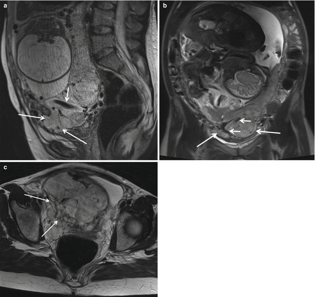
Fig. 1.1
Placenta percreta in a 28-year-old pregnant woman at 34 weeks’ gestation. Sagittal T2-FSE (a) and coronal T2-weighted HASTE images (b) show the bulging of the placenta (arrows) that invades an external layer of the bladder. Intraplacental hypointensive signal bands (newly formed vessels) are clearly seen (short arrows). Axial T2-weighted HASTE image (c) showing massive invasion of the right parametrium (arrows). The myometrial signal is absent in most part of the invaded area
Different studies compared the diagnostic performance of US and MRI in the diagnosis of PAD. One study retrospectively compared transvaginal US and MRI in 39 patients with abnormal placentation and showed 77 % sensitivity, 96 % specificity, 65 % PPV, and 98 % NPV for US and 88 % sensitivity, 100 % specificity, 100 % PPV, and 82 % NPV for MRI [8].
Another study with 13 patients showed comparable low sensitivities of US and MRI for the diagnosis of PAD (33 % and to 38 %, respectively) [9]. A multi-institutional study retrospectively evaluated 15 patients with confirmed abnormal placentation at delivery who had undergone both transabdominal sonography and MRI. Sonography had 93 % sensitivity, 71 % specificity, 74 % PPV, and 92 % NPV, whereas MRI had 80 % sensitivity, 65 % specificity, 67 % PPV, and 79 % NPV, an unremarkable difference [10].
A recent meta-analysis reviewed 18 studies involving 1010 pregnancies at risk for invasive placentation and found no difference in the sensitivity (P = 0.24) or specificity (P = 0.91) of US and MRI for the detection of PAD; however, based on the results of a single study included in the analysis, the authors suggested that MRI should always be considered to better assess both the depth and the topography of PAD [11].
In a report of 12 cases of PAD, MRI correctly gauged the depth of invasion in 3 cases underestimated by US; moreover, with reference to a standardized topographic classification that divides PADs into S1-PAD and S2-PAD (located anteriorly and posteriorly, respectively, to a plane perpendicular to the center of the so-called upper bladder axis), MRI correctly identified 3 cases of S2-PADs that were incorrectly scored as S1-PADs by US [12]. This result has great clinical implications; indeed, while S1-PADs are easily accessed during cesarean section with quick achievement of hemostasis, S2-PADs, being irrigated by deeply located vessels, often result in a massive hemorrhage that requires a complex surgical approach [13]. The MRI excellent characterization of PAD extension and invasiveness was confirmed by another report that specifically acknowledged the usefulness of these findings in the surgical planning of patients with PAD [14]. Overall, since MRI is superior to US in evaluating both the sector and the level of placental invasion, many authors propose its routine use for the implementation of the diagnostic algorithm after an US assigned diagnosis of PAD [15–17].
1.2 Placental Abruption
Placental abruption is the leading cause of vaginal bleeding in the third trimester of pregnancy, occurring in approximately 1 % of births, and accounts for up to 25 % of perinatal deaths; it originates from a premature separation of the placenta from the uterine wall and leads to the formation of retroplacental, subchorionic, or subamniotic hematomas [18]. US findings of placental abruption refer to the evidence of the hematomas but show an extremely poor diagnostic sensitiveness, with 25–50 % of false negatives; different reasons that concur to this poor performance include the similarity of the echotexture of a recent hemorrhage to that of the adjacent placenta and the small dimensions of some hematomas that spontaneously drain through the cervix [19–22].
MRI can dramatically improve the diagnosis of placental abruption. Diffusion- and T1-weighted sequences correctly identified 19 (100 %) and 18 (95 %) of 19 abruptions, with an extremely good interobserver agreement (k = 0.949) [23]. In this report, T1- and T2-weighted sequences were both required for complete tissue characterization. DWI accuracy in the detection of placental abruption was interpreted with reference to its sensitiveness to the susceptibility effects induced by the presence of blood breakdown products [24, 25], while the use of T2-weighted half-Fourier RARE and true FISP was substantiated by the coexisting presence, in cases of abruption, of acute/subacute bleeding and chronic ischemia [26].
The contemporary evaluation of T1-weighted, T2-weighted, and DWI images allowed the classification of hematomas as hyperacute (first few hours, intracellular oxyhemoglobin), acute (1–3 days, intracellular deoxyhemoglobin), early subacute (3–7 days, intracellular methemoglobin), and late subacute (14 days, extracellular methemoglobin) according to the paramagnetic effects of methemoglobin (Fig. 1.2). While the presence of a hematoma with hyperacute or acute MRI signal intensity characteristics anticipated the progression of the abruption to higher grades with the possible onset of fetal distress or maternal decompensation, the evidence of late subacute bleeding was reassuring since all these hematomas remained clinically stable over time. Therefore, MRI characterization of the hematoma structure can be of great utility for patient management as it represents a powerful instrument to portend a bad clinical outcome (Fig. 1.3).

Fig. 1.2
Placental abruption in a 35-year-old woman at 32 weeks of gestational age presenting with history of vaginal bleeding and unremarkable US findings. Sagittal gradient echo T1-weighted image (a) shows a marginal anterior hematoma (long arrows) with blood extending into the cervix; the hematoma appears hyperintense with respect to the placenta (short arrow). Sagittal DWI image (b) shows the markedly hyperintense anterior hematoma (long arrows) and a hypointense hemorrhage (short arrow) along the posterior uterine surface. On sagittal axial true FISP (c), the anterior hematoma signal intensity (long arrow) is similar to that of the placenta, while the posterior blood collection is hypointense (short arrow). Therefore, the different MR sequences are complementary and are all useful for a complete characterization of existing hemorrhage
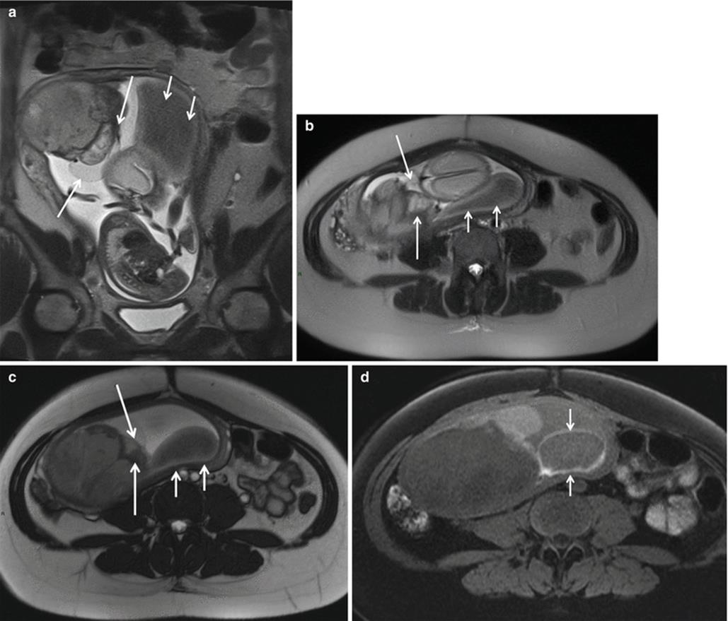
Fig. 1.3
Placental chorioangioma in patient at 24 weeks of gestational age and profuse vaginal bleeding. Coronal (a) and axial (b) T2-weighted HASTE and axial true FISP (c) images show an intrauterine mass peripherally located and protruding from the fetal surface of the placenta. Note the posterior hypointense subamniotic hematoma (short arrows). Axial T1-weighted fat-saturated (d) sequence shows the subamniotic hematoma (short arrows)
1.3 MRI in the Management of Twin-To-Twin Transfusion Syndrome
Twin-to-twin-transfusion syndrome (TTTS) results from a hemodynamical imbalance of placental vascular anastomosis connecting the circulation of the two fetuses. Demise of a co-twin in a monochorionic pregnancy places the surviving twin at significant risk of death or neurologic sequelae due to severe intracranial hypoperfusion [27–29]. Fetal MRI is being increasingly used to identify fetal brain abnormalities [30] and is more sensitive than US in detecting ischemic injury and therefore may have a great theoretical role in the context of TTTS.
In a series of 21 cases of TTTS complicated by the demise of one twin, intracranial abnormalities including polymicrogyria, germinolytic cysts, intracranial hemorrhage, ventriculomegaly, and delayed sulcation were evidenced in seven cases by antenatal MRI; notably, in four of these seven cases, US did not score any brain abnormality, while in two of the remaining three cases, MRI showed additional findings with respect to US [31]. In another study on 68 TTTS with single-twin demise, nine fetuses presented brain abnormalities at MRI (13 %); expert US and MRI findings agreed in only three out of nine cases, while in the other six US underestimated or missed the brain alterations; in this latter study, the diagnostic advantage of MRI was mainly attributed to its capability to image the presence of “reparative” polymicrogyria, a specific abnormality that results from abnormal neuronal migration and cortical organization [32]. Of note, in the two abovementioned studies, MRI examinations were performed only a few days after the US examination; this limits any possible bias related to the evolutive nature of these lesions and supports the proposition that fetal brain pathology in TTTS is genuinely better imaged by MRI.
An early diagnosis of cerebral involvement can greatly influence the consultation and management of the affected pregnancy when fetal death occurs shortly before delivery or after laser coagulation of placental anastomosis that are typically performed at 22–24 weeks of gestation; in this latter case, any brain abnormality would become evident by sonography or conventional MRI at least 2 weeks from the death of the co-twin, greatly limiting the choice for the termination of pregnancy due to the viability of the surviving fetus. The problem of an early diagnosis of ischemic brain damage in TTTS can be conveniently addressed by including DWI sequences in the MRI protocol. In a prospective cohort study, 34 TTTS with single fetal death were evaluated by fetal sonography, conventional MRI, and DWI between 1 and 6 days after the death of the co-twin. Overall, DWI evidenced the presence of cerebral abnormalities in nine cases with unremarkable neurosonographic findings; six of these cases were scored as normal when using conventional MRI [33].
MRI can grant significant information when tailored to image the brain of the recipient twin even before the death of the donor. Two cases of intraventricular hemorrhage and brain ischemia of the recipient twin were found in a series of 25 pregnancies complicated by TTTS; fetal US imaging, performed within 2 h from MRI, was negative in both cases. In the first case, the donor twin died within a week after imaging and ischemia likely developed as a result of inter-twin anastomosis and hypotension; the evidence of ischemia/hemorrhage in the second recipient twin was associated with the enlargement of the cerebral venous sinuses and the reversal of the a wave in the ductus venosus supporting the causative role of a hyperdynamic cardiac decompensation in the genesis of brain injury [34].
The present gold standard for the diagnosis of TTTS is the US staging of Quintero that monitors the progressive deterioration of the donor fetus rather the intraplacental unbalanced circulatory sharing between the donor and recipient fetuses [35]. The goal of correctly addressing the early phases of the syndrome may be possibly achieved by using MRI. In one report, 17 patients with TTTS and 17 controls at a similar gestational age (25.9 ± 2.8 weeks) underwent placental MRI and expert US examination; an accelerated maturation of some sectors of the placenta, putatively triggered by reduced oxygen extraction, increased pO2 and speeded villi maturation was unrecorded by US but clearly evident on T2-weighted images [36].
1.4 MRI in the Prediction and Diagnosis of Uterine Rupture
The overall rate of cesarean section (CS) has dramatically increased in developed countries over the last decade [37]. The WHO Global Survey on Maternal and Perinatal Health advocates a trial of vaginal birth after cesarean section (VBAC) [38] despite acknowledging an increased risk of uterine rupture [39].
Uterine dehiscence refers to separation of the uterine musculature without extravasation of intra-amniotic contents, whereas uterine rupture refers to a defect in the uterine musculature with extravasation of fetal parts and intra-amniotic contents into the peritoneal cavity. The risk of uterine dehiscence/rupture is related to the thickness of the lower uterine segment; however, a lower uterine segment that is less than 3.5 mm thick on an US performed at 37 weeks of gestational age predicts the occurrence of uterine rupture with a positive predictive value of only 11.8 % [40]. Different US cutoff values and measurement techniques were reviewed in a recent publication [41].
A precise evaluation of the uterine scar thickness and of the myometrial fibers organization might create an objective tool to reasonably select those pregnancies that can safely undergo a trial of VBAC. MRI with diffusion tensor imaging (MR-DTI) and fiber tracking reconstruction is a novel noninvasive imaging technique that was used to characterize tissue morphology and can highlight the fiber architecture of the uterus [42].
The ability of US and 3T MR-DTI with fiber tracking reconstruction to assess the uterine scar was compared in a group of in 24 patients with a history of one or two previous cesarean sections: scar morphology, subjectively classified as either “linear” or “retracting,” was found discordant between US and MRI 13 cases (54.1 %); the thickness of the myometrium upstream of the scar was significantly greater when measured by 3T-MR rather than US in women with linear scar morphology; and the disruption of uterine muscular structure was greater in patients with retracting scars independently from the number of previous cesareans. These results anticipate a possible great role of MRI in predicting the risk of uterine rupture during VBAC and seem to support the obstetric guidelines that state that VBAC can be attempted independently from the number of previous CS [43].
MRI allows a clear visualization of the uterine wall; therefore, it helps to diagnose antepartum uterine rupture in patients with indeterminate US evidence, alternatively showing the tear itself [44] or other uterine wall defects including uterine dehiscence and uterine sacculation [45] (Fig. 1.4).

Fig. 1.4
A 34-year-old woman, primipara with a history of previous myomectomies, was admitted at 26 weeks of gestation presenting with vomiting, acute abdominal pain, and tenderness in the upper quadrants. Axial T2-weighted HASTE (a, b) and T1-weighted fat-saturated GRE (c) sequences showed a posterior uterine rupture (arrows) with extravasation of amniotic sac in the peritoneum. Note hemoperitoneum (short arrows in b and c). Emergency laparotomy showed an irregular uterine rupture with 3 cm bleeding
1.5 MRI in the Study of Chronic Placental Insufficiency
MRI does not only highlight the morphologic alterations of the placenta but also offers a noninvasive tool to obtain a quantitative evaluation of the intraplacental circulation during the course of various placental bed disorders, including intrauterine growth restriction (IUGR) and preeclampsia (PIH) definitively inproving the informations granted by the Doppler interrogation of the uterine arteries.
The first pass of an MRI contrast agent through the placenta was first reported in pregnant patients who underwent MRI for non-obstetrical indications and described as fast and homogeneously diffuse throughout the parenchyma [46]. MRI requested for potentially serious indications provided a unique opportunity to characterize in vivo the uteroplacental blood flow in IUGR placentas and to correlate the intervillous circulation to the progressive deterioration of fetal condition [47]. In this study, IUGR placentas displayed many patchy unperfused areas, a slow intervillous circulation, and multiple intermittent stops indicating that dynamic phenomena (vasospasm) are major contributors to the deterioration of placental perfusion; these results are in good agreement with the notion that the spiral arteries are variably occluded in IUGR [48, 49] and that the different placenta sectors do not share a common circulation [50].
MRI evaluation of placental perfusion with contrast agents cannot be routinely employed since the clearance rate of gadolinium from the amniotic compartment has not been yet established. However, the increased availability of a larger repertoire of in vivo placental MRI acquisitions is beginning to offer unparalleled access to in utero placental micro- and macrostructure, metabolism, perfusion, and function [51].
Three-dimensional (3D) volumetric placenta MRI data appear more reliable than those provided by 3D US [52]. Using quantitative 3D volumetric MRI evaluation, one study demonstrated that placental volumetric growth increases exponentially between 17 and 38 weeks in normal pregnancies and that a small placental volume, when recorded in high-risk pregnancies, anticipates the occurrence of IUGR [53].
Diffusion-weighted magnetic resonance imaging (DWI) underscores the differences of water molecule diffusion in human tissues and can provide valuable information on perfusion abnormalities in the human placenta. Indeed, while a feasibility study could not effectively monitor the process of placental maturation by generated apparent diffusion coefficient (ADC) maps [54], another report interpreted the reduction in the ADC of IUGR placentas with reference to the presence of many hematomas and infarctions in tissue degeneration and scarring (Fig. 1.5) [55]. In another study, the addition of DWI analysis of the placenta to the evaluation of the uterine arteries by standard Doppler analysis improved the sensitivity to predict IUGR from 73 to 100 % with a maintained specificity of 99 % [56]. Intravoxel incoherent motion (IVIM) was used to calculate the placenta perfusion fraction in 19 controls in 13 cases of early PIH and 8 cases of late PIH; in these latter two groups placental perfusion fraction was, respectively, smaller and larger than that of controls at the same gestational age, suggesting a distinct pathophysiology of the two forms [57].

Fig. 1.5
A 34-year-old woman at 33 weeks of GA with IUGR fetus. (a) ADC map shows low signal intensity areas (arrows), and this corresponds to the areas of high signal intensity seen on diffusion image (b) (B = 800 s/mm2). Arterial spin labeling perfusion map (c) of the placenta shows multiple areas (blue) of reduction of the perfusion (the normal areas are in green and red). Findings from histopathologic examination of specimens from the placenta were consistent with ischemic endovasculitis
In another study, IVIM confirmed a significantly reduced placental blood flow in IUGR compared to controls [58]. A recent prospective MRI study compared controls to women at risk for PIH but with normal Doppler of the uterine artery who were followed up to delivery; in all pregnancies that delivered IUGR fetuses, placental perfusion fraction was significantly decreased, regardless of the estimated fetal weight at the time of MRI examination [59].
Diffusion tensor imaging (DTI) measures the restricted diffusion of water and produces cellular tract images. Maximum diffusivity, mean diffusivity, and fractional anisotropy are calculated and represent membrane density, cellularity, and microstructural integrity. A single study used this technique and suggested an altered microstructure of the IUGR placenta [60].
Flow-sensitive alternating inversion recovery (FAIR) imaging is used to measure the perfusion of a target organ, by applying a pulse sequence to a vascular network with unidirectional blood flow. The multidirectional blood flow of the placenta challenges the classic paradigm of FAIR imaging and limits the interpretation of classic perfusion measurements [61]. To date, only two studies have explored the use of in vivo FAIR imaging in the evaluation of placental perfusion with discordant results [59, 62].
The paramagnetic properties of oxygen can be used to study oxygenated and deoxygenated blood using oxygen-enhanced MRI (OE-MRI) and blood oxygen level-dependent (BOLD) imaging. One study attempted to decipher if maternal hyperoxia can result in detectable changes in placental oxygenation using OE-MRI and BOLD techniques and showed that in healthy uncomplicated pregnancies, the administration of supplemental oxygen to the mothers is associated with a significant increase in the placenta OE-MRI signal, i.e., to an increased tissue oxygenation [63].
Additional reports used BOLD MRI in healthy pregnancies to investigate the putative effects of placental hyperoxia on the fetal brain; in these studies, placental hyperoxygenation was not paralleled by changes in the oxygenation state of fetal brain, suggesting that oxygen extraction remains constant over wide variations of oxygen delivery as a result of a mechanism that protects the brain of healthy fetuses from the detrimental effects of hyperoxia [64, 65].
Magnetic resonance spectroscopy (MRS) can be used to study intracellular metabolism of different organs. Most commonly, MRS uses signals from hydrogen protons (1H) to determine the relative concentrations of target metabolites, but phosphorus (31P) has also been described for human tissue analysis. 31P-NMS evidenced an increased signal related to adenosine phosphodiesters in the human placenta with advancing gestational age, suggesting an increased cell membrane turnover in aging placenta [66]. Despite a wider availability, only one study explored the use of 1H-NMS in the study of human placenta reporting a low choline–lipid ratio in growth-restricted pregnancies; however, the significance of this finding was hampered by the high choline–lipid ratio that is normally present in the placenta and therefore limits a meaningful interpretation of the spectra and the possible clinical application of this technique (Figs. 1.6 and 1.7) [67].
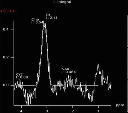
Fig. 1.6
2 × 2 × 4 cm voxel MRS acquired at 144 ms from the placenta of a healthy participant. Choline and lipid spectral peak demonstrated at frequencies of 3.21 ppm, and 1.3 ppm and 0.9 ppm per million (ppm), respectively

Fig. 1.7
2 × 2 × 4 cm voxel MRS acquired at 144 ms from the placenta of a patient with IUGR. In this case, lactate peak is visible as an inverted double peak at 1.32 ppm frequency (arrow). Lactate peak is produced by anaerobic metabolism
1.6 MRI in Preeclampsia/Eclampsia
Cerebral MRI is not necessary for most patients with PIH or eclampsia but can be considered in the presence of focal neurological deficits or prolonged coma; also, in cases of atypical PIH (onset before 20 weeks’ gestation or forms refractory to adequate magnesium sulfate therapy), it may improve the therapeutic planning through a better definition of the pathophysiology of the ongoing disorder. Hypertensive encephalopathy, occurring when high perfusion pressure exceeds cerebral blood flow autoregulation resulting in vasodilatation and brain edema, is considered as the model of central nervous system (CNS) perfusion abnormalities during PIH. In this regard, in a series of 9 normotensive controls and 12 untreated women with PIH, velocity-encoded phase contrast MRI of the middle and posterior cerebral arteries showed an increased cerebral artery blood flow in the affected pregnancies [68]. However, hypertensive encephalopathy does not represent the only possible pathophysiological derangement of cerebral circulation in PIH. Reversible posterior leukoencephalopathy syndrome (RPLS) is associated with PIH/eclampsia and clinically overlaps the classical picture of hypertensive encephalopathy (headache, altered mental functioning, seizures, and loss of vision) [69]. MRI of PRES demonstrates the presence of a symmetric edema that is prevalent in the hypoperfused junctional zones between the territories of the lateral and medial cerebral arteries and is generated by a hyperreactive cerebral vasoconstriction triggered by endothelial toxicity [70]. Indeed, many cases of PRES occur with minor increases of blood pressure, and the extent of brain edema does not correlate to the severity of hypertension [71]. The pathophysiological difference between hypertensive encephalopathy and PRES can be documented by MRI and have great clinical implications with reference to the use of magnesium sulfate, a commonly used drug for the treatment of PIH; indeed magnesium may be alternatively beneficial or detrimental according to the prevalence of cerebral vasodilatation or vasoconstriction since it is a potent vasodilator used in human to ameliorate the cerebral vasoconstriction triggered by a subarachnoid hemorrhage [71].
1.7 MRI in Clinical Pelvimetry and Prediction of Dystocia
MRI pelvimetry may represent a suitable approach in some rare circumstances that include patients that have either the fetus in breech presentation or a history of previous pelvic trauma but want to undergo a trial of labor [72]. One study evaluated eight singleton diabetic pregnant mothers who underwent MRI; a significant correlation was found between the MRI measurements obtained with different sequences and the actual shoulder diameter, with true FISP being the most accurate [73].
1.8 Cervical Incompetence and Prediction of Preterm Labor
Cervical incompetence, i.e., premature shortening of the cervix in the absence of uterine contractions, is due to the occurrence of premature cervical softening, a process that reduces cervical compliance through a disarrangement of collagen fibrils and an increased tissue hydration. The greatest obstetrical challenge of a sonographic short cervix consists in its poor anticipation of a forthcoming delivery, i.e., parturition within 7 days.
Cervical ripening, the cervical counterpart of active labor, is an accelerated process, totally independent from the slowly progressive shortening of the cervix, that occurs shortly before birth and is marked by an increased water permeability of cervical epithelia along with focal periglandular dissolution of the cervical connective tissue.
High ADC map intensity values in the subglandular area of the uterine cervix are significantly associated with the occurrence of delivery within 7 days (unpublished results). If confirmed in subsequent studies, this information will be of great clinical value when managing asymptomatic patients presenting at a critical gestational age with a sonographic short cervix.
1.9 MRI in the Evaluation of Maternal Acute Abdomen
A wide variety of diseases may present with acute abdominal pain during pregnancy and present diagnostic and therapeutic challenges. The diagnosis is often confounded by several common features of normal pregnancy including nonspecific pain, nausea, and vomiting and by the displacement of abdominal and pelvic structures that hampers the clinical examination. Moreover, leukocytosis is a normal finding in pregnancy and levels of C-reactive protein are higher than in nonpregnant women. Prompt diagnosis and treatment are essential for maternal and fetal well-being, and imaging is commonly requested to clarify the clinical picture and expedite diagnosis.
Given the established risks to the fetus from radiation exposure, only US and MRI are routinely considered for the diagnostic evaluation of the abdomen during pregnancy. In this regard, an increasing number of studies have substantiated the use of MRI as the technique of choice in the evaluation of pregnant patients with acute abdominal pain (Figs. 1.8 and 1.9).

Fig. 1.8
Obstruction in a 38-year-old woman at 30 weeks of gestational age presenting with acute abdominal pain and vomiting. Coronal (a) and axial (b) T2-weighted HASTE images show persistent focal transition point “beak sign” (arrows), indicative of proximal obstruction. These findings are suggestive of an adhesion, the presence of which was confirmed at laparoscopy
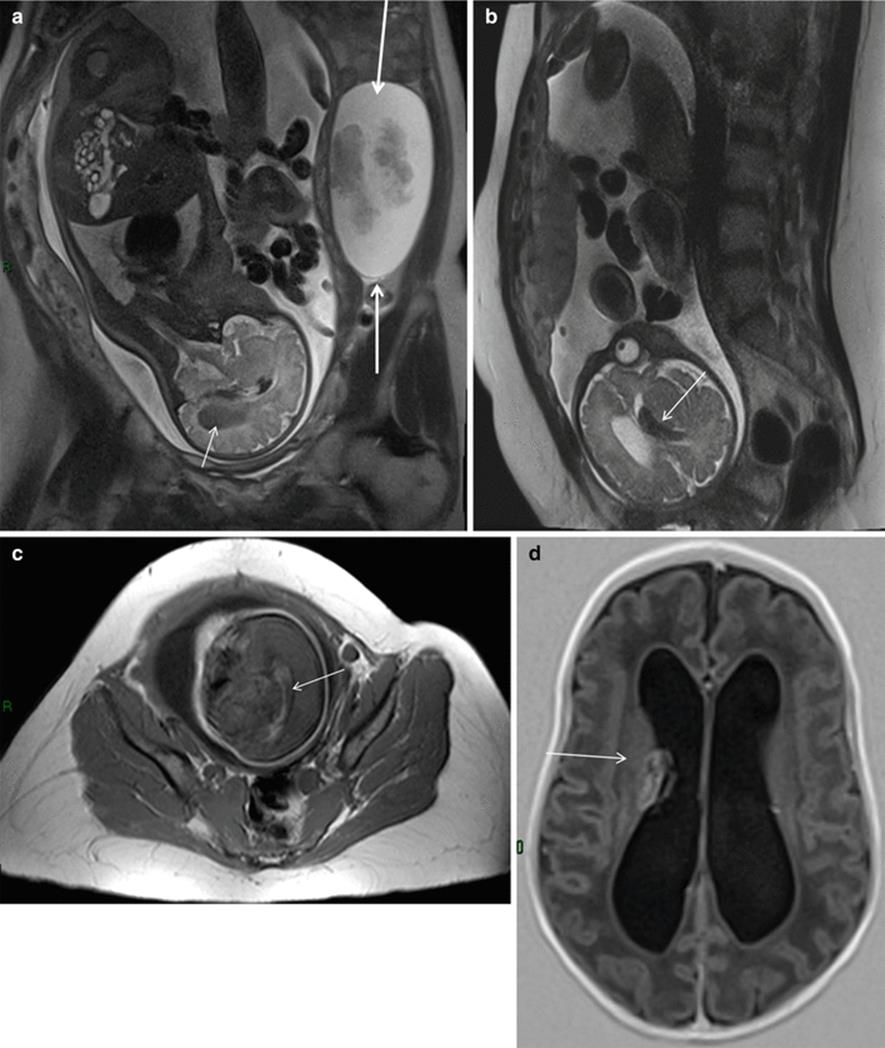
Fig. 1.9
Coronal T2-weighted HASTE MR image (a) shows a complex mass with fluid and solid components on the left ovary (borderline tumor) (arrows). Note low hyposignal into the right lateral ventricle (arrow), better seen on sagittal T2-weighted MR (b); axial T1-weighted sequence (c) shows hyperintense signal at the level of the right lateral ventricle, indicative of intraventricular hemorrhage. Axial T1-weighted MR image (d), performed in the newborn 10 days after birth, demonstrates a germinal matrix hemorrhage extended to the right ventricular system (arrow)
Acute appendicitis is the most common non-obstetrical surgical condition of the abdomen during pregnancy; it is often associated with perforation due to delayed diagnosis (43 % vs. 4–19 % in the general population) and to a significant fetal loss of (20–25 %). These data emphasize the need for accurate early diagnosis and timely surgical intervention [74–76]. Ultrasonography with graded compression is scarcely valuable for detecting acute appendicitis in pregnant women due to the infrequent visualization of the appendix; in one report, US detected the appendix in only 29 % of surgically proven cases occurring during the second and third trimesters of pregnancy [77]; in another study, US sensitivity and specificity to diagnose acute appendicitis during pregnancy were 78 % and 83 %, respectively [78].
MRI can dramatically improve the diagnosis of appendicitis [79] with a visualization rate of the appendix as high as 70 % and an extremely high specificity [80].
The overall sensitivity, specificity, positive predictive value, and negative predictive value of MRI for the diagnosis of acute appendicitis ranged from 90 to 100 %, 93 to 98 %, 61 to 82 %, and 99 to 100 %, respectively [81, 82].
This diagnostic performance was recently confirmed in a retrospective study from a university tertiary referral center [83].
Bowel obstruction in pregnancy is fairly uncommon (1 per 2,500 to 1 per 3,500 pregnancies), normally results from the mechanical effects exerted on the bowel by the enlarging gravid uterus or from the presence of adhesions, and is poorly characterized by US [74]. Magnetic resonance studies for bowel obstruction, particularly if performed with the use of multiplanar T2-weighted single-shot fast spin-echo (SSFSE) imaging can accurately depict the site of small bowel obstruction in up to 70 % of cases [84, 85] (Fig. 1.8).
Magnetic resonance enterography (MRE) is extremely valuable in the diagnostic evaluation for Crohn’s disease (CD) with reported sensitivity and specificity being 83–91 % and 86–100 %, respectively, in the presence of a bowel wall thickness greater than 3 mm and of increased signal intensity on T2-weighted images [86]; these evidences were particularly useful in the evaluation of the active state of CD in a group of 9 pregnant patients [87]. These results are particularly significant because up to one-third of women with inflammatory bowel disease (IBD) relapse during pregnancy as a consequence of a voluntary cessation or tapering of therapy, usually triggered by the fear of putative but undemonstrated teratogenic effects.
The assessment of the extent and severity of acute severe colitis and the identification of toxic dilatation of the colon are more challenging during pregnancy due to the preferential avoidance of ionizing radiation. Magnetic resonance colonography (MRC) performed in a pregnant patient according to a protocol that included multiplanar T1- and T2-weighted sequences and diffusion-weighted imaging (DWI) following intravenous (IV) administration of antispastics clearly evidenced the hallmarks of active ulcerative colitis (circumferential thickening together with a marked restricted diffusion in the involved segment of the colon) [88].
Symptomatic biliary tract disease in pregnancy is uncommon, despite the high prevalence of gallstones in pregnancy. Magnetic resonance cholangiopancreatography (MRCP) clearly evidenced the obstructive etiology in four out of eight pregnant patients with an undetermined biliary dilatation detected by US and was proposed as the most appropriate second-line imaging test to fully evaluate biliary dilatation during pregnancy [89].
Pregnancy-induced hepatic morbidity is rare and is usually seen in two forms: hepatic necrosis associated with the hemolysis, elevated liver enzymes, and low platelet count syndrome (HELLP) and acute fatty liver of pregnancy (AFLP). Hepatic involvement in HELLP syndrome is initiated by intravascular fibrin deposition (leading to sinusoidal obstruction) and hypovolemia, leading to hepatic ischemia/infarction and hemorrhage. The initial diagnosis of HELLP syndrome is usually made using a combination of biochemical criteria and clinical findings (right upper abdominal pain); urgent MRI can be performed to corroborate the diagnosis of HELLP by showing the presence of liver ischemia [90] or alternatively to evaluate the possible occurrence of a hematoma beneath Glisson’s capsule [91].
AFLP is initiated by a hampered intramitochondrial fatty acid oxidation leading to severe microvesicle steatosis and acute liver dysfunction. MRI performed with T1-weighted dual gradient-echo in-phase (IP) and out-of-phase (OOP) sequences is confirmatory of the diagnosis as it depicts a dramatic loss of hepatic signal intensity in OOP images [92].
Women with sickle cell disease who become pregnant are at increased risk for developing PIH and eclampsia [93]. Liver MRI in cases of sickle cell disease shows a low signal on T2*-weighted sequences: in the proper clinical setting, subtle increases of the T2 signal in the hepatic parenchyma can allow a presumptive diagnosis of end-organ ischemia and early preeclampsia [90].
Urolithiasis and urinary tract infection are the most common causes of abdominopelvic pain in pregnancy, and urolithiasis is the most common non-obstetric indication for hospitalization.
Ultrasound, despite its poor diagnostic performance, is the first imaging test for suspected urolithiasis in pregnancy; false negatives due to obstruction without dilatation are rare, but false positives are common because a physiological dilatation of the collecting system occurs in pregnancy [94]. Magnetic resonance urography (MRU) can rapidly acquire images of the upper urinary tract without the administration of intravenous contrast agents and is a valuable examination in pregnancy. In a series of 24 pregnant patients with symptomatic hydronephrosis, MRU clearly differentiated physiological urinary tract dilatation from abnormal dilatation related to urolithiasis by showing different indirect signs including renal enlargement, perirenal fluid collection, signal voids overlying the high signal of urine within a dilated ureter, the presence of a standing column of urine below the level of the pelvic brim, and an abrupt ending of the ureter [95]. MRU accuracy in detecting small ureteral calculi may be further improved by the use of thin-slice, high-resolution SSFSE with a positive predictive value of 80 % [96]. Finally, MRU can effectively score complications such as diffuse or focal pyelonephritis (visualized at as an enlarged edematous kidney or areas of lower signal intensity on T2-weighted images and restricted proton diffusion on the DWI, respectively) [97].
1.10 MRI to Improve Imaging of Fetal Structural Abnormalities
MRI can be extremely useful in the recognition of the developing fetal anatomy, detection of subtle fetal abnormalities, and evaluation of complex lesions.
The development of fetal MRI techniques predictably led to comparisons between the accuracies of MRI and US. A review consisting of 13 published studies comparing anomaly detection rate of US compared to MRI found that fetal MRI provided additional anatomical information in up to 45.3 % of the cases [98]. Many of these earlier studies presented significant bias because US was not usually performed in expert centers, their results were known to the radiologist and used in MRI interpretation, and significant intervals often elapsed between ultrasound and MRI (potentially allowing sufficient time for lesions evolution toward more identifiable forms). However, a number of recent prospective studies directly compared US and MRI and confirmed that this latter technique has a greater detection rate for many fetal anomalies [99, 100].
The most common clinical indication for fetal MRI is for the evaluation of suspected or proven CNS anomalies with improved detection in up to 55 % of cases (Figs. 1.10, 1.11, and 1.12) [100]. Thin MRI slices can be specifically targeted to study the possible occurrence of cortical malformations such as polymicrogyria, lissencephaly, and schizencephaly; other fetal cerebral parenchymal abnormalities including hemorrhage, gliosis, and white matter edema can also be easily detected [101].
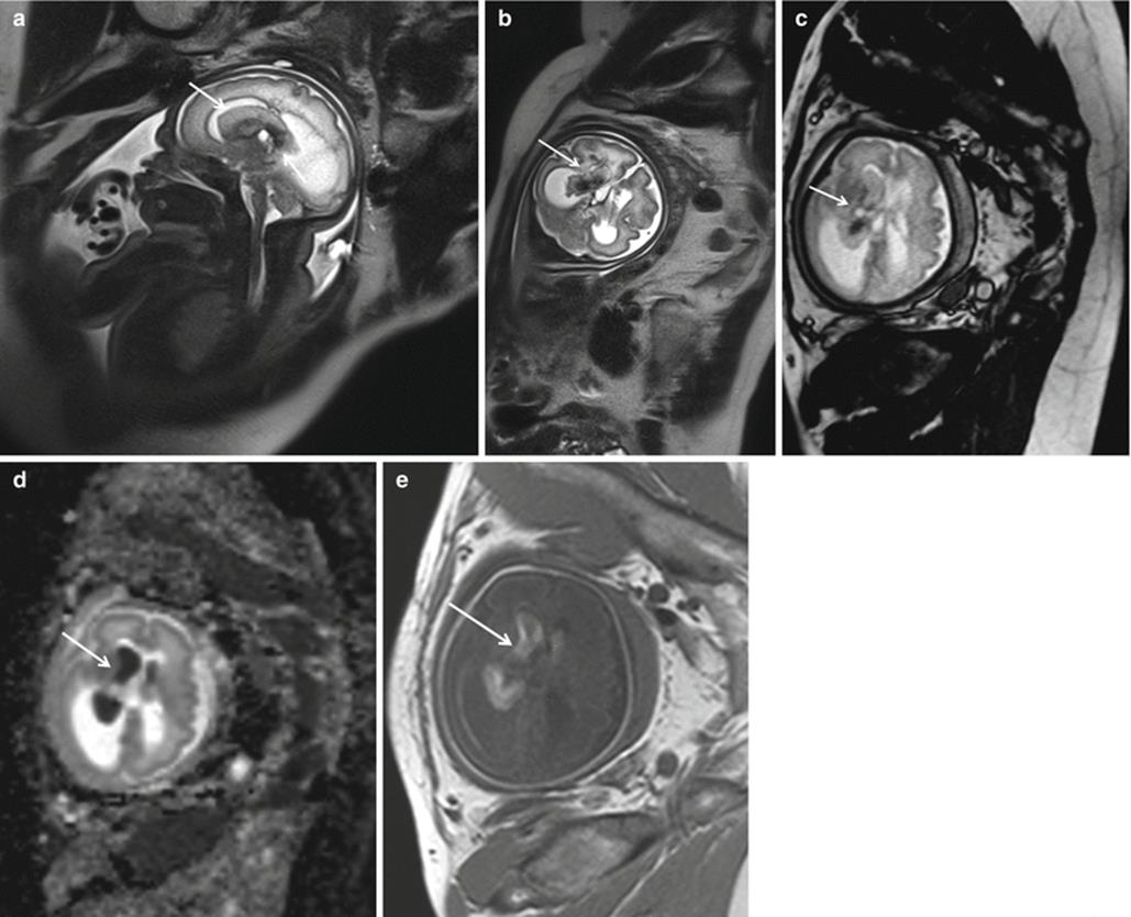
Fig. 1.10
Fetal magnetic resonance images of fetal choroid plexus papilloma with hemorrhage. Sagittal (a) and coronal (b) T2-weighted HASTE images at 33 weeks of gestation show a heterogeneous mass in the right choroid plexus (arrow), with hydrocephalus; absence of signal of hemosiderin on T2*-weighted imaging (c) at 33 weeks of gestation ruling out diagnosis of hematoma. The mass appears with restriction of the diffusion on ADC map (d). Note hyperintense signal on T1-weighted sequence (e) at the level of the right ventricle, indicative of intraventricular hemorrhage, a complication of plexus tumors
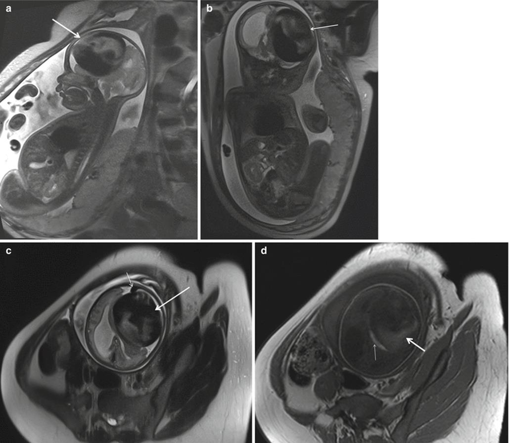
Fig. 1.11
Fetal brain MRI in a large dural arteriovenous (AV) fistula at 30 weeks of gestation. T2-weighted HASTE sagittal (a), coronal (b), and transverse (c) images, showing large extra-axial left frontal lesion (80x72x70mm), with marked hypointense areas due to flow artifact and dilatation of the adjacent cortical vein (short arrow in c). Note the mass effect on the brain parenchyma and on the left lateral ventricle that is compressed with dilation of the right lateral ventricle. Axial T1-weighted image (d) shows hyperintense signal areas in the lesion (arrow), indicative of hemorrhage with focal hyperintensity in the adjacent brain parenchyma (short arrow) due to retrograde leptomeningeal venous drainage

Fig. 1.12
Coronal (a) and sagittal (b) T2-weighted HASTE images of a 33-gestational-week fetus demonstrate bilateral clefts extending from the ventricle to the subarachnoid space, consistent with open lip schizencephalic defects (arrows). The adjacent sulcal pattern is abnormal, consistent with polymicrogyria. The cavum septum pellucidum is absent
Prenatal differentiation between forms of isolated ventriculomegaly and forms associated with other brain anomalies is vitally important given the poor prognosis of the latter group. In a large prospective multicenter study including 147 US-diagnosed cases of isolated ventriculomegaly, MRI found additional CNS abnormalities in 17 % of cases, agenesis of the corpus callosum being the most frequent [102]. Of note, additional MRI findings could be found also in 6.7 % of cases with isolated mild unilateral ventriculomegaly, confirming the importance of MRI as a backup test when a fetal cerebral abnormality is detected at US [103].
Fetal MRI can greatly improve the visualization of the posterior fossa abnormalities [104] including an early diagnosis, at 17–18th weeks of gestation, of the Joubert syndrome and related cerebellar disorders (JSRD) [105] and a detailed investigation of the Dandy–Walker spectrum of abnormalities [106].
MRI is extremely useful in the prenatal diagnostic workup of a vein of Galen aneurysmal malformation (VGAM) as it may properly detect the diffuse insults to the cerebral gray and white matter included in the so-called “melting brain syndrome” caused by chronic hypoxia and venous stasis [107].
Based on the available evidence, a strong recommendation can be made for the adjunctive use of prenatal MRI for the evaluation of fetal neck masses. Indeed, while masses of the fetal neck are relatively easily visualized by ultrasound, MRI can provide additional information in up to 75 % of cases [108], is useful in the identification of fetal goiter due to the characteristic signal intensity of the thyroid (hyperintense on T1-weighted images and isointense on T2-weighted images), and represents the modality of choice to assess the degree of infiltration of cystic hygroma into the surrounding structures improving the ability to plan and perform the ex utero intrapartum intubation (EXIT) [109]; in this procedure, the fetus is either ventilated or put on extracorporeal membrane oxygenation while still attached to umbilical cord circulation.
Some prospective studies confirm an improved detection rate of chest anomalies with fetal MRI versus US [99, 110]. A particular benefit of fetal chest MRI might be its accuracy in determining lung volume and liver position in congenital diaphragmatic hernias (CDH) [111]; in a retrospective study of 85 fetuses, the observed/expected total lung volume (O/E-TLV) and the percent herniated liver (%HL) calculated from the MRI images allowed a better prediction of newborn survival than the lung-to-head ratio (LHR) and the observed/expected LHR (O/E-LHR) obtained from US images [112].
MRI is particularly useful to assess the possible occurrence of pulmonary hypoplasia in cases of severe oligohydramnios as it can provide not only structural details but also biochemical and functional information on the lung parenchyma [113] MRI also represents a suitable noninvasive tool to assess the corticosteroid-induced lung maturation by recording a significant increase in the lung-to-liver signal intensity ratio [114] or lung ADC values [115].
MRI protocol for studying the fetal heart has been introduced recently and can identify positional anomalies of the heart, cardiac malformations associated with cardiomegaly, different sizes of the cardiac chambers, and cardiac tumors [116].
The high T1 signal of meconium that accumulates first in the rectum and later in the more proximal colon starting from the 18 weeks of gestation is crucial for the diagnosis of fetal bowel pathology [117]; of note, US images do not have the same accuracy as MRI in differentiating between meconium-filled distal ileum and colon [118, 119]. The absence of high T1 signal at the level of the rectum should raise the concern for microcolon or prompt the consideration of a variety of pathologies including distal ileal obstruction, microcolon–megacystis–intestinal hypoperistalsis syndrome, meconium plug syndrome, or Hirschsprung disease [120]. A fluid-filled rectum is most frequently seen in cases of urorectal septum malformations with urorectal fistula; urine from the fetal bladder fills the rectal pouch and prompts meconium precipitation in the fetal bowel with intraluminal formation of enteroliths [121].
The length, signal intensity on T2-weighted images, and ADC of the fetal kidney change significantly with gestational age [122]. A progressive increase in renal cortex/medulla signal intensity ratio with gestational age reaches its maximum at term [122, 123]. MRI can show morphological features of urinary diseases as obstructive uropathy (e.g., posterior urethral valve), renal tumors, and urinary tract anomalies and differentiate cystic lesions of the kidneys from a pathological dilatation of the collecting tubules that characteristically shows an increased signal intensity on T2-weighted images [123].
1.11 MRI in Fetal Functional Abnormalities
Congenital chloride diarrhea and sodium diarrhea are rare autosomal-recessive disorder associated with severe electrolyte disturbances [124, 125]. A successful prenatal diagnosis, essential to allow an immediate correction of the electrolyte unbalance and dehydration at delivery, can be obtained by MRI through the evidence of a moderately fluid-dilated small bowel and of an absent high T1 signal intensity in the colon and rectum; these findings suggest that a diluted meconium has been flushed out in the amniotic compartment, as confirmed by the associated polyhydramnios [126].
MRI is the only imaging technique able to demonstrate an abnormally high iron overload in the fetal liver parenchyma; this information can be of great importance when counseling patients carrying a fetus at risk of congenital hemochromatosis or of secondary forms of hemosiderosis triggered by hemolytic anemia (alloimmunization, parvovirus infection), biliary atresia (due to increased hemoglobin metabolism), or bowel abnormalities (altered integrity of the intestinal mucosae) [127].
1.12 MRI in the Evaluation of Fetal Cytomegalovirus (CMV) Infection
The main target of prenatal evaluation of congenital cytomegalovirus (CMV) infection is to assess the putative impairment of normal fetal brain development by the specific neurotrophic effect of CMV, which may result in reduced neuronal proliferation, incomplete neuronal migration, abnormal cortex organization, and inflammation of the astroglia.
A retrospective analysis of 49 fetuses with primary CMV infection compared the diagnostic performance of fetal US and MRI examinations, performed at ≥31 weeks of gestation or later, in the evaluation of nonspecific brain abnormalities (ventricular dilatation, intraventricular septa, ependymal cysts, calcifications, microcephaly). The best positive predictive value (88.9 %) was obtained by a combination of abnormal US and MRI findings; of note, the two techniques appeared complementary rather than mutually exclusive and US high negative predictive value (93.5 %) limited the usefulness of MRI to US positive cases. Since this study evaluated aspecific cerebral findings disclosed in late pregnancy, it may have introduced an important bias in favor of US, due to the intervallic progression of lesions and the long persistence of the infection [128].
Another study specifically addressed this issue; 38 fetuses with CMV infection were examined by using US and MRI at a mean gestational age of only 25 weeks with a targeted analysis for specific findings of CMV infection (cortical malformations, white matter lesions, cerebellar hypoplasia, and temporal lobe lesions); overall, MRI displayed 3 cases of cerebellar hypoplasia and cerebellar cortex anomalies, 5 cases of cortical abnormalities, and 14 cases with temporal lobe lesions that were undetected at US [129].
MRI volumetry was employed to measure brain and temporal lobe size in 27 CMV-infected fetuses and 52 gestational age-matched controls; temporal lobe volumes, normalized to whole brain, were significantly smaller in CMV-infected fetuses particularly when the infection occurred during the first and second trimesters [130].
The early detection of CMV cerebral lesions granted by MRI is very important because the presence of abnormal US and/or MRI findings early in gestation anticipates a rate of severe sequelae (deafness and neurodevelopmental delay) up to 25 % [131] and gives the opportunity to consider possible therapeutic approaches such as antiviral agents or hyperimmunoglobulin administration [131].
1.13 MRI in Fetal Surveillance
MRI can be used prospectively to identify poorly controlled diabetes and anticipate the development of macrosomia. In one study, 14 patients with diabetes and 12 controls were assessed at 24 weeks’ gestation and again at 34 weeks by standard ultrasound biometry followed by MRI. Fetal fat volume was determined from T1-weighted water-suppressed images using a semiautomated approach based on pixel intensity and taking into account partial volume effects. There was little fetal fat at 24 weeks’ gestation, but at 34 weeks, the fetal fat content was considerably higher in those fetuses destined to develop macrosomia [132].
A severely anemic fetus may be at increased risk infratentorial hemorrhage because the hyperdynamic circulation due to fetal anemia, in addition to hypoxia/ischemia, may damage brain vessels. In one report on four severely anemic fetuses, MRI improved US diagnosis of cerebellar injury showing the presence of an hemorrhage in the subcortical white matter and the vermis [133].
Abnormal development of the cardiovascular system may disrupt the preferential streaming of oxygen and glucose from the umbilical venous return to the brain and interfere with a normal development of this organ. Brain MRI scans performed between 20 and 38 weeks of gestation in 53 fetuses with an US prenatal diagnosis of congenital heart disease yielded abnormal results in 21 fetuses (39 %), mostly an asymmetry of the lateral ventricles [134]. Another study compared brain growth between 25 and 37 weeks of gestation in 30 control fetuses and 18 fetuses affected by the hypoplastic left heart syndrome (HLHS) and demonstrated a progressive falloff in the volumes of cortical gray matter, subcortical gray matter, and white matter during the third trimester in the affected fetuses [135]. Longitudinal surveillance of brain development in fetuses with congenital heart disease will certainly benefit from the metabolic assessment of the brain provided by magnetic resonance spectroscopy [136].
1.14 Conclusion
We reviewed various evidences supporting the notions that MRI can improve the management of many high-risk pregnancies such as those complicated by PAD, placental abruption, or acute abdominal pain; upgrades the counseling in different clinical conditions including trial of vaginal birth after cesarean section or CMV infection; is useful in the recognition of the developing fetal anatomy and in the detection of subtle fetal abnormalities or complex lesions; and may guide therapy, particularly when perinatal intensive care and surgery are considered or when delivery is expected to present unique challenges. Moreover, MRI offers a promising noninvasive method to interrogate multiple facets of placental development and function and to investigate the pathophysiology of severe disorders such as intrauterine growth restriction (IUGR) and preeclampsia (PIH).
References
1.
Baergren RN (2005) Placental shape aberrations. In: Baergren RN (ed) Manual of Benirschke and Kaufmann's pathology of the human placenta. Springer, New York, pp 208–221
2.
Sebire NJ, Sepulveda W (2008) Correlation of placental pathology with prenatal ultrasound findings. J Clin Pathol 61(12):1276–1284PubMed
3.
Oyelese Y, Smulian JC (2006) Placenta previa, placenta accreta, and vasa previa. Obstet Gynecol 107(4):927–941PubMed
4.
Maldjian C, Adam R, Pelosi M, Pelosi M 3rd, Rudelli RD, Maldjian J (1999) MRI appearance of placenta percreta and placenta accreta. Magn Reson Imaging 17(7):965–971PubMed
5.
Kim JA, Narra VR (2004) Magnetic resonance imaging with true fast imaging with steady-state precession and half-Fourier acquisition single-shot turbo spin-echo sequences in cases of suspected placenta accreta. Acta Radiol 45(6):692–698PubMed
6.
Lax A, Prince MR, Mennitt KW, Schwebach JR, Budorick NE (2007) The value of specific MRI features in the evaluation of suspected placental invasion. Magn Reson Imaging 25(1):87–93, Epub 2006 Nov 14PubMed
7.
Derman AY, Nikac V, Haberman S, Zelenko N, Opsha O, Flyer M (2011) MRI of placenta accreta: a new imaging perspective. AJR Am J Roentgenol 197(6):1514–1521PubMed
8.
Warshak CR, Eskander R, Hull AD, Scioscia AL, Mattrey RF, Benirschke K, Resnik R (2006) Accuracy of ultrasonography and magnetic resonance imaging in the diagnosis of placenta accreta. Obstet Gynecol 108(3 Pt 1):573–581PubMed
9.
Lam G, Kuller J, McMahon M (2002) Use of magnetic resonance imaging and ultrasound in the antenatal diagnosis of placenta accreta. J Soc Gynecol Investig 9(1):37–40PubMed
10.
Dwyer BK, Belogolovkin V, Tran L, Rao A, Carroll I, Barth R, Chitkara U (2008) Prenatal diagnosis of placenta accreta: sonography or magnetic resonance imaging? J Ultrasound Med 27(9):1275–1281PubMedCentralPubMed
11.
D'Antonio F, Iacovella C, Palacios-Jaraquemada J, Bruno CH, Manzoli L, Bhide A (2014) Prenatal identification of invasive placentation using magnetic resonance imaging: systematic review and meta-analysis. Ultrasound Obstet Gynecol 44(1):8–16PubMed
12.
Masselli G, Brunelli R, Casciani E, Polettini E, Piccioni MG, Anceschi M, Gualdi G (2008) Magnetic resonance imaging in the evaluation of placental adhesive disorders: correlation with color Doppler ultrasound. Eur Radiol 18(6):1292–1299PubMed
13.
Palacios Jaraquemada JM, Bruno C (2000) Gadolinium-enhanced MR imaging in the differential diagnosis of placenta accreta and placenta percreta. Radiology 216(2):610–611PubMed
14.
Palacios-Jaraquemada JM, Bruno CH, Martín E (2013) MRI in the diagnosis and surgical management of abnormal placentation. Acta Obstet Gynecol Scand 92(4):392–397PubMed
15.
Palacios Jaraquemada JM, Bruno CH (2005) Magnetic resonance imaging in 300 cases of placenta accreta: surgical correlation of new findings. Acta Obstet Gynecol Scand 84(8):716–724PubMed
16.
Baughman WC, Corteville JE, Shah RR (2008) Placenta accreta: spectrum of US and MR imaging findings. Radiographics 28(7):1905–1916PubMed
17.
Masselli G, Gualdi G (2013) MR imaging of the placenta: what a radiologist should know. Abdom Imaging 38(3):573–587PubMed
18.
Oyelese Y, Ananth CV (2006) Placental abruption. Obstet Gynecol 108(4):1005–1016PubMed
19.
Nyberg DA, Cyr DR, Mack LA, Wilson DA, Shuman WP (1987) Sonographic spectrum of placental abruption. AJR Am J Roentgenol 148(1):161–164PubMed
20.
Glantz C, Purnell L (2002) Clinical utility of sonography in the diagnosis and treatment of placental abruption. J Ultrasound Med 21(8):837–840PubMed
21.
Jaffe MH, Schoen WC, Silver TM, Bowerman RA, Stuck KJ (1981) Sonography of abruptio placentae. AJR Am J Roentgenol 137(5):1049–1054PubMed
22.
Yeo L, Ananth C, Vintzileos A (2004) Placenta abruption. In: Sciarra J (ed) Gynecology and obstetrics. Lippincott, Williams & Wilkins, Hagerstown
23.
Masselli G, Brunelli R, Di Tola M, Anceschi M, Gualdi G (2011) MR imaging in the evaluation of placental abruption: correlation with sonographic findings. Radiology 259(1):222–230PubMed
24.
Elsayes KM, Trout AT, Friedkin AM, Liu PS, Bude RO, Platt JF, Menias CO (2009) Imaging of the placenta: a multimodality pictorial review. Radiographics 29(5):1371–1391PubMed
25.
Kang BK, Na DG, Ryoo JW, Byun HS, Roh HG, Pyeun YS (2001) Diffusion-weighted MR imaging of intracerebral hemorrhage. Korean J Radiol 2(4):183–191PubMedCentralPubMed
26.
Elsasser DA, Ananth CV, Prasad V, Vintzileos AM, New Jersey-Placental Abruption Study Investigators (2010) Diagnosis of placental abruption: relationship between clinical and histopathological findings. Eur J Obstet Gynecol Reprod Biol 148(2):125–130PubMedCentralPubMed
27.
Grether JK, Nelson KB, Cummins SK (1993) Twinning and cerebral palsy: experience in four northern California counties, births 1983 through 1985. Pediatrics 92(6):854–858PubMed
28.
Pharoah PO, Adi Y (2000) Consequences of in-utero death in a twin pregnancy. Lancet 355(9215):1597–1602PubMed
29.
Ong SS, Zamora J, Khan KS, Kilby MD (2006) Prognosis for the co-twin following single-twin death: a systematic review. BJOG 113(9):992–998, Epub 2006 Aug 10PubMed
30.
Malinger G, Ben-Sira L, Lev D, Ben-Aroya Z, Kidron D, Lerman-Sagie T (2004) Fetal brain imaging: a comparison between magnetic resonance imaging and dedicated neurosonography. Ultrasound Obstet Gynecol 23(4):333–340PubMed
31.
Jelin AC, Norton ME, Bartha AI, Fick AL, Glenn OA (2008) Intracranial magnetic resonance imaging findings in the surviving fetus after spontaneous monochorionic cotwin demise. Am J Obstet Gynecol 199(4):398.e1-5PubMed
32.
Griffiths PD, Sharrack S, Chan KL, Bamfo J, Williams F, Kilby MD (2015) Fetal brain injury in survivors of twin pregnancies complicated by demise of one twin as assessed by in utero MR imaging. Prenat Diagn 35(6):583–591PubMed
33.
Hoffmann C, Weisz B, Yinon Y, Hogen L, Gindes L, Shrim A, Sivan E, Schiff E, Lipitz S (2013) Diffusion MRI findings in monochorionic twin pregnancies after intrauterine fetal death. AJNR Am J Neuroradiol 34(1):212–216PubMed
34.
Kline-Fath BM, Calvo-Garcia MA, O'Hara SM, Crombleholme TM, Racadio JM (2007) Twin-twin transfusion syndrome: cerebral ischemia is not the only fetal MR imaging finding. Pediatr Radiol 37(1):47–56, Epub 2006 Oct 25PubMed
35.
Quintero RA, Morales WJ, Allen MH, Bornick PW, Johnson PK, Kruger M (1999) Staging of twin-twin transfusion syndrome. J Perinatol 19(8 Pt 1):550–555PubMed
36.
Linduska N, Messerschmidt A, Dekan S, Brugger PC, Weber M, Pollak A, Prayer D (2012) Placental magnetic resonance imaging in monochorionic twin pregnancies. J Matern Fetal Neonatal Med 25(8):1419–1422PubMed
37.
Martin JA, Hamilton BE, Ventura SJ, Osterman MJ, Kirmeyer S, Mathews TJ, Wilson EC (2011) Births: final data for 2009. Natl Vital Stat Rep 60(1):1–70PubMed
38.
Souza JP, Gülmezoglu A, Lumbiganon P, Laopaiboon M, Carroli G, Fawole B, Ruyan P, WHO Global Survey on Maternal and Perinatal Health Research Group (2010) Caesarean section without medical indications is associated with an increased risk of adverse short-term maternal outcomes: the 2004–2008 WHO Global Survey on Maternal and Perinatal Health. BMC Med 8:71. doi:10.1186/1741-7015-8-71PubMedCentralPubMed
39.
Kennare R, Tucker G, Heard A, Chan A (2007) Risks of adverse outcomes in the next birth after a first cesarean delivery. Obstet Gynecol 109(2 Pt 1):270–276PubMed
40.
Rozenberg P, Goffinet F, Phillippe HJ, Nisand I (1996) Ultrasonographic measurement of lower uterine segment to assess risk of defects of scarred uterus. Lancet 347(8997):281–284PubMed
41.
Bujold E, Jastrow N, Simoneau J, Brunet S, Gauthier RJ (2009) Prediction of complete uterine rupture by sonographic evaluation of the lower uterine segment. Am J Obstet Gynecol 201(3):320.e1-6PubMed
42.
Fiocchi F, Nocetti L, Siopis E, Currà S, Costi T, Ligabue G, Torricelli P (2012) In vivo 3 T MR diffusion tensor imaging for detection of the fibre architecture of the human uterus: a feasibility and quantitative study. Br J Radiol 85(1019):e1009–e1017PubMedCentralPubMed
43.
Fiocchi F, Petrella E, Nocetti L, Currà S, Ligabue G, Costi T, Torricelli P, Facchinetti F (2015) Transvaginal ultrasound assessment of uterine scar after previous caesarean section: comparison with 3T-magnetic resonance diffusion tensor imaging. Radiol Med 120(2):228–238PubMed
44.
Hruska KM, Coughlin BF, Coggins AA, Wiczyk HP (2006) MRI diagnosis of spontaneous uterine rupture of an unscarred uterus. Emerg Radiol 12(4):186–188, Epub 2006 Feb 1PubMed
45.
Hasbargen U, Summerer-Moustaki M, Hillemanns P, Scheidler J, Kimmig R, Hepp H (2002) Uterine dehiscence in a nullipara, diagnosed by MRI, following use of unipolar electrocautery during laparoscopic myomectomy: Case report. Hum Reprod 17(8):2180–2182PubMed
46.
Marcos HB, Semelka RC, Worawattanakul S (1997) Normal placenta: gadolinium-enhanced dynamic MR imaging. Radiology 205(2):493–496PubMed
47.
Brunelli R, Masselli G, Parasassi T, De Spirito M, Papi M, Perrone G, Pittaluga E, Gualdi G, Pollettini E, Pittalis A, Anceschi MM (2010) Intervillous circulation in intra-uterine growth restriction. Correlation to fetal well being. Placenta 31(12):1051–1056PubMed
48.
Ness RB, Sibai BM (2006) Shared and disparate components of the pathophysiologies of fetal growth restriction and preeclampsia. Am J Obstet Gynecol 195(1):40–49, Epub 2006 Apr 21PubMed
49.
Kaufmann P, Black S, Huppertz B (2003) Endovascular trophoblast invasion: implications for the pathogenesis of intrauterine growth retardation and preeclampsia. Biol Reprod 69(1):1–7, Epub 2003 Mar 5PubMed
50.
Wallenburg HC, Hutchinson DL, Schuler HM, Stolte LA, Janssens J (1973) The pathogenesis of placental infarction. II An experimental study in the rhesus monkey placenta. Am J Obstet Gynecol 116(6):841–846PubMed
51.
Andescavage NN (2015) du Plessis A2, Limperopoulos C3. Advanced MR imaging of the placenta: Exploring the in utero placenta-brain connection. Semin Perinatol 39(2):113–123PubMed
52.
Duncan KR (2001) Fetal and placental volumetric and functional analysis using echo-planar imaging. Top Magn Reson Imaging 12(1):52–66PubMed
53.
Derwig IE, Akolekar R, Zelaya FO, Gowland PA, Barker GJ, Nicolaides KH (2011) Association of placental volume measured by MRI and birth weight percentile. J Magn Reson Imaging 34(5):1125–1130PubMed
54.
Manganaro L, Fierro F, Tomei A, La Barbera L, Savelli S, Sollazzo P, Sergi ME, Vinci V, Ballesio L, Marini M (2010) MRI and DWI: feasibility of DWI and ADC maps in the evaluation of placental changes during gestation. Prenat Diagn 30(12-13):1178–1184PubMed
55.
Bonel HM, Stolz B, Diedrichsen L, Frei K, Saar B, Tutschek B, Raio L, Surbek D, Srivastav S, Nelle M, Slotboom J, Wiest R (2010) Diffusion-weighted MR imaging of the placenta in fetuses with placental insufficiency. Radiology 257(3):810–819PubMed
56.
Sivrioğlu AK, Özcan Ü, Türk A, Ulus S, Yıldız ME, Sönmez G, Mutlu H (2013) Evaluation of the placenta with relative apparent diffusion coefficient and T2 signal intensity analysis. Diagn Interv Radiol 19(6):495–500PubMed
57.
Sohlberg S, Mulic-Lutvica A, Lindgren P, Ortiz-Nieto F, Wikström AK, Wikström J (2014) Placental perfusion in normal pregnancy and early and late preeclampsia: a magnetic resonance imaging study. Placenta 35(3):202–206PubMed
58.
Moore RJ, Strachan BK, Tyler DJ, Duncan KR, Baker PN, Worthington BS, Johnson IR, Gowland PA (2000) In utero perfusing fraction maps in normal and growth restricted pregnancy measured using IVIM echo-planar MRI. Placenta 21(7):726–732PubMed
59.
Derwig I, Lythgoe DJ, Barker GJ, Poon L, Gowland P, Yeung R, Zelaya F, Nicolaides K (2013) Association of placental perfusion, as assessed by magnetic resonance imaging and uterine artery Doppler ultrasound, and its relationship to pregnancy outcome. Placenta 34(10):885–891PubMed
60.
Javor D, Nasel C, Schweim T, Dekan S, Chalubinski K, Prayer D (2013) In vivo assessment of putative functional placental tissue volume in placental intrauterine growth restriction (IUGR) in human fetuses using diffusion tensor magnetic resonance imaging. Placenta 34(8):676–680PubMed
61.
Gowland P (2005) Placental MRI. Semin Fetal Neonatal Med 10(5):485–490PubMed
62.
Gowland PA, Francis ST, Duncan KR, Freeman AJ, Issa B, Moore RJ, Bowtell RW, Baker PN, Johnson IR, Worthington BS (1998) In vivo perfusion measurements in the human placenta using echo planar imaging at 0.5 T. Magn Reson Med 40(3):467–473PubMed
63.
Huen I, Morris DM, Wright C, Parker GJ, Sibley CP, Johnstone ED, Naish JH (2013) R1 and R2 * changes in the human placenta in response to maternal oxygen challenge. Magn Reson Med 70(5):1427–1433PubMed
64.
Huen I, Morris DM, Wright C, Sibley CP, Naish JH, Johnstone ED (2014) Absence of PO2 change in fetal brain despite PO2 increase in placenta in response to maternal oxygen challenge. BJOG 121(13):1588–1594PubMed
65.
Sørensen A, Peters D, Simonsen C, Pedersen M, Stausbøl-Grøn B, Christiansen OB, Lingman G, Uldbjerg N (2013) Changes in human fetal oxygenation during maternal hyperoxia as estimated by BOLD MRI. Prenat Diagn 33(2):141–145PubMed
66.
Sohlberg S, Wikström AK, Olovsson M, Lindgren P, Axelsson O, Mulic-Lutvica A, Weis J, Wikström J (2014) In vivo 31P-MR spectroscopy in normal pregnancy, early and late preeclampsia: a study of placental metabolism. Placenta 35(5):318–323PubMed
67.
Denison FC, Semple SI, Stock SJ, Walker J, Marshall I, Norman JE (2012) Novel use of proton magnetic resonance spectroscopy (1HMRS) to non-invasively assess placental metabolism. PLoS One 7(8), e42926PubMedCentralPubMed
68.
Zeeman GG, Hatab MR, Twickler DM (2004) Increased cerebral blood flow in preeclampsia with magnetic resonance imaging. Am J Obstet Gynecol 191(4):1425–1429PubMed
69.
Staykov D, Schwab S (2012) Posterior reversible encephalopathy syndrome. J Intensive Care Med 27(1):11–24PubMed
70.
Bartynski WS (2008) Posterior reversible encephalopathy syndrome, part 1: fundamental imaging and clinical features. AJNR Am J Neuroradiol 29(6):1036–1042PubMed
71.
Bartynski WS (2008) Posterior reversible encephalopathy syndrome, part 2: controversies surrounding pathophysiology of vasogenic edema. AJNR Am J Neuroradiol 29(6):1043–1049PubMed
72.
Levine D (2006) Obstetric MRI. Magn Reson Imaging 24(1):1–15
73.
Tukeva TA, Salmi H, Poutanen VP, Karjalainen PT, Hytinantti T, Paavonen J, Teramo KA, Aronen HJ (2001) Fetal shoulder measurements by fast and ultrafast MRI techniques. J Magn Reson Imaging 13(6):938–942PubMed
74.
Masselli G, Derchi L, McHugo J, Rockall A, Vock P, Weston M, Spencer J, ESUR Female Pelvic Imaging Subcommittee (2013) Acute abdominal and pelvic pain in pregnancy: ESUR recommendations. Eur Radiol 23(12):3485–3500PubMed
75.
Khandelwal A, Fasih N, Kielar A (2013) Imaging of acute abdomen in pregnancy. Radiol Clin North Am 51(6):1005–1022PubMed
76.
Masselli G, Brunelli R, Monti R, Guida M, Laghi F, Casciani E, Polettini E, Gualdi G (2014) Imaging for acute pelvic pain in pregnancy. Insights Imaging 5(2):165–181PubMedCentralPubMed
77.
Lehnert BE, Gross JA, Linnau KF, Moshiri M (2012) Utility of ultrasound for evaluating the appendix during the second and third trimester of pregnancy. Emerg Radiol 19(4):293–299PubMed
78.
van Randen A, Bipat S, Zwinderman AH, Ubbink DT, Stoker J, Boermeester MA (2008) Acute appendicitis: meta-analysis of diagnostic performance of CT and graded compression US related to prevalence of disease. Radiology 249(1):97–106PubMed
79.
Spalluto LB, Woodfield CA, DeBenedectis CM, Lazarus E (2012) MR imaging evaluation of abdominal pain during pregnancy: appendicitis and other nonobstetric causes. Radiographics 32(2):317–334PubMed
80.
Theilen LH, Mellnick VM, Longman RE, Tuuli MG, Odibo AO, Macones GA, Cahill AG (2015) Utility of magnetic resonance imaging for suspected appendicitis in pregnant women. Am J Obstet Gynecol 212(3):345.e1-6PubMed
81.
Rapp EJ, Naim F, Kadivar K, Davarpanah A, Cornfeld D (2013) Integrating MR imaging into the clinical workup of pregnant patients suspected of having appendicitis is associated with a lower negative laparotomy rate: single-institution study. Radiology 267(1):137–144PubMed
82.
Pedrosa I, Lafornara M, Pandharipande PV, Goldsmith JD, Rofsky NM (2009) Pregnant patients suspected of having acute appendicitis: effect of MR imaging on negative laparotomy rate and appendiceal perforation rate. Radiology 250(3):749–757PubMed
83.
Fonseca AL, Schuster KM, Kaplan LJ, Maung AA, Lui FY, Davis KA (2014) The use of magnetic resonance imaging in the diagnosis of suspected appendicitis in pregnancy: shortened length of stay without increase in hospital charges. JAMA Surg 149(7):687–693PubMed
84.
Masselli G, Brunelli R, Casciani E, Polettini E, Bertini L, Laghi F, Anceschi M, Gualdi G (2011) Acute abdominal and pelvic pain in pregnancy: MR imaging as a valuable adjunct to ultrasound? Abdom Imaging 36(5):596–603PubMed
85.
McKenna DA, Meehan CP, Alhajeri AN, Regan MC, O'Keeffe DP (2007) The use of MRI to demonstrate small bowel obstruction during pregnancy. Br J Radiol 80(949):e11–e14PubMed
86.
Masselli G, Gualdi G (2012) MR imaging of the small bowel. Radiology 264(2):333–348PubMed
87.
Stern MD, Kopylov U, Ben-Horin S, Apter S, Amitai MM (2014) Magnetic resonance enterography in pregnant women with Crohn's disease: case series and literature review. BMC Gastroenterol 14:146PubMedCentralPubMed
88.
Heetun ZS, Kavanagh D, Keegan D, Mulcahy H, Hyland J, Mc Mahon C, Doherty GA (2014) Magnetic resonance colonography for assessment of acute severe colitis in pregnancy. Ir J Med Sci 183(1):147–148PubMed
89.
Oto A, Ernst R, Ghulmiyyah L, Hughes D, Saade G, Chaljub G (2009) The role of MR cholangiopancreatography in the evaluation of pregnant patients with acute pancreaticobiliary disease. Br J Radiol 82(976):279–285PubMed
90.
Maher MM, Kalra MK, Lucey BC, Jhaveri K, Sahani DV, Hahn PF, O'Neill MJ, Mueller PR (2004) Haemolysis, elevated liver enzymes and low platelets syndrome: ultrasound and magnetic resonance imaging findings in the liver. Australas Radiol 48(1):64–68PubMed
91.
Heller MT, Tublin ME, Hosseinzadeh K, Fargiano A (2011) Imaging of hepatobiliary disorders complicating pregnancy. AJR Am J Roentgenol 197(3):W528–W536PubMed
92.
Farine D, Newhouse J, Owen J, Fox HE (1990) Magnetic resonance imaging and computed tomography scan for the diagnosis of acute fatty liver of pregnancy. Am J Perinatol 7(4):316–318PubMed
93.
Oteng-Ntim E, Meeks D, Seed PT, Webster L, Howard J, Doyle P, Chappell LC (2015) Adverse maternal and perinatal outcomes in pregnant women with sickle cell disease: systematic review and meta-analysis. Blood 125(21):3316–3325PubMed
94.
Masselli G, Derme M, Bernieri MG, Polettini E, Casciani E, Monti R, Laghi F, Framarino-Dei-Malatesta M, Guida M, Brunelli R, Gualdi G (2014) Stone disease in pregnancy: imaging-guided therapy. Insights Imaging 5(6):691–696PubMedCentralPubMed
95.
Spencer JA, Chahal R, Kelly A, Taylor K, Eardley I, Lloyd SN (2004) Evaluation of painful hydronephrosis in pregnancy: magnetic resonance urographic patterns in physiological dilatation versus calculous obstruction. J Urol 171(1):256–260PubMed
96.
White WM, Johnson EB, Zite NB, Beddies J, Krambeck AE, Hyams E, Marien T, Shah O, Matlaga B, Pais VM Jr (2013) Predictive value of current imaging modalities for the detection of urolithiasis during pregnancy: a multicenter, longitudinal study. J Urol 189(3):931–934PubMed
97.
Verswijvel G, Vandecaveye V, Gelin G, Vandevenne J, Grieten M, Horvath M, Oyen R, Palmers Y (2002) Diffusion-weighted MR imaging in the evaluation of renal infection: preliminary results. JBR-BTR 85(2):100–103PubMed
98.
Bekker MN, van Vugt JM (2001) The role of magnetic resonance imaging in prenatal diagnosis of fetal anomalies. Eur J Obstet Gynecol Reprod Biol 96(2):173–178PubMed
99.
Rajeswaran R, Chandrasekharan A, Joseph S, Venkata Sai PM, Dev B, Reddy S (2009) Ultrasound versus MRI in the diagnosis of fetal head and trunk anomalies. J Matern Fetal Neonatal Med 22(2):115–123PubMed
100.
Kul S, Korkmaz HA, Cansu A, Dinc H, Ahmetoglu A, Guven S, Imamoglu M (2012) Contribution of MRI to ultrasound in the diagnosis of fetal anomalies. J Magn Reson Imaging 35(4):882–890PubMed
101.
Saleem SN (2014) Fetal MRI: An approach to practice: A review. J Adv Res 5(5):507–523PubMedCentralPubMed
102.
Benacerraf BR, Shipp TD, Bromley B, Levine D (2007) What does magnetic resonance imaging add to the prenatal sonographic diagnosis of ventriculomegaly? J Ultrasound Med 26(11):1513–1522PubMedCentralPubMed
103.
Kandula T, Fahey M, Chalmers R, Edwards A, Shekleton P, Teoh M, Clark J, Goergen SK (2015) Isolated ventriculomegaly on prenatal ultrasound: What does fetal MRI add? J Med Imaging Radiat Oncol 59(2):154–162PubMed
104.
Guibaud L (2004) Practical approach to prenatal posterior fossa abnormalities using MRI. Pediatr Radiol 34(9):700–711, Epub 2004 Aug 4PubMed
105.
Saleem SN, Zaki MS (2010) Role of MR imaging in prenatal diagnosis of pregnancies at risk for Joubert syndrome and related cerebellar disorders. AJNR Am J Neuroradiol 31(3):424–429PubMed
106.
Malinger G, Lev D, Lerman-Sagie T (2009) The fetal cerebellum. Pitfalls in diagnosis and management. Prenat Diagn 29(4):372–380PubMed
107.
Wagner MW, Vaught AJ, Poretti A, Blakemore KJ, Huisman TA (2015) Vein of galen aneurysmal malformation: prognostic markers depicted on fetal MRI. Neuroradiol J 28(1):72–75PubMed
108.
Poutamo J, Vanninen R, Partanen K (1999) Ryynänen, Kirkinen P. Magnetic resonance imaging supplements ultrasonographic imaging of the posterior fossa, pharynx and neck in malformed fetuses. Ultrasound Obstet Gynecol 13(5):327–334PubMed
109.
Kathary N, Bulas DI, Newman KD, Schonberg RL (2001) MRI imaging of fetal neck masses with airway compromise: utility in delivery planning. Pediatr Radiol 31(10):727–731PubMed
110.
Amini H, Wikström J, Ahlström H, Axelsson O (2011) Second trimester fetal magnetic resonance imaging improves diagnosis of non-central nervous system anomalies. Acta Obstet Gynecol Scand 90(4):380–389PubMed
111.
Hubbard AM, Crombleholme TM, Adzick NS, Coleman BG, Howell LJ, Meyer JS, Flake AW (1999) Prenatal MRI evaluation of congenital diaphragmatic hernia. Am J Perinatol 16(8):407–413PubMed
112.
Bebbington M, Victoria T, Danzer E, Moldenhauer J, Khalek N, Johnson M, Hedrick H, Adzick NS (2014) Comparison of ultrasound and magnetic resonance imaging parameters in predicting survival in isolated left-sided congenital diaphragmatic hernia. Ultrasound Obstet Gynecol 43(6):670–674PubMed
113.
Kasprian G, Balassy C, Brugger PC, Prayer D (2006) MRI of normal and pathological fetal lung development. Eur J Radiol 57(2):261–270, Epub 2006 Jan 4PubMed
114.
Schmid M, Kasprian G, Kuessel L, Messerschmidt A, Brugger PC, Prayer D (2011) Effect of antenatal corticosteroid treatment on the fetal lung: a magnetic resonance imaging study. Ultrasound Obstet Gynecol 38(1):94–98PubMed
115.
Much CC, Schoennagel BP, Yamamura J, Buchert R, Kooijman H, Schätzle AK, Adam G, Wedegaertner U (2013) Diffusion-weighted MR imaging of fetal lung maturation in sheep: effect of prenatal cortisone administration on ADC values. Eur Radiol 23(7):1766–1772PubMed
116.
Saleem SN (2008) Feasibility of MRI of the fetal heart with balanced steady-state free precession sequence along fetal body and cardiac planes. AJR Am J Roentgenol 191(4):1208–1215PubMed
117.
Rubesova E (2012) Fetal bowel anomalies–US and MR assessment. Pediatr Radiol 42(Suppl 1):S101–S106PubMed
118.
Zalel Y, Perlitz Y, Gamzu R, Peleg D, Ben-Ami M (2003) In-utero development of the fetal colon and rectum: sonographic evaluation. Ultrasound Obstet Gynecol 21(2):161–164PubMed
119.
Veyrac C, Couture A, Saguintaah M, Baud C (2004) MRI of fetal GI tract abnormalities. Abdom Imaging 29(4):411–420, Epub 2004 May 12PubMed
120.
Munch EM, Cisek LJ Jr, Roth DR (2009) Magnetic resonance imaging for prenatal diagnosis of multisystem disease: megacystis microcolon intestinal hypoperistalsis syndrome. Urology 74(3):592–594PubMed
121.
Lubusky M, Prochazka M, Dhaifalah I, Horak D, Geierova M, Santavy J (2006) Fetal enterolithiasis: prenatal sonographic and MRI diagnosis in two cases of urorectal septum malformation (URSM) sequence. Prenat Diagn 26(4):345–349PubMed
122.
Witzani L, Brugger PC, Hörmann M, Kasprian G, Csapone-Balassy C, Prayer D (2006) Normal renal development investigated with fetal MRI. Eur J Radiol 57(2):294–302, Epub 2006 Jan 6PubMed
123.
Caire JT, Ramus RM, Magee KP, Fullington BK, Ewalt DH, Twickler DM (2003) MRI of fetal genitourinary anomalies. AJR Am J Roentgenol 181(5):1381–1385PubMed
124.
Höglund P, Sormaala M, Haila S, Socha J, Rajaram U, Scheurlen W, Sinaasappel M, de Jonge H, Holmberg C, Yoshikawa H, Kere J (2001) Identification of seven novel mutations including the first two genomic rearrangements in SLC26A3 mutated in congenital chloride diarrhea. Hum Mutat 18(3):233–242PubMed
125.
Orlowski J, Grinstein S (2004) Diversity of the mammalian sodium/proton exchanger SLC9 gene family. Pflugers Arch 447(5):549–565, Epub 2003 Jul 4PubMed
126.
Colombani M, Ferry M, Toga C, Lacroze V, Rubesova E, Barth RA, Cassart M, Gorincour G (2010) Magnetic resonance imaging in the prenatal diagnosis of congenital diarrhea. Ultrasound Obstet Gynecol 35(5):560–565PubMed
127.
Cassart M, Avni FE, Guibaud L, Molho M, D'Haene N, Paupe A (2011) Fetal liver iron overload: the role of MR imaging. Eur Radiol 21(2):295–300PubMed
128.
Benoist G, Salomon LJ, Mohlo M, Suarez B, Jacquemard F, Ville Y (2008) Cytomegalovirus-related fetal brain lesions: comparison between targeted ultrasound examination and magnetic resonance imaging. Ultrasound Obstet Gynecol 32(7):900–905PubMed
129.
Doneda C, Parazzini C, Righini A, Rustico M, Tassis B, Fabbri E, Arrigoni F, Consonni D, Triulzi F (2010) Early cerebral lesions in cytomegalovirus infection: prenatal MR imaging. Radiology 255(2):613–621PubMed
130.
Hoffmann C, Grossman R, Bokov I, Lipitz S, Biegon A (2010) Effect of cytomegalovirus infection on temporal lobe development in utero: quantitative MRI studies. Eur Neuropsychopharmacol 20(12):848–854PubMed
131.
Lipitz S, Yinon Y, Malinger G, Yagel S, Levit L, Hoffman C, Rantzer R, Weisz B (2013) Risk of cytomegalovirus-associated sequelae in relation to time of infection and findings on prenatal imaging. Ultrasound Obstet Gynecol 41(5):508–514PubMed
132.
Anblagan D, Deshpande R, Jones NW, Costigan C, Bugg G, Raine-Fenning N, Gowland PA, Mansell P (2013) Measurement of fetal fat in utero in normal and diabetic pregnancies using magnetic resonance imaging. Ultrasound Obstet Gynecol 42(3):335–340PubMed
133.
Simonazzi G, Bernabini D, Curti A, Bisulli M, Pilu G, Brill CB, Rizzo N, Berghella V (2015) Fetal cerebellar damage in fetuses with severe anemia undergoing intrauterine transfusions. J Matern Fetal Neonatal Med 12:1–4
134.
Mlczoch E, Brugger P, Ulm B, Novak A, Frantal S, Prayer D, Salzer-Muhar U (2013) Structural congenital brain disease in congenital heart disease: results from a fetal MRI program. Eur J Paediatr Neurol 17(2):153–160PubMed
135.
Clouchoux C, du Plessis AJ, Bouyssi-Kobar M, Tworetzky W, McElhinney DB, Brown DW, Gholipour A, Kudelski D, Warfield SK, McCarter RJ, Robertson RL Jr, Evans AC, Newburger JW, Limperopoulos C (2013) Delayed cortical development in fetuses with complex congenital heart disease. Cereb Cortex 23(12):2932–2943PubMed
136.
Brighina E, Bresolin N, Pardi G, Rango M (2009) Human fetal brain chemistry as detected by proton magnetic resonance spectroscopy. Pediatr Neurol 40(5):327–342PubMed